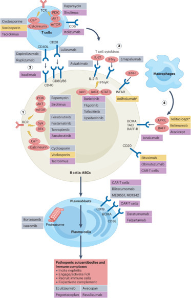
Summary There is currently scarce knowledge of the immunological profile of patients with latent autoimmune diabetes mellitus in the adult (LADA) when compared with healthy controls (HC) and patients with classical type 1 diabetes (T1D) and type 2 diabetes (T2D). The objective of this study was to investigate the cellular immunological profile of LADA patients and compare to HC and patients with T1D and T2D. All patients and age‐matched HC were recruited from Uppsala County. Peripheral blood mononuclear cells were isolated from freshly collected blood to determine the proportions of immune cells by flow cytometry. Plasma concentrations of the cytokine interleukin (IL)‐35 were measured by enzyme‐linked immunosorbent assay (ELISA). The proportion of CD11c+CD123– antigen‐presenting cells (APCs) was lower, while the proportions of CD11c+CD123+ APCs and IL‐35+ tolerogenic APCs were higher in LADA patients than in T1D patients. The proportion of CD3–CD56highCD16+ natural killer (NK) cells was higher in LADA patients than in both HC and T2D patients. The frequency of IL‐35+ regulatory T cells and plasma IL‐35 concentrations in LADA patients were similar to those in T1D and T2D patients, but lower than in HC. The proportion of regulatory B cells in LADA patients was higher than in healthy controls, T1D and T2D patients, and the frequency of IL‐35+ regulatory B cells was higher than in T1D patients. LADA presents a mixed cellular immunological pattern with features overlapping with both T1D and T2D. Introduction Latent autoimmune diabetes mellitus in the adult (LADA) 1 accounts for 9% of all adult onset diabetes 1. Phenotypically, LADA patients share characteristics of both type 1 diabetes (T1D) (antibodies against the insulin‐producing β cells) and type 2 diabetes (T2D) patients (aged >35 years and non‐insulin‐dependent at onset) 2, 3. LADA also shares genetic features of both T1D and T2D 4. Despite the presence of autoantibodies, predominantly against glutamic acid decarboxylase (GAD), the disease progress is much slower than in T1D. At present, the focus on the immunological characterization of LADA has concerned the presence of autoantibodies, with only a few studies of other components of the immune system, such as low‐grade proinflammatory markers or cellular immunology 4-6. So far there are no published reports describing the cellular immunological profile that characterizes LADA patients in comparison to healthy individuals, T1D as well as T2D patients, simultaneously. Innate immune cells, such as dendritic cells (DCs), are the main players during early development of autoimmune diabetes 7-10. Allen et al. have reported that plasmacytoid dendritic cells (pDCs) are expanded during the early development of T1D and enhance the islet autoantigen presentation to T cells through an immune complex capture 11. In line with this, it has been shown that interferon (IFN)‐α‐producing pDCs augment the T helper type 1 (Th1) response in T1D and the number of IFN‐α‐producing pDCs are increased in T1D patients compared to healthy controls 12. Therefore, it is of interest to investigate CD11c+CD123–human leucocyte antigen (HLA)‐DR+Lin– (CD11c+CD123–) antigen‐presenting cells (APCs), CD11c+CD123+HLA‐DR+Lin– (CD11c+CD123+) APCs and CD11c–CD123+HLA‐DR+Lin– (CD11c–CD123+) pDCs in LADA patients. Herein, these subsets of DCs were analysed using flow cytometry, as described previously 13. Neutrophils have been found to be reduced in recent‐onset T1D 14, 15, and CD15low neutrophils (low‐density granular neutrophils) have also been shown to have a pathogenic role in systemic autoimmunity 16. In accordance with this, Neuwrith et al. have reported a role of CD15low cells in T1D development 17. Alterations in the frequency of CD16 monocytes has been associated with diabetes complications 18. Natural killer (NK) cells participate in innate immune responses and in animal models of T1D, it has been reported that depletion of NK cells prevents the development of T1D 19, 20. An altered frequency of total number of NK cells has been found in LADA, but not in T2D, patients 21, 22. Furthermore, it has been reported in children with T1D that NK cells have defects in signalling and function that might contribute to the pathogenesis of T1D 23. NK cell subsets could be defined based on the relative expression of CD16 and CD56, i.e. CD56dim (CD56lowCD16+ and CD56lowCD16–) and CD56bright (CD56highCD16+ and CD56highCD16–), as reviewed by Poli et al. 24. CD56dim NK cells are considered to be mature NK cells and CD56bright cells are immature cells. CD56dim NK cells have cytotoxic properties while CD56bright NK cells are proinflammatory, as they produce IFN‐γ 25. In the present study we determined the proportion of both CD56dim and CD56bright NK cell subpopulations using flow cytometry. Adaptive immune cells such as T (CD4+ and CD8+) and B (CD19+) cells also play a major role in autoimmunity and metabolic disorders 26-30. An altered phenotype of CD4+, CD8+ and CD19+ cells has been reported in LADA patients 5, 31-33. Therefore, in this study we determined the proportions of these cells in LADA patients when compared to T1D and T2D patients. Immune cells that produce anti‐inflammatory cytokines, e.g. interleukin (IL)‐10, transforming growth factor (TGF)‐β and IL‐35, are known as regulatory immune cells, i.e. regulatory T (Treg) cells, regulatory B (Breg) cells and tolerogenic APCs 34-36. Treg cells have received a great deal of attention, as these cells play a major role in maintaining the homeostasis of the immune system and preventing the development of autoimmune diseases 37, 38. A reduced number of Treg cells has been reported in different autoimmune diseases, including T1D 39. Treg cells can be identified by using the cell surface markers CD4, CD25 and CD127 and the intracellular forkhead box (Fox) transcription factor FoxP3 40. Breg cells can be identified by using the cell surface markers CD19, CD24, CD38 and CD40 41. Treg cells, Breg cells and APCs produce IL‐35 34-36, 42-44. This cytokine is a novel anti‐inflammatory cytokine, which belongs to the IL‐12 cytokine family, and has two subunits, Ebi3 and IL‐12p35 42, 43. It has previously been reported that IL‐35 suppresses autoimmunity and plays a putative role in tumour biology 35, 36, 42, 45-47. We have found that the systemic administration of IL‐35 prevented the development of autoimmune diabetes in the multiple low‐dose streptozotocin (MLDSTZ) mouse model, and effectively reversed established disease in both the MLDSTZ and non‐obese diabetic mouse models of T1D by maintaining the normal suppressive phenotype of Treg cells 46. We also found that IL‐35 levels were higher in T1D patients with residual C‐peptide compared to T1D patients with no measurable residual C‐peptide 48. Thus, our previous findings indicate that IL‐35 may prevent the loss of β cell mass. The possible alteration in the frequencies of IL‐35‐producing cells in LADA patients has not yet been investigated. In this study we aimed to determine the proportions of different innate, adaptive and regulatory immune cells in order to elucidate the immunological profile that characterizes LADA in comparison to healthy controls, T1D and T2D patients. Materials and methods Informed consent The Uppsala County regional ethics board approved this study, and the reported investigations were carried out in accordance with the principles of the Declaration of Helsinki, as revised in 2000. All participants were provided with oral and written information about the study, and signed a written consent. Inclusion The All New Diabetes in Uppsala (ANDiU) study (www.andiu.se) is an ongoing study with the aim of describing the incidence of diabetes subgroups in Uppsala County, Sweden (population approximately 300 000). Patients newly diagnosed with diabetes (ADA criteria) are included. Upon registration patients are classified into diabetes subtypes according to criteria shared with the All New Diabetics in Scania (ANDIS) study (http://andis.ludc.med.lu.se; Principle Investigator: Leif Groop) in southern Sweden. Criteria for LADA diagnosis are age ≥35 years, GAD antibody (GADA) positivity (>20 IE/ml) and fasting (f)C‐peptide levels ≥0·3 nmol/l. T1D criteria are GADA positivity and fC‐peptide <0·3 nmol/l, whereas for T2D age ≥35 years, GADA negativity and fC‐peptide >0·72 nmol/l are used as diagnostic criteria. For all three diagnoses there should be no history of pancreatitis, pancreatic cancer or other causes of secondary diabetes. There are no unified criteria for LADA diagnosis and classification, but the criteria used in ANDiU and ANDIS are in line with previous literature 3. The exception is fC‐peptide, which is used in ANDiU and ANDIS as an indicator of remaining insulin production to separate LADA from T1D 49, 50. It replaces the insulin therapy criterion (i.e. no insulin therapy within 6–12 months of diagnosis) that is often used, which is open to subjectivity as it reflects the assessment made by the treating physician. From ANDiU we recruited patients classified as LADA and T2D. T1D patients and healthy controls were recruited at Uppsala University Hospital and Eriksberg Primary Health Care Clinic, Uppsala, respectively. T1D and T2D patients, as well as healthy controls, were age‐, sex‐ and body mass index (BMI)‐matched to the LADA patients. Exclusion criteria were ongoing hormonal treatment (ATC code H01‐03), use of non‐steroid anti‐inflammatory drugs (NSAID, ATC code M01), ongoing chemotherapy treatment (ATC code L01‐04) and other autoimmune disease (e.g. hypo/hyperthyroidism, rheumatic disease, vitiligo, psoriasis, coeliac disease, Addison’s disease and inflammatory bowel disease). Table 1 provides characteristics of the included patients and Supporting information, Fig. S1 shows a flow‐chart of the inclusion process of the study. Controls (n = 13) T1D (n = 16) LADA (n = 14) T2D (n = 16) Age (years) 61·6 ± 2·9 66·0 ± 1·4 65·7 ± 2·4 64·0 ± 2·0 Sex (M%/F%) 62%/38% 73%/27% 57%/43% 70%/30% BMI (kg/m2) 28·9 ± 4·0 26·5 ± 0·7 27·9 ± 0·5 29·1 ± 1·0 Age at onset n.a. 19·7 ± 1·8 63·6 ± 1·5b 60·0 ± 2·0b Disease duration n.a. 46·1 ± 2·2 4·9 ± 0·3b 5·3 ± 0·3a, b HbA1c (mmol/mol) 35·8 ± 0·7 58·9 ± 3·2a 45·8 ± 2·3b 52·27 ± 4·0a fC‐peptide (nmol/l) 1·10 ± 0·11 0·22 ± 0·13a 0·95 ± 0·14b 1·30 ± 0·12b, c fp‐glucose (mmol/l) 5·9 ± 0·1 11·7 ± 1·0a 8·0 ± 0·5b 9·1 ± 0·6a, b GAD antibody IgG (IE/ml) 0 ± 0 508 ± 205 337 ± 190 1 ± 0 IA‐2 antibody IgG (IE/ml) 1 ± 0 25 ± 7 387 ± 295 1 ± 0 Hb (g/l) 145 ± 2 142 ± 3 145 ± 3 141 ± 4 Lkc (109/l) 6·0 ± 0·4 5·8 ± 0·4 6·6 ± 0·8 5·4 ± 0·2 CRP (mg/l) 1·2 ± 0·4 1·6 ± 0·3 1·3 ± 0·2 2·4 ± 0·8 All values are given as mean ± standard error of the mean (s.e.m.). Multiple comparison of means by analysis of variance (anova), Tukey’s post‐hoc test. a P‐value versus controls < 0·05; b P‐value versus T1D < 0·05; c P‐value versus LADA < 0·05. GADA = glutamate decarboxylase 65 antibody; eGFR = creatinine estimated glomerular filtration rate; TSH = thyroid‐stimulating hormone; LADA = latent autoimmune diabetes mellitus in the adult (LADA); Ig = immunoglobulin; BMI = body mass index; n.a. = not available; HbAlc = haemoglobinA1c; CRP = C‐reactive peptide; fC‐peptide = fasting C‐peptide; fp‐glucose = fasting plasma glucose; Lkc = leucocyte count. Routine laboratory parameters Blood samples were collected after overnight fasting. All routine laboratory parameters were analysed at the Central Clinical Chemistry Laboratory at Uppsala University Hospital, which is certified by the External Quality Assessment Service (EQUALIS) (equalis.se/en). On inclusion, blood haemoglobin 51, blood leucocyte count (Lkc), plasma C‐reactive protein (CRP), serum transglutaminase [immunoglobulin (Ig)A] antibodies, serum thyroxine (T4), serum triiodothyronine (T3), serum thyroid‐stimulating hormone (TSH), serum TSH‐receptor 18 antibody, serum thyroid peroxidase antibody, plasma IgA antibody, serum cortisol, 21‐hydroxylase 18 antibody, serum GADA, fasting serum C‐peptide, fasting plasma glucose and blood haemoglobinA1c (HbA1c) were analysed. Glomerular filtration rate (GFR) was estimated based on creatinine‐levels calculated with the MDRD formula 52. Flow cytometry Freshly isolated peripheral blood mononuclear cells (PBMCs) were prepared using Histopaque‐1077 (Sigma, St Louis, MO, USA), as described earlier 48. PBMCs were stained with antibodies for surface markers (Supporting information, Table S1). The cells were thereafter fixed and permeabilized with fixation permeabilization buffer (eBioscience, San Diego, CA, USA) for intracellular markers (Supporting information, Table S1). The samples were run on LSR II Fortesa (Becton Dickinson, Franklin Lakes, NJ, USA) using DivaDacker software 53 and 1 million events were counted for analysis. Fluorescence minus one (FMO), isotype and single‐stained controls were used for gating strategies. The flow cytometry standard (FCS) files were analysed on FlowLogic software (Inivai Technologies, Mentone, Australia). Representative gating strategies are shown in Supporting information, Fig. S2–S5. IL‐35 measurements in plasma Plasma IL‐35 concentrations were determined by using an enzyme‐linked immunosorbent assay (ELISA) kit (Cloud‐Clone Corporation, Wuhan, China). Statistics Differences in means between healthy controls, T1D and T2D patients compared to LADA patients were calculated by one‐way analysis of variance (anova) with Dunnett’s post‐hoc test for immunological profiles. Comparisons between all groups were performed by anova with Tukey’s post‐hoc test for research subjects’ characteristics (Table 1). A P‐value < 0·05 was considered as a significant difference between groups. Statistical analyses were carried out in GraphPad Prism version 6.0TM. Results Innate immune cell responses We observed a lower proportion of CD11c+CD123– APCs in LADA patients than in healthy controls and T1D patients (Fig. 1a). Furthermore, the proportion of CD11c+CD123– APCs among HLA‐DR+Lin– cells was lower in LADA patients than in T1D patients (Fig. 1b), whereas the proportions of pDCs and pDCs among HLA‐DR+Lin– were not altered in LADA patients (Fig. 1c,d). We did not observe any difference in the proportion of CD11c+CD123+ APCs (Fig. 1e), but the proportion of CD11c+CD123+ cells among HLA‐DR+Lin– was higher in patients with LADA than in patients with T1D (Fig. 1f). We did not find any alteration in the proportions of CD15low neutrophils and CD16+ monocytes in LADA patients (Supporting information, Fig. S6a,b). We also examined different subsets of NK cells and did not find any alteration in LADA patients compared to the other groups regarding the proportions of CD3–CD56lowCD16+, CD3–CD56lowCD16– and CD3–CD56highCD16– NK cells (Fig. 2a–c). However, the proportion of CD3–CD56highCD16+ NK cells was higher in LADA patients compared to both healthy controls and T2D patients (Fig. 2d). We did not observe any alteration in the proportions of CD3+CD56+ NK T cells between the groups (Supporting information, Fig. S6c). Adaptive immune cell responses The proportions of CD3+, CD4+CD25– and CD8+ T cells were not altered in LADA patients when compared to the other groups (Supporting information, Fig. S7a–c). Similarly, the proportion of CD19+ cells was not altered (Supporting information, Fig. S7d). Regulatory immune cell responses The proportions of CD4+CD25+CD127–FoxP3+ cells (Treg cells) and CD4+CD25+CD127–FoxP3+Helios+thymic‐derived T cells (tTreg) were similar in all four groups (Supporting information, Fig. S8a,b). Similarly, the proportions of Treg cells as well as the tTreg cells among CD4+ T cells were similar in all four groups (Supporting information, Fig. S8c,d). However, the proportions of IL‐35+ cells among Treg and tTreg cells in patients with LADA were similar in patients with T1D and T2D, but lower when compared to healthy controls (Fig. 3a,b). We also analysed the proportion of CD19+CD24+CD40+CD38+ cells (Breg cells), and observed a higher proportion of these cells in LADA patients when compared to healthy controls and patients with T1D and T2D (Fig. 3c). The proportion of IL‐35+ cells among Breg cells was higher in LADA patients when compared to T1D patients (Fig. 3d). Subsequently, we analysed the proportion of IL‐35+ tolerogenic APCs. Interestingly, the proportion of IL‐35+ cells among APCs was higher in patients with LADA compared to patients with T1D (Fig. 3e). Patients with LADA had IL‐35 concentrations in blood plasma that were similar to those in patients with T1D and T2D, but lower compared to those in healthy controls (Fig. 3f). Discussion We determined the proportions of different immune cells to elucidate the immunological patterns that distinguish patients with LADA compared not only to healthy individuals, but also to patients with T1D and T2D. With regard to the innate and adaptive immune cells, we found that LADA shares common features with both T1D and T2D. The proportion of CD11c+CD123– APCs in LADA patients was between that observed in T1D and T2D patients. Similarly, the proportion of CD11c+CD123+ cells among the HLA‐DR+Lin– was highest in T2D patients and lowest in T1D patients, whereas the proportion in LADA patients was between that of T1D and T2D patients. Neutrophils have previously been claimed to play a role in the early development of both T1D 14, 15, 54 and T2D 55. In the present study, we did not find any alteration in the proportion of CD15low neutrophils in LADA patients when compared to the other study groups. It cannot be excluded that early transient alterations during disease development as described for T1D 14, 15 had occurred, as our study subjects with LADA had a mean disease duration of almost 5 years. It has been reported previously that the proportion of CD3–CD56+CD16+ NK cells is decreased in LADA patients 22. Instead, we observed an increased proportion of CD3–CD56highCD16+ NK cells in LADA patients compared to both healthy controls and T2D patients. The reason for the discrepancy between the studies is unknown, but it could be because we used freshly prepared PBMCs instead of cryopreserved cell preparations. CD56high or CD56bright NK cells produce IFN‐γ 25. The proportions of these NK cells were similar in LADA and T1D patients. This result illustrates that, like T1D, LADA is also a Th1 response disorder. The frequency of FoxP3+ Treg cells was similar in all groups. However, the proportion of IL‐35+ cells among Treg cells in patients with LADA was similar to those in patients with T1D and T2D, but lower when compared to healthy controls. This may suggest a common functional defect of Treg cells in LADA, T1D and T2D patients. This idea was further supported because the circulating concentrations of IL‐35 in patients with LADA were similar to patients with T1D and T2D, but lower than in than healthy controls. We have previously shown that decreased IL‐35 production by Treg cells is associated with a functional defect 46. Also, previous findings of a decreased frequency of FoxP3+ cells among CD4+CD25intermediate cells in LADA patients when compared to healthy controls 5, and a decreased Foxp3 gene expression in CD4+ T cells in LADA patients, are considered with defective Treg cell functionality 56. In patients with T1D, it has been reported that Treg cells lose their FoxP3 expression due to DNA methylation in the Foxp3 gene, which may cause a defective function in Treg cells and result in a decreased number of Treg cells 57. Interestingly, Li et al. also reported an abnormal DNA methylation in peripheral CD4+ T cells of LADA patients 33, which indicates a dysfunction of Treg cells 58. Breg cells play an important role in maintaining the autoimmune and/or inflammatory response 35, 36. We found that the frequency of Breg cells was higher in LADA patients compared to all other groups. These results are in agreement with a previous report, where Deng et al. found the lowest frequency of IL‐10‐producing B cells in T1D patients when compared to LADA and T2D patients 32. Our observed lower proportion of IL‐35+ cells among Breg cells in T1D than LADA patients suggests that the response of IL‐35+ Breg cells in LADA patients is similar to that of T2D patients, which might protect against β cell destruction by immunological insults in LADA patients. Although the proportions of IL‐35+ Breg cells and IL‐35+ tolerogenic APCs were higher in LADA than in T1D patients, the circulating concentrations of IL‐35 were similar in LADA and T1D patients. This could be due to other IL‐35‐producing immune cells, such as iTr35 59 and macrophages 60. These particular populations have not been investigated in the present study. APCs (CD123– APCs) and pDCs (CD123+ APCs) prime T cells to react against the insulin‐producing β cells. In the present study, we found that the numbers of APCs were lower in LADA than in T1D patients. This indicates that the initial immune response is lower in LADA patients, which might cause a delay in the development of an autoimmune response. Tolerogenic APCs protect β cells in animal models of T1D 44, and Dixon et al. reported that tolerogenic APCs produce IL‐35 in human peripheral blood 34. We analysed the frequency of IL‐35+ cells among APCs in our study and found that IL‐35+ cells were more frequent in LADA than in T1D patients, which may contribute to a milder disease development. In a previous study, we found that systemic treatment with IL‐35 in two different animal models of T1D prevented destruction of β cells 46. However, as this is an association study it is impossible to determine whether or not the immunological differences are causal. We observed changes in the proportions of different immune cells in LADA patients compared to healthy controls and other diabetes patients. Our conclusion is that the changes in APC and Breg cell numbers in LADA patients are more similar to those also observed in T2D patients, whereas the changes in NK cell numbers are more similar to those found in T1D patients. There are no differences in IL‐35 concentrations and IL‐35+ Treg cell numbers in between LADA, T1D and T2D patients, but in all three conditions these are lower than the healthy individuals. Thus, our analysis of several subsets of immune cells reveals that LADA patients present a mixture of cellular immunological changes with features overlapping with both T1D and T2D. Acknowledgements The technical assistance from research nurses Violeta Armijo Del Valle, Rebecka Hilmius and Karin Kjellström is gratefully acknowledged. The present study was supported financially by the Swedish Research Council (2017‐01343, 921‐2014‐7054), EXODIAB, the Swedish Diabetes Foundation, Diabetes Wellness Sverige, the Swedish Child Diabetes Fund, SEB Diabetesfonden, O.E. och Edla Johanssons vetenskapliga stiftelse and the Novo Nordisk Foundation. Disclosures The authors declare that there are no conflicts of interest associated with this paper. Author contributions M. M. and K. S. contributed to the design of the study, conducted experiments, acquired and analysed data, and participated in writing the manuscript. Z. L., D. E., J. S. and S. S. contributed to the design of the study, analysed data and participated in the writing of the manuscript. P. O. C. designed the study, analysed data and wrote the manuscript. All authors read and approved the final version. K. S. is the guarantor of this work and, as such, had access to all the data in the study and takes responsibility for the integrity of the data and the accuracy of the data analysis. Supporting Information References

|
Suggested by
Société Francaise d'Immunologie
onto AUTOIMMUNITY June 17, 2019 6:56 AM
|
No comment yet.
Sign up to comment


 Your new post is loading...

