AUTOIMMUNITY
19.9K views | +7 today
 
Scooped by Gilbert C FAURE
onto AUTOIMMUNITY
January 15, 2021 10:57 AM
Scoop.it!

Recent updates on the management of autoimmune hepatitis

INTRODUCTION Autoimmune hepatitis (AIH) is an immune-mediated inflammatory liver disease of non-self-limiting clinical course for which immunosuppressive agents are necessary in the majority of affected patients. The concept of the immunopathogenesis of AIH relies on autoreactive CD4 and CD8 T cells, whose emergence is induced after the break of self-tolerance by environmental triggers [1]. As the inflammation that AIH presents is likely to be characterized by a dynamic transition of the milieu of multiple effector immune cells in the liver, clinicians should take into consideration the chronological dynamics of disease manifestations or of distinct subtypes of disease, e.g., ranging from acute-onset, acute on chronic, and chronic insidious manifestation. The appropriate diagnosis and proper treatment strategy with special attention to the clinical subtypes of AIH must be considered to ensure favorable short- and long-term survival. In 2019, the American Association of the Study of Liver Disease (AASLD) published very comprehensive practice guidance and guidelines for AIH that updated the previous version published in 2010 [1]. The progress in our understanding of AIH is apparent in these guidelines, including their detailed description of a first-line treatment strategy based on the patient’s clinical manifestations. In this review, we summarize the recent updates regarding the management of AIH, focusing on the disease manifestations (Fig. 1) and the time frame of treatment and responses to treatment. EPIDEMIOLOGY AIH affects individuals of all ages from children to the very elderly, but it is most commonly identified in middle-aged women [2,3] in all ethnic groups. In 2016, a nationwide, hospital-based, epidemiological survey to approximate the prevalence of AIH was carried out in Japan. The estimated number of patients was 30,330 (95% confidence interval [CI], 29,592–31,069) and the calculated point prevalence of AIH per 100,000 population was 23.9 (95% CI, 23.3–24.5) [4]. Compared to the previous survey in 2004, the data revealed an almost threefold increase in the prevalence of AIH [4]. Among the widely varying nation-based prevalence data for adult AIH reported after 2000, e.g., from 4.0 (Singapore) [5] to 42.9 (Alaska) [6], a trend of increasing prevalence has been observed worldwide; for instance, from 10.7 in 2003 [7] to 17.3 in 2009 [8] in Sweden. The prevalence of AIH in Korea increased gradually from 2009 to 2013, although the incidence remained stable [2]. Alterations of environmental factors, including changes in lifestyle, might trigger the development of AIH, and environmental factors are likely to be linked to the increased male to female ratio of AIH in Japan from 1:6.9 in 2004 to 1:4.3 in 2016 as shown by the aforementioned survey [4]. Improved awareness of AIH among clinicians worldwide might also have contributed to the trend of increased prevalence, possibly resulting in a reduction of the number of otherwise undiagnosed patients, including adult male patients. DIAGNOSIS General considerations AIH is a disease without signature diagnostic features. The diagnosis of AIH requires 1) histological abnormalities (interface hepatitis), 2) characteristic laboratory findings (elevated serum hepatic enzymes, aspartate aminotransferase [AST] and alanine aminotransferase [ALT], and increased serum immunoglobulin G [IgG]), and 3) positive results of disease-defining autoantibodies, coupled with 4) the exclusion of other liver diseases that may resemble AIH, including viral hepatitis, hereditary, metabolic, cholestatic, or drug-induced liver injury (DILI). Anti-nuclear antibodies (ANA) and anti-smooth muscle antibodies should be tested in patients of all ages, and an additional test of anti-liver kidney microsomal type 1 is necessary in children for the characterization of type 2 AIH [1]. The clinical judgement is straightforward in typical AIH patients with the above-mentioned hallmarks, but atypical cases should be diagnosed with the aid of diagnostic scoring systems that were originally developed by the International AIH Group (IAIHG) in 1993 for the identification of patients with AIH for clinical research [9]. The revised IAIHG criteria reported in 1999 [10] and the simplified criteria proposed in 2008 [11] are commonly implemented in clinical practice, and they emphasize distinct diagnostic values. As the simplified scoring has superior specificity (90% vs. 73%) and accuracy (92% vs. 82%) compared to the revised scoring system [12], the former is preferable for the diagnosis of typical AIH cases. On the other hand, the revised scoring system is suitable for the reassessment of atypical cases with a low score in the simplified system, including cases of autoantibody-negative hepatitis and acute-onset AIH with normal IgG values [1]. Limitations to both scoring systems are evident (due to the lack of accuracy) for a diagnosis of AIH that is overlapped with a primary biliary cholangitis (PBC) [13], primary sclerosing cholangitis (PSC), or non-alcoholic fatty liver disease (NAFLD) [14], or fulminant liver failure. Histological findings The diagnosis of AIH requires liver biopsy results presenting compatible histological abnormalities. Typical histological features are indicative of (chronic) active hepatitis, comprising lymphoplasmacytic interface hepatitis, emperipolesis (intrusion of one intact lymphocyte into a hepatocyte), and hepatocyte rosettes. Gurung et al. [15] recently hypothesized that typical histological features are related to the severity of disease, but not to the etiology itself, and they reported the following as AIH-specific histological features: 1) Kupffer cell hyaline granules, 2) prominence of plasma cells in portal tracts, and 3) the relative predominance of plasma cells over lymphocytic inflammation. After Gurung et al. [15] adjusted the analysis results for the inflammatory grade, emperipolesis and rosette formation were similarly found in the disease control, chronic hepatitis C (CHC). The Kupffer cell hyaline granules were well-circumscribed, eosinophilic periodic acid-Schiff diastase-resistant deposits within Kupffer cells, and they were originally proposed as a specific histology in pediatric AIH [16]. Centrilobular necrosis is another histological AIH feature, presenting in a rather disease manifestation-specific manner in acute-onset AIH [17] and in acute liver failure (ALF). In ALF, central perivenulitis, plasma cell-enriched inflammatory infiltrate, and lymphoid follicles on a background of massive hepatic necrosis are the principle findings [18]. In clinical practice, the differential diagnosis of AIH in liver histology is routinely focused on DILI, including drug-induced AIH (DIAIH)-like liver injury. Though the rare presence of bridging fibrosis and the absence of advanced fibrosis are clues suggesting DILI, this is not the case for the differential diagnosis of acute-onset AIH over DILI. Histological findings of NAFLD and non-alcoholic steatohepatitis (NASH) are reported to be present in 17–30% of adult AIH patients [19,20]. These overlapping findings are indicative of patients who are at increased risk of liver-related mortality [19]. Conversely, characteristic laboratory findings with positive autoantibodies (especially in female patients) are sometimes refuted by the mere histology of NAFLD or NASH in the liver. Signature diagnostics for discriminating NASH with prominent periportal hepatitis from chronic active AIH are greatly anticipated. Noninvasive assessment of fibrosis The long-term outcome of AIH is associated with the stage of fibrosis. Since the evaluation of liver fibrosis by biopsy during the course of disease management is not feasible, noninvasive assessments have been conducted in clinical hepatology by using serum biomarkers, including the serum AST/platelet ratio index (APRI) and the fibrosis-4 (FIB-4) index [21]. However, a recent systemic review of the diagnostic accuracy of APRI and FIB-4 demonstrated their poor performance for detecting advanced fibrosis and cirrhosis in AIH [22]. Noninvasive assessment by liver stiffness has been shown to identify advanced fibrosis and cirrhosis in AIH with reasonable accuracy. The performance levels of vibration-controlled transient elastography (VCTE) and magnetic resonance elastography were indicated to be superior to those of the APRI and FIB-4, and VCTE was validated in a systemic review as providing good performance [22]. Considering that liver inflammation affects liver stiffness, the stiffness value at the initial diagnosis before the initiation of treatment with immunosuppressive agents is confounded by disease activity. In fact, the value of VCTE within 3 months after the start of treatment was significantly correlated with histological grading, but not with the fibrosis stage [23]. Thereafter, at least 6 months after the successful treatment of AIH, the area under the receiver operating curve of VCTE reached 1.0 [23]. Sustaining biochemical remission (normal ALT and normal IgG) and the use of VCTE help monitor and manage the disease course of AIH. A novel serum fibrosis marker, i.e., Mac-2 binding protein glycosylation isomer (M2BPGi), which was originally reported to be associated with the fibrosis stage in CHC patients [24], is likely to become an alternative to the use of VCTE; the M2BPGi value is influenced by both inflammation and fibrosis in AIH patients, in a similar way to VCTE [25]. A ‘one-serum parameter fits all’ approach to the evaluations of disease activity and fibrosis could be achievable with serum M2BPGi, but further studies are necessary to validate its utility. CLINICAL MANIFESTAIONS WITH SPECIAL ATTENTION TO DISEASE SUBTYPES Acute-onset AIH Acute-onset AIH is a clinically challenging disease subtype because a delayed diagnosis and delayed treatment, especially in the absence of typical serological findings, may lead to a poorer short-term prognosis. The prevalence of acute-onset AIH has been obtained in several cross-sectional studies worldwide. The 2019 AASLD practice guidance and guidelines state that 25–75% of individuals with AIH in western countries present with an acute onset and a disease duration <30 days [1,26]. A Korean study reported the prevalence 46.4%, using almost the same definition [27]. An Italian multicenter cohort applied arbitrary criteria, i.e., >10× the upper limit of normal (ULN) of transaminases and >5 mg/mL of bilirubin, and the study’s authors reported that 43% of their series of AIH patients were acute-onset [28]; among the patients who underwent liver biopsy, 64.8% fulfilled the histological criteria for acute-onset AIH, with the fibrosis stage lower than Ishak F2. In a Japanese nationwide cross-sectional study of AIH patients diagnosed in 2009–2013, the frequency of acute hepatitis without fibrosis (F0) was, on the other hand, almost 11% [3]. Acute-onset AIH may encompass two distinct clinical subgroups: 1) ‘genuine’ acute AIH with no chronic liver pathology (portal, followed by bridging fibrosis) and 2) acute exacerbation of chronic AIH. Even among the group of genuine acute AIH cases, dynamic histological changes—especially in the extent of portal fibrosis—are anticipated. As a trend, the median duration between disease onset and liver biopsy among Japanese AIH patients with acute presentation was longer in the F1–2 patients than in the F0 patients (29 vs. 15 days, P=0.052) [29]. Nevertheless, there were no significant differences between these two groups in laboratory data, AIH-related pathological findings, or disease outcomes after the introduction of prednisolone. Typical serological hallmarks of AIH (e.g., positive autoantibody or elevated serum IgG) are frequently absent in acute-onset AIH; in the above-mentioned Japanese cohort, 27% of the patients were ANA-negative (<×40) and >50% of them had normal serum IgG values [29]. As the absence of ANA and normal serum IgG were not associated with disease outcomes in that cohort, the prompt initiation of treatment with immunosuppressive agents was necessary to prevent progression to acute severe AIH (AS-AIH), and eventually ALF. AS-AIH is defined by the AASLD as AIH with jaundice, a prothrombin time (PT) international normalized ratio (INR) ≥1.5, and neither encephalopathy nor previously recognized liver disease (Table 1) [1]. A thorough diagnosis protocol that includes a transjugular liver biopsy is needed to differentiate AS-AIH from acute severe hepatitis with multiple other etiologies. In a cohort from the UK and France, 69% [30] and 59% [31] of the AS-AIH cases were reported to progress to ALF-AIH, respectively. A continuum of treatment strategies that are based on the benefit-to-risk ratio of glucocorticoid therapy should thus be seriously considered (Fig. 2). In the 2019 AASLD practice guidance, prednisone or prednisolone monotherapy (60 mg/day in adults) is recommended for AS-AIH [1], because no association with an increase in sepsis was demonstrated [32]. A short-term treatment response (within 1–2 weeks) in AS-AIH is indeed crucial to prevent disease progression. Zachou et al. [33] recently observed that high-dose intravenous (iv.) corticosteroids (either 1 g methylprednisolone for 3 consecutive days followed by iv. 1 mg/kg/day prednisolone, or iv. 1.5 mg/kg/day prednisolone) was safe and effective to treat AS-AIH patients (n=34; all were F0–2, and transaminases were >10× ULN). The complete response rate was higher than that in the non-AS-AIH group, with no case requiring liver transplantation (LT). ALF and acute on chronic liver failure (ACLF) AS-AIH with encephalopathy is defined as ALF caused by AIH (ALF-AIH) (Table 1). With regard to noninvasive diagnoses, heterogenous hypo-attenuated regions within the liver as visualized by unenhanced computed tomography (CT) is useful to differentiate patients with AS/ALF-AIH from those with viral-associated ALF [34]. The volumetric measurement of the liver on CT is also valuable, because the size of the liver was reported to be significantly reduced in non-acetaminophen cases of acute liver injury/ALF compared to the acetaminophen-induced cases [35]. A direct evaluation of indications for LT is recommended in ALF-AIH, because glucocorticoid therapy has not been associated with improved overall survival and is even harmful to patients with a model for end-stage liver disease (MELD) score >40 [36]. ACLF is caused by an AIH flare in previously diagnosed or undiagnosed chronic liver disease/cirrhosis (AIH-ACLF) patients. The Asian Pacific Association for the Study of Liver ACLF Consortium defines ACLF as patients with jaundice (bilirubin >5 mg/dL) and coagulopathy (PT [INR] ≥1.5), complicated by ascites and/or encephalopathy within 4 weeks after diagnosis. The consortium reported that 2.9% (n=82) of the ACLF cases diagnosed in 2012–2017 in nine Asian countries were regarded as having developed AIH as an acute insult; 97% of the patients exhibited IgG elevation (>1.1× ULN), whereas 49% were seronegative for autoantibodies [37]. Although 34% of the patients (n=28) being treated with a corticosteroid showed a significantly improved 90-day survival rate compared to those without treatment (75% vs. 48.1%, P=0.02), early stratification to corticosteroid therapy or LT is necessary [37]; predictors of an unfavorable response to corticosteroids were revealed to include a MELD score >27 and hepatic encephalopathy in advanced fibrosis (≥F3). DIAIH-like injury DILI can occasionally be diagnosed based on increased serum IgG and positive ANA. Even after the cessation of suspected drugs, such ‘AIH-mimic’ patients whose ALT elevation is persistent or progressive are indicated for treatment with immunosuppressive agents to prevent ALF. Remarkably, the short-term (1 week) response to corticosteroids was demonstrated to be more pronounced in the patients with AIH-mimic DILI compared to those with pure AIH [38]. AIH-mimic DILI and pathogenically DIAIH are difficult to differentiate by liver pathology, including the intensity of inflammatory infiltrates, the type of the predominant inflammatory cells, and the grade of fibrosis. In the 2019 AASLD practice guidance and guidelines, the term “DIAIH-like injury” was introduced as an alternative to DIAIH [1]. The majority of patients with DIAIH-like injury are acute-onset, and up to 30% of the cases are accompanied by hypersensitivity reaction [39]; the latency periods of minocycline and nitrofurantoin (the two most commonly implicated drugs) can exceed 12 months [40]. HLA-DR3 or -DR4 and cirrhosis at presentation are unusual [41]. Fulfilling Hy’s law, serum aminotransferase levels >3× ULN and total serum bilirubin >2× ULN as a predictor of ALF in patients with DILI [42], or failures of improvement in laboratory tests after medication discontinuation are the reasons for the implementation of glucocorticoids. The outcome of DIAIH-like injury has been shown to be excellent, and relapse after glucocorticoid withdrawal is rare [43]. Emerging liver injury that is related to the use of immune checkpoint inhibitors is distinct from DIAIH-like injury, as it lacks the typical serological and histological features of AIH [44,45]. Overlap manifestation with cholestatic liver disease or viral hepatitis The concurrences of AIH and PBC or AIH and PSC are not confirmed as specific pathological entities, but the identification of clinical ‘overlap’ among AIH patients is of importance, as these patients may not obtain sufficient benefit with only conventional AIH treatment. Concerning the AIH-PBC overlap, it should be noted that 5–35% of AIH patients, even in the absence of bile duct lesions, are reported to be positive for the serological hallmark of PBC, i.e., anti-mitochondrial antibodies (AMAs) [46]. Simultaneous and sequential AIH-PBC overlaps should be considered separately; the former is suspected in the presence of destructive cholangitis at the initial diagnosis, and the latter is suspected based on the occasional elevation of cholestatic enzymes after biochemical remission. The ‘Paris criteria’ for the identification of AIH-PBC overlap is valuable for apparent cases [47], but this criteria may miss cases with less severe cholestatic features [48,49]. The IAIHG’s position statement did not endorse the Paris criteria or even the revised AIH criteria regarding the diagnosis of AIH-PBC overlap [13]. Nevertheless, the reevaluation of suspected AIH-PBC overlap patients in light of their responses to immunosuppressive agents is likely practical and necessary. AIH-PSC overlap is diagnosed based on the following: 1) the typical features of AIH, 2) the absence of AMA, and 3) evidence of large-duct PSC by endoscopy or magnetic resonance imaging or of small-duct PSC, confirmed by ‘onion-skinning’ periductal fibrosis in a liver biopsy [49]. Concurrent ulcerative colitis with AIH is a critical indication for AIH-PSC overlap, especially in pediatric patients. The cases of hepatitis C virus-infected patients are occasionally accompanied by positive serum and histological markers of AIH, making a differential diagnosis of CHC-AIH overlap syndrome necessary. Direct-acting antiviral (DAA) therapy for CHC patients with AIH features was shown to significantly decrease ALT into the normal range, and serum markers of AIH in those patients began decreasing by 6 months post-treatment; >50% of the patients achieved complete resolution [50]. The CHC-AIH overlap syndrome may be a historical disease entity that is not likely to be diagnosed after the era of DAA. TREATMENT General considerations The purposes of the treatment of AIH are to first relieve symptoms, and then to achieve a biochemical response, control hepatic inflammation toward histological remission, prevent disease progression, and promote the regression of fibrosis. The ideal biochemical response, regarded as biochemical remission by the AASLD, is the normalization of the patient’s serum AST, ALT, and IgG levels to within the ULN (Table 1) [1]. A favorable treatment response in AIH patients assures overall survival that is comparable to that of general populations [51]. Due to the heterogenous manifestations of AIH, short- and long-term treatment responses with regard to liver-related adverse events should be defined in a personalized manner (Figs. 2, 3). Among AS-AIH, ALF, and ACLF patients, estimation of the early biochemical response within 7–14 days is necessary (Fig. 2) [1,32]. In contrast, the midterm biochemical response of patients with nonsevere acute-onset AIH or chronic insidious AIH, even with cirrhosis, can be evaluated at 4–8 weeks (Fig. 3) [1]. Biochemical remission is followed by a histological remission of disease activity. Sustaining biochemical remission for a long term (>1 year from treatment initiation) is thereafter a surrogate for favorable overall long-term survival [52]. Decreasing values of VCTE are favorable, even for the regression of fibrosis. First-line treatments The long-term outcome of patients with AIH has been shown to be improved with immunosuppressive treatment, both with corticosteroids alone and with a combination of a lower dose of corticosteroids and azathioprine (AZA) [53]; those regimens are consistently endorsed as a first-line treatment for AIH. The 2019 AASLD practice guidance and guidelines updated their recommended first-line treatment: either prednisone monotherapy (40–60 mg/day) or a combination of prednisone (20–40 mg/day) or budesonide (9 mg/day) and AZA (50–100 mg/day) [1]. The 2015 European Association for the Study of the Liver (EASL) guidelines propose 0.5–1 mg/kg/day predniso(lo)ne as the initial treatment, followed by a 50 mg/day AZA add-on [54]. The AASLD similarly indicates the appropriateness of a 2-week observation before the AZA is initiated, to confirm the patient’s steroid responsiveness and to evaluate his or her thiopurine-S methyl transferase (TPMT) status for the prevention of AZA-induced hepatitis. TPMT is an anabolizing enzyme for thiopurines, including AZA, and single nucleotide polymorphisms of TPMT genes that cause loss of enzymatic activity predispose patients, in particular European and African descendants, to thiopurine-related toxicity [1,55]. In Japan, AZA was finally approved for AIH treatment in 2018. Accordingly, the practice guidelines for AIH published by the Intractable Hepato-Biliary Diseases Study Group in Japan very recently added the recommendation to evaluate NUDT15 variant (but not TPMT) in patients who are to be treated with AZA, in order to exclude the possibility of thiopurine-induced early severe leukopenia and hair loss [55]. NUDT15 is a recently characterized nucleotide phosphatase that inactivates thiopurines. As the low- or intermediate activity diplotype was reported to be common in East Asian countries (22.6%) [55], the integration of NUDT15 variants in the dosing algorithm for AZA is regarded as most informative. Concerning the relevance of the starting dose of predniso(lo)ne to ensure remission, a retrospective observational study from nine centers in five European countries was performed and the results revealed no significant difference in the rate of normalization of transaminases at 6 months between groups with a higher (≥0.5 mg/kg/day) and lower (<0.5 mg/kg/day) initial dose of predniso(lo)ne [56]. With the aid of AZA as a maintenance therapy in the majority of patients (>85%), an initial lower dose significantly decreased the unnecessary exposure to predniso(lo)ne in patients with AIH. A synthetic steroid, i.e., budesonide has been shown to cause less systemic adverse effects, due to a 90% first-pass hepatic clearance rate. The AASLD investigated whether prednisone or predniso(lo)ne alone or in combination with AZA was superior to a combination of budesonide and AZA as the first-line treatment for patients with newly diagnosed AIH [1]. With an accompanying systemic review and meta-analysis, the AASLD demonstrated a higher rate of biochemical remission in the budesonide + AZA group compared to the prednisone + AZA group (odds ratio, 2.19; 95% CI, 1.30–3.67), and they described this finding as high-grade evidence [57]. Accordingly, the AASLD suggests budesonide in combination with AZA as a first-line therapy for child and adult AIH patients who do not have cirrhosis or acute severe AIH [1]; patients with cirrhosis are contraindicated for budesonide because portosystemic shunting may reduce the drug’s efficacy. The combination of AZA and either predniso(lo)ne or budesonide is now regarded as the most standard first-line therapy in western countries. Prednisone monotherapy, on the other hand, is likely to be appropriate for patients including those with DIAIH-like injury in whom the treatment duration is expected to be <6 months [1]. As corticosteroids are still the mainstay of the first-line treatment of AIH, the maintenance of bone during treatment is needed to limit treatment-related osteoporosis in patients with ongoing risk factors [58]. Bone mineral densitometry should be completed at baseline in those patients with repeated check-up every 2–3 years and supplementation with elemental calcium (1,000–1,200 mg/day) and vitamin D (400–800 IU/day) are recommended for all patients on glucocorticoid therapy [1,59]. Simultaneous bisphosphonate therapy is indeed indicated for patients with documented osteoporosis [60]. The determination of serum levels of 25-hydroxyvitamine D at diagnosis is justifiable, because vitamin D insufficiency (≤29 ng/mL) occurs frequently in patients with AIH (68–81%) [61,62] and even severe deficiency (<20 ng/mL) was reported to be documented in 20% of patients [62]. Second line-treatments The aims of second-line treatments for AIH are to manage refractoriness, incomplete biochemical response, and drug intolerance to first-line treatments (Figs. 2, 3). Anecdotally, second-line treatments have been performed with mycophenolate mofetil (MMF), calcineurin inhibitors (cyclosporin A, tacrolimus [TAC]), mercaptopurine, and biologics (e.g., infliximab). MMF is a DNA synthesis inhibitor and is indicated for immunosuppression after organ transplant or lupus nephritis. In a meta-analysis, the combination of MMF + prednisone was shown to be the most widely prescribed second-line treatment, achieving histological remission in 89% of the patients [63]. A recent report confirmed the effectiveness of MMF as a second-line therapy for patients who have failed standard therapy; the rate of induction of biochemical remission was 60% [64]. The AASLD performed a systemic review to compare the efficacies of MMF and TAC for treatment failure or incomplete biochemical response in adults and children: the AASLD 2019 conditional recommendation with low certainty suggests the use of MMP or TAC to achieve and maintain biochemical remission [1]. Exacerbation, recrudescence, and relapse During the course of maintenance therapy with corticosteroids/AZA, a substantial number of AIH patients spontaneously and asymptomatically experience biochemical exacerbation or recrudescence, i.e., an elevation of ALT coupled with or without an increase in IgG. The 2019 AASLD practice guidance and guidelines strictly define “relapse” as disease exacerbation that occurs after remission and drug withdrawal or by nonadherence [1]. Multiple relapses have been shown to be associated with worse outcomes [51], but the definitions of relapse in the literature differ from that issued by the AASLD, including the concept that biochemical remission may not have proceeded relapse. Following the AASLD rules regarding biochemical remission-induction with first-line or even second-line drugs could result in fewer exacerbations. Relapse after drug withdrawal (which usually occurs within 12 months) and exacerbation should be managed appropriately to induce re-(biochemical) remission with an increase in dosage or the reinstitution of immunosuppressive agents, or with the add-on of second-line drugs. In a case-control study, psychological stress was associated with relapse after drug taper-off or recrudescence [65]. Treatment withdrawal If AIH is a curable disease, the cessation of immunosuppressive agents is desirable. Could the cure for AIH be diagnosed based on serum biochemistry and liver histology, or both? Is the cure achievable in specific subgroups of patients? The answers to these clinical questions involve the feasibility of treatment withdrawal and simultaneously pursuing the lowest risk of drug-induced complications. Clinically, the duration and the degree of remission are the initial keys to the success of treatment withdrawal. A sustained biochemical remission of ≥2 years was proposed by the AASLD as the eligibility criterion for attempting a treatment withdrawal (Fig. 3) [1], in part because the inclusion of patients with only normalized ALT for 2 years resulted in almost universal relapse [66]. A further patient selection step should be included based on liver biochemistry, and/or on liver histology. Lower ALT and IgG values within the normal range were reported to be negative predictors of relapse; patients who achieved sustained remission for >1 year after drug withdrawal were all characterized by ALT values ≤0.5× ULN and IgG values ≤1,200 mg/dL [67]. Maintenance therapy before withdrawal was not associated with relapse: >80% of the patients were treated with AZA alone. A single-center study demonstrated that only 10% of their patients were eligible for treatment withdrawal and 5% reached sustained remission without treatment [67], highlighting AIH as generally a chronic disease demanding life-long maintenance therapy. Stringent biochemical remission for >2 years along with sustained low values of VCTE measured with an appropriate cut-off, may identify the patients who are at low risk of decompensation even when relapse occurs after treatment withdrawal. LT AIH with decompensated cirrhosis or ALF is indicated for LT. Among the listing of the Scientific Registry of Transplant Recipients (2002–2019) in the USA, 3.3% had AIH as the primary etiology [68]. In the trend analysis of the etiology of the Registry’s non-hepatocellular carcinoma LT listings, the rate of AIH during that period was stable. In the prospective multicenter European Liver Transplant Registry (1998–2017), the overall survival of patients after AIH-LT was reported to be similar to that of patients after alcohol-related cirrhosis-LT, but worse than that after PBC-LT and PSC-LT [69]; the 5- and 10-year patient and graft survival rates after AIH-LT were 79.4% and 70.8% and 73.25% and 63.4%, respectively. Compared to all of the other groups, the AIH-LT patients were at higher risk for infections—especially lethal fungal infections resulting in death and graft loss [69]. In AIH, living donor transplantation provided worse survival than that by donated LT after brain death [69]. The appropriateness of long-term glucocorticoid therapy after LT remains a matter of debate, in part because acute, steroid-resistant, and chronic rejection occurred more frequently in adult AIH patients who underwent LT compared to patients with other liver diseases [70], and also because of the chance of recurrence after LT [71]. A systemic review and meta-analysis of continuous glucocorticoid therapy in AIH-LT patients by the AASLD suggests that a gradual cessation of glucocorticoids could be considered after LT, with very low certainty [1]. UNMET NEEDS AND FUTURE PERSPERSPECTIVES The topics not addressed in this review include genetics, potential therapeutics based on the current understanding of the immune-pathogenesis of AIH, the inequity of AIH disease management worldwide, and patient-reported outcomes highlighted by the health-related quality of life. For example, the marked disparity in the prevalence of cirrhosis around the world, exemplified by the very high rate in South Asia [72], should be evaluated based on determinations of the patients’ genetic backgrounds and managed by the standardization of diagnosis and treatment. Improvements are anticipated regarding the accessibility to the flowchart of AIH diagnosis, with special attention to the differential diagnosis from emerging pandemic NASH. At the same time, the health-related quality of life of AIH patients, which was reported to be severely impaired [73-75], must be evaluated for future improvement from the standpoint of personalized management including appropriate first-line therapy even with potential therapeutics, and by the prediction of the success of treatment withdrawal. Using a multifaceted approach, we hepatologists are encouraged to achieve AIH patients’ total wellness.
No comment yet.
AUTOIMMUNITY
Pathology, Diagnosis and Therapies
Your new post is loading...
Scooped by Gilbert C FAURE
December 27, 2013 5:57 AM
Scoop.it!

Autoimmunity

Pathology, Diagnosis and Therapy

Gilbert C FAURE's insight:

Autoimmunity is indeed one of the biggest challenge of Immunology!

Understanding the mechanisms of this physiological immune phenomenon inducing such a diverse array of diseases, joint and muscular, digestive, endocrinological, neurological, cutaneous..


Among covered diseases

Lupus (>550 posts)

https://www.scoop.it/topic/autoimmunity?q=lupus

Scleroderma

https://www.scoop.it/topic/autoimmunity?q=scleroderma

Polymyositis

Sjögren's (>150)

https://www.scoop.it/topic/autoimmunity?q=sjogren


Diabetes (>400 posts)

https://www.scoop.it/topic/autoimmunity?q=diabetes

Mutiple sclerosis (>100 posts)

https://www.scoop.it/topic/autoimmunity?q=multiple+sclerosis

Thyroid disorders

https://www.scoop.it/topic/autoimmunity?q=thyroid


Improving the molecular and cellular tools in use for a few decades for diagnosis and follow-up of patients, perhaps screening in the future

Autoantibodies

https://www.scoop.it/topic/autoimmunity?q=autoantibody


Mastering the molecular and cellular mechanisms to treat patients.

Tregs

https://www.scoop.it/topic/autoimmunity?q=tregs


You can also find relevant informations on some other topics curated  here such as

Rheumatology

http://www.scoop.it/t/rheumatology-rhumatologie

Immunology and biotherapies

http://www.scoop.it/t/immunology-and-biotherapies

No comment yet.
Scooped by Gilbert C FAURE
June 28, 4:46 AM
Scoop.it!

Lupus patients in England in remission after pioneering NHS trial of GM therapy | George Niles Mekeel

Lupus patients in England in remission after pioneering NHS trial of GM therapy | George Niles Mekeel | AUTOIMMUNITY | Scoop.it
Wonderful news!

Five lupus patients in England are in remission after being treated with a revolutionary therapy that genetically modifies their own cells, in a medical breakthrough that could offer people a cure, doctors have said.

CAR (chimeric antigen receptor) T-cell therapy involves removing a type of white blood cell also called T lymphocytes, which are crucial for hunting out infected or damaged cells, and engineering them to spot and destroy disease. The T-cells are then fed back into the patient via an infusion to reset their immune system.

The therapy is already revolutionising cancer treatment. Now medics in London have successfully used the technique to effectively cure five NHS patients with severe lupus aged between 19 and 50.

CAR T-cell therapy, which patients only need to have once, could transform lupus treatment and remove the need for lifelong medication, doctors said.
No comment yet.
Scooped by Gilbert C FAURE
June 19, 1:19 PM
Scoop.it!

Systems serology identifies FcR-related autoantibody signatures and functions for Sjögren’s syndrome | EMBO Molecular Medicine | Springer Nature Link

Systems serology identifies FcR-related autoantibody signatures and functions for Sjögren’s syndrome | EMBO Molecular Medicine | Springer Nature Link | AUTOIMMUNITY | Scoop.it
Sjögren’s Syndrome (SjS) has historically been associated with classical anti-Ro60/SSA, Ro52/SSA and La/SSB, however they are lacking in one third of the patients, which induces delays in diagnosis, and their disease-contributing role is debated. Here we have applied a SjS-tailored Systems Serology approach to a cohort of 58 SjS and 16 non-SjS sicca syndrome patients, and 40 healthy individuals, involving a multiplex assay measuring antibody isotype, subclass, Fc Receptor and complement engagement to 14 SjS-related autoantigens, an antibody-glycosylation profiling assay and a phagocytosis cell-based assay. Via a machine learning approach, we have identified unique autoantibody signatures, including classical and non-classical autoantigens-related features especially involving autoantigen-specific Fc Receptor binding, with apparent functional consequences. These findings provide interesting insights into the autoantibody responses in SjS, possibly paving the way for improved diagnostics, especially in difficult-to-diagnose patients (e.g., seronegative SjS and non-SjS sicca syndrome patients), and novel therapeutic options targeting autoantibody-specific Fc/Fc Receptor-related effector functions.
No comment yet.
Scooped by Gilbert C FAURE
June 8, 4:10 AM
Scoop.it!

🎾 Dimanche, Alexander Zverev a soulevé la Coupe des Mousquetaires 🏆. Ce que les caméras n’ont pas filmé, c’est le deuxième match qu’il jouait en même temps. Celui contre sa propre… | Dr Riadh Ca...

🎾 Dimanche, Alexander Zverev a soulevé la Coupe des Mousquetaires 🏆. Ce que les caméras n’ont pas filmé, c’est le deuxième match qu’il jouait en même temps. Celui contre sa propre… | Dr Riadh Ca... | AUTOIMMUNITY | Scoop.it
🎾 Dimanche, Alexander Zverev a soulevé la Coupe des Mousquetaires 🏆.

Ce que les caméras n’ont pas filmé, c’est le deuxième match qu’il jouait en même temps. Celui contre sa propre glycémie.

Zverev est diabétique de type 1 depuis l’âge de 3 ans et demi. Une maladie auto-immune qui détruit les cellules du pancréas produisant l’insuline.

Résultat : son corps n’en fabrique plus une seule goutte. Sans injection, ce n’est pas une gêne,c’est un pronostic vital engagé.

Alors imaginez gérer cela pendant une finale de plus de 4 heures.

L’effort prolongé fait chuter la glycémie : le muscle consomme le glucose, la sensibilité à l’insuline grimpe. Mais le stress, l’adrénaline et le cortisol de la compétition la font, eux, remonter. Ajoutez la chaleur de juin sur l’ocre parisienne, qui accélère l’absorption de l’insuline et peut fausser les capteurs. La glycémie part dans les deux sens, sans prévenir.

Comment fait-il ?

Pas de pompe dernier cri. Zverev gère avec un capteur de glycémie en continu,qu’il consulte à chaque changement de côté et un simple stylo à insuline qu’il dégaine sur le court. Il a d’ailleurs dû batailler pour en obtenir le droit à Roland-Garros, qui le lui refusait au départ. « Si je ne le fais pas, ma vie est en danger », a-t-il répondu.

On lui avait pourtant dit, enfant, qu’une carrière de haut niveau serait impossible. Des médecins, « derrière leur bureau ». Ils avaient tort.

Et c’est là que la science a rattrapé le dogme. À condition de maintenir la glycémie dans la cible, une personne diabétique de type 1 peut atteindre une capacité aérobie comparable à celle d’une personne non diabétique. Le consensus international de référence (Riddell, The Lancet Diabetes & Endocrinology, 2017) en donne la recette :
- réduire l’insuline avant l’effort sans jamais la couper
- apporter 30 à 60 g de glucides par heure
- lire non pas le chiffre, mais la tendance 📈.

Team Novo Nordisk, équipe cycliste professionnelle composée exclusivement de diabétiques de type 1, le prouve chaque saison dans le peloton international.

Soyons clairs, en cardiologie/diabétologie : on ne « gagne » pas contre un diabète de type 1. La maladie est là le lendemain matin. Elle reste un facteur de risque cardiovasculaire majeur, à surveiller toute une vie.

Ce que Zverev démontre, ce n’est pas que la volonté guérit. C’est que la technologie et la discipline déplacent le plafond,celui que des décennies de prudence médicale avaient fixé bien trop bas.

Aujourd’hui, on gère le diabète de type 1. Demain, en guérira-t-on ?

Les avancées en cours méritent un post à part,j’y reviens bientôt.

À 9 ans, on lui a listé tout ce qu’il ne ferait jamais.

Dimanche, pendant que le public suivait le score, lui suivait une autre courbe,sur son capteur, entre deux jeux.

Les deux ont fini dans le vert.

Hôpital Américain de Paris
No comment yet.
Scooped by Gilbert C FAURE
May 13, 3:29 AM
Scoop.it!

Antiphospholipid antibodies and cardiovascular thrombosis | Nature Reviews Cardiology

Antiphospholipid antibodies (aPL) are directed against phospholipids and phospholipid-binding proteins. Laboratory assays used to detect aPL include serological tests for aPL against β2-glycoprotein 1, cardiolipin and other molecules, as well as functional assays for lupus anticoagulant. The presence of aPL can lead to endothelial dysfunction or a hypercoagulable state through prothrombotic and antifibrinolytic mechanisms. These processes, often in conjunction with a ‘second hit’, such as trauma, surgery, or other causes of hypercoagulability or stasis, can lead to venous or arterial thrombosis. The thrombotic risk associated with aPL is best recognized in thrombotic antiphospholipid syndrome, characterized by a persistently positive test for lupus anticoagulant or seropositivity for aPL associated with venous, arterial or microvascular thrombosis. However, aPL seropositivity and its clinical effect on thrombotic events have been increasingly recognized in a broader group of individuals who do not meet traditional research criteria for thrombotic antiphospholipid syndrome. In this Review, we provide an overview of the evidence related to aPL seropositivity in individuals with or without previous thrombosis and the clinical relevance of aPL seropositivity in predicting the risk of thrombotic cardiovascular events. We discuss potential management strategies and identify key knowledge gaps that warrant further research. Antiphospholipid antibodies (aPL) are associated with an elevated risk of thromboembolic events in patients with antiphospholipid syndrome and, increasingly, in those without previous thrombosis. In this Review, Bikdeli and colleagues discuss the clinical relevance of aPL seropositivity in predicting the risk of thrombotic cardiovascular events, summarize potential management strategies and identify key knowledge gaps that warrant further research.
No comment yet.
Scooped by Gilbert C FAURE
May 9, 5:28 AM
Scoop.it!

#pwme #myalgice #millionsmissing #severeme #mecfsconf2026 | Anil van der Zee

#pwme #myalgice #millionsmissing #severeme #mecfsconf2026 | Anil van der Zee | AUTOIMMUNITY | Scoop.it
Help!! They're attacking our balls!!

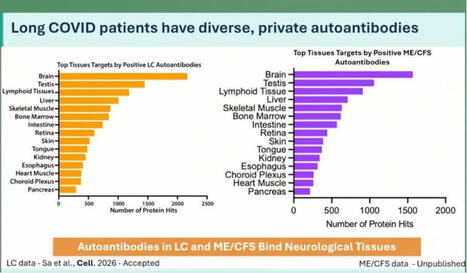
At the ME/CFS conference in Berlin today, Keyla Sá from the Akiko Iwasaki group at Yale showed that Long Covid patients have more autoantibodies targeting neurological tissue. This was apparently tested in both human and mouse tissue.

In ME there are even more autoantibodies than in Long Covid, but number 2 on the list are the cojones.

Interesting. 🍒

Maybe this helps explain why some men develop ME even though the disease is normally more common in women?

Testosterone seems to actually protect against this kind of immune misfire. We see this in research on trans men: https://lnkd.in/ewjxZsRZ

If that protection drops for any reason, the immune system can start attacking your own tissues?

Including your balls?

That’s bullocks!!

https://lnkd.in/eE-giXTv

#pwme #myalgicE #millionsmissing #severeME #MECFSConf2026
No comment yet.
Scooped by Gilbert C FAURE
April 10, 3:59 AM
Scoop.it!

Thymus May Be Critical for Longevity and Cancer Immunotherapy Response | Vincent Geenen

Thymus May Be Critical for Longevity and Cancer Immunotherapy Response | Vincent Geenen | AUTOIMMUNITY | Scoop.it
L'importance du thymus de plus en plus reconnue.
On est loin d'un organe qui n'avait plus d'importance après la puberté.
No comment yet.
Scooped by Gilbert C FAURE
March 26, 4:13 AM
Scoop.it!

#longcovid #autoimmunity #immunesystem #sarscov2 #infection #biology #antibodies | Melvin Sanicas

#longcovid #autoimmunity #immunesystem #sarscov2 #infection #biology #antibodies | Melvin Sanicas | AUTOIMMUNITY | Scoop.it
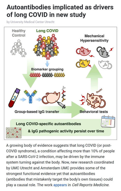
A new study coordinated by UMC Utrecht and Amsterdam UMC provides compelling evidence that #longCOVID may be driven by #autoimmunity - where the #immunesystem turns against the body.

▪️ Affecting over 10% of people after #SARSCoV2 #infection, long COVID is known for symptoms like fatigue, pain, and cognitive dysfunction. Yet its underlying #biology has remained unclear.

▪️In this study, researchers transferred IgG #antibodies from long COVID patients into mice triggering persistent pain-like symptoms that lasted weeks. Remarkably, antibodies collected from the same patients two years later produced the same effect, suggesting a long-lasting disease mechanism.

▪️Co-study leads Prof. Niels Eijkelkamp (UMC Utrecht) and Dr. Jeroen den Dunnen (Amsterdam UMC) highlight two key insights:
- Long COVID may be driven by persistent, pathogenic autoantibodies
- The condition is likely heterogeneous, with distinct biological subtypes

▪️The team also identified diverse autoantibodies targeting immune, neurological, and metabolic pathways - many persisting for years.

▪️These findings, published in Cell Reports Medicine by Cell Press, not only strengthen the case for an autoimmune basis of long COVID but also open the door to targeted treatments like immunoadsorption, plasmapheresis, and precision immunotherapy.

As research converges globally on similar findings, this marks a major step toward understanding - and treating - long COVID.

🗃️ See comments for reference.
No comment yet.
Scooped by Gilbert C FAURE
January 21, 4:18 AM
Scoop.it!

🔬 Histamine, mélanocytes et vitiligo : et si le chaînon manquant était oxydatif ? Le vitiligo est encore trop souvent présenté comme une maladie exclusivement auto-immune. Pourtant, les données… ...

🔬 Histamine, mélanocytes et vitiligo : et si le chaînon manquant était oxydatif ? Le vitiligo est encore trop souvent présenté comme une maladie exclusivement auto-immune. Pourtant, les données… ... | AUTOIMMUNITY | Scoop.it
🔬 Histamine, mélanocytes et vitiligo : et si le chaînon manquant était oxydatif ?
Le vitiligo est encore trop souvent présenté comme une maladie exclusivement auto-immune.
Pourtant, les données accumulées depuis plusieurs années dessinent une lecture plus intégrée, où stress oxydatif, neuro-inflammation et biologie de l’histamine jouent un rôle central.

🧬 Le mélanocyte : une cellule particulièrement vulnérable
Le mélanocyte est une cellule à haute activité métabolique, exposée en permanence à des espèces réactives de l’oxygène (ROS), notamment via la mélanogenèse. Sa survie dépend étroitement de l’équilibre redox local.

🧠 L’histamine : bien plus qu’un médiateur allergique
Dans la peau, l’histamine est libérée par les mastocytes, mais aussi modulée par les kératinocytes, les fibres nerveuses et l’immunité innée.
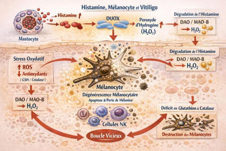
Elle agit comme un amplificateur inflammatoire et oxydatif, en particulier via l’activation des enzymes DUOX, productrices de peroxyde d’hydrogène (H₂O₂) et sa propre dégradation (DAO en extracellulaire, MAO-B en intracellulaire), qui génère elle aussi du H₂O₂.

Un paradoxe peu discuté
Même lorsque l’histamine est “correctement” dégradée, elle augmente la charge oxydative locale. Si les systèmes antioxydants, notamment glutathion et catalase sont insuffisants, le résultat est une accumulation de H₂O₂ toxique pour le mélanocyte.
🔁 Une boucle auto-entretenue
Histamine ↑ → ROS ↑ → stress oxydatif → souffrance mélanocytaire → signaux de danger → activation immunitaire → mastocytes → histamine ↑

🎯 Implication clinique majeure
Le vitiligo peut être relu comme une pathologie de déséquilibre histamino-oxydatif local, où l’auto-immunité apparaît souvent comme une conséquence plus que comme le point de départ.

👉 Cette approche ouvre des pistes complémentaires comme la réduction de la charge histaminique, la protection antioxydante ciblée, la modulation mastocytaire et la prise en compte du terrain neuro-immun et métabolique.
🔍 Changer de prisme, c’est parfois changer le pronostic.
Dr Lucie WETCHOKO
Source de réflexion:
1- constats de consultation
2- Rôle de l'histamine comme médiateur toxique dans la pathogenèse du vitiligo: 10.4103/0019-5154.119947
No comment yet.
Scooped by Gilbert C FAURE
January 14, 4:11 AM
Scoop.it!

The immunology of vitiligo | Nature Reviews Immunology

The immunology of vitiligo | Nature Reviews Immunology | AUTOIMMUNITY | Scoop.it
Vitiligo is an autoimmune disease of melanocyte destruction, which manifests as progressive, patchy loss of pigmentation in the skin. As one of most common autoimmune diseases, vitiligo inflicts a significant psychosocial burden. Research over the past two decades has revealed the underlying immune mechanisms of vitiligo, with key studies combining detailed analyses of patient tissue samples with mechanistic experiments in mouse models. Vitiligo has emerged as a prototypical CD8+ T cell-mediated autoimmune disease, with cooperation between innate immune cells, dendritic cells, T cells, keratinocytes and fibroblasts driving autoimmune pathology against the uniquely susceptible melanocyte target. The study of vitiligo has also revealed aspects of CD8+ T cell memory and resident memory against self-antigens. This work has drawn from, and contributed to, the study of melanoma immunology. Whereas drugs used for other autoimmune conditions have been largely ineffective in treating vitiligo, a growing base of knowledge recently led to the first successful FDA-approved immune-modulating drugs for vitiligo. This review focuses on the immunology of vitiligo: the mechanisms that drive melanocyte destruction, the biology of aberrant T cell responses against melanocytes and therapeutic means for counteracting this autoimmune condition. This Review from Turk and Huang discusses the immune processes involved in the development of vitiligo, an autoimmune disease in which melanocyte destruction causes loss of skin pigmentation. The authors highlight key studies from the past two decades that have shaped our understanding of vitiligo and led to newly approved immune-modulating drugs for the disease.
No comment yet.
Scooped by Gilbert C FAURE
December 29, 2025 9:16 AM
Scoop.it!

#autoimmunedisease #singlecellgenomics #immunology #drugdevelopment #translationalscience | Adam Arterbery, Ph.D.

#autoimmunedisease #singlecellgenomics #immunology #drugdevelopment #translationalscience | Adam Arterbery, Ph.D. | AUTOIMMUNITY | Scoop.it
Autoimmunity, decoded at single-cell resolution: toward mechanism-driven therapies and smarter diagnostics

Autoimmune diseases are often treated as distinct clinical entities, yet patients experience overlapping biology, variable responses to therapy, and chronic immune dysregulation. This new large-scale single-cell study delivers one of the clearest views yet into what is shared versus truly disease-specific across autoimmunity and why that distinction matters for drug development and patients.

By integrating over 1.3 million immune cells across six autoimmune diseases, the authors move beyond descriptive atlases to identify conserved, mechanistic immune programs that cut across diagnoses, alongside disease-selective features that may explain heterogeneity in outcomes and therapeutic response.

Key findings:
◾ A pan-autoimmune cytotoxic CD8 T cell program marked by clonal expansion and enhanced effector function is observed across multiple diseases, pointing to a shared axis of tissue damage driven by antigen-experienced T cells.

◾ CD14+ monocytes emerge as central orchestrators of humoral autoimmunity, promoting plasma cell survival through elevated TNFSF13B (BAFF) signaling, a pathway already clinically validated but now placed in a broader cross-disease cellular context.

◾ Systematic gene-cluster analysis disentangles cell-intrinsic programs from disease-driven transcriptional states, offering a roadmap for biomarker discovery that is more robust than single-gene approaches.

◾ A machine-learning classifier trained on these single-cell signatures achieves high accuracy in distinguishing autoimmune diseases, highlighting the diagnostic potential of immune-state–based classification rather than symptom-based labels.

This work reinforces a shift underway in immunology and therapeutics: diseases may differ at the organ level, but pathogenic immune circuits are often conserved. Targeting these shared circuits, such as cytotoxic CD8 T cell differentiation or monocyte-plasma cell crosstalk, could enable platform therapies spanning multiple indications, while disease-specific modifiers guide patient stratification and combination strategies.

Equally important, the study highlights how single-cell and AI-enabled frameworks can bridge discovery and clinic, enabling earlier mechanism-based diagnosis, smarter trial design, and clearer go/no-go decisions in immunology pipelines.

The challenge now is translation: validating which of these conserved programs are causal, druggable, and safely modulated in patients. Studies like this provide the systems-level blueprint needed to move autoimmune drug development from reactive suppression toward precise immune reprogramming.

Read the full article here: https://lnkd.in/eJFTRpBt

#AutoimmuneDisease #SingleCellGenomics #Immunology #DrugDevelopment #TranslationalScience
No comment yet.
Scooped by Gilbert C FAURE
December 19, 2025 4:35 AM
Scoop.it!

The NEJM has just released the usual end-of-the-year list of most influential papers of 2025. I found particularly amazing the paper and the editorial concerning allogeneic transplantation of… | ...

The NEJM has just released the usual end-of-the-year list of most influential papers of 2025. I found particularly amazing the paper and the editorial concerning allogeneic transplantation of… | ... | AUTOIMMUNITY | Scoop.it
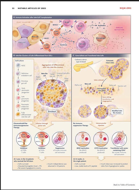
The NEJM has just released the usual end-of-the-year list of most influential papers of 2025.

I found particularly amazing the paper and the editorial concerning allogeneic transplantation of CRISPR genetically-modified Beta cells for type 1 diabetes. A groundbreaking clinical observation that could revolutionise completely this deadly disease. More to come in 2026!

You can download it for free.
No comment yet.
Scooped by Gilbert C FAURE
December 11, 2025 4:06 AM
Scoop.it!

#autoimmunity #kidney #kidneydisease #biomarkers | Nicola Ferrari

#autoimmunity #kidney #kidneydisease #biomarkers | Nicola Ferrari | AUTOIMMUNITY | Scoop.it
Autoantibody-triggered podocyte membrane budding drives autoimmune kidney disease

- Chronic kidney disease affects 1 in 10 people worldwide, with damage to specialized blood filter cells of the kidney, called podocytes, playing a critical role.

- In membranous nephropathy (MN), a major cause of nephrotic syndrome, circulating autoantibodies attack proteins on podocyte foot processes (FPs), damaging the kidney’s filtration barrier.

- This study shows that these autoantibodies trigger the formation of antigen-autoantibody aggregates on the podocyte FP plasma membrane.

- These aggregates bud off as stalked vesicles, termed autoimmunoglobulin-triggered extracellular vesicles (AIT-EVs), which are released into the urine.

- AIT-EVs carry disease-causing autoantibodies, their target antigens, essential FP proteins, and disease-associated stressors representing a mechanism for removing immune complexes (ICs) and waste. However, their excessive release leads to podocyte dysfunction and injury.

- The discovery of AIT-EVs bridges the gap between circulating autoantibodies and podocyte responses and can be harnessed in MN patients.

- Clinically, analysis of urinary AIT-EVs represents a novel non-invasive diagnostic tool to sensitively detect and monitor renal autoimmune activity in patients.

https://lnkd.in/ePD3_xDA

#autoimmunity #kidney #kidneydisease #biomarkers
No comment yet.
Scooped by Gilbert C FAURE
July 16, 10:22 AM
Scoop.it!

JCI Insight - Macrophage signaling associates with fibrogenic program activation in periductal fibroblasts in pediatric primary sclerosing cholangitis

JCI Insight - Macrophage signaling associates with fibrogenic program activation in periductal fibroblasts in pediatric primary sclerosing cholangitis | AUTOIMMUNITY | Scoop.it
Macrophage activity is correlated with advanced fibrosis in AILD. To investigate the association between macrophage activation and fibrosis-associated transcriptional programs across AILD, we performed RNA-Seq of cryopreserved tissue samples from 64 clinically indicated liver biopsies of pediatric...
No comment yet.
Scooped by Gilbert C FAURE
June 22, 10:42 AM
Scoop.it!

JCI Insight - Longitudinal clinical proteomics reveals pneumonia type–specific protein biomarkers and autoantibodies

JCI Insight - Longitudinal clinical proteomics reveals pneumonia type–specific protein biomarkers and autoantibodies | AUTOIMMUNITY | Scoop.it
Mass spectrometry reveals pneumonia type–specific protein signatures in bronchoalveolar fluid. To identify pneumonia type–specific biomarkers, we sampled BALF and plasma from individuals enrolled in the Successful Clinical Response in Pneumonia Therapy (SCRIPT) study at Northwestern Memorial...
No comment yet.
Scooped by Gilbert C FAURE
June 11, 1:45 PM
Scoop.it!

Autoantibodies against Cytokines — From Infection to Inflammation

No comment yet.
Scooped by Gilbert C FAURE
May 26, 3:59 AM
Scoop.it!

Inflammatory Myopathies and Autoantibodies Impact Diagnosis and Prognosis | NEJM Group posted on the topic

Inflammatory Myopathies and Autoantibodies Impact Diagnosis and Prognosis | NEJM Group posted on the topic | AUTOIMMUNITY | Scoop.it
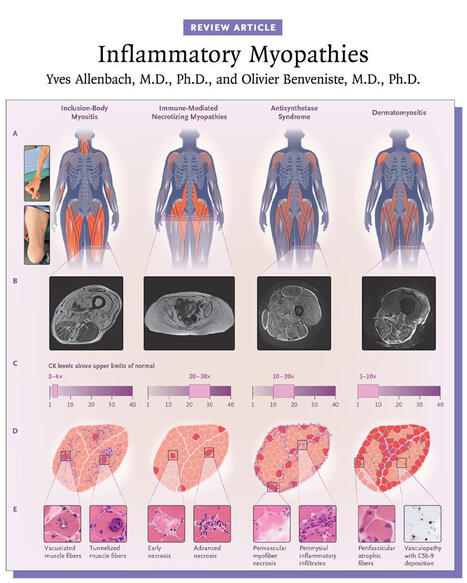
Inflammatory myopathies are typically associated with a myositis-specific autoantibody that determines the diagnosis and prognosis. Myositis subgroups have distinct pathomechanisms that now allow for targeted therapies. 👉 https://nej.md/49Iy9Oo

Inflammatory myopathies are a heterogeneous group of autoimmune diseases characterized by immune-mediated damage to skeletal muscle. They are classified into five major subtypes: inclusion-body myositis, immune-mediated necrotizing myopathies, antisynthetase syndrome, overlapping myositis, and dermatomyositis, each with distinct clinical features and outcomes.

Inclusion-body myositis and immune-mediated necrotizing myopathies primarily affect muscle, with prognosis largely determined by functional impairment, whereas antisynthetase syndrome, overlapping myositis, and dermatomyositis are systemic diseases that can involve the skin, joints, and lungs and may be life-threatening.

The majority of inflammatory myopathies are associated with myositis-specific autoantibodies, which inform diagnosis, subtype classification, and prognosis. Advances in understanding the distinct pathomechanisms underlying each subgroup now enable increasingly targeted therapeutic approaches.

Learn more in the Review Article “Inflammatory Myopathies” by Yves Allenbach, MD, PhD, and Olivier Benveniste, MD, PhD: https://nej.md/49Iy9Oo

#Neurology #Rheumatology
No comment yet.
Scooped by Gilbert C FAURE
May 13, 3:28 AM
Scoop.it!

Germinal-centre and extrafollicular B cell pathways in systemic lupus erythematosus | Nature Reviews Rheumatology

Germinal-centre and extrafollicular B cell pathways in systemic lupus erythematosus | Nature Reviews Rheumatology | AUTOIMMUNITY | Scoop.it
Autoantibody flares are important drivers of pathology in systemic lupus erythematosus (SLE), highlighting the pivotal role of B cells in initiating and propagating chronic autoimmunity. Although autoreactive specificities are a normal feature of the naive B cell repertoire, these cells are normally suppressed by layered tolerance checkpoints that limit inappropriate activation. Autoimmune-prone environments can lower these tolerance thresholds, rendering naive autoreactive B cells more sensitive to aberrant cues. Cytokines and other microenvironmental signals shape tissue niches that direct autoreactive B cells towards either germinal-centre or extrafollicular differentiation pathways. Germinal centres support the entry, selection and diversification of autoreactive B cells, with T cell help sustaining these repertoires. By contrast, naive autoreactive B cells entering the extrafollicular pathway exhibit an attenuated requirement for cognate T cell help and strong dependence on complement and TLR signalling. Emerging evidence continues to refine our understanding of germinal-centre and extrafollicular responses as complementary sources of autoreactive effector cells. With this progress, investigations into the origin, development, longevity and tissue dynamics of autoreactive memory B cells as chronic sources of autoantibodies are warranted. Although broad B cell depletion therapies have yielded benefit, a key challenge now is developing precision strategies that selectively target pathogenic B cell subsets. Autoreactive B cells normally held in check by tolerance checkpoints can be driven towards germinal-centre or extrafollicular differentiation in autoimmune environments. This Review examines the cellular, molecular and contextual cues that shape these pathways and considers their implications for systemic lupus erythematosus pathogenesis and therapy.
No comment yet.
Scooped by Gilbert C FAURE
April 22, 9:47 AM
Scoop.it!

Antiphospholipid syndrome classification - Rheumatology Notebooks | Podcast on

Listen to this episode from Rheumatology Notebooks on Spotify. Podcast on "2023 ACR/EULAR antiphospholipid syndromeclassification criteria".Created with NotebookLM.Intro and outro music created with Suno.Link to the paper: http:// dx.doi. org/ 10. 1136/ ard- 2023-224609
No comment yet.
Scooped by Gilbert C FAURE
April 10, 3:59 AM
Scoop.it!

New research suggests that amyotrophic lateral sclerosis, or ALS, may be partly an autoimmune disease. Scientists discovered that inflammatory immune cells called CD4+ T cells in people with ALS… |...

New research suggests that amyotrophic lateral sclerosis, or ALS, may be partly an autoimmune disease. Scientists discovered that inflammatory immune cells called CD4+ T cells in people with ALS… |... | AUTOIMMUNITY | Scoop.it
New research suggests that amyotrophic lateral sclerosis, or ALS, may be partly an autoimmune disease. Scientists discovered that inflammatory immune cells called CD4+ T cells in people with ALS mistakenly attack a protein found in neurons, known as C9orf72. This self-directed immune response appears to drive rapid disease progression, explaining why ALS can destroy motor neurons quickly and lead to severe physical decline.

The study also revealed two distinct patient groups. One group had highly inflammatory CD4+ T cells and shorter predicted survival, while the second group had more anti-inflammatory CD4+ T cells alongside the harmful ones. These protective T cells appear to regulate the immune response, slowing disease progression and allowing some patients to live significantly longer. Understanding this balance between harmful and protective T cells may help explain why survival in ALS varies dramatically, from just a couple of years to several decades in rare cases.

These findings open the door to potential new treatments that boost protective T cell responses and limit harmful inflammation. Future therapies might harness the immune system to slow ALS progression. The study also has implications for other neurodegenerative diseases, such as Parkinson’s, Huntington’s, and Alzheimer’s, where immune cell involvement may similarly influence disease outcomes.

Research Paper 📄
DOI: 10.1038/s41586-025-09588-6
No comment yet.
Scooped by Gilbert C FAURE
January 15, 4:04 AM
Scoop.it!

#immunology #scrna #omics #autoimmunity | Nicola Ferrari

#immunology #scrna #omics #autoimmunity | Nicola Ferrari | AUTOIMMUNITY | Scoop.it
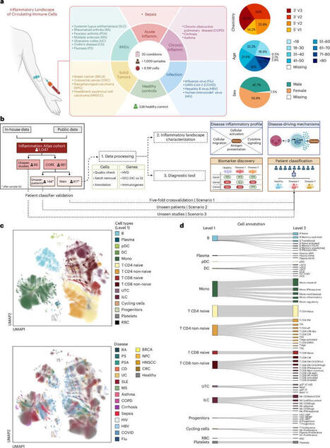
Interpretable inflammation landscape of circulating immune cells

- Inflammation represents an altered state within the immune system, which can manifest as either a protective or a pathological response. The cellular and molecular mediators of inflammation play pivotal roles in nearly every human disease.

- Although major progress has been made in characterizing inflammation in specific diseases, a global, holistic understanding is still elusive.

- In this study, the authors leveraged advances in single-cell transcriptomics to delineate inflammatory processes of circulating immune cells during infection, immune-mediated inflammatory diseases and cancer.

- A single-cell atlas of more than 6.5 million peripheral blood mononuclear cells from 1,047 patients (56% female, 43% male) and 19 diseases was generated allowing to learn a comprehensive model of inflammation in circulating immune cells.

- Beyond a disease-centered classification, the authors modeled the expression profiles of inflammatory molecules to uncover discriminative genes related to immune cell activation, migration, cytotoxic responses and antigen presentation activities.

- A classifier framework based on peripheral blood mononuclear cells (PBMCs) was generated, demonstrating the potential of circulating immune cells to contribute to precision medicine strategies for patients suffering from acute or chronic inflammation.

- As limitations of this study:
>Most samples derive from individuals of European ancestry, and expanding to ancestrally diverse populations will be essential to capture global immune variability.
>The classifier requires prospective validation in independent, multicenter cohorts to assess robustness and clinical applicability.
> Understanding the relationship between circulating and tissue-resident immune cells remains key for diagnostic translation.

https://lnkd.in/d7bKEg4T

#immunology #scRNA #omics #autoimmunity
No comment yet.
Scooped by Gilbert C FAURE
January 12, 6:41 AM
Scoop.it!

#scicomm | Kristin Rose Jutras, M.F.A.

#scicomm | Kristin Rose Jutras, M.F.A. | AUTOIMMUNITY | Scoop.it
More excellent #scicomm from Unbiased Science written by Aimee Pugh Bernard, PhD, Leigh Baxt, and Jess Steier. Learn more about the science behind Lupus and new treatments on the horizon for this autoimmune disease likened to "the wolf within."
No comment yet.
Scooped by Gilbert C FAURE
December 28, 2025 5:29 AM
Scoop.it!

Discovering Metabolic Diversity in Sjögren’s Syndrome

Discovering Metabolic Diversity in Sjögren’s Syndrome | AUTOIMMUNITY | Scoop.it
Recent advancements in the field of biomedical research have unveiled profound insights into Sjögren’s syndrome, a chronic autoimmune disorder primarily affecting the exocrine glands. This condition is notorious for causing significant dry mouth and dry eyes, but its effects extend far beyond...
No comment yet.
Scooped by Gilbert C FAURE
December 18, 2025 4:42 AM
Scoop.it!

#metabolichealth #fattyliver #cardiovascularhealth #prevention #publichealth | Erand Llanaj, PhD

#metabolichealth #fattyliver #cardiovascularhealth #prevention #publichealth | Erand Llanaj, PhD | AUTOIMMUNITY | Scoop.it
Fatty liver disease is not just a liver problem.
It is a whole-body metabolic disease.

A major review by Prof. Norbert Stefan, Hannele Yki-Järvinen and Brent A. Neuschwander-Tetri shows that metabolic dysfunction-associated steatotic liver disease affects nearly 4 in 10 adults worldwide and most people with it do not die from liver failure, but from heart disease and cancer…

Important:

• Fatty liver comes in different types, driven by metabolism, fat distribution, or genetics
• The liver actively worsens blood sugar, cholesterol, and clotting risk
• Weight loss helps many…but not everyone responds the same way
• Some treatments improve liver health even without weight loss

The key message is simple:

One-size-fits-all medicine does not work for fatty liver disease!!!

Prevention, early detection and treatment need to focus on metabolic health across the whole body, not just liver enzymes…

If we treat fatty liver early, we also reduce diabetes, heart attacks and strokes later 🤞🏼

What should come first in routine care: liver screening, metabolic screening or both?

If useful, save, share or add your perspective below.

Read original review in The Lancet Group: https://lnkd.in/g3m4tA3k

#metabolichealth #fattyliver #cardiovascularhealth #prevention #publichealth
No comment yet.
Scooped by Gilbert C FAURE
December 9, 2025 4:12 AM
Scoop.it!

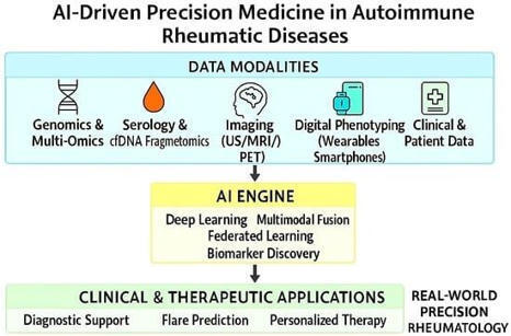
Emerging AI- and Biomarker-Driven Precision Medicine in Autoimmune Rheumatic Diseases: From Diagnostics to Therapeutic Decision-Making | Rheumato MDPI

Emerging AI- and Biomarker-Driven Precision Medicine in Autoimmune Rheumatic Diseases: From Diagnostics to Therapeutic Decision-Making | Rheumato MDPI | AUTOIMMUNITY | Scoop.it
🔥Don't miss this #FeaturePaper by Moawiah Naffaa, PhD and Ola A. Al-Ewaidat.

Emerging AI- and Biomarker-Driven Precision Medicine in Autoimmune Rheumatic Diseases: From Diagnostics to Therapeutic Decision-Making


🔗More details: https://brnw.ch/21wY2dx


#OpenAccess #AutoimmuneRheumaticDiseases #ArtificialIntelligence #Biomarkers #DigitalHealth
No comment yet.