Your new post is loading...

|
Scooped by
Gilbert C FAURE
August 2, 2021 4:00 AM
|
Th2 Cell Th2 cells stimulate B cell and eosinophil proliferation and reduce IFN-γ production by Th1 cells, thereby promoting humoral and allergic responses. From: Neurobiology of Disease, 2007 Related terms: View all Topics Effector CD4+ T Cells in the Intestines Craig L. Maynard, Casey T. Weaver, in Mucosal Immunology (Fourth Edition), 2015 Th2 Cells Th2 cells augment the eradication of parasitic helminthes that induce expression of IL-4 by innate immune cells, such as basophils and tissue-resident mast cells. IL-4 signaling to antigen-activated, previously naïve CD4 T cells results in activation of STAT6 and subsequent induction of the transcription factor GATA-3 (Bonecchi et al., 1998). Via secretion of IL-4, IL-5, and IL-13, Th2 cells orchestrate B cell class switching to IgE (Bonecchi et al., 1998), thereby priming basophils and mast cells for granule release, recruit eosinophils, and enhance mucus production, respectively. Human Th2 cells can be distinguished by surface expression of CCR4 and CRTH2 (Bonecchi et al., 1998; Abe et al., 1999; Nagata et al., 1999). Host Defenses in Skin Hui Xu, ... Craig A. Elmets, in Clinical Immunology (Fifth Edition), 2019 Th2 responses. Th2 cells are involved in type 2 immune responses, which are important for eradication of extracellular parasites and bacterial infection. They produce IL-4, IL-5, IL-10, and IL-13, which are important for the induction and development of humoral immune responses. IL-4 and IL-13 activate B-cell proliferation, Ig class-switching, and antibody production. Th2 cell-mediated inflammation is characterized by the presence of eosinophils and basophils, as well as extensive mast cell degranulation—a process dependent on cross-linking surface-bound IgE.24 IL-5 is a potent hematopoietic cytokine, which stimulates bone marrow production of eosinophils as well as activation and chemotaxis of eosinophils and basophils to affected tissue. In mice, Th2-cell deficiency profoundly increases susceptibility to Leishmania infection in skin. In humans, Th2 cells appear to play a critical role in the pathogenesis of atopic dermatitis (Chapter 44). A recent clinical trial with dupilumab, a fully human mAb that targets the IL-4 receptor-αα and blocks IL-4 and IL-13 signaling, improved atopic symptoms . Role of CD4+ T Cells in the Pathophysiology of Multiple Sclerosis Fumitaka Sato, ... Ikuo Tsunoda, in Multiple Sclerosis, 2016 Role of Th2 cells Th2 cells may play a protective role in MS, as Th2 immune responses have been shown to increase during remission in RRMS (Araki et al., 2003; Clerici et al., 2001). Decreased disease progression and exacerbation of MS during pregnancy have been associated with Th2-biased immune responses (Al-Shammri et al., 2004), although the exact mechanism remains unclear. Suppression of MS disease activities by immunomodulatory drugs, such as glatiramer acetate, has also been associated with enhanced Th2 immune responses (Weber et al., 2007). Experimentally, Th2 cells have been shown to regulate EAE and TMEV-IDD. In EAE induced with mouse spinal cord homogenate, injection of anti-IL-4 neutralizing mAb during the induction phase rendered resistant BALB/c mice susceptible to EAE (Constantinescu et al., 2001). The adoptive transfer of PLP-specific Th2 cell clones at the time of sensitization or disease onset prevented EAE in mice sensitized with PLP (Kuchroo et al., 1995). While T cell immunoglobulin mucindomain containing (TIM)2 has been shown to be preferentially expressed on the surface of Th2 cells and to negatively regulate Th2 immune responses, blockade of TIM-2/TIM-2 ligand interaction by administration of soluble TIM-2 fusion protein delayed the onset and decreased the severity of PLP-induced EAE by enhancing Th2 immune responses (Chakravarti et al., 2005). In TMEV-IDD, Th2 immune responses have also been demonstrated to suppress inflammatory demyelination in the CNS. Hill et al. (1998) demonstrated that during the early chronic phase of TMEV infection, infected mice treated with IL-4 developed less severe inflammatory demyelination compared with controls. Thus, the findings in EAE and TMEV-IDD suggest that Th1 cells could contribute to the pathogenesis of MS, while Th2 cells may play a protective role (Table 3). Cell-Mediated Defense against Infection Tobias M. Hohl, in Mandell, Douglas, and Bennett's Principles and Practice of Infectious Diseases (Eighth Edition), 2015 Th2 Cells Th2 cells express a range of cytokines that influence B-cell differentiation and antibody production, eosinophil recruitment, and mucus production. The signature cytokines produced by Th2 cells are IL-4, IL-5, and IL-13, but Th2 cells can also produce IL-9, IL-10, IL-25, and amphiregulin.20 Th2 responses are generated when naïve T cells are exposed to IL-4 at the time of T-cell priming. In the setting of low antigen concentrations, IL-4 can be produced by responding T cells.21 After antigenic challenge, IL-4 can also be produced by mast cells and basophils in the vicinity of T-cell priming.22,23 IL-4 signals naïve T cells via the STAT6 pathway to express GATA3, the master regulator of Th2 differentiation,24 a process that can be enhanced by IL-4– and STAT6-independent GATA3 activation,25 all of which drives the expression of additional downstream activators. Although Th2 cells are best known for causing or contributing to allergic diseases such as atopic dermatitis, allergic rhinitis, and asthma, Th2 cells also contribute to defense against infections, particularly helminth infections of the gastrointestinal tract.26 In this setting, eosinophil recruitment, IgE production, and mucus hypersecretion can enhance parasite expulsion in an IL-4 and IL-13 signaling–dependent manner, a notion that is supported by murine studies of Nippostrongylus brasiliensis infection.27,28 The secretion of amphiregulin by Th2 cells can stimulate intestinal epithelial cell proliferation and expulsion of Trichuris muris, a nematode that infects mice.29 Besides Th2 cells, tissue-resident and Th2 cytokine-secreting innate lymphoid cells represent a significant source of IL-13 during the early stages of parasitic infection and promote expulsion.30-32 Aberrant Th2 responses to pathogens that require IFN-γ and Th1 responses for control can result in progressive infections and lethality. For example, Leishmania major infection of certain mouse strains induces Th2 responses that result in progressive in vivo replication and host death.33,34 In contrast, mouse strains that respond to L. major with Th1 responses clear and survive experimental infections. The mechanisms that determine whether an L. major–specific T-cell response will be predominately Th1 or Th2 are complex.35 In some mouse strains, Th2 responses occur because of T-cell responses to one dominant antigen called LACK (Leishmania analogue of the receptors of activated C kinase).36 In the absence of a T-cell response to this specific antigen, the responding CD4+ T cells differentiate into Th1 cells. In humans, the type of disease associated with Mycobacterium leprae infection is also tied to CD4+ T-cell differentiation. Th1 differentiation is associated with tuberculoid leprosy, a paucibacillary infection in which IFN-γ–producing T cells enhance microbial killing. The induction of type I interferon and IL-10 signaling in innate immune cells during leprosy can antagonize IFN-γ–dependent protection.37 Th2 differentiation is associated with high tissue densities of M. leprae and more robust, but ineffective, antibody responses.38,39 T Cells and Their Effector Functions Ruben C. Fragoso, ... Steven J. Burakoff, in Encyclopedia of Cancer (Second Edition), 2002 IV.B.2 Th2 T Cells Th2 cells promote IgE production and eosinophil function, which are the key players in the pathogenesis of allergic inflammation and immunity against parasitic infections. Cytokines such as IL-4 and IL-5 released by Th2 cells stimulate, respectively, B-cell switching to the production of IgE antibody and activation of eosinophils. The coordinate actions of these effector mechanisms result in heightened immunity against, for example, helminthic parasites, which can be coated with IgE and destroyed by the toxic granular contents of eosinophils. The balance between Th1 and Th2 cells may serve to determine the outcome of an infection. The Th1-mediated response is an effective deterrent for the protozoan parasite Leishmania major. In strains of mice with a genetic predisposition to mount predominately Th2 responses, infection by L. major results in a severe cutaneous and systemic disease that cannot be eliminated effectively. In contrast, if mice were vaccinized with leishmania antigens coadministered with IL-12 to induce a Th1 response, the mice are protected from subsequent challenges with L. major. In an analogous manner, responses to Mycobacterium leprae in humans can have two sharply different outcomes depending on the polarization of Th cells. In lepromatous leprosy, a Th2-dominated response can result in diffuse and destructive lesions due to an ineffective response against M. leprae antigens. In contrast, patients who develop a strong Th1-mediated immunity have a less destructive disease called tuberculoid leprosy. T-Cell Immunity Shannon A. Carty, ... Gary A. Koretzky, in Hematology (Seventh Edition), 2018 Th2 Cells Th2 cells are critical for the immune response against extracellular parasites, such as helminths, through production of IL-4, IL-5, and IL-13. At initial sites of parasitic infection, epithelial cells of the target organs, including the skin, lungs, and intestines, and resident cells of the innate immune system sense parasite-derived products and produce Th2-inducing cytokines, including thymic stromal lymphopoietin (TSLP), IL-4, IL-25, and IL-33. These cytokines then act on innate immune cells, including basophils and DCs, as well as directly on naive CD4+ cells to promote Th2 differentiation. Recent work has provided insight into how cytokine signaling, particularly IL-4 signaling, promotes Th2 differentiation. Through interaction with its receptor, IL-4 activates STAT6. STAT6 plays a vital role in Th2 differentiation, as evidenced by the profound reduction in development of this lineage in Stat6-deficient mice. STAT6 activation leads to its nuclear translocation and subsequent induction of the transcription factor GATA3, which, like T-bet for Th1 cells, is considered the master regulator of Th2 differentiation. GATA3 regulates Th2 cytokine production by binding and activating the “Th2 locus,” which includes the genes encoding IL-4, IL-5, and IL-13. When GATA3 function is abrogated, Th2 differentiation is virtually absent both in vitro and in vivo. In mature differentiated Th2 cells, GATA3 deficiency results in loss of IL-5 and IL-13 production. GATA3 is both necessary and sufficient for Th2 differentiation because forced expression either by retroviral constructs or transgenic expression promotes Th2 differentiation and represses Th1 differentiation. Repression of Th1 development occurs at least partially through GATA3-dependent inhibition of STAT4, thus interfering with Ifng gene transcription. TCR signal strength also is involved in determining if a naive T cell will differentiate into a Th1 or Th2 cell. Studies in mice using altered peptide ligands that have decreased affinity for particular TCRs and experiments using limiting doses of antigen have demonstrated that diminished TCR stimulation promotes Th2 cell differentiation. Differences in costimulation also affect Th2 pathway differentiation. Mice deficient in CD28 or its ligand have a more pronounced defect in Th2 responses, suggesting that these molecules may play a greater role in promoting Th2 differentiation than Th1 differentiation. IL-4 produced by mature Th2 cells acts in a positive feedback loop to promote further Th2 cell differentiation in naive T cells as they encounter antigen. Th2-derived IL-4 also mediates IgE class switching in B cells. Soluble IgE binds to and crosslinks its high-affinity receptor FcεRI on basophils and mast cells, promoting production of histamine and serotonin as well as several cytokines, including IL-4, IL-13, and TNF-α. IL-5 produced from Th2 cells recruits eosinophils, whereas Th2-derived IL-13 promotes both the expulsion of helminths during parasitic infection and also the induction of airway hypersensitivity. Th2 responses are critical for immunity against extracellular parasites, but excessive Th2 responses are associated with the pathologic conditions of allergy and airway hypersensitivity. The increase in asthma in the developed world has been linked to an imbalance of Th subsets with skewing toward “Th2-ness” in the population. Additional work is necessary to more firmly establish a molecular immunologic link to the epidemiology of these diseases. Chronic Inflammation and Atherosclerosis Jan Nilsson, ... Andreas Edsfeldt, in Early Vascular Aging (EVA), 2015 Interleukin-10 Th2 cells, Tregs, B-cells, monocytes, and macrophages are all potential sources of IL-10. The anti-inflammatory effects of IL-10 are mediated by inhibition of T-cell proliferation, macrophage apoptosis, antigen presentation, collagenase expression, and inflammatory cytokine production. In mice, IL-10 deficiency is associated with increased inflammatory cell invasion, a greater plaque burden, and an increased inflammatory cytokine response [40]. Human studies on circulating IL-10 revealed that high plasma levels of IL-10 are associated with an improved outcome and a lower risk for recurrent events in patients with acute coronary syndromes [41,42]. Group 2 Innate Lymphoid Cells in the Regulation of Immune Responses Ben Roediger, Wolfgang Weninger, in Advances in Immunology, 2015 7.8 IL-4/IL-4Rα Like Th2 cells, ILC2 cells express a functional IL-4 receptor (Doherty et al., 2012; Motomura et al., 2014), at least in the lung, and have been shown to produce IL-13 and IL-9 in response to IL-4 in vitro (Motomura et al., 2014). IL-4 was also shown to augment IL-2-driven proliferation of ILC2 cells in vitro (Motomura et al., 2014), which may relate to the STAT6 dependency of ILC2 cell proliferation in vivo (discussed further below). Animal Models of Immunity to Female Genital Tract Infections and Vaccine Development Charu Kaushic, ... Kenneth W. Beagley, in Mucosal Immunology (Fourth Edition), 2015 Th2 Cells CD4+ Th2 cells do not protect against chlamydial infection (Wang et al., 1999; Yang, 2001; Hawkins et al., 2002) and can exacerbate pathology (Chen et al., 2010; Wang et al., 1999; Perry et al., 1997) because of suppression of Th1 immunity. However, activation of Th2 cells is important for the production of IgG and IgA, both of which reduce infection in vivo. Th2 cells also may act as regulators of the Th1 response to limit tissue pathology after resolution of infection (Debattista et al., 2003). Indeed, it has been suggested that a human vaccine to prevent ascending infection and tissue inflammation should aim to elicit primarily a Th2 response to limit collateral damage (Vicetti Miguel and Cherpes, 2012). This approach would certainly be contrary to the current dogma driving vaccine research (see below).
|
|
Scooped by
Gilbert C FAURE
July 30, 2021 11:47 AM
|
Memory T cells comprise the most abundant lymphocyte population in the body for the majority of one’s lifetime; however, our understanding of memory T cell generation, function and maintenance mainly derives from mouse studies, which cannot recapitulate ...
|
|
Scooped by
Gilbert C FAURE
July 27, 2021 3:22 AM
|
Exhausted CD8 T cells (TEX) are a distinct state of T cell differentiation associated with failure to clear chronic viruses and cancer. Immunotherapies such as PD-1 blockade can reinvigorate TEX cells, but reinvigoration is not durable. A major unanswered question is whether TEX cells differentiate into functional durable memory T cells (TMEM) upon antigen clearance. Here, using a mouse model, we found that upon eliminating chronic antigenic stimulation, TEX cells partially (re)acquire phenotypic and transcriptional features of TMEM cells. These ‘recovering’ TEX cells originated from the T cell factor (TCF-1+) TEX progenitor subset. Nevertheless, the recall capacity of these recovering TEX cells remained compromised as compared to TMEM cells. Chromatin-accessibility profiling revealed a failure to recover core memory epigenetic circuits and maintenance of a largely exhausted open chromatin landscape. Thus, despite some phenotypic and transcriptional recovery upon antigen clearance, exhaustion leaves durable epigenetic scars constraining future immune responses. These results support epigenetic remodeling interventions for TEX cell–targeted immunotherapies. Wherry and colleagues examine whether exhausted T cells (TEX) can differentiate into functional memory T cells (TMEM) when chronic antigen is withdrawn. Using the chronic LCMV infection mouse model, they show that ‘recovering’ TEX cells (REC-TEX) only partially recover immunophenotypic and functional characteristics of TMEM cells. The epigenomic status of REC-TEX cells more closely resembles that of TEX cells, and, upon rechallenge, the REC-TEX cells were still compromised in their ability to respond to virus.
|
|
Scooped by
Gilbert C FAURE
July 5, 2021 1:31 PM
|
Dermatitis encompasses a spectrum of inflammatory skin disorders with aberrant immune
responses classified as type 1, type 2, and/or type 3. Major advances in the understanding
of the pathogenesis of atopic dermatitis (AD) have shed new light on how innate immune
responses critically regulate type 2 inflammation and itch. This article highlights
the diverse ways by which type 2 immune cells regulate diseases beyond AD. The discovery
of human Mas-related G protein–coupled receptor X2 on mast cells has revealed novel
T cell–independent and immunoglobulin E–independent mechanisms of allergic contact
dermatitis–associated and urticarial itch, respectively.
|
|
Scooped by
Gilbert C FAURE
July 1, 2021 3:04 PM
|
Macrophages have a key role in shaping the tumour microenvironment (TME), tumour immunity and response to immunotherapy, which makes them an important target for cancer treatment1,2. However, modulating macrophages has proved extremely difficult, as we still lack a complete understanding of the molecular and functional diversity of the tumour macrophage compartment. Macrophages arise from two distinct lineages. Tissue-resident macrophages self-renew locally, independent of adult haematopoiesis3–5, whereas short-lived monocyte-derived macrophages arise from adult haematopoietic stem cells, and accumulate mostly in inflamed lesions1. How these macrophage lineages contribute to the TME and cancer progression remains unclear. To explore the diversity of the macrophage compartment in human non-small cell lung carcinoma (NSCLC) lesions, here we performed single-cell RNA sequencing of tumour-associated leukocytes. We identified distinct populations of macrophages that were enriched in human and mouse lung tumours. Using lineage tracing, we discovered that these macrophage populations differ in origin and have a distinct temporal and spatial distribution in the TME. Tissue-resident macrophages accumulate close to tumour cells early during tumour formation to promote epithelial–mesenchymal transition and invasiveness in tumour cells, and they also induce a potent regulatory T cell response that protects tumour cells from adaptive immunity. Depletion of tissue-resident macrophages reduced the numbers and altered the phenotype of regulatory T cells, promoted the accumulation of CD8+ T cells and reduced tumour invasiveness and growth. During tumour growth, tissue-resident macrophages became redistributed at the periphery of the TME, which becomes dominated by monocyte-derived macrophages in both mouse and human NSCLC. This study identifies the contribution of tissue-resident macrophages to early lung cancer and establishes them as a target for the prevention and treatment of early lung cancer lesions. Single-cell RNA sequencing and imaging of macrophages in human non-small cell lung cancer and in a mouse model of lung adenocarcinoma show that tissue-resident macrophages have a key role in early tumour progression.
|
|
Scooped by
Gilbert C FAURE
June 27, 2021 1:51 PM
|
Granulomas are complex cellular structures composed predominantly of macrophages and lymphocytes that function to contain and kill invading pathogens. Here, we investigated the single-cell phenotypes associated with antimicrobial responses in human leprosy granulomas by applying single-cell and spatial sequencing to leprosy biopsy specimens. We focused on reversal reactions (RRs), a dynamic process whereby some patients with disseminated lepromatous leprosy (L-lep) transition toward self-limiting tuberculoid leprosy (T-lep), mounting effective antimicrobial responses. We identified a set of genes encoding proteins involved in antimicrobial responses that are differentially expressed in RR versus L-lep lesions and regulated by interferon-γ and interleukin-1β. By integrating the spatial coordinates of the key cell types and antimicrobial gene expression in RR and T-lep lesions, we constructed a map revealing the organized architecture of granulomas depicting compositional and functional layers by which macrophages, T cells, keratinocytes and fibroblasts can each contribute to the antimicrobial response. Modlin and colleagues examined the skin lesions of human leprosy patients using single-cell RNA sequencing coupled to cellular spatial mapping. Their analysis maps the architecture of granulomas in leprosy lesions from patients with leprosy with localized disease (tuberculoid leprosy, reversal reaction) to those with progressive infection (lepromatous leprosy).
|
|
Rescooped by
Gilbert C FAURE
from Virus World
June 24, 2021 3:01 AM
|
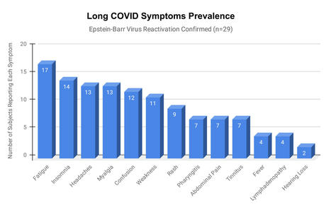
Epstein-Barr virus (EBV) reactivation resulting from the inflammatory response to coronavirus infection may be the cause of previously unexplained long COVID symptoms—such as fatigue, brain fog, and rashes—that occur in approximately 30% of patients after recovery from initial COVID-19 infection. The first evidence linking EBV reactivation to long COVID, as well as an analysis of long COVID prevalence, is outlined in a new long COVID study published in the journal Pathogens. "We ran EBV antibody tests on recovered COVID-19 patients, comparing EBV reactivation rates of those with long COVID symptoms to those without long COVID symptoms," said lead study author Jeffrey E. Gold of World Organization. "The majority of those with long COVID symptoms were positive for EBV reactivation, yet only 10% of controls indicated reactivation." The researchers began by surveying 185 randomly selected patients recovered from COVID-19 and found that 30.3% had long term symptoms consistent with long COVID after initial recovery from SARS-CoV-2 infection. This included several patients with initially asymptomatic COVID-19 cases who later went on to develop long COVID symptoms. The researchers then found, in a subset of 68 COVID-19 patients randomly selected from those surveyed, that 66.7% of long COVID subjects versus 10% of controls were positive for EBV reactivation based on positive EBV early antigen-diffuse (EA-D) IgG or EBV viral capsid antigen (VCA) IgM titers. The difference was significant (p < 0.001, Fisher's exact test). "We found similar rates of EBV reactivation in those who had long COVID symptoms for months, as in those with long COVID symptoms that began just weeks after testing positive for COVID-19," said coauthor David J. Hurley, Ph.D., a professor and molecular microbiologist at the University of Georgia. "This indicated to us that EBV reactivation likely occurs simultaneously or soon after COVID-19 infection." The relationship between SARS-CoV-2 and EBV reactivation described in this study opens up new possibilities for long COVID diagnosis and treatment. The researchers indicated that it may be prudent to test patients newly positive for COVID-19 for evidence of EBV reactivation indicated by positive EBV EA-D IgG, EBV VCA IgM, or serum EBV DNA tests. If patients show signs of EBV reactivation, they can be treated early to reduce the intensity and duration of EBV replication, which may help inhibit the development of long COVID. "As evidence mounts supporting a role for EBV reactivation in the clinical manifestation of acute COVID-19, this study further implicates EBV in the development of long COVID," said Lawrence S. Young, Ph.D., a virologist at the University of Warwick, and Editor-in-Chief of Pathogens. "If a direct role for EBV reactivation in long COVID is supported by further studies, this would provide opportunities to improve the rational diagnosis of this condition and to consider the therapeutic value of anti-herpesvirus agents such as ganciclovir." Original findings published in Pathogens (June 10, 2021): https://doi.org/10.3390/pathogens10060763
Via Juan Lama
|
|
Scooped by
Gilbert C FAURE
June 19, 2021 10:26 AM
|
Abstract Autopsies of patients who have died from COVID-19 have been crucial in delineating patterns of injury associated with SARS-CoV-2 infection. Despite their utility, comprehensive autopsy stu...
|
|
Scooped by
Gilbert C FAURE
June 15, 2021 5:00 AM
|
|
|
Scooped by
Gilbert C FAURE
June 13, 2021 3:40 AM
|
|
|
Scooped by
Gilbert C FAURE
June 11, 2021 3:10 AM
|
Les variants qui apparaissent toujours plus différenciés et plus tenaces sont-ils un péril pour la sortie du tunnel ?
|
|
Scooped by
Gilbert C FAURE
June 4, 2021 2:27 AM
|
medRxiv - The Preprint Server for Health Sciences
Via HAS-veille
|
|
|
Scooped by
Gilbert C FAURE
July 31, 2021 2:57 AM
|
Incontournables dans la coagulation, les plaquettes multiplient les rôles. Comme on le découvre, elles sont des éléments clés dans l’inflammation, l’immunité, la régénération des tissus et le cancer.
|
|
Scooped by
Gilbert C FAURE
July 29, 2021 4:54 AM
|
22–29 April 2021 is World Primary Immunodeficiency Week. Primary immunodeficiencies (PIs or PIDs) are caused by hereditary gene mutations that result in the disruption of normal immune function. There are over 380 different chronic PID disorders, ranging widely in severity. Severe combined immune deficiencies (SCIDs), which are characterised by the disturbed development of T
|
|
Scooped by
Gilbert C FAURE
July 8, 2021 6:22 AM
|
T follicular helper (Tfh) cells cognately guide differentiation of antigen-primed B cells in secondary lymphoid tissues. ‘Tfh-like’ populations not ex…

|
Suggested by
LIGHTING
July 2, 2021 6:49 AM
|
The pathophysiology of adverse events following programmed cell death protein 1 (PD-1) blockade, including tuberculosis (TB) and autoimmunity, remains poorly characterized. We studied a patient with inherited PD-1 deficiency and TB who died of pulmonary autoimmunity. The patient’s leukocytes did not express PD-1 or respond to PD-1-mediated suppression. The patient’s lymphocytes produced only small amounts of interferon (IFN)-γ upon mycobacterial stimuli, similarly to patients with inborn errors of IFN-γ production who are vulnerable to TB. This phenotype resulted from a combined depletion of Vδ2+ γδ T, mucosal-associated invariant T and CD56bright natural killer lymphocytes and dysfunction of other T lymphocyte subsets. Moreover, the patient displayed hepatosplenomegaly and an expansion of total, activated and RORγT+ CD4−CD8− double-negative αβ T cells, similar to patients with STAT3 gain-of-function mutations who display lymphoproliferative autoimmunity. This phenotype resulted from excessive amounts of STAT3-activating cytokines interleukin (IL)-6 and IL-23 produced by activated T lymphocytes and monocytes, and the STAT3-dependent expression of RORγT by activated T lymphocytes. Our work highlights the indispensable role of human PD-1 in governing both antimycobacterial immunity and self-tolerance, while identifying potentially actionable molecular targets for the diagnostic and therapeutic management of TB and autoimmunity in patients on PD-1 blockade. Dysregulated immune features in a patient with a homozygous loss-of-function mutation in PDCD1 suggest that IL-6, IL-23, STAT3 and RORγT might be potential targets for treatment of PD-1 blockade-induced autoimmunity.
|
|
Scooped by
Gilbert C FAURE
June 30, 2021 4:54 AM
|
Immunocompromised individuals, such as patients with haematological malignancies who have recently undergone haematopoietic stem cell transplantation, may not be protected effectively from COVID-1
|
|
Suggested by
LIGHTING
June 24, 2021 7:04 AM
|
Cancer immunotherapy offers substantive benefit to patients with various tumour types, in some cases leading to complete tumour clearance. However, many patients do not respond to immunotherapy, galvanizing the field to define the mechanisms of pre-existing and acquired resistance. Interferon-γ (IFNγ) is a cytokine that has both protumour and antitumour activities, suggesting that it may serve as a nexus for responsiveness to immunotherapy. Many cancer immunotherapies and chemotherapies induce IFNγ production by various cell types, including activated T cells and natural killer cells. Patients resistant to these therapies commonly have molecular aberrations in the IFNγ signalling pathway or express resistance molecules driven by IFNγ. Given that all nucleated cells can respond to IFNγ, the functional consequences of IFNγ production need to be carefully dissected on a cell-by-cell basis. Here, we review the cells that produce IFNγ and the different effects of IFNγ in the tumour microenvironment, highlighting the pleiotropic nature of this multifunctional and abundant cytokine. Vignali and colleagues discuss the pleiotropic roles of interferon-γ (IFNγ) in the tumour microenvironment; on the one hand, it acts as a ‘teammate’ to promote antitumour immune responses and, on the other hand, it acts as an ‘opponent’ promoting tumour growth and suppressing immune responses.
|
|
Scooped by
Gilbert C FAURE
June 24, 2021 3:00 AM
|
This study describes the multiple facets and molecular basis of excessive Th2 cytokine production by CD4+ αβ T lymphocytes as a possible mechanism underlying pe
|
|
Scooped by
Gilbert C FAURE
June 17, 2021 3:35 PM
|
|
|
Suggested by
LIGHTING
June 14, 2021 8:14 AM
|
The release of neutrophil extracellular traps (NETs), a process termed NETosis, avoids pathogen spread but may cause tissue injury. NETs have been found in severe COVID-19 patients, but their role in disease development is still unknown. The aim of this study is to assess the capacity of NETs to drive epithelial-mesenchymal transition (EMT) of lung epithelial cells and to analyze the involvement of NETs in COVID-19. Bronchoalveolar lavage fluid of severe COVID-19 patients showed high concentration of NETs that correlates with neutrophils count; moreover, the analysis of lung tissues of COVID-19 deceased patients showed a subset of alveolar reactive pneumocytes with a co-expression of epithelial marker and a mesenchymal marker, confirming the induction of EMT mechanism after severe SARS-CoV2 infection. By airway in vitro models, cultivating A549 or 16HBE at air-liquid interface, adding alveolar macrophages (AM), neutrophils and SARS-CoV2, we demonstrated that to trigger a complete EMT expression pattern are necessary the induction of NETosis by SARS-CoV2 and the secretion of AM factors (TGF-β, IL8 and IL1β). All our results highlight the possible mechanism that can induce lung fibrosis after SARS-CoV2 infection.
|
|
Scooped by
Gilbert C FAURE
June 12, 2021 4:39 AM
|
NK cells are considered an important component of innate immunity, which is the first line of defensing against tumors and viral infections in the absence of prior sensitization. NK cells express an array of germline-encoded receptors, which allow them to eliminate abnormal cells and were previously considered a homogenous population of innate lymphocytes, with limited phenotypic and functional diversity. Although their characteristics are related to their developmental origins, other factors, such as tumors and viral infections, can influence their phenotype. Here, we provide an overview of NK cells in the context of the tumor microenvironment, with a primary focus on their phenotypes, functions, and roles in tumor micro-environment. A comprehensive understanding of NK cells in the tumor microenvironment will provide a theoretical basis for the development of NK cell immunotherapy.
|
|
Scooped by
Gilbert C FAURE
June 8, 2021 3:08 PM
|
In this study, we found that patients suffering from severe COVID-19 presented with immature, activated blood neutrophils and were characterized by elevated plasma levels of G-CSF and CXCL8 tha
|
|
Scooped by
Gilbert C FAURE
June 2, 2021 2:20 PM
|
Pathways identified that either boost or hinder tumour dormancy.
|


 Scoop.it!