


Complement component 8 deficiency and its relationship with genes C8A and C8B have been added to Genetics Home Reference.


|
Scooped by
Gilbert C FAURE
onto Immunology December 20, 2015 1:47 PM
|
 Scoop.it!
Complement component 8 deficiency and its relationship with genes C8A and C8B have been added to Genetics Home Reference.

 Your new post is loading...
Scoop.it!
|
|
|
Scooped by
Gilbert C FAURE
November 6, 10:10 AM
|
Scoop.it!

🧬 How naïve are naïve T cells, really?
We often think of naïve T cells as the immune system’s blank slate: resting cells waiting for their first encounter with an antigen. But new research from UMC Utrecht challenges this textbook view.
🔬 In her PhD research, Elise Saager (UMC Utrecht) investigated how age, tissue location and immune challenges shape naïve T cells, revealing three important findings:
🔹 Primed for action: Naïve T cells in the gut, particularly early in life, already show distinct characteristics that may prepare them for immune activation.
🔹 Different activation thresholds: The molecule CD31 can make naïve T cells more responsive to weaker immune signals.
🔹 Remarkable resilience: Even after major immune disruptions, such as stem cell transplantation or early thymus removal, the T-cell system can preserve or recover much of its functional capacity.
💡 Why does this matter?
These findings deepen our understanding of immune development and may ultimately help explain differences in vaccine responses, immune ageing and inflammatory diseases.
👉 Discover how naïve T cells are less naïve than they seem: https://edu.nl/ynmfy
#Immunology #TCells #AdaptiveImmunity #ImmuneDevelopment #Vaccination #InfectionAndImmunity #UMCUtrecht
|
|
Scooped by
Gilbert C FAURE
November 5, 10:28 AM
|
Scoop.it!

Watch killer T cells swarm a tumor and destroy cancer cells!
These T cells don't naturally recognize the cancer, but we used a drug called a T-cell engager (TCE) to redirect them to attack. The TCE brings T cells together with cancer cells expressing a surface protein called HER2.
We captured 6 hours of cancer-cell killing in my lab, compressed into just 12 seconds. I've spent years studying how killer T cells destroy cancer, and one thing I've found fascinating is how complex the killing behavior becomes in 3D environments, like this tumor spheroid suspended in collagen.
Why do some attacks succeed while others fail? And can we engineer T cells to become even more effective killers?
I believe that to develop more powerful cancer immunotherapies, we need to understand not just how T cells recognize cancer, but what happens next. AI-powered analysis of advanced live-cell microscopy will allow us to understand complex killing behaviors. | 24 comments on LinkedIn
|
|
Scooped by
Gilbert C FAURE
October 22, 4:07 AM
|
Scoop.it!
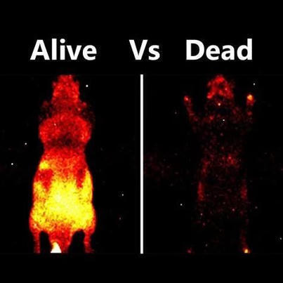
Did you know that a mouse glows slightly in total darkness?
But only if it is alive. Here’s the science behind it:
Every living mouse emits biophotons - in fact, it seems every living organism does.
You can’t see them with a naked eye, but only through ultra sensitive cameras.
Where do they come from?
This emission is a result of metabolic reactions happening inside the cells, especially mitochondria. Reactive oxygen species (ROS) are produced as a result of aerobic metabolism. These interact with cell components like DNA, lipids and proteins, causing them to enter an ‘excited’ state. When these biomolecules decay back to their ground state, they release that surplus energy as photons.
For decades, many biologists brushed this phenomenon off as useless metabolic noise. But new research shows it might tell us something.
It is possible that organelles like mitochondria are using these light pulses to coordinate cellular activities.
Scientists are also seeing this as a new way of real-time health monitoring. Damaged and stressed cells show distinct emission patterns compared to healthy ones.
It turns out light isn't just a metaphor for life. It might be part of the fundamental processes that keep it running.
Keep shining!
#STEM #PhDLife #Fascinating #BiologicalProcessesExplained | 29 comments on LinkedIn
|
|
Scooped by
Gilbert C FAURE
October 19, 10:35 AM
|
Scoop.it!
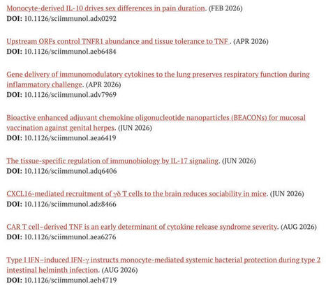
To celebrate International Cytokine & Interferon Society's annual meeting in Glasgow this week, #Science #Immunology has just released a free sample collection of recent papers with a focus on #cytokine biology! The collection will be available online to read until mid-December 2026! #Cytokines2026 Science Magazine
bit.ly/SciImm_free
|
|
Scooped by
Gilbert C FAURE
October 19, 4:30 AM
|
Scoop.it!

Your cells are powered by a microscopic world in constant motion.
Meet the mitochondria — far more than just the “powerhouses of the cell.”
Under the microscope, they reveal an incredible level of organization, movement, and adaptability.
They produce energy.
Mitochondria convert energy from nutrients into ATP, which powers countless cellular activities.
They constantly reshape themselves.
Through fusion and division, they adapt to changing energy demands and help maintain cellular health.
They carry their own DNA.
A fascinating evolutionary clue connecting mitochondria to ancient bacteria.
They help regulate cellular decisions.
From calcium signaling to programmed cell death, mitochondria influence how cells function and respond to stress.
But here’s what makes them truly remarkable:
Mitochondria are not static structures. They form a dynamic network that continuously reorganizes itself.
This movement helps cells distribute energy, exchange mitochondrial components, and isolate damaged regions for removal.
It’s a powerful reminder that even the smallest units of life depend on complex systems working together.
Sometimes, the most extraordinary engineering isn't found in machines.
It's happening inside every living cell.
|
|
Scooped by
Gilbert C FAURE
October 18, 7:11 AM
|
Scoop.it!
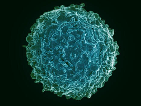
"Atypical" B cells (ABCs) have received increased attention in the scientific community as they are associated with many different types of diseases, such as autoimmune and infectious diseases, but also transplantation and ageing.
We have studied them in the context of malaria and just published a study in EMBO Journal describing how they do not constitute a uniform group but instead follow several developmental pathways that converge on a common late stage. This differentiation is associated with changes in surface markers and effector functions, such as cytokine secretion, antigen uptake, and plasma cell differentiation.
The findings may help explain why ABCs have previously been linked to different roles in the immune system.
Karolinska Institutet: https://lnkd.in/dFDmf-tz
Link to paper: https://lnkd.in/dczKxWcg
|
|
Scooped by
Gilbert C FAURE
October 16, 8:35 AM
|
Scoop.it!

Immune cells when migrate through tissue don't just interact with the ECM, they also make extensive contacts with other cells too. The crowded tissue environment isn't just matrix fibres, which is why this video is one of my most favorites. We could culture both ectodermal ⚪ and immune cells 🔴 together relatively easily to study their cell-cell interactions. These immune cells move up and down within the ectodermal tissue, physically contact surrounding cells as they navigate through the tissue. It gives me so much joy just looking at it because there is just so much hidden biology to be discovered.
#FluorescenceFriday
|
|
Scooped by
Gilbert C FAURE
October 4, 11:37 AM
|
Scoop.it!

Rethinking Immunity: How the Immune System Shapes Motherhood
We have long understood the immune system as a system of defense: protecting us from infection and, when dysregulated, contributing to disease. Exciting work from our laboratory and others is revealing a much broader role for immunity: helping the body adapt to major physiological transitions, including pregnancy and lactation.
During these remarkable periods, the maternal body undergoes profound transformations. We are beginning to understand that the immune system is not simply adapting to these changes, it is actively helping to orchestrate them.
At Institut Pasteur, our laboratory is proud to contribute to this rapidly developing field, one that has been overlooked for far too long and that has important implications for the long-term health of both mothers and children.
We discuss this emerging view of immunity in a new review just published in Science, which I had the great pleasure of writing with Dan Corral, a former member of our laboratory who recently joined INSERM and launched his own laboratory at the Infinity Institute in Toulouse.
A wonderful opportunity to rethink immunity not only as a system of defense, but as a fundamental partner in physiology and adaptation across life.
Read the paper : https://lnkd.in/etmvSpMh
Science Magazine Dan Corral INSERM
|
|
Scooped by
Gilbert C FAURE
October 2, 1:08 PM
|
Scoop.it!

🦠 Immune Cells in the Embryo 🐣
In this video, you’re seeing embryonic myeloid cells (red), among the earliest immune cells to emerge during development, spreading across developing frog tissue (blue).
New research shows that these cells aren’t navigating alone. The physical movement of surrounding tissue helps guide their migration.
🔴 As embryonic tissue spreads outward, early immune cells move along with it.
🧬 But they aren’t simply being carried by the flow. The cells reorganize their internal cytoskeleton and extend protrusions in the direction of tissue movement, allowing them to actively migrate with it.
🌊 This suggests that mechanical forces generated during development can provide directional cues to immune cells, helping them disperse throughout the embryo and establish an early line of defense.
Understanding how tissue movement guides immune cells could also reveal mechanisms involved in wound healing, inflammation, and cancer.
🎥 Video from work by Hoang Anh Le and colleagues, Department of Cell and Developmental Biology, University College London.
📄 Research published in Nature Cell Biology, September 2026.
#Immunology #CellBiology #ImmuneCells #CellMigration #Microscopy | 32 comments on LinkedIn
|
|
Scooped by
Gilbert C FAURE
November 6, 10:41 AM
|
Scoop.it!

Cytokines are critical in regulating immune responses and cellular behavior, playing dual roles in both normal physiology and the pathology of diseases such as cancer. These molecules, including interleukins, interferons, tumor necrosis factors, chemokines, and growth factors like TGF-β, VEGF, and EGF, can promote or inhibit tumor growth, influence the tumor microenvironment, and impact the efficacy of cancer treatments. Recent advances in targeting these pathways have shown promising therapeutic potential, offering new strategies to modulate the immune system, inhibit tumor progression, and overcome resistance to conventional therapies. In this review, we summarized the current understanding and therapeutic implications of targeting cytokine and chemokine signaling pathways in cancer. By exploring the roles of these molecules in tumor biology and the immune response, we highlighted the development of novel therapeutic agents aimed at modulating these pathways to combat cancer. The review elaborated on the dual nature of cytokines as both promoters and suppressors of tumorigenesis, depending on the context, and discussed the challenges and opportunities this presents for therapeutic intervention. We also examined the latest advancements in targeted therapies, including monoclonal antibodies, bispecific antibodies, receptor inhibitors, fusion proteins, engineered cytokine variants, and their impact on tumor growth, metastasis, and the tumor microenvironment. Additionally, we evaluated the potential of combining these targeted therapies with other treatment modalities to overcome resistance and improve patient outcomes. Besides, we also focused on the ongoing research and clinical trials that are pivotal in advancing our understanding and application of cytokine- and chemokine-targeted therapies for cancer patients.
|
|
Scooped by
Gilbert C FAURE
November 5, 10:50 AM
|
Scoop.it!

Recent studies suggest that the formation of tertiary lymphoid structures (TLSs) could be associated with better survival from certain cancers. In this Perspective, we propose that immunogenic cell death (ICD) can act as an upstream organizer of tumour-associated TLSs by coordinating vascular, stromal and immune reprogramming in the tumour microenvironment. ICD leads to the release of tumour antigens and damage-associated molecular patterns that activate innate pathways, such as cGAS–STING signalling, in dendritic cells, myeloid cells, endothelial cells and stromal fibroblasts. These activated cells release type I interferons, TNF and chemokines, such as CCL19, CCL21 and CXCL13, that promote endothelial cell activation and the differentiation of high endothelial venule-like structures. This enables the recruitment of naive and memory lymphocytes and the emergence of perivascular T cell–dendritic cell aggregates, which can form the basis of TLSs. Full structural maturation of TLSs, with segregated B cell and T cell zones, follicular dendritic cell networks and germinal centres, appears to require additional lymphotoxin β receptor (LTβR)-dependent stromal reprogramming. Together, this suggests a two-step model in which ICD first seeds inflammatory vascular niches and then, through LTβR-mediated tissue organization, supports the development of functional TLSs. The formation of tertiary lymphoid structures (TLSs) has been associated with better outcomes in patients with certain tumours. Here the authors discuss how immunogenic cell death can lead to the formation of TLSs and explain why this knowledge could help us design better cancer therapies.

|
|
Scooped by
Gilbert C FAURE
October 22, 3:48 AM
|
Scoop.it!
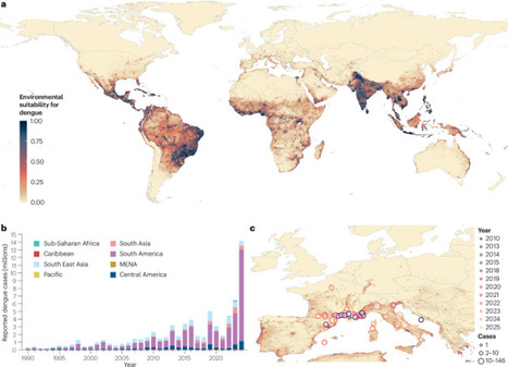
Dengue is an acute febrile disease caused by the Aedes mosquito-borne dengue viruses (DENV-1 to DENV-4). Annually, approximately 100 million people fall ill with dengue, which can cause debilitating pain and discomfort, reducing both physical functioning and quality of life. Some patients progress to life-threatening inflammation-driven circulatory failure. Disease burden remains primarily in urban centres; however, factors including increased travel and climate change are rapidly expanding the geographical distribution of dengue to rural and peri-urban environments. Dengue is diagnosed based on compatible clinical features, detection of viral RNA or antigens, or serological testing, depending on the phase of illness. There is no licensed antiviral or host-directed therapy for dengue; management is supportive, with close clinical monitoring and judicious fluid replacement to prevent circulatory failure while avoiding fluid overload. Thus, there is an increasing need for clinicians to be familiar with the clinical features, disease burden and management of dengue, along with the newly recognized post-acute sequelae. Although dengue vaccines have been licensed in several countries, their use has been restricted by uncertainties in dengue pathogenesis. Attention should be focused on how to use currently available vaccines to create the foundation of a larger strategy to mitigate dengue transmission and maximize public health impact. Dengue is an acute febrile disease caused by the Aedes mosquito-borne dengue viruses. In this Primer, Ooi et al. describe the epidemiology, mechanisms, diagnosis and management of dengue infection and highlight patients’ quality of life and questions for future research.
|
|
Scooped by
Gilbert C FAURE
October 19, 7:45 AM
|
Scoop.it!
What if one of our oldest immune cells makes its own steroids to keep itself in check?
Our new paper in Science Advances shows that mast cells do. Cyp11a1, the first and rate-limiting enzyme of steroid biosynthesis, is expressed in mast cells at baseline and rises on activation. It sits in a superenhancer alongside classic mast cell identity genes. When we deleted it, mast cells degranulated more, and mice had stronger anaphylactic responses.
Mast cells are ancient, likely older than the glands we usually credit with making steroid hormones. So a question remains open: did immune-cell steroidogenesis evolve before glandular steroidogenesis? It is time to explore how immune cells use endogenous steroids to control inflammation and immunity.
This paper also carries a story that is not in it: the story of a loving mother and a caring father. Co-first author Jhuma Pramanik and I are husband and wife. We finished this work through the long struggle with our son's mental health, and then through his loss and our grief. Much remains undone, but there was never any lack of effort, commitment or love for science. Division of Immunology, Department of Pathology, University of Cambridge, gave us a rare, positive and caring environment. I am deeply grateful to the department, my co-authors, colleagues and collaborators, who helped us through.
We dedicate this paper to the memory of our son, Samay Mahata.
Immunology Division, University of Cambridge Department of Pathology, University of Cambridge University of Cambridge, Klaus Okkenhaug, Rahul Roychoudhuri, Natalie Homer, Qiuchen Zhao, Soura Chakraborty, Yumi Kanemaru, Sanu K. Shaji, PhD. Silia Ayadi | 16 comments on LinkedIn
|
|
Scooped by
Gilbert C FAURE
October 18, 7:23 AM
|
Scoop.it!

Sugars attached to proteins and lipids help shape how pathogens interact with human cells and modulate immune responses. In our new editorial, we highlight findings from the Frontiers in Immunology Research Topic I co-edited with Héctor M. Mora Montes and Andrea Regina de Souza Baptista. The collection brings together more than 30 articles spanning fungal recognition, immune evasion, host susceptibility, diagnostics, and emerging therapies.
Read the editorial: https://lnkd.in/eVrYVnpx
If you want to contribute to Volume II: https://lnkd.in/ebFPEDtG
#glycans #Immunology #Mycology
|
|
Scooped by
Gilbert C FAURE
October 18, 5:26 AM
|
Scoop.it!

🧬 What Are Natural Killer Cells?
Natural Killer cells are a type of lymphocyte belonging to the innate immune system. They help identify and eliminate certain virus-infected cells and abnormal cells, including some cancer cells.
Unlike T cells, NK cells do not require prior sensitization to a specific antigen to kill their targets. Instead, they integrate signals from activating and inhibitory receptors to determine whether a cell should be attacked.
🔬 How Do NK Cells Recognize Their Targets?
NK cells use a balance of signals to distinguish healthy cells from potentially dangerous ones.
🔹 Missing-self recognition: Many healthy cells express MHC class I molecules, which engage inhibitory receptors on NK cells. Reduced MHC class I expression can remove this inhibitory signal and make a cell more susceptible to NK-cell attack.
🔹 Stress signals: Infected or cancerous cells may express stress-induced molecules that activate NK-cell receptors.
🔹 Receptor balance: NK cells integrate activating and inhibitory signals before deciding whether to kill a target cell.
⚔️ How Do NK Cells Kill Abnormal Cells?
1️⃣ Recognition: NK cells detect signals suggesting that a target cell is infected, stressed, or abnormal.
2️⃣ Perforin release: Perforin helps deliver cytotoxic proteins into the target cell.
3️⃣ Granzyme action: Granzymes activate pathways that trigger programmed cell death (apoptosis).
4️⃣ Target elimination: The abnormal cell dies, helping limit infection or remove potentially dangerous cells.
NK cells can also trigger apoptosis through death-receptor pathways and release cytokines such as interferon-gamma (IFN-γ), which help coordinate immune responses.
🛡️ Major Functions of NK Cells
✅ Destroy certain virus-infected cells.
✅ Recognize and kill some tumor cells.
✅ Produce cytokines that regulate immune activity.
✅ Support communication between innate and adaptive immunity.
✅ Participate in antibody-dependent cellular cytotoxicity (ADCC), in which NK cells recognize antibody-coated target cells through CD16.
🧪 NK Cells and Cancer Immunotherapy
NK cells are important in cancer research because of their ability to kill abnormal cells. Scientists are investigating NK-cell-based therapies, including engineered NK cells and CAR-NK therapy, as potential approaches to cancer treatment.
However, tumors can develop mechanisms to suppress NK-cell activity, and many NK-cell therapies remain under clinical investigation.
💡 Why Are NK Cells Important?
NK cells provide rapid immune surveillance and help control infections and abnormal cell growth. They demonstrate how the immune system can recognize cellular danger without relying exclusively on antigen-specific recognition.
follow #doublehelixdiaries for more such content
#NaturalKillerCells #NKCells #ImmuneSystem #CellBiology #Genetics #Biotechnology #LifeSciences
|
|
Scooped by
Gilbert C FAURE
October 14, 8:27 AM
|
Scoop.it!

Bacteria have immune systems to defend against their viruses.
Together with Nicholas M I Taylor, Philipp Popp, Inga Songailiene, Alexander Harms and colleagues, we investigated Zorya, a defense system that uses a proton-driven membrane complex related to the stator of the bacterial flagellum.
The animation follows the proposed Zorya-I mechanism from the phage encounter at the cell envelope to the action of its DNA-targeting proteins: ZorC binds DNA, while ZorD acts as a nuclease.
The movie combines structures and experimental findings with proposed steps that remain unresolved. In particular, the idea that the long ZorA tail reels in phage DNA remains a hypothesis.
Have a look! It is an interesting example of how related molecular machinery can serve very different functions.
Our paper: https://lnkd.in/dripiawz
#Microbiology #Phages #StructuralBiology | 18 comments on LinkedIn
|
|
Scooped by
Gilbert C FAURE
October 12, 8:55 AM
|
Scoop.it!
🚨Retrouvez en accès libre toutes les fiches graphiques de l’Internist Academy soutenue par le Fonds IMMUNOV
Plus de 200 sujets différents traités par les internes !!!
|
|
Scooped by
Gilbert C FAURE
October 7, 5:39 AM
|
Scoop.it!
This month’s cover image shows CD45+ immune cells (red) and EpCAM+ epithelial cells (blue) in malignant ascites from a patient with gastric cancer.
|
|
Scooped by
Gilbert C FAURE
October 5, 9:53 AM
|
Scoop.it!
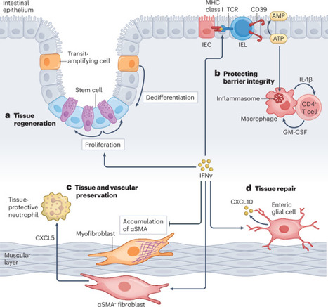
The effects of IFNγ on tissue physiology through structural cells - Nature Reviews Immunology
|
|
Scooped by
Gilbert C FAURE
October 3, 11:37 AM
|
Scoop.it!

CD1 proteins on so-called antigen-presenting cells display lipids from pathogens to T cells to drive protective immune responses, but they also trigger skin inflammation in response to some lipids.
Learn more: https://scim.ag/4xS88Wl
|
|
Scooped by
Gilbert C FAURE
September 23, 4:54 AM
|
Scoop.it!

For decades, #celldeath was often taught as essentially two processes: #apoptosis, an orderly form of cellular self-destruction, and #necrosis, associated with cellular injury, swelling and rupture.
▪️ We now know that the #biology is dramatically more complicated. Researchers have described more than 20 different forms or pathways of cell death, including apoptosis, necrosis, necroptosis, pyroptosis, ferroptosis, parthanatos, autophagy-dependent cell death, lysosome-dependent cell death, entotic cell death/entosis, NETosis or ETosis, anoikis, paraptosis, mitotic catastrophe, immunogenic cell death, cuproptosis, disulfidptosis, alkaliptosis, oxeiptosis, methuosis, lysozincrosis, erebosis and mitoxyperilysis, among others.
▪️These are not simply different names for the same endpoint. Some involve caspases, others iron-driven #lipid peroxidation, #copper toxicity, lysosomal disruption, oxidative stress, inflammatory pore formation or even the extrusion of extracellular traps.
▪️Why does this matter? Because how a #cell dies can be just as important as the fact that it dies. Different death pathways can be relatively silent or highly inflammatory, influence surrounding cells, activate or suppress immune responses, and ultimately affect diseases ranging from #infection and #cancer to #neurodegeneration and inflammatory disorders.
▪️Cell death is therefore not one biological endpoint reached by one or two roads. It is an expanding network of molecular pathways and understanding which pathway is operating may open entirely new opportunities for preventing disease or designing therapies.
🔗 See comments for reference.







Complement component 8 deficiency is a disorder that causes the immune system to malfunction, resulting in a form of immunodeficiency. Immunodeficiencies are conditions in which the immune system is not able to protect the body effectively from foreign invaders such as bacteria. People with complement component 8 deficiency have a significantly increased risk of recurrent bacterial infections, particularly by a bacterium called Neisseria meningitidis. Infection by this bacterium causes inflammation of the membranes surrounding the brain and spinal cord (meningitis). Although meningitis can be life-threatening, individuals with complement component 8 deficiency are less likely to die from the infection than people in the general population who contract it.