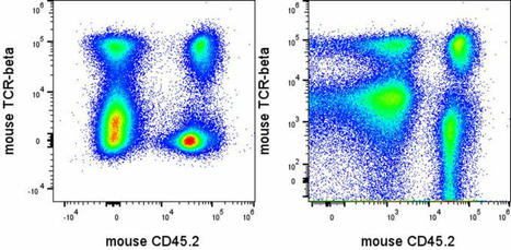
![Single-cell mass cytometry of differential immune an... [Science. 2011] - PubMed - NCBI | from Flow Cytometry to Cytomics | Scoop.it](https://img.scoop.it/R49xnLrZXb91oy4sAbghFTl72eJkfbmt4t8yenImKBVvK0kTmF0xjctABnaLJIm9)
PubMed comprises more than 21 million citations for biomedical literature from MEDLINE, life science journals, and online books. Citations may include links to full-text content from PubMed Central and publisher web sites.
Get Started for FREE
Sign up with Facebook Sign up with X
I don't have a Facebook or a X account


|
Scooped by
Gilbert C FAURE
onto from Flow Cytometry to Cytomics October 26, 2011 3:37 AM
|
PubMed comprises more than 21 million citations for biomedical literature from MEDLINE, life science journals, and online books. Citations may include links to full-text content from PubMed Central and publisher web sites.

 Your new post is loading...
 Your new post is loading...
Your new post is loading...
There can be tens of thousands of cells in one milliliter of culture medium. So how are cells counted?
Gilbert C FAURE's insight:
It is how techniques are taught or learned now! Thanks Youtube
Obviously, this first Scoop is not significant of the information you will find through the following 187 pages of this topic, with many recent informations of developments of flow cytometry, image cytometry... focusing particularly on the detection, quantification, characterization of rare events (CTCs https://www.scoop.it/topic/from-flow-cytometry-to-cytomics?q=ctc CECS, microvesicles...
in peripheral blood and biological fluids in the context of liquid biopsy https://www.scoop.it/topic/from-flow-cytometry-to-cytomics?q=liquid+biopsy
more than 3000 scoops, >5200 visitors, 10700 views (march 2015) juin 2019 more than 4400 scoops, >13K visitors, >31,6 K views august 2022, More than 4.7K scoops, more than 15.4K visitors
For data from the curator, see the Nancytomique topic. http://www.scoop.it/t/nancytomique
From
www

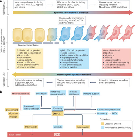
Cell plasticity is a crucial trait for cancer progression towards metastasis and treatment resistance. Research efforts from the past 20–30 years have revealed that the dynamic flux of the epithelial–mesenchymal transition (EMT) programme is one of the major underlying processes enabling cancer cell plasticity and greatly facilitates these major causes of cancer mortality. The spectrum of evidence ranges from extensive data from cell line and animal model studies across multiple cancer types through a rapidly expanding body of work demonstrating associations between EMT biomarkers and disease progression and mortality in patients. EMT is also implicated in resistance to most of the major treatment modalities, yet our efforts to harness this knowledge to improve therapeutic outcomes are currently in their early stages. In this Review, we describe clinical evidence supporting a role of EMT and the associated epithelial–mesenchymal plasticity in various stages of cancer in patients and discuss the subsequent clinical opportunities and challenges associated with attempts to implement this knowledge as novel therapies or clinical management approaches. Despite several decades of research that has revealed roles in the development and progression of many solid tumours, clinical translation of research targeting epithelial–mesenchymal transition (EMT) has thus far been limited. In this Review, the authors provide a summary of the role of EMT in cancer development and progression in the context of this lack of clinical translation, summarize the current status of direct or indirect EMT-modulating agents in clinical development, and highlight the major barriers to the development of EMT-related clinical interventions.
Gilbert C FAURE's insight:
https://www.scoop.it/topic/from-flow-cytometry-to-cytomics?q=emt bibliographie archivée sur
From
www
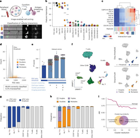
Cellular interactions are of fundamental importance, orchestrating organismal development, tissue homeostasis and immunity. Recently, powerful methods that use single-cell genomic technologies to dissect physically interacting cells have been developed. However, these approaches are characterized by low cellular throughput, long processing times and high costs and are typically restricted to predefined cell types. Here we introduce Interact-omics, a cytometry-based framework to accurately map cellular landscapes and cellular interactions across all immune cell types at ultra-high resolution and scale. We demonstrate the utility of our approach to study kinetics, mode of action and personalized response prediction of immunotherapies, and organism-wide shifts in cellular composition and cellular interaction dynamics following infection in vivo. Our scalable framework can be applied a posteriori to existing cytometry datasets or incorporated into newly designed cytometry-based studies to map cellular interactions with a broad range of applications from fundamental biology to applied biomedicine. Interact-omics, a high-throughput cytometry-based framework, resolves the cellular interaction landscape.
From
www
Out now! --> Cancer in a drop: Advances in liquid biopsy in 2024

From
www
Fostering the Implementation of Liquid Biopsy in Clinical Practice: European Liquid Biopsy Society (ELBS) 2024 Meeting Report by Klaus Pantel et al.

Proud to be part of the team that published this new #liquidbiopsy study! This study is the largest pooled analysis with globally collected individual patient…

Digoxin for reduction of circulating tumor cell cluster size in metastatic breast cancer: a proof-of-concept trial

Explorez nos modules avancés en #cytométrie en flux !

Marcus Jorelle's curator insight,
December 16, 2024 9:26 PM
Nosler Custom Competition Bullets 22 Caliber (224 Diameter) 69 Grain Hollow Point Boat Tail: A Comprehensive Guide ▎Introduction When it comes to precision shooting and competitive marksmanship, the choice of ammunition can significantly impact your performance. Among the leading options available on the market today, the Nosler Custom Competition Bullets in 22 caliber (224 diameter) with a 69-grain hollow point boat tail design stands out for its exceptional accuracy and reliability. In this article, we will explore the features, benefits, and applications of these premium bullets, helping you understand why they are a preferred choice for serious shooters. ▎Key Features of Nosler Custom Competition Bullets ▎1. Precision Engineering Nosler is renowned for its commitment to quality and precision. The Custom Competition Bullets are manufactured using advanced techniques that ensure uniformity in weight and dimensions. This precision manufacturing process results in tighter groupings and improved accuracy, making them ideal for competitive shooting. ▎2. Hollow Point Design The hollow point design of the Nosler Custom Competition Bullets enhances their terminal performance. When these bullets strike a target, the hollow cavity allows for controlled expansion, delivering maximum energy transfer and creating a larger wound channel. This feature is particularly beneficial for varmint hunting and target shooting scenarios where effective stopping power is essential. ▎3. Boat Tail Profile The boat tail design of these bullets contributes to their aerodynamic efficiency. The tapered rear end reduces drag during flight, allowing for flatter trajectories and improved stability. As a result, shooters can achieve greater accuracy at longer distances, making these bullets suitable for long-range shooting competitions. ▎4. Optimal Weight At 69 grains, these bullets strike an excellent balance between velocity and stability. This weight is particularly favored in competitive shooting disciplines, including High Power and F-Class matches. The 69-grain weight ensures that shooters can maintain a good ballistic coefficient while achieving consistent performance. ▎Benefits of Using Nosler Custom Competition Bullets ▎1. Enhanced Accuracy One of the primary advantages of using Nosler Custom Competition Bullets is their ability to deliver exceptional accuracy. The combination of precision engineering, uniformity, and optimal design features allows shooters to achieve tight shot groups, giving them a competitive edge. ▎2. Versatile Applications Whether you are participating in a shooting competition or engaging in varmint hunting, these bullets are versatile enough to meet various shooting needs. Their reliable performance makes them suitable for both target shooting and practical applications. ▎3. Consistency Consistency is critical in competitive shooting. Nosler’s rigorous quality control measures ensure that each bullet meets strict performance standards, providing shooters with confidence in their ammunition choice. ▎4. Increased Terminal Performance The hollow point design not only enhances accuracy but also improves terminal performance on impact. This makes the Nosler Custom Competition Bullets an excellent choice for shooters who require effective stopping power. ▎Conclusion In summary, the Nosler Custom Competition Bullets 22 Caliber (224 Diameter) 69 Grain Hollow Point Boat Tail represent a premium choice for shooters who demand precision, reliability, and performance. With their advanced engineering, versatile applications, and enhanced accuracy, these bullets are designed to meet the needs of both competitive shooters and hunters alike. Whether you’re aiming for the bullseye in a competition or seeking effective ammunition for varmint hunting, Nosler Custom Competition Bullets deliver the quality and consistency that serious shooters require. Invest in your shooting success with Nosler – where precision meets performance.https://canada-reloading.shop/product/federal-firestic…-powder-10-count/ https://canada-reloading.shop/product/fiocchi-primers-…09-w-type-1000ct/ https://canada-reloading.shop/product/fiocchi-small-pi…l-magnum-primers/ https://canada-reloading.shop/product/large-rifle-magn…formance-primers/ https://canada-reloading.shop/product/pmc-small-pistol…rimers-two-trays/ https://canada-reloading.shop/product/remington-10-percussion-caps-100/ https://canada-reloading.shop/product/remington-11-percussion-caps/ https://canada-reloading.shop/product/remington-209ml-…ader-primers-100/ https://canada-reloading.shop/product/remington-22617-…-caps-100-ct-tin/ https://canada-reloading.shop/product/rws-1075-plus-percussion-caps-11/ https://canada-reloading.shop/product/rws-4-flange-200-ct-musket-caps/ https://canada-reloading.shop/product/rws-11-percussio…250-to-1000-pack/ https://canada-reloading.shop/product/rws-musket-caps-…200-to-5000-pack/ https://canada-reloading.shop/product/schuetzen-musket…00-to-5000-count/ https://canada-reloading.shop/product/small-rifle-magn…formance-primers/ https://canada-reloading.shop/product/tulammo-large-ri…imers-1000-count/ https://canada-reloading.shop/product/tulammo-small-ri…imers-1000-count/ https://canada-reloading.shop/product/winchester-percussion-caps-11-100/ https://canada-reloading.shop/product/209-shotshell-wo…formance-primers/ https://canada-reloading.shop/product/cci-percussion-c…0-10-cans-of-100/ https://canada-reloading.shop/product/cci-primers-209-…-10-trays-of-100/ https://canada-reloading.shop/product/cci-primers-209m…-10-trays-of-100/ https://canada-reloading.shop/product/cheddite-shotshell-primer-209/ https://canada-reloading.shop/product/federal-209a-shotshell-primers/ https://canada-reloading.shop/product/federal-primers-…-10-trays-of-100/ https://canada-reloading.shop/product/fiocchi-primers-…-10-trays-of-100/ https://canada-reloading.shop/product/nobel-sport-shot…-10000-pack-case/ https://canada-reloading.shop/product/nobelsport-prime…-50-trays-of-100/ https://canada-reloading.shop/product/remington-premie…rs-209-shotshell/ https://canada-reloading.shop/product/rio-209-g-1000-shotshell-primers/ https://canada-reloading.shop/product/winchester-prime…-10-trays-of-100/ https://canada-reloading.shop/product/fort-smith-fsaap…ol-boxer-primers/ https://canada-reloading.shop/product/cci-small-pistol…-10-trays-of-100/ https://canada-reloading.shop/product/cci-small-pistol…-10-trays-of-100/ https://canada-reloading.shop/product/federal-premium-…-10-trays-of-100/ https://canada-reloading.shop/product/federal-premium-…-10-trays-of-100/ https://canada-reloading.shop/product/federal-small-pi…-10-trays-of-100/ https://canada-reloading.shop/product/federal-small-pi…-10-trays-of-100/ https://canada-reloading.shop/product/remington-small-…-10-trays-of-100/ https://canada-reloading.shop/product/remington-small-…-10-trays-of-100/ https://canada-reloading.shop/product/small-pistol-wol…formance-primers/ https://canada-reloading.shop/product/small-pistol-mag…formance-primers/ https://canada-reloading.shop/product/winchester-small…-10-trays-of-100/ https://canada-reloading.shop/product/winchester-small…-10-trays-of-100/ https://canada-reloading.shop/product/winchester-small…-10-trays-of-100/ https://canada-reloading.shop/product/winchester-usa-r…-10-trays-of-100/ https://canada-reloading.shop/product/fort-smith-fsaap…le-boxer-primers/ https://canada-reloading.shop/product/cci-small-rifle-…-10-trays-of-100/ https://canada-reloading.shop/product/cci-small-rifle-…-10-trays-of-100/ https://canada-reloading.shop/product/cci-small-rifle-…-10-trays-of-100/ https://canada-reloading.shop/product/cci-small-rifle-…-10-trays-of-100/ https://canada-reloading.shop/product/federal-premium-…-10-trays-of-100/ https://canada-reloading.shop/product/federal-small-ri…-10-trays-of-100/ https://canada-reloading.shop/product/magtech-small-ri…imers-1000-count/ https://canada-reloading.shop/product/pmc-small-rifle-magnum-primers/ https://canada-reloading.shop/product/remington-small-…-10-trays-of-100/ https://canada-reloading.shop/product/remington-small-…-10-trays-of-100/ https://canada-reloading.shop/product/hornady-brass-45…ess-3″-box-of-20/ https://canada-reloading.shop/product/hornady-brass-6-5-prc-box-of-50/ https://canada-reloading.shop/product/lapua-brass-30-0…field-box-of-100/ https://canada-reloading.shop/product/lapua-brass-338-…agnum-box-of-100/ https://canada-reloading.shop/product/lapua-brass-6-5-creedmoor/ https://canada-reloading.shop/product/norma-brass-shoo…magnum-box-of-50/ https://canada-reloading.shop/product/nosler-brass-6-5…dmoor-bag-of-100/ https://canada-reloading.shop/product/nosler-custom-br…osler-box-of-100/ https://canada-reloading.shop/product/nosler-custom-br…hester-box-of-50/ https://canada-reloading.shop/product/nosler-custom-br…oulder-box-of-50/ https://canada-reloading.shop/product/nosler-custom-br…magnum-box-of-25/ https://canada-reloading.shop/product/nosler-custom-co…-point-boat-tail/ https://canada-reloading.shop/product/once-fired-brass…00-bulk-packaged/ https://canada-reloading.shop/product/sierra-matchking…-point-boat-tail/ https://canada-reloading.shop/product/sig-sauer-brass-…ington-bag-of-50/ https://canada-reloading.shop/product/starline-brass-223-remington/ https://canada-reloading.shop/product/starline-brass-357-sig/ https://canada-reloading.shop/product/starline-brass-38-55-wcf-2-082″/ https://canada-reloading.shop/product/starline-brass-45-acp/ https://canada-reloading.shop/product/starline-brass-45-auto-rim-not-acp/ https://canada-reloading.shop/product/starline-brass-458-socom/ https://canada-reloading.shop/product/starline-brass-5-56x45mm-nato/ https://canada-reloading.shop/product/top-brass-premiu…300-aac-blackout/ https://canada-reloading.shop/product/top-brass-premiu…ired-brass-40-sw/ https://canada-reloading.shop/product/hodgdon-2024-ann…reloading-manual/ https://canada-reloading.shop/product/hornady-11th-edi…reloading-manual/ https://canada-reloading.shop/product/loadbooks-usa-12…reloading-manual/ https://canada-reloading.shop/product/lyman-pistol-rev…book-3rd-edition/ https://canada-reloading.shop/product/norma-reloading-manual-2nd-edition/ https://canada-reloading.shop/product/nosler-reloading-manual-9/ https://canada-reloading.shop/product/sierra-6th-editi…reloading-manual/ https://canada-reloading.shop/product/speer-reloading-manual-15/ https://canada-reloading.shop/product/swift-reloading-manual-2/ https://canada-reloading.shop/product/tom-rosters-hevi…nual-2nd-edition/ https://canada-reloading.shop/product/top-brass-premiu…ired-brass-40-sw/ https://canada-reloading.shop/product/federal-premium-…-point-boat-tail/ https://canada-reloading.shop/product/hornady-superfor…-v-max-box-of-20/ "Referring page title" "Referring page URL" "Language" "Platform" "Referring page HTTP code" "Domain rating" "UR" "Domain traffic" "Referring domains" "Linked domains" "External links" "Page traffic" "Keywords" "Target URL" "Left context" "Anchor" "Right context" "Redirect Chain URLs" "Redirect Chain status codes" "Type" "Content" "Nofollow" "UGC" "Sponsored" "Rendered" "Raw" "Lost status" "Drop reason" "Discovered status" "First seen" "Last seen" "Lost" "Author" "Links in group" "YouTube Creator Blog [UK]: Ride to Success with 63fixedtv, September’s Rising Partner" "https://youtubecreator-uk.googleblog.com/2012/09/ride-to-success-with-63fixedtv.html" "en" "" "200" "94" "5" "210429" "29" "180" "542" "0" "0" "http://ssgunstore.com/" "" "Buy sig sauer online" "" "" "" "text" "false" "true" "false" "false" "true" "true" "" "" "pagefound" "2022-07-11 14:53:04" "2024-05-29 07:01:22" "" "" "2" "沖縄対策本部■沖縄に現れた若き愛国ヒロインのスッキリする名スピーチ - 沖縄対策本部" "https://blog.goo.ne.jp/jiritsukokka/e/55ac732a803559c65c0865560848bb22" "" "" "200" "90" "6" "24746634" "22" "711" "1879" "0" "0" "https://ssgunstore.com/" "(" "Sig Sauer" ")" "" "" "text" "false" "true" "false" "false" "false" "true" "" "" "linkfound" "2021-07-09 02:55:40" "2024-05-29 17:13:03" "" "" "1" "Immagine Immagina | Germaine Muller" "http://www.germainemuller.altervista.org/?page_id=172" "it" "wordpress" "200" "90" "0.6" "2411712" "93" "5554" "13863" "0" "0" "https://ssgunstore.com/" "" "sig sauer sp2022" "" "" "" "text" "false" "true" "false" "false" "true" "true" "" "" "linkrestored" "2022-07-04 22:49:07" "2024-05-27 21:28:57" "" "" "1" "Up Against the Wall: How to Break Up Your Day so You Feel Great – Magellan Chiropractic" "http://jonesborochiropractor.flywheelsites.com/up-against-the-wall-how-to-break-up-your-day-so-you-feel-great/" "" "wordpress" "200" "88" "1.1" "15265" "2" "3840" "8378" "0" "0" "https://ssgunstore.com/" "" "sig sauer ar 15" "" "" "" "text" "false" "true" "true" "false" "true" "true" "" "" "pagefound" "2023-05-27 08:35:04" "2024-05-27 18:49:19" "" "" "2" "The Moo's News » Blog Archive » Click To Tweet" "http://fatcow.com/blog/?p=1137" "en" "ecommerce, wordpress" "200" "86" "19" "16248" "1158" "13023" "30474" "0" "6" "https://ssgunstore.com/" "" "sig sauer p226" "Says:" "" "" "text" "false" "true" "false" "false" "false" "true" "" "" "linkrestored" "2021-03-31 08:20:02" "2024-05-29 20:22:08" "" "" "2" "EINMANNDING – Kommentare zu Heute , Morgentext vom 28.06. 2017" "http://wordpress.p255834.webspaceconfig.de/?comments_popup=5817" "de" "" "200" "85" "9" "5551" "290" "12307" "21416" "0" "0" "https://ssgunstore.com/" "Kommentar von" "Trevor Mackey" "— 5. April 2021 zu 1:33" "" "" "text" "false" "true" "false" "false" "false" "true" "" "" "pagefound" "2024-01-05 06:21:59" "2024-04-30 15:24:48" "" "" "3" "maquete_2 – Adaptpolis" "http://adaptpolis.fa.ulisboa.pt/?attachment_id=334" "" "wordpress" "200" "82" "0.1" "273078" "111" "7539" "11901" "0" "0" "https://ssgunstore.com/" "" "sig sauer rattler" "" "" "" "text" "false" "true" "true" "false" "false" "true" "" "" "pagefound" "2023-01-06 19:43:36" "2024-05-29 15:48:06" "" "" "1" "What might a post-Wonga world look like? - University of Birmingham" "https://www.birminghamdev.bham.ac.uk/research/perspective/what-might-a-post-wonga-world-look-like" "en" "" "200" "80" "0" "29063" "0" "3994" "13855" "0" "0" "https://ssgunstore.com/" "At 5:25AM on 21 July 2021," "sig sauer guns shop" "wrote" "" "" "text" "false" "true" "false" "false" "false" "true" "" "" "pagefound" "2024-03-27 10:10:12" "2024-04-29 05:20:04" "" "" "1" "Baseball Talk » Blog Archive » Hamilton and Rangers agree to deal" "https://blogs.bgsu.edu/mlblog/2011/02/10/hamilton-and-rangers-agree-to-deal/" "en" "wordpress" "200" "80" "4.3" "234573" "141" "6158" "12700" "0" "4" "https://ssgunstore.com/" "to create this actual post amazing. Excellent process!My page ::" "sig sauer p228" "" "" "" "text" "false" "true" "true" "false" "false" "true" "" "" "linkfound" "2022-06-29 18:25:00" "2024-05-30 01:38:43" "" "" "1" "How Is A Man Freed After 23 Years? – Juris Magazine" "https://sites.law.duq.edu/juris/2013/03/27/how-is-a-man-freed-after-23-years/" "" "wordpress" "200" "78" "3.1" "110203" "47" "7446" "13671" "0" "3" "https://ssgunstore.com/" "" "Mammie Maguire" "" "" "" "text" "false" "true" "true" "false" "false" "true" "" "" "linkfound" "2023-11-04 16:44:53" "2024-05-29 08:38:20" "" "" "1" "buy ayahuasca - On Feet Nation" "https://www.onfeetnation.com/profiles/blogs/buy-ayahuasca-1?xg_source=activity" "en" "" "200" "77" "3.8" "8" "0" "23" "34" "0" "0" "https://ssgunstore.com/" "" "sig sauer, sig sauer p320, sig sauer p365, sig sauer p226, sig saue..." "" "http://ssgunstore.com/" "301" "text" "false" "true" "false" "false" "false" "true" "" "" "linkrestored" "2021-10-12 17:19:30" "2024-05-15 03:29:06" "" "" "8" "For the Love of Food: Get Me to the Greek" "http://healthyeating.sunnybrook.ca/2013/02/get-me-to-greek.html" "en" "" "200" "75" "4.6" "68539" "24" "217" "528" "0" "0" "https://ssgunstore.com/" "" "Firearms For sale, sig sauer p320 compact, sig sauer sp2022, sig sauer p320 for sale, sig sauer p365 for sale, sig sauer p250, sig sauer 365, sig sauer tread, sig sauer p226 legion, sig sauer mcx virtus, sig sauer p320 x5, sig sauer 380, sig sauer p210, sig sauer legion, sig sauer p320 m17, sig sauer pistols, sig sauer p320 x5 legion, sig sauer p228, sig sauer p226 price, sig sauer p365 price, sig ..." "" "" "" "text" "false" "true" "false" "false" "true" "true" "" "" "linkfound" "2023-07-06 15:43:32" "2024-05-28 15:57:43" "" "" "1" "EDDL 5141 – Benefits and Challenges Activity – sdeighton's Portfolio" "https://sdeighton-portfolio.eddl.tru.ca/2019/01/22/eddl-5141-benefits-and-challenges-activity/" "en" "wordpress" "200" "74" "4.4" "108624" "47" "4166" "7570" "0" "0" "https://ssgunstore.com/" "" "sig sauer m18" "" "" "" "text" "false" "true" "true" "false" "false" "true" "" "" "linkfound" "2022-06-30 18:15:14" "2024-05-28 23:46:27" "" "" "2" "31 companies participate in AIT Career Fair – AIT Research Portal" "https://research.ait.ac.th/2018/03/14/31-companies-participate-in-ait-career-fair/" "en" "wordpress" "200" "73" "4.2" "49074" "51" "7473" "12596" "0" "0" "https://ssgunstore.com/" "" "sig sauer tread" "" "" "" "text" "false" "true" "true" "false" "false" "true" "" "" "linkrestored" "2023-10-31 15:56:15" "2024-05-29 09:57:32" "" "" "2" "Majelis Ta’lim Uswatun Nisa Peringati Maulid - BIRO ADMINISTRASI PIMPINAN SETDA ACEH" "https://humas.acehprov.go.id/majelis-talim-uswatun-nisa-peringati-maulid/" "id, en" "ecommerce, wordpress" "200" "71" "1.8" "629960" "35" "2169" "3365" "0" "2" "https://ssgunstore.com/" "" "sig sauer p229" "" "" "" "text" "false" "true" "true" "false" "true" "true" "" "" "linkrestored" "2022-12-22 11:51:32" "2024-05-13 05:12:57" "" "" "1" "Rio Anhanduí pede socorro - Hidrologia, Erosão e Sedimentos - HEroS" "https://heros.ufms.br/rio-anhandui-pede-socorro/" "pt" "wordpress" "200" "70" "4.5" "123300" "79" "8555" "14327" "0" "1" "https://ssgunstore.com/" "" "sig sauer p365" "" "" "" "text" "false" "true" "true" "false" "true" "true" "" "" "linkrestored" "2021-04-07 22:14:28" "2024-05-28 21:29:27" "" "Pedro Zamboni" "2" "Olá, mundo! | IFA" "https://ifa.abf.com.br/index.php/2015/11/06/ola-mundo/" "pt" "wordpress" "200" "69" "2.4" "8739" "50" "7127" "11494" "0" "0" "https://ssgunstore.com/" "" "sig sauer sp2022" "1 de abril de 2021" "" "" "text" "false" "true" "false" "false" "false" "true" "" "" "pagefound" "2023-05-05 05:31:29" "2024-05-29 08:27:24" "" "" "1" "The Definitive Guide to Sig sauer firearms"

🩸Liquid Biopsy Milestones since 1869 ...
Researchers developed a microfluidic platform using surface-enhanced Raman spectroscopy (SERS) to detect single pancreatic cancer cells. The platform successfully differentiated between cancer stages, offering a promising tool for early cancer diagnosis through molecular analysis of individual...
The scale of the plots presenting your flow cytometry data are actually critically important in getting good results with high dimensional data analysis. The…
|

From
www
Heterogeneous circulating tumor cells (CTCs) have been implicated in the formation of new metastases. However, circulating cells expressing both tumor and immune cell proteins are often dismissed as insignificant findings in CTC studies. Two non-contemporaneous blood samples from a metastatic breast cancer patient were analyzed using an enrichment-free platform to identify canonical, epithelial-only CTCs (CD45-/cytokeratin + , epi.CTCs) and CD45 + /cytokeratin+ immune-like CTCs (im.CTCs). Single cells from both samples were subjected to copy number and protein expression profiling. A cohort of 36 metastatic breast cancer patients was then analyzed to search for additional cases with im.CTCs. Here, we identified and characterized a population of CTCs exhibiting an immune-like state. In two samples from an index patient, im.CTCs outnumbered epi.CTCs, comprising >97% of the CTC population. Single-cell copy number analysis of 43 im.CTCs and 30 epi.CTCs revealed clonal alterations across both populations, confirming a shared tumor origin. Furthermore, im.CTCs contained pseudo-diploid profiles that did not reflect dilution from the addition of a normal diploid genome, indicating that they were unlikely to have originated from tumor-immune cell fusion. Protein expression analysis showed that im.CTCs express CD45 as well as other immune-related markers, such as CD3 and CD4, and the cancer stemness marker, CD44. Subsequent analysis of a metastatic breast cancer cohort identified an additional patient harboring im.CTCs with the same tumor-derived, non-fusion genome as in the index case. Collectively, these genomic and proteomic features distinguish im.CTCs from previously reported circulating cells may represent a novel form of tumor cell plasticity. Tumor cells are known to take on features that allow them to survive and move to new sites. This variation can make it difficult to distinguish them from other cells in the blood. Using a platform to profile rare cells in blood samples, we identified a population of cells expressing cancer and immune cell proteins in a breast cancer patient. Genomics data confirmed that these cells originated from the tumor and that they were different from another cell type sharing a similar protein expression pattern. We analyzed additional samples and found a second patient with these immune-like tumor cells. These findings support the existence of a cancer-immune state that might play a role in helping tumor cells spread. Higa et al. analyzed circulating cells expressing cancer and immune cell markers in breast cancer patients. Based on genomic and protein expression profiling, they show that the cells were unlike previously described circulating tumor cells with immune-like phenotypes because they did not appear to arise from heterotypical cell fusion.

From
www
bioRxiv - the preprint server for biology, operated by Cold Spring Harbor Laboratory, a research and educational institution

❤️ Hot off the press!

What's New in Cancer treatment
Visualizing Basophil Activation in Allergy Diagnostics Using Cytolution
Marcus Jorelle's curator insight,
December 13, 2024 6:07 AM
Sierra MatchKing Bullets 22 Caliber (224 Diameter) 69 Grain Hollow Point Boat Tail: The Ultimate Choice for Precision Shooting ▎Introduction When it comes to precision shooting, every detail matters—from the rifle to the ammunition. Among the top choices for competitive shooters and enthusiasts alike are the Sierra MatchKing Bullets 22 Caliber (224 Diameter) 69 Grain Hollow Point Boat Tail. Renowned for their exceptional accuracy and performance, these bullets are designed to meet the rigorous demands of long-range shooting. In this article, we will explore the features, benefits, and applications of these premium bullets while incorporating essential SEO keywords to enhance your searchability. ▎What Are Sierra MatchKing Bullets? Sierra MatchKing bullets are specifically engineered for competitive shooting and precision applications. With a focus on accuracy, these bullets are crafted with meticulous attention to detail, ensuring they perform consistently in various conditions. The 22 caliber (224 diameter) designation makes them suitable for a range of rifles, while the 69 grain weight strikes a balance between velocity and stability. ▎Key Features of Sierra MatchKing Bullets 1. Hollow Point Design: The hollow point design enhances ballistic performance by promoting controlled expansion upon impact. This feature is particularly beneficial for target shooting, as it helps maintain accuracy at longer distances. 2. Boat Tail Configuration: The boat tail design reduces drag and improves aerodynamics, allowing for flatter trajectories and better stability in flight. This results in enhanced accuracy and reduced wind drift—critical factors for long-range shooters. 3. High-Quality Manufacturing: Sierra is known for its commitment to quality, and the MatchKing bullets are no exception. Each bullet undergoes rigorous testing to ensure consistency in weight, shape, and performance. ▎Benefits of Using Sierra MatchKing Bullets ▎1. Exceptional Accuracy The primary advantage of using Sierra MatchKing bullets is their unmatched accuracy. Whether you are participating in a competition or honing your skills at the range, these bullets are designed to deliver tight groupings, giving you confidence in your shots. ▎2. Versatility While primarily designed for match shooting, the 22 caliber 69 grain hollow point boat tail bullets can also be utilized for varmint hunting and other applications where precision is paramount. Their versatility makes them a valuable addition to any shooter’s arsenal. ▎3. Consistent Performance Thanks to their high-quality construction, MatchKing bullets provide consistent performance across various shooting conditions. This reliability ensures that you can trust your ammunition when it matters most. ▎Applications of Sierra MatchKing Bullets Sierra MatchKing bullets are ideal for: • Competitive Shooting: Perfect for benchrest competitions, high-power matches, and other precision shooting events. • Target Practice: Excellent choice for those looking to improve their marksmanship skills. • Varmint Hunting: Effective for taking down small game due to their accurate and controlled expansion. ▎How to Load Sierra MatchKing Bullets Loading Sierra MatchKing bullets requires careful attention to detail to maximize their potential. Here are some tips: 1. Choose the Right Powder: Select a powder that complements the bullet weight and your specific rifle. Refer to reloading manuals for recommended powder types and charges. 2. Set Proper OAL (Overall Length): Adjust the seating depth to achieve the optimal overall length for your specific firearm. This can significantly affect accuracy. 3. Test Different Loads: Experiment with various loads to find the combination that delivers the best performance in your rifle.https://canada-reloading.shop/product/federal-firestic…-powder-10-count/ https://canada-reloading.shop/product/fiocchi-primers-…09-w-type-1000ct/ https://canada-reloading.shop/product/fiocchi-small-pi…l-magnum-primers/ https://canada-reloading.shop/product/large-rifle-magn…formance-primers/ https://canada-reloading.shop/product/pmc-small-pistol…rimers-two-trays/ https://canada-reloading.shop/product/remington-10-percussion-caps-100/ https://canada-reloading.shop/product/remington-11-percussion-caps/ https://canada-reloading.shop/product/remington-209ml-…ader-primers-100/ https://canada-reloading.shop/product/remington-22617-…-caps-100-ct-tin/ https://canada-reloading.shop/product/rws-1075-plus-percussion-caps-11/ https://canada-reloading.shop/product/rws-4-flange-200-ct-musket-caps/ https://canada-reloading.shop/product/rws-11-percussio…250-to-1000-pack/ https://canada-reloading.shop/product/rws-musket-caps-…200-to-5000-pack/ https://canada-reloading.shop/product/schuetzen-musket…00-to-5000-count/ https://canada-reloading.shop/product/small-rifle-magn…formance-primers/ https://canada-reloading.shop/product/tulammo-large-ri…imers-1000-count/ https://canada-reloading.shop/product/tulammo-small-ri…imers-1000-count/ https://canada-reloading.shop/product/winchester-percussion-caps-11-100/ https://canada-reloading.shop/product/209-shotshell-wo…formance-primers/ https://canada-reloading.shop/product/cci-percussion-c…0-10-cans-of-100/ https://canada-reloading.shop/product/cci-primers-209-…-10-trays-of-100/ https://canada-reloading.shop/product/cci-primers-209m…-10-trays-of-100/ https://canada-reloading.shop/product/cheddite-shotshell-primer-209/ https://canada-reloading.shop/product/federal-209a-shotshell-primers/ https://canada-reloading.shop/product/federal-primers-…-10-trays-of-100/ https://canada-reloading.shop/product/fiocchi-primers-…-10-trays-of-100/ https://canada-reloading.shop/product/nobel-sport-shot…-10000-pack-case/ https://canada-reloading.shop/product/nobelsport-prime…-50-trays-of-100/ https://canada-reloading.shop/product/remington-premie…rs-209-shotshell/ https://canada-reloading.shop/product/rio-209-g-1000-shotshell-primers/ https://canada-reloading.shop/product/winchester-prime…-10-trays-of-100/ https://canada-reloading.shop/product/fort-smith-fsaap…ol-boxer-primers/ https://canada-reloading.shop/product/cci-small-pistol…-10-trays-of-100/ https://canada-reloading.shop/product/cci-small-pistol…-10-trays-of-100/ https://canada-reloading.shop/product/federal-premium-…-10-trays-of-100/ https://canada-reloading.shop/product/federal-premium-…-10-trays-of-100/ https://canada-reloading.shop/product/federal-small-pi…-10-trays-of-100/ https://canada-reloading.shop/product/federal-small-pi…-10-trays-of-100/ https://canada-reloading.shop/product/remington-small-…-10-trays-of-100/ https://canada-reloading.shop/product/remington-small-…-10-trays-of-100/ https://canada-reloading.shop/product/small-pistol-wol…formance-primers/ https://canada-reloading.shop/product/small-pistol-mag…formance-primers/ https://canada-reloading.shop/product/winchester-small…-10-trays-of-100/ https://canada-reloading.shop/product/winchester-small…-10-trays-of-100/ https://canada-reloading.shop/product/winchester-small…-10-trays-of-100/ https://canada-reloading.shop/product/winchester-usa-r…-10-trays-of-100/ https://canada-reloading.shop/product/fort-smith-fsaap…le-boxer-primers/ https://canada-reloading.shop/product/cci-small-rifle-…-10-trays-of-100/ https://canada-reloading.shop/product/cci-small-rifle-…-10-trays-of-100/ https://canada-reloading.shop/product/cci-small-rifle-…-10-trays-of-100/ https://canada-reloading.shop/product/cci-small-rifle-…-10-trays-of-100/ https://canada-reloading.shop/product/federal-premium-…-10-trays-of-100/ https://canada-reloading.shop/product/federal-small-ri…-10-trays-of-100/ https://canada-reloading.shop/product/magtech-small-ri…imers-1000-count/ https://canada-reloading.shop/product/pmc-small-rifle-magnum-primers/ https://canada-reloading.shop/product/remington-small-…-10-trays-of-100/ https://canada-reloading.shop/product/remington-small-…-10-trays-of-100/ https://canada-reloading.shop/product/hornady-brass-45…ess-3″-box-of-20/ https://canada-reloading.shop/product/hornady-brass-6-5-prc-box-of-50/ https://canada-reloading.shop/product/lapua-brass-30-0…field-box-of-100/ https://canada-reloading.shop/product/lapua-brass-338-…agnum-box-of-100/ https://canada-reloading.shop/product/lapua-brass-6-5-creedmoor/ https://canada-reloading.shop/product/norma-brass-shoo…magnum-box-of-50/ https://canada-reloading.shop/product/nosler-brass-6-5…dmoor-bag-of-100/ https://canada-reloading.shop/product/nosler-custom-br…osler-box-of-100/ https://canada-reloading.shop/product/nosler-custom-br…hester-box-of-50/ https://canada-reloading.shop/product/nosler-custom-br…oulder-box-of-50/ https://canada-reloading.shop/product/nosler-custom-br…magnum-box-of-25/ https://canada-reloading.shop/product/nosler-custom-co…-point-boat-tail/ https://canada-reloading.shop/product/once-fired-brass…00-bulk-packaged/ https://canada-reloading.shop/product/sierra-matchking…-point-boat-tail/ https://canada-reloading.shop/product/sig-sauer-brass-…ington-bag-of-50/ https://canada-reloading.shop/product/starline-brass-223-remington/ https://canada-reloading.shop/product/starline-brass-357-sig/ https://canada-reloading.shop/product/starline-brass-38-55-wcf-2-082″/ https://canada-reloading.shop/product/starline-brass-45-acp/ https://canada-reloading.shop/product/starline-brass-45-auto-rim-not-acp/ https://canada-reloading.shop/product/starline-brass-458-socom/ https://canada-reloading.shop/product/starline-brass-5-56x45mm-nato/ https://canada-reloading.shop/product/top-brass-premiu…300-aac-blackout/ https://canada-reloading.shop/product/top-brass-premiu…ired-brass-40-sw/ https://canada-reloading.shop/product/hodgdon-2024-ann…reloading-manual/ https://canada-reloading.shop/product/hornady-11th-edi…reloading-manual/ https://canada-reloading.shop/product/loadbooks-usa-12…reloading-manual/ https://canada-reloading.shop/product/lyman-pistol-rev…book-3rd-edition/ https://canada-reloading.shop/product/norma-reloading-manual-2nd-edition/ https://canada-reloading.shop/product/nosler-reloading-manual-9/ https://canada-reloading.shop/product/sierra-6th-editi…reloading-manual/ https://canada-reloading.shop/product/speer-reloading-manual-15/ https://canada-reloading.shop/product/swift-reloading-manual-2/ https://canada-reloading.shop/product/tom-rosters-hevi…nual-2nd-edition/ https://canada-reloading.shop/product/top-brass-premiu…ired-brass-40-sw/ https://canada-reloading.shop/product/federal-premium-…-point-boat-tail/ https://canada-reloading.shop/product/hornady-superfor…-v-max-box-of-20/ "Referring page title" "Referring page URL" "Language" "Platform" "Referring page HTTP code" "Domain rating" "UR" "Domain traffic" "Referring domains" "Linked domains" "External links" "Page traffic" "Keywords" "Target URL" "Left context" "Anchor" "Right context" "Redirect Chain URLs" "Redirect Chain status codes" "Type" "Content" "Nofollow" "UGC" "Sponsored" "Rendered" "Raw" "Lost status" "Drop reason" "Discovered status" "First seen" "Last seen" "Lost" "Author" "Links in group" "YouTube Creator Blog [UK]: Ride to Success with 63fixedtv, September’s Rising Partner" "https://youtubecreator-uk.googleblog.com/2012/09/ride-to-success-with-63fixedtv.html" "en" "" "200" "94" "5" "210429" "29" "180" "542" "0" "0" "http://ssgunstore.com/" "" "Buy sig sauer online" "" "" "" "text" "false" "true" "false" "false" "true" "true" "" "" "pagefound" "2022-07-11 14:53:04" "2024-05-29 07:01:22" "" "" "2" "沖縄対策本部■沖縄に現れた若き愛国ヒロインのスッキリする名スピーチ - 沖縄対策本部" "https://blog.goo.ne.jp/jiritsukokka/e/55ac732a803559c65c0865560848bb22" "" "" "200" "90" "6" "24746634" "22" "711" "1879" "0" "0" "https://ssgunstore.com/" "(" "Sig Sauer" ")" "" "" "text" "false" "true" "false" "false" "false" "true" "" "" "linkfound" "2021-07-09 02:55:40" "2024-05-29 17:13:03" "" "" "1" "Immagine Immagina | Germaine Muller" "http://www.germainemuller.altervista.org/?page_id=172" "it" "wordpress" "200" "90" "0.6" "2411712" "93" "5554" "13863" "0" "0" "https://ssgunstore.com/" "" "sig sauer sp2022" "" "" "" "text" "false" "true" "false" "false" "true" "true" "" "" "linkrestored" "2022-07-04 22:49:07" "2024-05-27 21:28:57" "" "" "1" "Up Against the Wall: How to Break Up Your Day so You Feel Great – Magellan Chiropractic" "http://jonesborochiropractor.flywheelsites.com/up-against-the-wall-how-to-break-up-your-day-so-you-feel-great/" "" "wordpress" "200" "88" "1.1" "15265" "2" "3840" "8378" "0" "0" "https://ssgunstore.com/" "" "sig sauer ar 15" "" "" "" "text" "false" "true" "true" "false" "true" "true" "" "" "pagefound" "2023-05-27 08:35:04" "2024-05-27 18:49:19" "" "" "2" "The Moo's News » Blog Archive » Click To Tweet" "http://fatcow.com/blog/?p=1137" "en" "ecommerce, wordpress" "200" "86" "19" "16248" "1158" "13023" "30474" "0" "6" "https://ssgunstore.com/" "" "sig sauer p226" "Says:" "" "" "text" "false" "true" "false" "false" "false" "true" "" "" "linkrestored" "2021-03-31 08:20:02" "2024-05-29 20:22:08" "" "" "2" "EINMANNDING – Kommentare zu Heute , Morgentext vom 28.06. 2017" "http://wordpress.p255834.webspaceconfig.de/?comments_popup=5817" "de" "" "200" "85" "9" "5551" "290" "12307" "21416" "0" "0" "https://ssgunstore.com/" "Kommentar von" "Trevor Mackey" "— 5. April 2021 zu 1:33" "" "" "text" "false" "true" "false" "false" "false" "true" "" "" "pagefound" "2024-01-05 06:21:59" "2024-04-30 15:24:48" "" "" "3" "maquete_2 – Adaptpolis" "http://adaptpolis.fa.ulisboa.pt/?attachment_id=334" "" "wordpress" "200" "82" "0.1" "273078" "111" "7539" "11901" "0" "0" "https://ssgunstore.com/" "" "sig sauer rattler" "" "" "" "text" "false" "true" "true" "false" "false" "true" "" "" "pagefound" "2023-01-06 19:43:36" "2024-05-29 15:48:06" "" "" "1" "What might a post-Wonga world look like? - University of Birmingham" "https://www.birminghamdev.bham.ac.uk/research/perspective/what-might-a-post-wonga-world-look-like" "en" "" "200" "80" "0" "29063" "0" "3994" "13855" "0" "0" "https://ssgunstore.com/" "At 5:25AM on 21 July 2021," "sig sauer guns shop" "wrote" "" "" "text" "false" "true" "false" "false" "false" "true" "" "" "pagefound" "2024-03-27 10:10:12" "2024-04-29 05:20:04" "" "" "1" "Baseball Talk » Blog Archive » Hamilton and Rangers agree to deal" "https://blogs.bgsu.edu/mlblog/2011/02/10/hamilton-and-rangers-agree-to-deal/" "en" "wordpress" "200" "80" "4.3" "234573" "141" "6158" "12700" "0" "4" "https://ssgunstore.com/" "to create this actual post amazing. Excellent process!My page ::" "sig sauer p228" "" "" "" "text" "false" "true" "true" "false" "false" "true" "" "" "linkfound" "2022-06-29 18:25:00" "2024-05-30 01:38:43" "" "" "1" "How Is A Man Freed After 23 Years? – Juris Magazine" "https://sites.law.duq.edu/juris/2013/03/27/how-is-a-man-freed-after-23-years/" "" "wordpress" "200" "78" "3.1" "110203" "47" "7446" "13671" "0" "3" "https://ssgunstore.com/" "" "Mammie Maguire" "" "" "" "text" "false" "true" "true" "false" "false" "true" "" "" "linkfound" "2023-11-04 16:44:53" "2024-05-29 08:38:20" "" "" "1" "buy ayahuasca - On Feet Nation" "https://www.onfeetnation.com/profiles/blogs/buy-ayahuasca-1?xg_source=activity" "en" "" "200" "77" "3.8" "8" "0" "23" "34" "0" "0" "https://ssgunstore.com/" "" "sig sauer, sig sauer p320, sig sauer p365, sig sauer p226, sig saue..." "" "http://ssgunstore.com/" "301" "text" "false" "true" "false" "false" "false" "true" "" "" "linkrestored" "2021-10-12 17:19:30" "2024-05-15 03:29:06" "" "" "8" "For the Love of Food: Get Me to the Greek" "http://healthyeating.sunnybrook.ca/2013/02/get-me-to-greek.html" "en" "" "200" "75" "4.6" "68539" "24" "217" "528" "0" "0" "https://ssgunstore.com/" "" "Firearms For sale, sig sauer p320 compact, sig sauer sp2022, sig sauer p320 for sale, sig sauer p365 for sale, sig sauer p250, sig sauer 365, sig sauer tread, sig sauer p226 legion, sig sauer mcx virtus, sig sauer p320 x5, sig sauer 380, sig sauer p210, sig sauer legion, sig sauer p320 m17, sig sauer pistols, sig sauer p320 x5 legion, sig sauer p228, sig sauer p226 price, sig sauer p365 price, sig ..." "" "" "" "text" "false" "true" "false" "false" "true" "true" "" "" "linkfound" "2023-07-06 15:43:32" "2024-05-28 15:57:43" "" "" "1" "EDDL 5141 – Benefits and Challenges Activity – sdeighton's Portfolio" "https://sdeighton-portfolio.eddl.tru.ca/2019/01/22/eddl-5141-benefits-and-challenges-activity/" "en" "wordpress" "200" "74" "4.4" "108624" "47" "4166" "7570" "0" "0" "https://ssgunstore.com/" "" "sig sauer m18" "" "" "" "text" "false" "true" "true" "false" "false" "true" "" "" "linkfound" "2022-06-30 18:15:14" "2024-05-28 23:46:27" "" "" "2" "31 companies participate in AIT Career Fair – AIT Research Portal" "https://research.ait.ac.th/2018/03/14/31-companies-participate-in-ait-career-fair/" "en" "wordpress" "200" "73" "4.2" "49074" "51" "7473" "12596" "0" "0" "https://ssgunstore.com/" "" "sig sauer tread" "" "" "" "text" "false" "true" "true" "false" "false" "true" "" "" "linkrestored" "2023-10-31 15:56:15" "2024-05-29 09:57:32" "" "" "2" "Majelis Ta’lim Uswatun Nisa Peringati Maulid - BIRO ADMINISTRASI PIMPINAN SETDA ACEH" "https://humas.acehprov.go.id/majelis-talim-uswatun-nisa-peringati-maulid/" "id, en" "ecommerce, wordpress" "200" "71" "1.8" "629960" "35" "2169" "3365" "0" "2" "https://ssgunstore.com/" "" "sig sauer p229" "" "" "" "text" "false" "true" "true" "false" "true" "true" "" "" "linkrestored" "2022-12-22 11:51:32" "2024-05-13 05:12:57" "" "" "1" "Rio Anhanduí pede socorro - Hidrologia, Erosão e Sedimentos - HEroS" "https://heros.ufms.br/rio-anhandui-pede-socorro/" "pt" "wordpress" "200" "70" "4.5" "123300" "79" "8555" "14327" "0" "1" "https://ssgunstore.com/" "" "sig sauer p365" "" "" "" "text" "false" "true" "true" "false" "true" "true" "" "" "linkrestored" "2021-04-07 22:14:28" "2024-05-28 21:29:27" "" "Pedro Zamboni" "2" "Olá, mundo! | IFA"
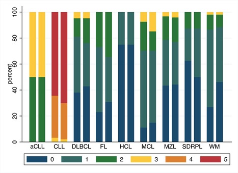
Le score de Matutes, décrit par le Dr Matutes du Royal Marsden Hospital à Londres en 1994, est un score diagnostique de la leucémie lymphoïde chronique (LLC), la forme la plus fréquente de leucémie chronique chez l'adulte. Il est basé sur l'étude de l’expression, par cytomérie en flux, de 5 marqueurs de surface des lymphocytes B: le CD5, le CD23, le FMC7, le CD79b et l'intensité de l'immunoglobuline de surface. Un point est ou non attribué en fonction de l’expression de chaque marqueur, et un score supérieur ou égal à 4 est très en faveur du diagnostic de LLC. Dans les combinaisons d’anticorps utilisées en cytométrie en flux pour le diagnostic des hémopathies lymphoïdes B, est systématiquement présent le CD20 qui permet d'isoler la population B, c’est également une cible thérapeutique. L’anticorps anti FMC7 reconnaissant un épitope de la protéine CD20, il y a compétition entre les anticorps et l’étude du FMC7 nécessite donc un tube spécifique, actuellement en 8 couleurs/8 antigènes.
Les cytomètres de routine hospitalière passant de 8-10 couleurs à 12-13 couleurs, Valérie Bardet et ses collaborateurs de du Service d'Hématologie-Immunologie-Transfusion des Hôpitaux Universitaires Paris Ile de France-Ouest (UVSQ/UPSaclay, Boulogne-Billancourt) se sont demandés s'ils pouvaient remplacer l’étude du FMC7 par celle de l’intensité du CD20, ceci afin de limiter le nombre d’anticorps utilisés et donc le coût et le temps des techniques. Leur étude parue dans le British Journal of Haematology a été réalisée sur une cohorte de 508 patients atteints d’hémopathie lymphoïde B chronique provenant des hôpitaux Ambroise Paré et Saint Louis. Elle a permis de montrer qu’une nouvelle modalité de calcul était possible avec des performances égales au score historiquement décrit.
Légende Figure : Comparaison des résultats du calcul du score avec le FMC7 (barre de gauche) ou le CD20 (barre de droite) dans différents types d'hémopathies B (CLL, chronic lymphocytic leukaemia; DLBCL, diffuse large B‐cell lymphoma; FL, follicular lymphoma; HCL, hairy cell leukaemia; MCL, Mantle cell lymphoma; MZL, marginal zone lymphoma; SDRPL, splenic diffuse red pulp small B‐cell lymphoma; WM, Waldenström macroglobulinaemia)
Contact : valerie.bardet@aphp.fr Via Life Sciences UPSaclay
Presented By: Jordi Petriz

Get everything you need to set up your flow and multicolor flow cytometry experiments with our comprehensive guide
|


Flow cytometry is an essential tool for dissecting the functional complexity of hematopoiesis. We used single-cell "mass cytometry" to examine healthy human bone marrow, measuring 34 parameters simultaneously in single cells (binding of 31 antibodies, viability, DNA content, and relative cell size). The signaling behavior of cell subsets spanning a defined hematopoietic hierarchy was monitored with 18 simultaneous markers of functional signaling states perturbed by a set of ex vivo stimuli and inhibitors. The data set allowed for an algorithmically driven assembly of related cell types defined by surface antigen expression, providing a superimposable map of cell signaling responses in combination with drug inhibition. Visualized in this manner, the analysis revealed previously unappreciated instances of both precise signaling responses that were bounded within conventionally defined cell subsets and more continuous phosphorylation responses that crossed cell population boundaries in unexpected manners yet tracked closely with cellular phenotype. Collectively, such single-cell analyses provide system-wide views of immune signaling in healthy human hematopoiesis, against which drug action and disease can be compared for mechanistic studies and pharmacologic intervention.