NACHT, LRR (leucine-rich repeat), and PYD (pyrin domain) domain-containing 6 (Nlrp6) is a member of the NLR (nucleotide-oligomerization domain-like receptor) family that patrols the cytosolic compartment of cells to detect pathogen- and damage-associated molecular patterns. Because Nlrp6 is a recently discovered inflammasome, details of its signaling mechanism, structural assembly, and roles in host defense have yet to be determined. To date, Nlrp6 has been proposed to perform a multitude of functions ranging from control of microbiota, maintenance of epithelial integrity, and regulation of metabolic diseases to modulation of host defense during microbial infections, cancer protection, and regulation of neuroinflammation. While recent studies have questioned some of the proposed functions of Nlrp6, Nlrp6 has been shown to form an inflammasome complex and cleaves interleukin-1β (IL-1β) and IL-18 during microbial infection, indicating that it is a bonafide inflammasome. In this review, we summarize the recent advancements in knowledge of the signaling mechanisms and structure of the Nlrp6 inflammasome and discuss the relevance of NLRP6 to human disease.
Natural killer (NK) cells are the major lymphocyte subset of the innate immune system. Their ability to mediate anti-tumor cytotoxicity and produce cytokines is well-established. However, the molecular mechanisms associated with the development of human or murine NK cells are not fully understood. Knowledge is being gained about the environmental cues, the receptors that sense the cues, signaling pathways, and the transcriptional programs responsible for the development of NK cells. Specifically, a complex network of transcription factors (TFs) following microenvironmental stimuli coordinate the development and maturation of NK cells. Multiple TFs are involved in the development of NK cells in a stage-specific manner. In this review, we summarize the recent advances in the understandings of TFs involved in the regulation of NK cell development, maturation, and effector function, in the aspects of their mechanisms, potential targets, and functions.
Le projet I-Covid a été lancé par des chercheurs de l'Inserm, de l'AP-HP et de Sorbonne Université du centre d’Immunologie et des maladies infectieuses (CIMI-Paris)* en avril 2020. Ce projet est le fruit d'une collaboration entre les équipes du Cimi-Paris et la biobanque de prélèvements réalisés chez des patients atteints de Covid-19, créée par le centre hospitalo-universitaire de la Pitié-Salpêtrière**. Guy Gorochov, coordinateur scientifique du projet et chef du service d'Immunologie à la Pitié-Salpêtrière, Christophe Combadière, chercheur Inserm, responsable du CIMI-Paris, et Béhazine Combadière, chercheuse Inserm, co-coordinatrice d'I-Covid, nous présentent leur travail.
* unité 1135 Inserm/CNRS/Sorbonne Université, Paris ** AP-HP
The interleukin-17 (IL-17) family is a relatively new family of cytokines consisting of 6 related factors (IL-17A–IL-17F), while the receptor family consists of 5 members: IL-17RA–IL-17RE.IL-17A is t...
What’s behind COVID-19’s selective onslaught? This article explores characteristics of the immune system during SARS-CoV-2 infection and why some comorbidities may be so risky.
The Hippo pathway responds to diverse extracellular cues and plays a key role in cell fate determination, tissue homeostasis and organ regeneration. Aberrant Hippo signaling, on the other hand, has been frequently implicated in diversified pathologies such as cancer and immune dysfunctions.
A key mystery about the COVID-19 pandemic is why the disease presents as mild or asymptomatic in the majority of the population, while presenting as severe or even life-threatening in approximately 15% of the population. An additional mystery is why COVID-19 generally presents more severely in adults than in children. A common underlying disease, Mast Cell Activation Syndrome (MCAS), may provide vital clues to solving these mysteries. MCAS is thought to be present in approximately 17% of the population, but most individuals who have MCAS remain undiagnosed. Our hypothesis is that many – perhaps even most – severe cases of COVID-19 can be attributed to pre-existing Mast Cell Activation Disease in those patients. Severe cases of COVID-19 are characterized by multi-system inflammation, caused by the body’s overreaction to the virus. Many times, it is this overreaction, not the virus itself, which becomes life-threatening. Mast cells which catastrophically over-activate in response to the virus could very well explain this phenomenon. Mast cells are tissue-resident immune system cells which act as the “sentries” of the immune system. They maintain numerous receptors on their surface, including histamine receptors as well as receptors to detect invading pathogens (PAMPS). When an invading pathogen (such as a virus) is detected, the mast cells release a variety of chemical mediators in a process called degranulation. These mediators both facilitate and coordinate the immune response, as well as provide signals to potentially every system in the body. In a normal individual, this degranulation process is well regulated and proportional to the detected threat. In an individual with MCAS, defective mast cells activate out of proportion with the need to defend the body from the perceived danger. This hyperreactivity can result in inappropriate continued activation even after the infection has passed. In our model, this underlying hyperreactivity is the “gun”, and the virus which causes COVID-19 merely “pulls the trigger”. In MCAS patients, some of the mast cell’s surface receptors (which typically function like an on/off switch) can become stuck in the “on” position. When this happens, MCAS patients can severely react to trace amounts of triggers which would be undetectable to normal individuals. When stuck in this “on” position, the mast cells will continue to release their proinflammatory mediators. This inappropriate activation in turn leads to the presentation of a multi-system inflammatory disease, affecting multiple, diverse systems in the body. Mast cells are central to the body’s immune response – as long as mast cells are activating in response to a perceived viral threat, they will both signal for more white blood cells to migrate to the infection site, and also cause the nearby tissue to expand in order to allow sufficient room for this migration to occur. Furthermore, the histamine released by mast cells activates the receptors on neighboring mast cells, causing them to degranulate, which then activates their neighboring mast cells in turn. In this manner, a cascading activation effect can sweep through all nearby mast cells, causing them to continually degranulate and release their mediators into the body. These inflammatory “cytokine storms”, which are characteristic in severe COVID-19, can affect such disparate systems as the lungs, the skin, and the blood. As a result, some of the unusual symptoms of the disease, such as blood clotting, skin rashes, and lung failure can all be explained by an underlying case of MCAS which becomes triggered by COVID-19. Further evidence for our hypothesis is seen in the correlation of the risk factors for severe COVID-19 with the most common symptoms of MCAS. High blood pressure is the primary comorbidity for COVID-19 patients who go on to develop Acute Respiratory Distress Syndrome. High blood pressure is also the second most common comorbidity seen in MCAS. Heartburn, the most common comorbidity seen in MCAS, has also been found to be highly prevalent in hospitalized COVID-19 patients. Severe COVID-19 is also correlated with a number of other inflammatory diseases, which are also comorbid with MCAS. The symptoms of the recently described Multisystem Inflammatory Syndrome in Children also fit an underlying diagnosis of MCAS extremely well. MCAS is frequently a lifelong, progressive disease, which tends to “step up” in severity over the patient’s lifetime. In the event our hypothesis is correct, this feature also describes why it is far more common to see severe COVID-19 in adults than in children. In our model, adults are far more likely to have severe MCAS, so they are far more likely to have severe COVID-19. Some of the treatments which are showing promise for severe COVID-19 – such as the drugs tocilizumab, famotidine, and vitamin D – are exactly the drugs one would expect to be effective if an underlying case of MCAS is present. The early evidence which shows famotidine (an H2 antihistamine sold under the brand name Pepcid) as a potentially effective treatment for severe COVID-19 is particularly striking. Two separate studies have found that famotidine use is associated with a 50% reduction in severe COVID-19 outcomes. Famotidine is a common over the counter medication which has no known anti-viral properties. However, it is commonly used to treat MCAS by suppressing overly active mast cells. If additional famotidine studies continue to substantiate the 50% reduction in severe COVID-19 outcomes, then it becomes imperative to determine the drug’s mechanism of action. In our model, famotidine is doing in COVID-19 patients what it is known to do in MCAS patients – block the histamine cascades, stabilize the patients mast cells and prevent the patient’s immune system from inappropriately overreacting. If an underlying case of MCAS is indeed the “gun” and the virus that causes COVID-19 merely “pulls the trigger”, then awareness of this connection is vital to proper treatment. This awareness can then lead to the necessary actions for better outcomes and a lower mortality rate during the COVID-19 pandemic. Mast Cell Activation Syndrome affects a large minority of the population, but it is a disease that most people have never heard of. If MCAS is indeed the key to the COVID-19 mystery, there may be no better time than this moment to raise the profile of this important disease. (Full Paper Below)
Acquista Online La Prescrizione Di Perdita Di Peso Crediamo che i farmaci a volte possano essere molto urgenti da assumere. Se hai urgente bisogno di farmaci, possiamo anche fornirti una consegna espressa,
Coronavirus in nonhuman primates We urgently need vaccines and drug treatments for coronavirus disease 2019 (COVID-19). Even under these extreme circumstances, we must have animal models for rigorous testing of new strategies. Rockx et al. have undertaken a comparative study of three human coronaviruses in cynomolgus macaques: severe acute respiratory syndrome–coronavirus (SARS-CoV) (2002), Middle East respiratory syndrome (MERS)–CoV (2012), and SARS-CoV-2 (2019), which causes COVID-19 (see the Perspective by Lakdawala and Menachery). The most recent coronavirus has a distinct tropism for the nasal mucosa but is also found in the intestinal tract. Although none of the older macaques showed the severe symptoms that humans do, the lung pathology observed was similar. Like humans, the animals shed virus for prolonged periods from their upper respiratory tracts, and like influenza but unlike the 2002 SARS-CoV, this shedding peaked early in infection. It is this cryptic virus shedding that makes case detection difficult and can jeopardize the effectiveness of isolation. Science, this issue p. 1012; see also p. 942 Abstract The current pandemic coronavirus, severe acute respiratory syndrome–coronavirus 2 (SARS-CoV-2), was recently identified in patients with an acute respiratory syndrome, coronavirus disease 2019 (COVID-19). To compare its pathogenesis with that of previously emerging coronaviruses, we inoculated cynomolgus macaques with SARS-CoV-2 or Middle East respiratory syndrome (MERS)–CoV and compared the pathology and virology with historical reports of SARS-CoV infections. In SARS-CoV-2–infected macaques, virus was excreted from nose and throat in the absence of clinical signs and detected in type I and II pneumocytes in foci of diffuse alveolar damage and in ciliated epithelial cells of nasal, bronchial, and bronchiolar mucosae. In SARS-CoV infection, lung lesions were typically more severe, whereas they were milder in MERS-CoV infection, where virus was detected mainly in type II pneumocytes. These data show that SARS-CoV-2 causes COVID-19–like disease in macaques and provides a new model to test preventive and therapeutic strategies. After the first reports of an outbreak of an acute respiratory syndrome in China in December 2019, a novel coronavirus, severe acute respiratory syndrome–coronavirus 2 (SARS-CoV-2), was identified (1, 2). As of 14 March 2020, over 140,000 cases were reported worldwide with more than 5400 deaths, surpassing the combined number of cases and deaths of two previously emerging coronaviruses, SARS-CoV and Middle East respiratory syndrome (MERS)–CoV (3). The disease caused by this virus, coronavirus disease 2019 (COVID-19), is characterized by a range of symptoms, including fever, cough, dyspnoea, and myalgia in most cases (2). In severe cases, bilateral lung involvement with ground-glass opacity is the most common chest computed tomography (CT) finding (4). Similarly to the 2002–2003 outbreak of SARS, the severity of COVID-19 disease is associated with increased age and/or a comorbidity, although severe disease is not limited to these risk groups (5). However, despite the large number of cases and deaths, limited information is available on the pathogenesis of this virus infection. Two reports on the histological examination of the lungs of three patients showed bilateral diffuse alveolar damage (DAD), pulmonary edema, and hyaline membrane formation, indicative of acute respiratory distress syndrome (ARDS), as well as characteristic syncytial cells in the alveolar lumen (6, 7), similar to findings during the 2002–2003 outbreak of SARS-CoV (8). The pathogenesis of SARS-CoV infection was previously studied in a nonhuman primate model (cynomolgus macaques) where aged animals were more likely to develop disease (9–13). In the current study, SARS-CoV-2 infection was characterized in the same animal model and compared with infection with MERS-CoV and historical data on SARS-CoV (9, 10, 12). First, two groups of four cynomolgus macaques [both young adult (young), 4 to 5 years of age; and old adult (aged), 15 to 20 years of age] were inoculated by a combined intratracheal (IT) and intranasal (IN) route with a SARS-CoV-2 strain from a German traveler returning from China. No overt clinical signs were observed in any of the infected animals, except for a serous nasal discharge in one aged animal on day 14 post inoculation (p.i.). No significant weight loss was observed in any of the animals during the study. By day 14 p.i., all remaining animals seroconverted as revealed by the presence of SARS-CoV-2–specific antibodies against the virus S1 domain and nucleocapsid proteins in their sera (fig. S1). As a measure of virus shedding, nasal, throat, and rectal swabs were assayed for virus by reverse transcription–quantitative polymerase chain reaction (RT-qPCR) and virus culture. In nasal swabs, SARS-CoV-2 RNA peaked by day 2 p.i. in young animals and by day 4 p.i. in aged animals and was detected up to at least day 8 p.i. in two out of four animals and up to day 21 p.i. in one out of four animals (Fig. 1A). Overall, higher levels of SARS-CoV-2 RNA were detected in nasal swabs of aged animals compared with young animals. SARS-CoV-2 RNA in throat swabs peaked at day 1 p.i. in young and day 4 p.i. in aged animals and decreased more rapidly over time by comparison with the nasal swabs but could still be detected intermittently up to day 10 p.i. (Fig. 1B). Low levels of infectious virus were cultured from throat and nasal swabs up to day 2 and 4, respectively (table S1). In support of virus shedding by these animals, environmental sampling was performed to determine potential contamination of surfaces. Environmental sampling indicated the presence of low levels of SARS-CoV-2 RNA on surfaces through both direct contact (hands) and indirect contamination within the isolator (table S2). SARS-CoV-2 RNA was only detected in a rectal swab from one animal on day 14 p.i., and no viral RNA was detected in whole blood at any time point throughout the study. On autopsy of four macaques on day 4 p.i., two had foci of pulmonary consolidation in the lungs (Fig. 2A). One animal (aged: 17 years) showed consolidation in the right middle lobe, representing less than 5% of the lung tissue. A second animal (young: 5 years) had two foci in the left lower lobe, representing about 10% of the lung tissue (Fig. 2A). The consolidated lung tissue was well circumscribed, red-purple, level, and less buoyant than normal. The other organs in these two macaques, as well as the respiratory tract and other organs of the other two animals, were normal. Virus replication was assessed by RT-qPCR on day 4 p.i. in tissues from the respiratory, digestive, urinary, and cardiovascular tracts and from endocrine and central nervous systems, as well as from various lymphoid tissues. Virus replication was primarily restricted to the respiratory tract (nasal cavity, trachea, bronchi, and lung lobes) with highest levels of SARS-CoV-2 RNA in lungs (Fig. 1C). In three out of four animals, SARS-CoV-2 RNA was also detected in ileum and tracheo-bronchial lymph nodes (Fig. 1C). The main histological lesion in the consolidated pulmonary tissues of both the young and aged animals involved the alveoli and bronchioles and consisted of areas with acute or more advanced DAD (Fig. 2B). In these areas, the lumina of alveoli and bronchioles were variably filled with protein-rich edema fluid, fibrin, and cellular debris; alveolar macrophages; and fewer neutrophils and lymphocytes (Fig. 2, C to E). There was epithelial necrosis with extensive loss of epithelium from alveolar and bronchiolar walls. Hyaline membranes were present in a few damaged alveoli. In areas with more advanced lesions, the alveolar walls were moderately thickened and lined by cuboidal epithelial cells (type II pneumocyte hyperplasia), and the alveolar lumina were empty. Alveolar and bronchiolar walls were thickened by edema fluid, mononuclear cells, and neutrophils. There were aggregates of lymphocytes around small pulmonary vessels. Moderate numbers of lymphocytes and macrophages were present in the lamina propria and submucosa of the bronchial walls, and a few neutrophils were detected in the bronchial epithelium. Regeneration of epithelium was seen in some bronchioles, visible as an irregular layer of squamous to high cuboidal epithelial cells with hyperchromatic nuclei. There were occasional multinucleated giant cells (syncytia) free in the lumina of bronchioles and alveoli (Fig. 2F) and, based on positive pan-keratin staining and negative CD68 staining, these appeared to originate from epithelial cells (Fig. 2F, inset). SARS-CoV-2 antigen expression was detected in moderate numbers of type I pneumocytes and a few type II pneumocytes within foci of DAD (Fig. 2, G and H, and fig. S2). The pattern of staining was similar to that in lung tissue from SARS-CoV–infected macaques (positive control). SARS-CoV-2 antigen expression was not observed in any of the syncytia. In addition, SARS-CoV-2 antigen expression was detected in nonlesional tissues of all lung lobes in three out of four macaques (both young and one aged) in a few type I and II pneumocytes, bronchial ciliated epithelial cells, and bronchiolar ciliated epithelial cells. The other aged macaque, without virological or pathological evidence of SARS-CoV-2 infection in the lungs, did have SARS-CoV-2 antigen expression in ciliated epithelial cells of nasal septum (Fig. 2I), nasal concha, and palatum molle, in the absence of associated histopathological changes. No SARS-CoV antigen expression was detected in other sampled tissues, including brain and intestine. To assess the severity of infection with SARS-CoV-2 compared with MERS-CoV, we inoculated young cynomolgus macaques (3 to 5 years of age) with MERS-CoV via the IN and IT route. All animals remained free of clinical signs. At day 21 p.i., all remaining animals (n = 2) seroconverted as revealed by the presence of MERS-CoV–specific antibodies in their sera by ELISA (fig. S3). MERS-CoV RNA was detected in nasal (Fig. 3A) and throat swabs (Fig. 3B) on days 1 to 11 p.i., with peaks on days 1 and 2 p.i., respectively. Low levels [between 1 and 85 median tissue culture infectious dose (TCID50) equivalent/ml] of MERS-CoV RNA were detected in rectal swabs on days 2 and 3 p.i. At autopsy of four macaques at day 4 p.i., three animals had foci of pulmonary consolidation, characterized by slightly depressed areas in the lungs, representing less than 5% of the lung tissue (Table 1). Similar to SARS-CoV-2 infection in both young and aged animals, on day 4 p.i., MERS-CoV RNA was primarily detected in the respiratory tract of inoculated animals (Fig. 3C). Infectious virus titers were comparable to those of SARS-CoV-2 infection, but lower compared to SARS-CoV infection, of young macaques (Table 1). In addition, MERS-CoV RNA was detected in the spleen (Table 1). Table 1 Comparative pathogenesis of SARS-CoV-2, MERS-CoV, and SARS-CoV infections in cynomolgus macaques. Max, maximum; Ref., reference. View this table: View popup View inline Consistent with the presence of virus in the lower respiratory tract at day 4 p.i., histopathological changes characteristic for DAD were observed in the lungs of inoculated animals (Fig. 3D). The alveolar septa were thickened owing to infiltration of neutrophils and macrophages, and to moderate type II pneumocyte hyperplasia and hypertrophy. In the alveolar lumina, there were increased numbers of alveolar macrophages and some edema fluid containing fibrin and some neutrophils (Fig. 3D). Few syncytial cells were seen in the alveolar lumina. MERS-CoV antigen was not detected in tissues on day 4 p.i. in any part of the respiratory tract. We therefore sampled four young macaques at day 1 p.i. At this time, we observed multifocal expression of viral antigen, predominantly in type II pneumocytes and occasionally in type I pneumocytes, bronchiolar and bronchial epithelial cells, and some macrophages (Fig. 3D). In summary, we inoculated young and aged cynomolgus macaques with a low-passage clinical isolate of SARS-CoV-2, which resulted in productive infection in the absence of overt clinical signs. Recent studies in human cases have shown that presymptomatic and asymptomatic cases can also shed virus (14, 15). Increased age did not affect disease outcome, but there was prolonged viral shedding in the upper respiratory tract of aged animals. Prolonged shedding has been observed in both SARS-CoV-2 and SARS-CoV patients (16, 17). SARS-CoV-2 shedding in our asymptomatic model peaked early in the course of infection, similar to what is seen in symptomatic patients (16). Also, SARS-CoV-2 antigen was detected in ciliated epithelial cells of nasal mucosae at day 4 p.i., which was not seen for SARS-CoV (10) or MERS-CoV infections (this study) in this animal model. Viral tropism for the nasal mucosa fits with efficient respiratory transmission, as has been seen for influenza A virus (18). This early peak in virus shedding for SARS-CoV-2 is similar to that of influenza virus shedding (19) and may explain why case detection and isolation may not be as effective for SARS-CoV-2 as it was for the control of SARS-CoV (20). SARS-CoV-2 was primarily detected in tissues of the respiratory tract; however, SARS-CoV-2 RNA was also detectable in other tissues such as intestines, in line with a recent report (21). Similar results regarding viral shedding and tissue and cell tropism were recently also reported after SARS-CoV-2 inoculation in rhesus macaques. However, unlike in our model, SARS-CoV-2 infection in rhesus macaques does result in transient respiratory disease and weight loss (22, 23). Two out of four animals had foci of DAD on day 4 p.i. The colocalization of SARS-CoV-2 antigen expression and DAD provides strong evidence that SARS-CoV-2 infection caused this lesion. The histological character of the DAD, including alveolar and bronchiolar epithelial necrosis, alveolar edema, hyaline membrane formation, and accumulation of neutrophils, macrophages, and lymphocytes, corresponds with the limited pathological analyses of human COVID-19 cases (6, 7). In particular, the presence of syncytia in the lung lesions is characteristic of respiratory coronavirus infections. Whereas MERS-CoV primarily infects type II pneumocytes in cynomolgus macaques, both SARS-CoV and SARS-CoV-2 also infect type I pneumocytes. Injury to type I pneumocytes can result in pulmonary edema, and formation of hyaline membranes (24), which may explain why hyaline membrane formation is a hallmark of SARS and COVID-19 (7, 10) but not frequently reported for MERS (25, 26). These data show that cynomolgus macaques are permissive to SARS-CoV-2 infection, shed virus for a prolonged period of time, and display COVID-19–like disease. In this nonhuman primate model, SARS-CoV-2 replicates efficiently in respiratory epithelial cells throughout the respiratory tract, including nasal cavity, bronchi, bronchioles, and alveoli. Replication in the upper respiratory tract fits with efficient transmission between hosts, whereas replication in the lower respiratory tract fits with the development of lung disease. An in-depth comparison of infection with SARS-CoV, MERS-CoV, and SARS-CoV-2 in this model may identify key pathways in the pathogenesis of these emerging viruses. This study provides a new infection model that will be critical in the evaluation and licensure of preventive and therapeutic strategies against SARS-CoV-2 infection for use in humans, as well as for evaluating the efficacy of repurposing species-specific existing treatments, such as pegylated interferon (12). Supplementary Materials science.sciencemag.org/content/368/6494/1012/suppl/DC1 Materials and Methods Figs. S1 to S3 Tables S1 and S2 References (27–30) MDAR Reproducibility Checklist http://www.sciencemag.org/about/science-licenses-journal-article-reuse This is an article distributed under the terms of the Science Journals Default License. References and Notes ↵ J. F. Chan, S. Yuan, K.-H. Kok, K. K.-W. To, H. Chu, J. Yang, F. Xing, J. Liu, C. C.-Y. Yip, R. W.-S. Poon, H.-W. Tsoi, S. K.-F. Lo, K.-H. Chan, V. K.-M. Poon, W.-M. Chan, J. D. Ip, J.-P. Cai, V. C.-C. Cheng, H. Chen, C. K.-M. Hui, K.-Y. Yuen, A familial cluster of pneumonia associated with the 2019 novel coronavirus indicating person-to-person transmission: A study of a family cluster. Lancet 395, 514–523 (2020). doi:10.1016/S0140-6736(20)30154-9pmid:31986261OpenUrlCrossRefPubMed ↵ C. Huang, Y. Wang, X. Li, L. Ren, J. Zhao, Y. Hu, L. Zhang, G. Fan, J. Xu, X. Gu, Z. Cheng, T. Yu, J. Xia, Y. Wei, W. Wu, X. Xie, W. Yin, H. Li, M. Liu, Y. Xiao, H. Gao, L. Guo, J. Xie, G. Wang, R. Jiang, Z. Gao, Q. Jin, J. Wang, B. Cao, Clinical features of patients infected with 2019 novel coronavirus in Wuhan, China. Lancet 395, 497–506 (2020). doi:10.1016/S0140-6736(20)30183-5pmid:31986264OpenUrlCrossRefPubMed ↵ E. Mahase, Coronavirus covid-19 has killed more people than SARS and MERS combined, despite lower case fatality rate. BMJ 368, m641 (2020). doi:10.1136/bmj.m641pmid:32071063OpenUrlFREE Full Text ↵ H. Shi, X. Han, N. Jiang, Y. Cao, O. Alwalid, J. Gu, Y. Fan, C. Zheng, Radiological findings from 81 patients with COVID-19 pneumonia in Wuhan, China: A descriptive study. Lancet Infect. Dis. 20, 425–434 (2020). doi:10.1016/S1473-3099(20)30086-4pmid:32105637OpenUrlCrossRefPubMed ↵ N. Chen, M. Zhou, X. Dong, J. Qu, F. Gong, Y. Han, Y. Qiu, J. Wang, Y. Liu, Y. Wei, J. Xia, T. Yu, X. Zhang, L. Zhang, Epidemiological and clinical characteristics of 99 cases of 2019 novel coronavirus pneumonia in Wuhan, China: A descriptive study. Lancet 395, 507–513 (2020). doi:10.1016/S0140-6736(20)30211-7pmid:32007143OpenUrlCrossRefPubMed ↵ Z. Xu, L. Shi, Y. Wang, J. Zhang, L. Huang, C. Zhang, S. Liu, P. Zhao, H. Liu, L. Zhu, Y. Tai, C. Bai, T. Gao, J. Song, P. Xia, J. Dong, J. Zhao, F.-S. Wang, Pathological findings of COVID-19 associated with acute respiratory distress syndrome. Lancet Respir. Med. 8, 420–422 (2020). doi:10.1016/S2213-2600(20)30076-Xpmid:32085846OpenUrlCrossRefPubMed ↵ S. Tian, W. Hu, L. Niu, H. Liu, H. Xu, S.-Y. Xiao, Pulmonary pathology of early-phase 2019 novel coronavirus (COVID-19) pneumonia in two patients with lung cancer. J. Thorac. Oncol. S1556-0864(20)30132-5 (2020). doi:10.1016/j.jtho.2020.02.010pmid:32114094OpenUrlCrossRefPubMed ↵ J. M. Nicholls, L. L. M. Poon, K. C. Lee, W. F. Ng, S. T. Lai, C. Y. Leung, C. M. Chu, P. K. Hui, K. L. Mak, W. Lim, K. W. Yan, K. H. Chan, N. C. Tsang, Y. Guan, K. Y. Yuen, J. S. Peiris, Lung pathology of fatal severe acute respiratory syndrome. Lancet 361, 1773–1778 (2003). doi:10.1016/S0140-6736(03)13413-7pmid:12781536 ↵ R. A. Fouchier, T. Kuiken, M. Schutten, G. van Amerongen, G. J. J. van Doornum, B. G. van den Hoogen, M. Peiris, W. Lim, K. Stöhr, A. D. M. E. Osterhaus, Koch’s postulates fulfilled for SARS virus. Nature 423, 240 (2003). doi:10.1038/423240apmid:12748632 ↵ T. Kuiken, R. A. M. Fouchier, M. Schutten, G. F. Rimmelzwaan, G. van Amerongen, D. van Riel, J. D. Laman, T. de Jong, G. van Doornum, W. Lim, A. E. Ling, P. K. S. Chan, J. S. Tam, M. C. Zambon, R. Gopal, C. Drosten, S. van der Werf, N. Escriou, J.-C. Manuguerra, K. Stöhr, J. S. M. Peiris, A. D. M. E. Osterhaus, Newly discovered coronavirus as the primary cause of severe acute respiratory syndrome. Lancet 362, 263–270 (2003). doi:10.1016/S0140-6736(03)13967-0pmid:12892955 S. L. Smits, A. de Lang, J. M. A. van den Brand, L. M. Leijten, W. F. van IJcken, M. J. C. Eijkemans, G. van Amerongen, T. Kuiken, A. C. Andeweg, A. D. M. E. Osterhaus, B. L. Haagmans, Exacerbated innate host response to SARS-CoV in aged non-human primates. PLOS Pathog. 6, e1000756 (2010). doi:10.1371/journal.ppat.1000756pmid:20140198OpenUrlCrossRefPubMed ↵ B. L. Haagmans, T. Kuiken, B. E. Martina, R. A. M. Fouchier, G. F. Rimmelzwaan, G. van Amerongen, D. van Riel, T. de Jong, S. Itamura, K.-H. Chan, M. Tashiro, A. D. M. E. Osterhaus, Pegylated interferon-alpha protects type 1 pneumocytes against SARS coronavirus infection in macaques. Nat. Med. 10, 290–293 (2004). doi:10.1038/nm1001pmid:14981511 ↵ B. Rockx, F. Feldmann, D. Brining, D. Gardner, R. LaCasse, L. Kercher, D. Long, R. Rosenke, K. Virtaneva, D. E. Sturdevant, S. F. Porcella, J. Mattoon, M. Parnell, R. S. Baric, H. Feldmann, Comparative pathogenesis of three human and zoonotic SARS-CoV strains in cynomolgus macaques. PLOS ONE 6, e18558 (2011). doi:10.1371/journal.pone.0018558pmid:21533129OpenUrlCrossRefPubMed ↵ C. Li, F. Ji, L. Wang, L. Wang, J. Hao, M. Dai, Y. Liu, X. Pan, J. Fu, L. Li, G. Yang, J. Yang, X. Yan, B. Gu, Asymptomatic and Human-to-Human Transmission of SARS-CoV-2 in a 2-Family Cluster, Xuzhou, China. Emerg. Infect. Dis. 26 (2020). doi:10.3201/eid2607.200718pmid:32228809OpenUrlCrossRefPubMed ↵ A. Kimball, K. M. Hatfield, M. Arons, A. James, J. Taylor, K. Spicer, A. C. Bardossy, L. P. Oakley, S. Tanwar, Z. Chisty, J. M. Bell, M. Methner, J. Harney, J. R. Jacobs, C. M. Carlson, H. P. McLaughlin, N. Stone, S. Clark, C. Brostrom-Smith, L. C. Page, M. Kay, J. Lewis, D. Russell, B. Hiatt, J. Gant, J. S. Duchin, T. A. Clark, M. A. Honein, S. C. Reddy, J. A. Jernigan, A. Baer, L. M. Barnard, E. Benoliel, M. S. Fagalde, J. Ferro, H. G. Smith, E. Gonzales, N. Hatley, G. Hatt, M. Hope, M. Huntington-Frazier, V. Kawakami, J. L. Lenahan, M. D. Lukoff, E. B. Maier, S. McKeirnan, P. Montgomery, J. L. Morgan, L. A. Mummert, S. Pogosjans, F. X. Riedo, L. Schwarcz, D. Smith, S. Stearns, K. J. Sykes, H. Whitney, H. Ali, M. Banks, A. Balajee, E. J. Chow, B. Cooper, D. W. Currie, J. Dyal, J. Healy, M. Hughes, T. M. McMichael, L. Nolen, C. Olson, A. K. Rao, K. Schmit, N. G. Schwartz, F. Tobolowsky, R. Zacks, S. Zane; Public Health – Seattle & King County; CDC COVID-19 Investigation Team, Asymptomatic and Presymptomatic SARS-CoV-2 Infections in Residents of a Long-Term Care Skilled Nursing Facility - King County, Washington, March 2020. MMWR Morb. Mortal. Wkly. Rep. 69, 377–381 (2020). doi:10.15585/mmwr.mm6913e1pmid:32240128OpenUrlCrossRefPubMed ↵ L. Zou, F. Ruan, M. Huang, L. Liang, H. Huang, Z. Hong, J. Yu, M. Kang, Y. Song, J. Xia, Q. Guo, T. Song, J. He, H.-L. Yen, M. Peiris, J. Wu, SARS-CoV-2 Viral Load in Upper Respiratory Specimens of Infected Patients. N. Engl. J. Med. 382, 1177–1179 (2020). doi:10.1056/NEJMc2001737pmid:32074444OpenUrlCrossRefPubMed ↵ J. S. Peiris, C. M. Chu, V. C. C. Cheng, K. S. Chan, I. F. N. Hung, L. L. M. Poon, K. I. Law, B. S. F. Tang, T. Y. W. Hon, C. S. Chan, K. H. Chan, J. S. C. Ng, B. J. Zheng, W. L. Ng, R. W. M. Lai, Y. Guan, K. Y. Yuen; HKU/UCH SARS Study Group, Clinical progression and viral load in a community outbreak of coronavirus-associated SARS pneumonia: A prospective study. Lancet 361, 1767–1772 (2003). doi:10.1016/S0140-6736(03)13412-5pmid:12781535 ↵ M. Richard, J. M. A. van den Brand, T. M. Bestebroer, P. Lexmond, D. de Meulder, R. A. M. Fouchier, A. C. Lowen, S. Herfst, Influenza A viruses are transmitted via the air from the nasal respiratory epithelium of ferrets. Nat. Commun. 11, 766 (2020). doi:10.1038/s41467-020-14626-0pmid:32034144OpenUrlCrossRefPubMed ↵ C. C. Li, L. Wang, H.-L. Eng, H.-L. You, L.-S. Chang, K.-S. Tang, Y.-J. Lin, H.-C. Kuo, I.-K. Lee, J.-W. Liu, E.-Y. Huang, K. D. Yang, Correlation of pandemic (H1N1) 2009 viral load with disease severity and prolonged viral shedding in children. Emerg. Infect. Dis. 16, 1265–1272 (2010). doi:10.3201/eid1608.091918pmid:20678321 ↵ X. Pang, Z. Zhu, F. Xu, J. Guo, X. Gong, D. Liu, Z. Liu, D. P. Chin, D. R. Feikin, Evaluation of control measures implemented in the severe acute respiratory syndrome outbreak in Beijing, 2003. JAMA 290, 3215–3221 (2003). doi:10.1001/jama.290.24.3215pmid:14693874 ↵ F. Xiao, M. Tang, X. Zheng, Y. Liu, X. Li, H. Shan, Evidence for gastrointestinal infection of SARS-CoV-2. Gastroenterology S0016-5085(20)30282-1 (2020). doi:10.1053/j.gastro.2020.02.055pmid:32142773OpenUrlCrossRefPubMed ↵W. D. Linlin Bao et al., Reinfection could not occur in SARS-CoV-2 infected rhesus macaques. bioRxiv 2020.03.13.990226 (2020). https://doi.org/10.1101/2020.03.13.990226. ↵V. J. Munster et al., Respiratory disease and virus shedding in rhesus macaques inoculated with SARS-CoV-2. bioRxiv 2020.03.21.001628 (2020). https://doi.org/10.1101/2020.03.21.001628. ↵ L. B. Ware, M. A. Matthay, The acute respiratory distress syndrome. N. Engl. J. Med. 342, 1334–1349 (2000). doi:10.1056/NEJM200005043421806pmid:10793167 ↵ W. J. Shieh, C.-H. Hsiao, C. D. Paddock, J. Guarner, C. S. Goldsmith, K. Tatti, M. Packard, L. Mueller, M.-Z. Wu, P. Rollin, I.-J. Su, S. R. Zaki, Immunohistochemical, in situ hybridization, and ultrastructural localization of SARS-associated coronavirus in lung of a fatal case of severe acute respiratory syndrome in Taiwan. Hum. Pathol. 36, 303–309 (2005). doi:10.1016/j.humpath.2004.11.006pmid:15791576 ↵ D. L. Ng, F. Al Hosani, M. K. Keating, S. I. Gerber, T. L. Jones, M. G. Metcalfe, S. Tong, Y. Tao, N. N. Alami, L. M. Haynes, M. A. Mutei, L. Abdel-Wareth, T. M. Uyeki, D. L. Swerdlow, M. Barakat, S. R. Zaki, Clinicopathologic, Immunohistochemical, and Ultrastructural Findings of a Fatal Case of Middle East Respiratory Syndrome Coronavirus Infection in the United Arab Emirates, April 2014. Am. J. Pathol. 186, 652–658 (2016). doi:10.1016/j.ajpath.2015.10.024pmid:26857507OpenUrlCrossRefPubMed ↵ V. S. Raj, H. Mou, S. L. Smits, D. H. W. Dekkers, M. A. Müller, R. Dijkman, D. Muth, J. A. A. Demmers, A. Zaki, R. A. M. Fouchier, V. Thiel, C. Drosten, P. J. M. Rottier, A. D. M. E. Osterhaus, B. J. Bosch, B. L. Haagmans, Dipeptidyl peptidase 4 is a functional receptor for the emerging human coronavirus-EMC. Nature 495, 251–254 (2013). doi:10.1038/nature12005pmid:23486063 B. L. Haagmans, J. M. A. van den Brand, L. B. Provacia, V. S. Raj, K. J. Stittelaar, S. Getu, L. de Waal, T. M. Bestebroer, G. van Amerongen, G. M. G. M. Verjans, R. A. M. Fouchier, S. L. Smits, T. Kuiken, A. D. M. E. Osterhaus, Asymptomatic Middle East respiratory syndrome coronavirus infection in rabbits. J. Virol. 89, 6131–6135 (2015). doi:10.1128/JVI.00661-15pmid:25810539 V. M. Corman, O. Landt, M. Kaiser, R. Molenkamp, A. Meijer, D. K. W. Chu, T. Bleicker, S. Brünink, J. Schneider, M. L. Schmidt, D. G. J. C. Mulders, B. L. Haagmans, B. van der Veer, S. van den Brink, L. Wijsman, G. Goderski, J.-L. Romette, J. Ellis, M. Zambon, M. Peiris, H. Goossens, C. Reusken, M. P. G. Koopmans, C. Drosten, Detection of 2019 novel coronavirus (2019-nCoV) by real-time RT-PCR. Euro Surveill. 25, 25 (2020). doi:10.2807/1560-7917.ES.2020.25.3.2000045pmid:31992387OpenUrlCrossRefPubMed ↵ N. M. A. Okba, M. A. Müller, W. Li, C. Wang, C. H. GeurtsvanKessel, V. M. Corman, M. M. Lamers, R. S. Sikkema, E. de Bruin, F. D. Chandler, Y. Yazdanpanah, Q. Le Hingrat, D. Descamps, N. Houhou-Fidouh, C. B. E. M. Reusken, B.-J. Bosch, C. Drosten, M. P. G. Koopmans, B. L. Haagmans, Severe acute respiratory syndrome coronavirus 2-specific antibody responses in coronavirus disease 2019 patients. Emerg. Infect. Dis. 26, 10.3201/eid2607.200841 (2020). doi:10.3201/eid2607.200841pmid:32267220OpenUrlCrossRefPubMed Acknowledgments: We thank L. de Meulder, A. van der Linden, I. Chestakova, and F. van der Panne for technical assistance. We also thank Y. Kap, D. Akkermans, V. Vaes, M. Sommers, and F. Meijers for assistance with the animal studies. Funding: This research is (partly) financed by the NWO Stevin Prize awarded to M.K. by the Netherlands Organisation for Scientific Research (NWO), H2020 grant agreement 874735-VEO to M.K., NIH/NIAID contract HHSN272201400008C to R.F., and H2020 grant agreement 101003651–MANCO to B.H. Author contributions: Conceptualization, B.R., T.K., R.F., M.K., RdS, M.F. B.H.; investigation, B.R., T.K., S.H., T.B., M.L., D.d.M., G.v.A., J.v.d.B., N.O., D.S., P.v.R., L.L.; resources, B.H., C.D., E.V., B.V., J.L.; supervision, B.R. and B.H.; writing, original draft, B.R., T.K., and B.H.; writing–review and editing, all authors; funding acquisition: S.H., B.H., R.F., and M.K. Animal welfare: Research was conducted in compliance with the Dutch legislation for the protection of animals used for scientific purposes (2014, implementing EU Directive 2010/63) and other relevant regulations. The licensed establishment where this research was conducted (Erasmus MC) has an approved OLAW Assurance no. A5051-01. Research was conducted under a project license from the Dutch competent authority and the study protocol (no. 17-4312) was approved by the institutional Animal Welfare Body. Competing interests: T.B., R.F., and B.H. are listed as inventors on a patent application related to MERS-coronavirus diagnostics, therapy and vaccines. Other authors declare no competing interests. Data and materials availability: All data are available in the main text or the supplementary materials. This work is licensed under a Creative Commons Attribution 4.0 International (CC BY 4.0) license, which permits unrestricted use, distribution, and reproduction in any medium, provided the original work is properly cited. To view a copy of this license, visit https://creativecommons.org/licenses/by/4.0/. This license does not apply to figures/photos/artwork or other content included in the article that is credited to a third party; obtain authorization from the rights holder before using such material.
Through the use of Drosophila genetics and the excellent in vivo imaging possible in the fly embryo, Davidson and Wood demonstrate that macrophages switch from engulfing debris via sheet-like lamellipods (lamellipodial phagocytosis) to extending phagocytic filopods (filopodial phagocytosis) when spatially confined, thus maintaining their critical clearance function in vivo.
CD4 T cells play a key role in the functioning of a healthy immune system. They assist B cells to make antibodies, activate the microbe killing capacity of macrophages and recruit other immune cells to infected or inflamed areas of the body.
Upon tissue injury or microbial invasion, a large number of neutrophils converge from blood to the sites of injury or infection in a short time. The migration through a limited number of paths through tissues and capillary networks seems efficient and ‘traffic jams’ are generally avoided. However, the mechanisms that guide efficient trafficking of large numbers of neutrophils through capillary networks are not well understood. Here we show that pairs of neutrophils arriving closely one after another at capillary bifurcations migrate to alternating branches in vivo and in vitro. Perturbation of chemoattractant gradients and the increased hydraulic resistance induced by the first neutrophil in one branch biases the migration of the following neutrophil towards the other branch. These mechanisms guide neutrophils to efficiently navigate through capillary networks and outline the effect of inter-neutrophil interactions during migration on overall lymphocyte trafficking patterns in confined environments. Neutrophils mobilize rapidly and travel through the vasculature and microcirculation en masse in response to inflammatory stimuli. Here the authors use a microfluidic device and intravital microscopy to show neutrophils move through tissues in a highly ordered pattern, taking turns to file into alternate branches at bifurcations.
Immunotherapy with checkpoint blockade induces rapid and durable immune control of cancer in some patients and has driven a monumental shift in cancer treatment. Neoantigen-specific CD8+ T cells are at the forefront of current immunotherapy strategies, and the majority of drug discovery and clinical trials revolve around further harnessing these immune effectors. Yet the immune system contains a diverse range of antitumour effector cells, and these must function in a coordinated and synergistic manner to overcome the immune-evasion mechanisms used by tumours and achieve complete control with tumour eradication. A key antitumour effector is the natural killer (NK) cells, cytotoxic innate lymphocytes present at high frequency in the circulatory system and identified by their exquisite ability to spontaneously detect and lyse transformed or stressed cells. Emerging data show a role for intratumoural NK cells in driving immunotherapy response and, accordingly, there have been renewed efforts to further elucidate and target the pathways controlling NK cell antitumour function. In this Review, we discuss recent clinical evidence that NK cells are a key immune constituent in the protective antitumour immune response and highlight the major stages of the cancer–NK cell immunity cycle. We also perform a new analysis of publicly available transcriptomic data to provide an overview of the prognostic value of NK cell gene expression in 25 tumour types. Furthermore, we discuss how the role of NK cells evolves with tumour progression, presenting new opportunities to target NK cell function to enhance cancer immunotherapy response rates across a more diverse range of cancers. This Review discusses the key role that natural killer (NK) cells play in driving an antitumour immune response throughout the progression of cancer from its initial development to its metastatic spread and eventual treatment, defined herein as the cancer–NK cell immunity cycle.
In the current COVID-19 pandemic a key unsolved question is the duration of acquired immunity in recovered individuals. The recent emergence of SARS-CoV-2 precludes a direct study on this virus, but the four seasonal human coronaviruses may reveal common characteristics applicable to all human coronaviruses.
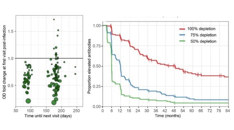
We monitored healthy subjects over a time span of 35 years (1985-2020), providing a total of 2473 follow up person-months, and determined a) the time to reinfection by the same seasonal coronavirus and b) the dynamics of coronavirus antibody depletion post-infection. An alarmingly short duration of protective immunity to coronaviruses was found. Reinfections occurred frequently at 12 months post-infection and there was for each virus a substantial reduction in antibody levels as soon as 6 months post-infection.
The first event in the initiation of an immune response is the capture and presentation of antigen to T cells. Such presentation involves two distinct steps: (1) display of the antigen, which requires uptake, processing and re-expression of the antigen in association with MHC molecules on the...
Abstract The emergence and evolution of the complement system and mast cells (MCs) can be traced back to sea urchins and the ascidian Styela plicata, respectively. Acting as a cascade of enzymatic ...
We developed and validated 2 species-independent protein-based assays to detect Middle East respiratory syndrome coronavirus functional antibodies that can block virus receptor-binding or sialic acid-attachment.
The T cells are shaped by thymic selection (positive and negative), where naive CD4+ and CD8+ T cells interact, and can be primed with "foreign" antigen in the secondary lymphoid tissues...
We aimed to use next‐generation sequencing (NGS) for the early diagnosis of primary immunodeficiency diseases (PIDs) and define its effects on medical management for an infant cohort in early life.A single‐centre study was conducted fro
To get content containing either thought or leadership enter:
To get content containing both thought and leadership enter:
To get content containing the expression thought leadership enter:
You can enter several keywords and you can refine them whenever you want. Our suggestion engine uses more signals but entering a few keywords here will rapidly give you great content to curate.


 Your new post is loading...