Immunology
199.2K views |
+0 today
Teaching and Learning Immunology. Information you never would have searched for!
Curated by
Gilbert C FAURE
 Your new post is loading...
 Scoop.it! bioRxiv - the preprint server for biology, operated by Cold Spring Harbor Laboratory, a research and educational institution 
Scoop.it!
T cell reactivity against SARS-CoV-2 was observed in unexposed people; however, the source and clinical relevance of the reactivity remains unknown. It is speculated that this reflects T cell memory to circulating ‘common cold’ coronaviruses. It will be important to define specificities of these T cells and assess their association with COVID-19 disease severity and vaccine responses. Recent studies have shown T cell reactivity to SARS-CoV-2 in 20–50% of unexposed individuals; it is speculated that this is due to T cell memory to common cold coronaviruses. Here, Crotty and Sette discuss the potential implications of these findings for disease severity, herd immunity and vaccine development.
Scoop.it!

From
www

Delayed skin-wound healing in AD-HIES patients. Aberrant postinfection healing of the lung parenchyma leading to formation of pneumatoceles and frequent postsurgery complications experienced by patients with AD-HIES (7–9) indicate major deficiencies in processes involved in wound healing and tissue remodeling. In order to gain insights into mechanisms underlying these abnormalities, we developed a human skin-wound–healing model in which we analyzed healing of skin biopsy wounds created by removing cylindrical pieces of skin using a 2-mm diameter punch biopsy tool (Figure 1A and Supplemental Figure 1; supplemental material available online with this article; https://doi.org/10.1172/JCI135490DS1). Histological examination of the traumatized tissue obtained by repeated 3-mm punch biopsy performed at day 7 after injury revealed histologic features consistent with a delayed wound-healing process in AD-HIES patients. In control patient wounds, abundant dermal collagen (highlighted by Masson’s trichrome stain) was present within the wound granulation tissue. By comparison, at day 7, AD-HIES wounds were still composed of mostly poorly organized granulation tissue, with nearly absent or minimal amounts of dermal collagen (Figure 1, A–C, and Supplemental Figure 1, C and D). While control fibroblasts in active granulation tissue during wound healing formed abundant collagen fibers (Figure 1C) and were surrounded by a higher density of blood vessels (Figure 1, D and E, and Supplemental Figure 1B), AD-HIES fibroblasts formed less collagen (Figure 1C) and the granulation tissue contained many fewer blood vessels (Figure 1, D and E). In summary, the analysis revealed abnormalities in skin-wound healing in AD-HIES patients that were characterized by delayed formation of granulation tissue, delayed reepithelialization, deficient neovascularization, and delayed restoration of dermal collagen by dermal fibroblasts. Deficiencies in these wound-healing processes may contribute to many of the AD-HIES pathologies, including inefficient repair of damaged soft tissues and structural abnormalities of connective tissues and arterial vessel walls. Figure 1 Delayed skin-wound healing in AD-HIES patients after tissue injury. (A) Overview of human cutaneous wound-healing model. A skin defect was created using a 2-mm-diameter skin punch biopsy tool (2 mm core biopsy of skin is excised to the depth of the deep dermis/dermal subcutaneous junction). Three or 7 days after the initial biopsy, a 3-mm skin punch biopsy of skin surrounding the original punch biopsy site was performed and the wound-healing process analyzed by Masson’s trichrome and immunohistochemistry. Upper: representative images of wounds at days 3 and 7. Lower: representative images of perpendicularly bisected biopsies. Dark blue staining indicates abundant collagen. Red color indicates early granulation tissue (GT) with much less collagen (asterisks). (See also Supplemental Figure 1, A and B.) (B–E) Delayed GT formation, collagen restoration, and vascularization during skin-wound healing in AD-HIES patients. Three AD-HIES patients and 3 matched controls were analyzed. (B) At day 7, AD-HIES sections contain larger areas of less mature GT that lacks collagen. Data are plotted as percentages of biopsy section area occupied by GT with no or low collagen (n = 3). See also Supplemental Figure 1, C and D. (C) AD-HIES fibroblasts generate less collagen at day 7 after injury. Representative Masson’s trichrome stain of GT (magnification of area labeled with asterisk in A). Scale bars: 50 μm. Collagen fibers show dark blue staining in the control, but weak to minimal staining in AD-HIES skin. (D) Decreased efficiency of new blood vessel formation in GT of AD-HIES. Representative images of staining for CD31 (a vascular endothelial cell marker) showed decreased vascularity and decreased endothelial cell staining in dermis. Scale bars: 1 mm. (E) Impaired vascularization and STAT3 signaling in GT of AD-HIES patients. Left: representative images of staining for CD31, STAT3, and p-STAT3. Scale bars: 50 μm. Locations of the images are indicated by asterisks in A. Right: Quantification of the images (see Methods for details) (n = 3). Decreased CD31 content indicates deficient angiogenesis during wound healing. While almost all cells in GT express STAT3 protein both in control and in AD-HIES, percentages of cells that express p-STAT3 are greatly reduced in AD-HIES. Data are represented as mean ± SEM. *P < 0.05; **P < 0.01, 2-tailed unpaired t test (B and E). See also Supplemental Figure 1. See Supplemental Table 1 for information about patient samples used. Characterization of STAT3 signaling in human primary skin fibroblast culture model. In order to better understand the mechanisms underlying the nonimmune abnormalities of AD-HIES, we isolated fibroblasts from skin biopsies of healthy volunteers and AD-HIES patients (Supplemental Table 1) and analyzed the changes that AD-HIES mutations introduced into cell signaling networks. We first confirmed intact STAT3 signaling and its responsiveness to IL-6 and TNF-α in our skin fibroblast cell culture model (Supplemental Figure 2). TNF-α overproduction by AD-HIES patient peripheral blood mononuclear cells, neutrophils, and dendritic cells upon stimulation by LPS and impaired antiinflammatory STAT3-mediated IL-10 signaling in response to Staphylococcus aureus and peptidoglycan have been previously shown (29–31). Additionally, previous studies identified an important role for the interaction between TNF-α and STAT3 signaling during the initial steps of establishing a proper inflammatory and immune response following tissue injury (32). Therefore, in addition to basal conditions, we used TNF-α to mimic the local environment to which fibroblasts would be exposed in the damaged tissues. Our analysis showed that control fibroblasts expressed phosphorylated STAT3 under basal conditions and that the phosphorylation was increased by its direct activator IL-6 and by TNF-α (Supplemental Figure 2A). The TNF-α–induced STAT3 phosphorylation was prevented by the IL-6R antagonist, indicating that the effect is mediated by an autocrine signaling loop through TNF-α–induced secretion of IL-6 (Supplemental Figure 2A). Indeed, measurement of IL-6 in cell culture medium confirmed that both control and AD-HIES skin fibroblasts increased IL-6 secretion in response to TNF-α (Supplemental Figure 2B). Following the verification of normal STAT3 signaling in the primary skin fibroblast culture model, we moved on to analyze the changes that AD-HIES mutations introduced in the STAT3 signaling networks. RNA-Seq identifies a deficiency in cell signaling related to wound healing, angiogenesis, and ECM remodeling in AD-HIES skin fibroblasts. To study signaling pathways that could be affected by STAT3 mutations in AD-HIES during wound healing and to identify promising therapeutic targets, we performed RNA-Seq and subsequent gene set enrichment analysis (GSEA) (33) (Pathway Studio, Elsevier) and pathway-enrichment analysis (Figure 2 and Supplemental Figure 3) using primary skin fibroblasts isolated from skin biopsies of patients with AD-HIES as well as from normal control volunteers (Supplemental Table 1). We compared global gene expression under basal conditions and after exposure to TNF-α. RNA-Seq analysis identified many genes differentially expressed in AD-HIES fibroblasts relative to in the control, both in basal conditions and after treatment with TNF-α (Figure 2A). Many transcriptional changes were similar to the changes identified in previous analyses of patient leukocytes (4, 29, 30, 34, 35), demonstrating overlaps in signaling pathways affected by AD-HIES STAT3 mutations in different cell types. Specifically, we found upregulation of many genes related to IFN signaling and host defense in skin fibroblasts from AD-HIES patients (Supplemental Table 2) and abnormal patterns of cytokine and chemokine expression (Supplemental Table 2). However, our analysis also identified additional pathways affected in AD-HIES skin fibroblasts that were consistent with AD-HIES nonimmune pathologies and seemed a promising target to investigate for a clinically impactful therapy for these pathologies. Thus, GSEA showed that multiple pathways related to wound healing, tissue development, and tissue remodeling were downregulated in AD-HIES skin fibroblasts, both in basal conditions and in the presence of TNF-α (Figure 2B). Targeted GSEA confirmed concordant decreased expression of genes related to both wound healing (Figure 2C) and the positive regulation of angiogenesis (Figure 2D). Further pathway analysis performed on genes that were differentially expressed in AD-HIES identified the ECM-remodeling pathway as the most affected (Supplemental Figure 3 and Supplemental Table 2). Decreased expression of several MMPs, ECM proteins, and growth factors that participate in ECM metabolism, tissue remodeling, and angiogenesis during wound healing (Supplemental Table 2 and refs. 36–38) is consistent with nonimmunological abnormalities of AD-HIES patients, such as reduced ability to repair damaged lungs after infection, connective tissue abnormalities, vascular abnormalities (7–11, 13, 14), and impaired skin-wound healing (Figure 1 and Supplemental Figure 1). The relevance of the identified signaling changes to AD-HIES patients’ nonimmunological features is supported by a number of hereditary connective tissue disorders with overlapping clinical presentations that are caused by mutations in ECM proteins regulated by the affected MMPs (Supplemental Figure 3, Supplemental Table 2, and refs. 39, 40). Thus, patients with Ehlers-Danlos syndromes, caused by mutations in collagens, present with joint hyperextensibility, facial dysmorphism, frequent fractures, aneurysms, and impaired wound healing that resemble some of the AD-HIES nonimmunological abnormalities (39, 41, 42). Figure 2 Deficiency in cell signaling related to wound healing, angiogenesis, and ECM remodeling in AD-HIES skin fibroblasts. Cultured skin fibroblasts generated from 3 AD-HIES patients and 3 normal volunteers were treated with or without TNF-α for 8 hours, and mRNA expression profiles were analyzed by RNA-Seq. (A) Overview of the differences in mRNA expression in AD-HIES and control (CT) skin fibroblasts. Expression data are plotted in the form of a volcano plot that plots fold difference versus P value of the difference for each expressed gene (n = 3). (B–D) Biological processes and pathways related to wound healing, including tissue remodeling, angiogenesis, and growth factor signaling, are downregulated in AD-HIES skin fibroblasts. (B) Biological processes that were identified by GSEA as significantly downregulated in AD-HIES skin fibroblasts. For each gene set, name of biological process is shown on the left with number of genes included in the set. Bars on the right plot P values for the gene set enrichment representing the degree of confidence for concordant decrease of expression of the genes in the set. Analysis was performed with Pathway Studio software. (C) Top: enrichment plot for the wound-healing gene signature (Gene Ontology [GO] 0042060) in AD-HIES compared with WT. Bottom: visualization of the expression of the signature genes. (D). Top: enrichment plot for the positive regulation of angiogenesis gene signature (GO 0045766) in AD-HIES compared with control. Bottom: visualization of the expression of the signature genes. GSEA for C and D was performed with Broad Institute software. See also Supplemental Figure 3 and Supplemental Table 2. See Supplemental Table 1 for information about patient samples used in these experiments. Normal signaling related to wound healing and angiogenesis in AD-HIES HUVECs. Endothelial cells are executers of angiogenesis in the wound-healing process that form new blood vessels in response to growth factors, cytokines, and ECM proteins released by immune cells and fibroblasts (43). Therefore, the attenuated vascularization of granulation tissue that we observed during skin-wound healing (Figure 1 and Supplemental Figure 1) could be caused by both deficient support with ECM proteins and proangiogenic factors released by immune cells and fibroblasts as well as by intrinsic deficiencies in the angiogenic responses of endothelial cells, which are themselves elicited by AD-HIES STAT3 mutations. Therefore, we next performed RNA-Seq of pathways dysregulated in HUVECs isolated from umbilical cords of healthy babies and babies born with AD-HIES STAT3 dominant negative mutations (Supplemental Figure 4). GSEA demonstrated that, as in skin fibroblasts (Supplemental Table 2) and immune cells (3, 4), AD-HIES HUVECs have increased expression of genes related to IFN signaling (Supplemental Figure 4, C and D). However, unlike fibroblasts, HUVECs did not show deficiencies in pathways related to wound healing and angiogenesis and even demonstrated significant upregulation of these pathways (Supplemental Figure 4, B and C). Consistently, AD-HIES HUVECs performed similarly to control HUVECs in an endothelial cell tube formation assay performed in medium fully supplemented with proangiogenic growth factors, suggesting preserved intrinsic angiogenic potential (Supplemental Figure 4E). The responses identified in these experiments analyzing HUVECs with AD-HIES STAT3 mutations are similar to previous findings from endothelial-specific Stat3-knockout mice showing exaggerated inflammatory response and normal performance in an endothelial cell tube formation assay (44). Therefore, we proceeded with more detailed studies of deficiencies that AD-HIES fibroblasts display in paracrine support of ECM remodeling and angiogenesis during repair of tissue damages. Confirmation of STAT3 dependence of changes detected by RNA-Seq in AD-HIES skin fibroblasts. By targeted quantitative PCR (qPCR), we confirmed reduced expression of MMP1, MMP3, MMP9, and VEGFR2 mRNAs in AD-HIES fibroblasts (Supplemental Figure 5A). We also confirmed that this deficiency is STAT3 dependent; it could be rescued by expressing WT STAT3 in the AD-HIES fibroblasts (Supplemental Figure 5B) and reproduced by shRNA-mediated knockdown of STAT3 in control fibroblasts (Supplemental Figure 5C). MMP1, MMP3, and MMP9 genes are direct targets of STAT3 (45). ChIP assay demonstrated that the binding efficiency of STAT3 to promoters of these genes is diminished in AD-HIES fibroblasts (Supplemental Figure 5D), confirming that their reduced expression in AD-HIES fibroblasts is STAT3 dependent. Decreased ability of AD-HIES skin fibroblasts to support angiogenesis in cell culture models. Following the identification via RNA-Seq of a global decrease in the gene expression of processes related to tissue remodeling and angiogenesis, we aimed to verify these findings functionally. We demonstrated, via RNA-Seq and qPCR, decreased mRNA expression of several MMPs (Supplemental Figure 3, Supplemental Figure 5A, and Supplemental Table 2) and major angiogenesis-related growth factors (FGF-2, PLGF, and VEGFA) in AD-HIES skin fibroblasts (Supplemental Table 2 and Figure 3A). This corresponded to decreased secretion of these MMPs and several growth factors by the fibroblasts into surrounding cell culture media (Figure 3B). Correspondingly, cell culture medium collected from AD-HIES fibroblasts had a reduced ability to support angiogenesis in the endothelial cell tube formation assay (Figure 3C). Figure 3 Decreased ability of AD-HIES skin fibroblasts to support angiogenesis in cell culture and mouse models. (A) AD-HIES skin fibroblasts (SFs) have decreased mRNA expression of angiogenic factors (n = 5). (B) AD-HIES skin fibroblasts secrete fewer angiogenic factors (n = 3–9). (C) Cell culture medium collected from AD-HIES fibroblasts has reduced ability to support angiogenesis in endothelial cell tube formation assay. HUVECs were cultured in culture medium collected from control and AD-HIES SFs. Left: representative images. Scale bars: 100 μm. Right: quantification of tubule structures (n = 8). (D) AD-HIES SFs fail to promote growth and vessel formation of AD-HIES iPSC–derived teratomas. iPSCs generated from 3 control and 3 AD-HIES SFs lines were injected alone or as mixture with SFs into hind limbs of NSG mice. In 8 weeks, teratoma weights (n = 10–18) and percentages of CD31+ cells (n = 7–11) were analyzed. See also Supplemental Figures 6 and 7. (E–G) AD-HIES fibroblasts fail to support restoration of blood perfusion after femoral artery ligation in NSG immunodeficient mice. HLI was induced by ligation of the femoral artery. Control, AD-HIES fibroblasts, or PBS was injected into calf muscle of ligated hind limb 1 day after surgery. (E and F) Estimation of blood flow by laser Doppler imaging. (E) Representative Laser Doppler images. (F) Quantification (n = 5–6). (G) Estimation of capillary density. Left: representative images of immunostaining with anti-CD31. Scale bars: 50 μm. Right: quantification (n = 3). Blood flow and capillary density are increased after injection of control fibroblasts but not after injection of AD-HIES fibroblasts or PBS. Data are represented as mean ± SEM. *P < 0.05; **P < 0.01; ***P < 0.001, Mann-Whitney rank sum test (A); 2-tailed unpaired t test (B); 1-way ANOVA followed by Holm-Šidák multiple comparisons test (C, D, and G); Kruskal-Wallis test followed by Dunn’s multiple comparisons test (F). See Supplemental Table 1 for information about patient samples used. Decreased ability of AD-HIES skin fibroblasts to support angiogenesis in iPSC-derived teratoma model. We next tested the proangiogenic potential of AD-HIES fibroblasts by studying their ability to promote the growth of teratomas derived from iPSCs after subcutaneous injection into NOD/SCID-γ (NSG) immunodeficient mice (46). For these experiments, we used iPSCs generated from our control and AD-HIES skin fibroblast cell lines (47). Surprisingly, compared with teratomas derived from control iPSCs, teratomas derived from AD-HIES iPSCs were small and avascular, suggesting a deficiency in blood vessel formation, thus providing us a model for investigating angiogenic capacities of human cells (fibroblasts) transplanted into human tissue (teratoma) (Figure 3D and Supplemental Figure 6A). Despite being very small, AD-HIES iPSC–derived teratomas contained structures representing all 3 lineages, indicating similar differentiation potential (Supplemental Figure 6B). Indeed, mixing of AD-HIES iPSCs with normal skin fibroblasts resulted in restoration of the teratoma growth rate (Figure 3D and Supplemental Figure 6C), accompanied by more efficient blood vessel formation, evidenced from increased proportion of CD31+ cells (Figure 3D and Supplemental Figure 7). However, AD-HIES skin fibroblasts failed to provide such support for teratoma growth (Figure 3D, Supplemental Figure 6C, and Supplemental Figure 7), consistent with the decreased ability of AD-HIES fibroblasts to secrete proangiogenic factors. Decreased ability of AD-HIES skin fibroblasts to support angiogenesis in mouse hind-limb ischemia model. To further investigate the ability of AD-HIES fibroblasts to support angiogenesis in the setting of tissue damage and remodeling, we used the murine model of hind-limb ischemia (HLI). In this model, surgical ligation of the femoral artery stops blood supply to the hind limb and stimulates angiogenesis in the calf muscles, leading to increased capillary density (48, 49). We tested the ability of AD-HIES fibroblasts to facilitate recapillarization after femoral artery ligation in NSG mice. At baseline, NSG immunodeficient mice have decreased recapillarization ability and require additional support with proangiogenic factors to be able to restore blood perfusion after femoral artery ligation (50). Injection of control fibroblasts into the calf muscle after femoral artery ligation stimulated angiogenesis, leading to restoration of blood perfusion (Figure 3, E and F) due to increased capillary density (Figure 3G). However, injection of AD-HIES skin fibroblasts failed to increase capillary density (Figure 3G) and did not restore blood perfusion (Figure 3, E and F). These results indicate that AD-HIES fibroblasts were not able to provide proangiogenic factors that were missing in immunodeficient mice due to lack of normal immune response initiated by injury, while normal skin fibroblasts were able to compensate for this deficiency by secreting necessary proangiogenic factors (Figure 3, E–G). HIF-1α–signaling deficiency in AD-HIES fibroblasts predicted by RNA-Seq. After confirmation of the RNA-Seq–predicted angiogenesis deficiency at a functional level, we sought to identify specific STAT3-dependent signaling targets that mediate such dysregulation. Among the biological processes that we identified by GSEA as significantly downregulated in AD-HIES skin fibroblasts was response to hypoxia (Figure 2B). HIF-1α, a master regulator of the hypoxic response (51–53), is also implicated in the regulation of a wide range of conditions characterized by ischemia and inflammation, including wound healing and tissue remodeling (54, 55). Indeed, GSEA showed a concordant decrease in mRNA expression of HIF-1α transcriptional targets in AD-HIES fibroblasts both in basal conditions and after stimulation with TNF-α (Supplemental Figure 8A, Figure 4A, and Supplemental Table 2), indicative of a STAT3-dependent decrease of HIF-1α transcriptional activity in AD-HIES skin fibroblasts. Indeed, Western blot analysis showed that the HIF-1α protein level was increased in response to TNF-α in control but not in AD-HIES fibroblasts (Figure 4B), paralleled by increased transcriptional activity in control fibroblasts but not in AD-HIES fibroblasts (Supplemental Figure 8B). At the same time, response to hypoxia was largely preserved: the HIF-1α protein level in the AD-HIES fibroblasts increased in response to hypoxia to the same degree as that of the control fibroblasts (Figure 4C) and transcriptional activity was also increased, but to slightly lower levels than in control fibroblasts (Supplemental Figure 8B). Figure 4 Deficient HIF-1α signaling in AD-HIES SFs at transcriptional and protein stabilization levels. (A) RNA-Seq pathway map showing decreased expression of many HIF-1α transcriptional targets in AD-HIES fibroblasts (P = 5.3E-12; FDR = 4.2E-9; GeneGo Metacore software). See Supplemental Table 2 for expression changes. (B) AD-HIES fibroblasts are deficient in ability to stabilize HIF-1α protein in response to cytokines. Cells were treated with TNF-α and IL-6 for 8 hours. Representative Western blot (upper panel) and quantification by densitometry (lower panel) are shown (n = 8). (C) AD-HIES SFs stabilize HIF-1α under hypoxic conditions (1% O2 for 8 hours). Representative Western blot and quantification (n = 3). (D) TNF-α increases STAT3 binding to promoter of HIF1α gene in control SFs but not in AD-HIES (ChIP; see Supplemental Figure 9 and Supplemental Table 3). Data are plotted as fold increase of STAT3 binding after treatment with TNF-α (see Methods) (n = 3). (E) HIF-1α mRNA levels were decreased in AD-HIES fibroblasts. (n = 6). (F–H) TNF-α decreased degradation rate of HIF-1α in control fibroblasts, but not in AD-HIES, indicating STAT3-dependent stabilization of HIF-1α and that this stabilization is STAT3 dependent. To measure rate of HIF-1α protein degradation, de novo protein synthesis was inhibited with cycloheximide (CHX). (F) Experimental design for estimation of HIF-1α stability. To increase level of HIF-1α, cells were treated with PHD inhibitor dimethyloxalylglycine (DMOG) that was washed out before addition of CHX. (G) Representative Western blot images showing dynamics of HIF-1α decrease after addition of CHX. (H) Quantification of Western blots (n = 3; asterisks, relative to control; pound signs, relative to 0 time). See also Supplemental Figure 8D. Data are represented as mean ± SEM. *,#P < 0.05; **P < 0.01; ***P < 0.001, 2-tailed unpaired t test (B, D, E, and H); 1-way ANOVA followed by Holm-Šidák multiple comparisons test (C). See Supplemental Table 1 for information about patient samples used. STAT3-dependent decrease in HIF-1α transcription in AD-HIES skin fibroblasts. We explored 2 possible mechanisms for the decreased expression level of HIF-1α protein in AD-HIES: decreased transcription and increased rate of degradation. Such mechanisms and possible STAT3 involvement have been demonstrated in a number of previous studies (56–58). The promoter of the HIF1α gene contains several STAT3-binding sites (Supplemental Figure 9) and binding of STAT3 to the HIF1α promoter has been demonstrated both in cancer cells (57) and mouse T cells (56). Our analysis showed that STAT3 binds to the HIF1α promoter in normal skin fibroblasts, but that binding is impaired in AD-HIES (Figure 4D). Consistently, a smaller amount of HIF-1α mRNA is present in AD-HIES fibroblasts (Figure 4E). Analysis of HIF-2α mRNA expression, the functions of which substantially overlap with those of HIF-1α (59), demonstrated that HIF-2α was expressed at lower levels compared with HIF-1α mRNA, was not differently expressed in control and AD-HIES cells, and was not affected by TNF-α (Figure 4E), suggesting that it is less likely to be a main contributor to AD-HIES phenotypes. To confirm the STAT3 dependence of reduced expression of HIF-1α, we performed knockdown and overexpression experiments. Consistent with the STAT3 dependency, siRNA-mediated knockdown of STAT3 in control fibroblasts decreased HIF-1α mRNA and protein levels (Supplemental Figure 10, A–C), while expression of WT STAT3 in the AD-HIES fibroblasts elevated HIF-1α mRNA and protein levels (Supplemental Figure 10, D–F). In addition, HIF-1α mRNA knockdown in control fibroblasts decreases MMPs and KDR mRNA (Supplemental Figure 8C). Similar results were seen with mRNA knockdown of STAT3 (Supplemental Figure 5C). Taken together, these results indicate that both STAT3 and HIF-1α regulate transcription of relevant genes. These results demonstrate that STAT3-dependent deficiency in the regulation of HIF-1α transcription in AD-HIES is a contributing mechanism to the decrease in target genes (Figure 4A, Supplemental Figure 8A, and Supplemental Table 2). STAT3-dependent decrease in HIF-1α protein stability in AD-HIES skin fibroblasts. Another possible mechanism for increase of HIF-1α protein level is one that operates in response to hypoxia through its stabilization due to decreased activity of prolyl hydroxylases (PHDs) that hydroxylate HIF-1α protein and target it for von-Hippel Lindau–dependent (VHL-dependent) ubiquitination and degradation (52). Therefore, in the next series of experiments, we determined whether AD-HIES mutations affected HIF-1α protein stability. Our measurements of HIF-1α protein degradation rates demonstrated that TNF-α stabilizes HIF-1α protein in normoxic conditions in control fibroblasts, but fails to do so in AD-HIES fibroblasts (Supplemental Figure 8D and Figure 4, F–H). Similar deficiency in stabilization of HIF-1α protein in response to TNF-α occurs when the level of STAT3 is decreased by siRNA in control fibroblasts, confirming that stabilization is STAT3 dependent (Supplemental Figure 10G). These results indicate that HIF-1α destabilization in AD-HIES contributes to deficiencies in its signaling network in response to TNF-α. Mechanistic analysis demonstrated that deficient HIF-1α signaling in AD-HIES skin fibroblasts is caused by dysregulation of its expression on both transcriptional and protein stability levels. Decreased HIF-1α expression during skin-wound healing and in coronary arteries of AD-HIES patients. Having found deficient HIF-1α signaling in cultured AD-HIES skin fibroblasts, we aimed to verify whether these findings can be confirmed in AD-HIES patients in vivo. Immunohistochemical analysis demonstrated decreased HIF-1α protein expression in granulation tissue of AD-HIES skin wounds (Figure 5A) and in coronary arteries from AD-HIES patients (Figure 5B). Consistent with RNA-Seq and qPCR results in fibroblasts, we found decreased expression of MMP1 and MMP3 mRNAs in AD-HIES coronary arteries (Figure 5C and Supplemental Figure 11B). Figure 5 Analysis of HIF-1α expression, vascularization, and ECM composition in skin wounds and coronary arteries of AD-HIES patients. (A) Decreased HIF-1α expression in granulation tissue during skin-wound healing of AD-HIES patients. Immunostaining of skin biopsy sections 7 days after initial biopsy. See Figure 1 for experiment details. Left: representative images. Scale bars: 50 μm. Right: quantification (see Methods) (n = 3). (B) Decreased HIF-1α expression in coronary arteries of AD-HIES patients. Immunostaining for HIF-1α protein. Scale bars: 500 μm. (C) Decreased levels of MMP1 and MMP3 mRNA in AD-HIES coronary arteries (in situ hybridization RNAscope assay). Top: representative images. Bottom: Quantification of MMP1 (n = 6 [control], n = 4 [AD-HIES]), and MMP3 (n = 9 [control], n = 5 [AD-HIES]) mRNA expression. Scale bars: 10 μm. See Supplemental Figure 11B for more images. (D) Decreased vasa vasorum perfusion in CA in adventitia. Left: representative images of CA sections stained for CD31 to identify vasa vasorum (black arrows). Scale bars: 100 μm. Right: image quantification: number of vasa vasorum vessels per mm2 of adventitia and blood perfusion presented as percentages of adventitia area occupied by vasa vasorum (n = 10 [control], n = 5 [AD-HIES]). (E) Increased elastic fibers (black) in adventitia of AD-HIES arteries. (F) Analysis of individual components of ECM by immunostaining with antibodies for elastin, collagen I, collagen IV, and laminin. Bottom: representative images of stainings. Top: quantification. Intensity of DAB staining is measured in the intima, media, and adventitia. Results presented relative to the average intensity in sections from control coronary arteries (n = 3–8). Scale bars: 100 μm. See also Methods and Supplemental Figure 11A for details of analysis. Data are represented as mean ± SEM. *P < 0.05; **P < 0.01; ***P < 0.001, 2-tailed unpaired t test. See Supplemental Table 1 for information about patient samples. Decreased blood perfusion by the vasa vasorum in the coronary artery walls of AD-HIES patients. To find out whether vascular structural abnormalities could be promoted by deficient angiogenesis, we analyzed the angiogenic response and perfusion of AD-HIES coronary arteries through the analysis of the vasa vasorum, a vascular network located in the adventitia that supplies oxygen and nutrients to larger arteries (diameter > 0.5 mm). Both the formation of the vasa vasorum during normal arterial growth and expansion of the vasa vasorum during pathological arterial wall remodeling are driven by local secretion of proangiogenic factors by medial smooth muscle cells (SMCs) and adventitial fibroblasts in response to hypoxia and inflammation (60). Vasa vasorum density as well as overall blood perfusion was reduced in AD-HIES coronary arteries as compared with control coronary arteries (Figure 5D), consistent with deficient angiogenesis. Altered ECM composition in the coronary artery walls of AD-HIES patients. Arterial tortuosity, dilatation, and aneurysms in patients with AD-HIES are not accompanied by inflammation or calcification. In addition, the response to atherosclerosis is abnormal (13, 14, 61), suggesting intrinsic ECM abnormalities as a likely etiology. Unlike in the skin-wound–healing process, in which fibroblasts play a major role in ECM remodeling, arterial wall ECM’s master regulators are SMCs. We hypothesized that the dysregulation of ECM remodeling and angiogenesis-related gene networks seen in AD-HIES fibroblasts (Figure 2 and Supplemental Figure 3) could be similar in SMCs and translate to altered ECM composition and mechanical properties of the arterial walls in AD-HIES patients. Indeed, immunohistochemical analysis of coronary arteries from AD-HIES patients showed an abnormal composition of major ECM components in the arterial wall (Figure 5, E and F, Supplemental Figure 11A, and ref. 62). Specifically, AD-HIES coronary arteries contained more collagen I and elastin in the adventitia, more collagen I and collagen IV in the intima, and fewer laminins in all layers of the arterial wall (Figure 5, E and F). Correlation analysis revealed strong associations between the abundance of elastin and the perfusion of the coronary artery wall (R = –0.82, P = 0.003), with clear separation of control and AD-HIES coronary artery (CA) walls on the correlation curve (Supplemental Figure 11C), suggesting existence of coordinated interdependent regulation of these structural components of the vessel walls in a STAT3-dependent manner. In summation, the changes in ECM composition and blood perfusion of the arterial walls mediated by STAT3-dependent signaling in medial SMCs and adventitial fibroblasts could be an underlying cause of the arterial tortuosity and aneurysms observed clinically in AD-HIES patients. Pharmacological stabilization of HIF-1α restores ability of AD-HIES skin fibroblasts to support angiogenesis in cell culture. Verification of decreased HIF-1α protein expression in AD-HIES patients in vivo during wound healing (Figure 5A) and in their coronary arteries (Figure 5B) suggested that pharmacological stabilization of HIF-1α could correct AD-HIES–related abnormalities. The degradation of HIF-1α protein, which occurs under normoxic conditions, is mediated by PHDs (63, 64). We used 2 inhibitors of PHDs, dimethyl fumarate (DMF) and daprodustat (26, 55), to stabilize expression of HIF-1α protein and to test the effect of HIF-1α signaling restoration on angiogenesis in AD-HIES. Treatment of AD-HIES fibroblasts with both daprodustat and DMF stabilized HIF-1α protein under normal cell culture conditions and hypoxia as well as in the presence of TNF-α (Figure 6, A and B). Stabilization of HIF-1α by DMF increased its transcriptional activity in a hypoxia response element (HRE) luciferase assay (Figure 6C). Both DMF and daprodustat were able to increase mRNA levels (Figure 6D) and secretion of proangiogenic factors (VEGF-A and FGF-2) in cell culture medium (Figure 6E). Consistently, cell culture media collected from AD-HIES fibroblasts after treatment with DMF acquired an ability that it otherwise lacked (Figure 3C) to support angiogenesis in an endothelial cell tube formation assay (Figure 6F). Increased VEGFA secretion by AD-HIES fibroblasts was a major contributor to such improvement, given that the neutralization of VEGFA by addition of the anti-VEGF monoclonal antibody bevacizumab prevented the beneficial effect of the DMF treatment (Figure 6F). Figure 6 Pharmacological stabilization of HIF-1α protein increases expression of proangiogenic factors and restores the ability of AD-HIES fibroblasts to support angiogenesis. To stabilize HIF-1α, cells were treated with 2 PHD inhibitors: DMF and daprodustat (DD). (A and B) DMF and DD stabilize HIF-1α under regular cell culture conditions, under hypoxia, and in the presence of TNF-α in AD-HIES fibroblasts. (A) Representative Western blot images. (B) Quantification (n = 3–4). (C) DMF increases HIF-1α transcriptional activity in both control and AD-HIES fibroblasts. Luciferase reporter assay with construct containing HRE was performed under basal conditions (n = 8). (D) DMF and DD increase mRNA levels of proangiogenic factors (n = 4–9). (E) DMF and DD increase secretion of proangiogenic factors by AD-HIES fibroblasts (15-hour treatment, n = 11). (F) DMF improves ability of cell culture medium collected from AD-HIES fibroblasts to support angiogenesis in an endothelial cell tube formation assay with VEGFA being a major contributor. To neutralize VEGFA, the cell culture medium was supplemented with bevacizumab (see Methods). Top: representative images of HUVECs taken 9 hours after seeding on Matrigel. Scale bars: 200 μm. Bottom: Quantification of different features of tubule structures (n = 7). Data are represented as mean ± SEM. *P < 0.05; **P < 0.01; ***P < 0.001, 1-way ANOVA followed by Holm-Šidák multiple comparisons test (B and F), 2-tailed unpaired t test in (C), Kruskal-Wallis test followed by Dunn’s multiple comparisons test (D and E). See Supplemental Table 1 for information about patient samples used. Vascular structural abnormalities and deficient angiogenesis and wound healing in an AD-HIES mouse model. Having found that HIF-1α stabilization is able to improve the proangiogenic properties of AD-HIES human skin fibroblasts in culture, we then tested the ability of HIF-1α stabilizers to correct angiogenesis and wound-healing deficiencies in the AD-HIES mouse model. The mouse model of AD-HIES that expresses a dominant negative mutation in STAT3 recapitulates multiple aspects of AD-HIES immunological features, including elevated serum IgE and the failure to generate Th17 cells (65). Nonimmunological features of AD-HIES have not been accessed in this model before. Casting of the coronary arterial network in the AD-HIES mice identified structural abnormalities in the coronary arteries that resemble vascular abnormalities seen in AD-HIES patients, such as increased tortuosity and abnormal branching (Figure 7, A–C, Supplemental Figure 12, A and B, and Supplemental Videos 1 and 2). Consistent with the AD-HIES angiogenesis defects identified in human fibroblasts, these mice have a diminished ability to activate recapillarization and restore blood perfusion after ligation of the femoral artery when compared with WT mice (Figure 7, D and E). Similarly to deficient skin-wound healing in AD-HIES patients (Figure 1 and Supplemental Figure 1), healing in the mouse model was also delayed (Figure 7, H and I, and Supplemental Figure 12, C–E). Figure 7 HIF-1α stabilizers improve angiogenesis and wound healing in AD-HIES mouse model. (A–C) Structural abnormalities of AD-HIES mouse CAs resemble AD-HIES patients. (A) Left: representative images of the hearts. Left CAs (LCAs) filled with silicon are seen. Right: representative images of CA 3D reconstruction. (B) Increased LCA tortuosity (n = 4). (C) Examples of abnormalities in secondary branches. (D and E) Angiogenesis abnormalities in AD-HIES mice: mice failed to restore blood perfusion after ligation of the femoral artery (FA). (D) Quantification by Doppler imaging (WT, n = 3; AD-HIES, n = 5). (E) Left: Representative images of CD31 staining. Scale bars: 50 µm. Right: quantification of capillary density (n = 3). (F–M) Stabilization of HIF-1α by PHD inhibitors improves angiogenesis and wound healing. Treatment with DMF by gavage: (F) improved recapillarization after FA ligation (left: representative images of CD31 staining, original magnification, ×100; right: quantification of capillary densities, n = 5) and (G) restored growth of teratomas from AD-HIES–derived iPSCs (n = 6). (H–M) Topical treatment with DMF and daprodustat (DD) improved deficient skin-wound healing. (H) Representative images. (I) Quantification of wound closure rates. (n = 8 for no-treatment groups; n = 4 for DMF and DD groups). Slower wound closure in AD-HIES mice was accelerated by DMF and DD. (J–M) Histological analysis of the wounds demonstrating (J) deficient reepithelialization and fibroblast infiltration, (K) increased STAT3 phosphorylation, and (L and M) increased HIF-1α expression by DMF treatment. Representative images and quantification of HIF-1α stainings are shown (n = 4). Data are represented as mean ± SEM. *P < 0.05; **P < 0.01; ***P < 0.001, 2-tailed unpaired t test (B, D, I, and M), 1-way ANOVA followed by Holm-Šidák multiple comparisons test (E–G). See also Supplemental Figure 12 and Supplemental Videos 1 and 2. HIF-1α stabilizers improve angiogenesis and wound healing in an AD-HIES mouse model. We then tested the ability of HIF-1α stabilizers to improve angiogenesis-related deficiencies in AD-HIES mice. Treatment by daily gavage with DMF improved recapillarization after ligation of the femoral artery in the AD-HIES mice (Figure 7F) and restored growth of teratomas from AD-HIES–derived iPSCs in NSG mice (Figure 7G). To study skin-wound healing in mice, we used a splinted wound-healing model (66, 67). Wound splinting minimizes skin contraction (a main mode of wound healing in mice) and stimulates healing through epithelialization and granulation tissue formation, which resembles the human skin-wound–healing process (Figure 7H and Supplemental Figure 12, C–E). We used 2 HIF-1α stabilizers, DMF and daprodustat, to decrease the possibility that off-target drugs effects would be the main contributors for observed changes. Topical treatment of wounds with both DMF and daprodustat accelerated wound closure in AD-HIES mice (Figure 7, H–J). Immunohistochemical analysis demonstrated tissue-wide phosphorylation of Stat3 in the wound area, but not in undamaged skin (Figure 7K), verifying the importance of Stat3-dependent signaling in the wound-healing process. Effectiveness of topical treatment with DMF for increasing the HIF-1α level in granulation tissue was verified by immunostaining (Figure 7, L and M). HIF-1α stabilizers improve TH17 differentiation. The ability of HIF-1α stabilization to normalize the STAT3 dominant negative phenotype was not limited to angiogenesis. DMF also improved the differentiation of naive STAT3LOF CD4+ T cells isolated from AD-HIES patients into Th17 cells, a process that is affected by HIF-1α activity (56) and is otherwise highly impaired in AD-HIES, potentially underlying AD-HIES patients’ susceptibility to Candida and staphylococcal infections (ref. 68 and Supplemental Figure 13).

good health's curator insight,
January 12, 2024 7:20 AM
Acquista Online La Prescrizione Di Perdita Di Peso
https://globalefarmacia.com/Prodotto/acquista-ozempic-online/
https://www.bing.it/url?q=https://globalefarmacia.com/Prodotto/acquista-ozempic-online/
Scoop.it!
medRxiv - The Preprint Server for Health Sciences
Scoop.it!

Abstract Inflammasomes are multiprotein complexes that activate inflammatory cytokines and induce pyroptosis in response to intracellular danger‐associated signals. NLRP1 and CARD8 are related germ...
Scoop.it!

A small subset of uninfected people also had SARS-CoV-2-fighting T cells, a finding that scientists are still trying to figure out.
Scoop.it!

The mammalian immune system implements a remarkably effective set of mechanisms for fighting pathogens1. Its main components are haematopoietic immune cells, including myeloid cells that control innate immunity, and lymphoid cells that constitute adaptive immunity2. However, immune functions are not unique to haematopoietic cells, and many other cell types display basic mechanisms of pathogen defence3–5. To advance our understanding of immunology outside the haematopoietic system, here we systematically investigate the regulation of immune genes in the three major types of structural cells: epithelium, endothelium and fibroblasts. We characterize these cell types across twelve organs in mice, using cellular phenotyping, transcriptome sequencing, chromatin accessibility profiling and epigenome mapping. This comprehensive dataset revealed complex immune gene activity and regulation in structural cells. The observed patterns were highly organ-specific and seem to modulate the extensive interactions between structural cells and haematopoietic immune cells. Moreover, we identified an epigenetically encoded immune potential in structural cells under tissue homeostasis, which was triggered in response to systemic viral infection. This study highlights the prevalence and organ-specific complexity of immune gene activity in non-haematopoietic structural cells, and it provides a high-resolution, multi-omics atlas of the epigenetic and transcriptional networks that regulate structural cells in the mouse. Structural cells implement a broad range of immune-regulatory functions beyond their roles as barriers and connective tissues, and they utilize an epigenetically encoded potential for immune gene activation in their rapid response to viral infection.
Scoop.it!
It’s now clear that nearly everyone who recovers from coronavirus disease 2019 (COVID-19) produces antibodies that specifically target SARS-CoV-2, the novel coronavirus that causes the infection. Yet many critical questions remain. A major one is: just how well do those particular antibodies neutralize the virus to fight off the infection and help someone recover from…
Scoop.it!
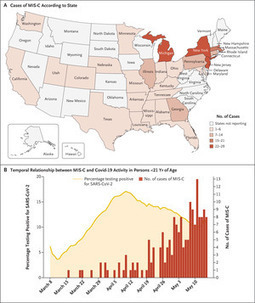
Abstract Background Understanding the epidemiology and clinical course of multisystem inflammatory syndrome in children (MIS-C) and its temporal association with coronavirus disease 201
|
Scoop.it!

Cathelicidins are short cationic peptides that are part of the innate immune system. At first, these peptides were studied mostly for their direct antimicrobial killing capacity, but nowadays they are more and more appreciated for their immunomodulatory functions. In this review, we will provide a comprehensive overview of the various effects cathelicidins have on the detection of damage- and microbe-associated molecular patterns, with a special focus on their effects on Toll-like receptor (TLR) activation. We review the available literature based on TLR ligand types, which can roughly be divided into lipidic ligands, such as LPS and lipoproteins, and nucleic-acid ligands, such as RNA and DNA. For both ligand types, we describe how direct cathelicidin-ligand interactions influence TLR activation, by for instance altering ligand stability, cellular uptake and receptor interaction. In addition, we will review the more indirect mechanisms by which cathelicidins affect downstream TLR-signaling. To place all this information in a broader context, we discuss how these cathelicidin-mediated effects can have an impact on how the host responds to infectious organisms as well as how these effects play a role in the exacerbation of inflammation in auto-immune diseases. Finally, we discuss how these immunomodulatory activities can be exploited in vaccine development and cancer therapies.
Scoop.it!
Extrapulmonary.... Click here to edit the content
Scoop.it!
bioRxiv - the preprint server for biology, operated by Cold Spring Harbor Laboratory, a research and educational institution
Scoop.it!
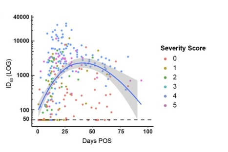
Antibody (Ab) responses to SARS-CoV-2 can be detected in most infected individuals 10-15 days following the onset of COVID-19 symptoms. However, due to the recent emergence of this virus in the human population it is not yet known how long these Ab responses will be maintained or whether they will provide protection from re-infection. Using sequential serum samples collected up to 94 days post onset of symptoms (POS) from 65 RT-qPCR confirmed SARS-CoV-2-infected individuals, we show seroconversion in >95% of cases and neutralizing antibody (nAb) responses when sampled beyond 8 days POS.
We demonstrate that the magnitude of the nAb response is dependent upon the disease severity, but this does not affect the kinetics of the nAb response. Declining nAb titres were observed during the follow up period. Whilst some individuals with high peak ID50 (>10,000) maintained titres >1,000 at >60 days POS, some with lower peak ID50 had titres approaching baseline within the follow up period. A similar decline in nAb titres was also observed in a cohort of seropositive healthcare workers from Guy′s and St Thomas′ Hospitals. We suggest that this transient nAb response is a feature shared by both a SARS-CoV-2 infection that causes low disease severity and the circulating seasonal coronaviruses that are associated with common colds.
This study has important implications when considering widespread serological testing, Ab protection against re-infection with SARS-CoV-2 and the durability of vaccine protection.
Preprint available at medRxiv (July 11, 2020): https://www.medrxiv.org/content/10.1101/2020.07.09.20148429v1 Via Juan Lama
Scoop.it!
Read the original article in full on F1000Research: Tissue-resident lymphocytes: weaponized sentinels at barrier surfaces...
Scoop.it!
Click here to edit the content Six months of coronavirus: the mysteries scientists are still racing to solve
Scoop.it!
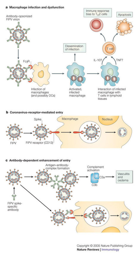
At the end of 2002, the first cases of severe acute respiratory syndrome (SARS) were reported, and in the following year, SARS resulted in considerable mortality and morbidity worldwide. SARS is caused by a novel species of coronavirus (SARS-CoV) and ...
Scoop.it!
Hundred-thousands of fungal species are present in our environment, including normal colonizers that constitute part of the human microbiota. The homeostasis of host-fungus interactions encompasses efficient fungal sensing, tolerance at mucosal surfaces, as well as antifungal defenses.
Scoop.it!
bioRxiv - the preprint server for biology, operated by Cold Spring Harbor Laboratory, a research and educational institution

televisionstand463@gmail.com's curator insight,
July 1, 2020 12:35 PM
https://timkush.com/
https://timkush.com/product/buy-london-pound-cake-online/ https://timkush.com/product/buy-gary-payton-cookies-online/ https://timkush.com/product/buy-runtz-carts-online/ https://timkush.com/product/buy-alpine-vapor-vape-pens-online/ https://timkush.com/product/buy-cotton-candy-dank-vapes-full-gram-cartridge/ https://timkush.com/product/buy-la-confidential-dank-vapes-full-gram-cartridge/ https://timkush.com/product/dank-vapes/ https://timkush.com/product/buy-white-widow-online/ https://timkush.com/product/buy-muha-meds-carts-online/ https://timkush.com/product/master-kush/ https://timkush.com/product/gorilla-glue-4/ https://timkush.com/product/gods-gift/ https://timkush.com/product/buy-lemon-tree-online/ https://timkush.com/product/afghan-kush/ https://timkush.com/product/buy-uk-cheese-online/ https://timkush.com/product/buy-do-si-dos-online/ https://timkush.com/product/girl-scout-cookies/ https://timkush.com/product/buy-lucky-charms-online/ https://timkush.com/product/buy-big-smokey-farms-tins-online/ https://timkush.com/product/buy-sexxpot-online/ https://timkush.com/product/buy-raw-garden-live-resin-cartridge/ https://timkush.com/product/blue-cookies/ https://timkush.com/product/pineapple-express/ https://timkush.com/product/buy-moon-rocks-online/ https://timkush.com/product/blue-dream/ https://timkush.com/product/buy-jungle-boys-online/ https://timkush.com/product/buy-710-kingpen-carts/ https://timkush.com/product/ak-47/ https://timkush.com/product/purchase-alaskan-thunder-fuck/ https://timkush.com/product/buy-brass-knuckles-vape-cartridges-online/ https://timkush.com/product/bubba-kush/ https://timkush.com/product/buy-backwoods-cigars/ https://timkush.com/product/buy-barewoods-online/ https://timkush.com/product/buy-blackberry-kush/ https://timkush.com/product/buy-blueberry-online/ https://timkush.com/product/buy-bruce-banner-strain/ https://timkush.com/product/buy-chemdawg-online/ https://timkush.com/product/buy-bloom-vape-cartridge-online/ https://timkush.com/product/buy-brass-knuckles-carts/ https://timkush.com/product/buy-cali-carts-online/ https://timkush.com/product/buy-cereal-carts-online/ https://timkush.com/product/buy-chronic-carts-online/ https://timkush.com/product/buy-cobra-extracts-online/ https://timkush.com/product/buy-cookies-carts-online-2/ https://timkush.com/product/buy-exotic-carts/ https://timkush.com/product/buy-glo-extracts-online/ https://timkush.com/product/buy-khalifa-kush-online/ https://timkush.com/product/buy-biscotti-weed-online/ https://timkush.com/product/buy-smart-bud-online/ https://timkush.com/product/buy-space-monkey-meds-online/ https://timkush.com/product/fire-og/ https://timkush.com/product/og-kush/ https://timkush.com/product/skywalker/ https://timkush.com/product/sfv-og/ https://timkush.com/product/tahoe-og-kush/ https://timkush.com/product/paris-og/ https://timkush.com/product/buy-plug-and-play-cartridge/ https://timkush.com/product/buy-heavy-hitters-online/ https://timkush.com/product/supreme-vape/ https://timkush.com/product/buy-kurvana-cartridges-online/ https://timkush.com/product/buy-palmas-cartridge-online/ https://timkush.com/product/soul-remedy-extract/ https://timkush.com/product/buy-cali-gold-extracts-online/ https://timkush.com/product/buy-mario-carts-online/ https://timkush.com/product/buy-kingpen-cartridge-online/ https://timkush.com/product/buy-litxtracts-cartridges-online/ https://timkush.com/product/buy-tko-carts-online/ https://timkush.com/product/buy-razzle-dazzle-clear-carts-online/ https://timkush.com/product/buy-west-coast-cure-carts-online/ https://timkush.com/product/buy-blueberry-muffin-dank-vapes-online/ https://timkush.com/product/durban-poison/ https://timkush.com/product/gorilla-glue/ https://timkush.com/product/sour-diesel/ https://timkush.com/product/buy-ghost-train-haze/ https://timkush.com/product/purple-kush/ https://timkush.com/product/gelato-41-2/ https://timkush.com/product/buy-gushers-online/ https://timkush.com/product/buy-haze-buy-haze-online/ https://timkush.com/product/buy-g-13-online/ https://timkush.com/product/tangie/ https://timkush.com/product/buy-trainwreck-buy-trainwreck-strain/ https://timkush.com/product/buy-magic-pipe-vape/ https://timkush.com/product/cbd-living-vape/ https://timkush.com/product/buy-runtz-online/ https://timkush.com/product/buy-hindu-kush/ https://timkush.com/product/buy-northern-lights/ https://timkush.com/product-category/flowers/flowers-indica/page/2/ https://timkush.com/product/buy-super-silver-haze/ https://timkush.com/product/buy-dankwoods-online/ https://timkush.com/product/buy-gaswoods-online/ https://timkush.com/product/dr-zodiaks-moonrock- cartridge/ |