Your new post is loading...

|
Scooped by
Gilbert C FAURE
May 24, 2020 11:56 AM
|
Since the discovery and definition of neutrophil extracellular traps (NETs) 14 years ago, numerous characteristics and physiological functions of NETs have been uncovered. Nowadays, the field continues to expand and novel mechanisms that orchestrate formation of NETs, their previously unknown...
|
|
Scooped by
Gilbert C FAURE
May 21, 2020 8:22 AM
|
On the basis of emerging evidence from patients with COVID-19, we postulate that endothelial cells are essential contributors to the initiation and propagation of severe COVID-19. Here, we discuss current insights into the link between endothelial cells, viral infection and inflammatory changes and propose novel therapeutic strategies. Here, Carmeliet and colleagues discuss the role of endothelial cells in inflammation and viral infection and propose novel therapeutic strategies for COVID-19.
|
|
Scooped by
Gilbert C FAURE
May 19, 2020 9:03 AM
|
Glycans play an important role in the course of SARS-CoV-2 infection and should not be overlooked when searching for ways to understand and control this new disease. Glycans might become a great biomarker tool to predict disease severity, response to therapy and recovery prognosis.
|
|
Scooped by
Gilbert C FAURE
May 18, 2020 10:27 AM
|
There’s still a lot to learn about SARS-CoV-2, the novel coronavirus that causes COVID-19. But it has been remarkable and gratifying to watch researchers from around the world pull together and share their time, expertise, and hard-earned data in the urgent quest to control this devastating virus.
|
|
Rescooped by
Gilbert C FAURE
from Buzz e-sante
May 18, 2020 3:38 AM
|
Immunai combine l'immunologie et l'IA pour développer de nouveaux médicaments et thérapies grâce à une plate-forme qui cartographie le système immunitaire. En levant 20 millions de dollars, la jeune pousse compte recruter de nouvelles personnes et financer des activités de R&D.
Via Rémy TESTON
|
|
Scooped by
Gilbert C FAURE
May 15, 2020 2:34 PM
|
New findings suggest past infections may offer some protection against the novel coronavirus

|
Suggested by
Société Francaise d'Immunologie
May 13, 2020 2:21 PM
|
Viruses, an international, peer-reviewed Open Access journal.
|
|
Scooped by
Gilbert C FAURE
May 12, 2020 3:50 AM
|
O'nyong-nyong virus (ONNV) is an arthritogenic alphavirus that caused two large epidemics in 1959 and 1996, affecting millions of people in Africa. More recently, sero-surveillance of healthy blood donors conducted in 2019 revealed high rates of unreported ONNV infection in Uganda. Due to similar clinical symptoms with other endemic mosquito-borne pathogens in the region, including chikungunya virus, dengue virus and malaria, ONNV infections are often un- or misdiagnosed. Elucidating the immunopathogenic factors of this re-emerging arbovirus is critical with the expanding geographic distribution of competent vectors. This study reports the establishment of an immune competent C57BL6/J mouse model to mechanistically characterize ONNV infection and assess potential treatment efficacy. This mouse model successfully recapitulated arthralgia and viremia profiles seen in ONNV patients. Furthermore, longitudinal in-vivo PET imaging with [18F]FB-IL-2 (CD25+CD4+ binding probe) and histopathological assessment in this model demonstrated the pathogenic role of CD4+ T cells in driving joint pathology. Concordantly, in vivo CD4+ T cell depletion, or suppression with fingolimod, an FDA-approved immunomodulating drug, abrogated CD4+ T cell-mediated disease. This study demonstrates the importance of this immune competent ONNV model for future studies on factors influencing disease pathogenesis, which could shape the discovery of novel therapeutic strategies for arthritogenic alphaviruses.
|
|
Scooped by
Gilbert C FAURE
May 9, 2020 11:50 AM
|
T-cell immunoglobulin and mucin domain containing 4 (Tim-4) is a phosphatidylserine receptor and is selectively expressed on antigen presenting cells. Recently, Tim-4 was reported to be expressed on iNKT cells, B1 cells, and tumor cells, suggesting it has multiple biological functions. In this review, we mainly summarize the expression and regulation of Tim-4 in immune cells including T cells, macrophages, dendritic cells, NKT cells, B cells, and mast cells. The expression of Tim-4 in these cells implies that Tim-4 might participate in immune related diseases. Emerging evidence emphasizes a substantial role for Tim-4 in maintaining homeostasis by regulating various immune responses, including viral infection, allergy, autoimmunity, and tumor immunity. Here, we collectively evaluated the role of Tim-4 in health and diseases. This summary will be extremely useful to fully understand the function of Tim-4 in the pathogenesis of immune related diseases, which would provide novel clues for the diagnosis and treatment of diseases.
|
|
Scooped by
Gilbert C FAURE
May 8, 2020 3:49 AM
|
Pigs are a major food source worldwide as well as major biomedical models for human physiology and therapeutics. A thorough understanding of porcine i…
|
|
Scooped by
Gilbert C FAURE
May 8, 2020 3:39 AM
|
Cells use complex containers to launch toxic chemicals at their victims
|
|
Scooped by
Gilbert C FAURE
May 6, 2020 12:07 PM
|
Plasma cells (PCs) represent the terminal differentiation step of mature B lymphocytes. These cells are most recognizable for their extended lifespan as well as their ability to secrete large amounts of antibodies (Abs) thus positioning this cell type as a key component of humoral immunity. However, it is now appreciated that PCs can have far reaching effects on pathologic as well as non-pathologic processes independent of Ab secretion. This is highlighted by recent studies showing that PCs function as key regulators of processes such as hematopoiesis as well as neuro-inflammation. In part, PCs accomplish this by integrating extrinsic signals from their environment which dictate their downstream functionality. Here we summarize the current understanding of PC biology focusing on their ever-growing functional repertoire independent of Ab production. Furthermore, we discuss potential applications of PC immunotherapy and its implementation for translational benefit.
|
|
Scooped by
Gilbert C FAURE
May 2, 2020 2:49 PM
|
KEY POINTS Patients with TB exhibit a high frequency of TRMs at different infection sites. These TRMs exhibit memory, with an activated and multifunctional phenotype. TRMs limit intracellular M. tuberculosis replication in macrophages. Abstract Tissue-resident memory T cells (TRMs) have a key role in mediating the host defense against tuberculosis (TB) in mice, but their human counterparts have not been well characterized. In this article, we recruited patients with TB and determined TRM frequency, trafficking, activation marker expression, and cytokine production by flow or mass cytometry at different infection sites, including peripheral blood, pleural fluid, bronchoalveolar lavage fluid, and lung. We found a high frequency of TRMs at all infection sites apart from the peripheral blood. These TRMs exhibited a memory phenotype, were highly activated (based on CD38 and HLA-DR expression), and expressed high levels of trafficking (CCR5 and CXCR6) and exhaustion (PD-1) markers. When stimulated with Mycobacterium tuberculosis, TRMs secreted cytokines, including IFN-γ, TNF-α, and IL-2, and exhibited a multifunctional phenotype. TRMs limited intracellular M. tuberculosis replication in macrophages. These data inform our current understanding of immunosurveillance at different infection sites in patients with TB. Introduction T cells have critical roles in mediating the host defense against Mycobacterium tuberculosis infection but are insufficient to clear the infection (1). No other suitable immune correlates for protective immunity against tuberculosis (TB), however, have been identified, perhaps because immune responses measured ex vivo in the peripheral blood might not faithfully reflect the immune response that occurs at the infection site (2). For example, tissue-resident memory T cells (TRMs) permanently reside in nonlymphoid tissues and cannot be detected in the peripheral blood (3). TRMs exhibit a distinct genetic signature as well as migration, retention, and functional maintenance characteristics that differ from those of peripheral T cells. These cells are typically defined by CD69, CD103, CD49a, and CD44 cell surface expression in both mice and humans (4). Increasing interest has been paid to the relevance of TRMs in disease pathogenesis because of their ability to mount a response against pathogens and accelerate pathogen clearance (5, 6). Recent studies performed in mice have indicated that TRMs in the lung are critical for TB protection (7); their human counterparts, however, remain relatively uncharacterized in TB. In this article, we aimed to address this knowledge gap by characterizing human TRMs present at infection sites, including the pleural fluid, bronchoalveolar lavage fluid (BALF), and the lung parenchyma from TB patients, in terms of their frequency, phenotype, and function. Based on previous studies, we defined TRMs by the coexpression of CD69 and CD103 (4). We show that TRMs are present at all infection sites in patients with TB. Importantly, TRMs are not detectable in the peripheral blood. These TRMs are activated, expressing a high level of T cell trafficking and exhaustion markers. M. tuberculosis Ag-specific TRMs secrete multiple cytokines and exhibit a multifunctional phenotype. Finally, we show that TRMs can limit intracellular M. tuberculosis replication in macrophages. These data support that TRMs exist in multiple infection sites. Given their absence from the peripheral blood, these findings inform us on the most appropriate sites to detect and study the role of the T cell response in the context of local TB infection. Our data provide a scientific basis for the future design of vaccines and immunotherapies for TB. Materials and Methods Subjects and clinical sample collection Ethical approval for this study was obtained from the Research Ethics Committee of the Shenzhen Third People’s Hospital (Shenzhen, China). All participants provided written informed consent for sample collection and the subsequent analyses. All participants were enrolled at the Shenzhen Third People’s Hospital between January 2017 and July 2019. A TB diagnosis was based on clinical symptoms, chest radiography, microscopy for acid-fast bacilli, M. tuberculosis culture and GeneXpert analysis of sputum and/or BALF and/or pleural effusion, and the response to anti-TB chemotherapy (8). All patients with comorbid HIV were excluded from the study. All patients with TB were undergoing their first round of anti-TB therapy. The cohort demographics and clinical information are provided in Supplemental Table I. Human blood and tissue cell isolation PBMCs were obtained by whole-blood gradient separation. Pleural fluid mononuclear cells (PFMCs) and BALF cells were separated by centrifugation, whereas RBCs were lysed using ACK lysis buffer (Life Technologies). Lung parenchyma tissues (Lung) collected during surgical procedures were incubated with 1 mg/ml collagenase type IV (Roche) and 1 mg/ml DNase I (Roche) for 1 h 37°C before filtering through a 70-mM nylon mesh. The homogenates were resuspended with Percoll (GE Healthcare Systems), and the cells were obtained by gradient separation. All the cells were stored in liquid nitrogen until further use. In vitro cell stimulation assay PFMCs (1 × 10⁶) were suspended in RPMI 1640 containing 10% FBS and then incubated with 10 μg/ml M. tuberculosis H37Rv lysate at 37°C for 24 h. During the final 4 h of the incubation, 10 μg/ml brefeldin A (Sigma-Aldrich) was added to the culture media. The cultured cells were then harvested for staining with Abs suitable for flow cytometry or cytometry by time-of-flight (CyTOF). FACS For cell surface labeling, 1 × 10⁶ cells were blocked with Fc-block reagent (BD Biosciences). Then, the following Abs were added and incubated for 30 min: CD8 (RPA-T8; BioLegend), CD69 (FN50; BioLegend), CD4 (RPA-T4; BD Biosciences), CD45RA (HI100; BD Biosciences), CCR7 (G043H7; BioLegend), and CD103 (Ber-ACT8; BD Biosciences). After incubation, the samples were washed and reconstituted in PBS for flow cytometric analysis on an FACSCanto II Flow Cytometer. For intracellular staining, the cells were fixed and permeabilized using Fix/Perm Buffer (BD Biosciences). Then, the following Abs were added to the cells for intracellular labeling: TNF-α (MAb11; BioLegend), IFN-γ (B27; BD Biosciences) and IL-2 (MQ1-17H12; BioLegend). The Abs were incubated for 60 min before the cells were washed and fixed with 1.6% paraformaldehyde. Data analysis was performed using FlowJo software version 10. Mass cytometry, CyTOF The Abs for CyTOF are listed in Supplemental Table II. All CyTOF staining was performed at room temperature, as previously described (9). In brief, the cells were incubated in 1 M cisplatin (Fluidigm) for 5 min. Fc-block reagent was then added to the cells for 10 min. Next, the cells were stained with Abs for 30 min for surface labeling and then washed once in CyTOF staining buffer (Fluidigm). For intracellular labeling, the cells were fixed and permeabilized using Fix/Perm Buffer (Fluidigm) prior to incubation with the appropriate Abs for 30 min. Then, the cells were washed twice by centrifugation with PBS, resuspended in 1.6% paraformaldehyde (Fluidigm) in the presence of 125 nM iridium intercalator (Fluidigm), and incubated overnight at 4°C. After three further washes, the cells were resuspended in PBS containing EQ calibration beads (Fluidigm) before analysis on a Helios mass cytometer. Data analysis was conducted using Cytobank (Cytobank, Mountain View, CA) and FlowJo (Tree Star, Ashland, OR). In vitro M. tuberculosis infection of monocyte-derived macrophages and intracellular M. tuberculosis growth assay Both M. tuberculosis infection and intracellular M. tuberculosis growth assays were performed as previously described (10). Briefly, monocytes (5 × 10⁴ per well) from patients with TB were cultured in RPMI 1640 medium supplemented with 10% FBS, human rIL-4 (BD Biosciences), and GM-CSF (Sigma-Aldrich) for 7 d. The H37Ra inoculum was added at a multiplicity of infection = 5. After overnight infection at 37°C, the extracellular H37Ra in the medium was removed. The memory (CD45RA−CCR7−) CD4+CD69−, CD4+CD69+, CD8+CD69−, and CD8+CD69− T cells were enriched from PFMCs by flow cytometry, and then 5 × 10⁵ cells per well were incubated with H37Ra-infected autologous macrophages. After incubation in 5% CO2 at 37°C for 3 d, the culture supernatants were aspirated, and the cells were lysed with lysis buffer (0.067% SDS in Middlebrook 7H9) prior to plating for CFU. Statistical analyses GraphPad Prism 6.0 software (GraphPad) was used for all statistical calculations. Heatmaps were displayed in R using the heatmap.2 function from the ggplot package. A p < 0.05 was considered statistically significant with a 95% confidence interval. The statistical analyses performed are indicated in the figure legends. Results TRMs at M. tuberculosis infection sites exhibit an effector memory phenotype CD69 and CD103 are two key markers used to identify TRMs (11, 12). We first determined the distribution of TRMs between different infection sites by flow cytometric analysis of CD4+ and CD8+ T cells based on CD69 and CD103 coexpression (Fig. 1A). We found that CD69+CD103+ T cells were significantly more frequent at sites of TB infection, (i.e., in the PFMCs [2–5%], BALF [13–30%], and lung [15–28%]) compared with in the PBMCs (0.05–0.3%) (Fig. 1B), suggesting that TRMs are present at different TB infection sites. FIGURE 1. TRMs are highly frequent at infection sites in patients with TB. (A) CD69 and CD103 expression in PBMCs, PFMCs, BALF, and lung parenchyma isolated from patients with TB, as assessed by flow cytometry. (B) The frequency of CD69+CD103+ cells in PBMCs (n = 19), PFMCs (n = 15), BALF (n = 30), and lung (n = 5). Unpaired Student t test compared with PBMCs. *p ≤ 0.05, **p ≤ 0.01. (C) The proportions of the four CD45RA/CCR7 subsets in CD69+CD103+ cells in the indicated compartments. T cells are typically subdivided into naive, central memory, effector memory (TEM) and late TEM cells based CD45RA and CCR7 expression (13). We found that both CD4+ and CD8+ TRMs in infection sites expressed low CCR7 and CD45RA levels. In addition, most TRMs (range 77.09–98.16%) exhibited a TEM phenotype (CD45RA−CCR7−) (Fig. 1C). These data indicate that a high frequency of TRMs in the CD4+ and CD8+ T cell population is present at infection sites in patients with TB. These TRMs exhibit a TEM-like phenotype. An immune landscape analysis indicates that TRMs present at TB infection sites are activated To comprehensively analyze the TRM immune landscape, we analyzed 28 markers for activation, trafficking, and exhaustion simultaneously by CyTOF (Supplemental Table II) (14). We determined the gating thresholds for each parameter by plotting each marker against a second marker known to express that protein (Supplemental Fig. 1A). We divided CD4+ and CD8+ T cells into four subsets based on CD69 and CD103 expression (CD69−CD103−, CD69−CD103+, CD69+CD103−, and CD69+CD103+) and then compared the frequencies of the different markers between the four subsets (Supplemental Fig. 1B). In this article, we found similar protein expression patterns presented by TRMs from different infection sites (Fig. 2A, 2B). For example, we found that CD69+CD103+ T cells presented a TEM phenotype (CD45RA−CCR7−) (Supplemental Fig. 1C, 1D). Moreover, CCR5 and CXCR6 expression by CD69+CD103+ subsets was significantly higher in CD8+ T cells from PFMCs, BALF, and lung compared with the other three subsets (Fig. 2C, 2D). FIGURE 2. TRMs in infection sites express high levels of T cell activation, tracking, and exhaustion markers. Heatmaps showing the average frequency of positive expression for the different markers within the indicated cell subsets found in PFMCs (n = 6), BALF (n = 6), and lung (n = 3) for CD4+ (A) and CD8+ (B) T cells. The frequencies of the indicated different marker-expressing CD4+ (C) and CD8+ (D) T cells found at found in PFMCs, BALF, and lung [related to (A)]. One-way ANOVA with Tukey multiple comparison. *p < 0.05, **p < 0.01, ***p < 0.001. Some markers did, however, show tissue-type and cell-type specificity. For example, the level of CD38 and HLA-DR expression in PFMCs by CD4+ and CD8+CD69+ subsets was higher than by CD69− subsets, and their frequencies in PFMCs were higher than in the BALF and lung. In CD4+T cells, both CCR5 and CXCR6 were expressed at significantly higher levels by CD69+ subsets than CD69− subsets in PFMCs and BALF but not in the lung. These findings might, however, be due to the small lung tissue sample size (n = 3) used in our study. Notably, we detected that more CD8+CD69+ subsets from PFMCs and BALF expressed significantly higher levels of CD161 and PD-1 than CD69− subsets (Fig. 2C, 2D). Taken together, the high frequency of CD38, HLA-DR, CCR5, CXCR6, CD161, and PD-1 expression by TRMs suggests that these cells at TB infection sites are activated. TRMs in PFMCs from TB patients can produce multiple cytokines and exhibit a highly cytotoxic profile in response to M. tuberculosis To identify M. tuberculosis Ag-specific TRMs at infection sites in patients with TB, we isolated and stimulated PFMCs with M. tuberculosis. Then, we monitored cytokine and chemokine production by the four CD69/CD103 T cell subsets (Fig. 3A). First, we found that the percentage of CD69+ cells in PFMCs was similar before and after M. tuberculosis stimulation. In CD4+ T cells, both CD69+ subsets in the PFMCs expressed high IFN-γ (∼30%), TNF-α (∼40%), IL-2 (∼15%), MIP1β (∼18%), GM-CSF (∼25%), and Tim-3 (∼20%) levels after in vitro stimulation. By contrast, the CD69−CD103− subset showed low to no cytokine and chemokine expression after M. tuberculosis stimulation (Fig. 3A). We verified these results by conventional flow cytometry in an independent cohort. In this article, we consistently detected IFN-γ, TNF-α and IL-2 expression by both CD69+ subsets (Supplemental Fig. 2A). However, IL-4, IL-5, IL-6, IL-10, IL-17, and IL-22 cytokine production was very low in all four subsets, regardless of M. tuberculosis stimulation (Fig. 3A). FIGURE 3. TRMs from the PFMCs express multiple cytokines in response to M. tuberculosis stimulation. PFMCs from patients with TB (n = 7) were stained with a panel of intracellular markers after stimulation or not with M. tuberculosis. (A) The frequencies of the indicated markers in four CD69/CD103 subsets for CD4+ and CD8+ T cells, as assessed by CyTOF. (B) The frequencies of the indicated cytokines (IFN-γ, IL-2, and/or TNF-α expressed alone or simultaneously) after M. tuberculosis stimulation across the different CD4+ and CD8+ T cell subsets. Paired Student t test compared with unstimulated cells (“−”). *p < 0.05, **p < 0.01. (C) The proportion of CD69+CD103+ T cells in PFMCs producing one (1+, gray), two (2+, blue), or three (3+, orange) cytokines (IFN-γ, IL-2, and/or TNF-α) after M. tuberculosis simulation. (D) Monocyte-derived macrophages isolated from untreated patients with active TB (n = 5) were infected with H37Ra overnight, and autologous CD69− and CD69+ T cells were purified and cocultured. The cultured cells were then lysed for CFU. Paired Student t test compared with mock (without purified cells added to autologous macrophages). *p < 0.05. We also found that CD8+CD69+ T cell subsets produced high cytokine levels following M. tuberculosis stimulation. The frequencies of these cytokines in CD8+ T cells were, however, lower than those in CD4+ T cells. Specifically, the frequency of IFN-γ–producing cells was 12.11% in CD8+ TRMs versus 37.27% in CD4+ TRMs (Fig. 3A). Interestingly, we detected high levels of granzyme B and perforin in both CD8+CD69+ subsets, regardless of M. tuberculosis stimulation (Fig. 3C). However, we found that only the CD4+CD69+CD103+ subset expressed granzyme B and perforin regardless of M. tuberculosis stimulation (Fig. 3A). We also found that the frequency of CD69+CD103+ T cells was very low in CD4+ (0.27 ± 0.05%) and CD8+ (0.98 ± 0.1183%) T cells from non-TB patients (Supplemental Fig. 2B). However, the TRM cytokine profile in non-TB patients significantly differed from that in patients with TB. In particular, the frequencies of cytokine-producing cells (such as IFN-γ, TNF-α, IL-2, GM-CSF, and MIP1β) in CD69+CD103+ cells were extremely low in PFMCs from non-TB patients, regardless of M. tuberculosis stimulation. In CD4+ T cells, the four subsets in the PFMCs expressed a low level of IFN-γ (∼1.6%), TNF-α (∼2.62%), IL-2 (∼0.77%), MIP1β (∼0.7%), GM-CSF (∼4.43%), and Tim-3 (∼1.50%) in response to M. tuberculosis stimulation. Similar data were found in CD8+ T cells (Supplemental Fig. 2C). These data support that TRMs found in the pleural fluid in patients with TB exhibit an M. tuberculosis Ag-specific Th1-cell response. TRMs in patients with TB exhibit a multifunctional phenotype based on multiple cytokine production Th1 cells can gain the capacity to secrete several cytokines; such “multifunctional” Th1 cells mainly produce IFN-γ, TNF-α, and IL-2 (15). The frequency of IFN-γ+TNF-α+IL-2+CD4+ T cells increases in patients with active TB and normalizes after anti-TB treatment (16, 17). Some studies performed in humans have shown that CD4+CD103+ T cells isolated from the human lung have the potential to elicit a rapid effector response to a secondary challenge and upregulate the genes required for multifunctional cytokine production (6, 18, 19). We found that in CD4+ T cells, the frequency of CD69+CD103+T cells producing two or three cytokines (IFN-γ, TNF-α, and/or IL-2) increased upon M. tuberculosis stimulation (Supplemental Fig. 2D). Specifically, we found that 13% CD69+CD103+T cells produced three cytokines compared with CD4+CD69− subsets (both were <1%) (Fig. 3B). In CD8+CD69+CD103+ T cells, we detected a low frequency (∼2.5%) of cells that produced three cytokines (Fig. 3B). We thus classified the M. tuberculosis–responsive cells in the four subsets in terms of their ability to produce three (3+), two (2+) or one (1+) cytokine(s) according to the IFN-γ, TNF-α, and IL-2 expression profile. In CD4+ T cells, the relative proportion of 3+ (IFN-γ+TNF-α+IL-2+) CD69+CD103+T cells within the total population of cytokine-producing cells was ∼28.71%; this value was much higher than the frequency of 3+ cells in the other three subsets (4.83–19.28%). Together, we classified 58.86% CD4+CD69+CD103+ T cells as 2+ and 3+ (Fig. 3C). More interestingly, we also classified∼40% of CD8+CD69+CD103+ T cells as 2+ and 3+ (Fig. 3C). TRMs limit intracellular M. tuberculosis replication in human macrophages Murine studies have shown that tissue-resident T cells likely have a protective role in TB (7, 20). We thus wondered whether TRMs can provide protective immunity in TB patients. Because macrophages are the predominant cell type to control M. tuberculosis, we asked whether TRMs could limit intracellular M. tuberculosis replication in human macrophages. Because of the limited number of CD69+CD103+ cells present in the pleural fluid, we isolated memory (CD45RA−CCR7−) CD69+ or CD69+ cells from CD4+ and CD8+ T cells in PFMCs by flow cytometry and cocultured these cells with M. tuberculosis–infected autologous macrophages. The CFUs of macrophages in coculture with CD4+CD69+ or CD8+CD69+ cells were significantly lower than those cultured alone. These data suggest that macrophages cocultured with TRMs from PFMCs showed a stronger ability to control intracellular M. tuberculosis replication. Interestingly, CD69+ cells from both CD4+ and CD8+ T cells seemed to limit intracellular M. tuberculosis growth (Fig. 3D). Thus, we conclude that CD69+ T cells in patients with active TB exhibit anti–M. tuberculosis effector function in terms of limiting intracellular M. tuberculosis replication in cultured macrophages. Discussion This study has characterized the TRMs profile (based on CD69 and CD103 coexpression) at different sites of infection in patients with TB. Our key findings derived from patients with TB are as follows: 1) CD4+ and CD8+ TRMs are highly expanded in nonlymphoid tissues but not in the peripheral blood; 2) TRMs found at TB infection sites exhibit memory and an activated phenotype; 3) CD4+ TRMs exhibit a cytotoxic phenotype similar to CD8+ T cells; 4) M. tuberculosis–specific TRMs exhibit a multifunctional phenotype based on multiple cytokine expression; and 5) TRMs limit intracellular M. tuberculosis replication in macrophages. We identified TRMs by CD69 and CD103 expression. Numerous other cell surface markers have been used to study TRMs in previous studies, including CD49a, CXCR6, CD101, PD-1, CD62L, KLRG1, and CX3CR1 (6, 21). Whereas CD69 is expressed by most TRMs, CD103 is primarily expressed by CD8+ T cells and not CD4+ T cells (4). Because of the wide acceptance of CD103 as a CD8+ TRM marker (5, 6, 21), we used CD103 expression to compare CD8+ TRMs with CD4+ TRMs. Others have used CD103 as an indicator of protective immunity after murine vaccination with bacillus Calmette-Guérin vaccination (22). The protective immunity elicited after a novel mucosal TB vaccination seems to be associated with the expression of CD4+ and CD8+CD103+ T cells in the lung (23). Some studies performed in the human context have found that CD4+ CD103+ T cells isolated from the human lung have the potential to elicit a rapid effector response to a secondary challenge and can upregulate the genes required for multifunctional cytokine production (6, 18, 19). Although these findings support that CD103 is a valid marker of TRMs, further studies are required to identify accurate molecular markers for TB-specific CD8+ and CD4+ TRMs. There is a growing body of evidence supporting that TRMs have a protective role during different pathogenic infections (21, 24). Although relatively few studies have directly investigated the protective role of TRMs in TB, one early study showed that parenchymal T cells exhibit greater control of M. tuberculosis infection than their intravascular counterparts (7). In addition, the protective role of TRMs in the lung was recently established after adoptive transfer of TRMs collected from mice that received the mucosal bacillus Calmette-Guérin vaccination. Notably, the CD69+CD103+ cells expanded in the vaccinated mice and promoted a high level of IFN-γ or IFN-γ/TNF-α/IL-2–producing TRMs-mediated cell responses, which seemed to act as surrogates for protective immunity against TB (22, 23). This earlier study, however, did not fully characterize the function of TRMs in human TB. Although more work is needed, our data strongly suggest that TRMs present in the infectious site of patients with TB have a protective role in the immune response to M. tuberculosis. At the infectious site, we found that TRMs exhibit memory, with an activated and multifunctional phenotype. CD4+ TRMs in the pleural fluid also expressed high levels of granzyme B and perforin, implicating that CD4+ TRMs exhibit a cytotoxic phenotype similar to CD8+ T cells in TB. Furthermore, both CD4+ and CD8+ M. tuberculosis Ag-specific TRMs exhibited a profile reminiscent of multifunctional Th1 cells, as they simultaneously expressed IFN-γ, TNF-α, and IL-2. Finally, although we had access to only a small number of samples in this study, these TRMs showed a strong protective immune response in TB. Because CD69 is also upregulated on recently activated T cells, we cannot definitively establish that all CD69+ cells analyzed are TRMs. Indeed, a key limitation of this study is that only tissues from patients with active TB were investigated because of the ethical and logistical challenges of performing the invasive procedures on asymptomatic latently infected individuals or the patients who have been cured from TB. It would be informative to examine in future whether the heightened susceptibility of HIV patients with low CD4+ T cell counts is associated with the reduction of the tissue-resident T cells described in this article. Based on our preliminary data obtained from TB patients thus far, we propose that CD69 and CD103 markers can be used to identify and isolate TRMs from human tissues. Studies that transfer these cells to humanized mice might now be established to study the function of TRMs in more detail. Overall, TRMs are not detectable in peripheral blood; therefore, analyses of the immune response using peripheral blood cells might miss this crucial cellular component and an arm of the immune response against M. tuberculosis infection. Ultimately, our data highlight the complexity of TRMs in human TB and give us a more-comprehensive and updated understanding of local immunity in patients with TB. Disclosures The authors have no financial conflicts of interest. Acknowledgments We thank Dr. Jessica Tamanini (Shenzhen University Health Science Center, ETediting) for editing the manuscript prior to submission. Footnotes This work was supported by grants from the National Science and Technology Major Project (2017ZX10201301-001 and 2017ZX10201301-008), the Natural Science Foundation of China (81671984, 81772150 and 81571951), the Science and Technology Project of Shenzhen (JCYJ20160427184123851), and the Jin Qi team of the Sanming Project of Medicine in Shenzhen (SZSM201412001). The online version of this article contains supplemental material. Abbreviations used in this article: BALF bronchoalveolar lavage fluid CyTOF cytometry by time-of-flight PFMC pleural fluid mononuclear cell TB tuberculosis TEM effector memory T cell TRM tissue-resident memory T cell. Received November 11, 2019. Accepted March 7, 2020. Copyright © 2020 by The American Association of Immunologists, Inc. This article is distributed under The American Association of Immunologists, Inc., Reuse Terms and Conditions for Author Choice articles. References ↵World Health Organization. 2018. Global Tuberculosis Report 2018. WHO Press, Geneva, Switzerland. ↵Seder, R. A., P. A. Darrah, M. Roederer. 2008. T-cell quality in memory and protection: implications for vaccine design. [Published erratum appears in 2008 Nat. Rev. Immunol. 8: 486.] Nat. Rev. Immunol. 8: 247–258.OpenUrlCrossRefPubMed ↵Topham, D. J., E. C. Reilly. 2018. Tissue-resident memory CD8+ T cells: from phenotype to function. Front. Immunol. 9: 515.OpenUrlCrossRef ↵Kumar, B. V., W. Ma, M. Miron, T. Granot, R. S. Guyer, D. J. Carpenter, T. Senda, X. Sun, S. H. Ho, H. Lerner, et al. 2017. Human tissue-resident memory T cells are defined by core transcriptional and functional signatures in lymphoid and mucosal sites. Cell Rep. 20: 2921–2934.OpenUrlCrossRef ↵Thome, J. J., D. L. Farber. 2015. Emerging concepts in tissue-resident T cells: lessons from humans. Trends Immunol. 36: 428–435.OpenUrlCrossRefPubMed ↵Mueller, S. N., L. K. Mackay. 2016. Tissue-resident memory T cells: local specialists in immune defence. Nat. Rev. Immunol. 16: 79–89.OpenUrlCrossRefPubMed ↵Sakai, S., K. D. Kauffman, J. M. Schenkel, C. C. McBerry, K. D. Mayer-Barber, D. Masopust, D. L. Barber. 2014. Cutting edge: control of Mycobacterium tuberculosis infection by a subset of lung parenchyma-homing CD4 T cells. J. Immunol. 192: 2965–2969. ↵Chen, X., Q. Yang, M. Zhang, M. Graner, X. Zhu, N. Larmonier, M. Liao, W. Yu, Q. Deng, B. Zhou. 2009. Diagnosis of active tuberculosis in China using an in-house gamma interferon enzyme-linked immunospot assay. Clin. Vaccine Immunol. 16: 879–884. ↵Amir, E. D., X. V. Guo, O. Mayovska, A. H. Rahman. 2018. Average overlap frequency: a simple metric to evaluate staining quality and community identification in high dimensional mass cytometry experiments. J. Immunol. Methods 453: 20–29.OpenUrl ↵Qiu, Y., J. Chen, H. Liao, Y. Zhang, H. Wang, S. Li, Y. Luo, D. Fang, G. Li, B. Zhou, et al. 2012. Tim-3-expressing CD4+ and CD8+ T cells in human tuberculosis (TB) exhibit polarized effector memory phenotypes and stronger anti-TB effector functions. PLoS Pathog. 8: e1002984. ↵Cibrián, D., F. Sánchez-Madrid. 2017. CD69: from activation marker to metabolic gatekeeper. Eur. J. Immunol. 47: 946–953.OpenUrlCrossRefPubMed ↵Wong, M. T., D. E. Ong, F. S. Lim, K. W. Teng, N. McGovern, S. Narayanan, W. Q. Ho, D. Cerny, H. K. Tan, R. Anicete, et al. 2016. A high-dimensional atlas of human T cell diversity reveals tissue-specific trafficking and cytokine signatures. Immunity 45: 442–456.OpenUrlCrossRefPubMed ↵Sallusto, F., D. Lenig, R. Förster, M. Lipp, A. Lanzavecchia. 1999. Two subsets of memory T lymphocytes with distinct homing potentials and effector functions. Nature 401: 708–712.OpenUrlCrossRefPubMed ↵Matos, T. R., H. Liu, J. Ritz. 2017. Research techniques made simple: mass cytometry analysis tools for decrypting the complexity of biological systems. J. Invest. Dermatol. 137: e43–e51.OpenUrl ↵Counoupas, C., J. A. Triccas. 2019. The generation of T-cell memory to protect against tuberculosis. Immunol. Cell Biol. 97: 656–663.OpenUrl ↵Qiu, Z., M. Zhang, Y. Zhu, F. Zheng, P. Lu, H. Liu, M. W. Graner, B. Zhou, X. Chen. 2012. Multifunctional CD4 T cell responses in patients with active tuberculosis. Sci. Rep. 2: 216.OpenUrlCrossRefPubMed ↵Caccamo, N., G. Guggino, S. A. Joosten, G. Gelsomino, P. Di Carlo, L. Titone, D. Galati, M. Bocchino, A. Matarese, A. Salerno, et al. 2010. Multifunctional CD4(+) T cells correlate with active Mycobacterium tuberculosis infection. Eur. J. Immunol. 40: 2211–2220.OpenUrlCrossRefPubMed ↵Oja, A. E., B. Piet, C. Helbig, R. Stark, D. van der Zwan, H. Blaauwgeers, E. B. M. Remmerswaal, D. Amsen, R. E. Jonkers, P. D. Moerland, et al. 2018. Trigger-happy resident memory CD4+ T cells inhabit the human lungs. Mucosal Immunol. 11: 654–667.OpenUrlCrossRef ↵Wilk, M. M., A. Misiak, R. M. McManus, A. C. Allen, M. A. Lynch, K. H. G. Mills. 2017. Lung CD4 tissue-resident memory T cells mediate adaptive immunity induced by previous infection of mice with Bordetella pertussis. J. Immunol. 199: 233–243. ↵Jeyanathan, M., J. Mu, S. McCormick, D. Damjanovic, C. L. Small, C. R. Shaler, K. Kugathasan, Z. Xing. 2010. Murine airway luminal antituberculosis memory CD8 T cells by mucosal immunization are maintained via antigen-driven in situ proliferation, independent of peripheral T cell recruitment. Am. J. Respir. Crit. Care Med. 181: 862–872.OpenUrlCrossRefPubMed ↵Ogongo, P., J. Z. Porterfield, A. Leslie. 2019. Lung tissue resident memory T-cells in the immune response to Mycobacterium tuberculosis. Front. Immunol. 10: 992.OpenUrl ↵Perdomo, C., U. Zedler, A. A. Kühl, L. Lozza, P. Saikali, L. E. Sander, A. Vogelzang, S. H. Kaufmann, A. Kupz. 2016. Mucosal BCG vaccination induces protective lung-resident memory T cell populations against tuberculosis. MBio 7: e01686-16. ↵Hart, P., A. Copland, G. R. Diogo, S. Harris, R. Spallek, W. Oehlmann, M. Singh, J. Basile, M. Rottenberg, M. J. Paul, R. Reljic. 2018. Nanoparticle-fusion protein complexes protect against Mycobacterium tuberculosis infection. Mol. Ther. 26: 822–833.OpenUrl ↵Snyder, M. E., D. L. Farber. 2019. Human lung tissue resident memory T cells in health and disease. Curr. Opin. Immunol. 59: 101–108.OpenUrl
|
|
|
Scooped by
Gilbert C FAURE
May 22, 2020 2:51 AM
|
|
|
Scooped by
Gilbert C FAURE
May 19, 2020 9:09 AM
|
abstracts immunology 2020
|
|
Scooped by
Gilbert C FAURE
May 19, 2020 3:19 AM
|
Although NLRs are structurally homologous to each other, they perform a diverse array of cellular functions and play key roles in the regulation of innate immune responses. In this research topic, our understanding of NLRP3 has received particular emphasis.
Key Points.Compared with cytotoxic T cells, NK cells share mechanisms for lytic granule release but more stringently control cytokine production.Analysis of C...
Via Krishan Maggon
|
|
Scooped by
Gilbert C FAURE
May 16, 2020 1:51 AM
|
bioRxiv - the preprint server for biology, operated by Cold Spring Harbor Laboratory, a research and educational institution
|
|
Scooped by
Gilbert C FAURE
May 15, 2020 2:54 AM
|
T cells and Covid-19 at last
Abstract
The rapid spread of SARS-CoV-2 has a significant impact on global health, travel and economy. Therefore, preventative and therapeutic measures are urgently needed. Here, we isolated neutralizing antibodies from convalescent COVID-19 patients using a SARS-CoV-2 stabilized prefusion spike protein. Several of these antibodies were able to potently inhibit live SARS-CoV-2 infection at concentrations as low as 0.007 μg/mL, making them the most potent human SARS-CoV-2 antibodies described to date. Mapping studies revealed that the SARS-CoV-2 spike protein contained multiple distinct antigenic sites, including several receptor-binding domain (RBD) epitopes as well as previously undefined non-RBD epitopes. In addition to providing guidance for vaccine design, these mAbs are promising candidates for treatment and prevention of COVID-19.
Competing Interest Statement
Amsterdam UMC has filed a patent application on COVID-19 monoclonal antibodies.
Paper in collection COVID-19 SARS-CoV-2 preprints from medRxiv and bioRxiv
Via Krishan Maggon
|
|
Scooped by
Gilbert C FAURE
May 10, 2020 4:39 AM
|
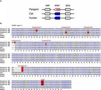
Zoonotic infections are an imminent threat to human health. Pangolins were recently identified as carriers and intermediate hosts of coronaviruses. Previous research has shown that infection with coronaviruses activates an innate immune response upon sensing of viral RNA by interferon-induced with helicase C domain 1 (IFIH1), also known as MDA5. Here, we performed a comparative genomics study of RNA sensor genes in three species of pangolins. DDX58/RIG-I, a sensor of cytoplasmic viral RNA and toll-like receptors (TLR) 3, 7, and 8, which bind RNA in endosomes, are conserved in pangolins. By contrast, IFIH1 a sensor of intracellular double-stranded RNA, has been inactivated by mutations in pangolins. Likewise, Z-DNA-binding protein (ZBP1), which senses both Z-DNA and Z-RNA, has been lost during the evolution of pangolins. These results suggest that the innate immune response to viruses differs significantly between pangolins and other mammals, including humans. We put forward the hypothesis that loss of IFIH1 and ZBP1 provided an evolutionary advantage by reducing inflammation-induced damage to host tissues and thereby contributed to a switch from resistance to tolerance of viral infections in pangolins.
|
|
Scooped by
Gilbert C FAURE
May 9, 2020 4:47 AM
|
|
|
Scooped by
Gilbert C FAURE
May 8, 2020 3:45 AM
|
|
|
Scooped by
Gilbert C FAURE
May 8, 2020 2:22 AM
|
Abstract
Sestrin proteins dampen TCR signaling and induce antigen-independent natural killer-like cytotoxicity in highly differentiated CD8 T cells.
Via Krishan Maggon
|