I’m pleased to share our latest publication in Cancer Cell International!
“Quantitative HER2 profiling on circulating tumor cells using an EpCAM-independent platform in metastatic breast cancer”
Our analysis highlights the dynamic heterogeneity of HER2 expression in metastatic breast cancer and supports the role of CTCs as a minimally invasive tool for real-time disease assessment.
🙏 Grateful to all co-authors, clinical collaborators, and especially to the patients who made this work possible.
We’re excited to share our latest paper exploring the tumor–immune interface in circulation through liquid biopsy: “Caught in the Act: Tumor–Immune Interactions in Circulation of Patients with Immune Marker–Positive Circulating Tumor Cells”
Circulating tumor cells (CTCs) and large extracellular vesicles (LEVs) are key components of liquid biopsy, offering minimally invasive insight into tumor biology. A particularly intriguing subset, immune marker–positive CTCs (im.CTCs), has been described clinically, yet their biological origin has remained unclear.
In this study, using high-resolution immunofluorescence microscopy and proteomic profiling of patient blood samples, we show direct physical interactions between white blood cells and both im.CTCs and im.LEVs, observed exclusively in patients with im.CTCs.
Together, these results support the hypothesis for in vivo membrane transfer as a mechanism of immune marker acquisition by CTCs and LEVs, with important implications for tumor–immune biology and the clinical interpretation of immune-positive CTCs in liquid biopsy.
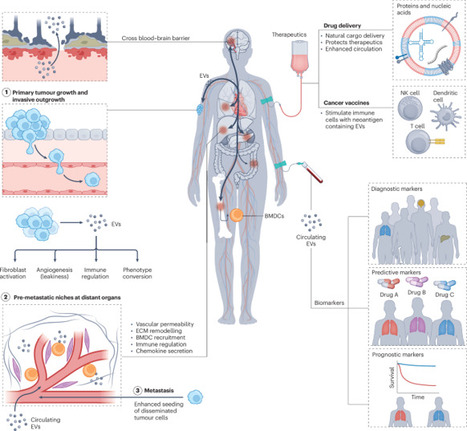
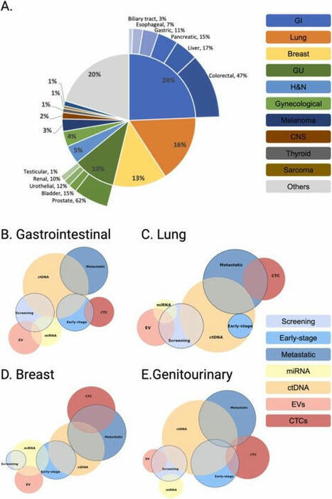
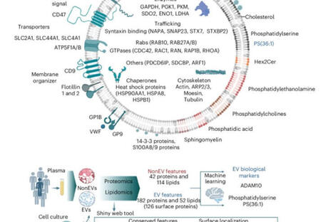
Extracellular vesicles (EVs) encompass a multitude of lipid bilayer-delimited particles, of which exosomes are the most widely studied. Bidirectional cell–cell communications via EVs have a pivotal role in the physiology of multicellular organisms. EVs carry biological cargoes (including proteins, RNA, DNA, lipids and metabolites) capable of mediating a range of pleiotropic cellular functions. Over the past decade, EVs released by cancer cells (onco-EVs) have been shown to promote cancer progression including tumour outgrowth and metastatic dissemination. Furthermore, the innate ability of EVs to protect vulnerable molecular cargoes (such as RNA, DNA or proteins) from enzymatic degradation, their presence in most biofluids and the ability to transverse biological barriers to reach distant organs make them ideal targeted drug delivery systems, including in patients with cancer. Many of these properties also support investigations of EVs as biomarkers with potential roles in both diagnosis and treatment monitoring. In this Review, we describe advances in the development of EVs as cancer therapeutics or biomarkers, including cancer vaccines, targeted drug delivery systems and immunotherapies, as well as potential roles in early cancer detection, diagnosis and clinical management. We also describe the potential of emerging technologies to support further discoveries as well as the clinical translation of EVs into diagnostic and therapeutic clinical tools. We highlight the potential of single-EV and onco-EV detection and discuss how advances in multi-omic and artificial intelligence-enabled integration are providing new biological insights and driving clinical translation. Extracellular vesicles (EVs), a diverse range of membrane-delimited particles, have multiple cellular functions and, when released by cancer cells, can promote tumour growth and metastatic dissemination. The authors of this Review describe advances in the development of EVs as biomarkers and cancer therapeutics, focusing on clinical translation of EVs into diagnostic and therapeutic clinical tools.
Ils ont trouvé ce qui se cache dans votre sang après le COVID (et c’est 19 fois pire que prévu) Des millions de personnes à travers le monde vivent depuis des mois, voire des années, avec des symptômes débilitants après avoir contracté la COVID-19. Fatigue écrasante, brouillard mental, essoufflement inexplicable. Pendant longtemps, la médecine n’avait aucune réponse à leur offrir. Aujourd’hui, une équipe franco-sud-africaine vient de lever le voile sur un phénomène microscopique qui pourrait enfin expliquer pourquoi certains corps refusent de tourner la page. Un ennemi invisible tapi dans le flux sanguin Le COVID long reste l’une des énigmes médicales les plus frustrantes de notre époque. Contrairement à une infection classique qui se résout en quelques semaines, cette condition transforme la vie de ses victimes en marathon d’épuisement sans ligne d’arrivée visible. Les médecins ont longtemps cherché à comprendre ce qui différencie biologiquement ces patients de ceux qui récupèrent normalement.
La réponse pourrait résider dans quelque chose d’infiniment petit : des microcaillots. Ces structures sanguines anormales ne ressemblent en rien aux caillots massifs responsables d’AVC ou de thromboses. Elles sont bien plus discrètes, suffisamment minuscules pour passer inaperçues aux examens standard, mais assez volumineuses pour obstruer progressivement les capillaires qui irriguent nos tissus les plus sensibles, notamment le cerveau.
Quand les défenses du corps deviennent une prison L’histoire se complique avec un deuxième acteur : les pièges extracellulaires de neutrophiles, plus sobrement appelés NETs. Ces structures constituent normalement une arme redoutable du système immunitaire. Lorsqu’un agent pathogène envahit l’organisme, certains globules blancs déploient ces filets collants composés d’ADN et d’enzymes pour capturer l’intrus. Une fois leur mission accomplie, ces pièges se dissolvent naturellement.
Sauf que chez les patients atteints de COVID long, quelque chose déraille. Les NETs prolifèrent en quantités anormales et refusent de disparaître. Pire encore, ils semblent avoir trouvé un complice inattendu dans les microcaillots. Cette association n’avait jamais été documentée auparavant dans la littérature médicale. Les résultats publiés dans le Journal of Medical Virology ont dépassé leurs attentes les plus pessimistes. Les patients atteints de COVID long présentaient un nombre de microcaillots multiplié par 19,7 par rapport à la médiane observée chez les sujets sains. Ces caillots étaient également significativement plus volumineux. Mais la véritable révélation résidait ailleurs : les NETs n’étaient pas simplement présents en parallèle des microcaillots, ils y étaient physiquement intégrés. https://lnkd.in/eZnhNd2S https://lnkd.in/eGm4-UBj | 14 comments on LinkedIn
The Flow Cytometry Market is undergoing a notable transformation, projected to grow from $7.4 billion in 2024 to $16.4 billion by 2034.This represents a...
Researchers at Moffitt Cancer Center have secured a monumental $22.4 million grant from the U.S. Department of War to ignite pioneering studies and clinical trials targeting leptomeningeal disease, an exceptionally dire complication arising from breast and other cancers.
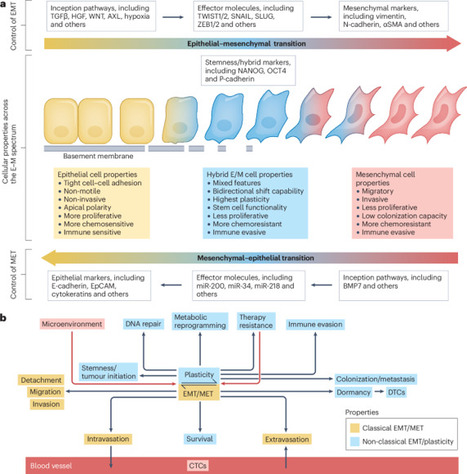
Cell plasticity is a crucial trait for cancer progression towards metastasis and treatment resistance. Research efforts from the past 20–30 years have revealed that the dynamic flux of the epithelial–mesenchymal transition (EMT) programme is one of the major underlying processes enabling cancer cell plasticity and greatly facilitates these major causes of cancer mortality. The spectrum of evidence ranges from extensive data from cell line and animal model studies across multiple cancer types through a rapidly expanding body of work demonstrating associations between EMT biomarkers and disease progression and mortality in patients. EMT is also implicated in resistance to most of the major treatment modalities, yet our efforts to harness this knowledge to improve therapeutic outcomes are currently in their early stages. In this Review, we describe clinical evidence supporting a role of EMT and the associated epithelial–mesenchymal plasticity in various stages of cancer in patients and discuss the subsequent clinical opportunities and challenges associated with attempts to implement this knowledge as novel therapies or clinical management approaches. Despite several decades of research that has revealed roles in the development and progression of many solid tumours, clinical translation of research targeting epithelial–mesenchymal transition (EMT) has thus far been limited. In this Review, the authors provide a summary of the role of EMT in cancer development and progression in the context of this lack of clinical translation, summarize the current status of direct or indirect EMT-modulating agents in clinical development, and highlight the major barriers to the development of EMT-related clinical interventions.
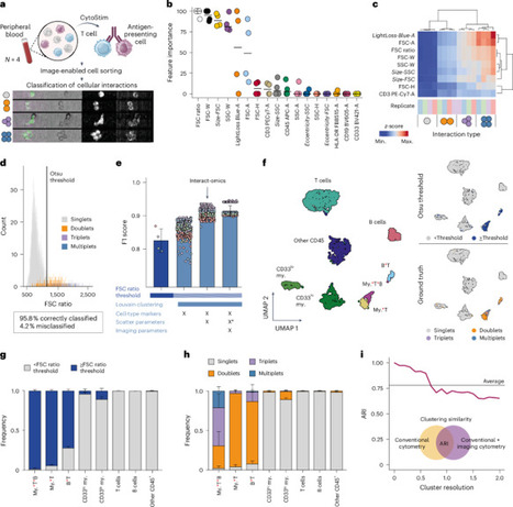
Cellular interactions are of fundamental importance, orchestrating organismal development, tissue homeostasis and immunity. Recently, powerful methods that use single-cell genomic technologies to dissect physically interacting cells have been developed. However, these approaches are characterized by low cellular throughput, long processing times and high costs and are typically restricted to predefined cell types. Here we introduce Interact-omics, a cytometry-based framework to accurately map cellular landscapes and cellular interactions across all immune cell types at ultra-high resolution and scale. We demonstrate the utility of our approach to study kinetics, mode of action and personalized response prediction of immunotherapies, and organism-wide shifts in cellular composition and cellular interaction dynamics following infection in vivo. Our scalable framework can be applied a posteriori to existing cytometry datasets or incorporated into newly designed cytometry-based studies to map cellular interactions with a broad range of applications from fundamental biology to applied biomedicine. Interact-omics, a high-throughput cytometry-based framework, resolves the cellular interaction landscape.
Fostering the Implementation of Liquid Biopsy in Clinical Practice: European Liquid Biopsy Society (ELBS) 2024 Meeting Report by Klaus Pantel et al. Journal of Experimental & Clinical Cancer Research Catherine Alix-Panabières
What a great way to start the year! We are delighted to present our latest review! If you want to know everything there is to know about the cellular biology of extracellular vesicle-receiving cells, check it out! https://lnkd.in/eZCnnV4G
Thank you to Léa Ripoll, Antje Zickler, Pieter Vader, Samir EL Andaloussi, Frederik Verweij for their contribution ! Happy reading and Happy new year !
CRCI2NA, CNRS, Inserm Grand Ouest, French Society for Extracellular Vesicles (FSEV), ISEV Admin,Vesiculab
And finally, the work of many researchers over a long period of time, driven by patience and passion, has led to this consensus on how, where, and when to include circulating tumor cells (CTCs) in clinical practice.Here we express our recognition and gratitude to all of them. The monitoring and follow-up of patients with metastatic cancer cannot be understood without considering the study of these cells.
Researchers have unlocked the molecular secrets of microscopic particles circulating in our blood, revealing in a landmark discovery how the body’s cells communicate in ways we’re only beginning to understand.
HPV-DeepSeek identifies circulating HPV DNA, PIK3CA mutations, and integration events in pretreatment blood samples with high sensitivity and specificity.
New research reveals significant circulatory alterations in patients approaching death, shedding light on a phenomenon that has remained poorly understood.
- The authors demonstrate that tumor infiltration of large vessels and a surge in circulating tumor cell clusters are consistent features of terminal disease progression across multiple types of solid cancer. These findings could reshape how to predict and manage advanced cancer.
- Retrospective autopsy analyses revealed that tumor emboli are present in nearly 90% of patients who die from solid cancers. In a prospective cohort of 21 patients at the end of life, the number of circulating tumor cells and clusters spiked sharply in the days preceding death, suggesting that vascular dissemination accelerates during the final stages of disease progression
- Perhaps most clinically relevant is the demonstration that the infiltration of tumors into large vessels is strongly associated with mortality regardless of whether patients have overt metastatic disease at the time of vascular infiltration, a provocative finding that challenges the current dogma of metastasis as the strongest predictor of mortality.
- The discovery of vascular infiltration as a pivotal event in the final stages of cancer opens promising avenues for both research and clinical intervention. The observation that large-vessel infiltration occurs independently of metastatic burden suggests that local treatments aimed at halting tumor growth near critical vasculature might be beneficial.
- Exploring these strategies in randomized clinical trials may provide valuable insights into their potential survival benefits, offering a new paradigm for managing end-stage cancer — whereby the treatment scope broadens from preventing metastatic spread to also preventing catastrophic vascular events.
Metastasis—the spread of cancer cells from a primary tumor to distant organs—is the principal cause behind over 90% of cancer-related mortality worldwide.Central to this insidious process are circulating tumor cells (CTCs), which detach from the primary malignancy and invade the bloodstream, thereby...
Heterogeneous circulating tumor cells (CTCs) have been implicated in the formation of new metastases. However, circulating cells expressing both tumor and immune cell proteins are often dismissed as insignificant findings in CTC studies. Two non-contemporaneous blood samples from a metastatic breast cancer patient were analyzed using an enrichment-free platform to identify canonical, epithelial-only CTCs (CD45-/cytokeratin + , epi.CTCs) and CD45 + /cytokeratin+ immune-like CTCs (im.CTCs). Single cells from both samples were subjected to copy number and protein expression profiling. A cohort of 36 metastatic breast cancer patients was then analyzed to search for additional cases with im.CTCs. Here, we identified and characterized a population of CTCs exhibiting an immune-like state. In two samples from an index patient, im.CTCs outnumbered epi.CTCs, comprising >97% of the CTC population. Single-cell copy number analysis of 43 im.CTCs and 30 epi.CTCs revealed clonal alterations across both populations, confirming a shared tumor origin. Furthermore, im.CTCs contained pseudo-diploid profiles that did not reflect dilution from the addition of a normal diploid genome, indicating that they were unlikely to have originated from tumor-immune cell fusion. Protein expression analysis showed that im.CTCs express CD45 as well as other immune-related markers, such as CD3 and CD4, and the cancer stemness marker, CD44. Subsequent analysis of a metastatic breast cancer cohort identified an additional patient harboring im.CTCs with the same tumor-derived, non-fusion genome as in the index case. Collectively, these genomic and proteomic features distinguish im.CTCs from previously reported circulating cells may represent a novel form of tumor cell plasticity. Tumor cells are known to take on features that allow them to survive and move to new sites. This variation can make it difficult to distinguish them from other cells in the blood. Using a platform to profile rare cells in blood samples, we identified a population of cells expressing cancer and immune cell proteins in a breast cancer patient. Genomics data confirmed that these cells originated from the tumor and that they were different from another cell type sharing a similar protein expression pattern. We analyzed additional samples and found a second patient with these immune-like tumor cells. These findings support the existence of a cancer-immune state that might play a role in helping tumor cells spread. Higa et al. analyzed circulating cells expressing cancer and immune cell markers in breast cancer patients. Based on genomic and protein expression profiling, they show that the cells were unlike previously described circulating tumor cells with immune-like phenotypes because they did not appear to arise from heterotypical cell fusion.
💫 Super excited to share our latest review just published in Nature Portfolio.
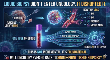
Liquid biopsies🩸, indicating the sampling of body fluids rather than solid-tissue biopsies, have the potential to revolutionize cancer care through personalized, noninvasive disease detection and monitoring. Circulating tumour DNA (#ctDNA) 🧬 and circulating tumour cells (#CTCs) are promising blood-based biomarkers in bladder cancer. Results from several studies have shown the clinical potential of ctDNA and CTCs in #bladder #cancer for prognostication, treatment-response monitoring, and early detection of minimal residual disease (#MRD) and disease recurrence. Following successful clinical trial evaluation, assessment of ctDNA and CTCs holds the potential to transform the therapeutic pathway for patients with bladder cancer — potentially in combination with the analysis of urinary tumour DNA — through tailored treatment guidance and optimized disease surveillance.
👏👏👏 Sia Viborg Lindskrog, Trine Strandgaard, Iver Nordentoft, Matthew Galsky, Tom Powles, Mads Agerbæk, Jørgen Bjerggaard Jensen, Catherine Alix-Panabières & Lars Dyrskjøt
🤩 Good reading !
CHU de Montpellier, Recherche et innovation – CHU de Montpellier, Renan Targhetta, Samir JABER Liquid Biopsy LCCRH Lab - Laboratoire Cellules Circulantes Rares Humaines 🩸 Aarhus University European Liquid Biopsy Society (ELBS) PANCAID, GUIDE.MRD OncoDaily | 11 comments on LinkedIn
To get content containing either thought or leadership enter:
To get content containing both thought and leadership enter:
To get content containing the expression thought leadership enter:
You can enter several keywords and you can refine them whenever you want. Our suggestion engine uses more signals but entering a few keywords here will rapidly give you great content to curate.


 Your new post is loading...