You Don’t Always Need a Blood Draw, Saliva, Urine & Spinal Fluid Are Next
Beyond Blood, Non-Blood Liquid Biopsy Fluids
The science behind it:
A 2026 meta-analysis covering 13,486 participants across nine specimen types found that non-blood liquid biopsy specimens show clinically meaningful diagnostic performance across diverse cancer types, with specimen-specific advantages for anatomically relevant malignancies. Aqueous humor achieved the highest individual performance with 96% sensitivity and 100% specificity for retinoblastoma detection. Cerebrospinal fluid demonstrated excellent performance across brain tumor types with 88.9% sensitivity.
In certain cases of advanced lung cancer and melanoma, clinically relevant alterations for targeted treatments could be identified with high sensitivity in non-blood biofluids, even when they were undetectable in peripheral blood.
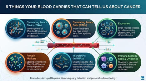
By analyzing tumor-derived components, including ctDNA, cfDNA, cfRNA, circulating microRNA, circulating tumor cells, tumor-educated platelets, and extracellular vesicles, liquid biopsies can detect minimal residual disease, monitor tumor burden, catch disease recurrence earlier, and guide therapy by identifying driver mutations and resistance mutations.
Plasma has become the most widely used and best-standardized biological fluid in liquid biopsy research and is now regarded as the core matrix for multi-cancer early detection and longitudinal disease monitoring.
🌹 Delighted to share our new article review published by HORIBA after my conference presentation at #HoribaKyoto in #Japan 🇯🇵: "Liquid Biopsy in Cancer Management: Advances, Challenges, and Emerging Clinical Applications" Doryan Masmoudi & Catherine Alix-Panabières
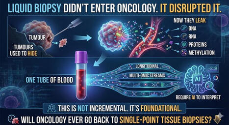
🩸 #LiquidBiopsy is transforming how we understand and manage cancer.
By analyzing tumor-derived components in blood—such as circulating tumor DNA (ctDNA) 🧬, circulating tumor cells (CTCs) 🟠 , and extracellular vesicles (EVs) 🫧 —this approach offers a minimally invasive and dynamic window into tumor heterogeneity and disease evolution.
🌹 Recent technological advances have significantly expanded its potential, from early detection and minimal residual disease (MRD) monitoring to treatment response evaluation and therapy guidance.
💫 Yet, key challenges remain. Biological complexity, technical variability, and the lack of standardized workflows continue to limit widespread clinical adoption. In this review, we explore: 🔬 Current methodologies and clinical applications 🔗 The complementary value of different circulating analytes 🚀 Emerging innovations, including multi-omics integration and AI-driven analysis 📊 Future priorities: standardization, multicenter trials, and integration into routine oncology practice
💥 The path forward is clear: collaboration, harmonization, and innovation will be essential to unlock the full clinical potential of liquid biopsy.
Atsushi Horiba, 中村博司 Hiroshi Nakamura, Tetsuya Matsuda, Motoaki Hamada, Kazunori YOSHIOKA, SEIICHIRO YOSHIOKA, Masayuki Adachi, Susumu Hayashi, Jun Nishimura Arnaud Pradel, Martine Clot, Damien Isèbe, Michael Bruckner, Lilou Garcia, Laurent Fullana CHU de Montpellier, Recherche et innovation – CHU de Montpellier, Renan Targhetta Liquid Biopsy LCCRH Lab - Laboratoire Cellules Circulantes Rares Humaines MedVallée Montpellier European Liquid Biopsy Society Admin PANCAID, PANLIPSY Project, GUIDE.MRD OncoDaily, ACADEMIE NATIONALE DE MEDECINE DE FRANCE SFPO - SOCIETE FRANCAISE DE PHARMACIE ONCOLOGIQUE
Metastatic relapse remains the main cause of treatment failure in pediatric osteosarcoma (OS), as conventional imaging often misses minimal residual or low-burden disease. Circulating tumor cells (CTCs) represent a promising non-invasive biomarker for monitoring tumor progression and heterogeneity.
🌹 ✨ International Day of Women and Girls in Science ✨ 🌹 February 11, 2026
✨ Let’s celebrate scientific women from yesterday, today, and tomorrow! ✨
☀️ On this meaningful day, we are proud to highlight the outstanding work of 3 women scientists from Liquid Biopsy LCCRH Lab - Laboratoire Cellules Circulantes Rares Humaines – CHU de Montpellier, whose dedication and excellence are helping advance translational oncology research.
📰 Their study has just been published in the highly regarded Journal of Experimental & Clinical Cancer Research (IF= 12.8), which trusted us with the publication of: “Reconstructing the Metastatic Journey: Functional Circulating Tumor Cells and Disseminated Tumor Cells-based Models for Translational Oncology.”
🕊️ This achievement reflects their scientific rigor, commitment, and passion for better understanding metastasis to ultimately improve patient care.
📖 The article is open access — feel free to read and share it!
🌺 Today, and 🌺 every day, let’s continue to support, empower, and inspire women and girls in science.
Recherche et innovation – CHU de Montpellier, Anne FERRER-VILLENEUVE, Renan Targhetta, Pôle Biologie Pathologie du CHU de Montpellier, Laurence LACHAUD Liquid Biopsy LCCRH Lab - Laboratoire Cellules Circulantes Rares Humaines, Sanglier caroline, Laure Cayrefourcq Mauro Castelli MedVallée Montpellier, Quinzaine Franco-Allemande d'Occitanie Association Des étoiles dans la mer, vaincre le glioblastome, Laetitia Levère European Liquid Biopsy Society, PANCAID, GUIDE.MRD OncoDaily Aurélia Brégnac ACADEMIE NATIONALE DE MEDECINE DE FRANCE
We have built something that I believe our field urgently needs and that I would like to share: www.liquidbiopsyinfo.com.
When I talk to oncologists about liquid biopsy, the same question keeps coming up:
Where do I find a clear overview of the clinical evidence for my specific clinical scenario?
The honest answer, until now, has been: there is no single place. The data is scattered. The field moves too fast. And the gap between what the science shows and what reaches daily clinical practice is growing.
www.liquidbiopsyinfo.com is a free, structured resource covering liquid biopsy evidence across 20+ cancer types from MRD detection to molecular profiling. It helps to decide where measuring MRD makes sense and what findings mean.
Built by LIQOMICS, a team that has worked on ctDNA diagnostics for years.
We are transparent about our approach: we use AI to keep pace with the volume of new publications, and we know that this requires expert oversight to meet the standards clinicians and patients deserve. That is why this is an open invitation.
If you work in oncology, liquid biopsy, or clinical research and you think this kind of resource matters I would genuinely value your perspective.
🩸 ❓🩸 Do you want to learn more about the history of circulating tumor cell (CTC) lines and xenograft models, developed over the past 2️⃣2️⃣ years from CTCs isolated directly from cancer patients — and their biological insights, clinical relevance, and translational applications?
✨ This new review is for you !!!! GOOD READING !!!!
❤️ Proud to highlight the outstanding work of these women scientists from the Liquid Biopsy LCCRH Lab - Laboratoire Cellules Circulantes Rares Humaines (CHU de Montpellier), whose dedication and scientific excellence continue to advance translational oncology research.
📰 Our study has just been published in the excellent Journal of Experimental & Clinical Cancer Research (IF 12.8) : ✨ “Reconstructing the Metastatic Journey: Functional Circulating Tumor Cells and Disseminated Tumor Cells-based Models for Translational Oncology.” ✨
🕊️ A comprehensive overview of how functional CTC/DTC models help us better understand metastasis biology and move closer to improving patient care.
📖 The article is open access — feel free to read and share it!
Recherche et innovation – CHU de Montpellier, Renan Targhetta, Pôle Biologie Pathologie du CHU de Montpellier Liquid Biopsy LCCRH Lab - Laboratoire Cellules Circulantes Rares Humaines, Sanglier caroline, Laure Cayrefourcq Mauro Castelli MedVallée Montpellier, Quinzaine Franco-Allemande d'Occitanie Fondation ARC pour la recherche sur le cancer European Liquid Biopsy Society, PANCAID, GUIDE.MRD OncoDaily Aurélia Brégnac ACADEMIE NATIONALE DE MEDECINE DE FRANCE
What a great way to start the year! We are delighted to present our latest review! If you want to know everything there is to know about the cellular biology of extracellular vesicle-receiving cells, check it out! https://lnkd.in/eZCnnV4G
Thank you to Léa Ripoll, Antje Zickler, Pieter Vader, Samir EL Andaloussi, Frederik Verweij for their contribution ! Happy reading and Happy new year !
CRCI2NA, CNRS, Inserm Grand Ouest, French Society for Extracellular Vesicles (FSEV), ISEV Admin,Vesiculab
And finally, the work of many researchers over a long period of time, driven by patience and passion, has led to this consensus on how, where, and when to include circulating tumor cells (CTCs) in clinical practice.Here we express our recognition and gratitude to all of them. The monitoring and follow-up of patients with metastatic cancer cannot be understood without considering the study of these cells.
Liquid Biopsy Found Cancer Where Tissue Biopsy Missed It, In 5,954 Patients
NCI-MATCH Trial cDNA vs. Tissue, ASCO Annual Meeting, May 2026, Chicago
Rini Pauly of the Frederick National Laboratory for Cancer Research presented results at ASCO 2026 (May 29–June 2, Chicago) comparing mutations found in paired tumors and circulating tumor DNA from 5,954 patients with advanced cancers screened for NCI-MATCH, one of the largest precision medicine platform trials sponsored by the National Cancer Institute.
In some cases, the ctDNA results revealed mutations where the tissue biopsy did not, likely due to tumour heterogeneity in advanced cancers, where a single tumour may harbour multiple mutations that a single-site tissue biopsy simply misses. This is one of the most powerful real-world arguments for liquid biopsy: it samples the whole tumour landscape simultaneously, not just one corner of it.
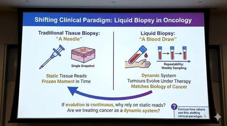
Liquid biopsy enables comprehensive tumor profiling without the need for traditional tissue biopsies. Over the past decade, research in this field has expanded exponentially, leading to integration into clinical practice for specific cancer types, including lung and breast cancer.
Liquid biopsy currently serves as a complement rather than a replacement for traditional tissue biopsy, especially when histopathological context, tissue architecture, or precise tumor type and location are critical for diagnosis and initial treatment. As technologies improve and standardized protocols develop, liquid biopsies are set to become central to personalized oncology.
In a cohort study, Liao and colleagues investigate whether using a lower flow-cytometry threshold (0.05%), compared with the conventional 0.1% cutoff, to define minimal residual disease improves risk stratification in pediatric acute myeloid leukemia.
It began in 1869, when Thomas Ashworth, an Australian physician, looked through a microscope 🔬 and saw tumor-like cells circulating in the blood of a patient with metastatic cancer.
Today, we know these as circulating tumor cells, or CTCs.
Twenty years later, Stephen Paget gave oncology one of its most powerful ideas: the “seed and soil” hypothesis 🌱🌍 He proposed that metastasis was not random. Cancer cells may travel through the blood, but they grow only when they find the right microenvironment.
In many ways, Ashworth saw the seed. Paget imagined the soil.
For decades, the field waited for technology to catch up ⏳
That changed when CTC detection became more standardized, especially with the FDA-approved CellSearch® system in 2004. For the first time, clinicians and researchers had a practical way to capture and enumerate CTCs from blood.
Then, in 2010, Klaus Pantel and Catherine Alix-Panabières gave the field a name that would define a new era: “liquid biopsy.” 💡
Today, liquid biopsy is often used almost interchangeably with circulating tumor DNA, or ctDNA 🧬 But the term was originally coined in the context of CTCs — the idea that blood could provide a noninvasive, real-time window into cancer biology.
As sequencing became faster, cheaper, and more sensitive ⚡ ctDNA moved to the forefront.
It allowed us to: ✔ Detect mutations ✔ Monitor resistance ✔ Assess minimal residual disease
All without repeated tissue biopsies.
But liquid biopsy is bigger than ctDNA.
In 2026, the International Society of Liquid Biopsy (@ISLB) has given the field a broader and more detailed definition 📘 https://lnkd.in/dktDMVaT
🩸 Liquid biopsy now includes the analysis of:
→ Cells → Nucleic acids → Proteins → Metabolites → Extracellular vesicles → Other molecules released into body fluids
This is an important correction.
Liquid biopsy is not one analyte. It is not one platform. It is not only blood.
It's a dynamic way to study disease through accessible body fluids.
The future will be multi-analyte and longitudinal 🔄
CTCs inform us of viable metastatic cells. ctDNA may reveal genomic evolution. Extracellular vesicles, proteins, RNA, metabolites, and immune signals may complete the picture.
The next chapter will not be about replacing tissue biopsy entirely.
It will be about asking better questions, earlier:
❓ Is disease still present? ❓ Is resistance emerging? ❓ Is treatment working? ❓ Can relapse be detected before imaging?
From Ashworth’s microscope 🔬 to Paget’s hypothesis 🌱 to Pantel and Alix-Panabières’ 2010 terminology 📖 to ISLB’s 2026 definition
Liquid biopsy has become one of oncology’s most powerful ideas:
A less invasive, more dynamic way to understand cancer as it changes.
Recent research highlights an important shift in how we understand #cancer spread: #tumor cells do not always act alone.
In many cases, they travel in clusters - working together like a coordinated team. Much like individuals versus a group in any high-stakes environment, these clusters are more resilient, better protected, and significantly more effective at establishing new growth elsewhere in the body.
This challenges a long-standing assumption in treatment design, which has largely focused on targeting single cells. The emerging insight is clear: addressing complex problems may require not just targeting individual actors, but disrupting the systems and networks that enable them to thrive.
Prostate cancer is characterized by multifocality, inter- and intra-patient tumour heterogeneity, and differences in risk of progression to metastatic disease, castration resistance and lethality, which can make prognosis challenging. Consequently, sampling methods that provide accurate insight into disease phenotype to facilitate risk-stratification of patients are crucial. The variable biology of prostate cancer seems to be recapitulated in the phenotypic heterogeneity of circulating tumour cells (CTCs). CTC sampling offers a liquid biopsy method to achieve minimally invasive longitudinal sampling for disease monitoring. CTC analysis has also offered a crucial insight into aggressive phenotypes, disease metastasis and treatment response, particularly in clinical trials. The clinical use of CTC count for prognosis in advanced prostate cancer has been approved by the FDA, but is not routinely used clinically, as these cells are technically challenging to isolate and analyse. However, methodological advances continue to improve CTC enrichment and profiling. Understanding the clinical utility of CTCs and future innovations is crucial to incorporating CTCs into the clinical management of prostate cancer. Circulating tumour cells (CTCs) offer a minimally invasive biopsy strategy for prostate cancer monitoring. This Review discusses the use of CTCs at all stages of prostate cancer development and treatment, from CTC isolation and enrichment strategies to the prognostic and clinical utility of these cells in prostate cancer.
If you’ve ever struggled with identifying dendritic cell subsets by flow cytometry (and honestly… who hasn’t?), this one’s for you. In my recent blog for Proteintech Group I’ve put together a practical, step-by-step guide that walks through: • DC subsets (cDC1, cDC2, pDCs, moDCs) • Key surface markers • Smart panel design tips • Clean gating strategies that actually work Whether you’re setting up a new panel or troubleshooting an existing one, this guide is meant to save you time (and frustration). 👉 Read the blog here: https://lnkd.in/eWBBuUBn Would love to hear how you approach DC phenotyping in your lab 👇 #FlowCytometry #Immunology #DendriticCells #Proteintech
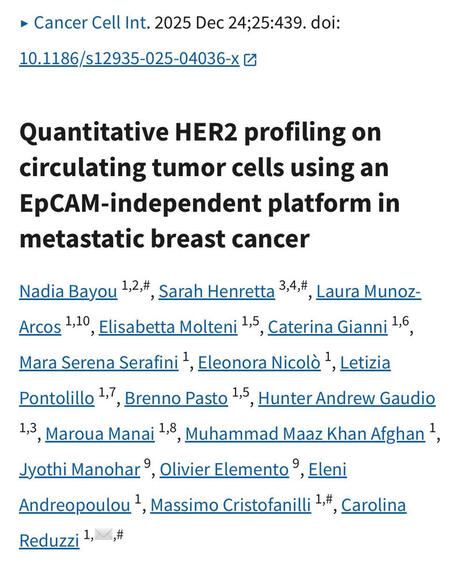
I’m pleased to share our latest publication in Cancer Cell International!
“Quantitative HER2 profiling on circulating tumor cells using an EpCAM-independent platform in metastatic breast cancer”
Our analysis highlights the dynamic heterogeneity of HER2 expression in metastatic breast cancer and supports the role of CTCs as a minimally invasive tool for real-time disease assessment.
🙏 Grateful to all co-authors, clinical collaborators, and especially to the patients who made this work possible.
We’re excited to share our latest paper exploring the tumor–immune interface in circulation through liquid biopsy: “Caught in the Act: Tumor–Immune Interactions in Circulation of Patients with Immune Marker–Positive Circulating Tumor Cells”
Circulating tumor cells (CTCs) and large extracellular vesicles (LEVs) are key components of liquid biopsy, offering minimally invasive insight into tumor biology. A particularly intriguing subset, immune marker–positive CTCs (im.CTCs), has been described clinically, yet their biological origin has remained unclear.
In this study, using high-resolution immunofluorescence microscopy and proteomic profiling of patient blood samples, we show direct physical interactions between white blood cells and both im.CTCs and im.LEVs, observed exclusively in patients with im.CTCs.
Together, these results support the hypothesis for in vivo membrane transfer as a mechanism of immune marker acquisition by CTCs and LEVs, with important implications for tumor–immune biology and the clinical interpretation of immune-positive CTCs in liquid biopsy.
To get content containing either thought or leadership enter:
To get content containing both thought and leadership enter:
To get content containing the expression thought leadership enter:
You can enter several keywords and you can refine them whenever you want. Our suggestion engine uses more signals but entering a few keywords here will rapidly give you great content to curate.


 Your new post is loading...