Your new post is loading...

|
Scooped by
Gilbert C FAURE
January 15, 2025 1:38 PM
|
#Autoimmunity | #Tregs | Differential roles of human CD4+ & CD8+ regulatory T cells in controlling self-reactive immune responses | Fundamental new insights by… | 10 comments on LinkedIn
|
|
Scooped by
Gilbert C FAURE
January 2, 2025 8:30 AM
|
Stem cells reside in specialized microenvironments, termed niches, at several different locations in tissues1–3. The differential functions of heterogeneous stem cells and niches are important given the increasing clinical applications of stem-cell transplantation and immunotherapy. Whether hierarchical structures among stem cells at distinct niches exist and further control aspects of immune tolerance is unknown. Here we describe previously unknown new hierarchical arrangements in haematopoietic stem cells (HSCs) and bone marrow niches that dictate both regenerative potential and immune privilege. High-level nitric oxide-generating (NOhi) HSCs are refractory to immune attack and exhibit delayed albeit robust long-term reconstitution. Such highly immune-privileged, primitive NOhi HSCs co-localize with distinctive capillaries characterized by primary ciliated endothelium and high levels of the immune-checkpoint molecule CD200. These capillaries regulate the regenerative functions of NOhi HSCs through the ciliary protein IFT20 together with CD200, endothelial nitric oxide synthase and autophagy signals, which further mediate immunoprotection. Notably, previously described niche constituents, sinusoidal cells and type-H vessels2–10 co-localize with less immune-privileged and less potent NOlow HSCs. Together, we identify highly immune-privileged, late-rising primitive HSCs and characterize their immunoprotective niches comprising specialized vascular domains. Our results indicate that the niche orchestrates hierarchy in stem cells and immune tolerance, and highlight future immunotherapeutic targets. Previously undescribed hierarchical arrangements in haematopoietic stem cells and their niches that mediate both regenerative potential and immune privilege are identified.
|
|
Scooped by
Gilbert C FAURE
December 4, 2024 1:56 PM
|
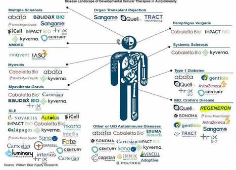
𝐂𝐞𝐥𝐥 𝐓𝐡𝐞𝐫𝐚𝐩𝐲 𝐋𝐚𝐧𝐝𝐬𝐜𝐚𝐩𝐞 𝐢𝐧 𝐀𝐮𝐭𝐨𝐢𝐦𝐦𝐮𝐧𝐢𝐭𝐲 🏄♂️
30 companies below categorised by 12 disease areas and other/undiscovered… | 26 comments on LinkedIn
|
|
Scooped by
Gilbert C FAURE
November 29, 2024 10:33 AM
|
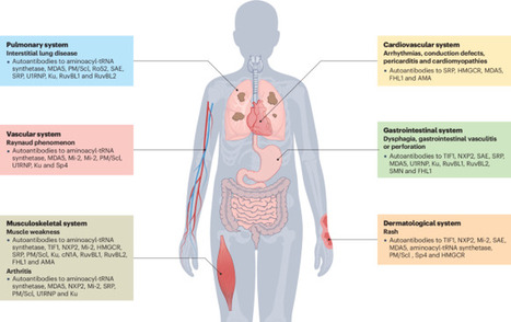
Myositis-specific autoantibodies (MSAs) have become pivotal biomarkers for idiopathic inflammatory myopathies and have revolutionized understanding of the heterogeneous disease spectrum that affects both adults and children. The discovery and characterization of MSAs have substantially enhanced patient stratification based on clinical phenotype, thereby facilitating more precise diagnosis and ultimately improving management strategies. Advances in immunoassay technologies in the past 20 years have further propelled the field forward, enabling the detection of a growing repertoire of autoantibodies with high specificity and sensitivity; however, evolving research over the past decade has revealed that even within antibody-defined subsets, considerable clinical diversity exists, suggesting a broader spectrum of disease manifestations than previously acknowledged. Challenges persist, particularly among patients who are seronegative, where the failure to identify certain rare MSAs stems from the use of diverse detection methodologies and inadequate consensus-guided standardization and validation protocols. Bridging these diagnostic gaps is crucial for optimizing patient care and refining prognostic stratification in idiopathic inflammatory myopathies. This Review provides an update on autoantibodies associated with idiopathic inflammatory myopathies in both adults and children. The authors also discuss methods of autoantibody detection and the advantages and limitations of each technique.
|
|
Scooped by
Gilbert C FAURE
November 9, 2024 4:37 AM
|
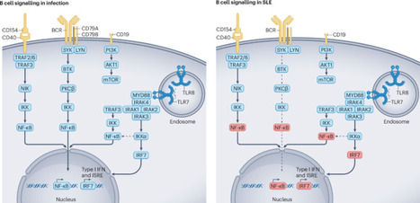
BCR-independent memory B cell reactivation via TLR7 or TLR8 activation, type I interferon production, immune complex formation and T helper cell signalling is central in SLE pathogenesis. Dörner and Lipsky discuss the potential of targeting these pathways to eliminate autoreactive memory B cells...
|
|
Scooped by
Gilbert C FAURE
October 3, 2024 4:54 AM
|
Idiopathic inflammatory myopathies (IIMs) encompass a spectrum of autoimmune diseases characterized by muscle inflammation and systemic involvement. This review aimed to synthesize current evidence on the clinical significance and pathogenic mechanism
|
|
Scooped by
Gilbert C FAURE
August 14, 2024 3:31 AM
|
Guest Blog: A Major Health Crisis: The Alarming Rise of Autoimmune Disease By Molly Murray, President and CEO, Autoimmune Association Observed every March, Autoimmune Awareness Month aims to enhance awareness […]
|
|
Scooped by
Gilbert C FAURE
August 10, 2024 5:31 AM
|
|
|
Scooped by
Gilbert C FAURE
August 3, 2024 2:35 AM
|
|
|
Scooped by
Gilbert C FAURE
July 20, 2024 7:08 AM
|
Sjögren syndrome or Sjögren disease is a chronic form of autoimmune epithelitis characterized by lymphocytic infiltration of the exocrine glands, particularly the salivary and lacrimal glands, leading to progressive glandular dysfunction and subsequent xerostomia and xerophthalmia. Other common manifestations include pain and fatigue, various systemic manifestations and non-Hodgkin’s lymphoma. Sjögren syndrome is therefore a complex and disabling disease associated with a reduced quality of life and with considerable long-term damage. Most of the available treatments are merely symptomatic with limited efficacy in both preventing glandular damage and suppressing systemic disease activity. In the past 10 years, great progress has been made in understanding the pathophysiology of Sjögren syndrome, opening new avenues towards a more targeted and individualized therapeutic approach to the disease. Indeed, several randomized controlled trials have just been completed or are poised to commence evaluating the effectiveness of novel drugs targeting both innate and adaptive immune pathways, including pro-inflammatory cytokines, the type I interferon system, B cell activation, B cell and T cell co-stimulation pathway, and ectopic germinal centre formation. Novel clinical trials are also ongoing exploring various targeted approaches (that is, IgG recycling inhibition, nuclease therapy and CAR-T cell therapy) for Sjögren syndrome. Sjögren syndrome is a chronic autoimmune disease affecting exocrine glands, causing dryness and systemic symptoms. Treatment has been primarily symptomatic, but advances in our understanding of its pathophysiology offer promise for targeted therapies, aiming for personalized care and improved outcomes.
|
|
Scooped by
Gilbert C FAURE
July 17, 2024 4:46 AM
|
The genetic contribution of protein-coding variants to immune-mediated diseases (IMDs) remains underexplored. Through whole exome sequencing of 40 IMDs in 350,770 UK Biobank participants, we identified 162 unique genes in 35 IMDs, among which 124 were novel genes. Several genes, including FLG which is associated with atopic dermatitis and asthma, showed converging evidence from both rare and common variants. 91 genes exerted significant effects on longitudinal outcomes (interquartile range of Hazard Ratio: 1.12-5.89). Mendelian randomization identified five causal genes, of which four were approved drug targets (CDSN, DDR1, LTA, and IL18BP). Proteomic analysis indicated that mutations associated with specific IMDs might also affect protein expression in other IMDs. For example, DXO (celiac disease-related gene) and PSMB9 (alopecia areata-related gene) could modulate CDSN (autoimmune hypothyroidism-, psoriasis-, asthma-, and Graves’ disease-related gene) expression. Identified genes predominantly impact immune and biochemical processes, and can be clustered into pathways of immune-related, urate metabolism, and antigen processing. Our findings identified protein-coding variants which are the key to IMDs pathogenesis and provided new insights into tailored innovative therapies. The genetic contributions of protein-coding variants to immune-mediated diseases (IMDs) have yet to be fully explored. Here, the authors conduct a large whole-exome association study, identifying 162 genes associated with 35 IMDs, including 124 that had not been previously reported.
|
|
Scooped by
Gilbert C FAURE
May 22, 2024 7:16 AM
|
Vaccination is not limited to infectious diseases. Discover the fascinating world of vaccination against autoimmune diseases
|
|
Scooped by
Gilbert C FAURE
May 3, 2024 7:45 AM
|
|
|
|
Rescooped by
Gilbert C FAURE
from Virus World
January 5, 2025 5:31 AM
|
Aberrant immune responses to viral pathogens contribute to pathogenesis, but our understanding of pathological immune responses caused by viruses within the human virome, especially at a population scale, remains limited. We analyzed whole-genome sequencing datasets of 6,321 Japanese individuals, including patients with autoimmune diseases (psoriasis vulgaris, rheumatoid arthritis (RA), systemic lupus erythematosus (SLE), pulmonary alveolar proteinosis (PAP) or multiple sclerosis) and coronavirus disease 2019 (COVID-19), or healthy controls. We systematically quantified two constituents of the blood DNA virome, endogenous HHV-6 (eHHV-6) and anellovirus. Participants with eHHV-6B had higher risks of SLE and PAP; the former was validated in All of Us. eHHV-6B-positivity and high SLE disease activity index scores had strong correlations. Genome-wide association study and long-read sequencing mapped the integration of the HHV-6B genome to a locus on chromosome 22q. Epitope mapping and single-cell RNA sequencing revealed distinctive immune induction by eHHV-6B in patients with SLE. In addition, high anellovirus load correlated strongly with SLE, RA and COVID-19 status. Our analyses unveil relationships between the human virome and autoimmune and infectious diseases. Analysis of the blood DNA virome in patients with COVID-19 and autoimmune disease associates endogenous HHV-6 (eHHV-6) and high anellovirus load with increased disease risk, most notably for systemic lupus erythematosus. eHHV-6 carriers show a distinct immune response. Published in NAt. Genetics (Jan. 3, 2025): https://doi.org/10.1038/s41588-024-02022-z
Via Juan Lama
|
|
Scooped by
Gilbert C FAURE
December 5, 2024 4:04 AM
|
WEDNESDAY, Dec. 4, 2024 (HealthDay News) -- Inflammatory bowel disease (IBD) in older adults has become a global public health burden, according to a study published in the Jan. 31, 2025, issue of Autoimmunity Reviews.
|
|
Rescooped by
Gilbert C FAURE
from Immunology
December 3, 2024 4:37 AM
|
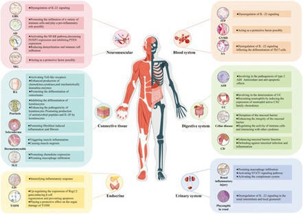
As a pivotal cytokine, IL-22 plays a crucial role in the pathogenesis of autoimmune diseases. In this comprehensive review, we initially provide an overvie

|
Suggested by
LIGHTING
November 16, 2024 5:00 AM
|
Early and accurate diagnosis is crucial to prevent disease development and define therapeutic strategies. Due to predominantly unspecific symptoms, diagnosis of autoimmune diseases (AID) is notoriously challenging. Clinical decision support systems (CDSS) are a promising method with the potential to enhance and expedite precise diagnostics by physicians. However, due to the difficulties of integrating and encoding multi-omics data with clinical values, as well as a lack of standardization, such systems are often limited to certain data types. Accordingly, even sophisticated data models fall short when making accurate disease diagnoses and presenting data analyses in a user-friendly form. Therefore, the integration of various data types is not only an opportunity but also a competitive advantage for research and industry. We have developed an integration pipeline to enable the use of machine learning for patient classification based on multi-omics data in combination with clinical values and laboratory results. The application of our framework resulted in up to 96% prediction accuracy of autoimmune diseases with machine learning models. Our results deliver insights into autoimmune disease research and have the potential to be adapted for applications across disease conditions.
|
|
Scooped by
Gilbert C FAURE
November 5, 2024 3:55 AM
|
Sixteen presentations of preclinical studies funded by the Lupus Research Alliance (LRA) to advance understanding of lupus and potential treatment pathways,...
|
|
Scooped by
Gilbert C FAURE
September 7, 2024 2:55 AM
|
Systemic lupus erythematosus (SLE) is a prototypic autoimmune disease that primarily affects young women and causes a wide range of inflammatory manifestations. The hallmark of SLE is the production of antibodies to components of the cell nucleus (anti-nuclear antibodies [ANAs]).
|
|
Scooped by
Gilbert C FAURE
August 12, 2024 4:27 AM
|
Myositis with anti-Ku-autoantibodies is a rare inflammatory myopathy associated with various connective tissue diseases. Histopathological studies have identified inflammatory and necrotizing aspects, but a precise morphological analysis and pathomechanistic disease model are lacking. We therefore aimed to carry out an in-depth morpho-molecular analysis to uncover possible pathomechanisms. Muscle biopsy specimens from 26 patients with anti-Ku-antibodies and unequivocal myositis were analyzed by immunohistochemistry, immunofluorescence, transcriptomics, and proteomics and compared to biopsy specimens of non-disease controls, immune-mediated necrotizing myopathy (IMNM), and inclusion body myositis (IBM). Clinical findings and laboratory parameters were evaluated retrospectively and correlated with morphological and molecular features. Patients were mainly female (92%) with a median age of 56.5 years. Isolated myositis and overlap with systemic sclerosis were reported in 31%, respectively. Isolated myositis presented with higher creatine kinase levels and cardiac involvement (83%), whereas systemic sclerosis-overlap patients often had interstitial lung disease (57%). Histopathology showed a wide spectrum from mild to pronounced myositis with diffuse sarcolemmal MHC-class I (100%) and -II (69%) immunoreactivity, myofiber necrosis (88%), endomysial inflammation (85%), thickened capillaries (84%), and vacuoles (60%). Conspicuous sarcoplasmic protein aggregates were p62, BAG3, myotilin, or immunoproteasomal beta5i-positive. Proteomic and transcriptomic analysis identified prominent up-regulation of autophagy, proteasome, and hnRNP-related cell stress. To conclude, Ku + myositis is morphologically characterized by myofiber necrosis, MHC-class I and II positivity, variable endomysial inflammation, and distinct protein aggregation varying from IBM and IMNM, and it can be placed in the spectrum of scleromyositis and overlap myositis. It features characteristic sarcoplasmic protein aggregation on an acquired basis being functionally associated with altered chaperone, proteasome, and autophagy function indicating that Ku + myositis exhibit aspects of an acquired inflammatory protein-aggregate myopathy.
|
|
Scooped by
Gilbert C FAURE
August 7, 2024 9:13 AM
|
Chimeric antigen receptor (CAR) T cells are highly effective at targeting and eliminating cells of the B cell lineage. CAR T cell therapy has become a standard-of-care treatment for patients with relapsed or refractory B cell malignancies. In addition, the administration of genetically modified T cells with the capacity to deplete B cells and/or plasma cells has tremendous therapeutic potential in autoimmune diseases. In the past few years, CD19-based and B cell maturation antigen (BCMA)-based CAR T cell therapies have been applied to various B cell-mediated autoimmune diseases including systemic lupus erythematosus, idiopathic inflammatory myopathy, systemic sclerosis, neuromyelitis optica spectrum disorder, myasthenia gravis and multiple sclerosis. The scientific rationale behind this approach is that deep depletion of B cells, including autoreactive B cell clones, could restore normal immune function, referred to as an immune reset. In this Review, we discuss important aspects of CAR T cell therapy in autoimmune disease, including considerations relating to patient selection, safety, efficacy and medical management. These considerations are based on the early experiences of CAR T cell therapy in autoimmune diseases, and as the field of CAR T cell therapy in autoimmune diseases continues to rapidly evolve, these issues will remain subject to ongoing refinement and adaptation. CAR T cell therapy shows promise for achieving long-term drug-free remission in various autoimmune diseases. This Review discusses the ongoing challenges and unanswered questions of CAR T cell therapy in autoimmune diseases, including pre-procedural, procedural and post-procedural considerations.
|
|
Scooped by
Gilbert C FAURE
August 2, 2024 1:04 PM
|
|
|
Scooped by
Gilbert C FAURE
July 18, 2024 10:48 AM
|
|
|
Scooped by
Gilbert C FAURE
June 21, 2024 7:42 AM
|
Identification of HSP60 as an antigenic target of a subset of synovial B cell–derived anti-FLS RA-rmAbs with partial cross-reactivity to other stromal-related antigens. Of 71 rmAbs generated from 4 ELS+ RA synovial tissues, we previously showed that 10 RA-rmAbs display anti-FLS immunoreactivity with variable immunofluorescence patterns (Figure 1A and Supplemental Figure 1; supplemental material available online with this article; https://doi.org/10.1172/JCI169754DS1). Liquid chromatography tandem mass spectrometry (LC-MS/MS) analysis of a 60 kDa band of total protein detected via Western blotting by several RA-rmAbs targeting RA-FLSs identified HSP60 as one of the most abundant proteins (among >100 detected by the LC-MS/MS analysis), with a high amount of sequence coverage (68%) across the full length of HSP60 (Figure 1B). HSP60 is a molecular chaperone expressed in different intracellular compartments (e.g., cytosol) as well as extracellularly that migrates to an approximately 58 kDa position in SDS-PAGE (Figure 1C). Figure 1Synovial RA-rmAbs display immunoreactivity to RA-FLS. (A) Schematic summarizing the origin and reactivity characterization of the RA-rmAbs (n = 71) against NETs/cit-histones and RA-FLS. Schematic in A was created using BioRender.com. (B) RA-FLS protein extract was probed with an anti-FLS RA-rmAb. A protein of approximately 60 kDa was excised and analyzed by MS. The analysis detected a high amount of sequence coverage (68%) across the full length of the HSP60 protein in the RA-FLS protein extract. (C) rhHSP60 and RA-FLS protein extracts were subjected to Western blotting and probed with an anti–human HSP60 antibody. (D) Representative immunofluorescence images of RA-FLS incubated with the RA-rmAbs, showing immunoreactivity of 3 RA-rmAbs to FLS-derived antigens (green). RA056/11.95.2 was used as a negative control. Nuclei were stained with DAPI (blue). Original magnification, ×20. (E) Representative immunofluorescence image of RA-FLS showing expression of HSP60 (red). Original magnification, ×40. Therefore, we next performed immunofluorescence studies to determine whether HSP60 and/or other stromal-related antigens were the target of the subset of RA-rmAbs reactive to RA-FLSs (Figure 1D and Supplemental Figure 1). We confirmed the expression of HSP60 in RA-FLSs by Western blotting of the RA-FLS total protein extract and by immunofluorescence staining (Figure 1, C and E). We then quantitatively assessed in an ELISA the binding of the anti-FLS RA-rmAbs to a recombinant human HSP60 (rhHSP60) protein. We identified 3 RA-rmAbs (RA057/11.35.1, RA056/11.76.1, RA056/11.48.2) that showed binding to the rhHSP60 in a dose-dependent manner (Figure 2A). The RA057/11.35.1 rmAb showed the strongest reactivity to rhHSP60. In contrast, 1 RA-rmAb, RA056/11.95.2, which did not bind to RA-FLS (Figure 1D) and was thus used as a negative control, failed to show any binding to rhHSP60 (Figure 2A). The observed immunoreactivity against HSP60 was not due to polyreactivity, as none of the 3 RA-rmAbs displayed polyreactivity to multiple structurally unrelated antigens (Supplemental Figure 2) (2, 20, 21). We also investigated whether the 3 RA-rmAbs with anti-HSP60 immunoreactivity displayed enhanced binding to an in vitro citrullinated form of HSP60 (cit-rHSP60) (Supplemental Figure 3A). As shown in Supplemental Figure 3B, only RA056/11.76.1 rmAbs displayed increased binding to cit-rHSP60. Next, the 3 RA-rmAbs were tested for their reactivity in ELISAs to other RA-associated antigens including anti–cyclic citrullinated peptide (anti-CCP) ELISA, citrullinated fibrinogen, and the FLS-associated antigens calreticulin and vimentin, which are known RA autoantigens and migrate to a similar 60 kDa position in RA-FLS protein extract electrophoresis. As shown in Supplemental Figure 4, we also observed a variable degree of reactivity of the anti-HSP60 RA-rmAb to calreticulin and vimentin, whereas only anti-RA056/11.76.1 rmAb displayed low citrullinated fibrinogen–binding activity. We next conducted a competitive binding assay to further confirm whether HSP60 was recognized by the RA-rmAbs. As shown in Figure 2B, incubation of the RA-rmAbs with soluble rhHSP60 significantly reduced, but did not completely abrogate, the binding to HSP60 in the ELISA. In order to support the HSP60/RA-rmAb binding results, IP assays were performed. As shown in Figure 2C, IP of rhHSP60 with 2 RA-rmAbs (RA057/11.35.1 and RA056/11.76.1) showed a band of approximately 60 kDa, confirming the ELISA data. The absence of signal for the RA-rmAb RA057/11.48.2 might be explained by the low level of binding to HSP60 observed in the ELISA. Figure 2RA-rmAbs with anti-FLS reactivity recognize HSP60. (A) Binding of RA057/11.35.1, RA056/11.76.1, and RA056/11.48.2 rmAbs and a negative RA-rmAb (RA056/11/95.2) to rhHSP60. All RA-rmAbs were tested at a concentration of 100 mg/mL followed by 6 serial dilutions (1:2). (B) Competitive binding of the 3 RA-rmAbs to rhHSP60 preincubated with or without soluble HSP60 (competitor). Results are expressed as a percentage of HSP60 binding. (C) IP results of rhHSP60 and the RA-rmAbs. As a control, rhHSP60 alone was loaded. (D) Representative immunofluorescence image showing staining for HSP60 (red) and RA-rmAbs (green). Nuclei were stained with DAPI (blue). Original magnification, ×63. The data are the results of 3 independent experiments. *P < 0.05, ***P < 0.001, and ****P < 0.0001, by 1-way ANOVA with multiple comparisons. We next sought to confirm that the RA-rmAbs target FLS-derived HSP60 by immunofluorescence staining. As shown in Figure 2D, double-immunofluorescence staining with the 3 RA-rmAbs in combination with an anti–human HSP60 antibody demonstrated a partial cellular colocalization with the HSP60. We quantified the degree of colocalization using ImageJ (NIH), with a Pearson’s correlation coefficient of r = 0.4–0.5, which suggested a positive correlation (22, 23). Since the staining morphology of our anti-FLS rmAbs suggested partial binding to cytoskeletal proteins, either through direct biding or interaction with HSP60 (24), we performed double-immunofluorescence staining of the RA-rmAbs with an anti–human F-actin antibody showing variable but largely incomplete colocalization for the 3 RA-rmAbs with a structural FLS antigen (Supplemental Figure 5). Anti-FLS antibodies accelerate arthritis in the CIA mouse model. We explored whether a cocktail of anti-FLS antibodies containing the RA057/11.35.1 and RA056/11.76.1 anti-HSP60 antibodies could have a functional role in the CIA mouse model. CIA was induced by intradermal injection of 2 mg/mL bovine collagen type II on day 0, followed by a boost on day 21. Anti-FLS RA-rmAbs were administered i.p. at 4 time points. As a control, we used both PBS and a mix of RA-rmAbs targeting histone proteins, which were used as a negative control (2, 25). We found that the anti-FLS RA-rmAbs were able to enhance the arthritis score and increase inflammation in the paws compared with the control-treated group (Figure 3A). In contrast, the mice treated with anti-histone RA-rmAbs (2) showed a reduction in the arthritis score (Figure 3A), similar to previous data (25). Histological evaluation of the hind paws showed a large influx of inflammatory cells and signs of cartilage damage in the anti-FLS RA-rmAbs group versus the PBS- and anti-histone RA-rmAb–treated groups (Figure 3B and Supplemental Figure 6). Moreover, micro-CT (μCT) analysis confirmed the increased bone erosion induced by the anti-FLS RA-rmAbs compared with both the control- and anti–histone RA-rmAb–treated group (Figure 3, C–E). Figure 3Effect of anti-FLS RA-rmAbs in CIA. (A) Graph shows the arthritis scores for CIA DBA/1 mice treated i.p. with anti-FLS rmAbs (n = 20), PBS (controls, n = 40), and anti-histone rmAbs (n = 20). Anti-FLS rmAbs were administered on days 17, 28, 38, and 49. The anti-FLS rmAbs cocktail consisted of RA056/11.76.1, RA057/11.35.1, and RA057/11.89.1 antibodies. The anti-histone rmAbs cocktail consisted of RA015/11.58, RA015/11.88, and RA015/11.91 antibodies. Results are from 2 independent experiments. The scores are reported as the mean of the sum of the score ± SEM assessed for each paw of the mouse. The P values correspond to the treatment × time element from the ANOVA linear model between groups. (B) Representative IHC (top) and safranin-O (SO) (bottom) images of control- and anti-FLS–treated mice. Scale bars: 2.5 mm. Original magnification, ×1.5 (enlarged insets, control) and ×23 (anti-FLS). (C) Representative μCT scan of a hind paw from a CIA mouse and the scoring system used to evaluate bone erosion (erosion score). The score was given for each digit (color-coded area), with a maximum score of 24. 0 = normal; 1 = signs of pathological bone changes; 2 = heavy pathological bone changes. (D) Representative μCT scan images of control-treated, anti-FLS–treated (n = 5) and anti-histone–treated (n = 5) mice. (E) Graph shows the μCT erosion score for CIA DBA/1 mice treated with PBS, anti-FLS RA-rmAbs, or anti-histone RA-rmAbs. Five representative hind paws were used for each group. The scoring was done blindly by 3 independent evaluators and is reported as the mean of the sum ± SEM. *P < 0.05 and **P < 0.01, by 1-way ANOVA with multiple comparisons. Antigenic targets of anti-FLS rmAbs such as HSP60 are preferentially expressed in the lympho-myeloid pathotype, colocalize with synovial ELSs, and are upregulated in FLSs derived from leukocyte-rich RA joints. The distribution of HSP60 within RA synovial tissue and in matched synovial fluid (SF) and serum samples from patients with RA was examined by bulk RNA-Seq, immunofluorescence staining, and ELISA, respectively. RNA-Seq expression for HSPD1 (gene name for HSP60) within synovial tissue was compared between the lympho-myeloid, diffuse-myeloid, and pauci-immune fibroid histological groups in patients with early or established RA using bulk RNA-Seq data generated by us from synovial biopsies obtained as part of the Pathobiology of Early Arthritis Cohort (PEAC) study and the R4RA randomized clinical trial (26–28). HSPD1 was elevated in the lympho-myeloid versus the pauci-immune fibroid and diffuse-myeloid in both early (P = 0.0012) and established RA (P = 0.0015) patients (Figure 4, A and B). Moreover, at the protein level, HSP60 showed a preferential distribution around lymphocyte aggregates and periaggregates within the RA synovial tissue (Figure 4, C and D). In addition, we reanalyzed GeoMx Digital Spatial Profiler (DSP) data from the R4RA trial to characterize the spatial positioning of the HSPD1 transcript in association with the different RA histopathotypes (27). Consistent with the bulk RNA-Seq data, HSPD1 expression was significantly higher in the lympho-myeloid group versus the diffuse-myeloid group (P < 0.0001; Figure 4E). Like the HSP60 distribution within the RA synovial tissue determined by immunofluorescence, the spatial positioning of HSPD1 was observed mainly around lymphocyte aggregates independently of the sublining and lining areas (Figure 4, F and G). Moreover, we detected similar levels of HSP60 protein expression in the synovial sublining and lining fibroblasts, with HSP60 protein preferentially observed around lymphocyte aggregates, as previously mentioned (Figure 4C). This observation was supported by scRNA-Seq data from the Accelerating Medicines Partnership (AMP) RA database, in which HSPD1 was found to be highly expressed in fibroblasts from the patients with leukocyte-rich RA (29) (Figure 4H). However, HSPD1 expression was also detected in other cell types, like monocytes (Figure 4H). Finally, we observed higher levels of HSP60 protein in the SF of patients with RA compared with HSP60 levels at the systemic level (Figure 4I). Accumulation of HSP60 in the SF was preferentially observed in patients with RA (n = 20) versus patients with osteoarthritis (OA) (n = 11) (Figure 4J). Figure 4HSP60 expression in synovial tissue and SF of patients with RA. Synovium HSPD1 gene evaluation compared across histological pathotype in patients with early (A) and established (B) RA. Unit y axis: regularized log-normalized read. (C) Representative immunofluorescence and IHC images of synovial tissue from patients with RA showing staining for HSP60 (yellow)/CD90+ (green)/CD55+ (red) synovial fibroblasts and for CD20 (B cells), CD3 (T cells), and CD138 (plasma cells). Nuclei were stained with DAPI (blue). Original magnification, ×2 and ×10 (enlarged insets). (D) Quantification of HSP60 differential expression in RA synovial tissue (RA patients, n = 3) by QuPath analysis. Statistical analysis was done by Friedman’s test. (E) HSPD1 was differentially expressed in the lympho-myeloid (lymphoid) versus diffuse-myeloid (myeloid) pathotypes using GeoMx DSP data. (F) HSPD1 differential expression in the sublining, lining, and aggregated area using GeoMx DSP data. In E and F, statistical analysis was performed using the Mann-Whitney U test. (G) HSPD1 differential expression in the sublining and lining in diffuse-myeloid (myeloid) versus lympho-myeloid (lymphoid) pathotypes. Scatterplot shows individual regions of interest (ROIs)/. Statistical analysis was done using the Kruskal-Wallis test. (H) scRNA-Seq profiling of HSPD1 in synovial fibroblasts in leukocyte-rich/leukocyte-poor RA and OA patients (https://immunogenomics.io/ampra/). (I) Graph shows HSP60 levels (pg/mL) in matched sera and SF from patients with RA (n = 14). Statistical analysis was done with the Wilcoxon test. (J) Box plot displays SF HSP60 levels in RA (n = 20) versus OA (n = 11) patients. Statistical analysis was done using the Mann-Whitney U test. Each data point represents an individual patient. *P < 0.05, **P < 0.01, ***P < 0.001, and ****P < 0.0001. Similar to HSP60, calreticulin (CALR) gene expression was significantly higher in the lympho-myeloid pathotype in bulk synovial RNA-Seq results and in spatial association with lymphoid aggregates using GeoMx analysis of patients with established RA (Supplemental Figure 7, A–D), supporting the conclusion that stromal-derived autoantigens involved in the cellular stress response were upregulated in RA patients with the lympho-myeloid pathotype and contributed to the generation of pathogenic autoantibodies. Anti-HSP60 antibodies are elevated in the SF of patients with RA. We then investigated the prevalence of anti-HSP60 antibodies in matched SF and serum samples from patients with RA. Serum and SF anti-HSP60 antibodies were measured by ELISA using rhHSP60. As shown in Figure 5A, binding of IgG, IgM, and IgA to HSP60 in SF was positively correlated to RA serum levels. We detected a significant difference of SF IgG, IgM, and IgA anti-HSP60 antibodies between patients with OA or RA, with an increase of IgM levels observed in the SF of patients with RA (Figure 5B and Supplemental Figure 8, A and B). Only 10% of the RA SF tested did not have anti-HSP60 antibodies. Ten percent of RA SF was characterized by the presence of IgG, IgM, and IgA binding to HSP60 (triple-positive), with 25% double-positive and 55% single-positive binding (Supplemental Figure 8C). Finally, patients with RA were divided into ACPA+ (serum, n = 11; SF, n = 10) and ACPA– (serum, n = 9; SF, n = 4) groups using a conventional anti-CCP2 test. As shown in Figure 5C, the levels of antibodies against HSP60 in ACPA+ RA patients were significantly increased for IgA antibodies, with only a trend for IgG and IgM anti-HSP60 antibodies for both serum and SF samples. Figure 5Expression of anti-HSP60 antibodies in serum and SF of patients with RA or OA. (A) Graphs show the correlation of matching RA SF IgG, IgM, and IgA binding to HSP60 with RA sera IgG, IgM, and IgA binding to HSP60. (B) Box plots display the levels of IgG, IgM, and IgA anti-HSP60 antibodies in SF of RA (n = 20) versus OA (n = 11) patients. (C) Scattered dot plots show IgG, IgM, and IgA binding to HSP60 in SF and sera of ACPA+ versus ACPA– RA patients. For B and C, the results are expressed as absorbance at 450 nm, and each data point represents an individual patient. *P < 0.05, **P < 0.01, and ****P < 0.0001, by Mann-Whitney U test. Synovial HSP60 expression is predictive of the clinical response to rituximab and is modulated by B cell depletion in good clinical responders. Given the high correlation of HSP60 with the presence of ELSs in the RA joints, we assessed the longitudinal effect of rituximab therapy on synovial HSPD1 expression before and after B cell depletion in the R4RA biopsy-driven randomized trial (Figure 6A) (27, 28). Baseline HSPD1 gene expression was significantly higher in the B cell–rich subset of patients with RA and was associated with good clinical responders, as defined by the R4RA trial’s clinical primary endpoint, the Clinical Disease Activity Index (CDAI), and by the secondary endpoint, the Disease Activity Score 28–C-reactive protein (DAS28-CRP) (Figure 6, B and C). Hence, we next explored the longitudinal effect of rituximab on HSPD1 within synovial tissue before and after B cell depletion. Using mixed-effects models applied to paired synovial samples (n = 29 individuals, n = 58 samples), we first observed a significant downregulation of HSPD1 expression 16 weeks after treatment with rituximab, but not following tocilizumab therapy (Figure 6D). For tocilizumab, baseline HSPD1 gene expression was not associated with good clinical responders, as defined by the R4RA trial clinical primary endpoint CDAI and by the secondary endpoint DAS28-CRP (Supplemental Figure 9). Interestingly, a significant downregulation of synovial HSPD1 expression upon B cell depletion was selectively observed in good responders compared with nonresponders and moderate responders (Figure 6, E and F) using mixed-effects models applied to paired synovial samples only, with similar results if all samples were included in the models (Supplemental Figure 10A). We observed similar results also for CALR expression (Supplemental Figure 7, E–G), suggesting a common gene expression profile linked to cellular stress response mechanisms in RA-FLS with local release of autoantigens. Figure 6HSP60 analysis of paired pre- and post-rituximab treatment synovial biopsies. (A) Schematic representation of the R4RA clinical trial (27, 28). At baseline, patients with RA underwent a synovial biopsy of a clinically active joint for histological evaluation. Following the synovial biopsy, patients were randomized 1:1 to either the rituximab or tocilizumab treatment group. An optional repeated synovial biopsy of the same joint previously sampled was performed at 16 weeks, followed by histological and transcriptomic evaluation. A CDAI of 50% was used as a primary endpoint to define responders and nonresponders to treatment at 16 weeks. Patients were initially followed up every 4 weeks, with the end of the trial at 48 weeks. Numbers in parentheses represent patients with available RNA-Seq data. (B) Synovium HSPD1 variance-stabilizing transformation (VST) gene expression in patients stratified by histology into B cell–poor (B poor) and B cell–rich (B rich) categories. (C) HSPD1-normalized synovial gene expression levels assessed at baseline in CDAI nonresponders versus responders and DAS28-CRP moderate/nonresponders versus responders to rituximab treatment. (D) HSPD1 RNA-Seq counts were assessed at baseline and 16 weeks following rituximab and tocilizumab treatment in paired synovial biopsies (rituximab, n = 29 individuals, n = 58 samples; tocilizumab, n = 15 individuals, n = 30 samples). (E and F) HSPD1 counts were assessed at baseline and 16 weeks following rituximab treatment in paired synovial biopsies. In D–F, gray data points represent RNA-Seq counts between paired samples for individuals. Statistical analysis was performed using a negative binomial mixed-effects model on RNA-Seq counts. Overlaid green/purple data points show estimated marginal means of the fitted mixed-effects model, with error bars showing 95% CIs for the fixed effects of the mixed-effects model. To assess the clinical response, the following parameters were used: CDAI 50% improvement and DAS28-CRP European Alliance of Associations for Rheumatology (EULAR) response. RTX, rituximab. Finally, in the R4RA cohort we observed a positive correlation between serum anti-HSP60 IgG with CDAI, tender joint count, and DAS28-ESR at baseline (Supplemental Figure 10B), even though IgG anti-HSP60 antibody levels did not change over time after rituximab treatment (Supplemental Figure 11).
|
|
Scooped by
Gilbert C FAURE
May 4, 2024 1:45 PM
|
Antiphospholipid syndrome (APS) consists of thrombotic, non-thrombotic and obstetric clinical manifestations developing in individuals with persistent antiphospholipid antibodies (aPL). Although researchers have made progress in characterizing different clinical phenotypes of aPL-positive people, the current approach to clinical management is still mostly based on a ‘one size fits all’ strategy, which is derived from the results of a limited number of prospective, controlled studies. With the 2023 publication of the ACR–EULAR APS classification criteria, it is now possible to rethink APS, to lay the groundwork for subphenotyping through novel pathophysiology-informed approaches, and to set a future APS research agenda guided by unmet needs in clinical management. In this Review, Knight and Erkan consider how the 2023 ACR–EULAR classification criteria for antiphospholipid syndrome (APS) can guide future research to subphenotype APS by understanding its pathophysiology, paving the way for the personalized and proactive management of individuals with APS.
|


 Scoop.it!