Your new post is loading...

|
Scooped by
Gilbert C FAURE
October 17, 2022 11:45 AM
|
|
|
Scooped by
Gilbert C FAURE
October 21, 4:09 AM
|
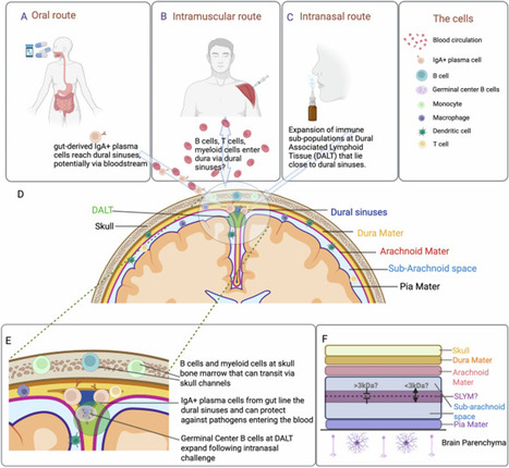
Download PDF Subjects Bioinformatics Innate immunity Skull bone marrow (BM) has emerged as an important immune interface with the central nervous system (CNS). Park and colleagues identify germinal center-like lymphoid structures within skull BM that can generate adaptive immune responses to CNS-derived antigens and contribute to anti-tumor immunity in experimental glioma.The skull bone marrow (BM) is increasingly recognized as an immune interface with the central nervous system (CNS). Vascular channels and diploic veins connect skull BM with the dura mater, providing routes for the exchange of cells and soluble molecules between these compartments.1,2 Cerebrospinal fluid can access skull BM niches, while skull BM supplies myeloid cells and immature B cells to the meninges and, under some conditions, the CNS parenchyma.3,4,5 Organized lymphoid hubs containing germinal center (GC) B cells have also been identified around meningeal venous plexuses, where they support local humoral immune responses.6 More recently, neuron-derived proteins were shown to accumulate at skull-associated borders, where they are sampled by resident immune cells.7 Despite these anatomical and functional connections, whether skull BM can organize antigen-specific adaptive immune responses to CNS-derived antigens has remained unclear. Park and colleagues now identify GC-like lymphoid structures in skull BM and examine their contribution to CNS immunosurveillance.8Comparing BM from different bones, the authors identified an enrichment of IL-21+CXCR5+ follicular helper T (Tfh) cells in skull BM relative to the sternum and femur. These cells expressed Th1-associated markers and formed clusters with B cells in discrete regions of the skull. Skull BM also contained increased numbers of class-switched and GC-like B cells, IgG+ plasmablasts, and antibody-secreting cells. Further characterization revealed organized aggregates containing T cells, B cells, antigen-presenting cells (APCs), CXCL13, B cell activating factor, follicular dendritic cells, and activation-induced cytidine deaminase-expressing B cells. GC-like B cells and antibody-secreting cells underwent clonal expansion and somatic hypermutation and depended on CD4+ T cells, IL-21, and CD40-CD40L signaling. These findings identify a specialized lymphoid environment within skull BM capable of supporting a local humoral immune response.8What drives these responses? Gut microbiota and dietary antigens contributed to the generation of IgA-producing and GC-like B-cell populations but did not fully account for their presence. Park and colleagues therefore asked whether CNS-derived antigens could also reach and activate this compartment. Using neuronal expression of secreted ovalbumin (OVA), they detected antigen in skull BM and observed activation of antigen-specific CD4+ T and B cells.8 Recent work by Chayama and colleagues provides an interesting comparison. Using neuronal protein tracing, they found that brain-derived proteins reach skull-associated niches under physiological conditions.7 However, antigen-sampling skull B cells in that study expressed genes associated with antigen processing and presentation together with a predominantly tolerogenic transcriptional program. Earlier work also identified a CNS antigen-dependent tolerance checkpoint in the adjacent meninges, where developing B cells reactive to the CNS-specific antigen myelin oligodendrocyte glycoprotein were selectively eliminated.9 Access of CNS antigens to skull-associated immune niches, therefore, does not necessarily result in immune activation. The conditions and anatomical site in which antigen is encountered may determine the response that follows.Park and colleagues next examined whether these structures participate in immunity against brain tumors. Following intracranial implantation of CT2A glioma cells, T- and B-cell responses increased within skull BM. Using CT2A tumors expressing HEL-OVA together with antigen-specific lymphocytes, they detected early activation of tumor-reactive T and B cells in skull BM.8 These responses persisted in mice lacking conventional secondary lymphoid organs, although they were diminished and remained dependent on cells recruited from the periphery. The skull BM can therefore support local adaptive immune responses, but it does not appear to function independently of the peripheral immune system.The authors next asked whether these responses influence tumor control. Local blockade of CD40L disrupted skull GC-like structures, reduced intratumoral lymphocyte accumulation and CD8+ T-cell activation, and shortened survival. Conversely, combining a CD40 agonist with IL-21 and IFNγ enhanced skull B-cell responses, increased immune cell infiltration and cytotoxic activity within tumors, and prolonged survival.8 Under these experimental conditions, manipulating skull-associated immune responses can alter immunity within the brain and influence disease outcome (Fig. 1).How well these findings extend to naturally arising brain tumors remains uncertain. Glioblastoma (GBM) is generally considered an immunologically “cold” tumor, with an immune environment dominated by myeloid cells and relatively limited productive T-cell immunity. Evidence for skull-associated anti-tumor immunity has nevertheless been found in patients. Dobersalske and colleagues identified tumor-reactive CD8+ T cells in cranial BM from patients with GBM, including T-cell receptor clonotypes shared between cranial marrow and matched tumors.10 The extent to which these cells contribute to tumor control remains uncertain. Immune checkpoint blockade has also produced disappointing results in large clinical trials, with nivolumab failing to improve overall survival compared with bevacizumab in recurrent GBM.11 These clinical observations do not rule out a contribution from skull-associated immunity, but they show that tumor antigen recognition in GBM rarely translates into an effective anti-tumor response.The glioma models used in the study warrant some consideration. Tumors were established by intracranial transplantation, and some experiments employed cells engineered to express defined model antigens.8 These approaches are useful for resolving antigen-specific responses but may not fully reproduce tumors that arise spontaneously within the CNS. Whether skull-associated responses are similarly engaged by tumors with lower immunogenicity, and how their contribution compares with conventional lymphoid organs and other CNS border tissues, remain unresolved.An important question is how antigen encounter within skull BM is translated into an immune response. CNS-derived proteins reach skull-associated immune niches under physiological conditions,7 while studies of the skull and adjacent meninges have identified mechanisms that may favor B cell tolerance to CNS-derived antigens.7,9 What determines whether CNS antigen encounter promotes tolerance or productive adaptive immunity? Does inflammation or tissue injury alter this balance? Are similar skull-associated responses generated during infection, autoimmunity, or neurodegeneration? It will also be important to determine how much skull BM contributes to CNS immunity relative to the meninges and conventional secondary lymphoid organs, and whether comparable antigen-responsive lymphoid structures function in the human skull.Park and colleagues establish that skull BM contains organized lymphoid structures capable of responding to CNS-derived antigens and influencing immunity in experimental glioma. What remains less clear is when these responses become functionally important. Defining the signals that balance tolerance and immunity within this niche and determining its contribution relative to other CNS and peripheral immune compartments will be important for establishing the role of skull BM in CNS immunosurveillance. ReferencesMonaghan, K. L. et al. Nature 652, 1298–1307 (2026).Article PubMed Google Scholar Zanluqui, N. G. & McGavern, D. B. Trends Immunol. 45, 738–749 (2024).Article PubMed PubMed Central Google Scholar Brioschi, S. et al. Science 373, eabf9277 (2021).Article PubMed PubMed Central Google Scholar Cugurra, A. et al. Science 373, eabf7844 (2021).Article PubMed PubMed Central Google Scholar Mazzitelli, J. A. et al. Nat. Neurosci. 25, 555–560 (2022).Article PubMed PubMed Central Google Scholar Fitzpatrick, Z. et al. Nature 628, 612–619 (2024).Article PubMed PubMed Central Google Scholar Chayama, Y. et al. Cell 189, 4737–4755.e17 (2026).Article PubMed PubMed Central Google Scholar Park, J. H. et al. Nature 658, 196–204 (2026).Article PubMed PubMed Central Google Scholar Wang, Y. et al. Immunity 54, 2784–2794.e6 (2021).Article PubMed Google Scholar Dobersalske, C. et al. Nat. Med. 30, 2947–2956 (2024).Article PubMed PubMed Central Google Scholar Reardon, D. A. et al. JAMA Oncol. 6, 1003–1010 (2020).Article PubMed PubMed Central Google Scholar Download referencesAcknowledgementsThis work was supported by the intramural program at the National Institute of Neurological Disorders & Stroke (NINDS) within the National Institutes of Health (NIH). The contributions of the NIH author(s) are considered Works of the United States Government. The findings and conclusions presented in this paper are those of the author(s) and do not necessarily reflect the views of the NIH or the U.S. Department of Health and Human Services.Author informationAuthors and AffiliationsViral Immunology and Intravital Imaging Section, National Institute of Neurological Disorders and Stroke (NINDS), National Institutes of Health, Bethesda, MD, USANagela G. Zanluqui, Yvonne L. Latour & Dorian B. McGavernAuthorsNagela G. ZanluquiView author publicationsSearch author on:PubMedGoogle ScholarYvonne L. LatourView author publicationsSearch author on:PubMedGoogle ScholarDorian B. McGavernView author publicationsSearch author on:PubMedGoogle ScholarCorresponding authorCorrespondence to Dorian B. McGavern.Additional informationPublisher’s note Springer Nature remains neutral with regard to jurisdictional claims in published maps and institutional affiliations.Rights and permissionsReprints and permissionsAbout this articleCite this articleZanluqui, N.G., Latour, Y.L. & McGavern, D.B. A neighborhood watch within the skull. Cell Res (2026). https://doi.org/10.1038/s41422-026-01310-zDownload citationPublished21 October 2026Version of record21 October 2026DOIhttps://doi.org/10.1038/s41422-026-01310-z
|
|
Scooped by
Gilbert C FAURE
October 18, 7:12 AM
|
🧠 Exercise Protects the Brain from Aging 🏃♂️
As we age, the hippocampus, a brain region essential for learning and memory, undergoes structural and cellular changes that contribute to cognitive decline. Research suggests that physical exercise may help counteract these changes by supporting neuroplasticity, vascular health, and the formation of new neurons.
A scientific review published in Ageing Research Reviews examines how exercise influences the aging hippocampus, particularly through adult hippocampal neurogenesis (AHN), the process by which new neurons are generated from neural stem cells.
How Exercise May Protect the Aging Brain:
🧠 Preserving Hippocampal Volume: Exercise may help maintain hippocampal structure, counteracting age-related atrophy.
🩸 Improving Brain Vasculature: Physical activity promotes vascular remodeling, supporting oxygen and nutrient delivery to brain tissue.
🧬 Increasing Neurotrophic Factors: Exercise enhances signaling involving BDNF, VEGF, and IGF-1, which support neuronal survival, plasticity, and vascular health.
🔬 Supporting Neurogenesis: Preclinical studies suggest exercise can activate dormant neural stem cells, increase their proliferation, and improve the survival of new neurons.
💡 Supporting Learning and Memory: These adaptations may help preserve hippocampal-dependent cognitive functions throughout aging.
Why does this matter:
Exercise does more than improve cardiovascular and muscular health. It influences the brain’s molecular and cellular environment, potentially helping protect against age-related cognitive decline.
While much of the evidence for exercise-induced neurogenesis comes from animal studies, and adult neurogenesis in humans remains under investigation, these findings highlight the importance of physical activity for healthy brain aging.
📄 Research: Cognition on the move: Examining the role of physical exercise and neurogenesis in counteracting cognitive aging
Researchers: Sahand Farmand, Andrea Du Preez, Curie Kim, Chiara de Lucia, Marc-David Ruepp, Brendon Stubbs, and Sandrine Thuret.
Image credit: Farmand et al., Ageing Research Reviews.
#Neuroscience #Neuroplasticity #ExerciseScience | 11 comments on LinkedIn
|
|
Scooped by
Gilbert C FAURE
September 26, 5:27 AM
|
Glioblastoma is one of the worst cancers in the world.
But these authors developed a unique CAR-T that cleared the disease by disarming the tumor microenvironment.
Context:
Glioblastoma has resisted virtually every immunotherapy approach, in part because the tumor microenvironment is saturated with immunosuppressive myeloid cells that actively disarm incoming T cells.
What The Authors Did
- The Singh lab used multi-omic analysis across 83 GBM patient samples to identify GPNMB (glycoprotein non-metastatic melanoma protein B) as uniquely expressed on both glioma stem cells and a specific subset of macrophages, while sparing pro-inflammatory macrophages and healthy brain tissue.
- They engineered GPNMB-targeting CAR T cells and tested them in patient-derived xenograft (PDX) models, a GL261 syngeneic immune-competent model, and a humanized NOG-EXL model.
Key Results
- Complete tumor clearance in PDX models lasting 120+ days post-treatment
- Durable tumor control in mice for more than 100 days
AND ON TOP OF THAT...
- Rescued CD133-targeting CAR T cells that had previously failed against CD133+ tumor relapse, by eliminating the myeloid shield first
This is one of the first examples of a single CAR construct that simultaneously addresses both the malignant AND ALSO immunosuppressive compartments in a solid tumor.
Eliminating the myeloid shield may be necessary, not optional, for any CAR-T therapy to work in GBM.
Kudos to the authors, great work!
(and shout out to the Canadians involved, just down the road in Hamilton)
What are your thoughts on this?
Drop them in the comments.
Article link:
https://lnkd.in/g32hzZys | 17 comments on LinkedIn
|
|
Scooped by
Gilbert C FAURE
September 18, 5:04 AM
|
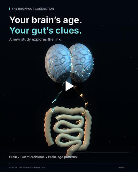
Your brain may appear older than it really is. And scientists at UCLA say that gap might have something to do with your gut. In a new paper published in eBioMedicine, researchers looked at brain images from about 1,500 men and women, both younger and middle aged.
Using a measure of how various areas of the brain talk to one another while resting, they calculated the brain’s age and then contrasted it with the individuals’ real ages.
Those whose brains appeared older than their years also scored lower on tests of memory and cognition, and exhibited more symptoms of depression.
This is when the gut enters the picture.
For a smaller group, researchers looked at fecal specimens. They discovered links between the brain-age gap, certain strains of gut bacteria, and metabolites in the stool.
Their results suggested mechanisms involving the immune system, blood vessels, neuronal communication, and energy metabolism.
I hope the dialogue around the microbiome can expand beyond a list of good and bad microbes. The presence of certain organisms is important. But linking them to a chemical landscape in the gut, as well as other parts of the body, seems like a far more intriguing topic.
This study was cross-sectional. It didn’t track patients into dementia. It can’t establish that a certain microbial composition leads to an older brain or that changing those bacteria would make the brain younger. Nor can it tell you if a colonoscopy would help identify someone at risk for dementia.
we should stop limiting the discussion of the gut to gas and defecation. Are we being too circumspect about the microbiome? Or are we jumping the gun? To me, that’s the conversation we should be having. | 12 comments on LinkedIn
|
|
Scooped by
Gilbert C FAURE
August 16, 3:51 AM
|
🧠 Alzheimer’s disease: should we also consider it a disease of innate immunity?
For decades, Alzheimer’s disease has been viewed primarily through the accumulation of amyloid-β (Aβ) and pathological tau protein. A more recent perspective is broadening this paradigm: innate immunity may be a central driver of the disease rather than merely a consequence of neurodegeneration.
The AD² hypothesis (Alzheimer’s disease as an autoimmune disease) proposes that Aβ may initially participate in a protective immune response. Infections, trauma, metabolic stress and other cellular injuries generate PAMPs and DAMPs, activating the brain’s innate immune machinery.
When this response persists, microglia, initially protective, may shift toward a dysfunctional phenotype. This can lead to chronic production of pro-inflammatory cytokines, inflammasome activation and recruitment of the complement system, particularly C1q and C3.
Complement may subsequently contribute to abnormal synaptic tagging and microglia-mediated synaptic elimination, progressively promoting synaptic dysfunction, neuronal loss and neurodegeneration.
Neuronal injury then releases additional DAMPs, further stimulating innate immunity and creating a self-perpetuating vicious cycle in which an initially protective immune response becomes chronic, dysregulated and potentially damaging to self.
This is the essence of the emerging concept of “innate autoimmunity”: Alzheimer’s disease may not simply be a proteinopathy, but a disorder in which innate immunity, microglia, inflammasomes and complement actively participate in neurodegenerative progression.
⚠️ Importantly, this remains an emerging pathophysiological hypothesis rather than a universally accepted classification of Alzheimer’s disease as an autoimmune disorder.
Rethinking Alzheimer’s disease through the lens of innate immunity may ultimately open new avenues for diagnosis, prevention and immune-targeted therapies.
|
|
Scooped by
Gilbert C FAURE
July 30, 3:42 AM
|
Can your voice help detect disease? 🎙️🤖🩺
Excited to share a major milestone in the field of AI-powered vocal biomarkers!
Researchers and clinicians from the Department of Precision Health (DoPH) at the Luxembourg Institute of Health and the University of South Florida (USF) have led an international effort to establish the first consensus-based framework and definitions for vocal biomarkers: a crucial step toward making voice-based disease detection more reliable, reproducible, and ready for clinical practice.
Led by Dr. Guy Fagherazzi (Head of the Department of Precision Health at LIH and Chair of eVoiceNet - COST Action Network) and Dr. Yael Bensoussan (USF Health Morsani College of Medicine and co-lead of Bridge2AI-Voice), this initiative brought together 24 experts from Europe and North America to create a shared scientific language for the field.
Why does this matter? 👇
🗣️ Our voices contain valuable information about our health
📊 AI can detect subtle changes linked to conditions such as Parkinson's disease, Alzheimer's disease, depression, heart failure, and type 2 diabetes
🤝 A common framework will accelerate collaboration, improve validation, and help bring trustworthy voice-based technologies from research labs to clinical care
🔗 Read the full article here and discover how AI and voice are shaping the future of precision health:
https://lnkd.in/edHx32SK
#AI #VoiceAI #VocalBiomarkers #DigitalHealth #PrecisionHealth #HealthcareInnovation #ClinicalResearch #MachineLearning #LIH #eVoiceNet #Bridge2AI #HealthTech
|
|
Scooped by
Gilbert C FAURE
July 29, 1:03 PM
|
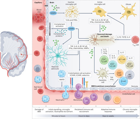
The blood–brain barrier (BBB) is a unique specialization of central nervous system (CNS) capillary endothelial cells that controls molecular traffic between the blood and the brain. In doing so, the BBB maintains a safe and homeostatic environment for proper neuronal function. The BBB restricts drug delivery to the CNS, while BBB defects contribute to neurodevelopmental and neurodegenerative disorders. In this Review, we discuss the distinct features of CNS capillary endothelial cells that form the BBB. Meanwhile, interactions between capillary endothelial cells and surrounding pericytes, astrocytes and with the extracellular matrix of the basement membrane, are crucial for BBB formation and maintenance. We examine how molecular regulators of capillary endothelial cells and signalling pathways that control cell–cell interactions render the BBB a dynamic and selective interface. We highlight emerging areas of BBB research, including heterogeneity of brain vascular permeability and technology development. Finally, we discuss how insights from BBB biology are shaping therapeutic strategies targeting the BBB. This Review summarizes the latest advances in blood–brain barrier (BBB) research, highlighting how emerging findings on the modulation of BBB function and heterogeneity by cellular interactions and signalling pathways might shape BBB-targeted therapeutics.
|
|
Scooped by
Gilbert C FAURE
June 13, 4:31 AM
|
The Autism Medical & Research Summit (AMRS) is an international, congress dedicated to high-support autism
|
|
Scooped by
Gilbert C FAURE
February 13, 4:38 AM
|
The immune landscape at the brain’s borders is being reconsidered. In Nature Medicine (2024), we contributed to this broader effort by examining the clinical relevance of immune-brain interactions in patients with glioblastoma.
Since then, accumulating evidence suggests that the skull bone marrow and adjacent meninges form an active immune and hematopoietic compartment directly interfacing with the CNS.
This cranial niche appears to be more than anatomically adjacent – it may actively shape immune surveillance, neuroinflammation, and brain tumor biology.
A key question now is how this interface can be translated into clinical strategies: from refined biomarker approaches and localized immune modulation to new concepts in CNS drug delivery and, potentially, cell-based therapies informed by cranial immune biology.
Selected recent original research:
🔗 Nat Med (2024): https://lnkd.in/efgufXDM
🔗 Cancer Cell (2024): https://lnkd.in/emWxuMv2
🔗 Cell (2026): https://lnkd.in/eAZEGAtq
🔗 Cell (2026): https://lnkd.in/eg4qjubf
🔗 Nat Genet (2026): https://lnkd.in/eDSCYrxQ
#Neuroimmunology #CNS #BrainTumors #CellTherapy #TranslationalResearch
|
|
Scooped by
Gilbert C FAURE
January 14, 1:06 PM
|
Targeting TREM2 has emerged as an attractive therapeutic strategy for Alzheimer’s disease, with multiple monoclonal antibodies and small-molecule agonists advancing into clinical development in recent years.
TREM2 is a microglial transmembrane receptor that plays key roles in innate immune signaling, microglial survival, and responses to amyloid-β and tau pathology. While early antibody programs have yet to demonstrate clear clinical benefit, they have generated valuable insights into target biology, pharmacology, and clinical trial design.
This target review covers the evolving TREM2 landscape, including lessons from first-generation antibodies, emerging small-molecule agonists, and how recent clinical and patent activity is renewing momentum around this target.
Explore our target review on emerging TREM2 agonists.
|
|
Scooped by
Gilbert C FAURE
January 7, 4:51 AM
|
|
|
Scooped by
Gilbert C FAURE
January 2, 5:08 AM
|
Emerging evidence highlights the crucial role of peripheral immune cells in maintaining brain homeostasis and their influence on the pathology of Alzheimer disease (AD). Genome-wide association studies have identified numerous AD risk variants in genes expressed by immune cells, implicating innate and adaptive immune pathways in disease progression. Advances in neuroimmunology have revealed that immune cell crosstalk involving T cells, B cells, monocytes and/or macrophages and neutrophils can modulate the hallmark features of AD, including amyloid plaque accumulation, tau pathology and chronic neuroinflammation. Mechanistic insights suggest that chronic peripheral inflammation, immune exhaustion, metabolic dysfunction and epigenetic reprogramming exacerbate neurodegeneration in AD by promoting toxic inflammation and impairing protein clearance in the brain. These findings may catalyse the development of novel immunomodulatory strategies, such as immune checkpoint inhibition and cytokine targeting, among others, for AD. This Review examines peripheral immune alterations in AD, evaluates related therapeutic opportunities and highlights key knowledge gaps, particularly the need for human-derived data to advance translational progress. Future research should prioritize personalized approaches that integrate genetic risk, immune profiling and ageing to inform next-generation therapies for AD. The importance of peripheral immunity in Alzheimer disease has gained much traction in recent years, mainly due to multiple genome-wide association studies identifying risk loci associated with genes expressed predominantly in the periphery. This review discusses the importance of peripheral immune cells in shaping brain physiology and their role in Alzheimer disease.
|
|
|
Scooped by
Gilbert C FAURE
October 24, 4:23 AM
|
Does sleep deprivation affect the brain's ability to clear waste, and what might that mean for Alzheimer’s disease?
In our latest preprint, we combine glymphatic MRI with Bayesian inference and mathematical modelling to estimate CSF tracer clearance rates across the living human brain.
🧠 Regional brain clearance is strongly organised and broadly consistent across individuals. It is among the fastest in the entorhinal cortex, where tau pathology typically first appears, and declines across the Braak hierarchy.
😴 Just one night of sleep deprivation was associated with approximately one-third lower clearance across the brain, while the regional pattern remained largely unchanged.
📈 The overall clearance level and its spatial pattern play distinct roles in simulated tau propagation. Using these estimated clearance maps in a mathematical model of tau spreading across the connectome, we found that the overall clearance level sets the pace of simulated pathology, while the spatial pattern of clearance modifies local vulnerability and the course of progression.
Spatially organised clearance may be a missing piece of the neurodegeneration puzzle. Our findings suggest that both how fast the brain clears waste and the spatial pattern of clearance may be important for understanding the progression of proteinopathies.
A truly interdisciplinary collaboration across Oxford Mathematics, Simula Research Laboratory, Oslo University Hospital and beyond. Thank you to my brilliant co-authors Travis Thompson, Hadrien Oliveri, Vegard Vinje, Geir Ringstad, Per Kristian Eide, Alain Goriely and Marie E. Rognes.
📄 https://lnkd.in/ezSCbhgz
#Neuroscience #SleepResearch #Glymphatic #AlzheimersDisease #MathematicalModelling
|
|
Scooped by
Gilbert C FAURE
October 19, 4:26 AM
|
● Neurodegenerative diseases pose major clinical challenges partly due to the underappreciation of the brain’s vascular and clearance systems. Neurovascular dysfunction and glymphatic impairment are early contributors to disease onset, preceding established markers such as protein aggregation.
● The convergence of
■ BBB dysfunction,
■ Glymphatic impairment, and
■ Neuroinflammation, creates a self-perpetuating cycle that accelerates neurodegeneration through mechanisms that remain largely untargeted by current therapeutic approaches. This neurovascular dysfunction represents one of the earliest detectable changes in neurodegeneration, often preceding classical pathological markers by years or decades.
● Impaired glymphatic clearance is an important cause of metabolite accumulation in Alzheimer’s disease, as the disease is characterized by the abnormal accumulation of amyloid-β protein creating neuritic plaques and hyperphosphorylated tau protein forming neurofibrillary tangles. Multisensory gamma stimulation has been shown to promote glymphatic clearance, as glymphatic transport clears parenchymal metabolites, including pathogenic proteins such as amyloid-β. The glymphatic system, critically dependent on astrocytic AQP4 water channels for CSF-interstitial fluid exchange, becomes compromised when neurovascular integrity fails.
● Blood-brain barrier BBB breakdown, pericyte dysfunction, and compromised cerebral perfusion precede protein aggregation in multiple neurodegenerative disorders. Glymphatic dysfunction, characterized by aquaporin-4 (AQP4) depolarization and abnormalities in meningeal lymphatic vessels, impairs the clearance of neurotoxic metabolites.
● Novel therapeutic opportunities include the preservation of pericyte function, restoration of AQP4 polarity, enhancement of meningeal lymphatic drainage via vascular endothelial growth factor-C (VEGF-C)/vascular endothelial growth factor receptor-3 VEGFR-3 signaling, and targeted modulation of microRNA and complement pathways that regulate neuroinflammation. By targeting the earliest vascular and glymphatic disruptions, emerging therapeutic strategies may halt or delay disease progression before irreversible neuronal loss occurs. This neurovascular-glymphatic approach represents an unexplored frontier that complements traditional protein-centric therapeutic paradigms, offering new possibilities for early intervention in neurodegenerative disorders.
● The presented review identifies these three critical knowledge gaps that represent unprecedented therapeutic opportunities. These interconnected pathways offer novel pharmacological targets that could halt or reverse the neurodegenerative process before irreversible neuronal damage occurs.
https://lnkd.in/dT3fjHjh
This study highlights cross-sectional and longitudinal analyses of the GS in COVID-19 patients.
https://lnkd.in/d6X95FKg
#covid #glymphatic #neuroinflammation #BBB
|
|
Scooped by
Gilbert C FAURE
October 16, 10:45 AM
|
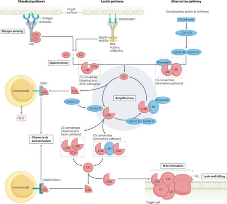
Complement comprises a group of plasma and membrane proteins that provide an effective bridging function for innate and adaptive humoral immunity. Understanding complement pathophysiology is fundamental given that inappropriate complement function in host defence can lead to infectious diseases and inefficient disposal of altered, damaged or senescent cells can lead to or enhance autoimmune neurological processes. Although the rising number of approved drugs targeting complement pathways remains primarily focused on diseases with complement-fixing pathogenic antibodies (such as myasthenia gravis and neuromyelitis optica spectrum disorder), a robust pipeline of emerging treatments holds promise for expanding complement-targeted therapies to a broader spectrum of autoimmune neurological diseases, such as multiple sclerosis and even neurodegenerative diseases such as Alzheimer disease or amyotrophic lateral sclerosis. This Review presents insights into complement biology as it relates to the development or initiation of autoimmune and possibly degenerative diseases affecting the central and peripheral nervous systems or muscle. The effects, merits, risks and challenges of marketed drugs or biologic agents in ongoing phase I–III clinical trials engineered to inhibit proximal or distal components of the complement cascade are also discussed. Anti-complement therapeutics are destined to change the treatment of autoimmune neurologic conditions in which the therapeutic landscape is now becoming crowded with biologic agents targeting other key autoimmunity factors. This Review outlines the main components of the complement cascade associated with initiating or propagating the progression of key autoimmune neurological diseases. The authors also highlight how current and emerging complement-targeted therapeutics are destined to redefine treatment paradigms.
|
|
Scooped by
Gilbert C FAURE
September 21, 10:01 AM
|
Children as young as one who caught mild COVID-19 came out with measurably altered brains, showing a larger left superior parietal cortex, a thickened left occipital cortex, disrupted network clustering, and an enlarged choroid plexus, according to a Chinese study of 19 infected toddlers against 22 healthy peers.
Researchers in Xiamen scanned kids aged 1 to 6 roughly a month after mild infection, fever, cough, and the occasional headache, and found the left superior parietal cortex had gained area, volume, and gyrification while the left lateral occipital cortex had thickened. The brain's structural covariance network showed a significantly lower clustering coefficient and the network hubs shifted: healthy kids carried hub activity in the precuneus, frontal cortex, and insula, regions built for integration and judgment, while the COVID group's hubs sat in the postcentral and precentral cortex, territory built for reflex and touch.
Choroid plexus volume, a marker of cerebrospinal fluid production, ran significantly higher in the COVID group, which the authors read as a signal of neuroinflammation. The authors frame the cortical growth as possible compensation, a young brain flexing plasticity to absorb viral damage, but the hub data undercuts that comfort, because a brain that answers infection by relocating its command centers out of association cortex and into motor strip is being reorganized around the pathogen. The researchers' own compensation language sits on top of a network topology that demotes the higher-order regions built for integration and swaps them for territory built for reflex.
The study is small, cross-sectional, and drawn from one hospital, so it cannot say whether this reshaping resolves, persists, or compounds as these children grow. Mild pediatric COVID, the version this pandemic's messaging waved off as a non-event, left a measurable mark on a one-year-old's brain before that brain had finished building itself.
https://lnkd.in/gaP9E8un
|
|
Scooped by
Gilbert C FAURE
August 27, 7:23 AM
|
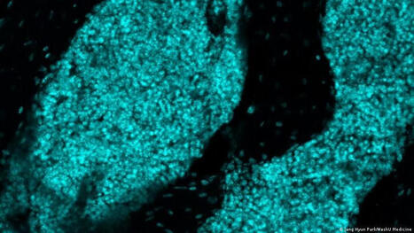
"Immune hubs" that actively defend the brain have been discovered in mouse skull marrow. Similar cells exist in human skulls, but their role remains unclear.
|
|
Scooped by
Gilbert C FAURE
August 1, 2:12 AM
|
New research from Trinity College Dublin is changing how scientists think about Alzheimer’s disease.
Researchers have discovered that microglia, the brain’s immune cells, do more than drive inflammation. They also help keep the brain’s electrical activity stable. When these cells were reduced in an Alzheimer’s model, brain activity became more abnormal and seizure-like episodes increased, suggesting that some experimental treatments could have unintended consequences.
The study, led by Dr Hugh Delaney with senior authors Prof Mark Cunningham (School of Medicine, Trinity College Dublin) and Prof colm cunningham (Trinity School Biochemistry and Immunology), highlights the complexity of Alzheimer’s disease and the importance of understanding the brain’s own protective mechanisms.
The findings suggest that future therapies should target harmful inflammation while preserving the beneficial roles these cells play in maintaining healthy brain function.
Published in the journal Brain, this research is an important step towards developing safer and more effective treatments for the more than 55 million people worldwide living with Alzheimer’s disease.
Congratulations to Hugh, Mark, Colm and all the Trinity researchers involved in this important work. To read the full article, click here: https://lnkd.in/dTAMwtnq
|
|
Scooped by
Gilbert C FAURE
July 30, 3:21 AM
|
Charlie Rice's lab at Rockefeller has discovered that the brain prepares for a potential viral attack in advance, thanks to a signaling network that transmits advanced intelligence about infection from distal regions of the body.
Once set in motion by viral RNA, this interferon-powered network protects against viruses such as #WestNileVirus, as well as other viruses that cause #neuroinflammation—many with infection numbers on the rise.
“We discovered that the blood-brain barrier acts as a sort of central immune signaling hub between the body and the brain,” says first author Tyler Lewy, a former graduate student in Rice’s lab. “The presence of virus-associated molecules in the foot is enough to trigger the network.”
The findings deepen our understanding of the immune system’s intricate surveillance apparatus and could inform new approaches to preventing vector-borne neuroinflammation.
🔗: https://bit.ly/4hdtQ28
|
|
Scooped by
Gilbert C FAURE
June 13, 11:51 AM
|
In a 2025 #ScienceImmunology Review, researchers discussed how interactions between the nervous and immune systems could impact neurological disorders and allergy-related behaviors like food avoidance.
Learn more: https://scim.ag/4jcCmMr #ScienceMagArchives
|
|
Scooped by
Gilbert C FAURE
February 19, 2:05 AM
|
🎓 Série : Extraits du Congrès "NeuroImmunologie"
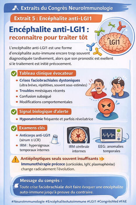
🔎 Extrait 5🧠 Encéphalite anti-LGI1 : reconnaître pour traiter tôt
L’encéphalite anti-LGI1(Leucine-Rich Glioma-Inactivated 1), est une forme d’encéphalite auto-immune encore trop souvent diagnostiquée tardivement, alors que son pronostic est excellent si le traitement est initié précocement.
📌 Ce qu’il faut retenir :
✅ Tableau clinique évocateur
Crises faciobrachiales dystoniques (ultra brèves, répétitives, souvent sous-estimées)
Troubles mnésiques récents
Confusion subaiguë
Modifications comportementales
✅ Signal biologique d’alerte
Hyponatrémie fréquente et parfois révélatrice
✅ Examens clés
Anticorps anti-LGI1 (sérum ± LCR)
IRM : hypersignaux temporaux internes
EEG : anomalies temporales
⚠️ Point important : les antiépileptiques seuls sont généralement insuffisants.
👉 L’immunothérapie précoce (corticoïdes, IgIV, plasmaphérèse) change radicalement l’évolution.
👥 Profil typique : homme > 50 ans, mais le diagnostic doit rester ouvert devant tout tableau compatible.
🎯 Message du congrès :
Toute crise faciobrachiale doit faire évoquer une encéphalite auto-immune jusqu’à preuve du contraire.
La reconnaissance précoce évite les séquelles cognitives et améliore significativement le pronostic.
#NeuroImmunologie #EncéphaliteAutoimmune #LGI1 #Neurologie #Médecine #CongrèsMédical #Epilepsie #Neuroinflammation #Dibcenter
|
|
Scooped by
Gilbert C FAURE
January 30, 6:26 AM
|
New review from our team published in Nature Communications/ Nature Portfolio – “Incorporating meningeal immunity into vaccine development”. We discuss growing evidence that strategies can be developed to deliberately engage meningeal immune compartments, offering new opportunities for immunizations targeting CNS pathogens, optimizing opioid vaccine approaches, and improving our understanding of neuroinflammation.
Open access: https://lnkd.in/ePbrwy8Y
#Neuroimmunology #VaccineDesign #Adjuvant
|
|
Scooped by
Gilbert C FAURE
January 13, 7:49 AM
|
Repeated concussions aren’t just “minor” head injuries - they can trigger lasting #braindamage through the #immunesystem.
From contact sports to military training, repeated head impacts increase the risk of #mooddisorders, #memoryloss, movement problems, and neurodegenerative diseases such as chronic traumatic encephalopathy (CTE), seen in former NFL players including Aaron Hernandez. While these risks are well known, the biological mechanisms have remained unclear.
▪️ A team at the Medical University of South Carolina (MUSC) - led by Stephen Tomlinson, PhD and Silvia Guglietta, PhD - has shown that the immune system, specifically the complement system, plays a central role in driving long-term brain damage after repeated mild concussions. The study, led by first author Khalil Mallah, PhD, with Carsten Krieg, PhD, and colleagues including Devin Hatchell (SCTR Institute), was published in Signal Transduction and Targeted Therapy.
▪️ Using a newly developed murine model that mimics repeated concussions in humans, the researchers demonstrated that excessive complement activation over-stimulates #microglia, the brain’s immune cells. Instead of just clearing damaged tissue, these cells begin “over-pruning” synapses and neurons, disrupting brain connectivity and cognition. Importantly, blocking complement activation with an experimental inhibitor (CR2-Crry) reduced inflammation, preserved neurons and synapses, and improved cognitive outcomes.
💡 Why this matters: there are currently no treatments that target the underlying #inflammation following concussion. Because complement inhibitors are already being explored in other diseases, this work opens a realistic path toward disease-modifying therapies to protect athletes, service members, and others exposed to repeated #headinjury.
🗃️ See comments section for reference.
|
|
Scooped by
Gilbert C FAURE
January 3, 11:30 AM
|
Restless legs syndrome (RLS) and periodic limb movements (PLMs) are increasingly recognized as risk factors for cerebrovascular and cardiovascular diseases, particularly stroke. Conversely, stroke can precipitate or exacerbate the symptoms of RLS and PLMs. This Review explores the shared pathophysiological mechanisms linking RLS and PLMs to cerebrovascular pathology, highlighting a bidirectional relationship. We discuss mechanisms including neurotransmitter dysregulation, autonomic dysfunction, inflammation, oxidative stress, hypoxia and genomic and proteomic factors. Furthermore, we summarize emerging evidence and provide new insights on the potential clinical relevance of RLS in cerebrovascular risk assessment and management. Restless legs syndrome and periodic limb movements are increasingly recognized as risk factors for stroke and other cerebrovascular and cardiovascular diseases and vice versa. This Review explores the shared pathophysiological mechanisms that could underly this bidirectional relationship.
|
|
Scooped by
Gilbert C FAURE
December 18, 2025 12:56 PM
|
Stroke remains a leading cause of disability owing to the irreversible neuronal loss that it causes and the limited regenerative capacity of the CNS. Although reperfusion therapies such as thrombolysis and mechanical thrombectomy can restore blood flow after stroke, their stringent eligibility criteria leave many patients without treatment options. The immune response, involving complex interactions between brain-resident and peripheral immune cells, has a critical role in stroke recovery. Stem cell-based therapies, particularly those involving neural stem cells and mesenchymal stem cells, may be able to reshape the inflammatory microenvironment after stroke, mitigating secondary injury and promoting tissue repair. However, the precise mechanisms driving their effects remain incompletely understood, hindering clinical translation. In this Review, we highlight the bidirectional crosstalk between stem cells and immune cells (including microglia, T cells and peripheral immune cells) and discuss how these interactions influence neuroinflammation, neural plasticity and circuit remodelling in stroke recovery. We examine key determinants of stem cell therapy efficacy, emphasizing the role of stem cell–immune cell interactions, and discuss targeted strategies to enhance immune modulation and neuroprotection. In this Review, Saef Izzy and colleagues examine the therapeutic potential of stem cells in stroke, with a focus on neural and mesenchymal stem cells. They explore how these stem cells interact with brain immune cells to modulate the inflammatory microenvironment, restore blood–brain barrier integrity and promote tissue repair following a stroke.
|