Your new post is loading...

|
Scooped by
Gilbert C FAURE
October 21, 4:09 AM
|
Download PDF Subjects Bioinformatics Innate immunity Skull bone marrow (BM) has emerged as an important immune interface with the central nervous system (CNS). Park and colleagues identify germinal center-like lymphoid structures within skull BM that can generate adaptive immune responses to CNS-derived antigens and contribute to anti-tumor immunity in experimental glioma.The skull bone marrow (BM) is increasingly recognized as an immune interface with the central nervous system (CNS). Vascular channels and diploic veins connect skull BM with the dura mater, providing routes for the exchange of cells and soluble molecules between these compartments.1,2 Cerebrospinal fluid can access skull BM niches, while skull BM supplies myeloid cells and immature B cells to the meninges and, under some conditions, the CNS parenchyma.3,4,5 Organized lymphoid hubs containing germinal center (GC) B cells have also been identified around meningeal venous plexuses, where they support local humoral immune responses.6 More recently, neuron-derived proteins were shown to accumulate at skull-associated borders, where they are sampled by resident immune cells.7 Despite these anatomical and functional connections, whether skull BM can organize antigen-specific adaptive immune responses to CNS-derived antigens has remained unclear. Park and colleagues now identify GC-like lymphoid structures in skull BM and examine their contribution to CNS immunosurveillance.8Comparing BM from different bones, the authors identified an enrichment of IL-21+CXCR5+ follicular helper T (Tfh) cells in skull BM relative to the sternum and femur. These cells expressed Th1-associated markers and formed clusters with B cells in discrete regions of the skull. Skull BM also contained increased numbers of class-switched and GC-like B cells, IgG+ plasmablasts, and antibody-secreting cells. Further characterization revealed organized aggregates containing T cells, B cells, antigen-presenting cells (APCs), CXCL13, B cell activating factor, follicular dendritic cells, and activation-induced cytidine deaminase-expressing B cells. GC-like B cells and antibody-secreting cells underwent clonal expansion and somatic hypermutation and depended on CD4+ T cells, IL-21, and CD40-CD40L signaling. These findings identify a specialized lymphoid environment within skull BM capable of supporting a local humoral immune response.8What drives these responses? Gut microbiota and dietary antigens contributed to the generation of IgA-producing and GC-like B-cell populations but did not fully account for their presence. Park and colleagues therefore asked whether CNS-derived antigens could also reach and activate this compartment. Using neuronal expression of secreted ovalbumin (OVA), they detected antigen in skull BM and observed activation of antigen-specific CD4+ T and B cells.8 Recent work by Chayama and colleagues provides an interesting comparison. Using neuronal protein tracing, they found that brain-derived proteins reach skull-associated niches under physiological conditions.7 However, antigen-sampling skull B cells in that study expressed genes associated with antigen processing and presentation together with a predominantly tolerogenic transcriptional program. Earlier work also identified a CNS antigen-dependent tolerance checkpoint in the adjacent meninges, where developing B cells reactive to the CNS-specific antigen myelin oligodendrocyte glycoprotein were selectively eliminated.9 Access of CNS antigens to skull-associated immune niches, therefore, does not necessarily result in immune activation. The conditions and anatomical site in which antigen is encountered may determine the response that follows.Park and colleagues next examined whether these structures participate in immunity against brain tumors. Following intracranial implantation of CT2A glioma cells, T- and B-cell responses increased within skull BM. Using CT2A tumors expressing HEL-OVA together with antigen-specific lymphocytes, they detected early activation of tumor-reactive T and B cells in skull BM.8 These responses persisted in mice lacking conventional secondary lymphoid organs, although they were diminished and remained dependent on cells recruited from the periphery. The skull BM can therefore support local adaptive immune responses, but it does not appear to function independently of the peripheral immune system.The authors next asked whether these responses influence tumor control. Local blockade of CD40L disrupted skull GC-like structures, reduced intratumoral lymphocyte accumulation and CD8+ T-cell activation, and shortened survival. Conversely, combining a CD40 agonist with IL-21 and IFNγ enhanced skull B-cell responses, increased immune cell infiltration and cytotoxic activity within tumors, and prolonged survival.8 Under these experimental conditions, manipulating skull-associated immune responses can alter immunity within the brain and influence disease outcome (Fig. 1).How well these findings extend to naturally arising brain tumors remains uncertain. Glioblastoma (GBM) is generally considered an immunologically “cold” tumor, with an immune environment dominated by myeloid cells and relatively limited productive T-cell immunity. Evidence for skull-associated anti-tumor immunity has nevertheless been found in patients. Dobersalske and colleagues identified tumor-reactive CD8+ T cells in cranial BM from patients with GBM, including T-cell receptor clonotypes shared between cranial marrow and matched tumors.10 The extent to which these cells contribute to tumor control remains uncertain. Immune checkpoint blockade has also produced disappointing results in large clinical trials, with nivolumab failing to improve overall survival compared with bevacizumab in recurrent GBM.11 These clinical observations do not rule out a contribution from skull-associated immunity, but they show that tumor antigen recognition in GBM rarely translates into an effective anti-tumor response.The glioma models used in the study warrant some consideration. Tumors were established by intracranial transplantation, and some experiments employed cells engineered to express defined model antigens.8 These approaches are useful for resolving antigen-specific responses but may not fully reproduce tumors that arise spontaneously within the CNS. Whether skull-associated responses are similarly engaged by tumors with lower immunogenicity, and how their contribution compares with conventional lymphoid organs and other CNS border tissues, remain unresolved.An important question is how antigen encounter within skull BM is translated into an immune response. CNS-derived proteins reach skull-associated immune niches under physiological conditions,7 while studies of the skull and adjacent meninges have identified mechanisms that may favor B cell tolerance to CNS-derived antigens.7,9 What determines whether CNS antigen encounter promotes tolerance or productive adaptive immunity? Does inflammation or tissue injury alter this balance? Are similar skull-associated responses generated during infection, autoimmunity, or neurodegeneration? It will also be important to determine how much skull BM contributes to CNS immunity relative to the meninges and conventional secondary lymphoid organs, and whether comparable antigen-responsive lymphoid structures function in the human skull.Park and colleagues establish that skull BM contains organized lymphoid structures capable of responding to CNS-derived antigens and influencing immunity in experimental glioma. What remains less clear is when these responses become functionally important. Defining the signals that balance tolerance and immunity within this niche and determining its contribution relative to other CNS and peripheral immune compartments will be important for establishing the role of skull BM in CNS immunosurveillance. ReferencesMonaghan, K. L. et al. Nature 652, 1298–1307 (2026).Article PubMed Google Scholar Zanluqui, N. G. & McGavern, D. B. Trends Immunol. 45, 738–749 (2024).Article PubMed PubMed Central Google Scholar Brioschi, S. et al. Science 373, eabf9277 (2021).Article PubMed PubMed Central Google Scholar Cugurra, A. et al. Science 373, eabf7844 (2021).Article PubMed PubMed Central Google Scholar Mazzitelli, J. A. et al. Nat. Neurosci. 25, 555–560 (2022).Article PubMed PubMed Central Google Scholar Fitzpatrick, Z. et al. Nature 628, 612–619 (2024).Article PubMed PubMed Central Google Scholar Chayama, Y. et al. Cell 189, 4737–4755.e17 (2026).Article PubMed PubMed Central Google Scholar Park, J. H. et al. Nature 658, 196–204 (2026).Article PubMed PubMed Central Google Scholar Wang, Y. et al. Immunity 54, 2784–2794.e6 (2021).Article PubMed Google Scholar Dobersalske, C. et al. Nat. Med. 30, 2947–2956 (2024).Article PubMed PubMed Central Google Scholar Reardon, D. A. et al. JAMA Oncol. 6, 1003–1010 (2020).Article PubMed PubMed Central Google Scholar Download referencesAcknowledgementsThis work was supported by the intramural program at the National Institute of Neurological Disorders & Stroke (NINDS) within the National Institutes of Health (NIH). The contributions of the NIH author(s) are considered Works of the United States Government. The findings and conclusions presented in this paper are those of the author(s) and do not necessarily reflect the views of the NIH or the U.S. Department of Health and Human Services.Author informationAuthors and AffiliationsViral Immunology and Intravital Imaging Section, National Institute of Neurological Disorders and Stroke (NINDS), National Institutes of Health, Bethesda, MD, USANagela G. Zanluqui, Yvonne L. Latour & Dorian B. McGavernAuthorsNagela G. ZanluquiView author publicationsSearch author on:PubMedGoogle ScholarYvonne L. LatourView author publicationsSearch author on:PubMedGoogle ScholarDorian B. McGavernView author publicationsSearch author on:PubMedGoogle ScholarCorresponding authorCorrespondence to Dorian B. McGavern.Additional informationPublisher’s note Springer Nature remains neutral with regard to jurisdictional claims in published maps and institutional affiliations.Rights and permissionsReprints and permissionsAbout this articleCite this articleZanluqui, N.G., Latour, Y.L. & McGavern, D.B. A neighborhood watch within the skull. Cell Res (2026). https://doi.org/10.1038/s41422-026-01310-zDownload citationPublished21 October 2026Version of record21 October 2026DOIhttps://doi.org/10.1038/s41422-026-01310-z
|
|
Scooped by
Gilbert C FAURE
August 16, 3:51 AM
|
🧠 Alzheimer’s disease: should we also consider it a disease of innate immunity?
For decades, Alzheimer’s disease has been viewed primarily through the accumulation of amyloid-β (Aβ) and pathological tau protein. A more recent perspective is broadening this paradigm: innate immunity may be a central driver of the disease rather than merely a consequence of neurodegeneration.
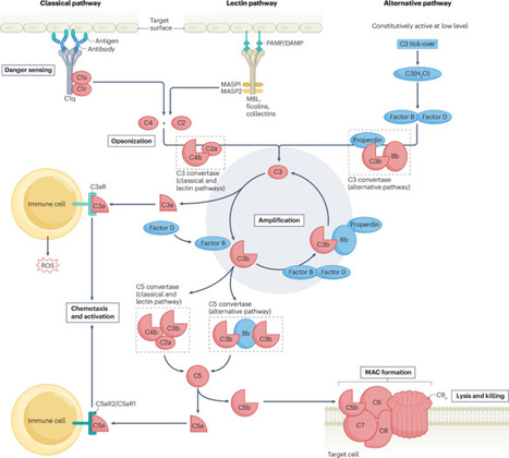
The AD² hypothesis (Alzheimer’s disease as an autoimmune disease) proposes that Aβ may initially participate in a protective immune response. Infections, trauma, metabolic stress and other cellular injuries generate PAMPs and DAMPs, activating the brain’s innate immune machinery.
When this response persists, microglia, initially protective, may shift toward a dysfunctional phenotype. This can lead to chronic production of pro-inflammatory cytokines, inflammasome activation and recruitment of the complement system, particularly C1q and C3.
Complement may subsequently contribute to abnormal synaptic tagging and microglia-mediated synaptic elimination, progressively promoting synaptic dysfunction, neuronal loss and neurodegeneration.
Neuronal injury then releases additional DAMPs, further stimulating innate immunity and creating a self-perpetuating vicious cycle in which an initially protective immune response becomes chronic, dysregulated and potentially damaging to self.
This is the essence of the emerging concept of “innate autoimmunity”: Alzheimer’s disease may not simply be a proteinopathy, but a disorder in which innate immunity, microglia, inflammasomes and complement actively participate in neurodegenerative progression.
⚠️ Importantly, this remains an emerging pathophysiological hypothesis rather than a universally accepted classification of Alzheimer’s disease as an autoimmune disorder.
Rethinking Alzheimer’s disease through the lens of innate immunity may ultimately open new avenues for diagnosis, prevention and immune-targeted therapies.
|
|
Scooped by
Gilbert C FAURE
from Virus World
December 14, 2023 2:28 AM
|
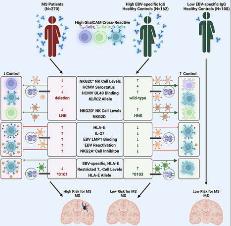
Highlights - Control of autoimmunity by NKG2C+ NK cell responses is severely impaired in MS patients
- MS-patient-derived GlialCAM-specific cells evade control via inhibitory HLA-E/NKG2A axis
- MS patients are predominantly infected with EBV variants that highly upregulate HLA-E
- Specific cytotoxic T cell responses can control EBV-infected GlialCAM-specific B cells
Summary Multiple sclerosis (MS) is a demyelinating disease of the CNS. Epstein-Barr virus (EBV) contributes to the MS pathogenesis because high levels of EBV EBNA386–405-specific antibodies cross react with the CNS-derived GlialCAM370–389. However, it is unclear why only some individuals with such high autoreactive antibody titers develop MS. Here, we show that autoreactive cells are eliminated by distinct immune responses, which are determined by genetic variations of the host, as well as of the infecting EBV and human cytomegalovirus (HCMV). We demonstrate that potent cytotoxic NKG2C+ and NKG2D+ natural killer (NK) cells and distinct EBV-specific T cell responses kill autoreactive GlialCAM370–389-specific cells. Furthermore, immune evasion of these autoreactive cells was induced by EBV-variant-specific upregulation of the immunomodulatory HLA-E. These defined virus and host genetic pre-dispositions are associated with an up to 260-fold increased risk of MS. Our findings thus allow the early identification of patients at risk for MS and suggest additional therapeutic options against MS. Published in Cell (December 12, 2023):
Via Juan Lama
|
|
Scooped by
Gilbert C FAURE
October 17, 2022 11:45 AM
|
|
|
Scooped by
Gilbert C FAURE
October 9, 2021 10:52 AM
|
For many many years we have been fed a diet of MS is an autoimmune disease of T cell reactivity to myelin basic protein.This has been lapped up by researchers and with any dissenting voices their …...
|
|
Scooped by
Gilbert C FAURE
June 8, 2021 3:59 AM
|
Abstract Objective To investigate whether anti-CD20 B-cell-depleting monoclonal antibodies (ɑCD20 mAbs) inhibit the formation or retention of meningeal ectopic lymphoid tissue (mELT) in a murine model of multiple sclerosis (MS). Methods We used a spontaneous chronic experimental autoimmune encephalomyelitis (EAE) model of mice with mutant T-cell and B-cell receptors specific for myelin oligodendrocyte glycoprotein (MOG), which develop meningeal inflammatory infiltrates resembling those described in MS. ɑCD20 mAbs were administered in either a preventive or a treatment regimen. The extent and cellular composition of mELT was assessed by histology and immunohistochemistry. Results ɑCD20 mAb, applied in a paradigm to either prevent or treat EAE, did not alter the disease course in either condition. However, ɑCD20 mAb depleted virtually all B cells from the meningeal compartment but failed to prevent the formation of mELT altogether. Because of the absence of B cells, mELT was less densely populated with immune cells and the cellular composition was changed, with increased neutrophil granulocytes. Conclusions These results demonstrate that, in CNS autoimmune disease, meningeal inflammatory infiltrates may form and persist in the absence of B cells. Together with the finding that ɑCD20 mAb does not ameliorate spontaneous chronic EAE with mELT, our data suggest that mELT may have yet unknown capacities that are independent of B cells and contribute to CNS autoimmunity. Glossary ɑCD20 mAb=anti-CD20 monoclonal antibody; BCR=B-cell receptor; EAE=experimental autoimmune encephalomyelitis; mELT=meningeal ectopic lymphoid tissue; MOG=myelin oligodendrocyte glycoprotein; MPO=myeloperoxidase; MS=multiple sclerosis; RRMS=relapsing-remitting multiple sclerosis; PMS=progressive MS; SLO=secondary lymphoid organ; LFB-PAS=Luxol fast blue and periodic acid-Schiff reaction; TCR=T-cell receptor B-cell-depleting strategies with anti-CD20 monoclonal antibodies (ɑCD20 mAbs), such as rituximab, ocrelizumab, or ofatumumab, are among the most potent therapies currently available in the treatment of relapsing-remitting multiple sclerosis (RRMS). Modest efficacy has also been shown in progressive MS (PMS), specifically active primary PMS, and early, relapse-independent progression in RRMS.1,-,4 Their success highlights the importance of B cells for MS pathogenesis. Among other factors, the therapeutic effect of B-cell depletion may be mediated by interrupting antigen presentation by B cells to T cells in the CNS.5,-,9 Leukocytic infiltrates, forming meningeal ectopic lymphoid tissue (mELT), are found in patients with PMS and to a lesser extent in RRMS. They correlate with an earlier disease onset, a more rapid disease progression, and subpial cortical damage.10,-,15 In general, ectopic lymphoid tissue (ELT) is formed by aggregates of B and T cells, which range from tight-cell clusters to highly organized structures and resemble secondary lymphoid organs (SLOs).16,-,20 In a model of MS, it was demonstrated that B-cell repertoire expansion occurs in mELT, suggesting germinal center activity in the CNS.21 Hereby, mELT may facilitate smoldering autoimmunity in the CNS parenchyma sequestered beyond the blood-brain barrier. Yet, it remains unclear whether and how mELT contributes to early or progressive MS. Because B cells are an integral part of mELT and ɑCD20 mAb that targets B cells not only in the periphery but also in the meninges,22 it seems possible that ɑCD20 mAb inhibits the formation or retention of mELT. Assuming that mELT plays a role in MS pathogenesis, preventing or suppressing mELT may contribute to the efficacy of B-cell depletion in MS. In this study, we investigated the effects of ɑCD20 mAb on mELT in a B-cell-dependent and T-cell-dependent experimental autoimmune encephalomyelitis (EAE) model of MS. Approximately, 50% of 2D2 (TCRMOG) × Th (IgHMOG) mice develop spontaneous chronic EAE and exhibit meningeal clusters of T and B cells in the spinal cord, reminiscent of those found in MS.23,-,25 In this model, we depleted B cells to investigate whether ɑCD20 mAb prevents mELT formation. Surprisingly, our results showed that, despite depleting virtually all B cells from the meninges, mELT still formed or persisted. Materials and Methods Mice 2D2xTh mice, developing spontaneous chronic EAE with mELT, were generated as previously described by crossing myelin oligodendrocyte glycoprotein (MOG)-specific T-cell receptor (TCR) transgenic 2D2 (TCRMOG) mice with MOG-B-cell receptor (BCR) knockin Th mice (IgHMOG).23,24 Mice were bred and maintained in-house at the Center of Preclinical Research (CPR) at Klinikum rechts der Isar (Technical University of Munich) under specific pathogen-free conditions, according to FELASA recommendations with free access to water and standard rodent chow. EAE and Clinical Assessment 2D2xTh mice develop spontaneous chronic EAE with an incidence of approximately 30–50% in our facility. Clinical scores of mice were evaluated on a daily basis and assessed as follows: 0 = no clinical signs, 1 = tail weakness, 1.5 = additional impaired tail erection, 2 = ataxia and hind limb weakness, 2.5 = hind limb paresis, 3 = high-grade hind limb paresis, 3.5 = additional minor weakness of the fore limbs, 4 = additional fore limb paresis, 4.5 = tetraplegia, and 5 = moribund. ɑCD20 mAb Treatment 100 µg of antimouse CD20 mAb (clone 18B12, provided by F. Hoffmann-La Roche, Switzerland) in a murine IgG2a Fc-format or antihen egg lysozyme isotype control (Crown bio, Cat: C0006) in 100 µL PBS was injected intraperitoneally (i.p.) at day 0, 7, 14, and 21 for a total of 4 doses. Mice were grouped for prevention or treatment of EAE. In an attempt to prevent EAE, mice received the first injection at the age of 26 ± 3 days (n = 18 in both groups). For the evaluation of the therapeutic potential, mice were treated when reaching a clinical score of ≥3 (n = 11 ɑCD20 mAb-treated, n = 14 isotype-treated). Mice were killed 28 days after the first injection. Reference mice at the onset of disease (figure 3C) were dissected immediately after reaching an EAE score of ≥3 without treatment. Cell Isolation and Flow Cytometric Analysis Blood was freshly obtained by terminal cardiac puncture and mixed with 500 µL 2 mM EDTA. Cervical lymph nodes were carefully dissected and passed through a 30-µm cell strainer. Samples were washed with RPMI medium, and erythrocytes were lysed using BD Pharm Lyse Buffer (BD Biosciences). Cells were washed again, and the number of living cells was determined using a hemocytometer (Cellometer Auto 2000, Nexcelom Bioscience). Single-cell suspensions were stained with antibodies in PBS containing 2% fetal calf serum for 20 minutes at 4°C in the dark. Fc-receptors were blocked to prevent nonspecific antibody binding. Dead cells were excluded using the LIVE/DEAD Fixable Aqua Dead Cell Stain Kit (Invitrogen). Cells were washed twice and acquired on a CytoFLEX flow cytometer (Beckman Coulter). The following antimouse antibodies were used: CD16/CD32 (2.4G2), CD45R/B220 (RA3-6B2, FITC), and CD3e (145-2C11, PE) (BD Bioscience). Data were analyzed using FlowJo version 10 (TreeStar). Histology, Immunohistochemistry, and Imaging Spinal cord, spleen, and inguinal lymph nodes were perfused before dissection, fixed with 4% paraformaldehyde, and paraffin embedded. The spinal cord was additionally decalcified with Osteosoft (Sigma-Aldrich) 72 hours before embedding. To determine the degree of meningeal infiltrates, the spinal cord was sectioned horizontally into 13–16 tissue blocks (thickness: 2 × 5 mm cervical, subsequently 2.5 mm) per mouse and stained with hematoxylin and eosin according to standard protocols. The stained tissues were digitized (Leica AT2 scanner) and examined using Image Scope (Pathology Slide Viewing Software, version 12.4.3, Leica Biosystems). Infiltrates with a size of ≥1 × 104 µm2 per section were considered as mELT. The cell density was analyzed using a nuclear algorithm (Nuclear v9, Image Scope; Leica). To characterize the cellular composition of mELT, immunohistochemistry was performed on representative sections with antimouse CD45R/B220 (BD, 550286, 1:50), CD3 (DCS, CI597R0, 1:50), and myeloperoxidase (MPO; Thermo Fisher, RB-373-A, 1:50). Mouse spleens served as positive controls. Numbers of B220+ and CD3+ positive cells were determined using a membrane algorithm (Membrane v9, Image Scope; Leica). In the CNS, MPO+ cells were counted manually. In the periphery, MPO+ cells were quantified by applying a nuclear algorithm (Membrane v9, Image Scope; Leica). Two cross-sections of the cervicothoracic, the thoracolumbar, and the lower lumbar part were stained with Luxol fast blue and periodic acid-Schiff reaction (LFB-PAS) according to standard protocols. LFB-PAS staining was analyzed semiquantitatively by dividing spinal cord sections into quadrants and counting quadrants with demyelination. All histologic evaluations were performed in a blinded manner. Detection of Anti-MOG Antibodies Antirecombinant mouse MOG (1–125) (Eurogentec, Cat: AS-55150-100) IgG was measured with a noncommercial ELISA in duplicates as described,26 using MOG at a final concentration of 10 µg/mL and horseradish peroxidase-labeled goat antimouse IgG (Thermo Fisher Scientific). The optical density was measured at a wavelength of 450 nm with 540 nm as a reference wavelength using the ELISA reader (infinite M200 PRO, TECAN). Statistical Analysis Data are shown as mean ± SEM or 95% CI. Statistical significance between groups was analyzed using the unpaired Student t test. For correlation analysis, simple linear regression was calculated for each group. A value of p ≤ 0.05 was considered significant with *p < 0.05, **p < 0.01, ***p < 0.001, and ****p < 0.0001. Standard Protocol Approvals, Registrations, and Patient Consents All animal experiments were approved by the competent authority, Regierung von Oberbayern, Munich, Germany (ROB-55.2-2532.Vet_02-16-100). Data Availability Data not published within this article will be made available by request from any qualified investigator. Results ɑCD20 mAb Efficiently Depletes B Cells in the Periphery but Does Not Affect the Clinical Course of Spontaneous Chronic EAE To analyze the effects of ɑCD20 mAb on mELT in the context of EAE, we crossed MOG Ig-VH knock-in (IgHMOG, Th) mice, in which B cells express a MOG-specific BCR heavy chain, with MOG-specific TCR transgenic mice (TCRMOG, 2D2). As previously described, 2D2xTh mice develop spontaneous chronic EAE with B-cell-rich and T-cell-rich mELT along the spinal cord.23,24 2D2xTh mice were treated weekly for a total of 4 intraperitoneal (i.p.) doses with antimurine CD20 or isotype control mAb. Two treatment paradigms were pursued in parallel: (1) to prevent EAE and mELT formation, mice received the first dose as soon as they were weaned about 26 days after birth (figure 1A) and (2) to treat fully established EAE, ɑCD20 mAb was first administered after mice developed an EAE score of ≥3 (figure 3A). ɑCD20 mAb efficiently depleted B cells in the peripheral blood (figure 1B.a, upper panel) and lymph nodes (figure 1B.b, lower panel). However, its application neither attenuated the rather severe and chronic course of EAE compared with the control groups in either condition (figures 1C and 3B) nor changed the incidence of EAE. In total, 44% of preventatively treated mice developed spontaneous chronic EAE, which was similar to 39% of control treated mice (figure 1D). Figure 1 Spontaneous EAE Occurs in the Absence of Peripheral B Cells in Mice Preventatively Treated With ɑCD20 mAb (A) Experimental setup. Mice were treated with a weekly dose of 100 μg of ɑCD20 mAb (n = 18) or isotype control (n = 18) after weaning. (B) Flow cytometric analysis of B220+ and CD3+ cells in blood (B.a) and lymph nodes (B.b) after 4 weeks of treatment. The representative flow cytometry contour plot (left) and quantification (right). (C) Daily evaluated EAE scores over the experimental period. Data are shown as mean ± SEM. (D) Incidence of mice developing clinical signs of disease. When not stated differently, data shown as mean ±95% CI. *p < 0.05; ****p < 0.0001; statistical significance between groups was analyzed using the Student t test. ɑCD20 mAb = anti-CD20 monoclonal antibody; EAE = experimental autoimmune encephalomyelitis; i.p. = intraperitoneally; n/N = mice with EAE/all mice observed. mELT Formation and Retention Occurs in the Absence of B Cells in Preventatively Treated Mice To investigate whether mELT forms and persists under B-cell-depleting conditions, we analyzed cross-sections along the spinal cord of 2D2xTh mice, which developed clinical signs of EAE during the observation/treatment period. With a mean EAE duration of 19 days (range 13–24 days) in the ɑCD20 mAb-treated group and 16 days (range 3–21 days) in the isotype-treated group, all mice showed mELT in at least 5 of 12–16 segments with a maximum along the thoracolumbar part of the spinal cord (figure 2A). Infiltrates were considered as mELT when their size was ≥1 × 104 µm2 per section. Surprisingly, the mean area of mELT per section, including sections in which no mELT was detected, was similar in both groups with a mean of 81,152 µm2 per section in ɑCD20 mAb and 103,968 µm2 in isotype-treated controls (figure 2B, upper panel). Hence, despite the absence of B cells in circulation and cervical lymph nodes, structures of mELT formed. With B cells being a main component of mELT,21,23,24 we performed B220 immunohistochemistry on sections of the thoracolumbar part of the spinal cord. We found that, although mELT was present to a similar extend, mELT of ɑCD20 mAb-treated mice only exhibited 0.59% of B220+ B cells observed in controls (figure 2B, lower panel). Figure 2 mELT Formation Occurs in the Absence of B Cells in Mice Preventatively Treated With ɑCD20 mAb After weaning, 2D2xTh mice received a weekly dose of 100 µg ɑCD20 mAb (n = 18) or isotype control mAb (n = 18) and were observed for 28 days (A + B) 8 of 18 aCD20 mAb-treated mice and 7 of 18 isotype mAb-treated mice developed EAE. One aCD20 mAb-treated mouse died prematurely and had to be excluded from the histologic analysis. (A) Quantitative distribution of mELT along the spinal cord with 12–16 cross-sections per animal analyzed. (B) Representative HE-stained (upper left panel) and B220-stained (lower left panel) sections of the thoracolumbar part of the spinal cord and corresponding close-up views of mELT. Quantification of the mean area of mELT including all sections (upper right panel) and the number of B220+ cells in mELT, analyzed in 2 randomly selected sections per animal, for all mice (lower right panel). (C) Analysis of histologic signs of disease in mice without clinically manifest EAE (11 of 18 isotype mAb-treated mice and 10 of 18 aCD20 mAb-treated mice). Representative HE sections given in (a) and (b) for mice with no pathologies, in (c) and (d) for mice with minor cellular infiltrates in the meninges, and in (e) and (f) for mice with mELT. (D) Quantitative analysis of the size and distribution of mELT along the spine in mice without clinically manifest EAE. In total, 12–16 sections analyzed per animal. Scale bars: 250 µm (overviews) and 100 µm (close-ups). When not stated differently, data shown as mean ±95% CI. *p < 0.05; ****p < 0.0001; statistical significance between groups was analyzed using the Student t test. ɑCD20 mAb = anti-CD20 monoclonal antibody; EAE = experimental autoimmune encephalomyelitis; HE = hematoxylin & eosin; mELT = meningeal ectopic lymphoid tissue; n/N = mice showing mELT or noted feature/total number of mice analyzed; ns = not significant. Interestingly, in both groups, mice without clinically apparent EAE over the treatment period of 28 days also showed histologic signs of disease in approximately 60% of cases, ranging from scattered cellular infiltrates in the meninges in some cases to mELT in 30% of the ɑCD20 mAb-treated mice and 36% of the isotype controls (figure 2C). In these clinically unremarkable animals, mELT was also mainly found in the thoracolumbar part of the spinal cord and was comparable in size in both groups (figure 2D). Our data indicate that the absence of B cells in preventatively treated 2D2xTh mice is not sufficient to prevent mELT formation and that not only mELT formation but also its retention may be independent of B cells. Moreover, mELT formation in our model occurs before developing clinical signs of EAE. ɑCD20 mAb Treatment Does Not Influence the Size of mELT In a second experiment, more closely resembling a clinical scenario, mice were randomized into groups treated with ɑCD20 or control mAbs after disease onset, i.e., when having developed an EAE score of ≥3 (aged on average 47.6 days in the ɑCD20 mAb and 48.4 days in the isotype-treated group). As a baseline, we used mice which were euthanized without further treatment immediately after having developed a score of ≥3 (figure 3A). At that stage of disease (onset), all mice already exhibited extensive mELT with a distinct fraction of B220+ B cells (figure 3C), confirming that mELT was sufficiently present before we started our intervention. After 4 weeks of treatment, distribution of mELT throughout the spinal cord was almost identical in-between groups, with all mice exhibiting mELT in at least 1 of 14–16 cross-sections analyzed (figure 3D). Again, we did not observe a difference in the mean area of mELT in both groups (figure 3E). Similar to what was observed in autopsy cases of MS, the extent of mELT correlated with disease severity in control mice. However, this was not statistically significant in ɑCD20 mAb-treated animals (figure 3F). Figure 3 ɑCD20 mAb Applied After EAE Onset Does Not Change the Clinical Course of Spontaneous EAE or the Size of mELT (A) Experimental setup. As soon as 2D2xTh mice developed a score of ≥3, they received a weekly dose of 100 µg of ɑCD20 (n = 11) or isotype control mAb (n = 14) over a period of 28 days. Another 5 mice were euthanized and dissected directly after developing a clinical score of ≥3, without receiving treatment. They served as histologic reference at disease onset for the remainder of the mice for which treatment commenced at that stage. (B) Clinical course of EAE during application of ɑCD20 or isotype mAb. Data expressed as mean ± SEM. (C) Representative HE (upper left panel) and B220 (lower left panel) staining of the spinal cord mELT of mice at EAE onset and respective quantification in all mice (right panels). (D) Representative HE-stained cross-sections of the thoracolumbar part of the spinal cord of ɑCD20 mAb-treated and isotype-treated mice (left) and representation of the distribution of mELT along the spine with 14–16 sections analyzed per animal (right). (E) Quantification of the mean area of mELT per section. (F) Correlation of the mean area of mELT per section with the mean EAE score over the observation period. Simple linear regression was calculated for each group. Scale bars, 250 µm. When not indicated differently, data shown as mean ± 95% CI. Statistical significance between groups was analyzed using the Student t test. ɑCD20 mAb = anti-CD20 monoclonal antibody; EAE = experimental autoimmune encephalomyelitis; HE = hematoxylin & eosin; i.p. = intraperitoneally; mELT = meningeal ectopic lymphoid tissue; n/N = mice showing mELT/total number of mice analyzed; ns = not significant. Persisting High Titers of Anti-MOG Antibodies and Extensive Demyelination in B-Cell-Depleted Mice Reflects the Lasting Severe and Chronic Course of EAE Although B cells were effectively reduced not only in the periphery but also in the meningeal compartment, MOG-specific total IgG titers remained high even after 4 weeks of B-cell depletion (figure 4A). Correlating with the clinical disease course, the spinal cord was similarly affected by demyelination in ɑCD20 mAb-treated mice compared with controls (figure 4B). Together with persisting B-cell-free mELT, these observations may contribute to the lack of a clinical benefit of B-cell depletion in our model. Figure 4 B-Cell-Depleted Spontaneous Chronic EAE Mice Exhibit High Titers of Anti-MOG Antibodies and Extensive Demyelination, Reflecting Their Severe Disease Course 2D2xTh mice received 4 doses of ɑCD20 mAb (n = 11) or isotype control (n = 10) as soon as they reached an EAE score of ≥3. (A) Antimurine MOG IgG serum levels determined by ELISA (dilution 1:300). Data are shown as individual data points and mean ± 95% CI. (B) LFB-PAS staining of the spinal cord, showing representative thoracolumbar cross-sections and corresponding close-ups. For each mouse, 2 randomly chosen cross-sections per spinal cord segment—cervicothoracic (n = 5 for ɑCD20 mAb and n = 6 for isotype), thoracolumbar (n = 8 per group), and lumbar (n = 7 for ɑCD20 mAb and n = 6 for isotype)—were examined. Data are presented as minimum and maximum values and mean. Scale bars: 250 µm and 100 µm (close-ups). Statistical significance between groups was analyzed using the Student t test. ɑCD20 mAb = anti-CD20 monoclonal antibody; EAE = experimental autoimmune encephalomyelitis; LFB-PAS = Luxol fast blue and periodic acid-Schiff reaction; MOG = myelin oligodendrocyte glycoprotein; ns = not significant; OD = optical density. ɑCD20 mAb Treatment Changes the Density and Composition of the Cellular Content of mELT When we analyzed the cellular composition of mELT more closely, we found that, within the treatment paradigm, mELT in ɑCD20 mAb-treated mice was populated with 24.2% less cells (figure 5A). The count of B220+ B cells, which was the largest immune cell fraction in control mice, was drastically reduced on ɑCD20 mAb treatment, suggesting that the absence of B cells led to this reduced cellular density in mELT (figure 5B, upper panel). We did not notice a relevant change in CD3+ cells (figure 5B, middle panel), whereas the number of MPO positive neutrophil granulocytes was significantly increased under ɑCD20 mAb therapy (figure 5B, lower panel). Comparing these changes in mELT with SLOs, we observed that in the spleen and inguinal lymph nodes, B220+ B cells were also largely eliminated. MPO+ cells seemed to be increased, but this did not reach statistical significance. In contrast to mELT, however, the amount of CD3+ T cells was slightly increased in B-cell-depleted lymph nodes (figure 6). Figure 5 ɑCD20 mAb B-Cell Depletion in Spontaneous Chronic EAE Alters Cellular Density and Composition of mELT 2D2xTh mice received 4 doses of ɑCD20 mAb (n = 11) or isotype control (n = 10) as soon as they reached an EAE score of ≥3. (A) HE staining of representative spinal cord sections of (a) isotype-treated and (b) ɑCD20 mAb-treated mice visualizing the cellular density of mELT (c) and (d) show representative samples of the computer-aided quantification of nuclei in mELT with the corresponding graphs. (B) Quantification of B cells (B220), T cells (CD3), and neutrophil granulocytes (MPO) in spinal cord mELT. mELT of 1–4 randomly chosen cross-sections per mouse were analyzed. Scale bars: 50 µm (A) and 100 µm (B). Data are presented as individual data points and mean ± 95% CI. *p ≤ 0.05; **p < 0.01; ****p < 0.0001; statistical significance between groups was analyzed using the Student t test. ɑCD20 mAb = anti-CD20 monoclonal antibody; EAE = experimental autoimmune encephalomyelitis; HE = hematoxylin & eosin; mELT = meningeal ectopic lymphoid tissue; MPO = myeloperoxidase; ns = not significant. Figure 6 ɑCD20 mAb Efficiently Depletes B Cells in SLOs 2D2xTh mice received 4 doses of ɑCD20 mAb (n = 11) or isotype control (n = 10) as soon as they reached an EAE score of ≥3. Quantification of B cells (B220), T cells (CD3), and neutrophil granulocytes (MPO) in spleens and inguinal lymph nodes. One randomly chosen cross-section per organ (n = 5 mice per group) was evaluated. Scale bars: 250 µm. Data are presented as individual data points and mean ±95% CI. *p ≤ 0.05; ****p < 0.0001; statistical significance between groups was analyzed using the Student t test. ɑCD20 mAb = anti-CD20 monoclonal antibody; EAE = experimental autoimmune encephalomyelitis; LN = lymph node; MPO = myeloperoxidase; ns = not significant; SLO = secondary lymphoid organ; SPL = spleen. Together, our findings show that ɑCD20 mAb, to some degree, depletes B cells in mELT in mice with spontaneous chronic EAE, yet mELT persists. The role and function of mELT devoid of B cells remains unclear. Discussion Although its exact role in MS pathogenesis remains unclear, meningeal inflammation has gained widespread attention in recent years. Because B cells are a major component of mELT and B-cell-depleting mAbs are among the most successful MS therapeutics today, the question how B-cell-depleting therapies affect mELT is obvious. In this study, we systematically addressed this question in a murine model of MS. Not unexpectedly, B-cell depletion did not change the disease course in our spontaneous chronic EAE model. It has previously been reported that ɑCD20 mAb did not significantly change the course of EAE in the same model because high titers of MOG-binding antibodies in conjunction with MOGTCR 2D2 T cells could support clinical disease in the absence of B cells.26 Importantly, ɑCD20 mAb does not target antibody-producing plasma cells. It could be debated that spontaneous chronic EAE in 2D2xTh mice, because of the MOG specificity of both T and B cells, is not dynamic enough to allow being modified by pharmaceutical interventions. However, laquinimod ameliorated spontaneous chronic EAE and reduced meningeal B-cell aggregates and thereby demonstrated its plasticity.27 Thus, other factors are likely to contribute. Although clinical effects were neglectable, we found that ɑCD20 mAb depleted virtually all B cells not only from SLOs and blood but also from mELT. It has been shown before that B cells infiltrating the meninges in EAE could be efficiently depleted by ɑCD20 mAb administered either intraperitoneally or intrathecally.22 This study used a model, however, which does not feature large meningeal B-cell aggregates such as mELT. A case report of a patient with gastrointestinal mantle cell lymphoma, who developed progressive multifocal leukoencephalopathy after rituximab therapy, showed that 8 months after the last dose, no B cells were found in perivascular spaces.28 By contrast, a case report of a young man with aggressive MS treated with rituximab and with no detectable peripheral B cells found some B cells in the parenchyma and in perivascular cuffs on CNS autopsy.29 Regarding B-cell-depleting antibodies used to treat MS, it is an encouraging finding that ɑCD20 mAb could deplete B cells present in mELT and prospectively prevent colonization of the meningeal compartment by B cells. Yet, open questions remain. The most unexpected finding in our study was that mELT formed or remained in the absence of B cells when mice were B-cell-depleted after EAE was fully established and even when treated before EAE onset. The size and extent of mELT was even comparable whether they contained B cells or not. This shows that, in our model, B cells are not essential for the induction or maintenance of mELT development. Yet, in the absence of B cells, mELT was less densely packed with cells and the cellular composition was altered. ɑCD20 mAb-treated mice had drastically fewer B220+ B cells, comparable amounts of CD3+ T cells, and significantly more MPO+ neutrophil granulocytes in mELT. A recent study showed that neutrophil granulocytes pioneer mELT formation and precede B cells in the meningeal compartment.30 In addition, several other cell types have been proposed as candidates to induce ELT formation, including IL-22 or IL-17 secreting immune cells, dendritic cells, and CD3−CD4+CD45+ lymphoid tissue inducer cells.16,31,-,35 Our own data showed that shortly after EAE onset, B220+ B cells were already present in mELT, but after 4 weeks of isotype control treatment, their amount had roughly doubled. Thus, B cells seem to fully populate mELT at later stages. This raises the question, which role B cells play in (later stage) mELT and whether mELT without B cells still has a pathogenic role. Generally, our mice would be expected to benefit from B-cell depletion, like it has been shown in another B-cell-dependent model without mELT or in MS itself.1,-,3,36 If mELT lacking B cells still contribute to autoimmunity, this could explain why the EAE disease course is unchanged in ɑCD20 mAb-treated 2D2xTh mice. As discussed above, other inherent properties of the model could contribute, such as the presence of high titers of MOG-binding antibodies and MOGTCR T cells with a potentially altered activation status under ɑCD20 mAb therapy. However, the notion that mELT without B cells persist in EAE and its persistence correlates with sustained severe disease, may give rise to interesting considerations: B-cell-depleting therapies are disproportionately more effective in RRMS than PMS. It could be speculated that future therapeutic approaches targeting mELT directly, instead of B cells, may be more effective in treating PMS. We observed that approximately one-third of 2D2xTh mice without clinically manifest EAE develop a minor degree of mELT both in the presence or absence of B cells. What does this imply regarding the role of mELT? Naturally, it remains unknown whether for these mice disease was imminent and the occurrence of mELT only preceding clinical signs. Alternatively, it is possible that 2D2xTh mice live with mELT without developing a phenotype. The latter would suggest that mELT may be a rather irrelevant epiphenomenon of encephalitogenic B and T cells being present in this model or that mELT may even have a regulatory role. One limitation of our study is that we depleted B cells relatively soon, i.e., before the onset of EAE or at an early stage of disease, where B cells are already present in mELT but to a lesser extent. We believe that it is reasonable to conclude from our results that B cells already present in mELT were depleted by ɑCD20 mAb. However, we could not formally assess the respective contributions of preventing B-cell influx from the periphery or direct depletion in the CNS itself to our finding that mELT was devoid of B cells after several weeks of treatment with ɑCD20 mAb. It is very challenging to study meningeal pathologies in patients in vivo or even postmortem. Thus, animal models are instrumental, and yet, the transferability of any findings in such models to MS must be questioned continuously. Our model of spontaneous chronic EAE has several characteristics that make it a suitable tool. EAE develops without an external stimulus, such as immunization or an adjuvant, and mice form meningeal inflammatory aggregates. Furthermore, mELT is readily available in high quality and quantity. However, it is a highly artificial model which is genetically determined through B-cell-mediated and T-cell-mediated autoimmunity against MOG. MS, by contrast, is not known to have a single autoantigen and certainly does not feature such a highly polarized adaptive immune system. Therefore, studies in human subjects treated with ɑCD20 mAb are required to confirm our results. In conclusion, we provide evidence that systemically applied ɑCD20 mAbs efficiently deplete B cells in meningeal inflammatory aggregates, and yet, mELT still form and persist with an altered cellular composition. The unchanged severe course of EAE, despite near complete B-cell depletion, both in the periphery and the meningeal compartment, may point to a yet unknown function of mELT, which is independent of B cells. Future studies are required, which aim to better understand the role and pathogenic relevance of mELT with and without B cells. Study Funding This work was supported by the Deutsche Forschungsgemeinschaft (DFG, German Research Foundation) SFB TRR 130 TP17 to H. Radbruch. K. Lehmann-Horn received research support from the Deutsche Forschungsgemeinschaft (SFB-TR-128 and LE 3079/3-1), the Hertie Foundation (MyLab program), and the US National Multiple Sclerosis Society (NMSS) (G-1508-07064). Disclosure R.M. Brand, V. Friedrich, J. Diddens, M. Pfaller, F.R. de Franchis, H. Radbruch, and K. Steiger report no disclosures. B. Hemmer has served on scientific advisory boards for Novartis; he has served as DMSC member for AllergyCare, Polpharma, and TG therapeutics; he or his institution has received speaker honoraria from Desitin; his institution received research grants from Regeneron for MS research. He holds part of two patents: one for the detection of antibodies against KIR4.1 in a subpopulation of patients with MS and the other for genetic determinants of neutralizing antibodies to interferon. All conflicts are not relevant to the topic of this study. K. Lehmann-Horn has received research support (to TUM) from Novartis, honoraria, and compensation for travel expenses from Novartis, F. Hoffmann-La Roche, and Merck Serono. Go to Neurology.org/NN for full disclosures. Acknowledgment The authors would like to thank Melina Pekic Hajdarbasic, Beatrix Lunk, Olga Seelbach, Marion Mielke, Ulrike Mühlthaler (all TUM), and Vera Wolf and Sandra Meier (both Charité) for excellent technical assistance. The authors acknowledge support from F. Hoffmann-La Roche for providing the antimouse CD20 antibody 18B12. R.M. Brand is a fellow of the Hertie Foundation (medMS Doctoral Program). Appendix Authors Footnotes Go to Neurology.org/NN for full disclosures. Funding information is provided at the end of the article. The Article Processing Charge was funded by the authors. Editorial, page e1000 Received January 25, 2021. Accepted in final form March 8, 2021. Copyright © 2021 The Author(s). Published by Wolters Kluwer Health, Inc. on behalf of the American Academy of Neurology. This is an open access article distributed under the terms of the Creative Commons Attribution-NonCommercial-NoDerivatives License 4.0 (CC BY-NC-ND), which permits downloading and sharing the work provided it is properly cited. The work cannot be changed in any way or used commercially without permission from the journal. References 1.↵Hauser SL, Waubant E, Arnold DL, et al. B-cell depletion with rituximab in relapsing-remitting multiple sclerosis. N Engl J Med. 2008;358(7):676-688.OpenUrlCrossRefPubMed 2.↵Hauser SL, Bar-Or A, Comi G, et al. Ocrelizumab versus interferon beta-1a in relapsing multiple sclerosis. N Engl J Med. 2017;376(3):221-234.OpenUrlCrossRefPubMed 3.↵Montalban X, Hauser SL, Kappos L, et al. Ocrelizumab versus placebo in primary progressive multiple sclerosis. N Engl J Med. 2017;376(3):209-220.OpenUrlCrossRefPubMed 4.↵Ancau M, Berthele A, Hemmer B. CD20 monoclonal antibodies for the treatment of multiple sclerosis: up-to-date. Expert Opin Biol Ther. 2019;19(8):829-843.OpenUrl 5.↵Molnarfi N, Schulze-Topphoff U, Weber MS, et al. MHC class II-dependent B cell APC function is required for induction of CNS autoimmunity independent of myelin-specific antibodies. J Exp Med. 2013;210(13):2921-2937. 6.↵Sabatino JJ Jr., Probstel AK, Zamvil SS. B cells in autoimmune and neurodegenerative central nervous system diseases. Nat Rev Neurosci. 2019;20(12):728-745.OpenUrl 7.↵von Budingen HC, Palanichamy A, Lehmann-Horn K, Michel BA, Zamvil SS. Update on the autoimmune pathology of multiple sclerosis: B-cells as disease-drivers and therapeutic targets. Eur Neurol. 2015;73(3-4):238-246.OpenUrlCrossRefPubMed 8.↵Weber MS, Hemmer B. Cooperation of B cells and T cells in the pathogenesis of multiple sclerosis. Results Probl Cell Differ. 2010;51:115-126.OpenUrlPubMed 9.↵Zamvil SS, Hauser SL. Antigen presentation by B cells in multiple sclerosis. N Engl J Med. 2021;384(4):378-381.OpenUrl 10.↵Serafini B, Rosicarelli B, Magliozzi R, Stigliano E, Aloisi F. Detection of ectopic B-cell follicles with germinal centers in the meninges of patients with secondary progressive multiple sclerosis. Brain Pathol. 2004;14(2):164-174.OpenUrlCrossRefPubMed 11.↵Magliozzi R, Howell O, Vora A, et al. Meningeal B-cell follicles in secondary progressive multiple sclerosis associate with early onset of disease and severe cortical pathology. Brain. 2007;130(pt 4):1089-1104.OpenUrlCrossRefPubMed 12.↵Howell OW, Reeves CA, Nicholas R, et al. Meningeal inflammation is widespread and linked to cortical pathology in multiple sclerosis. Brain. 2011;134(pt 9):2755-2771.OpenUrlCrossRefPubMed 13.↵Lucchinetti CF, Popescu BF, Bunyan RF, et al. Inflammatory cortical demyelination in early multiple sclerosis. N Engl J Med. 2011;365:2188-2197.OpenUrlCrossRefPubMed 14.↵Choi SR, Howell OW, Carassiti D, et al. Meningeal inflammation plays a role in the pathology of primary progressive multiple sclerosis. Brain. 2012;135(pt 10):2925-2937.OpenUrlCrossRefPubMed 15.↵Reali C, Magliozzi R, Roncaroli F, Nicholas R, Howell OW, Reynolds R. B cell rich meningeal inflammation associates with increased spinal cord pathology in multiple sclerosis. Brain Pathol. 2020;30(4):779-793.OpenUrl 16.↵Pitzalis C, Jones GW, Bombardieri M, Jones SA. Ectopic lymphoid-like structures in infection, cancer and autoimmunity. Nat Rev Immunol. 2014;14:447-462.OpenUrlCrossRefPubMed 17.↵Corsiero E, Nerviani A, Bombardieri M, Pitzalis C. Ectopic lymphoid structures: powerhouse of autoimmunity. Front Immunol. 2016;7:430.OpenUrlCrossRef 18.↵Pikor NB, Prat A, Bar-Or A, Gommerman JL. Meningeal tertiary lymphoid tissues and multiple sclerosis: a gathering place for diverse types of immune cells during CNS autoimmunity. Front Immunol. 2015;6:657.OpenUrlCrossRef 19.↵Jones GW, Jones SA. Ectopic lymphoid follicles: inducible centres for generating antigen-specific immune responses within tissues. Immunology. 2016;147(2):141-151.OpenUrlCrossRefPubMed 20.↵Bell L, Lenhart A, Rosenwald A, Monoranu CM, Berberich-Siebelt F. Lymphoid aggregates in the CNS of progressive multiple sclerosis patients lack regulatory T cells. Front Immunol. 2019;10:3090.OpenUrl 21.↵Lehmann-Horn K, Wang SZ, Sagan SA, Zamvil SS, von Budingen HC. B cell repertoire expansion occurs in meningeal ectopic lymphoid tissue. JCI Insight. 2016;1(20):e87234.OpenUrl 22.↵Lehmann-Horn K, Kinzel S, Feldmann L, et al. Intrathecal anti-CD20 efficiently depletes meningeal B cells in CNS autoimmunity. Ann Clin Transl Neurol. 2014;1(7):490-496.OpenUrl 23.↵Krishnamoorthy G, Lassmann H, Wekerle H, Holz A. Spontaneous opticospinal encephalomyelitis in a double-transgenic mouse model of autoimmune T cell/B cell cooperation. J Clin Invest. 2006;116(9):2385-2392.OpenUrlCrossRefPubMed 24.↵Bettelli E, Baeten D, Jager A, Sobel RA, Kuchroo VK. Myelin oligodendrocyte glycoprotein-specific T and B cells cooperate to induce a Devic-like disease in mice. J Clin Invest. 2006;116(9):2393-2402.OpenUrlCrossRefPubMed 25.↵Dang AK, Tesfagiorgis Y, Jain RW, Craig HC, Kerfoot SM. Meningeal infiltration of the spinal cord by non-classically activated B cells is associated with chronic disease course in a spontaneous B cell-dependent model of CNS autoimmune disease. Front Immunol. 2015;6:470.OpenUrl 26.↵Kinzel S, Lehmann-Horn K, Torke S, et al. Myelin-reactive antibodies initiate T cell-mediated CNS autoimmune disease by opsonization of endogenous antigen. Acta Neuropathol. 2016;132(1):43-58.OpenUrlCrossRefPubMed 27.↵Varrin-Doyer M, Pekarek KL, Spencer CM, et al. Treatment of spontaneous EAE by laquinimod reduces Tfh, B cell aggregates, and disease progression. Neurol Neuroimmunol Neuroinflamm. 2016;3(5):e272. 28.↵Martin MdP, Cravens PD, Winger R, et al. Depletion of B Lymphocytes from cerebral perivascular spaces by rituximab. Arch Neurol. 2009;66(8):1016-1020.OpenUrlCrossRefPubMed 29.↵esfandi S, Salimian S, Corboy J, Alvarez E. Persistent B lymphocytes in multiple sclerosis plaques after rituximab treatment (P5.341). Neurology. 2017;88(16 suppl):P5.341.OpenUrl 30.↵Parker Harp CR, Archambault AS, Cheung M, et al. Neutrophils promote VLA-4-dependent B cell antigen presentation and accumulation within the meninges during neuroinflammation. Proc Natl Acad Sci USA. 2019;116(48):24221-24230. 31.↵Pipi E, Nayar S, Gardner DH, Colafrancesco S, Smith C, Barone F. Tertiary lymphoid structures: autoimmunity goes local. Front Immunol. 2018;9:1952.OpenUrlCrossRefPubMed 32.↵Neyt K, Perros F, GeurtsvanKessel CH, Hammad H, Lambrecht BN. Tertiary lymphoid organs in infection and autoimmunity. Trends Immunol. 2012;33(6):297-305.OpenUrlCrossRefPubMed 33.↵Jones GW, Hill DG, Jones SA. Understanding immune cells in tertiary lymphoid organ development: it is all starting to come together. Front Immunol. 2016;7:401.OpenUrl 34.↵Gago da Graca C, van Baarsen LGM, Mebius RE. Tertiary lymphoid structures: diversity in their development, composition, and role. J Immunol. 2021;206(2):273-281. 35.↵Carragher DM, Rangel-Moreno J, Randall TD. Ectopic lymphoid tissues and local immunity. Semin Immunol. 2008;20(1):26-42.OpenUrlCrossRefPubMed 36.↵Weber MS, Prod'homme T, Patarroyo JC, et al. B-cell activation influences T-cell polarization and outcome of anti-CD20 B-cell depletion in central nervous system autoimmunity. Ann Neurol. 2010;68(3):369-383.OpenUrlCrossRefPubMed
|
|
Scooped by
Gilbert C FAURE
May 19, 2021 11:39 AM
|
Abstract Objective The contemporary diagnosis of paraneoplastic neurologic syndromes (PNSs) requires an increasing understanding of their clinical, immunologic, and oncologic heterogeneity. The 2004 PNS criteria are partially outdated due to advances in PNS research in the last 16 years leading to the identification of new phenotypes and antibodies that have transformed the diagnostic approach to PNS. Here, we propose updated diagnostic criteria for PNS. Methods A panel of experts developed by consensus a modified set of diagnostic PNS criteria for clinical decision making and research purposes. The panel reappraised the 2004 criteria alongside new knowledge on PNS obtained from published and unpublished data generated by the different laboratories involved in the project. Results The panel proposed to substitute “classical syndromes” with the term “high-risk phenotypes” for cancer and introduce the concept of “intermediate-risk phenotypes.” The term “onconeural antibody” was replaced by “high risk” (>70% associated with cancer) and “intermediate risk” (30%–70% associated with cancer) antibodies. The panel classified 3 levels of evidence for PNS: definite, probable, and possible. Each level can be reached by using the PNS-Care Score, which combines clinical phenotype, antibody type, the presence or absence of cancer, and time of follow-up. With the exception of opsoclonus-myoclonus, the diagnosis of definite PNS requires the presence of high- or intermediate-risk antibodies. Specific recommendations for similar syndromes triggered by immune checkpoint inhibitors are also provided. Conclusions The proposed criteria and recommendations should be used to enhance the clinical care of patients with PNS and to encourage standardization of research initiatives addressing PNS. Glossary AMPAR=α-amino-3-hydroxy-5-methyl-4-isoxazolepropionic acid receptor; ANNA=antineuronal nuclear antibody; CASPR2=contactin-associated protein-like 2; CBA=cell-based assay; CRMP5=collapsin response-mediator protein 5; DNER=delta/notch-like epidermal growth factor-related receptor; EM=encephalomyelitis; GABABR=gamma-aminobutyric-acid B receptor; GAD65=glutamic acid decarboxylase 65; ICI=immune checkpoint inhibitor; IHC/IF=immunohistochemistry/immunofluorescence; irAE=immune-related adverse event; LE=limbic encephalitis; LEMS=Lambert-Eaton myasthenic syndrome; LGI1=leucine-rich glioma-inactivated 1; mGluR=metabotropic glutamate receptor; NSCLC=non-SCLC; OMS=opsoclonus-myoclonus syndrome; PCA=Purkinje cell antibody; PNS=paraneoplastic neurologic syndrome; SCLC=small-cell lung cancer; SNN=sensory neuronopathy; SPS=stiff-person syndrome; VGCC=voltage-gated calcium channel Paraneoplastic neurologic syndromes (PNSs) are remote effects of cancer with an immune-mediated pathogenesis.1,2 The diagnosis of PNS can be difficult and requires careful exclusion of direct involvement of the nervous system by cancer, such as brain metastasis or carcinomatous meningitis, and indirect involvement caused by coagulopathy, treatment-related neurotoxicity, metabolic problems, or infections.1,3 PNSs develop in approximately 1 of 300 patients with cancer.3 Few population-based epidemiologic studies have been performed in the field of PNS. Yet, stated incidence varies from 1.6 to 8.9 per million person-years, suggesting that underdiagnosis and underreporting are still relevant issues.3,4 It is likely that the expanding use of immune checkpoint inhibitors (ICIs) in oncologic practice will lead to an increased frequency of similar syndromes.5,6 In 2004, a set of recommended diagnostic criteria for PNS were defined by a panel of international experts and became the standard for clinical and research purposes.7 Since then, several advances in the field of PNS suggest that this is an opportune moment to update the 2004 criteria: first, the characterization of new intraneuronal proteins as targets of autoantibodies in PNS; second, the discovery of pathogenic antibodies against neuronal surface antigens in neurologic syndromes that can occur with or without cancer, a group which emphasizes the need for a new definition of an onconeural antibody; and third, some of the 2004 fundamental criteria needed to be redefined or modified. For example, the definition of definite PNS solely based on the presence of onconeural antibodies is no longer adequate. Similarly, in the elderly population, where the prevalence of some tumors is high (e.g., prostate cancer), the use of criteria that rely on generic tumor association may overestimate the real burden of PNS. In September 2019, a group of international experts (PNS-Care panel) was convened and charged with revising the diagnostic criteria of PNS to benefit clinical decision making, epidemiologic, and research purposes and to address the ancillary issues outlined above. The following report, which includes a newly developed clinical scoring system (PNS-Care Score), represents the panel's consensus recommendations. Methods The PNS-Care panel initially consisted of 14 investigators from 8 different countries; all members of the panel are neurologists with clinical and research expertise in PNS and related syndromes. The panel started with the premise that revised consensus diagnostic criteria for PNS were required to improve clinical care and support research. The group established 3 levels of certainty in the diagnosis of PNS (i.e., possible, probable, and definite PNS) according to the coherence between clinical phenotype, antibody, and cancer. In assessing the diagnostic process, the panel reviewed the experience and caveats with detection and interpretation of neuronal antibodies. In addition, new recommendations were considered for neurologic syndromes developing in the context of ICI treatment. It was agreed that several neurologic disorders that can occur in association with cancer are not included in the current diagnostic criteria, such as inflammatory myopathies (dermatomyositis, polymyositis, and necrotizing myopathies), myasthenia gravis, polyneuropathies associated with monoclonal gammopathies, and paraneoplastic retinopathy, optic neuritis, and cochlea-vestibulopathy. Well-designed diagnostic criteria already exist for most of these entities, which are historically not included within the spectrum of PNS. An initial draft of the guidelines was discussed during the inaugural meeting in Lyon (France) and subsequently underwent several iterations via electronic communication. The last version was then sent to all 14 members, in addition to 5 additional international experts, for final review and comment. All 19 PNS-Care panel members endorsed the final guidelines. Data Availability Data sharing is not applicable to this article as no new data were created or analyzed in this study. Results Definition of Paraneoplastic Neurologic Syndromes PNSs are defined as neurologic disorders that (1) can affect any part of the nervous system, often presenting with stereotyped clinical manifestations; (2) occur in association with cancer; and (3) have an immune-mediated pathogenesis that is supported by the frequent presence of specific neuronal antibodies. The 3 parts of this definition represent the main axes of discussion by the panel and constitute the structure of the present guidelines. High-Risk Neurologic Phenotypes There are no absolute pathognomonic neurologic presentations associated with PNS. However, the panel recognizes specific clinical presentations, here defined as “high-risk phenotypes” and previously known as “classical PNS,” frequently have a paraneoplastic etiology. In these phenotypes, cancer represents an important trigger, and therefore, their clinical recognition should lead to a search for an underlying cancer. The extent of cancer search may depend on the demographic characteristics of the patient (age, sex) and the type of neuronal antibody (see below). Despite numerous advances in the field of PNS in the last 16 years, including the discovery of new antibodies and novel clinical manifestations, the panel agrees that there are no new descriptions of high-risk phenotypes, and therefore, the list is as below: Encephalomyelitis Limbic encephalitis Rapidly progressive cerebellar syndrome Opsoclonus-myoclonus Sensory neuronopathy Gastrointestinal pseudo-obstruction (enteric neuropathy) Lambert-Eaton myasthenic syndrome Encephalomyelitis The term encephalomyelitis (EM) should be used only in patients with clinical dysfunction at multiple sites of the nervous system, including also peripheral involvement such as dorsal root ganglia, peripheral nerve or nerve roots, as recommended in the 2004 PNS criteria.7 These additional areas of involvement should be included in the description of the phenotype, for example, EM with dorsal root ganglionitis or sensory neuronopathy (SNN) or EM with peripheral neuropathy. EM almost always associates with small-cell lung cancer (SCLC) with Hu (also called antineuronal nuclear antibody 1, ANNA-1) or CV2/collapsin response-mediator protein 5 (CRMP5) antibodies.8,9 Limbic Encephalitis Limbic encephalitis (LE) usually presents with short-term memory loss, seizures, and psychiatric manifestations rapidly progressing in less than 3 months. The diagnostic criteria of LE were updated in 2016,10 and in this phenotype, the most advances have been made in terms of antibody discovery since 2004. At that time, paraneoplastic and autoimmune LEs were clearly underdiagnosed, and the frequency of reported cases was substantially lower compared with rapidly progressive cerebellar syndromes and sensory neuronopathies.11 Importantly, some of the most frequent cell surface antibodies associate with typically nonparaneoplastic forms of LE, such as leucine-rich glioma-inactivated 1 (LGI1) or contactin-associated protein-like 2 (CASPR2) antibodies.10 Therefore, the historical concept of LE as a phenotype predominantly associated with cancer has changed dramatically in the last 10 years. 12,13 However, because of multiple variants of LE, such as less common forms almost always associated with cancer, this disorder has been retained as a high-risk phenotype. This is important for 2 reasons: first, because the neurologic presentation of paraneoplastic and nonparaneoplastic cases can be undistinguishable, and second, because some of the associated antibodies (e.g., gamma-aminobutyric-acid B receptor [GABABR]14,15 and α-amino-3-hydroxy-5-methyl-4-isoxazolepropionic acid receptor [AMPAR]16,17) can manifest as paraneoplastic LE in more than 50% of the cases. Although the presence of onconeural antibodies, such as anti-Hu and anti-Ma2, almost always occurs in adults and associate with an underlying cancer, the detection of Hu antibodies in children with LE is exceptionally rare and usually does not associate with cancer.18 Rapidly Progressive Cerebellar Syndrome This disorder, previously known as subacute cerebellar degeneration, is characterized by a rapidly progressive cerebellar syndrome, without substantial cerebellar atrophy at early stages of the disease. Cases with hyperacute onset, unilateral onset, or slowly progressive and insidious clinical course mimicking neurodegenerative diseases have also been reported,19,20 but in general, the patients rapidly develop a severe and bilateral cerebellar syndrome limiting activities of daily living in less than 3 months. The panel decided to avoid the term paraneoplastic cerebellar degeneration when referring to the clinical syndrome because the presentation of the cases with or without cancer can be indistinguishable. Although gait ataxia may be the main or sole initial feature, truncal and limb involvement later in the course of the disease are needed to define it as rapidly progressive cerebellar syndrome. Extracerebellar dysfunction, predominantly involving brainstem, may accompany the cerebellar features. Isolated cerebellar symptoms are typical of Yo (also known as PCA-1, Purkinje cell antibody 1)21 and Tr/delta/notch-like epidermal growth factor-related receptor (DNER) antibodies.22,23 Unlike LE, newly identified antibodies for paraneoplastic (and nonparaneoplastic autoimmune) rapidly progressive cerebellar syndrome have been reported only in isolated case reports or small series of patients (table e-1, links.lww.com/NXI/A491). Future research may help to clarify which aspects of cerebellar dysfunction correlate more with specific antibodies. Opsoclonus-Myoclonus Syndrome Opsoclonus-myoclonus syndrome (OMS) is characterized by involuntary, high-frequency, chaotic multidirectional saccadic movements without intersaccadic pauses, and nonrhythmic action myoclonus, often involving the trunk, limbs, and head. Additional features include cerebellar involvement (dysarthria and trunk ataxia) and encephalopathy (ranging from confusion to coma). Two main etiologies for OMS include paraneoplastic and idiopathic mechanisms, although there is increasing evidence suggesting that the latter is usually an immune-mediated, postinfectious process. Paraneoplastic OMS in children accounts for 50% of cases and is closely associated with neuroblastoma.24 Paraneoplastic OMS in adults frequently associates with SCLC or breast cancer. Patients with breast cancer and paraneoplastic OMS usually have Ri antibodies (also known as ANNA-2).20,25,26 Compared with adults with nonparaneoplastic OMS, those with paraneoplastic OMS are more likely to be older, develop encephalopathy, and have a poorer outcome.25 In young women, OMS may appear in association with ovarian teratomas without neuronal antibodies.27 Sensory Neuronopathy SNN refers to a phenotype caused by involvement of the sensory neurons of the dorsal root ganglia and manifesting with sensory deficits sometimes accompanied by motor symptoms due to additional involvement of motor nerve roots of peripheral nerves. SNN diagnosis (regardless of etiology) should follow previously reported criteria.28 The potential causes of SNN are diverse, including Sjögren syndrome or platinum-based chemotherapy, but a paraneoplastic origin should be especially considered if patients have inflammatory CSF or motor involvement.29 The terms sensorimotor/sensory neuropathy, polyradiculopathy, or polyradiculoneuropathy should be used when the clinical and electrophysiologic findings indicate additional involvement of the peripheral nerves or nerve roots. The most frequent antibody specificity for SNN is Hu, followed by CV2/CRMP5 and amphiphysin antibodies.8,30,31 Gastrointestinal Pseudo-obstruction This term applies to a clinical picture characterized by recurrent episodes of abdominal pain, distension, constipation, and/or vomiting, without evidence of mechanical obstruction.32 An abnormal gastric emptying or small bowel manometry confirms the diagnosis. Gastrointestinal pseudo-obstruction is due to a myenteric plexus dysfunction and may occur along with other features of autonomic involvement, SNN, or EM. The identification of Hu antibodies suggests a paraneoplastic origin,8,32 whereas antibodies against ganglionic acetylcholine receptor are more frequently seen in nonparaneoplastic cases.33 Lambert-Eaton Myasthenic Syndrome Lambert-Eaton myasthenic syndrome (LEMS) is characterized by the progressive development of proximal muscle weakness that usually starts in the lower limbs and follows with involvement of the upper limbs, distal muscles, and finally the ocular and bulbar muscles. About 90% of patients have symptoms of autonomic dysfunction, which is a hallmark of LEMS, including dry mouth, erectile dysfunction, and constipation. In addition to muscle weakness and dysautonomia, patients have decreased or absent muscle reflexes, which improve after repeat exercise or maximal voluntary contraction.34 Clinical suspicion must be confirmed with electrophysiologic studies.35 Antibodies against P/Q type voltage-gated calcium channels (VGCCs) are present in nearly 90% of the patients, although their detection is not needed for the diagnosis. These antibodies occur similarly in the paraneoplastic and nonparaneoplastic forms of the disease.34 Conversely, antiglial nuclear antibodies (or SOX1 antibodies) are strongly associated with SCLC or paraneoplastic syndromes associated with SCLC; therefore, their detection in patients with LEMS strongly suggests the presence of an underlying SCLC.36 In addition, the Dutch-English LEMS tumor association prediction score is based on clinical criteria and is useful in the discrimination between paraneoplastic and nonparaneoplastic LEMS.37 Intermediate-Risk Phenotypes Intermediate-risk phenotypes are neurologic disorders that can occur with or without cancer. The recognition of these phenotypes should prompt consideration of a PNS, particularly when no alternative explanation is found, and patients should be tested for neuronal specific antibodies. The Panel proposes to consider a possible intermediate-risk phenotype when the onset is rapidly progressive (<3 months) or there are inflammatory findings in the CSF or brain/spine MRI. The panel acknowledges that the list of possible intermediate-risk phenotypes is far from complete but listed below are some of the most suggestive ones: Encephalitis other than well-defined LE can be considered as intermediate risk phenotype if diagnostic criteria for possible autoimmune encephalitis are fulfilled and antibodies of high or intermediate risk are detected (see below and tables 1 and 2).10 This applies especially for those cases with multifocal or diffuse involvement not restricted to the limbic system, such as anti-mGluR5 (metabotropic glutamate receptor 5; associated with Hodgkin lymphoma),38 or anti-GABAAR encephalitis (gamma-aminobutyric-acid A receptor; associated with malignant thymoma in adult patients).39 View inline View popup Table 1 High-Risk Antibodies (>70% Associated With Cancer) View inline View popup Table 2 Intermediate-Risk Antibodies (30%–70% Associated With Cancer) A condition with well-defined diagnostic criteria but unusual oncologic associations is anti-NMDAR encephalitis.10 The panel considers this disease as an intermediate-risk phenotype because the presence of an associated tumor highly depends on age and sex.40 Children of both sexes and young adult men rarely have tumors but women aged between 18 and 35 years often have an ovarian teratoma, with frequencies ranging between 35% and 50%. In most cases, the teratoma is mature and therefore benign, yet pathologic studies show that they contain NMDAR-expressing neural tissue and often structures that may act as ectopic germinal centers, with tumor-resident NMDAR antibody–producing B cells, directly contributing to the PNS.41,42 Immature ovarian teratomas are less common but more frequent than in the general population,41 and other malignant tumors occur almost exclusively in elderly patients.43,44 Brainstem encephalitis usually presents with oculomotor abnormalities and bulbar symptoms (dysarthria, dysphagia), sometimes accompanied by abnormal movements or cerebellar dysfunction. Brainstem encephalitis may co-occur with LE and is strongly associated with Ma2 antibodies, usually with underlying testicular tumors or non-SCLC (NSCLC).45 Diencephalic involvement may accompany brainstem encephalitis in patients with Ma2 antibodies, characterized by excessive daytime sleepiness/narcolepsy, hyperphagia, hyperthermia, and endocrine abnormalities.45 The presence of bulbar dysfunction and central hypoventilation is characteristic of Hu antibodies,46 whereas OMS and jaw dystonia are common with Ri antibodies.20,47 Sensorineural deafness is frequent in brainstem encephalitis associated with KLHL11 antibodies and testicular cancer or teratomas.48,-,50 Morvan syndrome is defined by peripheral nerve hyperexcitability along with encephalopathy characterized by behavioral change, hallucinations, dysautonomia, and sleep disorders, especially agrypnia excitata. Importantly, the co-occurrence of LE and neuromyotonia should not be considered as a synonym of Morvan syndrome. Malignant thymoma is the tumor more commonly associated with Morvan syndrome, frequently accompanied by myasthenia gravis.51,52 Morvan syndrome is almost always associated with CASPR2 antibodies, sometimes with concurrent LGI1 and netrin 1 receptor antibodies.51,52 Isolated myelopathy as a paraneoplastic manifestation of cancer may have a variable clinical evolution and usually presents with longitudinally extensive, symmetric, tract or gray-matter specific abnormalities in MRI studies. It is mainly associated with breast and lung carcinomas, and with CV2/CRMP5 and amphiphysin antibodies.53 However, patients may not have neuronal antibodies, and in these cases, the possibility of a paraneoplastic origin should be considered when MRI is suggestive, and there are no alternative diagnoses. Stiff-person syndrome (SPS) is characterized by painful muscular spasms that can be spontaneous or triggered by activity or external sensory stimuli and occurs in association with stiffness due to coactivation of agonist and antagonist muscles. Paraneoplastic SPS is mostly associated with amphiphysin antibodies and breast cancer. Compared with the nonparaneoplastic SPS, usually associated with glutamic acid decarboxylase 65 (GAD65) antibodies, patients with amphiphysin-related paraneoplastic SPS are older and frequently have neck and upper limb involvement.54 Although some patients with anti–GAD65-associated SPS may have cancer, a paraneoplastic etiology should not be considered unless GAD65 is found expressed by the tumor cells. Besides focal variants of SPS (such as stiff-leg syndrome) that show the same antibody and tumor associations than classic SPS, another disorder lying within the SPS spectrum is progressive encephalomyelitis with rigidity and myoclonus, which usually presents with hyperekplexia, brainstem dysfunction, and dysautonomia, and is related mostly to glycine receptor antibodies in a nonparaneoplastic context.55,56 Paraneoplastic polyradiculoneuropathies have typically an axonal pattern and often present with concurrent CNS involvement. Pain, dysautonomia, and distribution (symmetric or asymmetric) are variable. The most frequent antibodies are CV2/CRMP5,30 amphiphysin,31 and PCA-2/microtubule-associated protein 1B,57 usually in the context of SCLC, or breast cancer also in association with amphiphysin antibodies. In patients with cancer, the development of neuropathies that fulfill the criteria of Guillain-Barré syndrome or chronic inflammatory demyelinating polyneuropathy should not be considered paraneoplastic unless a high-risk antibody is identified (table 1). Cancer Associated With PNS and Cancer Screening The panel agreed that the demonstration of a causal, not coincidental, association between the underlying tumor and the neurologic phenotype is crucial for the definite diagnosis of PNS. Although some progress has been made in the characterization of this pathogenic link, such as the identification of specific mutations or amplifications in the genes encoding for onconeural antigens in tumors of patients with PNS,58 in clinical practice, this link is suggested by: Epidemiologic Associations Clinical series indicate that distinct types of PNS preferentially associate with certain types of cancers, regardless of the presence or absence of antibodies, and type of antibody. For example, rapidly progressive cerebellar syndrome in postmenopausal women is frequently paraneoplastic, and the tumors more frequently involved are breast and ovarian cancer (in this case, patients usually have Yo antibodies). Another example is OMS in children with neuroblastoma (in this case, patients do not have a specific antibody). Antibody Associations Antibodies are important to guide the search for an underlying tumor. In the context of PNS, 3 groups of antibodies can be considered according to the frequency of cancer association regardless of their eventual pathogenic effect. The first group includes antibodies that occur very frequently (>70%) in patients with an underlying cancer (table 1). In the 2004 PNS criteria, these antibodies were defined as onconeural antibodies to emphasize the link between cancer and brain. However, it is now clear that some antibodies associate less frequently with cancer, for example, AMPAR and NMDAR, and the target antigens are expressed in both the neurons and the tumor. On the other hand, some of the antigens of classical onconeural antibodies (such as Tr/DNER) are not expressed in the associated tumor (Hodgkin lymphoma). For this reason, the panel proposes to substitute the term onconeural, which implies the obligatory expression of the antigen by the nervous system and cancer, for the term high risk. Most high-risk antibodies target intracellular antigens and are currently considered not to be directly pathogenic but only good biomarkers of PNS. The second group of antibodies occur in association with cancer in 30%–70% of cases (table 2). Finally, the third group of antibodies have a much lower (<30%), or absent, association with cancer (table 3). In cases of PNS without antibodies, the involvement of a tumor is more difficult to demonstrate as it may be coincidental and not pathogenically linked. The tumors more frequently associated with PNS irrespective of the antibody status are SCLC, breast cancer, ovarian cancer, NSCLC, and lymphomas.13,59,60,e1 View inline View popup Table 3 Lower-Risk Antibodies (<30% Associated With Cancer) In clinical practice, the indicated antibody associations with cancer have important clinical implications. For a specific phenotype, for example, LE, the presence of one antibody vs another suggests the likelihood of having a tumor or not and directs the tumor search. For example, patients with LE and LGI1 antibodies rarely have a tumor, whereas at least 50% of patients with GABABR antibodies have SCLC,14,15 more than 50% of patients with AMPAR antibodies have thymoma, lung, or breast cancer,16,17 and most (>85%) patients with Hu antibodies have SCLC.8 These antibody-tumor associations can show age and sex dependence; for example, NMDAR antibodies rarely associate with tumors in young children or young adult male patients.40 Very rarely, Hu18 or GABABRe2, e3 antibodies can be identified in children with epileptic encephalopathy, and these patients almost never have cancer. Expression of Neural Antigens by the Tumor In some clinical research settings (e.g., unexpected tumor and antibody concurrence), the demonstration that the tumor expresses the cognate antigen is critical to pathogenically associate it with the PNS (i.e., Yo antibodies in a man with gastric adenocarcinoma).e4 Similar studies are needed when there is limited experience with the PNS or associated antibody (i.e., mGluR2 antibodies and rapidly progressive cerebellar syndrome with sarcoma).e5 Cancer screening should be promptly undertaken when a PNS is suspected and should be guided by the type of phenotype or antibody (tables 1 and 2). Patients can have more than 1 tumor; thus, if the identified tumor is atypical for the type of suspected phenotype or antibody, additional studies for a second tumor should be considered.e6 Recommendations according to the type of suspected tumor are shown in table e-2 (links.lww.com/NXI/A491).21,e6-e16 When initial tumor screening is negative, it should be repeated every 4–6 months for 2 years in patients with high-risk phenotypes along with high-risk antibodies (table 1). The panel decided to establish this time frame based on the members' clinical experience and the evidence from the literature showing that a vast majority of the tumors are diagnosed within 2 years after PNS onset8,21,-,23,26,30,31,45,48,e15-e18; however, this is a general recommendation that should be adapted to every individual case according to risk factors, clinical evolution, and medical resources. The same applies to patients with high-risk phenotypes along with intermediate risk antibodies (table 2) who show particular demographic characteristics (older age and smoking) or have concurrent antibodies with strong cancer association (e.g., P/Q VGCC and SOX1 antibodies in LEMS). For patients who do not fulfill these criteria, and those with lower risk antibodies (table 3), a comprehensive screening for cancer by the time of initial diagnostic assessment is sufficient. Tumor rescreening could be considered in some clinical settings, such as patients refractory to treatment or with relapsing neurologic diseases (e.g., anti-NMDAR encephalitis). Neuronal Antibodies as Biomarkers in PNS Although PNS can be diagnosed without neuronal antibody testing (e.g., pediatric OMS and neuroblastoma; LEMS and SCLC), the demonstration of neuronal antibodies is of extraordinary help in the diagnosis of PNS, and these antibodies have become very important biomarkers of PNS. Gold standard detection methods include rodent brain tissue immunohistochemistry/immunofluorescence (IHC/IF) accompanied by confirmatory studies using immunoblot with recombinant proteins (for most antibodies directed to intracellular antigens) or cell-based assays (CBAs, for antibodies against cell surface or synaptic proteins).e19,e20 Brain immunohistochemistry is not useful for 2 antibodies (P/Q type VGCC, glycine receptor antibodies), and the utility of tissue immunohistochemistry is unclear (pending to be better defined) for SOX1 antibody. Recommendations for antibody testing are shown in table 4. View inline View popup Table 4 Recommendations for Antibody Testing in PNS Sensitivity and specificity for serum or CSF analysis vary among different antibodies; it is therefore recommended to perform antibody testing in both samples. Laboratory studies using CBA with serum only have similar problems of false-positive and -negative results. For all suspected autoimmune or paraneoplastic encephalitis associated with antibodies against neuronal surface antigens, screening of CSF should be obligatory to avoid mistakes. Patients with neuronal surface antibodies detected in serum only (CSF negative) should be re-examined in a research laboratories or with confirmatory tissue IHC/IF before considering a definite diagnosis. On the other hand, some antibodies (e.g., against LGI1) are best detected in serum, with CSF showing lower sensitivity. Despite the indicated gold standard techniques mentioned above (brain IHC/IF and CBA), very few laboratories use both techniques. Commercial kits that test multiple antibodies may be helpful. However, often, the kits detect antibodies of limited clinical value for diagnosis of PNS along with antibodies with well-known clinical and cancer associations that have been validated across different specialized centers. Although commercial line blot kits for antibody detection are useful for the initial screening of patients, the number of false-positive and -negative results is particularly high for line blots assessing Yo, Ma2, CV2/CRMP5, and SOX1 antibodies.e21-e25 Unexpected antibody results based on the type of neurologic phenotype, tumor, or patient's age and sex should raise concern for false-positive results and be reassessed with additional studies, preferably in research laboratories. Similarly, reference laboratories should perform the antibody testing in patients with high suspicion for PNS but negative routine screening of antibodies in clinical or commercial laboratories.e21,e22 Several antibodies, mostly related to rapidly progressive cerebellar syndrome, are not well characterized yet, because they have been described recently, in small series or isolated case reports, or there is limited experience across different research laboratories (table e-1, links.lww.com/NXI/A491). Further studies involving larger series are needed to confirm the clinical and oncologic associations of these new antibodies and require input of research laboratories to forward the accuracy of patient diagnoses. Diagnostic Criteria for PNS The diagnosis of PNS requires the reasonable exclusion of alternative causes that sometimes are much more prevalent. The differential diagnosis of PNS is wide, as it includes infections, autoimmune nonparaneoplastic diseases, tumors, neurodegenerative disorders, and toxic/metabolic disturbances. Most of these alternative diagnoses are epidemiologically more frequent than PNS, and some of them are treatable; therefore, there is an important need to readily identify them. The differential diagnosis should be based on the clinical presentation and patient's demographic features (table e-3, links.lww.com/NXI/A491). After that, 3 levels of diagnostic certainty are proposed (possible, probable, and definite PNS) based on a scoring system (PNS-Care Score) that considers the type of clinical phenotype, presence or absence of neuronal antibodies, and presence or absence of cancer (table 5). View inline View popup Table 5 PNS-Care Score The panel recognizes that the proposed criteria may underestimate the occurrence of cases of PNS without neuronal antibodies, but the use of these biomarkers provides unambiguous diagnostic certainty and enables to homogenize samples for research purposes. According to these criteria, the diagnosis of definite PNS (score ≥8) includes the presence of a high- or intermediate-risk phenotype (as previously described) along with a high- or intermediate-risk antibody, and the presence of cancer. The presence of cancer is mandatory to define definite PNS. If the cancer is unusual for the type of antibody found, the diagnosis of definite PNS requires the demonstration of antigen expression by the tumor. The panel proposed as exception the OMS associated with neuroblastoma or SCLC in which there is no specific antibody association. Therefore, although this syndrome provides a score of 7, it should be considered definite PNS when associated with these tumors. The panel also acknowledges that the present criteria do not identify as definite PNS neurologic syndromes associated with cancer and low-risk antibodies even if tumor cells express the neuronal antigen recognized by the antibody (e.g, neuromyelitis optica with aquaporin 4 antibodies and concurrent lung adenocarcinoma that expresses aquaporin 4). Note that the diagnostic level of probable or possible PNS (table 5) may change over time according to the length of follow-up, greater or less than 2 years. For example, a patient with LEMS, VGCC antibodies but no cancer at diagnosis has a score of 6 (probable). If an SCLC is found 18 months later, the diagnosis will be upgraded to definite (score 9), but if no cancer is found after >2 years, the diagnosis will be downgraded to possible (score 5). The Era of Immune Checkpoint Inhibitors ICIs enhance antitumor immunity by blocking immune checkpoint molecules expressed in T lymphocytes and tumor cells including programmed cell death protein 1, its ligand, and cytotoxic T-lymphocyte–associated antigen 4.e26 The adoption of ICI treatment in oncologic practice has led to increased survival and long-term remissions, even in patients with extensive metastatic cancer.e26 The major downside of ICIs is the possibility of developing immune-related adverse events (irAEs),e27 including severe (grade 3 or higher) neurologic syndromes (1%–3% of the cases).5,e28 These include the worsening of preexisting and de novo development of autoimmune neurologic diseases. The panel recommends that the first step in approaching these disorders is to determine whether the syndrome fulfills the above-mentioned criteria for PNS, after having excluded other alternative etiologies (e.g., carcinomatous meningitis).e29 Both peripheral and CNS complications have been described.e30,e31 There is already evidence that specific neurologic syndromes (e.g., those associated with Ma2 and Hu antibodies) can be triggered by cancer immunotherapy.6,e32 Nevertheless, a substantial proportion of cases remain seronegative despite comprehensive screening, and the detection of antibodies is not required for the diagnosis of irAEs. Although classical PNSs are known to precede the discovery of cancer, neurologic syndromes triggered by ICIs by definition develop when the cancer is already diagnosed, in general shortly after the initiation of ICIs. Of interest, for the few patients who developed antibody-associated irAEs and samples taken before ICI introduction were available, the retrospective analysis revealed the presence of Ma2 or Hu antibodies before the onset of PNS in 4 cases,6,e33-e35 similarly to what it was observed in 3 cases of ICI-triggered myasthenia gravis.e36-e38 The optimal management of ICI-induced neurologic autoimmunity has not been established and is beyond the scope of this diagnostic guidelines, but the panel recommended the following considerations: (1) neuronal antibody testing needs to be routinely performed in all patients developing neurologic irAEs resembling high or intermediate risk PNS; (2) patients with current or previous PNS are at a higher risk of developing neurologic worsening if treated with ICIs, and therefore, the risk/benefit ratio of ICI should be carefully weighted in this setting. For example, 50% of cases with preexistent PNS worsened during ICI treatment in a recent studye39; (3) future studies should address the potential value of assessing neuronal antibodies before starting ICIs, particularly in patients harboring cancers with tendency to associate with PNS (e.g., lung, breast, and ovary cancer); (4) close neurologic follow-up of antibody-positive cases is recommended. Final Comments The evaluation of suspected PNS and their management requires detailed (and evolving) knowledge, so as to permit timely and accurate diagnosis of these uncommon disorders. Unambiguous diagnostic criteria facilitate both timely diagnosis (which may affect the neurologic and oncologic outcome) and avoidance of overdiagnosis and unnecessary treatments. In addition, these criteria represent an important research tool for epidemiologic studies and to analyze the value of new antibodies for the diagnosis of PNS. For the reasons stated in the introduction, modification of the 2004 criteria was necessary to accommodate the new knowledge generated in the last 16 years. The update criteria presented here (1) include novel phenotypes and immune-mediated pathogenic mechanisms identified since 2004; (2) emphasize a causal (and not merely chronological) association with cancer; and (3) require the demonstration of neuronal antibodies using gold standard techniques. These 3 elements represent the core of the present criteria of PNS that we hope will be of help to clinicians and researchers. Study Funding This study is supported by FRM (Fondation pour la recherche médicale, DQ20170336751) and has been developed within the BETPSY project, which is supported by a public grant overseen by the French National Research Agency (ANR), as part of the second “Investissements d'Avenir” program (reference ANR-18-RHUS-0012). This research was funded in whole, or in part, by the Wellcome Trust (Grant number 104079/Z/14/Z). One of the authors (M.J.T.) of this publication is a member of the European Reference Network for Rare Immunodeficiency, Autoinflammatory and Autoimmune Diseases—Project ID No 739543. S.R. Irani is supported by the Wellcome Trust (104079/Z/14/Z), the UCB-Oxford University Alliance, BMA Research Grants—Vera Down grant (2013) and Margaret Temple (2017), Epilepsy Research UK (P1201), the Fulbright UK-US commission (MS-Society research award), and by the NIHR Oxford Biomedical Research Centre. The views expressed are those of the author(s) and not necessarily those of the NHS, the NIHR, or the Department of Health. Disclosure F. Graus receives royalties from licensing fees to Euroimmun for the use of IgLON5 as a diagnostic test. A. Vogrig reported receiving a fellowship grant from the European Academy of Neurology (EAN). S. Muñiz-Castrillo, J.-C.G. Antoine, V. Desestret, D. Dubey, and B. Giometto report no disclosures relevant to the manuscript. S.R. Irani is a coapplicant and receives royalties on patent application WO/210/046716 (U.K. patent no., PCT/GB2009/051441) entitled “Neurological Autoimmune Disorders.” The patent has been licensed for the development of assays for LGI1 and other VGKC-complex antibodies. S.R. Irani has filed a patent on a Diagnostic Strategy to improve specificity of antibody detection. S.R. Irani has received honoraria from UCB, MedImmune, ADC Therapeutics, Immunovant, Brain, Medlink Neurology and research support from CSL Behring, UCB, and ONO Pharma. B. Joubert, F. Leypoldt, A. McKeon, H. Prüss, D. Psimaras, L. Thomas, M.J. Titulaer, C.A. Vedeler, and J. Verschuuren report no disclosures relevant to the manuscript. J. Dalmau receives royalties from licensing fees to Athena Diagnostics for a patent for the use of Ma2 as an autoantibody test; licensing fees to Euroimmun for patents for the use of NMDA receptor and GABAB receptor as autoantibody tests; licensing fees for the use of DPPX, GABAA receptor, and IgLON5 antibodies as diagnostic tests; and has received a research grant from Euroimmun. J. Honnorat receives royalties from licensing fees to Athena Diagnostics, Euroimmun, and Ravo Diagnostika for a patent for the use of CV2/CRMP5 as diagnostic tests. None of the funding sources had any influence in the preparation of this Position Paper. Go to Neurology.org/NN for full disclosures. Appendix Authors Footnotes Go to Neurology.org/NN for full disclosures. Funding information is provided at the end of the article. ↵* These authors contributed equally to this work. The Article Processing Charge was funded by Wellcome Trust. Editorial, page e1015 Received November 18, 2020. Accepted in final form February 22, 2021. Copyright © 2021 The Author(s). Published by Wolters Kluwer Health, Inc. on behalf of the American Academy of Neurology. This is an open access article distributed under the terms of the Creative Commons Attribution License 4.0 (CC BY), which permits unrestricted use, distribution, and reproduction in any medium, provided the original work is properly cited. References 1.↵Honnorat J, Antoine JC. Paraneoplastic neurological syndromes. Orphanet J Rare Dis. 2007;2(1):22.OpenUrlCrossRefPubMed 2.↵Darnell RB, Posner JB. Paraneoplastic syndromes involving the nervous system. N Engl J Med. 2003;349(3):1543-1554.OpenUrlCrossRefPubMed 3.↵Vogrig A, Gigli GL, Segatti S, et al. Epidemiology of paraneoplastic neurological syndromes: a population-based study. J Neurol. 2019;267(1):26-35.OpenUrl 4.↵Hébert J, Riche B, Vogrig A, et al. Epidemiology of paraneoplastic neurologic syndromes and autoimmune encephalitides in France. Neurol Neuroimmunol Neuroinflamm. 2020;7(6):e883. 5.↵Graus F, Dalmau J. Paraneoplastic neurological syndromes in the era of immune-checkpoint inhibitors. Nat Rev Clin Oncol. 2019;16(9):535-548.OpenUrlPubMed 6.↵Vogrig A, Fouret M, Joubert B, et al. Increased frequency of anti-Ma2 encephalitis associated with immune checkpoint inhibitors. Neurol Neuroimmunol Neuroinflamm. 2019;6(6):e604. 7.↵Graus F, Delattre JY, Antoine JC, et al. Recommended diagnostic criteria for paraneoplastic neurological syndromes. J Neurol Neurosurg Psychiatry. 2004;75(8):1135-1140. 8.↵Graus F. Anti-Hu-associated paraneoplastic encephalomyelitis: analysis of 200 patients. Brain. 2001;124(pt 6):1138-1148.OpenUrlCrossRefPubMed 9.↵Yu Z, Kryzer TJ, Griesmann GE, Kim K, Benarroch EE, Lennon VA. CRMP-5 neuronal autoantibody: marker of lung cancer and thymoma-related autoimmunity. Ann Neurol. 2001;49(2):146-154.OpenUrlCrossRefPubMed 10.↵Graus F, Titulaer MJ, Balu R, et al. A clinical approach to diagnosis of autoimmune encephalitis. Lancet Neurol. 2016;15(4):391-404.OpenUrlCrossRefPubMed 11.↵Giometto B, Grisold W, Vitaliani R, et al. Paraneoplastic neurologic syndrome in the PNS Euronetwork database: a European study from 20 centers. Arch Neurol. 2010;67(3):330-335.OpenUrlCrossRefPubMed 12.↵Dubey D, Pittock SJ, Kelly CR, et al. Autoimmune encephalitis epidemiology and a comparison to infectious encephalitis: autoimmune Encephalitis. Ann Neurol. 2018;83(1):166-177.OpenUrlCrossRefPubMed 13.↵Graus F, Escudero D, Oleaga L, et al. Syndrome and outcome of antibody-negative limbic encephalitis. Eur J Neurol. 2018;25(8):1011-1016.OpenUrlCrossRef 14.↵Hoftberger R, Titulaer MJ, Sabater L, et al. Encephalitis and GABAB receptor antibodies: novel findings in a new case series of 20 patients. Neurology. 2013;81(17):1500-1506.OpenUrlCrossRefPubMed 15.↵Jeffery OJ, Lennon VA, Pittock SJ, Gregory JK, Britton JW, McKeon A. GABAB receptor autoantibody frequency in service serologic evaluation. Neurology. 2013;81:(10)882-887.OpenUrlCrossRefPubMed 16.↵Hoftberger R, van Sonderen A, Leypoldt F, et al. Encephalitis and AMPA receptor antibodies: novel findings in a case series of 22 patients. Neurology. 2015;84(24):2403-2412.OpenUrlPubMed 17.↵Joubert B, Kerschen P, Zekeridou A, et al. Clinical spectrum of encephalitis associated with antibodies against the α-amino-3-hydroxy-5-methyl-4-isoxazolepropionic acid receptor: case series and review of the literature. JAMA Neurol. 2015;72(10):1163.OpenUrl 18.↵Honnorat J, Didelot A, Karantoni E, et al. Autoimmune limbic encephalopathy and anti-Hu antibodies in children without cancer. Neurology. 2013;80(24):2226-2232.OpenUrlCrossRefPubMed 19.↵Vogrig A, Bernardini A, Gigli GL, et al. Stroke-like presentation of paraneoplastic cerebellar degeneration: a single-center experience and review of the literature. Cerebellum. 2019;18(5):976-982.OpenUrl 20.↵Simard C, Vogrig A, Joubert B, et al. Clinical spectrum and diagnostic pitfalls of neurologic syndromes with Ri antibodies. Neurol Neuroimmunol Neuroinflamm. 2020;7(3):e699. 21.↵McKeon A. Purkinje cell cytoplasmic autoantibody type 1 accompaniments: the cerebellum and beyond. Arch Neurol. 2011;68(10):1282.OpenUrlCrossRefPubMed 22.↵Bernal F, Shams'ili S, Rojas I, et al. Anti-Tr antibodies as markers of paraneoplastic cerebellar degeneration and Hodgkin's disease. Neurology. 2003;60(2):230-234.OpenUrlCrossRefPubMed 23.↵de Graaff E, Maat P, Hulsenboom E, et al. Identification of delta/notch-like epidermal growth factor-related receptor as the Tr antigen in paraneoplastic cerebellar degeneration. Ann Neurol. 2012;71(6):815-824.OpenUrlCrossRefPubMed 24.↵Pranzatelli MR, Tate ED, McGee NR. Demographic, clinical, and immunologic features of 389 children with opsoclonus-myoclonus syndrome: a cross-sectional study. Front Neurol. 2017;8:468.OpenUrl 25.↵Armangué T, Sabater L, Torres-Vega E, et al. Clinical and immunological features of opsoclonus-myoclonus syndrome in the era of neuronal cell surface antibodies. JAMA Neurol. 2016;73(4):417.OpenUrl 26.↵Pittock SJ, Lucchinetti CF, Lennon VA. Anti-neuronal nuclear autoantibody type 2: paraneoplastic accompaniments. Ann Neurol. 2003;53(5):580-587.OpenUrlCrossRefPubMed 27.↵Armangue T, Titulaer MJ, Sabater L, et al. A novel treatment-responsive encephalitis with frequent opsoclonus and teratoma. Ann Neurol. 2014;75(3):435-441.OpenUrl 28.↵Camdessanché JP, Jousserand G, Ferraud K, et al. The pattern and diagnostic criteria of sensory neuronopathy: a case-control study. Brain. 2009;132(pt 7):1723-1733.OpenUrlCrossRefPubMed 29.↵Camdessanché JP, Jousserand G, Franques J, et al. A clinical pattern-based etiological diagnostic strategy for sensory neuronopathies: a French collaborative study. J Peripher Nerv Syst JPNS. 2012;17(3):331-340.OpenUrl 30.↵Dubey D, Lennon VA, Gadoth A, et al. Autoimmune CRMP5 neuropathy phenotype and outcome defined from 105 cases. Neurology. 2018;90(2):e103-e110.OpenUrl 31.↵Dubey D, Jitprapaikulsan J, Bi H, et al. Amphiphysin-IgG autoimmune neuropathy: a recognizable clinicopathologic syndrome. Neurology. 2019;93(20):e1873-e1880.OpenUrl 32.↵Lee HR, Prather CM. Paraneoplastic gastrointestinal motor dysfunction: clinical and laboratory characteristics. Am J Gastroenterol. 2001;96(2):7.OpenUrl 33.↵Li Y, Jammoul A, Mente K, et al. Clinical experience of seropositive ganglionic acetylcholine receptor antibody in a tertiary neurology referral center. Muscle Nerve. 2015;52(3):386-391.OpenUrlPubMed 34.↵Titulaer MJ, Lang B, Verschuuren JJ. Lambert-Eaton myasthenic syndrome: from clinical characteristics to therapeutic strategies. Lancet Neurol. 2011;10(12):1098-1107.OpenUrlCrossRefPubMed 35.↵Oh SJ, Kurokawa K, Claussen GC, Ryan HF. Electrophysiological diagnostic criteria of Lambert-Eaton myasthenic syndrome. Muscle Nerve. 2005;32(4):515-520.OpenUrlCrossRefPubMed 36.↵Graus F, Vincent A, Pozo-Rosich P, et al. Anti-glial nuclear antibody: marker of lung cancer-related paraneoplastic neurological syndromes. J Neuroimmunol. 2005;165(1-2):166-171.OpenUrlCrossRefPubMed 37.↵Titulaer MJ, Maddison P, Sont JK, et al. Clinical Dutch-English Lambert-Eaton Myasthenic syndrome (LEMS) tumor association prediction score accurately predicts small-cell lung cancer in the LEMS. J Clin Oncol. 2011;29(7):902-908. 38.↵Spatola M, Sabater L, Planagumà J, et al. Encephalitis with mGluR5 antibodies: symptoms and antibody effects. Neurology. 2018;90(22):e1964-e1972.OpenUrl 39.↵Spatola M, Petit-Pedrol M, Simabukuro MM, et al. Investigations in GABA A receptor antibody-associated encephalitis. Neurology. 2017;88(11):1012-1020.OpenUrlPubMed 40.↵Titulaer MJ, McCracken L, Gabilondo I, et al. Treatment and prognostic factors for long-term outcome in patients with anti-NMDA receptor encephalitis: an observational cohort study. Lancet Neurol. 2013;12(2):157-165.OpenUrlCrossRefPubMed 41.↵Chefdeville A, Treilleux I, Mayeur ME, et al. Immunopathological characterization of ovarian teratomas associated with anti-N-methyl-D-aspartate receptor encephalitis. Acta Neuropathol Commun. 2019;7:38.OpenUrl 42.↵Makuch M, Wilson R, Al-Diwani A, et al. N-methyl-D-aspartate receptor antibody production from germinal center reactions: therapeutic implications. Ann Neurol. 2018;83(3):553-561.OpenUrlPubMed 43.↵Titulaer MJ, McCracken L, Gabilondo I, et al. Late-onset anti-NMDA receptor encephalitis. Neurology. 2013;81(12):1058-1063.OpenUrlCrossRefPubMed 44.↵Bost C, Chanson E, Picard G, et al. Malignant tumors in autoimmune encephalitis with anti-NMDA receptor antibodies. J Neurol. 2018;265(10):2190-2200.OpenUrl 45.↵Dalmau J, Graus F, Villarejo A, et al. Clinical analysis of anti-Ma2-associated encephalitis. Brain. 2004;127(pt 8):1831-1844.OpenUrlCrossRefPubMed 46.↵Saiz A, Bruna J, Stourac P, et al. Anti-Hu-associated brainstem encephalitis. J Neurol Neurosurg Psychiatry. 2008;80(4):404-407.OpenUrl 47.↵Pittock SJ, Parisi JE, McKeon A, et al. Paraneoplastic jaw dystonia and laryngospasm with antineuronal nuclear autoantibody type 2 (anti-Ri). Arch Neurol. 2010;67(9):1109-1115.OpenUrlCrossRefPubMed 48.↵Mandel-Brehm C, Dubey D, Kryzer TJ, et al. Kelch-like protein 11 antibodies in seminoma-associated paraneoplastic encephalitis. N Engl J Med. 2019;381:47-54.OpenUrlCrossRefPubMed 49.↵Maudes E, Landa J, Muñoz-Lopetegi A, et al. Clinical significance of Kelch-like protein 11 antibodies. Neurol Neuroimmunol Neuroinflamm. 2020;7(3):e666. 50.↵Dubey D, Wilson MR, Clarkson B, et al. Expanded clinical phenotype, oncological associations, and immunopathologic insights of paraneoplastic Kelch-like protein-11 encephalitis. JAMA Neurol. 2020;77(11):1-10.OpenUrl 51.↵Irani SR, Pettingill P, Kleopa KA, et al. Morvan syndrome: clinical and serological observations in 29 cases. Ann Neurol. 2012;72(2):241-255.OpenUrlCrossRefPubMed 52.↵Muñiz-Castrillo S, Joubert B, Elsensohn MH, et al. Anti-CASPR2 clinical phenotypes correlate with HLA and immunological features. J Neurol Neurosurg Psychiatry. 2020;91(10):1076-1084. 53.↵Flanagan EP, McKeon A, Lennon VA, et al. Paraneoplastic isolated myelopathy: clinical course and neuroimaging clues. Neurology. 2011;76(24):2089-2095.OpenUrlCrossRefPubMed 54.↵Murinson BB, Guarnaccia JB. Stiff-person syndrome with amphiphysin antibodies. Neurology. 2008;71(24):1955-1958.OpenUrlCrossRef 55.↵Carvajal-González A, Leite MI, Waters P, et al. Glycine receptor antibodies in PERM and related syndromes: characteristics, clinical features and outcomes. Brain. 2014;137(pt 8):2178-2192.OpenUrlCrossRefPubMed 56.↵Hinson SR, Lopez-Chiriboga AS, Bower JH, et al. Glycine receptor modulating antibody predicting treatable stiff-person spectrum disorders. Neurol Neuroimmunol Neuroinflamm. 2018;5(2):e438. 57.↵Jitprapaikulsan J, Klein CJ, Pittock SJ, et al. Phenotypic presentations of paraneoplastic neuropathies associated with MAP1B-IgG. J Neurol Neurosurg Psychiatry. 2020;91(3):328-330.OpenUrlFREE Full Text 58.↵Small M, Treilleux I, Couillault C, et al. Genetic alterations and tumor immune attack in Yo paraneoplastic cerebellar degeneration. Acta Neuropathol (Berl). 2018;135(4):569-579.OpenUrl 59.↵Ducray F, Demarquay G, Graus F, et al. Seronegative paraneoplastic cerebellar degeneration: the PNS Euronetwork experience. Eur J Neurol. 2014;21(5):731-735.OpenUrl 60.↵Gultekin SH, Rosenfeld MR, Voltz R, et al. Paraneoplastic limbic encephalitis: neurological symptoms, immunological findings and tumour association in 50 patients. Brain. 2000;123(pt 7):1481-1494.OpenUrlCrossRefPubMed
|
|
Scooped by
Gilbert C FAURE
April 21, 2020 4:09 AM
|
T cells that react to alpha-synuclein are most abundant in the early stages of Parkinson's disease but tend to disappear as the disease progresses. Signs of autoimmunity can appear in Parkinson's patients up to ten years before a diagnosis of the neurodegenerative disease.
|
|
Scooped by
Gilbert C FAURE
January 14, 2020 7:50 AM
|
Abstract At steady state, the CNS parenchyma has few to no lymphocytes and less potent Ag-presentation capability compared with other organs. However, the meninges surrounding the CNS host diverse populations of immune cells that influence how CNS-related immune responses develop. Interstitial and cerebrospinal fluid produced in the CNS is continuously drained, and recent advances have emphasized that this process is largely taking place through the lymphatic system. To what extent this fluid process mobilizes CNS-derived Ags toward meningeal immune cells and subsequently the peripheral immune system through the lymphatic vessel network is a question of significant clinical importance for autoimmunity, tumor immunology, and infectious disease. Recent advances in understanding the role of meningeal lymphatics as a communicator between the brain and peripheral immunity are discussed in this review. Footnotes This work was supported by grants from the National Institutes of Health (AG034113, AG057496, and NS096967) to J.K. Abbreviations used in this article: AQP4 aquaporin-4 water channel DC dendritic cell EAE experimental autoimmune encephalomyelitis ISF interstitial fluid. Received July 17, 2019. Accepted November 15, 2019. Copyright © 2020 by The American Association of Immunologists, Inc.
|
|
Scooped by
Gilbert C FAURE
October 1, 2019 1:27 PM
|
Research ArticleImmunologyNeuroscience Free access | 10.1172/JCI128475 Early adaptive immune activation detected in monozygotic twins with prodromal multiple sclerosis Eduardo Beltrán,1 Lisa Ann Gerdes,1 Julia Hansen,1 Andrea Flierl-Hecht,1 Stefan Krebs,2 Helmut Blum,2 Birgit Ertl-Wagner,3,4 Frederik Barkhof,5,6 Tania Kümpfel,1 Reinhard Hohlfeld,1,7 and Klaus Dornmair1,7 First published September 30, 2019 - More info Abstract Multiple sclerosis (MS) is a disabling disease of the CNS. Inflammatory features of MS include lymphocyte accumulations in the CNS and cerebrospinal fluid (CSF). The preclinical events leading to established MS are still enigmatic. Here we compared gene expression patterns of CSF cells from MS-discordant monozygotic twin pairs. Six “healthy” co-twins, who carry a maximal familial risk for developing MS, showed subclinical neuroinflammation (SCNI) with small MRI lesions. Four of these subjects had oligoclonal bands (OCBs). By single-cell RNA sequencing of 2752 CSF cells, we identified clonally expanded CD8+ T cells, plasmablasts, and, to a lesser extent, CD4+ T cells not only from MS patients but also from subjects with SCNI. In contrast to nonexpanded T cells, clonally expanded T cells showed characteristics of activated tissue-resident memory T (TRM) cells. The TRM-like phenotype was detectable already in cells from SCNI subjects but more pronounced in cells from patients with definite MS. Expanded plasmablast clones were detected only in MS and SCNI subjects with OCBs. Our data provide evidence for very early concomitant activation of 3 components of the adaptive immune system in MS, with a notable contribution of clonally expanded TRM-like CD8+ cells. Graphical Abstract Introduction Multiple sclerosis (MS) is the most frequent inflammatory disease of the central nervous system (CNS). A variety of lymphocyte subtypes invade the brain parenchyma, where they sustain chronic inflammation and contribute to characteristic lesion formation. The current concepts of MS pathogenesis and treatment have been mainly shaped by autoimmune animal models, with a strong emphasis on autoreactive CD4+ T effector lymphocytes (1–4). In contrast to animal models, it has been difficult to investigate the early phase of the disease process in humans, because the diagnosis of MS requires the occurrence of a first clinical episode, which is known to be preceded by an undetermined period of subclinical neuroinflammation. It has therefore been very difficult to distinguish between early, potentially primary, and later, potentially secondary, immunological mechanisms. Here we took a 2-pronged approach to investigate the earliest identifiable stages of MS, which we refer to as subclinical neuroinflammation (SCNI), in familially predisposed subjects. First, we identified a small cohort of monozygotic twins in whom one sibling has clinically definite MS and the other is clinically “healthy” but has MRI and/or immunological evidence for SCNI. Second, we applied single-cell whole transcriptome sequencing (scRNA-Seq) to analyze cerebrospinal fluid (CSF) samples from MS-discordant twin pairs and controls. In contrast to traditional flow cytometry, which is restricted to very limited panels of antibodies against preselected markers, this technology gives an unbiased overview of expression patterns of thousands of genes expressed in each single cell at the time of lumbar puncture. This technology offers novel insights into the complexities of the human immune system and of disease-relevant cell populations (5, 6). We found that clonal expansions of CD8+ T cells, B cells, and CD4+ T cells are conspicuous even in the prodromal stage of MS in familially predisposed subjects. Strikingly, the majority of clonal expansions occur in the CD8+ T cell compartment. In direct comparison with nonexpanded CD8+ T cells, the clonally expanded CD8+ T cells show distinct signs of acute activation, express an array of cytotoxic effector molecules, and phenotypically resemble tissue-resident memory T (TRM) cells (7, 8). This pattern was most pronounced in subjects with definite MS, less pronounced but detectable in subjects with SCNI, and absent in healthy controls and nonexpanded cells from all subjects. In the CD4+ compartment, a few clonal expansions are present, which also display an activated phenotype. Furthermore, the presence of clonally expanded B cells in the plasmablast cluster is strictly correlated with the presence of oligoclonal immunoglobulin bands (OCBs). Viewed together, our findings demonstrate that even the earliest experimentally approachable stage of MS is characterized by a synergistic activation of the main components of the adaptive immune system, with a striking contribution of recently activated, clonally expanded CD8+ T cells with a TRM phenotype. Results Landscape of lymphocytes analyzed by scRNA-Seq. To obtain an overview of the different lymphocyte populations, we isolated single lymphocytes by flow cytometry, determined their transcriptomes by scRNA-Seq, and applied dimensional reduction analysis (t-distributed stochastic neighbor embedding [t-SNE]) to the gene expression data (see Figure 1A for workflow). Altogether, we analyzed 3084 single lymphocyte cells from 16 subjects (Figure 1B and Table 1). We spiked 2752 CSF cells with 332 cells from peripheral blood, since blood cells are well characterized; these cells served as “guideposts” to initially match the unbiased clustering to known immune cell types. In all subsequent analyses, blood cells were excluded from the data set. Throughout, we used conditions that allowed us to identify distinct T and B cell clones according to their matching T and B cell antigen receptor (TCR, BCR) chains (Figure 1A). After combining the gene markers for each individual cluster identified through unbiased clustering with the flow cytometry data, we grouped the cells into several clusters designated T cell cluster I, T cell cluster II, plasmablasts, B cells, plasmacytoid dendritic cells (pDCs), all other subfamilies of dendritic cells (DCs), monocytes, and natural killer (NK) cells (Figure 1B and Supplemental Table 1). Further, CD4+ and CD8+ T cells were identified by the presence of at least one TCR chain as identified by scRNA-Seq. It should be noted that the absolute numbers of CD4+ cells do not reflect the relative proportion of sorted (analyzed) cells present in the original CSF samples (see legend to Figure 1A). Figure 1 Cellular composition of lymphocytes upon analysis by scRNA-Seq. (A) Flowchart of the analysis of human CSF by scRNA-Seq. Because CD4+ cells make up the majority of CSF cells, they were collected by magnetic beads and single CD4+ cells were isolated by flow cytometry. The remaining non-CD4+ cells were separately isolated by single-cell index sorting using different markers. Thus, the ratio of CD4+ and non-CD4+ cell numbers does not reflect the ratio of absolute cell numbers in the CSF samples. Nevertheless, ratios and cell numbers within the non-CD4+ populations are comparable. Whole transcriptomes of each single cell were determined by next-generation sequencing (NGS) with a read length of 2 × 150 bp that allows identification of the hypervariable regions of TCRs and BCRs together with their corresponding V families. Thus, not only transcriptome profiles of each single cell are determined, but also matching α:β TCR and H:L BCR chains. This allows tracking of distinct clones. (B) t-SNE projection of transcriptome data from 2752 single CSF cells and 332 PBMCs from 16 patients, profiled in 9 main clusters. For better visualization, background areas were shaded manually to indicate major cell populations, although some cells will appear in “foreign” areas. Each dot corresponds to one single cell, colored according to the respective cell cluster. DCs, pDCs, and monocytes were not specifically labeled during flow cytometry analyses; therefore up to 6 transcripts were used as discriminators and are listed next to each cluster. (C) Heatmap showing normalized mean expression levels of discriminative gene sets for T cell cluster I CD4+ (lane 1) and CD8+ cells (lane 2), and cluster II CD4+ (lane 3) and CD8+ cells (lane 4). (D) t-SNE projection of all index-sorted CD8+ T cells. (E) t-SNE projection of all index-sorted CD4+ T cells. Table 1 Clinical characteristics of all subjects We then compared the expression profiles of genes that discriminate T cell clusters I and II (Figure 1C). The heatmap and corresponding t-SNE projections (Figure 1, D and E) show that T cell cluster I mainly (but not exclusively) contains CD4+ T cells, whereas T cell cluster II mainly contains CD8+ T cells. In each cluster, the dominant population expresses a characteristic panel of lead transcripts (e.g., IL-7 receptor in CD4+ T cells or cytotoxic molecules [granzymes] in CD8+ T cells). Further, each cluster contains a subpopulation of the nondominant phenotype (CD8+ cells in cluster I, CD4+ cells in cluster II). High levels of the chemokine receptor CCR7 and the cell adhesion molecule SELL confirm that cells profiled in T cell cluster I (mostly CD4+) have a central memory (TCM) phenotype (9). In contrast, the mRNA profiles of discriminative genes of T cell cluster II (mostly CD8+) suggest an effector/cytotoxic phenotype. In Figure 1, D and E, TCR+ cells are displayed in accordance with their flow cytometry information as CD8+ or CD4+ T cells. Composition of CSF cell populations from SCNI, MS, and control subjects. Based on the overall cellular landscape determined by scRNA-Seq (Figure 1B), we compared the cellular distribution of CSF samples from 4 different groups of subjects: SCNI (n = 6), clinically definite MS (MS; n = 4), noninflammatory controls (NIC; n = 4), and autoimmune encephalitis (Enc; n = 2) (Figure 2, A–D, and Table 1). Our study cohort included 8 pairs of MS-discordant monozygotic twins. We were able to obtain CSF samples from all 8 clinically “healthy” co-twins, and from 4 of the MS-affected co-twins (Table 1). Among the 8 clinically healthy co-twins (who have a maximally high familial risk of developing MS), 6 subjects showed MRI evidence for SCNI. In addition, 4 of the 6 had OCBs (Table 1). None of the healthy co-twins had ever been treated with an immunosuppressive or immunomodulatory drug. Figure 2 Cellular composition of CSF samples in different disease stages of MS and controls. t-SNE projections of CSF samples from subjects with NIC (A), SCNI (B), MS (C), and Enc (D). Clusters were defined as in Figure 1B but blood cells were removed. CD4+ and CD8+ T cells are colored according to the index-sorting information obtained by flow cytometry. As seen in Figure 2, A–D, all groups show considerable similarities in the overall cellular distribution. In all groups, T cells contribute the majority of cells, and the distribution between T cell clusters I and II is preserved. B cells were also detectable in all groups, and plasmablasts were only missing in the NIC group. Notably, plasmablasts were already present in SCNI. When analyzing individual SCNI patients (Table 1), we found that plasmablasts were detected only in subjects who had OCBs. Further, we found increased numbers of DCs and pDCs not only in MS but also in SCNI. These results show that t-SNE projections do not distinguish different stages of disease, except for the presence of plasmablasts, which are strictly correlated with OCBs and are already a distinct feature of subjects with SCNI. Clonal expansions of T cells and plasmablasts. In addition to clustering CSF cells according to their genome-wide expression profiles, our approach provides information about the antigen-specific, paired α:β TCR and H:L BCR chains expressed by individual lymphocytes. This enabled us to detect clonal B and T cell expansions in clusters containing B or T cells in the t-SNE projections (Figure 3, A and B). Strong clonal expansions were detectable in the plasmablast cluster of MS, where 90% of all clones were expanded, but also in SCNI, where 20% were expanded. Expanded CD4+ and CD8+ clones were found predominantly in T cell cluster II. The numbers of identified nonexpanded and expanded T cell clones are listed in Table 2. Strong clonal expansions were also found in the CD8+ T cell population of all inflammatory cases (SCNI, MS, Enc), whereas lower percentages of expansions were observed in the CD4+ population. For example, 29% of all CD8+ but only 9% of all CD4+ T cells from MS patients belonged to expanded clones, and a similar preponderance of expanded CD8+ cells was seen in SCNI and Enc (Table 2). Note that the ratios of expanded versus nonexpanded cells are given in percent independently for CD4+ and CD8+ cells. They are therefore not related to the absolute cell numbers or to CD4/CD8 ratios. Regarding the distribution of expanded T cells between clusters I and II, most CD8+ clones from SCNI subjects tended to group in the upper right region of cluster II, whereas clones from MS patients were found predominantly in the lower right and central right region (Figure 4, A and B). A similarly uneven distribution is seen for CD4+ clones. Comparison of TCR sequences of CSF-resident CD8+ T cells with sequences of corresponding memory and naive CD8+ T cells from peripheral blood revealed clonal overlaps of expanded clones preferentially in SCNI and healthy subjects, whereas fewer overlaps were observed in MS patients (Supplemental Figure 1; supplemental material available online with this article; https://doi.org/10.1172/JCI128475DS1). Whether these differences represent statistical fluctuations or systematic differences of phenotypic properties remains to be determined in future investigations with larger numbers of patients and cells. Taken together, clonal expansions occur in all cellular compartments of adaptive immunity, namely B cells, CD8+ T cells, and, to a lesser extent, CD4+ T cells. In particular, such clonal expansions are a consistent and conspicuous feature observed in subjects with SCNI. Figure 3 Analysis of clonally expanded lymphocyte cells. t-SNE projection of all CSF cells where either CD4+ (A) or CD8+ (B) cells were removed. Cells of the adaptive immune system with at least 1 detectable TCR or BCR chain are shown in light reddish color. Cells of the innate immune system are shown in gray. Expanded clones, i.e., T and B cells that express identical α:β TCR or H:L BCR chains, are highlighted in dark red for CD8+ T cells, blue for CD4+ T cells, and green for B cells. Dot sizes correlate with clonal frequencies: normal-sized dots indicate that the clone was found 2 times; larger dots indicate that it was found 3 or more times. Figure 4 Analysis of clonally expanded T cells in SCNI and MS. t-SNE projection of all CD8+ and CD4+ T cells from subjects with SCNI (A) and MS (B). Expanded T cell clones that express identical α:β TCR are highlighted in dark red for CD8+ T cells and blue for CD4+ T cells. Dot sizes correlate with clonal frequencies: normal-sized dots indicate that the clone was found 2 times; larger dots indicate that it was found 3 or more times. Table 2 Numbers of identified nonexpanded and expanded T cell clones Differentially regulated transcripts in expanded versus nonexpanded T cell clones. Clonal expansion is usually interpreted as evidence for pathogenic relevance of the expanded lymphocyte clones. Therefore, we compared normalized expression levels of differentially expressed genes related to T cell egress, tissue retention, inhibition, transcription factors, cytokines, and migration and activation of expanded and nonexpanded CD8+ T cell clones (Figure 5). The top panel of this heatmap shows expression of homing receptors and molecules involved in T cell egress, including CCR7, SELL (CD62L), TCF7 (TCF-1), and S1PR1. These markers are gradually downregulated from NIC, via nonexpanded cells from SCNI and MS subjects, to expanded clones from SCNI and MS subjects. In the latter case, all markers are downregulated, indicating a fully established TRM phenotype (10–12). Figure 5 Heatmap of gene expression levels of selected function-associated genes in CD8+ T cells. CD8+ T cells were selected based on flow cytometry staining for CD8 and presence of at least one TCR chain as determined by next-generation sequencing. Gene expression of 25 marker genes for homing, migration, and activation is shown for nonexpanded CD8+ T cells and expanded CD8+ T cell clones from SCNI, MS, and Enc subjects. No distinction is made for NIC, as the number of expanded clones is too low. Color scheme is based on Z score distribution from –2.5 (blue) to 2.5 (red). In addition, CD69 (second panel), which has been postulated as a marker of TRM as well as being an early activation marker (13, 14), is upregulated in the MS expanded CD8+ T cell clones but not in SCNI. Consistently, other tissue retention molecules, i.e., ITGAE (CD103), ITGA1 (CD49a), and CRTAM, which play an important role in TRM development (14), are upregulated in MS as compared with SCNI, but no significant differences are seen between expanded and nonexpanded clones. Typical TRM gene signatures also include upregulation of inhibitors of T cell activation, including PDCD1 (PD-1), CD101, CTLA4 (third panel), and increased expression of CXCR6 (bottom panel), a recently identified key marker of TRM cells (14). Strikingly, our data again show upregulation of these molecules in the expanded CD8+ T cell population in MS but not in SCNI subjects. Further, this expression profile is consistent with upregulation of the key regulator of the universal TRM transcriptional program ZNF683 (Hobit), which causes downregulation of KLF2 (fourth panel) (15, 16), and finally downregulation of the egress-related transcripts. Such a pattern is not seen explicitly in SCNI. The cytokine expression panel (fifth panel) resembles TRM characteristics in expanded CD8+ clones from MS patients and additional signs of T cell activation as indicated by GZMH, high levels of CXCR6 and CCR5, and proinflammatory cytokines including IFNG and IL-2 (10, 14, 17). Other activation and migration markers, such as SLAMF7, FCRL6, ADGRE5 (CD97) (bottom panel), and HLA class II, were also upregulated in CD8+ T cells from MS patients. In SCNI, no increases or only minor increases of these markers were seen. Similar, though not as concise, patterns are detected by comparison of expanded and nonexpanded CD4+ T cell clones (Supplemental Figure 2). As seen for CD8+ T cells, the bias toward TRM increases from NIC via SCNI to expanded clones in MS. Together, these findings indicate that expanded and nonexpanded T cell clones display different expression profiles. Markers for T cell egress, tissue retention, and inhibition together with the cytokine pattern suggest a TRM phenotype in expanded T cell clones from MS patients, particularly in the CD8 compartment. In MS, the expanded CD8+ T cells express markers of cellular activation and cytotoxic activity. Conspicuous clonal expansions are also present in SCNI, but — compared with MS — they show a less distinct phenotype and activation profile. Recruitment of expanded TRM CD8+ cells into the CSF of MS patients. Because heatmaps only show average values of all cells, we next analyzed the strong downregulation of S1PR1 and upregulation of CXCR6 in single expanded TRM CD8+ cells from different subject groups by violin plots (Figure 6, A and B). S1PR1 is significantly downregulated only in expanded CD8+ cells from MS patients, and CXCR6 is upregulated only in expanded cells from MS and Enc patients. CXCR6 is the sole receptor for CXCL16, which can mediate lymphocyte recruitment, in particular of CD8+ T cells, and is induced by the proinflammatory cytokine IFNG (18). Here we found significant upregulation of CXCL16 in DCs and monocytes (Figure 6C) from all subject groups (Figure 6D). Notably, IFNG is also strongly upregulated in expanded CD8+ T cells of MS (Figure 5), whereas it is not upregulated in all other subject groups, including SCNI. Taken together, these findings indicate that the CXCR6-CXCL16 axis may be involved in the recruitment and maintenance of clonally expanded CD8+ T cells in the CSF of MS patients. Figure 6 Violin plots show gene expression of CD8+ T cells on the single-cell level. Each dot represents a single cell. Statistically significant gene expression is observed only if a violin-shaped fitting area can be calculated. (A) The homing marker S1PR1 is expressed on all T cells but is downregulated when a cell adopts a TRM phenotype. This is only the case for expanded CD8+ T cells from MS patients. (B) The TRM marker CXCR6 is upregulated only in expanded CD8+ T cells from MS and Enc patients. (C) The marker CXCL16 is the sole ligand of CXCR6. It is upregulated only in DCs and monocytes. (D) CXCL16 is expressed in DCs (left panel) and monocytes (right panel) from all patient groups. Comparison of clonal expansions within monozygotic, MS-discordant twin pairs. We were able to obtain CSF from 4 MS-discordant twin pairs (Table 1). This allowed us to compare different disease conditions on a genetically identical background. Thus, in contrast to Figure 5, where data from all subjects were analyzed together, we now compared distinct twin pairs. In 2 twin pairs, the clinically unaffected twin had SCNI (Figure 7 and Supplemental Figure 3), whereas in the 2 other pairs, the clinically unaffected twin had no evidence of subclinical inflammation (NIC; Supplemental Figures 4 and 5). Thus, we compared the gene expression pattern of CD8+ T cells individually for each pair: AR-MS versus AR-H, who later developed radiologically isolated syndrome, i.e., MRI-based evidence for dissemination in space (Figure 7); AU-MS versus AU-H (Supplemental Figure 3); AV-MS versus AV-H (healthy) (Supplemental Figure 4); and BJ-MS versus BJ-H (Supplemental Figure 5). The individual gene expression patterns fit to the average patterns shown in Figure 5 with signatures of TRM cells, expression of a panel of cytotoxic molecules, higher proinflammatory marker cytokines, and markers of activation including MHC class II. These patterns are particularly pronounced in expanded CD8+ T cells from MS patients as compared with nonexpanded cells, and with SCNI or healthy twins. This is remarkable since all MS patients received different medication (Table 1). The only exception is pair BJ-MS, BJ-H (Supplemental Figure 5), in whom the pattern of the teriflunomide-treated MS patient BJ-MS differs from those of all other probands. Together, these findings in genetically identical, yet disease-discordant twins support the notion that clonal activation and expansion, particularly of CD8+ T cells, is a distinct, very early event in the chain of pathogenic events leading from prodromal SCNI to clinically manifest MS. Figure 7 Heatmap of gene expression levels of nonexpanded and expanded CD8+ T cells from 1 monozygotic twin pair. Expression levels of the same genes as in Figure 5 are shown for the twin pair AR-H and AR-MS. Discussion Here we investigated early cellular CSF profiles in the prodromal stage of MS in subjects with a maximally high familial risk of developing clinically manifest MS. We identified 8 subjects who were clinically unaffected but had an identical twin sibling who had clinically definite MS. Only 2 of them did not show any evidence of latent neuroinflammation, whereas 6 showed MRI signs of SCNI and 4 additionally showed OCBs. This allowed us to investigate SCNI using freshly isolated CSF cells for index sorting and subsequent scRNA-Seq, including identification of clonally distributed B and T cell antigen receptor chains at the single-cell level. Compared with traditional approaches that often focus on particular, predefined subsets and lineages of lymphocytes, scRNA-Seq provides an unbiased overview and therefore offers novel insights into the complexity of human (auto)immune reactions. SCNI as defined here is not identical with conventional radiologically isolated syndrome (RIS). The concept of RIS is based on radiological criteria that have changed over time. According to the most recent criteria, the diagnosis of RIS requires at least 1 MRI lesion in at least 2 predefined areas (19, 20). As RIS is usually discovered by chance, strict radiological criteria are required in order to distinguish neuroinflammatory MRI changes from those commonly observed in other conditions like migraine or ischemia. Rather than relying on chance detection, in our study we searched for SCNI in a cohort of subjects who had a maximal familial risk of developing MS. In this way we identified 6 clinically “healthy” co-twins who showed small MRI lesions (Table 1). It is noteworthy that these subjects represent a spectrum of SCNI changes (Table 1): two (AR-H and BF-H; Table 1) fulfilled the current RIS criteria of dissemination in space, whereas the other 4 SCNI subjects, 2 of whom had positive OCBs, did not fulfill the current RIS criteria and thus may be considered “pre-RIS.” Indeed, one of the SCNI subjects (AR-H) meanwhile showed MRI evidence for dissemination in time (Table 1). Like conventional RIS, SCNI may be regarded as an early stage in the pathogenesis of MS, although it is impossible to know exactly when lesion initiation started. One of the most conspicuous features we observed in our t-SNE projections of CSF cells from subjects with SCNI was the early and consistent presence of clonally expanded, activated CD8+ T cells. CD8+ cells have long been implicated in the pathogenesis of human MS because they predominate in MS brain lesions and show signs of clonal expansion, persistence, and pervasiveness, all of which support their pathogenic relevance (3, 21–28). However, CD8+ T cells have not been extensively studied in animal models, and therefore, they do not play a prominent role in the CD4+ T cell–centered scenario derived from classical experimental autoimmune encephalomyelitis models (29). Indeed, it has been argued that the CD8+ predominance in MS lesions might reflect the recruitment of bystander cells, and therefore a secondary event in the pathogenesis. Our findings strongly support the notion that clonal activation and expansion of CD8+ T cells represent crucial, early steps in the disease process. We identified striking differences between expanded and nonexpanded CD8+ T cell clones. Regarding nonexpanded T cell clones, we did not observe much variation between NIC, SCNI, MS, and Enc subjects in the t-SNE projections. Slight differences were seen when we analyzed specific homing, migration, and activation markers. In striking contrast, we observed major alterations in t-SNE projections and markers when we analyzed clonally expanded T cells, which were detected in all subjects with neuroinflammation, although to a different extent. Thus, the percentages of expanded clones as compared with nonexpanded clones were about 2-fold higher in MS and Enc than in SCNI, and higher in SCNI than in NIC. In all conditions, CD8+ T cell expansions were more frequent than CD4+ expansions. In both populations, however, there were subtle differences between SCNI and MS in the t-SNE projections of the expanded clones, indicating altered cellular phenotypes. Because dimensional reductions were calculated on thousands of highly diverse genes, the resulting clusters may not reflect the classical lineages that are defined by a few selected markers. Gene expression analysis of markers related to migration, homing, and activation of CD8+ T cells revealed that expression of egress markers (S1PR1, CCR7, SELL, TCF7) was normal in NIC but strongly downregulated in expanded CD8+ TRM cell clones from MS patients. In nonexpanded MS cells, they were weakly downregulated, suggesting that this population is sensing the inflammatory milieu and is possibly “on the road” to adopting a TRM phenotype. The expression levels of egress markers in SCNI reveal that nonexpanded CD8+ T cells are indistinguishable from NIC cells. Strikingly, expanded CD8+ T cells from SCNI, though fewer in number than expanded CD8+ T cells from MS, downregulated CCR7 and SELL and upregulated proinflammatory cytokines, indicating that they are in transition to an effector phenotype. This is also reflected by different positions in the t-SNE projections (Figure 4, A and B). Comparison of egress markers and t-SNE projections therefore illustrates the gradual transition from a TCM to an effector phenotype, moving from NIC and nonexpanded SCNI to expanded T cell SCNI and nonexpanded MS, to a fully established TRM phenotype of CD8+ T cells in definite MS. This interpretation is supported by the gradual upregulation of inhibition and retention markers (such as PDCD1 or CD69), cytokines, and activation markers (such as IFNG or CXCR6), moving from NIC and nonexpanded SCNI, to expanded SCNI and nonexpanded MS, and finally to definite MS. This pattern is compatible with a phenotype change of CD8+ T cells from TCM to TRM during the course of the disease. Four of the six SCNI subjects had positive CSF OCBs. This is in agreement with numerous previous reports that OCBs represent an early immunological marker of neuroinflammation and indeed serve as a prognostic marker in subjects with RIS (30). The presence of OCBs was strictly correlated with the presence and expansion of plasmablasts in our t-SNE projection. This is consistent with previous observations that plasmablasts are characteristic components of the B cell compartment in MS CSF (31, 32), and supports our previous observation that intrathecal B cells are a cellular source of OCBs (33), which at least in some cases were shown to be involved in removal of debris from cells that died by prior events (34). Together, our data suggest that deleterious inflammatory events may already be ongoing at a cryptic stage of the disease, possibly even earlier than the SCNI stage investigated here. Extensive analyses of intrathecal B cell clonal pedigrees suggested an ongoing intrathecal antigen-driven B cell maturation process in MS, including immunoglobulin class switching and somatic hypermutation (35–37). These processes likely depend on help by CD4+ T cells. However, our scRNA-Seq analyses of CSF CD4+ T cells did not reveal any distinct signatures of known CD4+ T cell lineages such as T follicular helper cells, regulatory T cells, Th1, Th2, or Th17 effector cells, which are usually defined by flow cytometry. This by no means implies that CD4+ T cells are not crucially involved. However, with the introduction of scRNA-Seq, the borders between traditional cell lineages are beginning to blur, and an unexpected complexity and dynamic nature of the cellular immune system are beginning to emerge (38, 39). A still unanswered key question in MS pathogenesis is how putatively autoreactive lymphocytes are recruited to the CNS and which target cells they communicate with. It has long been known that T cells survey the brain parenchyma also in noninflammatory conditions (40), and recently it has been shown that many of them are TRM cells (41). Here we found that expanded CD8+ T cells from MS and Enc patients strongly upregulated CXCR6. CXCR6 has been implicated in various human diseases (17, 42–44), and notably, it has a significant role in recruiting activated CD8+, but not CD4+, T cells, under pathological conditions (42, 43). Its only ligand is CXCL16. Strikingly, we detected highly elevated levels of CXCL16 transcription in monocytes and DCs in the CSF. For CD4+ T cells, we also observed upregulation of CXCR6 predominantly in expanded clones of SCNI and MS subjects. This might also be triggered by mononuclear phagocytes and/or DCs serving as antigen-presenting cells. Consistent with earlier reports (45, 46), we observed DCs and pDCs already in the CSF of NIC with a marked predominance of myeloid DCs over pDCs, but cell numbers were significantly increased in the CSF of patients with neuroinflammatory diseases, such as MS. This is not yet an ultimate proof of a functional interaction of CD8+ T cells with mononuclear phagocytes and/or DCs, but it may trigger further investigations. An obvious limitation of our study is the small sample size. For example, we did not include subjects with clinically isolated syndrome because they are usually sampled in close relation to clinical activity. In our study, all subjects were sampled outside clinical activity, as this could turn out to be a significant confounding factor that hampers comparability of data and needs to be addressed in future studies with larger cohorts. The same applies to the potential confounding effects of various MS treatments. In contrast to the subjects with SCNI, who had never been exposed to any immunomodulatory therapy, the group of MS patients was heterogeneous with regard to treatment. Surprisingly, however, there were no significant differences in the overall CSF cell populations, clonal expansions, and gene expression levels of the MS patients irrespective of their diverse treatments. The only significant deviation from the reasonably conserved overall expression pattern was seen in MS patient BJ-MS, who was under teriflunomide treatment, which inhibits lymphocyte proliferation and therefore changes gene expression patterns and phenotypes. Obviously, these treatment-related initial findings need validation in larger cohorts. Our results provide a first glimpse into the complexity of CSF cells at a very early stage of neuroinflammation as it occurs in clinically healthy subjects who are at a maximally high familial risk because they have a monozygotic co-twin affected by MS. In these subjects, we observed evidence for early involvement of the major components of the adaptive immune system, B cells, CD4+ T cells, and CD8+ T cells. Both in SCNI and in MS, conspicuous changes were observed in clonally expanded CD8+ T cells, though these changes were more pronounced in MS than in SCNI. The observed alterations in gene expression levels suggest that activation and clonal expansion of CD8+ T cells are not just a late epiphenomenon but rather reflect early mechanisms of MS pathogenesis. Expanded CD8+ T cell clones show a TRM phenotype and may be assumed to relate directly to disease progression as seen in tumors and infections (7, 8, 10). It is tempting to speculate that expanded CD8+ clones might also play a major role in CNS lesions, where they are the dominant lymphocyte population, persist, and are shared between CNS and CSF (22–27). Although our cohort is small and should be enlarged for drawing final conclusions, our results underline the need to study antigens and communication partners of intrathecally expanded CD8+ T cell clones in much greater detail. These results have implications not only for the pathogenesis but also for the therapy of MS. Methods Study subjects and procedures. Twins were recruited in Germany. Inclusion criteria for study participation were met for monozygotic twins with an MS diagnosis according to the revised McDonald criteria (20). Monozygotic twin pairs with clinical discordance for MS visited the outpatient department at the Institute of Clinical Neuroimmunology, Munich, for a detailed interview, neurological examination, MRI investigations, and optional CSF sampling. Twins either volunteered for CSF sampling or were specifically offered CSF sampling if suspicious lesions were detected on MRI. For inclusion in the present analysis, CSF samples had to be available of either both co-twins or the clinically healthy co-twin only, resulting in 4 monozygotic twin pairs and 4 additional clinically healthy co-twins with signs of SCNI in MRI and/or CSF. Zygosity was confirmed by genotyping of 17 highly polymorphic microsatellite markers and by next-generation sequencing of 33 SNPs. CSF samples from control subjects with encephalitis (Enc) and noninflammatory controls (NIC) were provided during routine diagnostic testing or serial CSF collection to treat idiopathic intracranial hypertension. All twins underwent MR imaging on a 3 Tesla MR scanner (Magnetom Skyra, Siemens Healthineers) using a 20-channel phased-array head and neck coil. The sequence protocol included a 3D-FLAIR (TI 1800 ms, TR 5000 ms, TE 388 ms, flip angle 90°; 1.0 mm3 isotropic voxels) and a 3D T1-weighted sequence using a 3D magnetization-prepared rapid-acquisition gradient-echo (MP-RAGE) sequence (TR 1600 ms, TE 2.15 ms, flip angle 9°; 1.0 mm3 isotropic voxels). Flow cytometry single-cell index sorting. Fresh CSF and blood samples were processed within 1 hour after collection. CSF samples (3–6 mL) were centrifuged at 300 g for 10 minutes and CD4+ cells isolated using the EasySep human CD4 Positive Selection Kit II (STEMCELL Technologies) according to the manufacturer’s instructions. In parallel, non-CD4+ cells were stained for the following lymphocyte cell surface markers: CD3 (eBioscience, clone OKT3; diluted 1:50), CD8 (BioLegend, clone SK1; diluted at 1:50), CD56 (BioLegend, clone HCD56; diluted 1:40), CD19 (BioLegend, clone HIB19; diluted at 1:40), CD27 (BioLegend, clone O323; diluted 1:40), CD38 (eBioscience, clone HB7; diluted 1:40), and Fc receptor blocking (Miltenyi Biotec) in a total volume of 100 μL PBS with 2% FBS. After incubation for 30 minutes at 4°C, both CD4+ and non-CD4+ cells were washed twice, resuspended in 300 μL of the above buffer, and single-cell-sorted using a FACSAria Fusion (BD Biosciences) sorter. Single cells were sorted only according to their forward and side scatter properties, and no further gates were used to keep the ratios between all non-CD4+ cells unchanged. Cells were sorted into lysis buffer in 96-well plates using index sorting. Index sorting enabled us to sort all single cells while retaining the complete phenotype of every single cell sorted into the plate. For profiling of blood lymphocytes, PBMCs were isolated using Ficoll-Paque density gradient centrifugation. Single-cell suspensions were stained using the same markers as described above for CD8+ cells plus anti-CD4 (BioLegend, clone SK3; diluted 1:25). Then, single CD4+ T cells (CD3+CD4+), CD8+ T cells (CD3+CD8+), B cells (CD3–CD19+), and CD3–CD19– cells were sorted into 96-well plates. scRNA-Seq. The Smart-Seq2 protocol was performed on sorted single cells as described previously (47), with minor modifications. Briefly, PCR preamplification products were purified using Ampure XP beads in a 0.6:1 ratio. For the tagmentation reaction (48), 0.8 ng DNA was used as input. Then, PCR purification was carried out using Ampure XP beads in a 0.75:1 ratio. Multiplex sequencing of 96 single-cell libraries was performed on a Hiseq 1500 lane in 150-bp paired-end mode, giving an average sequence depth of 2 million reads per cell. A total of 3362 CSF cells and 384 PMBCs were sequenced, and 2752 and 332 passed quality control. scRNA-Seq raw data were deposited in the NCBI’s Sequence Read Archive database (SRA), and processed gene expression data can be accessed from the Gene Expression Omnibus (GEO) database under accession numbers SRP180896 and GSE127969. scRNA-Seq data analyses. Short sequencing reads were aligned to the UCSC hg38 transcriptome using HISAT2. These alignments were used as input in featureCounts to count mapped reads in all samples. As quality control steps, we removed transcripts detected in fewer than 3 cells and kept only cells with 200 to 6000 detected transcripts. Further, we disregarded cells in which the proportion of mitochondrial transcripts was higher than 5%. Following visual inspection, we further discarded cells located in a cluster characterized by mitochondrial and ribosomal gene expression. Transcripts per million (TPM) data were log-transformed [log(TPM+1)] for all downstream analyses. Detection of highly variable genes, unbiased graph-based clustering of single-cell data, and identification of markers for each cluster were performed with Seurat (49). See Supplemental Methods (online) for more details, including the Seurat R script used to analyze the single-cell data. In addition, scRNA-Seq data were aligned to the TCR and Ig genes. The sorted reads were subjected to MiGEC-CdrBlast (https://migec.readthedocs.io) for extraction of TCR and Ig chains, leading to the identification of CDR3 sequences of TCR and BCR chains. To compare single–T cell repertoires from CSF with T cells from peripheral blood, we analyzed the TCR repertoire of T cells present in 14 mL blood collected into EDTA containing tubes as described previously (50). Statistics. The χ2 test was used to assess the differences in size of both nonexpanded and expanded T cells in each of the study groups. A P value less than 0.05 was considered statistically significant. Study approval. The study was approved by the local ethics committee of the Ludwig Maximilian University of Munich, and all participants gave written informed consent, according to the principles of the Declaration of Helsinki. Author contributions EB, LAG, and JH share first authorship. EB and LAG both contributed to study design and, as EB also performed bioinformatics analysis, analyzed data and contributed to writing, he is listed first. See below for more detailed contributions. EB, LAG, RH, and KD designed the study and experiments. EB and JH performed flow cytometry experiments, PCR, and sample preparation for next-generation sequencing analysis. SK and HB performed Illumina next-generation sequencing. EB performed bioinformatics analysis. LAG, AFH, FB, TK, and RH characterized and contributed patients’ samples. BEW performed MRI examinations and analyzed data. KD, RH, and EB analyzed data and wrote the manuscript with contributions of all other authors. All authors reviewed and approved the manuscript. Supplemental material View Supplemental data Acknowledgments We are grateful to all subjects who participated in this study. We thank Edgar Meinl and Naoto Kawakami for comments on the manuscript, Nicolaus König for valuable support, and Katja Anslinger for zygosity determination, and acknowledge the Core Facility Flow Cytometry unit at the Biomedical Center, Ludwig Maximilian University of Munich, for providing equipment. The study was supported by the German Research Council through CRC128-A5, DO 420/3-1 DACH, the Munich Cluster for Systems Neurology (SyNergy), the Dr. Leopold and Carmen Ellinger Foundation, the Gemeinnützige Hertie Stiftung, the national division of the German MS Society (DMSG), the Bavarian Association of the German MS society (DMSG-Landesverband Bayern), the Federal Ministry of Education and Research (BMBF) organization Krankheitsbezogenes Kompetenznetz Multiple Sklerose, and the association Verein zur Therapieforschung für MS Kranke e.V. FB is supported by the National Institute for Health Research Biomedical Research Centre at University of College London Hospitals. Footnotes Conflict of interest: The authors have declared that no conflict of interest exists. Copyright: © 2019, American Society for Clinical Investigation. Reference information: J Clin Invest. https://doi.org/10.1172/JCI128475. References Dendrou CA, Fugger L. Immunomodulation in multiple sclerosis: promises and pitfalls. Curr Opin Immunol. 2017;49:37–43. View this article via: PubMed CrossRef Google Scholar Hohlfeld R, Dornmair K, Meinl E, Wekerle H. The search for the target antigens of multiple sclerosis, part 1: autoreactive CD4+ T lymphocytes as pathogenic effectors and therapeutic targets. Lancet Neurol. 2016;15(2):198–209. View this article via: PubMed CrossRef Google Scholar Hohlfeld R, Dornmair K, Meinl E, Wekerle H. The search for the target antigens of multiple sclerosis, part 2: CD8+ T cells, B cells, and antibodies in the focus of reverse-translational research. Lancet Neurol. 2016;15(3):317–331. View this article via: PubMed CrossRef Google Scholar Reich DS, Lucchinetti CF, Calabresi PA. Multiple sclerosis. N Engl J Med. 2018;378(2):169–180. View this article via: PubMed CrossRef Google Scholar Papalexi E, Satija R. Single-cell RNA sequencing to explore immune cell heterogeneity. Nat Rev Immunol. 2018;18(1):35–45. View this article via: PubMed CrossRef Google Scholar Stubbington MJT, Rozenblatt-Rosen O, Regev A, Teichmann SA. Single-cell transcriptomics to explore the immune system in health and disease. Science. 2017;358(6359):58–63. View this article via: PubMed CrossRef Google Scholar Molodtsov A, Turk MJ. Tissue resident CD8 memory T cell responses in cancer and autoimmunity. Front Immunol. 2018;9:2810. View this article via: PubMed Google Scholar Park CO, Kupper TS. The emerging role of resident memory T cells in protective immunity and inflammatory disease. Nat Med. 2015;21(7):688–697. View this article via: PubMed CrossRef Google Scholar Kivisäkk P, et al. Human cerebrospinal fluid central memory CD4+ T cells: evidence for trafficking through choroid plexus and meninges via P-selectin. Proc Natl Acad Sci U S A. 2003;100(14):8389–8394. View this article via: PubMed CrossRef Google Scholar Amsen D, van Gisbergen KPJM, Hombrink P, van Lier RAW. Tissue-resident memory T cells at the center of immunity to solid tumors. Nat Immunol. 2018;19(6):538–546. View this article via: PubMed CrossRef Google Scholar Mackay LK, Kallies A. Transcriptional regulation of tissue-resident lymphocytes. Trends Immunol. 2017;38(2):94–103. View this article via: PubMed CrossRef Google Scholar Matloubian M, et al. Lymphocyte egress from thymus and peripheral lymphoid organs is dependent on S1P receptor 1. Nature. 2004;427(6972):355–360. View this article via: PubMed CrossRef Google Scholar Cibrián D, Sánchez-Madrid F. CD69: from activation marker to metabolic gatekeeper. Eur J Immunol. 2017;47(6):946–953. View this article via: PubMed CrossRef Google Scholar Kumar BV, et al. Human tissue-resident memory T cells are defined by core transcriptional and functional signatures in lymphoid and mucosal sites. Cell Rep. 2017;20(12):2921–2934. View this article via: PubMed CrossRef Google Scholar Mackay LK, et al. Hobit and Blimp1 instruct a universal transcriptional program of tissue residency in lymphocytes. Science. 2016;352(6284):459–463. View this article via: PubMed CrossRef Google Scholar Skon CN, Lee JY, Anderson KG, Masopust D, Hogquist KA, Jameson SC. Transcriptional downregulation of S1pr1 is required for the establishment of resident memory CD8+ T cells. Nat Immunol. 2013;14(12):1285–1293. View this article via: PubMed CrossRef Google Scholar Zaid A, et al. Chemokine receptor-dependent control of skin tissue-resident memory T cell formation. J Immunol. 2017;199(7):2451–2459. View this article via: PubMed CrossRef Google Scholar van Lieshout AW, et al. Regulation of CXCL16 expression and secretion by myeloid cells is not altered in rheumatoid arthritis. Ann Rheum Dis. 2009;68(6):1036–1043. View this article via: PubMed CrossRef Google Scholar De Stefano N, et al. Radiologically isolated syndrome or subclinical multiple sclerosis: MAGNIMS consensus recommendations. Mult Scler. 2018;24(2):214–221. View this article via: PubMed CrossRef Google Scholar Thompson AJ, et al. Diagnosis of multiple sclerosis: 2017 revisions of the McDonald criteria. Lancet Neurol. 2018;17(2):162–173. View this article via: PubMed CrossRef Google Scholar Friese MA, Fugger L. Autoreactive CD8+ T cells in multiple sclerosis: a new target for therapy? Brain. 2005;128(pt 8):1747–1763. View this article via: PubMed Google Scholar Machado-Santos J, et al. The compartmentalized inflammatory response in the multiple sclerosis brain is composed of tissue-resident CD8+ T lymphocytes and B cells. Brain. 2018;141(7):2066–2082. View this article via: PubMed CrossRef Google Scholar Skulina C, et al. Multiple sclerosis: brain-infiltrating CD8+ T cells persist as clonal expansions in the cerebrospinal fluid and blood. Proc Natl Acad Sci U S A. 2004;101(8):2428–2433. View this article via: PubMed CrossRef Google Scholar van Nierop GP, et al. Phenotypic and functional characterization of T cells in white matter lesions of multiple sclerosis patients. Acta Neuropathol. 2017;134(3):383–401. View this article via: PubMed CrossRef Google Scholar Held K, et al. αβ T-cell receptors from multiple sclerosis brain lesions show MAIT cell-related features. Neurol Neuroimmunol Neuroinflamm. 2015;2(4):e107. View this article via: PubMed CrossRef Google Scholar Junker A, et al. Multiple sclerosis: T-cell receptor expression in distinct brain regions. Brain. 2007;130(pt 11):2789–2799. View this article via: PubMed Google Scholar Salou M, et al. Expanded CD8 T-cell sharing between periphery and CNS in multiple sclerosis. Ann Clin Transl Neurol. 2015;2(6):609–622. View this article via: PubMed CrossRef Google Scholar Babbe H, et al. Clonal expansions of CD8(+) T cells dominate the T cell infiltrate in active multiple sclerosis lesions as shown by micromanipulation and single cell polymerase chain reaction. J Exp Med. 2000;192(3):393–404. View this article via: PubMed CrossRef Google Scholar Lassmann H. Pathogenic mechanisms associated with different clinical courses of multiple sclerosis. Front Immunol. 2018;9:3116. View this article via: PubMed Google Scholar Lebrun C, Kantarci OH, Siva A, Pelletier D, Okuda DT, , RIS Consortium. Anomalies characteristic of central nervous system demyelination: radiologically isolated syndrome. Neurol Clin. 2018;36(1):59–68. View this article via: PubMed CrossRef Google Scholar Cepok S, et al. Short-lived plasma blasts are the main B cell effector subset during the course of multiple sclerosis. Brain. 2005;128(pt 7):1667–1676. View this article via: PubMed Google Scholar Michel L, Touil H, Pikor NB, Gommerman JL, Prat A, Bar-Or A. B cells in the multiple sclerosis central nervous system: trafficking and contribution to CNS-compartmentalized inflammation. Front Immunol. 2015;6:636. View this article via: PubMed Google Scholar Obermeier B, et al. Matching of oligoclonal immunoglobulin transcriptomes and proteomes of cerebrospinal fluid in multiple sclerosis. Nat Med. 2008;14(6):688–693. View this article via: PubMed CrossRef Google Scholar Brändle SM, et al. Distinct oligoclonal band antibodies in multiple sclerosis recognize ubiquitous self-proteins. Proc Natl Acad Sci U S A. 2016;113(28):7864–7869. View this article via: PubMed CrossRef Google Scholar Beltrán E, et al. Intrathecal somatic hypermutation of IgM in multiple sclerosis and neuroinflammation. Brain. 2014;137(pt 10):2703–2714. View this article via: PubMed Google Scholar Palanichamy A, et al. Immunoglobulin class-switched B cells form an active immune axis between CNS and periphery in multiple sclerosis. Sci Transl Med. 2014;6(248):248ra106. View this article via: PubMed CrossRef Google Scholar Stern JN. B cells populating the multiple sclerosis brain mature in the draining cervical lymph nodes. Sci Transl Med. 2014;6(248):248ra107. View this article via: PubMed CrossRef Google Scholar Neu KE, Tang Q, Wilson PC, Khan AA. Single-cell genomics: approaches and utility in immunology. Trends Immunol. 2017;38(2):140–149. View this article via: PubMed CrossRef Google Scholar Nguyen A, Khoo WH, Moran I, Croucher PI, Phan TG. Single cell RNA sequencing of rare immune cell populations. Front Immunol. 2018;9:1553. View this article via: PubMed Google Scholar Ransohoff RM, Engelhardt B. The anatomical and cellular basis of immune surveillance in the central nervous system. Nat Rev Immunol. 2012;12(9):623–635. View this article via: PubMed CrossRef Google Scholar Smolders J, et al. Tissue-resident memory T cells populate the human brain. Nat Commun. 2018;9(1):4593. View this article via: PubMed CrossRef Google Scholar Günther C, Carballido-Perrig N, Kaesler S, Carballido JM, Biedermann T. CXCL16 and CXCR6 are upregulated in psoriasis and mediate cutaneous recruitment of human CD8+ T cells. J Invest Dermatol. 2012;132(3 pt 1):626–634. View this article via: PubMed Google Scholar Sato T, et al. Role for CXCR6 in recruitment of activated CD8+ lymphocytes to inflamed liver. J Immunol. 2005;174(1):277–283. View this article via: PubMed CrossRef Google Scholar van der Voort R, et al. Elevated CXCL16 expression by synovial macrophages recruits memory T cells into rheumatoid joints. Arthritis Rheum. 2005;52(5):1381–1391. View this article via: PubMed CrossRef Google Scholar de Graaf MT, et al. Central memory CD4+ T cells dominate the normal cerebrospinal fluid. Cytometry B Clin Cytom. 2011;80(1):43–50. View this article via: PubMed Google Scholar Pashenkov M, Huang YM, Kostulas V, Haglund M, Söderström M, Link H. Two subsets of dendritic cells are present in human cerebrospinal fluid. Brain. 2001;124(pt 3):480–492. View this article via: PubMed Google Scholar Picelli S, Faridani OR, Björklund AK, Winberg G, Sagasser S, Sandberg R. Full-length RNA-seq from single cells using Smart-seq2. Nat Protoc. 2014;9(1):171–181. View this article via: PubMed CrossRef Google Scholar Baym M, Kryazhimskiy S, Lieberman TD, Chung H, Desai MM, Kishony R. Inexpensive multiplexed library preparation for megabase-sized genomes. PLoS One. 2015;10(5):e0128036. View this article via: PubMed CrossRef Google Scholar Butler A, Hoffman P, Smibert P, Papalexi E, Satija R. Integrating single-cell transcriptomic data across different conditions, technologies, and species. Nat Biotechnol. 2018;36(5):411–420. View this article via: PubMed CrossRef Google Scholar Beltrán E, et al. Shared T cell receptor chains in blood memory CD4+ T cells of narcolepsy type 1 patients. J Autoimmun. 2019;100:1–6. View this article via: PubMed CrossRef Google Scholar Version history Version 1 (September 30, 2019): Electronic publication
|
|
Scooped by
Gilbert C FAURE
June 15, 2019 6:41 AM
|
Abstract Autoimmune diseases of the central nervous system (CNS...
|
|
Scooped by
Gilbert C FAURE
May 5, 2019 5:28 AM
|
Summary The importance of the gut microbiome in the regulation of non‐infectious diseases has earned unprecedented interest from biomedical researchers. Widespread use of next‐generation sequencing techniques has prepared a foundation for further research by correlating the presence of specific bacterial species with the onset or severity of a disease state, heralding paradigm‐shifting results. This review covers the mechanisms through which a dysbiotic gut microbiota contributes to the pathological symptoms in an autoimmune neurodegenerative disorder, multiple sclerosis (MS). Although the central nervous system (CNS) is protected by the blood–brain barrier (BBB), it is unclear how gut dysbiosis can trigger potential immunological changes in the CNS in the presence of the BBB. This review focuses on the immunoregulatory functionality of microbial metabolites, which can cross the BBB and mediate their effects directly on immune cells within the CNS and/or indirectly through modulating the response of peripheral T cells to stimulate or inhibit pro‐inflammatory chemokines and cytokines, which in turn regulate the autoimmune response in the CNS. Although more research is clearly needed to directly link the changes in gut microbiome with neuroinflammation, focusing research on microbiota that produce beneficial metabolites with the ability to attenuate chronic inflammation systemically as well as in the CNS, can offer novel preventive and therapeutic modalities against a wide array of inflammatory and autoimmune diseases. Introduction Multiple sclerosis (MS) is a neurodegenerative autoimmune disorder in which the host immune system recognizes the myelin sheath surrounding axon terminals to be immunogenic, resulting in breakdown of tolerance. This triggers chronic inflammation within the central nervous system (CNS), lesion formation, demyelination of axons, along with breakdown in the blood–brain barrier (BBB), and gradual paralysis stemming from an inability to perpetuate action potentials over time. Multiple sclerosis has various forms with varying degrees of severity possessing distinct phenotypic characteristics; however, most cases share a common theme of varying symptoms at early stages followed by unrelenting neurodegeneration over time.1 The inflammation in the CNS in patients with MS is mediated primarily by the T helper type 1 (Th1) and Th17 T‐cell subsets producing pro‐inflammatory cytokines, which leads to infiltrating monocytes and macrophages in the CNS following damage to the BBB. These infiltrating mononuclear cells carry out these inadvertent effector functions resulting in neurodegeneration.2 Experimental autoimmune encephalomyelitis (EAE) is a CD4+ T‐cell‐mediated autoimmune disease induced in model organisms (primarily mouse and rat) in the research setting.3, 4 EAE is widely accepted as a reference standard disease model for in vivo laboratory research geared towards studying the physiology and potential treatments of MS. EAE is inducible in mice through administration of antigens from the myelin sheath along with an adjuvant, which leads to induction of inflammatory T helper cells that infiltrate the CNS. These cells typically include Th1 and Th17 subsets that produce cytokines and chemokines, including interleukin‐17 (IL‐17), tumour necrosis factor‐α (TNF‐α), interferon‐γ and CCL5, observed both locally in the CNS as well as systemically in circulation.2, 5-7 Along with the up‐regulation of pro‐inflammatory markers following induction of EAE, there is suppression of functionality of an anti‐inflammatory T‐cell subset known as T regulatory (Treg) cells.8 Treg cells are CD4+ cells characterized primarily by their expression of the transcription factor Foxp3 and production of the inhibitory cytokines such as IL‐10 and transforming growth factor‐β.3 The production of these suppressive cytokines inhibiting T helper cell proliferation and function is the key mechanism by which Treg cells accomplish their effector function of inducing immune tolerance.9 In addition to producing inhibitory cytokines, Treg cells accomplish their functions through cell–cell contact.9 Treg cells have garnered great interest as a potential focal point for MS treatment due to their induction of tolerance displaying protective effects against demyelination.10 The microbiota is a microbial collective composed of commensal organisms and opportunistic pathogens alike that reside along barrier sites of the host organism. The microbial diversity along these barrier sites plays a regulatory role in digestion, immune system activation, protection from development of opportunistic pathogens, and various other physiological processes.11-13 The importance of a tightly regulated microbiota on host physiology has gained a greater appreciation over recent years due largely to novel technologies giving a previously unavailable ability to culture fastidious organisms, rapidly sequence various macromolecules, and characterize microbe–host interactions with greater detail. The commensal relationship between host and microbial community possesses a highly complicated web of modulatory cross‐talk among organisms and has been recognized as an undeniable factor affecting host response in disease states.14 The count of living microbial cells, as well as genes expressed by those cells, possesses a towering presence over the host it resides within. The relative parity between these numbers is disputed due to difficulties in calculating definitive quantities in an ever‐changing microbiota; however, the ratio is believed to be at the very least a 10‐fold difference favouring the microbial flora.15 Gut–brain axis in MS and EAE Interactions between the host microbiota and the gut‐associated lymphoid tissue (GALT) help shape the immune system both locally and systemically throughout the host.16 The GALT has become a focal point in research targeting autoimmune disorders because of Treg activation by bacterial strains inducing immune tolerance within host organisms.17 The gut–brain axis is a term used to describe the interactions between host and microbiota residing within the gut and the lingering effects between these interactions that take effect within the CNS. The biodiversity present within the gastrointestinal tract has a collective metabolome very different from that of the human host that regulates the availability of nutrients for the host.18 The result of bacterial metabolism under dysbiotic conditions can either prevent nutrients from reaching the host through bacterial sequestering or provide an excess of unwanted xenobiotic metabolites with detrimental effects. The study of outward products of bacterial metabolism in the gut–brain axis is relatively new but rapidly expanding. In combination with microbial metabolism, the interactions between bacteria and the immune cells are an important aspect of the gut–brain axis and are often dependent on the products of bacterial metabolism. Interventional therapies focusing on regulation of microbial diversity as well as the metabolic traits of the microbiome have started showing progress in the treatment of MS, including the use of established non‐pathogenic probiotic regimens in the treatment of EAE.19 Although there are numerous reviews on the immune mechanisms that regulate the pathogenesis of EAE and the role of the microbiome in shaping the immune response in the gut, there is a paucity of reviews that help to understand the effects of microbiota in the gut–brain axis. It was therefore the primary objective of the current review to elucidate the complex interactions between the microbial metabolome found in the gut and how it can regulate immune response in the CNS in EAE and MS. Amino acid metabolism Cysteine metabolism Desulfovibrionaceae are strictly anaerobic bacteria that desulfinate the amino acid ‘cysteine’ for downstream purposes as a primary carbon source.20 The presence of Desulfovibrionaceae within the human gut has been shown in healthy colons; however, intestinal dysbiosis resulting from elevated levels of Desulfovibrionaceae has been shown in studies by sequencing the intestinal microbiota in both patients with MS and EAE‐induced mice.21, 22 An overabundance of Desulfovibrionaceae plays a vital role in the host metabolism due to its sequestering of the amino acid cysteine within the gastrointestinal tract. Abnormal availability of the amino acid cysteine can have widespread effects throughout the host body because it is a building block required for host production of sulphur‐containing bioactive compounds, such as glutathione. The rate‐limiting step of glutathione production, which is a tripeptide antioxidant, is the condensation reaction of cysteine with glutamate to form γ‐glutamylcysteine.23 Glutathione levels in particular are garnering focus due to their role as the primary countermeasure against reactive oxygen species (ROS) ‐related damage within the CNS. Currently, clinical studies are being performed to better understand the pathogenic role of underlying glutathione deficiencies in MS patients at different stages of the disease.24 Damage related to ROS in glutathione‐deficient patients has been shown to be reversible by the dietary supplementation of cysteine and glycine.25 Supplementation of sulphur‐containing precursor molecules was also shown to reduce oxidative stress in studies focusing on EAE‐induced mice.7 These studies suggest that the link between the bioavailability of these precursor molecules and the effector function of glutathione to eliminate elevated levels of ROS are directly related in both mice and humans. Elevated levels of reactive oxygen species within the CNS arise from residential microglia, infiltrating macrophages and general mitochondrial activity in combination with faulty countermeasures to rid the tissues of free radicals. The multifaceted role of ROS in MS pathogenesis has been gaining a greater appreciation within the research community in recent years26 due to elevated concentrations of ROS in CNS tissues playing potential roles in the initiation of lesions, recruitment of lymphocytes and the phagocytosis of myelin. High levels of superoxide in the CNS initiate lesions along the BBB due to disruption of the tight cell–cell junctions within cerebral endothelial cells as well as instigating a chemotaxis of monocytes towards the BBB.27 The phagocytosis of myelin results in the loss of action potentials being perpetuated throughout the nervous system (causing the symptoms of paralysis) and is carried out by infiltrating macrophages. This phagocytosis of myelin sheathing has been shown to be dependent on the concentration of ROS present in the surrounding microenvironment.28 In addition to an elevated ROS production, there is an increased expression of genes involved in the detoxification of ROS in an effort to alleviate self‐inflicted damage caused by inflammation targeting the CNS.29 This increased demand for antioxidant molecules locally within the CNS exposes and further exacerbates a potential susceptibility, if substantial levels of antioxidant precursor molecules are not readily available. Collectively, the above studies suggest that pathological symptoms directly caused by accumulation of ROS during an inflammatory response in the CNS can be attributed at least in part to a lack of sulphur‐containing precursor molecules related to either diet or bacterial dysbiosis. Dysbiosis arising from an overabundance of Desulfovibrionaceae within the host gut, provides a linkable mechanism of cysteine sequestering inducing a host deficiency of the sulphur‐containing bioactive antioxidant glutathione. This glutathione deficiency aids in an abundance of ROS, which degrade the BBB, aid in the recruitment of immune cells to the CNS and are required for myelin phagocytosis. Tryptophan metabolism Bacterial metabolism of tryptophan can be linked to the modulation of inflammation through the production of indole‐based compounds.30 The enzyme tryptophanase, unique to bacteria, breaks down the amino acid residues into indole, ammonia and pyruvate for further bacterial use as an energy source or building block.31 The outward indole by‐products of this reaction can come with a variety of side chains attached to the third carbon. Some of the known side‐chain functional groups include, but are not limited to, acetate, aldehyde and propionate.30 These indole‐based by‐products have the capability to bind as well as activate the aryl hydrocarbon receptor (AHR) present in host lymphocytes, similar to plant‐derived indoles.32 The AHR is a cytosolic receptor that upon activation undergoes conformational changes, binds a carrier protein to import it into the nucleus, and carries out an effector function within the nucleus as a transcription factor.33 The ligands for AHR are primarily planar hydrophobic molecules. Each of the mentioned indole by‐products containing unique side chains possesses a unique binding efficiency to the AHR ligand‐binding site. AHR ligands come from both endogenous and exogenous sources. Endogenous molecules produced by the host add a layer of complexity to the biological activity of ligands as environmental products and can either work as agonists activating the receptor, or as antagonists occupying the binding site with low efficacy to defer the basal levels of endogenous ligand activation.34 This contribution of various indole‐based compounds from the microbiome, which then compete with endogenous ligands produced by the host, results in disruption of balance between commensal organisms and host immune tissue. Modulation of AHR activity through ligand binding by environmental pollutants such as 2,3,7,8‐tetrachlorodibenzodioxin to dietary indoles has been shown primarily to lead to induction of Treg cells.35-37 However, some endogenous ligands such as 6‐formylindolo[3,2‐b]carbazole have also been shown to induce Th17 cells.32, 38, 39 The ability of AHR ligands to induce Treg cells versus Th17 cells may depend on their capacity to cause epigenetic changes including microRNA,37 and clearly more research is needed to understand the complexity of the receptor. Studies focusing on AHR activation and inhibition in EAE have resulted in polarized effects of either amelioration or perpetuation of symptoms, through induction of Treg cells and Th17 cells, respectively.40-42 It has become apparent that AHR activation via bacterial derivatives of tryptophan metabolism has a net positive effect on symptomatic outcomes of EAE. It was shown in one study that using a tryptophan‐depleted diet worsened the overall disease state of EAE in mice, whereas tryptophan supplementation resulted in an amelioration of symptoms.41 This alleviation of symptoms within the CNS was found to be mediated in part by repressing the expression of Nos2 and Ccl2 in astrocytes.41 An up‐regulation of Nos2 and Ccl2 expression within astrocytes aggravated EAE symptoms due to a localized pro‐inflammatory phenotype. Ccl2 is a cytokine involved in chemotaxis of memory T cells and monocytes to peripheral tissue sites, helping to initiate local inflammatory responses.43 Nos2, commonly known as nitric oxide synthase, produces the reactive molecule nitric oxide (NO), which contributes to vasodilatation and is used as a ROS in targeted killing.44 There does appear to be promise in the protective nature of tryptophan by‐products against autoimmune inflammation in the CNS through the gut–brain axis. However, this area of research is relatively new and will require much more focus in the coming years to pinpoint the exact roles of these dynamic processes in EAE. Carbohydrate metabolism Short‐chain fatty acid production from dietary fibres The gut microbiota also plays a key role in the production of short chain fatty acids (SCFAs) through the fermentation of carbohydrates that are indigestible to the host organism. These SCFAs direct inflammatory responses throughout the body by modulating the chromatin structure within the nuclei of lymphocytes favouring gene products that result in the proliferation of anti‐inflammatory Foxp3+ Treg cells.45, 46 SCFAs regulate T‐cell populations through the inhibition and activation of histone‐modifying enzymes that either up‐regulate (through acetylation) or down‐regulate (through deacetylation) transcription at proximal promoter regions. Treg induction by SCFA is accomplished through the inhibition of histone deacetylase activity near the Foxp3 promoter region while simultaneously promoting acetylation of histone 3 at the Foxp3 promoter region.47 The actual SCFA binding sites among CD4+ T cells for inhibition of histone deacteylase enzymes are currently disputed and could occur through either intracellular binding affecting the mTOR pathway,48 or through the cell surface G‐protein‐coupled receptors such as GPR41 and GPR43, specific for SCFAs.49 The SCFA ligand binding site on CD4+ T cells is currently debated due to GPR41 and GPR43 double knockouts having seemingly no effect on Treg cell proliferation as well as SCFAs being entirely permeable through the plasma membrane requiring no need for active transport.50 Butyrate production stems from prokaryotic fermentation of acetyl CoA precursors originating from dietary fibre catalysis. A diverse collection of species within the gastrointestinal tract have shown capabilities to ferment acetyl CoA into butyrate within the colon, however there is a primary focus on the Clostridium clusters XIVa and IV and the phylum Bacteroidetes, which both produce large quantities of butyrate.51, 52 Clinical studies focusing on sequencing the microbiome have shown a relative depletion of Bacteroidetes as well as the Clostridium clusters XIVa and IV in human samples from patients with MS when compared with healthy individuals.53 The direct effect of butyrate induction within the colon has been tested through both high‐fibre diet supplementation as well as direct oral butyrate treatments. Observed in both of these routes of butyrate supplementation was an amelioration of clinical symptoms of EAE using in vivo scoring algorithms combined with an increase in the numbers of Foxp3+ CD4+ T cells.45 Propionic acid is another SCFA produced from bacterial fermentation of indigestible fibres that could play a role in the prevention and relief of symptoms in inflammatory disorders. Propionic acid is produced by fermentation within the succinate pathway and modulates histone modifications at the Foxp3 promoter region in a manner similar to butyrate.46, 52 A large portion of the intestinal flora has been linked to the production of propionic acid, including the Gram‐negative phylum Bacteroidetes and the Gram‐positive phylum Firmicutes, which contains the genus Clostridium. Studies have shown that faecal transplants high in propionic acid in combination with propionic‐acid‐producing bacterial species have been beneficial in ameliorating the clinical symptoms of EAE in mice.54 Short‐chain fatty acids are found within the body in staggering concentrations for biologically active compounds, relatively ~150 mm when pooled as a collective within the large intestine of healthy individuals, making changes in compositional and collective abundance more than sufficient to have changes in the phenotypic properties of lymphocytes within the GALT.52 Lipid metabolism Polyunsaturated fatty acids Another mechanism modulating the inflammatory response throughout the host is perpetuated by the metabolism of polyunsaturated fatty acids, namely omega‐6 and omega‐3 fatty acids, from host cells as well as bacterial cells within the gut. These long hydrocarbon chains are directly involved in the regulation of metabolic endotoxaemia, a disorder in which an overabundance of lipopolysaccharide (LPS) ‐producing gut bacteria leads to activation of Toll‐like receptor 4 (TLR4), producing widespread chronic low‐grade inflammation.55 Omega‐3 fatty acids display an anti‐inflammatory phenotype when ingested in higher proportions than omega‐6 fatty acids due to the activation of intestinal alkaline phosphatase, an antimicrobial peptide that targets Gram‐negative organisms while simultaneously promoting the growth of Gram‐positive organisms.56 In addition to reducing Gram‐negative abundance, intestinal alkaline phosphatase exhibits anti‐toxic effects by hydrolysing phosphate groups coupled to Lipid‐A present on degraded LPS, reducing its binding efficiency to the TLR4 100‐fold.56 TLR4 activation leads to cryopyrin inflammasome activation releasing subsequent amounts of pro‐inflammatory cytokines, TNF‐α, IL‐1 and IL‐6 circulating in host organisms.57 This chronic low‐grade systemic inflammation connects the effects of microbial dysbiosis instigated by host diet to perpetual worsening of autoimmune disorders. This potential explanation coincides with the geographic distribution of diets high or low in omega‐3 fatty acids. Regions of the world with diets rich in omega‐3 fatty acids tend to have fewer instances of MS in comparison with those of the world that have a lower ratio of omega‐3 fatty acids to omega‐6 fatty acids,58 adding a potential geographical proof to polyunsaturated fatty acids as a contributing factor. With focus on polyunsaturated fatty acids as a non‐invasive intervention in the treatment of MS, clinical trials have tested the effects of supplementation of unsaturated fatty acids and found that there was a positive trend in the attenuation of symptoms, although it did not produce statistically significant results.59 Saturated long‐chain fatty acids Saturated long‐chain fatty acids, such as lauric acid, have been shown to have antimicrobial properties in high concentrations.60 These lipid molecules have a tendency to primarily target Gram‐positive microbes. They have even been implicated recently as a potential topical therapy for inflammatory skin conditions caused by Gram‐positive organisms such as Propionibacterium acnes.60 Because saturated fatty acids form an increasingly substantial portion of energy‐supplying molecules in westernized diets, there has been growing interest in the role of lauric acid in EAE due to shaping the microbiota with antimicrobial properties as well as shaping the GALT with TLR4 agonist properties similar to that of LPS.61 In vitro experiments supplementing media with saturated long‐chain fatty acids, namely lauric acid, showed an up‐regulation of Th17 cells and Th1 cells alongside a down‐regulation of Treg cells.62 In depth analysis of the transcriptome from these experiments showed an increase in pro‐inflammatory markers (TNF‐α, interferon‐γ and Csf2) as well as a decrease in the anti‐inflammatory marker Foxp3. Interestingly, these differential cytokine levels coincided with an up‐regulation of the AHR, which shows a potential increased susceptibility to both endogenous and microbiota‐produced compounds to compete as antagonists/agonists to drive further T‐cell proliferation towards either an inflammatory or anti‐inflammatory phenotype. To focus on the effects of microbial by‐products, researchers included faecal filtrates from saturated long‐chain fatty acid‐fed mice to cell cultures in Th17 cell polarizing conditions, which resulted in higher proportions of activated Th17 cells than that of control groups when compared with media that were supplemented with the faecal filtrates of mice fed a normal diet.62 Sequencing analysis from these experiments showed decreased abundance of Bacteroidetes families as well as of Prevotellaceae. Both of these bacterial populations have previously been implicated in having protective roles against the progression of EAE symptoms through production of the SCFA butyrate,51, 52 which in turn up‐regulates Treg cell proliferation. Passage through the BBB Passage of compounds from the peripheral blood supply into the CNS through the vascular endothelium composing the BBB is dependent on characteristics such as lipid solubility, tertiary structure, concentration, molecular weight and charge of the compound.63 Generally compounds that can freely pass through the endothelium are low‐molecular‐weight lipid‐soluble molecules with little to no charge. Majority of the bioactive compounds previously covered in this review are either theorized to be BBB permeable due to having all of the key characteristics of BBB‐positive compounds, or have been proven to be BBB permeable through previous experimentation. Cytokines, generally hydrophilic in nature, present in the peripheral blood can modulate immunological functions within the CNS due to transporter‐protein‐mediated passage through the BBB and deposition into the CNS.64-66 The passage of cytokines produced by peripheral lymphatic tissue through the BBB endothelium presents a bridge for BBB‐negative compounds to have lasting effects on the physiology of the CNS through the modulation of peripheral lymphocytes. Intravenously injected indole compounds similar in structure to those produced by bacterial tryptophan metabolism have been shown to be able to freely pass through the BBB when present in the peripheral blood in high enough concentrations.67 While structurally similar to many BBB‐positive compounds, the indole‐based microbial by‐products of tryptophan metabolism have not been characterized as BBB‐negative or ‐positive in vivo. The antioxidant glutathione is known to be BBB‐positive due to well‐defined transport proteins along the BBB that permit the deposition of glutathione in the non‐oxidized state into the CNS.68 LPS has been shown to be minimally invasive through the BBB endothelium of mice in vivo.69 The neuroinflammatory effects of LPS can be accomplished by TLR activation in peripheral tissues inducing secondary effects within the CNS through BBB‐positive mediators such as pro‐inflammatory cytokines. SCFAs are BBB‐positive, used by the CNS, and are integral in the development of proper permeability within the BBB.70, 71 It is currently unknown how much of the protective effects displayed by SCFAs in EAE are the result of direct interaction with the CNS versus interactions in the periphery. Summary and conclusion Multiple sclerosis is a debilitating autoimmune disease and, similar to 80 other autoimmune disorders, has no cure and limited treatment options aimed at reducing the clinical symptoms. Although there is increasing evidence suggesting that gut microbiota may play a critical role in the development or progression of MS, the precise mechanisms through which the gut microbiome can influence immune functions in the CNS despite the presence of BBB is unclear. This review systematically analysed the role of microbial metabolites and how they may regulate the immune functions of the host both within and outside the CNS, thereby providing evidence to support the hypothesis on the gut–brain axis (Fig 1). So far, research on the role of microbiota in MS has been fruitful in developing a better understanding of the pathogenesis of MS, but much more research is clearly needed in this field. Specifically, whether MS results from alterations in the microbiota caused by environmental factors and whether the microbiota influence the severity of the disease through regulation of immunological tolerance, needs additional research. Focusing research on probiotic organisms that produce beneficial metabolites or introducing changes in diet or other environmental factors to induce dysbiosis that is associated with induction of Treg cells or other immunosuppressive cytokines to attenuate chronic inflammation, can offer novel preventive and therapeutic modalities against a wide array of inflammatory and autoimmune diseases. Additionally, identification of specific deleterious microbiota triggering or promoting MS, and approaches to replace them with those that dampen chronic inflammation, can yield microbially based therapies that can help patients with minimally invasive procedures as well as cutting down on the costs of expensive long‐term medications and their associated toxicity. Disclosures The authors declare that there are no conflicts of interest. Funding NIH grants R01ES019313, R01MH094755, R01AI123947, R01AI129788, P01AT003961, P20GM103641, and R01AT006888. References Citing Literature Number of times cited according to CrossRef: 7 Eric M. Brown, Douglas J. Kenny and Ramnik J. Xavier, Gut Microbiota Regulation of T Cells During Inflammation and Autoimmunity, Annual Review of Immunology, 10.1146/annurev-immunol-042718-041841, 37, 1, (599-624), (2019). Crossref Daniel M. Altmann, Establishing the new playbook for interactions among microbiota, bacterial metabolites, adaptive immunity, autoimmune disease and metabolic syndrome, Immunology, 154, 4, (533-534), (2018). Wiley Online Library Daniel M. Altmann, Neuroimmunology and neuroinflammation in autoimmune, neurodegenerative and psychiatric disease, Immunology, 154, 2, (167-168), (2018). Wiley Online Library Antonio Inserra, Claudio Alberto Mastronardi, Geraint Rogers, Julio Licinio and Ma-Li Wong, Neuroimmunomodulation in Major Depressive Disorder: Focus on Caspase 1, Inducible Nitric Oxide Synthase, and Interferon-Gamma, Molecular Neurobiology, 10.1007/s12035-018-1359-3, (2018). Crossref Matthew Lacorcia and Clarissa U. Prazeres da Costa, Maternal Schistosomiasis: Immunomodulatory Effects With Lasting Impact on Allergy and Vaccine Responses, Frontiers in Immunology, 10.3389/fimmu.2018.02960, 9, (2018). Crossref Leila Abdelhamid and Xin Luo, Retinoic Acid, Leaky Gut, and Autoimmune Diseases, Nutrients, 10.3390/nu10081016, 10, 8, (1016), (2018). Crossref Monica J Killen, Susan Giorgi-Coll, Adel Helmy, Peter JA Hutchinson and Keri LH Carpenter, Metabolism and inflammation: implications for traumatic brain injury therapeutics, Expert Review of Neurotherapeutics, 10.1080/14737175.2019.1582332, (1-16), (2019). Crossref
|
|
Scooped by
Gilbert C FAURE
March 12, 2019 5:52 AM
|
Summary Neuroimmunology as a separate discipline has its roots in the fields of neurology, neuroscience and immunology. Early studies of the brain by Golgi and Cajal, the detailed clinical and neuropathology studies of Charcot and Thompson’s seminal paper on graft acceptance in the central nervous system, kindled a now rapidly expanding research area, with the aim of understanding pathological mechanisms of inflammatory components of neurological disorders. While neuroimmunologists originally focused on classical neuroinflammatory disorders, such as multiple sclerosis and infections, there is strong evidence to suggest that the immune response contributes to genetic white matter disorders, epilepsy, neurodegenerative diseases, neuropsychiatric disorders, peripheral nervous system and neuro‐oncological conditions, as well as ageing. Technological advances have greatly aided our knowledge of how the immune system influences the nervous system during development and ageing, and how such responses contribute to disease as well as regeneration and repair. Here, we highlight historical aspects and milestones in the field of neuroimmunology and discuss the paradigm shifts that have helped provide novel insights into disease mechanisms. We propose future perspectives including molecular biological studies and experimental models that may have the potential to push many areas of neuroimmunology. Such an understanding of neuroimmunology will open up new avenues for therapeutic approaches to manipulate neuroinflammation. Introduction Neuroimmunology encompasses fundamental and applied biology, immunology, chemistry, neurology, pathology, psychiatry and virology of the central nervous system (CNS). Scientists in the field study the interactions of the immune and nervous system during development, homeostasis and response to injuries with the major aim of developing approaches to treat or prevent neuroimmunological diseases. The immune system has been generally regarded as autonomous and the brain protected by the blood–brain barrier, (BBB) and in the words of Rudyard Kipling (Barrack‐room ballads, 1892), ‘never the twain shall meet’. In the past decades these dogmas have been strongly challenged and dispelled with the wealth of evidence showing that not only does the nervous system receive messages from the immune system, but that signals from the brain regulate immune functions that subsequently control inflammation in other tissues 1. Communication between the immune system and the CNS is exemplified by the finding that many molecules associated with the immune system are widely expressed and functional in the nervous system and vice versa. Cross‐talk between microglia and neurones is known to be essential for maintaining homeostasis, yet such cross‐talk also occurs between oligodendrocytes and microglia 2. Disturbance in this communication due to peripheral infections in mice are known to trigger microglia activation and augment neurodegeneration 3. Similarly, recent experimental studies show that maternal infections lead to long‐term changes in microglia and abnormal brain development in the offspring 4, 5. Despite this evidence, it is surprising that the term ‘neuroimmunology’ was only first used on PubMed in 1982, coinciding with the first Neuroimmunology Congress in Stresa, Italy (Fig. 1) and following the launch of the Journal of Neuroimmunology in 1981. Although neuroimmunology research has focused on multiple sclerosis (MS; using the search term ‘neuroimmunology’, 43% of papers on PubMed in 2018 were on MS), immune responses are also observed in Guillain–Barré syndrome (GBS), white matter diseases, psychiatric disorders, infections, trauma and neurodegenerative diseases traditionally considered to be ‘cell autonomous’ (Table 1). Disease Clinical characteristics Immune involvement Ref ADEM Lethargy, visual problems, paralysis associated with viral infection or vaccination Demyelination, inflammation, axonal loss, hypertrophic astrocytes, activated microglia 6 ALS motor neurone disease Fatal motor neurone disease affecting the motor neurones leading to weakness of voluntary muscles Systemic immune activation, microglia activation and hypertrophic astrocytes. Complement deposition 7-9 AD Progressive cognitive decline. Amyloid plaques, synaptic loss and neurofibrillary tangles. Anti‐inflammatory drugs associated with reduced risk Microglia, astrocytes, complement and cytokines in plaques. Aβ binds and activates microglia. Aβ reactive T cells in blood, immunoglobulin in CSF 10, 11 Autoimmune encephalitis Psychiatric symptoms may predominate Autoantibodies directed against neuronal surface proteins including adhesion molecules, ion channels and receptors used as biomarkers of disease 12, 13 CFS Chronic dysfunction including fatigue, headaches and cognitive impairment PET imaging shows microglia activation. Immune dysregulation in cytokine profiles and T and B cells, immunoglobulin and natural killer cell cytotoxicity 14 CNS vasculitis Fatigue, impaired cognition, speech problems, seizures, paralysis Inflammation of blood vessels in the CNS 15 Depression Anxiety, cognitive impairment, panic attacks. Changes in serotonergic or glutamatergic transmission Increased T cells and cytokines. Injection of inflammatory mediators, e.g. interleukin‐2 and interferon gamma induce symptoms of depression 16, 17 Epilepsy Seizures associated with cognitive and psychological sequelae Innate and adaptive immune responses. Antibodies deposits on BBB. Anti‐inflammatory agents control forms of epilepsy 18, 19 GBS Acute paralytic neuropathy. High cerebrospinal fluid protein levels Disease seen following Zika virus infection Pathogenic antibodies to gangliosides arise due to molecular mimicry in Campylobacter jejuni lipo‐oligosaccharide infection 20, 21 HD and other polyQ diseases Mutant huntingtin protein (or other polyQ) aggregates. Neostriatal atrophy and neuronal loss in putamen and caudate nucleus Microglia express mutant huntingtin (and other polyQ) protein are dysfunctional. Expression of complement components in associated with severe atrophy 22 Infections Encephalitis, encephalomyelitis, meningitis, polyradiculitis or polyneuritis Immune responses to infectious agent Some viruses induce immunosuppression (e.g. HIV, EBV, Herpes simplex virus) 23 Leucodystrophies e.g. X‐ALD: progressive cognitive and motor function impairment and eventually total disability. Accumulated levels of very long chain fatty acids (VLCFA) X‐ALD: severe lymphocytic response. VLCFA impair monocytes. Activated microglia and astrocytes become dystrophic 24, 25 MS Relapsing remitting or progressive neurological dysfunction. Oligoclonal cerebrospinal fluid bands Demyelination and axonal loss in CNS associated with innate and adaptive immune cell activation 26 MG and other channel‐opathies Clinical features depend on antibody e.g. synaptic dysfunction, neuronal excitability due to inhibition of ion channel function Antibody‐mediated disorders of the neuromuscular junction, e.g. antibodies to AChR in MG 27, 28 Neuromyelitis optica (Devic’s disease) Inflammatory disorder affecting optic nerves and spinal cord Presence of antibodies to aquaporin 4 in 80% cases damage astrocytes 29 Paraneoplastic disorders Immune mediated disorders triggered by tumour expressing neuronal antigens. Clinical manifestations depend on target of antibody Disease associated with antibody deposits on neuromuscular junction, Purkinje cell or peripheral nerves. T cells and immunoglobulin in cerebrospinal fluid 30 Parkinson’s disease Progressive movement disorder associated with loss of dopaminergic neurones Microglia and astrocyte activation associated with neuronal loss. IL‐1b gene polymorphisms associated with early onset. CD4+ and CD8 T cells in animal models 31 SLE, PSS, diabetes, gluten ataxia SLE: cognitive decline, depression, seizures, chorea. PSS: optic neuritis, vasculitis, results neurological syndrome. Gluten ataxia: cerebellar ataxia and atrophy SLE: vasculitis, autoantibodies, immune complexes 30 PSS: inflammation mimicking MS. Gluten ataxia: loss of Purkinje cells associated with immune activation Stroke Blockage of blood vessel or haemorrhage deprives CNS of oxygen resulting in various levels of unconsciousness Systemic and local inflammation triggered to clear debris 32 Traumatic spinal injury Contusions and bruising due to fracture or dislocation leading to paralysis, or degrees of dysfunction below level of injury Injury triggers inflammation that may contribute to secondary tissue damage 33 Neuroinfections Virus Clinical characteristics Neuroimmune involvement Ref HIV dementia Cognitive changes HIV‐infected monocytes and T cells produce chemokines and cytokines 34 Arbovirus Depends on infection Virus infects neurones, local immune response, microglia and macrophages present viral antigens to T cells. Antibodies may control spread 35, 36 TBE, e.g. Zika Depends on infection, e.g. Zika virus: microcephaly, GBS and CNS disorders Role of myeloid cells in facilitating viral spread and pathology 37 Rabies Encephalitis Immune responses crucial to clear neurotrophic virus 38 HSV Fever can induce anti‐NMDAR encephalitis Innate and adaptive immune responses control infection. Virus evades CD8+ T cells. TLR‐3 polymorphisms associated with susceptibility 39 EBV Febrile illness, meningeal signs, epileptic insults, depression polyradiculomyelitis, cognitive disorders, encephalitis EBV‐related lymphomas in CNS. Increased mononuclear leucocytes. Evidence that EBV infection is linked to MS and CFS 40, 41 SSPE Fatal complication of measles infection. Latency period of 4–10 years leading to coma Immaturity of immune response leads to widespread infection 42 CFS = chronic fatigue syndrome; HSV = herpes simplex virus; NMDAR = N‐methyl‐D‐aspartate receptor; PSS = primary Sjögren’s syndrome; SSPE = subacute sclerosing panencephalitis; TBE = tick‐borne encephalitis virus; AChR = acetylcholine receptor; AD = Alzheimer’s disease; ADEM = acute demyelinating encephalomyelitis virus; ALS = amyotrophic lateral sclerosis; CNS = central nervous system; CSF = cerebrospinal fluid; EBV = Epstein–Barr virus; GBS = Guillain–Barré syndrome; HD = Huntington’s disease; MS = multiple sclerosis; MG = myasthenia gravis; SLE = systemic lupus erythematosus; TLR = Toll‐like receptor. One of the greatest misconceptions that impeded progress in neuroimmunology was the idea that the blood–brain barrier (BBB) and the perceived immunological privilege of the brain prevent cross‐talk between the CNS and immune systems. This long‐standing dogma has been challenged by recent studies and the discovery of glymphatics and meningeal lymphatic vessels 43. Although this paradigm shift is a recent advancement in thinking of nervous‐immune system cross‐talk, such changes in the field, beginning over 150 years earlier, have been generally linked to technological advances, some of which have yielded Nobel Prizes in neuroimmunology (Table 2), including the development of mutant and transgenic mice to examine disease mechanisms, stem cell technologies and the novel CRISPR/cas9 system, that allows gene editing enabling personalized treatments. Year Recipient Topic Influence on neuroimmunology field 1901 Emile A. Behring Serum therapy Opened a new road in medical science for treating diseases 1906 Camillo Golgi and Santiago Ramón y Cajal Structure of the nervous system Impregnation method allowed microscopy of neuroglia 1908 Ilya I. Metchnikoff and Paul Ehrlich Recognition of work on immunity. Metchnikoff discovered types and functions of phagocytes. Ehrlich identified types of blood leucocytes Formulating the concept of antibody: antigens complexes Antibodies are the foundation for immunohistochemistry and for some therapies 1919 Jules Bordet Discoveries relating to immunity Interaction of antibodies and complement. Of diagnostic importance and understanding mechanisms of cell death 1927 Julius Wager‐Jauregg Therapeutic value of malaria inoculation in the treatment of dementia paralytica The link between infection, inflammation and neurological diseases 1945 Alexander Fleming, Ernst B. Chain and Howard W. Florey Discovery of penicillin and treatment for various infectious diseases Key approach to managing bacterial infections including central nervous system (CNS) diseases, e.g. brain abscesses 1951 Max Theiler Yellow fever and how to combat it Controlling arboviruses using live attenuated viruses. Paved the way for controlling neurotrophic viruses 1953 Watson and Crick Structure of DNA Understanding genetic disorders and potential of gene therapy 1954 John F. Enders, Thomas H. Weller and Frederick C. Robbins Ability of poliomyelitis viruses to grow in cultures of various types of tissue In‐vitro testing of vaccines, neutralizing antibodies, typing infectious agents and cytopathic effects 1960 Frank Macfarlane Burnet and Peter B. Medawar Acquired immunological tolerance Self/non‐self‐discrimination led to approaches to induce tolerance to self‐antigens in neuroinflammatory diseases 1972 Gerald M. Edelman and Rodney R. Porter Discoveries concerning the chemical structure of antibodies Role of antibodies in disease, use in technologies, e.g. vaccine development, enzyme‐linked immunosorbent assay 1976 Baruch S. Blumberg and D. Carleton Gajdusek New mechanisms for the origin and dissemination of infectious diseases Idea of persistent infections and slow viruses (spongiform encephalopathies) 1980 Baruj Benacerraf, Jean Dausset and George D. Snell Genetically determined structures on the cell surface regulating immunological reactions Relevance of major histocompatibility complex (MHC) to developing neuroinflammatory disorders, e.g. DR2 in multiple sclerosis 1984 Niels K. Jerne, Georges J.F. Köhler and César Milstein Specificity in development and control of the immune system. Principle for production of monoclonal antibodies Development of monoclonal antibody (mAb) for therapies in neuroinflammatory diseases. mAb for characterizing immune molecules and role in diseases using immunohistochemistry 1987 Susumu Tonegawa Genetic principle for generation of antibody diversity Autoantibodies to peripheral nervous system (PNS) and CNS surface proteins, e.g. ion channels, receptors, myelin, axons 1996 Peter C. Doherty and Rolf M. Zinkernagel specificity of the cell mediated immune defence MHC class I and II restricted immune response applicable to infections and autoimmunity 1997 Stanley B. Prusiner Prions: a new biological principle of infection Modes of action may be applicable to neurodegenerative diseases 2002 Sydney Brenner, H. Robert Horvitz and John E. Sulston Genetic regulation of organ development and programmed cell death Cell death mechanism key to regulating neuronal development, neurodegeneration and control of immune responses 2003 Paul C. Lauterbur and Sir Peter Mansfield Magnetic resonance imaging Imaging neuroinflammatory diseases and response to therapy 2006 Andrew Z. Fire and Craig C. Mello RNA interference: gene silencing by double‐stranded RNA Therapeutic approaches targeting aberrant gene associated with neurological disorders 2007 Mario R. Capecchi, Martin J. Evans and Oliver Smithies Principles for introducing gene modifications in mice using embryonic stem cells The approach allows the study specific gene function and to create animal models for, e.g. neuroinflammatory diseases 2011 Bruce A. Beutler, Jules A. Hoffmann and Ralph M. Steinman Discoveries concerning activation of innate immunity (B.A.B., J.A.H.). Role of dendritic cells in adaptive immunity (R.M.S.) How innate and adaptive immune responses are activated are key to understanding and manipulation of immune responses to control diseases 2012 John B. Gurdon and Shinya Yamanaka Mature cells can be reprogrammed to become pluripotent Stem cells will facilitate regeneration within the nervous system to replace damaged cells and tissues Here, we review the developments in neuroimmunology since its roots in the first descriptions of immunological processes and neurological diseases, as well as the development of technologies and clinical trials for such diseases. Important events are given in major timelines or eras, along with the Nobel Prizes considered relevant by their impact on the field of neuroimmunology. The review also includes a perspective on the future of neuroimmunology that should herald prospective approaches to understanding these diseases, and we address several outstanding questions in the field. The long‐term goal of this rapidly developing field of neuroimmunology is to further the understanding of how immune responses shape the nervous system during development and ageing, how such responses lead to neurological diseases, and ultimately to develop new pharmacological treatments. These aspects are thus the major topics of the International Society of Neuroimmunology meetings (ISNI) (Fig. 1) and the educational topics of the global schools in neuroimmunology. Historical beginnings The first descriptions of many neuroinflammatory disorders come from personal notes, early authors and diarists. The earliest report purported to be MS was in an Icelandic woman (in approximately 1200) and Saint Lidwina of Schiedam (1380–1433), while the detailed personal diaries of Sir Augustus d’Esté, born in 1794 (grandson of King George III of England) and the British writer W. N. P. Barbellion (1889–1919) reveal their daily struggle with symptoms of MS 44, 45. Examples of early reports of other neuroinflammatory diseases include Sir Thomas Willis, credited with the first description of myasthenia gravis (MG) in 1672 46 (Fig. 2), as well as in early medical documents and diaries descriptions of encephalitis. Neuroinflammatory disorders were also documented in (albeit) fictional characters in novels such as those by Charles Dickens 47, 48. Early detailed descriptions of many neurological diseases expanded in the early 1800s (Fig. 2), due in part to Jean‐Martin Charcot (1825–1893), who systematically identified many neurological diseases including Charcot–Marie–Tooth, MS, Parkinson’s disease (PD; only later in 1872 was Parkinson credited for his earlier description, Fig. 2) and amyotrophic lateral sclerosis (ALS), by linking the clinical disease in patients with detailed studies of the anatomy and microscopy of diseased tissues 49. The link between neurology and immunology gained momentum with the refinement of the microscope and development of staining techniques to allow detailed studies of tissue. For example, the identification of different types of glial cells in the CNS and peripheral nervous system (PNS) was aided by the use of chemicals to enhance the microscopic visibility of nerve cells 50, 51, approaches for which Camillo Golgi and Santiago Ramon y Cajal received the Nobel Prize for Medicine in 1906 (Table 2). It was also with these new staining techniques that Alois Alzheimer identified the pathology underlying dementia that later became known as Alzheimer’s disease (AD) (1906) 52, and allowed Dawson to perform detailed microscopic examinations of MS (1916) 53 showing inflammation around blood vessels in CNS lesions. Purkinje is credited for the first descriptions of neurones in 1837 54, and only later did Golgi describe glial cells (1871), although Virchow had introduced the name ‘neuroglia’ and created the concept that nerve cells are held together by ‘glia’ (meaning glue) in 1856 55. Alongside the descriptions of neurological disease, various aspects of immunology were also investigated (Fig. 2). Metchnikoff revealed the rudimentary immune cells in freshwater starfish (1880) 56, and used the term ‘phagocytosis’, which became the basis of his research for which he was awarded the Nobel Prize in 1908 with Paul Ehrlich for discovery of blood leucocytes (Table 2). Later, Rio‐Hortega showed that cells in the brain (microglia) were able to phagocytose (1919) 57. In the same year, Jules Bordet was awarded the Nobel Prize for identifying factors (antibodies) in blood arising after vaccination 58, although it was not until 70 years ago that B cells were found to be important producers of antibodies in 1948 59. Immunology at the time was focused on the vaccine development for infectious diseases after the published work on the first vaccine for smallpox by British physician Edward Jenner in 1796 60. More relevant for the neuroimmunological field was the discovery of the vaccine for the neurotrophic rabies virus by Louis Pasteur (1885) 61 and the vaccine for polio by Jonas Edward Salk (1953) 62. Importantly, Pasteur used dried virus‐infected rabbit spinal cord for immunization which occasionally induced a post‐vaccine encephalomyelitis in humans. That the disease did not reflect rabies indicated that brain components in the vaccine were antigenic. In the 1940s adjuvants were developed to potentiate vaccines, and several vaccines as well as infections have been linked to neuroinflammatory diseases such as, for example, e.g. MS and acute disseminated encephalomyelitis (ADEM) (Table 1). The serendipitous finding of post‐rabies vaccination encephalitis was later exploited for immunization strategies to deliberately induce experimental autoimmune diseases (Fig. 2). Of relevance to the immune privilege nature of the CNS, in 1890 Gilman Thomson showed that brain cells can be transplanted without being rejected, many years before Sir Frank Macfarlane Burnet and Peter B. Medawar’s seminal studies, for which they received the Nobel prize in 1960 (Table 2). 1960–1980 Further to the identification and description of diseases, this era prompted the development of precise criteria for diagnosis of neuroinflammatory diseases, as well as examining the pathological mechanisms underlying disease and testing therapeutic approaches (Fig. 3). Technically, the development of computed tomography scans, positron emission spectroscopy (PET) and magnetic resonance imaging (MRI) allowed the first images of living brain, revolutionizing the diagnosis of neuroinflammatory diseases and allowing non‐invasive monitoring of disease progression as well as response to therapy. There was a surge in discoveries related to antibodies after the antibody structure was discovered (1959) 63. In this era associations were made linking antibodies to diseases such as MG and other neuroinflammatory diseases 64. For some diseases the target of the antibodies were identified 65, and the impact of pathogenic antibodies shown in vitro 66. A key development in the immunology field was the generation of monoclonal antibodies (mAb) 67. Not only were mAb key to the development of assays such as enzyme‐linked immunosorbent assay and other techniques key to linking immune cells to neurological diseases 68, this advancement also allowed development of specific therapeutic approaches in which mAb were designed to block or deplete specific cells of the immune system. The involvement of immune responses in neurological diseases prompted new approaches to treat disease and development of animal models of human diseases. While adjuvants developed in the 1940s were essential for inducing clinical disease in the case of experimental autoimmune encephalitis (EAE) 69 and experimental autoimmune neuritis (EAN), injection of antibodies to acetylcholine receptor (AchR) and from patients with myasthenia gravis (MG) induced experimental disease in rabbits. The therapy used for antibody‐mediated diseases included plasma exchange 70, while broad immunosuppressive approaches, e.g. adrenocorticotrophic hormone, were implemented for MS [Food and Drug Administration (FDA)‐approved in 1978]. Study of the immune system differentiated between cellular and humoral immunity and recognized T and B cell interactions, as well as the discovery of the first interleukins. Key to further developments in immune‐mediated diseases was Zinkernagel and Doherty’s finding (1974) that elimination of virus‐infected cells killer T cells required not only to recognize the virus but also the major histocompatibility complex (MHC) molecule of the host 71. Around this time the realization grew that cells later named as dendritic cells, due to their morphology, were intricately linked with adaptive immune responses, a notion that would later earn Steinman the Nobel Prize 72. Studies in this era supporting Cajal’s idea, that glia assist neurones, were aided by the development of the electron microscope and electrophysiological studies, although how this impacted on neuroinflammatory disease was as yet unknown. 1981–2000 This era saw major steps in putting neuroimmunology on the map as a new field with the launch of the Journal of Neuroimmunology by Cedric Raine and colleagues (1981), the first PubMed term of neuroimmunology (1981), the initiation of Neuroimmunology Congresses in Stresa, Italy (1982), the foundation of the ISNI (1987) and the launch of the Journal of Clinical and Experimental Neuroimmunology in 1988. If the previous era was dedicated to the role of antibodies in disease for which Tonegawa received the Nobel Prize in 1987 (Table 2) 73, this era was that of T cells in neuroimmunology and the recognition of the importance of innate immunity (Fig. 3). Following Doherty and Zinkernagel’s discovery in 1974, for which they were awarded the Nobel Prize in 1996, major steps were made in identifying the T cell receptor (1983–1987) 74, 75 (Table 2), classification of T cells (1986) 76, the role of MHC peptide complex in triggering T cell responses (1991) 77 and how T cells are regulated (1995) 78 or modified using altered peptide ligands (1998) 79. Models also made use of the emerging field of transgenic mice designed to express human proteins such as human leucocyte antigens (HLA), T cells expressing specific T cell receptors (TCRs), markers such as green fluorescent protein (GFP) to allow tracking of cells or generated to lack specific molecules (knock‐out or deficient mice). Many of the studies examining the pathogenic role of T cells focused on the EAE model of MS (1981–1984) 80-82 although inflammation was also reported in depression (1983) 83 and neurodegenerative diseases, e.g. AD, which up to that point had been widely assumed to be due to neuronal degeneration. While many studies focused on immune‐mediated damage, studies also revealed the importance of the immune response in shaping neuronal development. For example, while microglia were reported to be crucial for synaptic pruning, new studies from the Shatz laboratory revealed that neuronal expression of MHC class I was key to long‐term structural and synaptic modifications 84. The focus on pathogenic T cells in EAE models of MS increased and experiments using antibodies to block TCRs were performed 85, 86. Further studies highlighted the importance of other myelin antigens as targets for the demyelinating response and induction of chronic relapsing clinical disease to model the disease course in MS more clearly 87. Although T cells were at the forefront of many studies, therapeutic approaches targeting pathogenic antibodies such as trials using intravenous immunoglobulin (IVIg) in GBS, or use of therapeutic mAB to block adhesion molecules on immune cells, revealed the importance of cell trafficking across the BBB 88. Although such approaches were effective in animal models, blocking immune cell entry in the CNS in humans had serious side effects. Other strategies focused on repairing damage in the nervous systems were examined. These strategies included transplanting oligodendrocyte progenitor cells for remyelination 89 and stem cells that, although originally designed to replace damaged cells, they were later recognized to be neuroprotective via the release of growth factors and immune modulatory molecules (i.e. therapeutic plasticity) 90. This era saw the emergence of the human immunodeficiency virus (HIV), the isolation of HTLV‐1‐like retrovirus from tropical spastic paraparesis cases, the link between Campylobacter jejuni infection and GBS and the Nobel Prize to Prusiner for his studies on prions as new infectious particles promoting neurological disease (Table 2). These findings clearly highlighted the role of infectious agents in triggering neuroinflammatory disorders, although it was unclear how the different infections triggered disease. One innovative concept at the time was proposed by Janeway (1989) 91, suggesting that microbes act via receptors on innate immune cells. Only later was this concept validated by the discovery of Toll‐like receptors (TLR) and other innate receptors, as well as dendritic cells (Nobel Prize: Beutler, Hoffman, Steinman 2011). Further revelations were made in 1994, when Matzinger proposed the ‘danger model’ (1994) to include the concept that changes in the host’s tissues due to ‘dangerous’ situations, i.e. trauma or disease, could also activate innate immunity 92. Another technological leap during this era was the use of genetic engineering that enabled the generation of mice expressing antigen‐specific TCR, such as against the myelin basic protein, and humanized mice expressing certain HLA haplotypes in an attempt to understand how human genes contributed to neuroinflammmatory diseases. 2001–2018 Accumulating evidence during the last two decades shows that immune senescence is associated with late‐onset neurodegenerative diseases such as AD, PD, spinal cerebellar ataxia, ALS and Huntington’s disease, thus broadening the range of diseases falling within the neuroimmunology field (Table 1). Further evidence that the immune response is also key to neuronal development was highlighted by the finding that the complement component C1q is expressed by synapses of postnatal but not adult neurones 93 (Fig. 4). Studies in this era have also expanded ideas of how microbes, such as the newly emerging Zika virus, the re‐emergence of Ebola and the gut microbiome, influence susceptibility to neuroinflammatory disease. In line with this, clinical trials have highlighted the need to develop more specific approaches in neuroimmune diseases other than broad immunosuppression or blocking cells from entering into the CNS, in order to avoid the emergence of opportunistic infections. Thus, specific approaches such as cell depletion therapies (e.g. of B cells in MS), tolerance‐inducing strategies and the use of stem cells have been a major focus in MS, while gene therapy approaches have been initiated in an attempt to correct genetic mutations in ALS 94 (Fig. 4). Probing neuroinflammatory diseases has been aided with improved higher‐resolution MRI, single photon emission computed tomography and PET ligands 95, 96, and optical coherence tomography to visualize the progression of disease in patients and for some modes the contribution of inflammation. Similarly, in‐vivo optical imaging, for example of GFP‐labelled T cells, glia or transplanted human induced pluripotent stem cells (iPSC), in experimental models has greatly influenced our knowledge of the cross‐talk between the immune and nervous systems 97. Although mainly limited to in‐vitro and animal studies, genetic modification has proved to be an indispensable tool to study gene function in normal development and disease and has yielded several Nobel Prizes in this area (2006, Fire and Mello; 2007, Capecchi, Evans, Smithies). Breakthroughs in this era include the generation of human iPSCs for which Gurdon and Yamanaka received the Nobel Prize in 2012; gene‐targeting approaches and genome‐editing tools, the most effective for interrogation of neuroimmune disease being the CRISPR/Cas9 system (derived from clustered regularly interspaced short palindromic repeats) originating from early discoveries in bacteria 98. While yet to prove applicable to human disorders, such gene editing has allowed genetic manipulation of iPSC from humans, ALS models and elimination of viral infections by targeting viral genomes. Future perspectives While current therapies aim to modulate neuroinflammation arising during the disease, future approaches should aim at disease prevention. For some diseases, the aetiological agents are known, and thus vaccination strategies are key for disease prevention. In other cases, the specific genes or environmental agents triggering disease require clarification. Prophylactic approaches for genetic disorders could exploit genetic modification during development, while cell therapy strategies may aid regeneration of the damaged nervous system. Exploitation of infections agents may also be beneficial, as demonstrated by the recent clinical trial using a non‐pathogenic poliovirus for treating glioblastomas 99. For disease prevention, rapid and specific diagnosis as well as adequate ways to monitor the disease course and response to therapy are crucial. Thus, advancements in biomarker research will be key to faster diagnosis and more efficient monitoring in clinical trials, speeding up drug development and reducing costs. Biomarkers of neuroimmunological diseases may include markers of BBB disruption, demyelination, oxidative stress and excitotoxicity, axonal/neuronal damage, gliosis, remyelination and repair, but should also focus on markers of altered immune function such as cytokines, chemokines, antibodies, adhesion molecules, antigen presentation and changes in cellular subpopulations 100. Ideally, detection and collection of new biomarkers will be minimally invasive, specific for the disease and reflect response to therapy Additionally, well‐characterized tissue biobanks will be crucial to these advancements in biomarkers. Another important aspect of future neuroimmunology research and developments will be in disease modelling. The highly effective CRISPR/cas9 system will allow precision engineering of the genome, and has the potential to speed up the generation of transgenic animal models, generating single‐gene mutations in adult animals. Model systems making use of iPSCs from patients will also allow better translation of fundamental research data to the clinic. Further advances in CRISPR/cas9 or similar systems to increase transgene efficiency or to regulate gene expression using inducible expression systems will allow genes to be regulated once gene editing is completed. Such approaches will herald better treatments in the form of personalized medicine, gene editing (taking into account the ethical issues) and improved clinical trial design. The increase in data generated by next‐generation sequencing is expected to aid identification of genetic variants in neuroimmunological diseases. Such data are already contributing to designing algorithms, development of pharmacogenomics and personalized medicine. These approaches will be fundamental in reducing risks in drug development by avoiding adverse drug reactions, and minimizing cost by limiting drug administration solely to those patients who will benefit 101. While drug discovery is increasingly costly and prolonged, artificial intelligence (AI) may be key to reversing this trend. AI will use previously collected data and molecular dynamic predictions to reduce the number of compounds to be screened, repurpose compounds, predict interactions between compounds and their target and refine clinical trial populations 102. Advancements in targeted drug delivery will also reduce side‐effect profiles of compounds and aid in those compounds that will readily cross the BBB 103. Both Big Pharma and academia have the potential to increase drug discovery efficiency by embracing AI, pharmacogenomics, personalized medicine and targeted drug delivery to provide future treatments of neuroimmunological diseases. Conclusions The field of neuroimmunology has evolved from early studies recognizing that immune responses are present in the CNS and PNS during disease, to sophisticated approaches for manipulation of the immune system. The list of neuroimmune diseases has expanded from the prototypical cases of MS, GBS and MG to incorporate diseases considered to be purely neurological such as AD, PD, ALS as well as behavioural and mood disorders. Neuroimmunology has evolved to encompassed less disease‐orientated fields by addressing how the immune system impacts upon the developing nervous systems during pregnancy, how neural stem cells play an immune regulatory role, the contribution of immune‐senescence to ageing, how microbiota influence the immune system, and how this impacts upon development and susceptibility to neurological diseases. Understanding the delicate balance between the beneficial and pathological effects of the immune system with neuronal development and diseases has already allowed the development of rational approaches for treating neuroimmune disorders. Further advances are expected to address the following points. How pathogenic (auto)antibodies arise and how they contribute to immune‐mediated neurological disorders While the source of pathogenic antibodies in paraneoplastic neurological syndrome (PNS) are well described, a significant number of neurological diseases in which pathogenic antibodies directed to neuronal structures are not related to cancer. Uncovering how these antibodies arise, how they enter the nervous systems and approaches to inhibit antibody formation will be key to developing effective therapeutic approaches. The role of memory B cells in autoimmune diseases For several autoimmune disorders, e.g. MS, rheumatoid arthritis and Graves’ disease, among others, an association has been made between Epstein–Barr virus (EBV) and development of disease. The recent awareness that effective therapies target memory B cells makes the hypothesis that EBV triggers autoreactive B cells and/or antibodies is very compelling. Exactly how EBV triggers autoimmune neurological diseases will be an important step in understanding neuroimmunological diseases such as MS. Inflammaging and neurological diseases The term ‘inflammaging’ has been used to describe the chronic, low‐grade inflammation associated with ageing. Senescence in the immune and nervous systems covers a multitude of factors, including lowered response to vaccination, decline in effective autophagy and increased susceptibility to cancer and autoimmune diseases. Why such changes occur will be aided by studying healthy aged cohorts of different backgrounds and races and highlight how environmental factors such as diet, gut microbiota or genes and lifestyle contribute to the immune imbalance associated with ‘inflammaging’. A key question will thus be: ‘Can we manipulate the immune response to combat the effects of ageing?’. Neuroimmunology of pregnancy and development Maternal stress or infections during pregnancy have been linked to impaired cognitive development and psychiatric disorders in the offspring. The recent emergence of Zika virus has underscored not only how the brain may be shaped by infections during development, but that such infections may predispose to autoimmune diseases later in life. A future challenge will thus be to understand how maternal immune factors, including immune cells and cytokines, influence brain development in utero and modulate the beneficial factors to enhance brain development to prevent and limit the detrimental effects of the immune system that may contribute to behavioural and mood disorders. Human stem cell technology and personalized medicine The advances in reprogramming somatic cells into iPSCs has allowed the culture of patient‐specific stem cells, e.g. neuronal stem cells (NSC), to study the disease specific pathways. This technology will allow the development of human in‐vitro models to study disease and patient‐specific pathways. More importantly, these models should also allow approaches to modulate disease‐specific factors aiding personalized medicine. For some neuroimmunological diseases the use of NSC has already proved effective in experimental settings to not only repair the nervous system but examine an unexpected trait by which NSC modulate immune responses. While in its infancy, gene‐editing approaches are expected to develop to the point that genetic neurological diseases may be treatable and modulate the immune and nervous systems to combat neuroimmunological disease, and in the meantime allow standardization of iPSC cells. Acknowledgements This review was written in celebration of the 60 years’ anniversary of the foundation of the British Society of Immunology, and following the 13th ISNI meeting in 2018 in Brisbane. We thank our colleagues in the neuroimmunology field who have contributed to this review in terms of discussions and their invaluable insight into neuroimmune diseases. Specifically, we thank Dr Hans van Noort and Dr Gareth Pryce for their input with critical feedback on the paper. To the neuroimmunologists whom we have not mentioned due to lack of space, we warmly encourage them and readers to make suggestions of omissions. Disclosures None. References
|
|
|
Scooped by
Gilbert C FAURE
October 16, 10:45 AM
|
Complement comprises a group of plasma and membrane proteins that provide an effective bridging function for innate and adaptive humoral immunity. Understanding complement pathophysiology is fundamental given that inappropriate complement function in host defence can lead to infectious diseases and inefficient disposal of altered, damaged or senescent cells can lead to or enhance autoimmune neurological processes. Although the rising number of approved drugs targeting complement pathways remains primarily focused on diseases with complement-fixing pathogenic antibodies (such as myasthenia gravis and neuromyelitis optica spectrum disorder), a robust pipeline of emerging treatments holds promise for expanding complement-targeted therapies to a broader spectrum of autoimmune neurological diseases, such as multiple sclerosis and even neurodegenerative diseases such as Alzheimer disease or amyotrophic lateral sclerosis. This Review presents insights into complement biology as it relates to the development or initiation of autoimmune and possibly degenerative diseases affecting the central and peripheral nervous systems or muscle. The effects, merits, risks and challenges of marketed drugs or biologic agents in ongoing phase I–III clinical trials engineered to inhibit proximal or distal components of the complement cascade are also discussed. Anti-complement therapeutics are destined to change the treatment of autoimmune neurologic conditions in which the therapeutic landscape is now becoming crowded with biologic agents targeting other key autoimmunity factors. This Review outlines the main components of the complement cascade associated with initiating or propagating the progression of key autoimmune neurological diseases. The authors also highlight how current and emerging complement-targeted therapeutics are destined to redefine treatment paradigms.
|
|
Scooped by
Gilbert C FAURE
July 24, 2025 10:50 AM
|
|
|
Scooped by
Gilbert C FAURE
November 10, 2022 4:51 AM
|
Growing evidence links COVID-19 with acute and long-term neurological dysfunction. However, the pathophysiological mechanisms resulting in central nervous system involvement remain unclear, posing both diagnostic and therapeutic challenges.
|
|
Scooped by
Gilbert C FAURE
October 12, 2021 2:55 AM
|
The study was supported by an unrestricted research grant from Merck-Serono.
|
|
Scooped by
Gilbert C FAURE
June 8, 2021 2:13 PM
|
Multidimensional single-cell analyses of T cells have fueled the debate about whether there is extensive plasticity or ‘mixed’ priming of helper T cell subsets in vivo. Here, we developed an experimental framework to probe the idea that the site of priming in the systemic immune compartment is a determinant of helper T cell–induced immunopathology in remote organs. By site-specific in vivo labeling of antigen-specific T cells in inguinal (i) or gut draining mesenteric (m) lymph nodes, we show that i-T cells and m-T cells isolated from the inflamed central nervous system (CNS) in a model of multiple sclerosis (MS) are distinct. i-T cells were Cxcr6+, and m-T cells expressed P2rx7. Notably, m-T cells infiltrated white matter, while i-T cells were also recruited to gray matter. Therefore, we propose that the definition of helper T cell subsets by their site of priming may guide an advanced understanding of helper T cell biology in health and disease. Korn and colleagues demonstrate that the site of T cell priming (gut versus skin draining lymph nodes) dictates their effector phenotypes and homing to distal sites of immunopathology.
|
|
Scooped by
Gilbert C FAURE
May 21, 2021 1:17 PM
|
The realization that autoantibodies can contribute to dysfunction of the brain has brought about a paradigm shift in neurological diseases over the past decade, offering up important novel diagnostic and therapeutic opportunities. Detection of specific autoantibodies to neuronal or glial targets has resulted in a better understanding of central nervous system autoimmunity and in the reclassification of some diseases previously thought to result from infectious, ‘idiopathic’ or psychogenic causes. The most prominent examples, such as aquaporin 4 autoantibodies in neuromyelitis optica or NMDAR autoantibodies in encephalitis, have stimulated an entire field of clinical and experimental studies on disease mechanisms and immunological abnormalities. Also, these findings inspired the search for additional autoantibodies, which has been very successful to date and has not yet reached its peak. This Review summarizes this rapid development at a point in time where preclinical studies have started delivering fundamental new data for mechanistic understanding, where new technologies are being introduced into this field, and — most importantly — where the first specifically tailored immunotherapeutic approaches are emerging. Here, Harald Prüss discusses how autoantibodies can contribute to neurological diseases. The identification of specific autoantibodies to neuronal and glial targets has increased our understanding of autoimmunity in the central nervous system and led to the reclassification of some diseases previously thought to result from infection or ‘idiopathic’ causes.
|
|
Scooped by
Gilbert C FAURE
August 17, 2020 4:09 AM
|
Abstract Objective To investigate the involvement of interleukin (IL)-26 in neuroinflammatory processes in multiple sclerosis (MS), in particular in blood-brain barrier (BBB) integrity. Methods Expression of IL-26 was measured in serum, CSF, in vitro differentiated T helper (TH) cell subsets, and postmortem brain tissue of patients with MS and controls by ELISA, quantitative PCR, and immunohistochemistry. Primary human and mouse BBB endothelial cells (ECs) were treated with IL-26 in vitro and assessed for BBB integrity. RNA sequencing was performed on IL-26–treated human BBB ECs. Myelin oligodendrocyte glycoprotein35–55 experimental autoimmune encephalomyelitis (EAE) mice were injected IP with IL-26. BBB leakage and immune cell infiltration were assessed in the CNS of these mice using immunohistochemistry and flow cytometry. Results IL-26 expression was induced in TH lymphocytes by TH17-inducing cytokines and was upregulated in the blood and CSF of patients with MS. CD4+IL-26+ T lymphocytes were found in perivascular infiltrates in MS brain lesions, and both receptor chains for IL-26 (IL-10R2 and IL-20R1) were detected on BBB ECs in vitro and in situ. In contrast to IL-17 and IL-22, IL-26 promoted integrity and reduced permeability of BBB ECs in vitro and in vivo. In EAE, IL-26 reduced disease severity and proinflammatory lymphocyte infiltration into the CNS, while increasing infiltration of Tregs. Conclusions Our study demonstrates that although IL-26 is preferentially expressed by TH17 lymphocytes, it promotes BBB integrity in vitro and in vivo and is protective in chronic EAE, highlighting the functional diversity of cytokines produced by TH17 lymphocytes. Glossary BBB=blood-brain barrier; CLDN=claudins; DEG=differentially expressed gene; EAE=experimental autoimmune encephalomyelitis; EC=endothelial cell; GM-CSF=granulocyte macrophage colony-stimulating factor; GSEA=gene set enrichment analysis; HDs=healthy donors; IL=interleukin; JAM=junctional adhesion molecule; MOG=myelin oligodendrocyte glycoprotein; mRNA=messenger RNA; OCLN=occludin; OND=other neurologic disease controls; rh=recombinant human; TEER=transendothelial electrical resistance; TH=T helper; TJ=tight junction; ZO=zonula occludens The abundance of immune cells and their products in CNS lesions and in the CSF of patients with multiple sclerosis (MS), together with the genetic risk conferred by immune gene variants, supports the concept that MS is an autoimmune disorder.1,–,5 It is strongly believed that myelin-reactive CD4+ T helper (TH) lymphocytes are activated by an environmental trigger, after which they cross the blood-brain barrier (BBB), invade the CNS, and initiate a chronic, relapsing inflammatory cascade leading to tissue damage.3,6,–,8 These TH lymphocytes produce cytokines such as interleukin (IL)-17, IL-22, and granulocyte macrophage colony-stimulating factor (GM-CSF); the former 2 being involved in BBB breakdown8 and the latter stimulating CCR2+ monocytes, which promote tissue damage.9 IL-26 was first discovered as an inducible gene in Herpesvirus saimiri–transformed human T lymphocytes,10 but has since been described as a TH17-associated cytokine that is regulated by IL-1β, IL-23, and RORγt.5,11,12 IL-26 is part of the IL-10 cytokine family and binds to a heterodimeric receptor composed of the IL-10R2 and IL-20R1 chains, which results in activation of STAT1 and STAT3.13,14 Although the IL26 gene is absent in rodents, Ohnuma et al15 have recently demonstrated that recombinant human (rh) IL-26 can bind to and function through the murine receptor complex. In recent years, studies regarding the role of IL-26 in various immunologic diseases have started to emerge.15,–,20 In chronic inflammatory diseases including Crohn disease, rheumatoid arthritis, chronic hepatitis C infection, and chronic graft-versus-host disease, IL-26 appears to play a proinflammatory role, acting on epithelial cells, monocytes, natural killer cells, and fibroblasts, respectively.15,–,17,19 It was also recently found to be involved in antibacterial host defense.18,20,–,22 However, the role of IL-26 in MS, where TH17 lymphocytes are believed to play a major role, has not yet been investigated. In the current study, we hypothesized that IL-26 has a proinflammatory role and is involved in BBB disruption. We report that the concentration of IL-26 is elevated in the serum and CSF of untreated patients with MS compared with healthy individuals and controls, respectively. We further demonstrate that IL-26–expressing TH lymphocytes are found within perivascular infiltrates in the brain of patients with MS. In contrast to our hypothesis, we demonstrate that the administration of IL-26 in mice with experimental autoimmune encephalomyelitis (EAE) reduces disease severity compared with control-treated mice. This is associated with a barrier-promoting effect of IL-26 through upregulation of tight junction molecules. Overall, our results demonstrate that IL-26, preferentially produced by TH17 lymphocytes, has a beneficial effect on barrier function during neuroinflammation and reduces clinical and pathologic disease burden in sterile autoimmune CNS inflammation. Methods Ethics Blood was obtained from patients with MS and healthy donors (HDs) after written informed consent and ethical approval (Centre Hospitalier de l'Université de Montréal research ethic committee approval number BH07.001). Patients were classified according to the 2010 McDonald's revised criteria for MS diagnosis (Polman et al., 2011), as previously described (Larochelle et al., 2012). Other neurologic disease controls (OND) consisted of migraine, glioblastoma, and subjective neurologic symptoms with normal neurologic examination, normal MRI, and normal CSF analysis. With informed consent and ethical approval (Centre Hospitalier de l'Université de Montréal research ethic committee approval number BH07.001), human temporal lobe material was obtained from patients who underwent surgical treatment for intractable temporal lobe epilepsy and used for isolation of primary BBB endothelial cells (ECs). Human fetal brain tissue was obtained with ethical approval (Centre Hospitalier de l'Université de Montréal research ethic committee approval number HD07.002) for isolation of astrocytes. All animal procedures were approved by the CHUM Animal Care Committee in accordance with the guidelines of the Canadian Council on Animal Care (approval number N15035APs). Data availability Data not provided in the article because of space limitations can be shared at the request of other investigators for purposes of replicating procedures and results. Results IL-26 is preferentially expressed by TH17 lymphocytes To address the phenotype of TH lymphocytes expressing IL-26, we cultured human naive (CD45RA+) TH lymphocytes from HDs in TH1, TH2, or TH17 differentiating conditions. We found that TH17 lymphocytes expressed the highest level of IL26 messenger RNA (mRNA) after 3 days in culture, which correlated with expression of IL17 (figure e-1, A and B, links.lww.com/NXI/A295). TH1 lymphocytes also expressed IL26 mRNA, but only after 6 days in culture. To identify the cytokine (or combination of cytokines) that induces expression of IL26 mRNA in naive TH lymphocytes, we treated naive TH lymphocytes from HDs with TH17-inducing cytokines daily for 6 days. We found that the combination of IL-1β, IL-23, and transforming growth factor (TGF)-β1 induced the highest level of IL26 mRNA expression in these lymphocytes (figure e-1C) and that IL-6, alone or in combination with other cytokines, did not significantly affect IL26 gene expression. To address whether IL26 is also expressed in memory TH lymphocytes, we cultured CD45RO+ TH lymphocytes from HDs in TH1- or TH17-polarizing conditions. IL-26 mRNA (figure e-1D) and protein (figure e-1E) expression was highest in TH17 lymphocytes, generated from memory CD4+ lymphocytes after 6 days in culture, when compared with TH1 lymphocytes or ex vivo memory TH lymphocytes. Because intracellular flow cytometry with commercially available antibodies does not work in our hands, we used immunocytofluorescent microscopy to analyze coexpression of IL-26 with IL-17 and RORγt in TH17 differentiated lymphocytes of HDs (figure e-1F). Quantification revealed a greater enrichment of IL-26+ cells in IL-17–expressing TH lymphocytes compared with interferon (IFN)-γ–expressing TH lymphocytes (figure e-1G). In addition, levels of IL26 mRNA significantly correlated with mRNA levels of TH17 markers IL17, IL22, RORC, and MCAM, but not with Csf2 (figure e-1H). To investigate whether IL-26 is primarily associated with TH17 lymphocytes and not with other immune cells, we measured its levels via qPCR in different ex vivo or activated/differentiated immune cell subsets collected from HDs. The highest IL26 mRNA levels were detected in TH17 lymphocytes polarized from memory TH lymphocytes (figure e-2A), whereas lower IL26 mRNA levels were detected in CD8+ T cytotoxic 17 (TC17) lymphocytes and TCR-stimulated memory TH lymphocytes without addition of polarizing cytokines. B lymphocytes, monocytes, M1 and M2 macrophages, and immature and mature dendritic cells (DCs) did not have detectable IL26 mRNA levels. IL-26 is elevated in the serum and CSF of untreated patients with MS To determine whether IL-26 is augmented in relevant biological compartments during MS disease, we measured the concentration of IL-26 in the serum and CSF of controls and untreated patients with MS. IL-26 was significantly elevated in the serum of untreated patients with relapsing-remitting MS compared with HDs (figure 1A). In addition, IL-26 was approximately twofold higher in the CSF of untreated patients with MS compared with OND (figure 1B). Using MS patient–derived TH17 differentiated lymphocytes, we also found that IL26 mRNA significantly correlated with expression of IL17, IL22, RORC, and MCAM (figure 1C), as previously demonstrated using the blood of HDs. In addition, expression of IL26 mRNA also correlated with expression of Csf2 mRNA in TH17 lymphocytes of patients with MS (figure 1C). Finally, immunohistofluorescence imaging of active MS brain lesions (18 lesions, 6 distinct patients) revealed innumerable CD4+ T lymphocytes expressing IL-26 in perivascular infiltrates (figure 1D). Figure 1 IL-26 is increased in the serum and CSF of untreated patients with MS and is expressed by CNS-infiltrating lymphocytes (A) IL-26 protein levels in the serum of HDs (n = 6) and untreated patients with RRMS (MS, n = 9). (B) IL-26 protein levels in the CSF of persons with OND (n = 8) and untreated patients with MS (n = 10). (C) Relative expression of IL26 mRNA plotted against the relative expression of other T helper (TH)17-associated genes (IL17, IL22, Csf2, RORC, and MCAM) in ex vivo CD4+CD45RO+ T lymphocytes, TH1- and TH17-polarized lymphocytes (after 6 days in culture) from 5 to 18 patients with MS. mRNA expression is relative to 18S mRNA and was assessed by qPCR. (D) Autopsy-derived MS CNS material was stained with Luxol Fast Blue/H&E (LHE, left panel) to identify lesions (dashed line). Colocalization of IL-26 (red) with CD4+ cells (green) and TOPRO-3 (blue, nuclei) in MS lesions is shown (left panel). As a control, CNS material was stained with an isotype control and secondary antibody (red) or secondary antibody alone (green, right panel). Images shown are representative of immunostainings on CNS samples from 6 patients with MS (3 tissue blocks per patient). Scale bars: 25 μm. Data are presented as mean ± SEM (A and B). *p < 0.05; **p < 0.01. Statistical tests: Student 2-tailed t test (A and B) and Pearson correlation (C). HDs = healthy donors; IL = interleukin; mRNA = messenger RNA; OND = other neurologic disease controls; RRMS = relapsing-remitting MS. IL-26 increases BBB tightness through upregulation of tight junction molecules Our group previously demonstrated that the TH17-associated cytokines, IL-17A and IL-22, increase BBB permeability and facilitate TH lymphocyte migration across the BBB.8 Because IL-26 is preferentially expressed by TH17 lymphocytes, we hypothesized that it has a similar effect on the BBB. To investigate whether BBB ECs can respond to IL-26 stimulation, we first assessed expression of the IL-10R2 and IL-20R1 chains. Indeed, we detected both IL-26 receptor chains on primary cultures of human BBB ECs in vitro (figure 2A) as well as on CNS-ECs in situ (figure 2, B and C). Inflammatory conditions did not significantly affect the expression of either chain (figure 2A). Further investigation of the 2 receptor chains demonstrated their spatial colocalization on ECs in situ both in normal-appearing white matter and in MS lesions (figure 2, B and C), suggesting that these cells can respond to IL-26. Figure 2 Presence of IL-10R2 and IL-20R1 on BBB ECs (A) IL-26 receptor expression on resting (CTL) and inflamed (IFNγ and TNFα, I/T) human BBB ECs by Western blot. Representative image of n = 4 experiments is shown. (B) Autopsy-derived MS CNS material stained for IL-10R1 (red) and IL-20R2 (green) and with TOPRO-3 (blue, nuclei). Representative plots of IL-20R1 and IL-10R2 signal intensity and colocalization of both receptor chain (upper graphs) in areas 1 and 2 are shown for NAWM (left) or MS lesions (right). (C) Semiquantitative analysis of the signal intensity from 10 µm z-stack reconstructions along the line markers in the images shown in (B), for IL-20R1 and IL-10R2, in NAWM and MS lesion. Mean fluorescence intensity was averaged from 3 distinct measurements on each vessel (n = 33 blood vessels). Images shown are representative of immunostainings on brain lesions of 6 patients with MS (1–2 tissue blocks per patient). Scale bars: 50 μm for lesion and 30 μm for NAWM. Data are represented as mean ± SEM (C). Statistical tests: Student 2-tailed t test (C). BBB ECs = blood-brain barrier endothelial cells; CTL = control; IL = interleukin; NAWM = normal-appearing white matter. To assess the effect of IL-26 on human BBB ECs, we investigated the transcriptome of resting (control [CTL]) vs rhIL-26 treated BBB ECs. In total, 459 differentially expressed genes (DEGs; false discovery rate < 0.1 & |logFC| > 0.25) were identified, all in the 100 ng/mL condition (figure e-3, A and B, links.lww.com/NXI/A295). To understand what functional pathways might be associated with these transcriptional changes, a ranked gene-set enrichment analysis (GSEA) approach was used to alleviate the limitations of setting predefined numerical thresholds of significance to form discrete gene sets (See Methods section). Our data demonstrate that rhIL-26 treatment resulted mostly in a general downregulation of pathways, notably those involved in the signaling of proinflammatory cytokines tumor necrosis factor-α, IFN-γ, IFN-α, and IL-6, as well as in apoptosis and adipogenesis (figure 3A, figure e-3C). The most significant enrichment was associated with a downregulation of oxidative phosphorylation and was consistent in both conditions (figure e-3C). This suggests that IL-26 could mediate cellular metabolism through decrease in energy production. Of interest, using the ranked approach for GSEA shows that although there are no DEGs in the 10 ng/mL condition according to predefined criteria, the pathways associated with the transcriptomic variation is consistent with the 100 ng/mL condition, indicating that the response in both conditions is correlated. We indeed observe correlation between the fold changes calculated in each condition (R = 0.62, p < 2.2 × 10⁻¹⁶). Because BBB integrity is highly dependent on the expression of tight junction (TJ) molecules (e.g., junctional adhesion molecules [JAM], claudins [CLDN], occludin [OCLN], and zonula occludens [ZO]),23 we hypothesized that some of these molecules would also be downregulated on IL-26 stimulation. Therefore, we decided to focus on the expression profiles of specific BBB-associated TJ molecules within the same transcriptome data. This allowed us to observe that stimulation with IL-26 upregulates the mRNA levels of TJP1 (ZO-1), OCLN, and CLDN18, while also downregulating F11R (JAM-1) levels (figure 3B). CLDN5 mRNA was below the detection limit. Collectively, these data indicate that unlike IL-17, IL-26 has a unique effect on BBB ECs where it downregulates proinflammatory pathways and upregulates TJ molecules. Figure 3 IL-26 enhances BBB function in vitro (A and B) Human BBB ECs derived from 1 donor were treated with 0, 10, or 100 ng/mL recombinant human (rh)IL-26 in triplicate for 24 hours. RNA sequencing was performed on messenger RNA (mRNA) isolated from these samples. (A) A heatmap of the enrichment scores for pathways found in the hallmark curated database is shown. Only the pathways that are significant in at least 1 condition are shown (adjusted p value < 0.25). Gray tiles are nonsignificant, and the enrichment is not shown for clarity purposes. (B) The boxplots show the distributions of the voom-normalized expression of claudin 18 (CLDN18), ZO-1 (TJP1), JAM-1 (F11R), and occludin (OCLN) mRNA levels using the same samples as for (A). Normalization was performed with limma/voom. (C) JAM-1 protein expression by human BBB ECs pretreated with or without 5 μg/mL anti–IL-10R2 or 5 μg/mL anti–IL-20R1. After 1 hour, 100 ng/mL rhIL-26 or control was added for 24 hours. Samples were analyzed by Western blot (n = 3). Protein expression, relative to beta-actin, was calculated and normalized to the respective controls (upper panels, white dotted line). (D) In vitro permeability was investigated using a modified Boyden chamber assay. The permeability coefficients of 3-kDa, 10-kDa, and 70-kDa fluorescently labeled dextran through untreated (CTL), 100 ng/mL IL-26, or 40% ACM (positive control) treated monolayer of human BBB ECs (n = 4 experiments). Each condition was performed in triplicate. (E) Transendothelial electrical resistance of human BBB EC monolayers was measured continuously for 92 hours. ACM (positive control) or rhIL-26 was added 24 hours after BBB EC plating (dotted line, left panel and arrow right panel). The right panel is an enlarged representation of the insert box in the left panel. Representative graph of n = 4 experiments is shown. All conditions were performed in triplicate. Data are represented as mean ± SEM (C and D). *p < 0.05; ***p < 0.001. Statistical tests: Student 2-tailed t test (C), a 1-way analysis of variance (ANOVA) (D), and repeated-measures 1-way ANOVA followed by the Dunnett multiple comparison test (E). ACM = astrocyte-conditioned medium; BBB ECs = blood-brain barrier endothelial cells; CTL = control; IL = interleukin; SEM = standard error of the mean. To confirm our observations at the protein level, we performed Western blot analysis on IL-26–treated BBB ECs and observed an increase in JAM-1 protein levels compared with CTL BBB ECs (figure 3C and figure e-4A, links.lww.com/NXI/A295, p = 0.053). The ZO-1 protein level did not change (figure e-4B, p = 0.431). To confirm that JAM-1 upregulation is due to IL-26 signaling via its heterodimeric receptor, we pretreated BBB ECs with blocking antibodies against IL-10R2 or IL-20R1 before rhIL-26 was added. The IL-26–induced upregulation in the JAM-1 level in BBB ECs was prevented by pretreatments with anti–IL-10R2 or anti–IL-20R1 (figure 3C and figure e-4A). To investigate whether IL-26 has a functional effect on BBB integrity, we performed an in vitro permeability assay using modified Boyden chambers. Briefly, we treated primary human BBB ECs, grown to confluency on inserts, with rhIL-26, and measured their permeability to 3-, 10- and 70-kDa dextrans via spectroscopy. Of interest, rhIL-26–treated human BBB ECs acquired a significantly lower permeability coefficient to each of the dextrans compared with untreated BBB ECs (figure 3D), suggesting that IL-26 indeed has a positive effect on BBB integrity compared with IL-17 and IL-22. Consistent with these observations, the transendothelial electrical resistance (TEER) of human BBB ECs monolayers was also significantly and sustainably increased by rhIL-26 treatment to the same extent as the positive control (astrocyte-conditioned medium; figure 3E). IL-26 therapy reduces BBB permeability during neuroinflammation Although mice do not express IL-26, they do express a functional receptor, which responds to rhIL-26.15 Previous studies on IL-26 have shown that results obtained from mouse models using rhIL-26 provide information, which is supportive of human in vitro or in situ studies.15,24,25 Before investigating the contribution of IL-26 on BBB permeability in vivo, we first sought to confirm whether rhIL-26 affects mouse BBB ECs in vitro. Mouse brain ECs were isolated and treated with 100 ng/mL rhIL-26, and subsequent expression of TJ molecules JAM-1, ZO-1, and CLDN5 was assessed. In line with our aforementioned observations, we found that rhIL-26–treated mouse BBB ECs significantly upregulated the expression of both JAM-1 and CLDN5 (figure 4A), whereas it had no effect on ZO-1 (not shown). In addition, rhIL-26 enhanced the organizational structure of the intercellular TJ strands between mouse BBB ECs (figure 4A). To study the impact of IL-26 on the BBB in vivo, we analyzed its effect on BBB permeability in EAE mice immunized with myelin oligodendrocyte glycoprotein (MOG)35–55 and treated with 200 ng rhIL-26 daily IP from day 5–24 postimmunization. In situ analysis of endogenous serum proteins extravasation into the CNS revealed that rhIL-26 treatment significantly reduced extravasation of both IgG (figure 4B) and fibrinogen (figure 4C) in spinal cords of EAE animals compared with Hanks balanced salt solution (control)-treated mice. Figure 4 IL-26 treatment reduces fibrinogen and IgG leakage in EAE mice (A) Monolayers of mouse BBB ECs immunostained for tight junction molecules JAM-A (red) and claudin-5 (green) and nuclei (blue) after treatment with IFN-γ and TNF-α (I/T), 100 ng/mL IL-26, or left untreated (24 hours treatment). Representative images of n = 4 experiments are shown. JAM-A and Claudin-5 mean fluorescent intensity across the mouse BBB EC membrane was quantified at 30 different places per image. Three fields of view were randomly analyzed and quantified for each condition and for each experiment (n = 240 measurements per condition). Scale bars: 100 μm. (B and C) MOG35–55 immunized C57BL/6 mice were injected IP with either HBSS or 200 ng recombinant human IL-26 in HBSS daily from day 5 to day 24. Immunohistofluorescence of IgG (green, B, central panel), fibrinogen (green, C, central panel), and PECAM-1 (red, left panel) was performed on spinal cords of EAE mice at day 27 postinduction. Fluorescence of IgG (B, right panel) and fibrinogen (C, right panel) extravasation was quantified (n = 3 animals per group and 3 sections per mouse). Scale bars: 25 μm. Data are represented as mean ± SEM (A–C). *p < 0.05; **p < 0.01; ***p < 0.001. Statistical tests: 1-way analysis of variance followed by the Dunnett multiple comparison test (A) and Student 2-tailed t test (B and C). BBB ECs = blood-brain barrier endothelial cells; EAE = experimental autoimmune encephalomyelitis; HBSS = Hanks balanced salt solution; IL = interleukin; MOG = myelin oligodendrocyte glycoprotein; SEM = standard error of the mean. IL-26 therapy reduces EAE severity and limits infiltration of pathogenic T lymphocytes To evaluate whether the protective effects of IL-26 on the BBB in EAE translate into a clinically detectable benefit, we monitored the symptoms of EAE mice treated with IL-26 daily for 45 days. We found that rhIL-26–treated EAE mice experienced a less severe clinical disease course compared with control-treated mice, particularly during the chronic phase of the disease (figure 5, A and B), but also at the peak of the disease. Surprisingly, this beneficial outcome was associated with an increased number of CNS-infiltrating TH lymphocytes at peak of disease, and a decreased number during the chronic phase, compared with control-treated mice (figure 5C). A deeper analysis of these lymphocytes revealed that the administration of IL-26 resulted in an infiltration of both conventional and regulatory (CD4+Foxp3+CD25+) T lymphocytes (Treg) at peak of disease (figure e-5A, links.lww.com/NXI/A295 and figure 5C). When analyzing cytokine production by different TH subsets, we found that the proportion and number of IL-17–expressing TH lymphocytes were significantly reduced in the CNS of rhIL-26–treated animals, whereas the number and proportion of IL-10–expressing TH lymphocytes were increased compared with control animals (figure 5D). No clear effect was observed for IFN-γ+IL-17+ TH lymphocytes, but a tendency toward a lower proportion of GM-CSF+IL-17+ TH lymphocytes was observed (figure 5D). In the myeloid compartment within the CNS at peak of disease, no significant differences were found for the number of inflammatory (Ly6C+) or patrolling (Ly6C−) monocytes between IL-26–treated and control animals (figure e-5, B and C). In addition, no differences were observed for the numbers of Ly6Cint neutrophils (figure e-5D). Of interest, the number of microglia was decreased in the chronic phase of IL-26–treated mice compared with control mice (figure e-5E). Figure 5 IL-26 treatment ameliorates clinical symptoms of EAE (A) MOG35–55 immunized C57BL/6 mice injected IP with either 200 μL HBSS (black squares) or 200 μL containing 200 ng recombinant human IL-26 in HBSS (gray open circles) were scored (left panel) and weighed (normalized to day 0; right panel) daily. IL-26 treatment was started at day 5 after disease induction and was given daily until day 24. (B) Distribution and sum of the clinical scores for the 2 different treatment groups. Each dot is 1 animal. (C) The CNS of perfused mice was collected, and infiltrating immune cells were isolated, counted, and analyzed by flow cytometry at onset (day 7), peak (day 15), and chronic (day 27) phases of disease. (D) The CNS, spleen, and LN of perfused mice were collected, and immune cells were isolated, counted, and analyzed by flow cytometry for intracellular cytokine staining at peak (day 15) of disease. Bars to the left of the dotted line represent cell percentages (left y-axis), and bars to the right of the dotted line represents total number of cells (right y-axis). Data are represented as mean ± SEM (A–D). *p < 0.05. Statistical analyses performed: Student 2-tailed t test of the area under the curve (A); Student 2-tailed t test (B–D). Data shown are representative of 6 independent EAE experiments with 9–30 animals per group (A and B). n = 3 animals per group (C and D). EAE = experimental autoimmune encephalomyelitis; GM-CSF = granulocyte macrophage colony-stimulating factor; HBSS = Hanks balanced salt solution; IL = interleukin; LN = lymph nodes; MOG = myelin oligodendrocyte glycoprotein; SEM = standard error of the mean. Discussion Although IL-26 is suspected to be proinflammatory and antimicrobial,15,–,17,19,20 the exact role of this cytokine in the context of neuroinflammatory disorders has not been identified. Here, we demonstrated that IL-26, in contrast to IL-17 and IL-22, produced by TH17 lymphocytes, reduced BBB permeability through upregulation of TJ molecules, thereby reducing clinical severity in EAE mice treated with rhIL-26 in vivo. Our data demonstrate that IL26 mRNA expression was induced in naive TH lymphocytes by a combination of the TH17-polarizing cytokines IL-1β, IL-23, and TGF-β1, whereas IL-6, a nonredundant cytokine for TH17 differentiation, seems to have barely an effect on IL-26 levels. In addition, IL26 transcript expression correlated strongly with TH17 markers. Overall, these data support its preferential association with the TH17 lineage, although broader regulation across TH lineages cannot be ruled out, especially because our postmortem MS brain analyses show that the majority of TH lymphocytes in perivascular cuffs express IL-26. Together with our data showing nondetectable mRNA levels in B cells, monocytes, macrophages, and DCs, this suggests that TH lymphocytes (especially TH17 lymphocytes) are the most relevant source of IL-26 in MS. Because both IL-17 and IL-22 have been shown to be involved in BBB disruption,8 we wanted to determine whether the same held true for IL-26. Surprisingly, IL-26 enhanced BBB integrity by reducing permeability of BBB ECs both in vitro and in vivo. This seemed to be a direct effect because both IL-26 receptor chains were found to be expressed on BBB ECs. We further demonstrated that this reduction in leakage was due to an increased expression of TJ molecules, especially JAM-1 and CLDN5. Although our pathway analysis indicated an increase in TJP1, this was not seen on the protein level. A possible explanation could be that the effect on protein levels might take longer than 24 hours. Of interest, F11R levels seemed to be decreased, whereas protein levels were increased after 24 hours of IL-26 treatment. This could also be related to the time point of protein detection. However, it is also possible that the translation rate is increased on IL-26 treatment. Protein levels are, however, the most important readout for TJ molecules and hold the most biological relevance for BBB integrity. These results in combination with the observed increase in IL-26 levels in MS suggest a compensatory mechanism to restore BBB integrity and reduce BBB leakage. A barrier-promoting effect was already shown for IL-10,26–28 which shares one of the receptor chains of the IL-26 receptor, suggesting that the barrier-promoting effect of IL-26 is established through signaling via the IL-10R2 subunit. We have confirmed that both receptor units, IL-10R2 and IL-20R1, are critically involved in this barrier-enhancing effect. Because it has been shown that activation of both receptor units signals via the STAT1/STAT3 pathway,14 the barrier-strengthening effect of IL-26 seems to also be mediated via this pathway. Recently, Itoh et al.29 showed a possible opposite effect of IL-26 on vasculature in a psoriasis model. This difference might either be due to a secondary inflammatory effect via the epidermal keratinocytes and dermal fibroblast or a direct effect. To study this, they used human umbilical vein ECs, which were able to respond to IL-26 stimulation. However, these cells did not express the IL-20R1, which was confirmed by the lack of p-STAT3 upregulation. Our data show that human primary BBB ECs do express the IL-20R1 and that IL-26 signals via this receptor. The fact that both cell types react differently to IL-26 might not be very surprising because numerous studies have already reported differences with regard to permeability, TEER, migration, and chemokine production.30,31 A previous study has shown that IL-26 is not expressed in mice, although they do express a functional IL-26 receptor.15 Therefore, murine cell lines or mouse models have been used to further unravel the mechanisms of IL-26 and to study the effect of IL-26 in vivo.15,24,25 Moreover, the results of these studies were in line with findings obtained in human in vitro experiments. To assess the role of IL-26 in vivo in the context of neuroinflammation, we administered rhIL-26 to EAE mice immunized with MOG35–55. Mice treated with IL-26 displayed a milder disease course and a reduced BBB permeability. Most surprisingly, there were a higher number of IL-10–producing CNS-infiltrating TH lymphocytes in the IL-26–treated group. In parallel, we also observed a reduction in CNS-infiltrating pathogenic TH lymphocytes in IL-26–treated mice (IL-17+IFN-γ+). Whether these findings are the result of (1) suppression of pathogenic TH lymphocytes by infiltrating Tregs, (2) skewing of pathogenic TH lymphocytes toward Tregs by modification in the cytokine environment induced by IL-26, (3) a direct effect of IL-26 on polarization of TH lymphocytes, or finally (4) an effect of IL-26 on the expression of CAMs by BBB ECs, has yet to be investigated. However, it is unlikely that IL-26 directly skews TH lymphocytes toward a regulatory phenotype because these cells do not express the IL-20R1. This is supported by our data showing that rhIL-26 does not influence TH17 lymphocyte polarization (figure e-6A, links.lww.com/NXI/A295). In addition, a study by Oral et al.32 also shows that IL-26 did not affect naive TH lymphocyte differentiation, proliferation, or viability. An indirect effect on TH lymphocytes, via antigen-presenting cells, can not be excluded. Recent studies show that IL-26 can bind to genomic and mitochondrial self-DNA and bacterial DNA and activate the Toll-like receptor 9 or directly activate stimulator of interferon genes in myeloid cells.20,33 Nevertheless, this effect of IL-26 induces IFN-α, IL-6, and IL-1β, which trigger a proinflammatory, rather than an anti-inflammatory, response. Alternatively, IL-26 could also act on other immune or CNS cells, which would, in turn, skew TH lymphocytes toward an IL-10–producing phenotype. Finally, as Tregs have been reported to migrate faster across BBB ECs than non-Tregs,34 we therefore speculate that IL-26, produced by CNS-infiltrating TH lymphocytes in the context of neuroinflammation, could affect the recruitment of IL-10–producing TH lymphocytes across the CNS barriers by a yet unknown mechanism. In conclusion, our data reveal that IL-26 is a protective cytokine, which is preferentially produced by TH17 lymphocytes on inflammation. During neuroinflammation, the administration of IL-26 promotes BBB functions through upregulation of TJ molecules, increases the number of IL-10–producing CNS-infiltrating TH lymphocytes, and reduces the number of CNS-infiltrating pathogenic TH lymphocytes. These findings might have important therapeutic implications for patients with MS, which will be validated in future studies. Study funding This work was funded by a grant from the Canadian Institutes of Health Research (MOP136980). A. Prat holds a senior Canada Research Chair in Multiple Sclerosis. M. Charabati held a postdoctoral studentship from the Fonds de recherche du Québec–Santé (FRQS) and is now supported by a doctoral scholarship from the Multiple Sclerosis Society of Canada (MSSC). E. Peelen and S. Zandee held a postdoctoral studentship from the FRQS/MSSC Partnership Award. B. Broux held a postdoctoral studentship from Fonds Wetenschappelijk Onderzoek–Vlaanderen. L. Hachehouche held PhD studentships from the MSSC, FRQS, and Neuroinflammation Training Program. Disclosure The authors report no disclosures relevant to the manuscript. Go to Neurology.org/NN for full disclosures. Acknowledgment The authors are grateful to Dr. D. Gauchat (Flow Cytometry Core Facility, CRCHUM, Montreal, QC, Canada) for her technical support with flow cytometry and Dr. A. Cleret-Buhot (Cell Imaging Core Facility, CRCHUM, Montreal, QC, Canada) for her technical support with confocal microscopy. They thank Dr. E. Haddad (Pediatric Immunologist, CHU Sainte-Justine Research Center) for providing human fetal CNS material. Appendix Authors Footnotes Go to Neurology.org/NN for full disclosures. Funding information is provided at the end of the article. ↵* These authors contributed equally to the manuscript. ↵‡ These authors should be considered co–last authors. The Article Processing Charge was funded by the authors. Received April 18, 2020. Accepted in final form July 13, 2020. Copyright © 2020 The Author(s). Published by Wolters Kluwer Health, Inc. on behalf of the American Academy of Neurology. This is an open access article distributed under the terms of the Creative Commons Attribution-NonCommercial-NoDerivatives License 4.0 (CC BY-NC-ND), which permits downloading and sharing the work provided it is properly cited. The work cannot be changed in any way or used commercially without permission from the journal. References 1.↵Weiner HL. Multiple sclerosis is an inflammatory T-cell-mediated autoimmune disease. Arch Neurol 2004;61:1613–1615.OpenUrlCrossRefPubMed 2.↵Nylander A, Hafler DA. Multiple sclerosis. J Clin Invest 2012;122:1180–1188.OpenUrlCrossRefPubMed 3.↵Broux B, Stinissen P, Hellings N. Which immune cells matter? The immunopathogenesis of multiple sclerosis. Crit Rev Immunol 2013;33:283–306.OpenUrlCrossRefPubMed 4.↵Beecham AH, Patsopoulos NA, Xifara DK, et alInternational Multiple Sclerosis Genetics Consortium, Beecham AH, Patsopoulos NA, Xifara DK, et al. Analysis of immune-related loci identifies 48 new susceptibility variants for multiple sclerosis. Nat Genet 2013;45:1353–1360.OpenUrlCrossRefPubMed 5.↵Manel N, Unutmaz D, Littman DR. The differentiation of human T(H)-17 cells requires transforming growth factor-beta and induction of the nuclear receptor RORgammat. Nat Immunol 2008;9:641–649.OpenUrlCrossRefPubMed 6.↵Hafler DA. Multiple sclerosis. J Clin Invest 2004;113:788–794.OpenUrlCrossRefPubMed 7.↵Codarri L, Greter M, Becher B. Communication between pathogenic T cells and myeloid cells in neuroinflammatory disease. Trends Immunol 2013;34:114–119.OpenUrlCrossRefPubMed 8.↵Kebir H, Kreymborg K, Ifergan I, et al. Human TH17 lymphocytes promote blood-brain barrier disruption and central nervous system inflammation. Nat Med 2007;13:11731175.OpenUrl 9.↵Croxford AL, Lanzinger M, Hartmann FJ, et al. The cytokine GM-CSF drives the inflammatory signature of CCR2 monocytes and licenses autoimmunity. Immunity 2015;43:502–514.OpenUrlCrossRefPubMed 10.↵Knappe A, Hor S, Wittmann S, Fickenscher H. Induction of a novel cellular homolog of interleukin-10, AK155, by transformation of T lymphocytes with herpesvirus saimiri. J Virol 2000;74:3881–3887. 11.↵Wilson NJ, Boniface K, Chan JR, et al. Development, cytokine profile and function of human interleukin 17-producing helper T cells. Nat Immunol 2007;8:950–957.OpenUrlCrossRefPubMed 12.↵Donnelly RP, Sheikh F, Dickensheets H, Savan R, Young HA, Walter MR. Interleukin-26: an IL-10-related cytokine produced by Th17 cells. Cytokine Growth Factor Rev 2010;21:393–401.OpenUrlCrossRefPubMed 13.↵Sheikh F, Baurin VV, Lewis-Antes A, et al. Cutting edge: IL-26 signals through a novel receptor complex composed of IL-20 receptor 1 and IL-10 receptor 2. J Immunol 2004;172:2006–2010. 14.↵Hor S, Pirzer H, Dumoutier L, et al. The T-cell lymphokine interleukin-26 targets epithelial cells through the interleukin-20 receptor 1 and interleukin-10 receptor 2 chains. J Biol Chem 2004;279:33343–33351. 15.↵Ohnuma K, Hatano R, Aune TM, et al. Regulation of pulmonary graft-versus-host disease by IL-26+CD26+CD4 T lymphocytes. J Immunol 2015;194:3697–3712. 16.↵Dambacher J, Beigel F, Zitzmann K, et al. The role of the novel Th17 cytokine IL-26 in intestinal inflammation. Gut 2009;58:1207–1217. 17.↵Corvaisier M, Delneste Y, Jeanvoine H, et al. IL-26 is overexpressed in rheumatoid arthritis and induces proinflammatory cytokine production and Th17 cell generation. PLoS Biol 2012;10:e1001395.OpenUrlCrossRefPubMed 18.↵Che KF, Tengvall S, Levanen B, et al. Interleukin-26 in antibacterial host defense of human lungs. Effects on neutrophil mobilization. Am J Respir Crit Care Med 2014;190:10221031.OpenUrl 19.↵Miot C, Beaumont E, Duluc D, et al. IL-26 is overexpressed in chronically HCV-infected patients and enhances TRAIL-mediated cytotoxicity and interferon production by human NK cells. Gut 2015;64:1466–1475. 20.↵Meller S, Di Domizio J, Voo KS, et al. TH17 cells promote microbial killing and innate immune sensing of DNA via interleukin 26. Nat Immunol 2015;16:970–979.OpenUrlCrossRefPubMed 21.↵Scala E, Di Caprio R, Cacciapuoti S, et al. A new T helper 17 cytokine in hidradenitis suppurativa: antimicrobial and proinflammatory role of interleukin-26. Br J Dermatol 2019;181:1038–1045.OpenUrl 22.↵Woetmann A, Alhede M, Dabelsteen S, et al. Interleukin-26 (IL-26) is a novel antimicrobial peptide produced by T cells in response to staphylococcal enterotoxin. Oncotarget 2018;9:19481–19489.OpenUrlCrossRef 23.↵Luissint AC, Artus C, Glacial F, Ganeshamoorthy K, Couraud PO. Tight junctions at the blood brain barrier: physiological architecture and disease-associated dysregulation. Fluids Barriers CNS 2012;9:23.OpenUrlCrossRefPubMed 24.↵Bao A, Che KF, Bozinovski S, et al. Recombinant human IL-26 facilitates the innate immune response to endotoxin in the bronchoalveolar space of mice in vivo. PLoS One 2017;12:e0188909.OpenUrlCrossRefPubMed 25.↵Peng YJ, Wang CY, Lin YH, et al. Interleukin 26 suppresses receptor activator of nuclear factor kappaB ligand induced osteoclastogenesis via down-regulation of nuclear factor of activated T-cells, cytoplasmic 1 and nuclear factor kappaB activity. Rheumatology (Oxford) 2016;55:2074–2083.OpenUrlCrossRefPubMed 26.↵Chen X, Duan XS, Xu LJ, et al. Interleukin-10 mediates the neuroprotection of hyperbaric oxygen therapy against traumatic brain injury in mice. Neuroscience 2014;266:235–243.OpenUrl 27.Liu Z, Zhang P, Ma Y, et al. Lactobacillus plantarum prevents the development of colitis in IL-10-deficient mouse by reducing the intestinal permeability. Mol Biol Rep 2011;38:1353–1361.OpenUrlCrossRefPubMed 28.↵Mazzon E, Puzzolo D, Caputi AP, Cuzzocrea S. Role of IL-10 in hepatocyte tight junction alteration in mouse model of experimental colitis. Mol Med 2002;8:353–366.OpenUrlPubMed 29.↵Itoh T, Hatano R, Komiya E, et al. Biological effects of IL-26 on T cell-mediated skin inflammation, including psoriasis. J Invest Dermatol 2019;139:878–889.OpenUrl 30.↵Man S, Ubogu EE, Williams KA, Tucky B, Callahan MK, Ransohoff RM. Human brain microvascular endothelial cells and umbilical vein endothelial cells differentially facilitate leukocyte recruitment and utilize chemokines for T cell migration. Clin Dev Immunol 2008;2008:384982.OpenUrlCrossRefPubMed 31.↵Shukaliak JA, Dorovini-Zis K. Expression of the beta-chemokines RANTES and MIP-1 beta by human brain microvessel endothelial cells in primary culture. J Neuropathol Exp Neurol 2000;59:339–352.OpenUrlPubMed 32.↵Oral HB, Kotenko SV, Yilmaz M, et al. Regulation of T cells and cytokines by the interleukin-10 (IL-10)-family cytokines IL-19, IL-20, IL-22, IL-24 and IL-26. Eur J Immunol 2006;36:380–388.OpenUrlCrossRefPubMed 33.↵Poli C, Augusto JF, Dauve J, et al. IL-26 confers proinflammatory properties to extracellular DNA. J Immunol 2017;198:3650–3661. 34.↵Schneider-Hohendorf T, Stenner MP, Weidenfeller C, et al. Regulatory T cells exhibit enhanced migratory characteristics, a feature impaired in patients with multiple sclerosis. Eur J Immunol 2010;40:3581–3590.OpenUrlCrossRefPubMed
|
|
Scooped by
Gilbert C FAURE
March 17, 2020 3:01 PM
|
Understanding neuroimmunological disorders is essential for developing new diagnostic
and therapeutic strategies. Rodent models have provided valuable insights, but are
sometimes equated with their human counterparts. Here, we summarize how novel technologies
may enable an improved human-focused view of immune mechanisms. Recent studies have
applied these new technologies to the brain parenchyma, its surrounding cerebrospinal
fluid, and peripheral immune compartments. Therapeutic interventions have also facilitated
translational understanding in a reverse way.
|
|
Scooped by
Gilbert C FAURE
December 22, 2019 1:41 PM
|
IL-17-producing CD8+ (Tc17) cells are enriched in active lesions of patients with multiple sclerosis (MS), suggesting a role in the pathogenesis of autoimmunity. Here we show that amelioration of MS by dimethyl fumarate (DMF), a mechanistically elusive drug, associates with suppression of Tc17 cells. DMF treatment results in reduced frequency of Tc17, contrary to Th17 cells, and in a decreased ratio of the regulators RORC-to-TBX21, along with a shift towards cytotoxic T lymphocyte gene expression signature in CD8+ T cells from MS patients. Mechanistically, DMF potentiates the PI3K-AKT-FOXO1-T-BET pathway, thereby limiting IL-17 and RORγt expression as well as STAT5-signaling in a glutathione-dependent manner. This results in chromatin remodeling at the Il17 locus. Consequently, T-BET-deficiency in mice or inhibition of PI3K-AKT, STAT5 or reactive oxygen species prevents DMF-mediated Tc17 suppression. Overall, our data disclose a DMF-AKT-T-BET driven immune modulation and suggest putative therapy targets in MS and beyond. Dimethyl fumarate (DMF) is a therapy for multiple sclerosis (MS) with undetermined mechanism of action. Here the authors find that clinical response to DMF associates with decrease in IL-17-producing CD8+ T cells (Tc17), delineate molecular pathways involved, and show that DMF suppresses Tc17 pathogenicity in a mouse model of MS.
|
|
Scooped by
Gilbert C FAURE
June 17, 2019 2:25 AM
|
Summary Mental illness exerts a major burden on human health, yet evidence‐based treatments are rudimentary due to a limited understanding of the underlying pathologies. Clinical studies point to roles for the immune system in psychiatric diseases, while basic science has revealed that the brain has an active and multi‐cellular resident immune system that interacts with peripheral immunity and impacts behavior. In this perspective, we highlight evidence of immune involvement in human psychiatric disease and review data from animal models that link immune signaling to neuronal function and behavior. We propose a conceptual framework for linking advances in basic neuroimmunology to their potential relevance for psychiatric diseases, based on the subtypes of immune responses defined in peripheral tissues. Our goal is to identify novel areas of focus for future basic and translational studies that may reveal the potential of the immune system for diagnosing and treating mental illnesses Introduction: proposing a new framework Psychiatric diseases massively impact human health 1 but lack reliable diagnostic and prognostic biomarkers, molecularly targeted therapies and definitive treatments. The dire combination of severe disease burden and limited treatment efficacy makes it critical to increase our mechanistic understanding of psychiatric disease etiology. Causal connections between the immune system and brain function have been hypothesized and studied for many years, and an abundant literature has identified correlations between measures of immune function and psychiatric diseases in humans, yet the meaning of these associations remains largely mysterious. How does the immune system contribute to causing or modifying psychiatric disease? Answering this question has been challenging due to several distinct properties of the central nervous sytem (CNS) compared to other organs. First, the CNS environment differs from the blood and other peripheral tissues due to its network properties, regional complexity, distinct metabolic demands and a blood–brain barrier that restricts exposure to the circulation. The result is a homeostatic immune set‐point and dynamic range quite different from other tissues, leading to the common view that the brain is ‘immune privileged’. In addition, while the classic immune triggers in peripheral tissues – viruses, damage and pathogen‐associated molecular patterns – have been studied since the time of van Leeuwenhoek's ‘animalcules’, the relevant triggers that modulate immune tone in the brain remain undiscovered. Consequently, potential links between the immune system and psychiatrically relevant triggers such as stress and brain trauma are poorly understood. However, a greater problem is that our understanding of the neuroimmunology of psychiatric diseases is not up to date with recent advances in basic immunology. Clinical studies measure inflammatory markers that are sensitive but not necessarily specific or relevant to the brain environment [e.g. interleukin (IL)‐6, C‐reactive protein (CRP)]. The notion of regulatory, adaptive and stimulus‐specific immune responses has not gained sufficient traction. It is now clear that there is a complex immune environment in the brain. The resident immune cells of the brain, known as microglia, can both promote synapses and engulf them, as well as directly influence neurotransmission. Immune modulators such as tumor necrosis factor (TNF), transforming growth factor (TGF)‐β, IL‐33 and many others are made and tightly regulated within the brain parenchyma, where they act on microglia, neurons and other glia. Within the brain meninges, there are immune cells in both the myeloid and lymphoid lineages that may influence brain function. There has been great progress in identifying these varied cellular constituents of the brain's immune system. As such, there is an opportunity to understand more clearly their roles in the healthy brain and in psychiatric diseases. In this perspective paper, we will first highlight studies associating immune dysfunction with human psychiatric disease. We will then review the immune landscape of the brain, with attention to data from animal models that links tissue‐resident and subtype‐specific immune signaling to neuronal function and behavior. We propose that a conceptual framework linking advances in basic neuroimmunology to their potential relevance for psychiatric diseases will reveal novel areas of focus for future basic and translational studies. The immune system and psychiatric diagnoses: what human studies tell us A broad human literature has implied a role for immune dysfunction in psychiatric diseases (reviewed in 2-6). These data strongly argue that brain–immune interactions are important; however, specific molecular underpinnings have not yet emerged. Which immune cells and cytokines are preferentially relevant in different psychiatric diagnoses? What brain regions are affected and how? Are brain–immune connections unfolding primarily in the brain or the periphery? Below we present vignettes from clinical literature that highlight evidence for immune dysfunction, but also make clear the need for more specific investigations that can target the questions above. The evidence for immune dysfunction in a psychiatric syndrome is perhaps best exemplified in the schizophrenia literature. Schizophrenia is characterized by severely disordered thinking (including hallucinations and delusional beliefs) and cognitive deficits, and affects 1% of the population worldwide. It has its roots in neurodevelopment 7, 8 and is correlated with thinning of the cortical gray matter where synapses reside 9. There are epidemiological links between viral infections, autoimmune diseases and schizophrenia diagnosis 10. Altered blood cytokines and circulating immune cell abnormalities have also been identified, but with incompletely controlled confounders 11, 12. Critically, a large genomewide association study of schizophrenia 13 prominently implicated the major histocompatibility complex class 1 (MHC1) locus, which contains some ~250 immune genes, providing the first evidence of a possible causal association. These data intersect with years of evidence from rodent models that the MHC1 pathway regulates synaptic refinement (reviewed in 14 and discussed in the ‘Signaling molecules regulating CNS immunity’ section below). Another gene in the MHC locus – complement C4 protein – accounts for a small but significant increase in schizophrenia risk, correlated with the presence of gain‐of‐function alleles 15. These data intersect with molecular studies in mouse, suggesting that the complement system promotes pruning of synapses by brain microglia. This convergence of human genetics with a specific molecular mechanism may be consistent with Irwin Feinberg's ‘pruning hypothesis’ of excess synaptic pruning in schizophrenia 8, 16. Although caution must be taken in interpreting still preliminary findings, this cross‐modal observation provides a clear foothold for future research. Autism spectrum disorder (ASD) is another example in which a potential role of the immune system has been much debated. ASD is characterized by social and language deficits that present in early childhood, although the literature on several fronts seems to indicate that the aberrant neurodevelopment begins much earlier, in early to mid‐fetal stages 17.The genetics of autism are complex, but so far the evidence of immune involvement is less clear than for schizophrenia 17. Some epidemiological data suggest links between maternal infections during pregnancy and later risk for autism 18, 19. There is some clinical evidence of alterations in immune function in autism, both in peripheral blood (decreased TGF‐β1 and increased IL‐1β) and in brain gene expression 20, 21, reviewed in 5. How these postnatal findings might relate mechanistically to deficits in mid‐gestation is not yet clear. Gestational immune insults have been studied in rodents using a maternal immune activation model (MIA) of behavioral disturbance that is sometimes described as a mouse model of autism and schizophrenia. The model requires injection of the viral mimic poly(I:C) into pregnant mouse dams at embryonic day 12·5. The timing matters, suggesting that there is a sensitive period in mid‐fetal development for this type of immune disruption. The MIA model generates some social deficits reminiscent of autism symptoms. We would argue, however, that findings should be interpreted with caution, for several reasons. First, data from different groups has been challenging to standardize 22. Secondly, poly(I:C) in mice is an imperfect phenocopy of human anti‐viral immune responses. Most importantly, and not limited to studies of neuroimmunity, there is strong reason for caution when relying on mouse behavior as a model for human behavioral diseases. Conversely, some studies of the MIA model have provided relevant insight into how type III immune responses modulate embryonic brain development (see ‘Signaling molecules regulating CNS immunity’ section below). Thus, there are opportunities to improve current animal models to reflect the construct validity most consistent with the clinical data (mid‐gestational infection), while being cautious about over‐reliance on face validity (mouse behavior). Studies of anxiety and mood disorders anchored in the field of psychoneuroimmunology have produced a similarly intriguing but incomplete picture. Type I interferons used clinically induce depressive symptoms responsive to antidepressant therapy 23-25. Some preliminary immunological biomarkers in major depressive disorder (MDD) have been described, including increased CD4/CD8 T cell ratios and serum IL‐6 26. Among mood and anxiety disorders, post‐traumatic stress disorder (PTSD) is perhaps the most mechanistically accessible – for example, acute stress responses can be elicited and characterized in clinical studies 27. Chronic PTSD is correlated with increased IL‐6, IL‐1β and TNF‐α 28. It is important to note that many of these same cytokines are known to cause an identifiable pattern of ‘sickness behavior’ in animal models (see ‘Signaling molecules regulating CNS immunity’ section, below). This model has yielded insight into how cytokines acutely affect brain function, but at the same time it is clear that sickness behavior in humans is not the same thing as depression, a chronic and episodic condition with strong social triggers. Distinguishing acute and chronic effects of immune function in the brain is an ongoing challenge. If some aspect of psychiatric disorders is indeed due to excess or dysregulated inflammation, it is possible that defining the homeostatic role of regulatory immune responses in the brain (see ‘The interface between peripheral and CNS immunity’ section below) could yield insight into why some individuals are more resilient to stressors associated with mood and anxiety disorders. Complex as it is, one key theme is that the intersection of immune dysfunction and psychiatric disease may inform how environmental factors contribute to disease risk. In most mental illness, social stressors are a major contributor to disease. In addition to the infectious correlations described above, other immune activators such as traumatic brain injury (TBI) may be risk factors for suicide 29, schizophrenia 30, 31 and dementia 32. In ‘The interface between peripheral and CNS immunity’ section below we will discuss how to conceptualize the impact of TBI in terms of type II immune responses in the brain. We also review the notion that stress may generate a unique type of immune response. It is our hope that a framework focused on specific subtypes of immune dysfunction will encourage new hypotheses that can advance clinical questions in a meaningful way. An overview of the brain and its immune environment: diverse cells mediate co‐ordinated responses The brain is arguably the most regionally complex tissue in the body. Its neurons are supported by an almost equal number of glial cells that shape and support neuronal function. These include astrocytes that provide metabolic support and promote synapse formation, oligodendrocytes that generate the protective myelin sheath and microglia, the resident macrophages of the brain. Neurons form precise intracellular connections termed synapses. Electrical signals determined by the connectivity and strength of these synapses drive local and brain‐wide oscillations that determine communication between brain regions. Variables at all these levels ultimately generate behavior. As different brain regions mediate unique functions (e.g. movement, higher‐order cognition) these regions are non‐redundant. Thus, the ability to compensate for damage or tissue loss is extremely limited. Preserving the pattern and strength of synaptic connections between neurons is essential to all brain function. For many years, the idea that immune signaling could contribute to brain function gained little traction, based on the notion that the brain is ‘immune privileged’. Indeed, at first pass this appears to be the case. Early studies demonstrated reduced clearance of tumors injected in the brain relative to other organs, suggesting suppressed cellular immune responses 33. Immune entry into the brain is tightly regulated by the blood brain barrier (BBB), a neurovascular unit that controls movement of ions, molecules and cells between the blood and CNS 34. The brain is protected from trauma, encased in bone and floating within a protective cushion of cerebrospinal fluid (CSF). This CSF is held in place by a meningeal barrier similar to the pleural cavities around peripheral organs. Although access to the brain is tightly regulated, it is not static. Permeability can be altered during physiological states such as sleep 35, as well as during injury, stress or pathology. As such, much is still unknown about how immune information can cross brain barriers. Nonetheless, there is a full complement of tissue‐resident innate and adaptive lymphocytes in the brain meninges and in the vasculature (36, Fig. 1). In addition, there is evidence that lymphocyte‐derived cytokines impact neural circuit activity and behavior. In ‘Signaling molecules regulating CNS immunity’ below, we discuss the tissue‐resident immunity of the brain, and we also review evidence that peripheral immune signals may cross barriers to affect brain function. Tissue‐resident immunity of the brain: cellular players It is increasingly clear that the immune system plays important homeostatic roles which are best appreciated by studying the tissue under homeostatic conditions 37. As discussed in ‘The immune system and psychiatric diagnoses: what human studies tell us’ section, there is clear evidence of correlations between immune perturbation and psychiatric disease. However, when compared to neurological diseases such as multiple sclerosis and neurodegeneration, there is no discrete ‘lesion’ that can be studied to determine etiology. Thus, there is much to be learned by defining the homeostatic roles of the immune system in the developing and adult brain. Microglia, resident immune cells of the brain Much immunity is local, regulated at the level of tissue resident macrophages, which are in homeostasis with their resident tissue 38, 39 and directly respond to tissue cues to promote remodeling. Microglia are highly ramified brain‐resident macrophages that comprise approximately 10% of brain cells. Their dynamic processes physically contact neurons and glia, surveying the entire volume of the brain every few hours 40, 41. Like macrophages in other tissues, they are phagocytic and broadly sensitive to immune signals. However, microglia also have unique attributes, such as the expression of many platelet genes implicated in hemostasis 42 and a very long lifespan (4·2 years in humans 43.) Microglia originate from primitive hematopoietic progenitors in the yolk sac and arrive in the fetal head as the neural plate begins to form. These colonize the brain parenchyma for life, with little or no contribution from circulating cells 44-46. Microglia dynamically engage with neurons and glia, and are strongly programmed by the brain environment (including tonic TGF‐β signaling 47-50 ) to adopt a brain‐specific state 51. Not surprisingly, they perform critical local immune functions during development, homeostasis and in disease states. One aspect of microglia especially relevant to psychiatric disease is their role in the refinement of neural circuit circuits through effects on synapse numbers. The pruning hypothesis of schizophrenia postulates that the physiological decrease in synapse numbers observed in the adolescent human brain 52represents a period of ‘pruning’ of excess synaptic connections, and that over‐pruning leads to the progressive loss in prefrontal cortical gray matter observed in schizophrenic patients 9. Synapse loss has also been reported in human MDD 53. Defective pruning during development has been associated with abnormal circuit function in several brain regions 54-56 and is implicated in Rett's syndrome 57-61. During early postnatal development of the visual thalamus, microglia were shown to engulf complement tagged synapses, a candidate molecular mechanism for inappropriate pruning in psychiatric diseases. Although it is not clear whether its role in development extends beyond this early, non‐experience dependent synapse engulfment, complement deficiency has been strongly implicated in synapse loss during neurodegeneration and viral infection 62-64. A human genetics study recently found that complement pathway gain of function is positively correlated with schizophrenia (see ‘The immune system and psychiatric diagnoses: what human studies tell us’ section), and that complement protein C4 also participates in developmental synapse pruning of the visual system in mouse 65. Could complement dependent phagocytosis by microglia underlie the pruning hypothesis? Complement and microglia are implicated in synapse pruning in psychiatrically relevant brain regions such as the hippocampus. Microglial engulfment of synapses, however, has yet to be clearly shown in prefrontal cortex during a time window compatible with developmental synapse loss in schizophrenia. The nature of microglial engulfment is also not fully understood – although synaptic material is clearly observed in phagocytic vesicles inside microglia, it remains to be seen whether synapse engulfment is an active microglia‐driven process. Live imaging studies have demonstrated a ‘nibbling’ of synaptic material termed trogocytosis 66, that is hypothesized to promote stabilization of synapses. In adult motor cortex, dendritic spine formation rather than elimination is defective after depletion of microglia, raising the possibility of multiple synaptic functions for microglia 67. In the search for new treatments, better tools and molecular candidates are essential to resolve the role of microglia at synapses. Other known microglial functions have not been deeply studied in the context of psychiatric diseases but hold great promise as contributors. For example, microglia appear to play a role in myelination and the health of the brain's myelinating cells, oligodendrocytes. Mice depleted of microglia using genetic and pharmacological strategies show abnormal myelination during development, and have abnormal numbers of oligodendrocyte precursors in adulthood 68. Multiple groups have identified a transcriptionally distinct microglial subset during development that is associated with white matter, and could support normal myelination, potentially by secretion of insulin‐like growth factor 1 (IGF1) and/or phagocytosis of oligodendrocyte lineage cells 69-71. As white matter abnormalities have been reported in diverse psychiatric diseases, this is a promising candidate mechanism for further study. Microglia also participate actively in phagocytic clearance of apoptotic neurons 72, 73. Although neuronal cell death is observed in many psychiatric diseases, particularly schizophrenia, the role of this microglial clearance function, which is critical to normal brain development, remains unexplored. In summary, direct relationships between established microglial functions and psychiatric disease remain theoretical but compelling. As the only immune cells within the brain parenchyma, microglia should continue to be a major focus in the search for links between immunity and behavior. Several studies in mouse suggest that microglia may directly affect behavior. For example, absence of homeobox protein Hox‐B8 (Hoxb8) or progranulin in microglia leads to obsessive–compulsive disorder (OCD)‐like symptoms in rodents, such as excessive grooming 74, 75, while absence of Cx3cr1 leads to ASD‐like symptoms 76. A recent study also observed a sex‐specific role for microglial phagocytosis in male social play 77, setting the stage for further study of molecular mechanisms by which microglia cause psychiatric symptoms. Other cell types Astrocytes are implicated in the pathogenesis of many psychiatric diseases, particularly through their contributions to synapse formation, function and elimination. Although not immune cells, it is clear that astrocytes are also essential to the immune response. Similar to stromal cells in other tissues, astrocytes are critical to the structural integrity of healthy brain tissue and the formation of glial scars after injury. Glial scars are essential to protecting brain tissue from further damage, but excessive glial scarring by reactive astrocytes leads to a fibrotic condition classically thought to impair tissue regeneration (reviewed in 78). Similar to non‐professional phagocytes in other tissues, astrocytes have also been shown to engulf synapses in early postnatal development 79. Astrocytes can both secrete and sense immune signals. For example, microglial‐secreted inflammatory mediators (IL‐α, TNF, C1q) can induce a reactive astrocyte state that is associated with neurodegenerative diseases, characterized by decreased production of synaptogenic factors and impaired phagocytosis 80. Further, astrocytes can produce cytokines that affect the function of microglia and other brain cells. This is relevant both during homeostasis 54 and in response to inflammation 81. As discussed in the sections below, astrocytes and many other cell types, including neurons and endothelial cells, may directly sense immune signals. Decoding this multi‐cellular response is an important area for future study. Signaling molecules regulating CNS immunity TNF Nearly all brain cells can respond to tissue resident immune signals. TNF‐α, a cytokine made by multiple local cell types and associated with type 1 immunity, also plays important physiological roles in the brain that appear to be distinct from its roles as an inflammatory mediator in the circulation. Several studies using TNF receptor (TNFR) and TNF‐deficient animals show that, in development, TNF signaling restricts neurogenesis 82-84 and dendrite maturation 85, possibly even acting through mother's milk on offspring to impact adult behavior 86. The TNF receptor superfamily member Tnfrsf12a is induced in neurons in response to visual experience and is involved in activity‐dependent synaptic refinement 87. A separate series of studies in adult hippocampus demonstrate that TNF promotes activity‐dependent synaptic strengthening through AMPA receptor (α‐amino‐3‐hydroxy‐5‐methyl‐4‐isoxazolepropionic acid) trafficking 88-90 and impacts cognition 91. Together, these studies suggest that TNF‐α physiologically impacts brain function by restricting synaptic plasticity and stabilizing synapses. Major histocompatibility complex Interestingly, signaling through the MHC‐1 pathway has also been shown to restrict brain plasticity. Loss of MHC‐1 or its receptor pirB lead to defects in synaptic refinement in the visual system 92, 93, presumably through neuron‐to‐neuron communication. Although it is unclear exactly how MHC‐1 deficiency causes synaptic defects at the molecular level, genetic links between the MHC1 locus and schizophrenia make this a worthy target for future study. TNF and MHC1 both have the general effect of restricting synapse plasticity during homeostasis. To what extent should these be considered ‘co‐opted’ (e.g. non‐immune) roles of these signaling pathways? This probably depends on whether signaling is primarily between neurons, or also includes microglia and other immune cells. Additional insight will come from defining how these physiological roles in neural circuits may be engaged in adaptive or maladaptive ways when homeostasis is perturbed. Such approaches could have clear implications for understanding gene–environment interactions in brain diseases. IL‐1 family members Emerging data shows that IL‐1 family members produced locally in the brain play important homeostatic roles, in addition to their more well‐studied roles in the context of inflammation. We recently found that the IL‐1 family cytokine IL‐33 made by astrocytes physiologically regulates microglial synapse elimination 59. This homeostatic function is distinct from its reparative role in the context of injury 94, 95, Alzheimer's disease and ischemic stroke 95-97. Another IL‐1 family member, IL‐18, is also locally expressed in the brain, including in the emotion and threat regulating centers in the basolateral amygdala and has been proposed to mediate behavioral responses to chronic stress 98. While both IL‐33 and IL‐18 seem to have physiological as well as inflammatory roles, IL‐1 itself has largely been studied for its roles in response to immune activation, as occurs with lipopolysaccharide (LPS) injection. IL‐1β or LPS lead to sickness behavior – a response to infection that includes social withdrawal, anorexia and anhedonia 99. Sickness behavior has been used by some as a rodent model for depression. Although of unclear construct validity, it has yielded important insights into how cytokines affect brain function. IL‐1β can be made by activated macrophages, although its relevant sources in the brain or periphery in the context of sickness behavior are not clear. However, analysis of its downstream targets has yielded compelling recent data on its potential mechanisms of action. This highlights multiple cellular targets of IL‐1 signaling in the brain, particularly endothelial cells, which are required for IL‐1's effects on sickness behavior by permitting leukocyte infiltration 100, 101. Other studies show that IL‐1 induction is responsible for impaired hippocampal neurogenesis and impaired spatial learning 81. These data indicate an important role of this cytokine in brain–peripheral interactions in the context of inflammation. In summary, several tissue resident signals and cytokines have fundamental physiological and pathological roles in regulating cognition, memory, neurogenesis and behavior. These alterations are commonly observed in psychiatric diseases, and so are appealing pathways for future study. Below we discuss how distinct subtypes of immune responses may provide additional levels of immune regulation in the brain. The interface between peripheral and CNS immunity Lymphocyte functional heterogeneity is central to the specificity of the immune response. Phenotypically distinct subsets of lymphocytes respond to unique immune triggers and secrete specific cytokines that co‐ordinate downstream immune responses 102, 103. Data from lymphocyte‐deficient animals has suggested that lymphocytes may be physiologically relevant to brain function, although with somewhat conflicting behavioral outcomes 104-106. Recent advances reinvigorate the question of whether specific subsets of lymphocytes may contribute to heterogeneous immune responses in the brain. One is the identification of lymphocytes in the meningeal spaces of the brain and the discovery of a brain lymphatic system that could mediate communication between innate and adaptive immune responses 107-109. Another is evidence that specific lymphocyte‐derived cytokines impact brain development, neural function and behavior (Fig. 2). A third is the identification of new channels for immune–CNS communication such as the gut–brain axis and infiltration of circulating cells into the brain. Below, we highlight recent examples of this concept and its implications for understanding brain–peripheral interactions. Type 1 immunity and social behavior Type I immunity is classically elicited by tumors and intracellular microbes, in response to cytokines such as IL‐12/‐15/‐18. In the periphery, these cytokines recruit and activate lymphocytes [natural killer (NK), innate lymphoid cells (ILC)1 and CD4 T cells] which then cause the release of effector cytokines such as interferon (IFN)‐γ and TNF. However, there is evidence that these cytokines have behaviorally relevant roles. A recent study demonstrated a physiological role for the canonical type 1 effector cytokine IFN‐γ in promoting social behavior, using rodent loss of function models in a three‐chamber social interaction test 110. Another study indicated that absence of IFN‐γ promotes hippocampal plasticity and cognition 111. Mechanistically, some studies suggest that IFN‐γ acts directly on inhibitory interneurons to modulate function 110, 112, thus leading to increased circuit excitability. However, other mechanisms, including developmental roles, are also possible. Is this role of IFN‐γ an accidental side effect of endogenous viral protection systems 112, or does it play an adaptive role in linking immunity to behavior? Further defining the mechanistic links between these different behavioral observations in models of IFN‐γ could be relevant to understanding the epidemiological links between viral infections and psychiatric diseases. Type II immunity – a focus on traumatic brain injury Type II responses, classically elicited by large extracellular parasites and allergens, are in fact responsive to a diverse array of stimuli that signal via receptors that recognize tissue damage 103. These responses lead to alternative macrophage activation, mediated by cytokines such as Il‐4 and IL‐13 and the epidermal growth factor receptor (EGFR) ligand amphiregulin. Type II responses are typically delayed, rather than acute, consistent with a chronic process of tissue repair that occurs after an injury. Evidence for a physiologic role of type II immunity in brain function comes from studies of IL‐4 knock‐out animals, which have impaired spatial learning which is partially rescued by transplant of wild‐type T cells 104. Type II innate lymphoid cells are required for efficient repair after spinal cord injury, as is the upstream cytokine IL‐33 94, 113, 114, which also has physiological roles as discussed in the ‘Tissue‐resident immunity of the brain: cellular players’ section; as discussed in that section, traumatic brain injury is a risk factor for schizophrenia as well as behavioral disturbances, including suicidality and impulsivity. It is interesting to speculate that type II remodeling functions could be relevant links between brain immunity and behavior. Thus, defining how type II responses induced after brain injury impact behaviorally relevant brain circuitry could provide important mechanistic insights and therapeutic targets. Type III immunity in neurodevelopment Type III responses are classically triggered by extracellular bacteria (both commensals and invasive pathogens), and impact tissues via effector cytokines such as IL‐17A and IL‐22. Physiological roles for IL‐17 have not been demonstrated in the mammalian brain, although IL‐17 has been proposed as a neuromodulator in Caenorhabditis elegans 115. However, recent work suggests that IL‐17 is a critical mediator of the neurodevelopmental abnormalities associated with maternal immune activation by the viral mimic poly(I:C). The authors found that IL‐17 produced by maternal Th17 cells caused cortical malformations reminiscent of human autism cases 116, as well as social behavioral defects mediated by the maternal microbiome, which promotes Th17 cell differentiation 117, 118 . Other studies implicate the broadly proinflammatory cytokine IL‐6 in the behavioral effects of maternal immune activation (MIA) 119, 120 via IL‐6 derived from the placenta 119. Resolving the different contributions of these cytokines in brain development will be essential areas of study. Pregnancy, which is a powerful modulator of immune responses, will be an important variable to be considered. However, we would suggest that pursuing signaling pathways from well‐defined cellular sources and with clear targets (e.g. IL‐17) are likely to be more straightforward. Studying signals that are comparatively pleiotropic and can be produced by and impact multiple cell types (e.g. IL‐6) will require carefully targeted genetic approaches. Regulatory immune responses and their connection to stress In all the data described above, the role of the regulatory arm of the immune system in psychiatric disease remains understudied. Regulatory T cells (Treg) are present in most organs, including in the brain meninges. They play key roles in preventing autoimmunity and dampening excessive immune responses via effector cytokines (e.g. IL‐10, IL‐35), acting as a sponge for the proinflammatory cytokine IL‐2 and inhibiting dendritic cell activation. An immense clinical literature, briefly surveyed in the ‘The immune system and psychiatric diagnoses: what human studies tell us’ section, suggests that many psychiatric disorders can be associated with markers of increased inflammation. To what extent might stress generically suppress immune responses, perhaps via dysfunction of meningeal or peripheral Tregs? Do Tregs secrete cytokines that could impact the function of microglia or other cells? Exploring the sensitivity or function of Tregs in meninges, gut or circulation could provide mechanistic clues and markers that exceed the sensitivity of measuring transient bursts of short‐lived inflammatory cytokines in peripheral blood. Alternately, could some forms of stress or trauma generate a unique subtype of immune response (‘Type X’) distinct from established categories? Answering this question requires further study, but is greatly facilitated by our growing ability to understand complex cellular and network immune responses via ‘omic’ approaches. The gut–brain axis in modulation of immune tone A discussion of immune–brain interactions is incomplete without considering the direct neuronal links between brain and periphery. The peripheral nervous system includes sensory, motor and autonomic nerves. It is outside the blood–brain barrier and thus a key potential modulator of neuroimmunity. The sympathetic and parasympathetic nervous systems control most autonomic functions, including digestion and secretions. Of these peripheral connections, the gut has emerged as a key link between peripheral and central immunity. Second only to the brain in its number of neurons, the gut also has an active immune system. Recent data suggest that type II immune responses in the gut are responsive to signals from the autonomic nervous system. Norepinephrine (the major mediator of sympathetic ‘fight or flight’ responses) can suppress ILC2 function and type II immunity. Conversely, the neuropeptide neuromedin U, which is produced by cholinergic parasympathetic neurons, can promote type II remodeling responses 121, 122. Acute stress is a state of sympathetic activation and hyperarousal in response to immediate threat. It is intriguing to speculate that suppressing energetically costly remodeling functions during acute stress could be beneficial in the short term, but detrimental to learning and cognition in the long term. Investigating this hypothesis could reveal immune links between acute stress and the development of chronic stress disorders such as PTSD. The gut microbiome is an increasingly recognized modulator of peripheral immune responses and, by extension, the brain 123. Gut microbiota may modulate autism symptoms 124, and their relevance to psychiatric diseases can cytokine‐dependent 118 or act directly through bacterial metabolites. For example, specific spore‐forming bacteria in both mouse and human gut directly promote serotonin biosynthesis by host enterochromaffin cells 125. The human commensal Bacteriodes is protective against behavioral deficits seen in the MIA model 124. Finally, bacterial species that reduce gamma‐glutamyl amino acids may be responsible for some of the benefits of the ketogenic diet, widely used in intractable epilepsy and, despite weaker evidence, recommended by some for treatment of autism 126. While much remains to be determined about direct versus indirect effects of the microbiome on the brain, defining the underlying mechanisms represents an exciting area for further study with many potential therapeutic implications. What are the physical and anatomical links between gut and brain? One candidate is the parasympathetic vagus nerve, which consists of 80% afferent fibers and is one of the major pathways transmitting signals from gut to brain 127. Data from vagal nerve transection studies indicate that IL‐1‐mediated sickness behavior requires intact connections between the parasympathetic nervous system and brain (reviewed in 128, 129). Clearly, the vagus is poised to sense signals from gut immune cells and the microbiota. Vagal stimulation has demonstrated efficacy in the management of treatment‐resistant depression 130. This raises interesting questions as to what extent this could involve modulation of immune responses or its impact on the brain. Breaching of boundaries Another variation on brain immune responses occurs when normal boundaries between brain and periphery are breached. In homeostasis, microglia constitute nearly all brain resident immune cells at rest and are not replaced from the blood. However, circulating myeloid cells commonly enter the brain in inflammatory diseases such as multiple sclerosis (MS), malignancy and stroke. Animal models show that infiltrating macrophages have clear effects on disease pathogenesis. They are known to exacerbate clinical symptoms in an MS model, and can even cause disease when infiltrating cells are deficient for Tgfbr2 50, 131. In most cases infiltrating cells are associated with structural brain damage and disappear after disease resolution, probably by apoptosis 131. Recent evidence, however, points to the unexpected long‐term residence of brain‐engrafted peripheral myeloid cells after pediatric bone marrow transplantation, in schizophrenia and in AD 42, 132-134. Despite this, bone marrow‐derived macrophages cannot adopt a true microglial identity. Instead, they show transcriptional overlap with microglia from models of neuroinflammation and neurodegeneration, including abnormal expression of genes associated with phagocytosis and antigen presentation. Whether they exert meaningful effects on psychiatric disease pathogenesis requires further study, but represents an exciting potential treatment target. Conclusions: linking bench to bedside in future work Work in recent years makes it clear that defining the complexity of neuroimmune cross‐talk has clear implications for understanding both homeostasis and disease. Ongoing work should continue to focus on defining basic mechanisms of immune function in homeostasis, in valid animal models of neuropsychiatric disease or after defined perturbations that are known risk factors for psychiatric diagnoses. Understanding this complex cellular cross‐talk will benefit greatly from high‐dimensional data that define more effectively the immune composition in the meninges, brain parenchyma and intracerebral vasculature and its response to challenges. As the basic science moves towards translation, new challenges will arise. First, what should we be measuring in clinical populations? Measurements of individual pan‐inflammatory cytokines in peripheral blood have so far not led to reliable biomarkers. Much more needs to be understood about how immune processes happening centrally manifest in peripheral tissues. Might the chromatin profile or other definable features of circulating immune cells provide clues as to the ‘immune history’ of the body or brain? Could we one day biopsy the cervical lymph nodes draining meningeal lymphatics as a proxy for meningeal immunity? Could imaging biomarkers such as translocator protein (TSPO) 135, but much more targeted, be used to visualize immune processes within the human brain with molecular precision? All these are possibilities that close collaboration between basic scientists and clinicians could one day make feasible. Secondly, and most importantly, how can we move towards new treatment targets for these devastating illnesses? In addition to advances in basic science, some clues could arise from defining the immune impact of current clinical therapies. With future advances in basic mechanisms, it is possible that immune modulation could be used to promote brain repair and cure the long‐term effects of trauma. It is conceivable that cellular therapies and antibody‐based treatments such as those being developed for cancer and rheumatoid arthritis could one day be the standard of care for mental illnesses. It is probable that our current symptom‐based diagnostic categories will be completely rewritten as the mechanistic heterogeneity of these diseases is revealed. Defining the links between immune function and the brain will yield novel and unexpected avenues to diagnosis and treatment. Acknowledgements Sincere thanks to Drs Mariko Bennett and Ari Molofsky for helpful discussions, feedback and comments on the manuscript. The authors acknowledge funding from NIMH (K08 MH112120‐01 to F. C. B. and DP2MH116507 and R01MH119349 to A. V. M.), the Pew Charitable Trusts, the Brain and Behavior Research Foundation and the Burroughs Wellcome fund. We regret that many excellent references were not cited due to space constraints. References
|
|
Scooped by
Gilbert C FAURE
June 8, 2019 1:59 PM
|
Summary Genes encoding antigen‐presenting molecules within the human major histocompatibility complex (MHC) account for the highest component of genetic risk for many neurological diseases, such as multiple sclerosis, neuromyelitis optica, Parkinson's disease, Alzheimer's disease, schizophrenia, myasthenia gravis and amyotrophic lateral sclerosis. Myriad genetic, immunological and environmental factors may contribute to an individual's susceptibility to neurological disease. Here, we review and discuss the decades long research on the influence of genetic variation at the MHC locus and the role of immunogenetic killer cell immunoglobulin‐like receptor (KIR) loci in neurological diseases, including multiple sclerosis, neuromyelitis optica, Parkinson's disease, Alzheimer's disease, schizophrenia, myasthenia gravis and amyotrophic lateral sclerosis. The findings of immunogenetic association studies are consistent with a polygenic model of inheritance in the heterogeneous and multifactorial nature of complex traits in various neurological diseases. Future investigation is highly recommended to evaluate both coding and non‐coding variation in immunogenetic loci using high‐throughput high‐resolution next‐generation sequencing technologies in diverse ethnic groups to fully appreciate their role in neurological diseases. Introduction Rare or common neurological diseases are associated with an impaired central nervous system (CNS) and/or peripheral nervous system (PNS). Neurological disease impacts approximately 1 billion individuals around the world, comprising individuals of all age groups and races, in diverse geographical locations and with diverse socio‐economic status.1 Together, neurological diseases represent 7·1% of the total global burden of disease, evaluated in disability‐adjusted life years, across all causes and ages.2 Despite significant progress in the management of many neurological diseases, we are still lacking complete and coherent models of pathogenesis, and as a result the repertoire of available therapies is imperfect. Recent advances in genomic sciences have set in place the foundation for understanding and decoding the rules of inheritable risk for chronic neurological diseases, which may translate into improved prognosis of outcomes and new therapeutic options. The genetic signals associated with susceptibility to the majority of neurological diseases remains inadequately explained due to the heterogeneous and multifactorial nature of these complex traits.3 Myriad genetic, immunological and environmental factors may contribute to an individual's susceptibility to neurological disease. However, the clear implication of genetic factors in the causation of many neurological diseases has been gleaned from heritability studies in families, twins and adopted individuals.3 Familial studies have reported increased incidence of several neurological diseases in offspring or siblings of affected individuals, twin studies have reported higher disease concordance in monozygotic than in dizygotic twins and studies in adopted individuals have suggested a high disease concordance in monozygotic twins raised in diverse environments. Genes encoding antigen‐presenting molecules within the human major histocompatibility complex (MHC) account for the highest component of the genetic risk for many neurological diseases. Risk or protection for a variety of neurological diseases, including multiple sclerosis, neuromyelitis optica, Parkinson's disease, Alzheimer's disease, schizophrenia, myasthenia gravis and amyotrophic lateral sclerosis, has been mapped to this region (Fig. 1). However, the precise mechanisms underpinning these effects in these neurological diseases remain elusive. Here, we aim to review and discuss the decades long research on the influence of genetic variation at immunogenetic loci in neurological disease. We focus primarily on the well‐studied loci of the MHC, and later turn our attention to the killer immunoglobulin‐like receptor (KIR) complex, whose protein products functionally interact with some loci of the MHC. The human major histocompatibility complex region and neurological disease The balance of innate and adaptive immunity is now appreciated as an important component in the determination of neurological disease outcome. The molecules encoded by the major histocompatibility complex (MHC) regions regulate the innate and adaptive arms of human immune response through antigen presentation, inflammation regulation and the complement system and the impact of this region in various immune‐mediated conditions, including neurological diseases, has long been recognized.4-9 The human MHC gene family maps to chromosome 6. With a size of nearly 5 Mbp it encodes approximately 165 protein‐coding genes, many of them immune‐related,10 and comprises approximately 0·13% of the human genome.9 After the first discovery of the mouse MHC in 193611 the human equivalent, the human leucocyte antigen (HLA), was subsequently mapped and extensively studied for both gene content and allelic variation. Thus, the HLA region became the most investigated region in vertebrate genomes. This region is considered the densest region of the human genome and with the effort of the MHC sequencing consortium the complete sequence and gene map of this region was first generated in 1999.12 The HLA region is characterized by an extreme level of polymorphism and extensive patterns of linkage disequilibrium (LD), which varies among populations. The genes of this region are divided into five subregions: (i) the extended class I, (ii) class I, (iii) class III, (iv) class II and (v) extended class II regions.9 The extended MHC region comprises greater than 400 annotated genes and pseudogenes.10 The HLA class I region consists of three classical loci, HLA‐A, HLA‐B and HLA‐C, along with three non‐classical loci: HLA‐G, HLA‐E and HLA‐F. The non‐classical HLA class I molecules are characterized by a more limited degree of polymorphism compared to the their classical counterparts.9 HLA class I molecules are expressed on all nucleated cells and their main function is presentation of non‐self antigens originated from intracellular sources to cytotoxic (CD8+) T‐cells for killing of the antigen‐presenting cells (APCs).13 Similarly, the class II region of HLA comprises three classical loci, HLA‐DP, HLA‐DQ and HLA‐DR, along with two non‐classical loci, HLA‐DO and HLA‐DM.9 The genes of classical HLA class II loci are expressed on the surface of professional APCs, which generally present antigens of extracellular origin,14 such as those derived from food (metabolites) or bacteria for the presentation to helper (CD4+) T‐cells. The HLA class III region consists of inflammatory regulatory genes, such as complement (C2, C4, CFB), cytokine genes (e.g. TNF, LTA, LTB) and other genes with non‐immune or unknown functions.9 The CNS is considered an immune privileged site, and it was long considered that typical neurons did not express HLA class I. However, this notion was rejected following the detection of HLA class I mRNA and/or protein expression in various neuronal populations, comprising motor nuclei, substantia nigra pars compacta,15, 16 dorsal root ganglia neurons,17 dopaminergic nigral cells,18 developing and adult hippocampal pyramidal cells,19, 20 sensory neurons of the vomeronasal organ,21, 22 brainstem,15, 18 and spinal,15, 23 motor neurons and cortical pyramidal cells.16, 20 More recently, a direct link has been established for HLA class I in functional and structural synapse pruning in the CNS.24, 25 Further, the capacity of microglia, the brain's resident macrophage, to present antigen through the class II MHC to T‐cells permits these typically quiescent cells to perform an important role in determining the clinical outcome of various neurological diseases. The roles of microglia in several neurological diseases are well documented.26-28 Taken together, the HLA loci are vital for shaping cellular adaptive immune responsiveness, and their impact upon human health and disease has long been appreciated. During the course of four decades, the impact of variation in HLA has been studied with respect to neurological disease. A time‐line of the crucial findings of MHC and KIR loci in relevance to neurological diseases are presented in Fig. 2. In the following sections, we discuss the role of HLA class I and II molecules in these diverse and often debilitating diseases (Table 1). Neurological diseases MHC class II MHC class I References Predisposing Protective Predisposing Protective Multiple sclerosis DRB1*15:01, DRB1*15, DRB1*08:01, DRB1*04:05, DRB1*03:01; DRB1*13:03; DRB1*13∼DQA1*05:01∼DQB1*03:01 DRB1*14:01, DRB1*11, DRB1*13‐DQB1*06:03, DQA1*01:01‐DRB1*15:01, DQB1*03:01‐ DQB1*03:02 A*03, *0301; B*07 A*02:01; B*44:02, *44, *38:01, *55:01; C*07, *05 5, 29, 30, 35-41, 50-55, 57-64 Neuromyelitis optica DPB1*05:01, DPB1*03:01, DRB1*12, DRB1*16:02, DRB1*03 DRB1*09:01 – – 66-76 Parkinson DRA, DRB5, DRB1, DRB1*04, DRB1*04:03, DRB1*03, DRB1*03:01 DRB1*04:06, DRB1*04:04, DQA1*03:01 B*07:02, *17, *18; C*07:02 C*03:04 4, 77, 78, 84, 86, 87, 89 Alzheimer's DR1, DR2, DR3, DRB1*03, DPB1, DRB5‐DRB1, DRA DR4, DR6 A*02 B*07:02, A*03:01 8, 94-101, 104 Schizophrenia DRB1*01:01, DRB1*03:01:01, DRB1*03:01:02, DQA1 DRB1*03:01, DRB1*04, DRB1*06 B*08:01, C*01:02 A*03, *011, *02; B*27, *51 107, 108, 111, 114, 117, 118 Myasthenia gravis DQB1*05:02, DRB1*03, DRB1*04, DQB1*02, DQB1*03, DRB1*09, DRB1*15:01, DQB1*05:02, DRB1*16, DQA1*03:02/DQB1*03:03:02 DRB1*08, DRB1*13:01, DQA1*05:01 B*08, C*07:01 – 63, 121-125, 130, 132, 133, 138 Amyotrophic lateral sclerosis – – A*03, A*02, A*28; B*40, B*35, C*04 A*09, HLA‐F 7, 140-142, 145, 146 HLA and multiple sclerosis The neurological disease most clearly and consistently associated with variation in the HLA region is multiple sclerosis (MS). The first evidence for the association of HLA class I antigens with MS was published in 1972,29 with risk for MS initially reported to be associated with HLA‐A*03 and HLA‐B*0729, 30 on the basis of their serological specificity.29-32 It later became apparent that these class I alleles were part of an extended class I and class II haplotype, associated with the serological determinant Dw2,33 later renamed DR2.34 The advancement in HLA genotyping approaches and continuous investigation of this region in MS ultimately revealed that the DR2 specificity has two distinctive molecular allotypes, DR*15 and DR*16, and the correlation with MS was pinpointed to DRB1*15:01,35 a subtype of DRB1*15.36 In illustration of the strength and consistency of this association in individuals with European ancestry, a Human Genome Epidemiology (HuGE) report reviewing 72 published studies from 1993 to 2004 observed a significantly increased frequency of DRB1*15:01 among MS patients in the vast majority.37 Reports of non‐association of DRB1*15:01 with MS, in almost every instance, was performed in cohorts of non‐European ancestry. Recently, GWAS performed in collaboration with the International Multiple Sclerosis Genetics Consortium (IMSGC) and the Wellcome Trust Case Control Consortium 2 (WTCCC2) project confirmed that the main susceptibility signal for MS maps to the DRB1 in the class II region of the MHC, and describes up to 10·5% of the genetic variance underlying risk.5 DRB1*15:01 revealed the strongest effect with an average odds ratio (OR) of 3·08, and all additional DRB1 associations emerge to describe less than 2% of the residual variance.5 Similar to other autoimmune diseases, DRB1*15:01 in MS susceptibility adheres to an additive model in a dose‐dependent manner with zero, one or two copies of the causal allele accounting for increased risk, respectively.38, 39 Along with the augmented risks for DRB1*15:01 homozygous genotypes, an epistatic effect for MS risk has been reported for carriers of the DRB1*15:01/*08:01 heterozygous genotype, with an augmented risk compared to other heterozygous DRB1*15:01 genotypes,40 while DRB1*08:01 alone was not observed to be a risk allele. However, a report in an Ashkenazi Jewish cohort suggested an independent association of DRB1*08:01 when considering clinical subgroups, with a weak significant signal observed only in primary progressive patients.41 Moreover, DRB1*15:01 is the most consistently reported MS susceptibility marker of disease severity. An effect of age and gender along with DRB1*15:01 has also been reported, and it was suggested that female MS patients carrying the DRB1*15:01 haplotype have an earlier age of disease onset.42, 43 In an attempt to correlate DRB1*15:01 with disease progression or severity, this allele was associated with the existence of oligoclonal bands and increased immunoglobulin (Ig)G levels in the cerebrospinal fluid (CSF) of MS cases.44, 45 In contrast, there has been no consistent reporting of other MS predisposing DRB1 alleles with respect to disease progression or severity.38, 46 DRB1*15:01 is most often observed in European populations as a segment of an extended haplotype with DQA1*01:02 and DQB1*06:02, and therefore it has been considered challenging to discriminate the main casual allele or locus. Imputation of classical HLA alleles from single nucleotide polymorphism (SNP) data demonstrated DRB1 as a primary risk locus in Europeans, and revealed that the majority of the effect attributed to DQB1*06:02 can be elucidated primarily by association with DRB1*15:01.47 As the HLA region displays varied patterns of linkage disequilibrium between populations, cross‐population analysis can be explanatory in unravelling the predisposing locus from a multilocus association. Examination of the African American MS cohort indicated risk to be strongly attributable to DRB1*15.48 In the same study, the evaluation of alternate DQB1*06:02 haplotypes without DRB1*15 suggested no difference between cases and controls, eliminating DQB1*06:02 as the primary allele of the association signal.48 This observation has been strengthened by a study in a population from Martinique with African ancestry.49 Non‐European studies suggested a correlation of DRB1*15 and DRB1*04:05 with MS in Japanese50 and Asian populations, respectively.50, 51 The same studies have reported the association of DRB1*04:05 with a clinically diverse disease course described by earlier onset age, decreased severity50 and a lack of brain lesions.51 Similarly, in Europeans the detection of a correlation of DRB1*04:05 with MS in Sardinian,52, 53 Sicilian53 and African American54 populations provided a coherent model for DR4 with MS aetiology. Additionally, the correlation of DRB1*03:01 and DRB1*13:03 was first detected in Sardinian55 and Israeli Ashkenazi and non‐Ashkenazi Jewish MS41 patients, respectively, but this allele is extremely common throughout Europe, Africa and Asia. In contrast to DRB1*03:01, DRB1*13:03 has been rarely found at population frequencies higher than 3% worldwide, but the correlation with MS shows effect sizes.56 A study in Canadian multiplex MS families found over‐transmission of the DRB1*13˜DQA1*05:01˜DQB1*03:01 haplotype;57 however, this study did not evaluate the DRB1 locus at high resolution. This haplotype is almost constantly linked with the DRB1*13:03 allele in Europeans, whereas the other common subtypes of DRB1*13 allele, such as of DRB1*13:01 and DRB1*13:02, are usually located on other DQA1˜DQB1 haplotypes; these additional DR*13 haplotypes were not observed to be over‐transmitted in the Canadian study. Protective effects for HLA have also been observed in some studies. The protective effect of HLA class II alleles was seen for DRB1*14:01 in the European MS cohort,39, 40 while DRB1*11 was protective in both a Brazilian58 and a Canadian MS cohort.59 Similarly, the DRB1*13˜DQB1*06:03 haplotype was protective in Finnish60 and Canadian MS families.57 Another study confirmed interactions involving pairs of HLA class II alleles: DQA1*01:01‐ DRB1*15:01 and DQB1*03:01‐ DQB1*03:02, with a protective effect in MS.61 In the same study, HLA class I‐mediated protection has also been observed for HLA‐A*02:01, HLA‐B*44:02, HLA‐B*38:01 and HLA‐B*55:01.61 More recently, HLA‐B*44‐mediated protection was also reported in MS.62 Similarly, an imputation study revealed protection by the high‐resolution HLA‐B*44:02 genotype in MS.63 Finally, HLA‐B*44:02 was observed in LD with HLA‐C*05, which independently demonstrated protection for MS in the absence of DRB1 risk alleles;64 thus, it is challenging to discriminate whether these observations reflect a single effect through strong LD. HLA and neuromyelitis optica Neuromyelitis optica (NMO) was initially considered a variant of MS, but the identification of antibodies for aquaporin 4 (AQP4) or NMO IgG considerably transformed clinical discernment of the disease as an independent entity.65 This led investigators to evaluate the potential role of the HLA region in the aetiology of NMO. Numerous reports in Japanese populations, where the prevalence of NMO is higher than in European populations, suggested DPB1*05:01 as a predisposing allele and DRB1*09:01 with a protective effect.66-68 These results were confirmed in a replication study on a southern Han Chinese NMO cohort.69 Later, additional HLA class II alleles were found to be predisposing in NMO, including DPB1*03:01,70 DRB1*1271 and DRB1*16:02.68, 69 Meanwhile, in contrast to Asian populations,69 DPB1*05:01 revealed no association with NMO in a French population, but DRB1*03 was shown to be a susceptibility marker.66, 67, 72 Similarly, DRB1*03 has been also observed with increased risks for NMO among Brazilian mulattos,73 Afro‐Caribbeans74 and Mexican Mestizos,75 but not in Muslim Arabs.76 It is important to consider that as DRB1*03 is comparatively less frequent and DPB1*05:01 is more frequent in Asian population, there may be inadequate power to detect the risk for DRB1*03 in Asian populations and DPB1*05:01 in European populations, yielding these varied results. HLA and Parkinson's disease The association of HLA with Parkinson's disease (PD) was first reported more than 4 decades ago.77 This study reported an increased risk for PD attributable to the HLA‐B*17 and ‐B*18 antigens.77 However, subsequent studies failed to replicate the association of HLA class I antigens with PD.4, 78 Genomewide association studies (GWAS) provided a new angle for investigating common complex traits such as PD.4, 78 Evaluation of more than a million SNPs in large sample sizes considerably enhanced the statistical power of the associations. Breakthrough discoveries made by two GWAS recognized SNPs in the HLA‐DR region to be associated with PD, confirming the immune component in pathogenesis of PD.4, 78 Hamza et al.4 suggested the association of rs3129882, a non‐coding variant in HLA‐DRA with PD in Americans of European ancestry, while a large‐scale imputation‐based approach applied in a meta‐analysis of five GWAS with data generated from US and European cohorts identified chr6: rs32588205 A/G SNP located in the intronic region of HLA‐DRB5 locus with augmented risk for PD under an additive model.78 Because the HLA‐DRA locus is mostly monomorphic and less often identified in HLA disease association studies, attempts to replicate this observation have generated varied and conflicting results.79-85 Similarly, the HLA‐DRB5 locus is in strong LD with HLA‐DRB1 and only present in approximately 20% of the population, and thus this association has also been challenging to decipher. In an attempt to replicate the finding of a GWAS‐reported association of rs3129882 in HLA‐DRA locus, two studies in different populations used a candidate gene approach and confirmed the association of rs3129882 variants with increased risks of PD. The first was conducted in a small cohort of 284 Chinese Han cases and 258 controls from Mainland China,79 and the second in 520 Iranian cases and 520 controls.80 Meanwhile, a meta‐analysis of five case–control studies with a total of 2230 PD cases and 2262 controls from Mainland China, Taiwan, Singapore and Malaysia reported no association of HLA‐DRA rs3129882 variants with PD.85 Subsequently, a study performed in three different European‐ancestry cases and controls from the United States, Ireland and Poland in a comparatively larger cohort of 1313 cases and 1305 controls observed no association of rs3129882 variants with PD under an additive or dominant model.81 However, the same study found a protective effect of GG genotype in the Irish, Polish and combined cohorta under a recessive model.81 Finally, no association with rs3129882 was reported in two GWAS: the first conducted in a relatively homogeneous Ashkenazi Jewish (AJ) population from New York comprised of 2050 cases and 1836 controls;82 and the second in the largest single PD GWAS cohort of 3400 cases and 29 000 controls.83 A French case–control PD study revealed an association of rs660895 within the HLA‐DRB1 locus, which is significantly more polymorphic than HLA‐DRA and, unlike HLA‐DRB5, present in all individuals and is frequently associated with disease.84This study used an imputation approach to infer HLA alleles from SNP data, and suggested a protective effect for DRB1*04.84 Another study reported an association of DRB1*03 with increased risks to PD in individuals with European ancestry.86 Subsequently, these results were confirmed in another study performed on 567 PD Han Chinese patients and 746 controls from Guangdong province of the China, and suggested the strongest association for PD causation with DRB1*03:01, the most common subtype of DRB1*03 allele.87 Meanwhile, the same study has also found a decreased frequency of DRB1*04:06 in PD cases compared to controls, suggesting a protective effect.87 Interestingly, the DRB1*04:06 allele, a subtype of DRB1*04, is rare in European populations; however, it is common in Asian populations (http://www.allelefrequencies.net).88 DRB1*04:03, another subtype of DRB1*04, has been reported to be more frequent among PD cases in Han Chinese.87 Whether DRB1*04:06 displays a susceptible effect in European ancestry populations still needs to be evaluated. A more recent and large study implicated structural and regulatory variants in the HLA region.89 This study suggested that rs3129882 located in intron‐1 and the closely linked rs9268515 and rs2395163 SNPs positioned in intergenic region remained significant regardless of HLA alleles.89 Further, this study used an imputation approach and suggested an increased risk for B*07:02 ˜ C*07:02 ˜ DRB5*01 ˜ DRB1*15:01 ˜ DQA1*01:02 ˜ DQB1*06:02 haplotype and a protective effect for C*03:04, DRB1*04:04 and DQA1*03:01 alleles.89 However, when they conditioned on the associated SNPs, only C*03:04 and DRB1*04:04 alleles remained significant.89 Finally, this study concluded that rs3129882 and rs2395163 SNPs are in expression quantitative trait loci (eQTLs) for HLA‐DR and HLA‐DQ, and suggested that HLA gene expression might impact PD.89 HLA and Alzheimer's disease Like MS and PD, the first report of a role for HLA in Alzheimer's disease (AD) was published in the 1970s.90 Since then, multiple studies have evaluated the role of HLA class I91-94 and class II genes in AD.95-99 The early findings of an association with HLA‐A*02 in AD were inconsistently replicated. While some studies confirmed a role for this antigen,94, 100 others failed to replicate any association.91 Most of these studies suffered from small sample sizes, but two most well‐powered studies failed to find any association of HLA‐A*02 with AD.92, 93 More recently, a trend for association of SNPs in the HLA‐A locus with atrophy of brain structures has been reported, although the corrected P‐values in this study would be considered marginal.94 The role of HLA class II antigens has also been investigated in the pathophysiology of AD. Curran et al.95 showed that DR1, DR2 and DR3 antigens, in the absence of apolipoprotein E (APOE) risk alleles, are associated with an increased risk for developing late‐onset AD, whereas DR4 or DR6 antigens appear to be associated with a decreased risk of AD. Aisen et al.96 suggested that DR4 might exert a protective influence in AD via modulation of glial activity. A later study also showed that risk for AD in older late‐onset cases is associated with DRB1*03 in APOE4‐negative individuals.97 However, as is the case for HLA‐A*02, these studies have suffered from small sample sizes. Large‐scale GWAS have also provided evidence for the involvement of HLA class II in AD. Analysis of genomewide copy number variation (CNV) suggested a susceptible association for DPB1 in AD.98 A subsequent GWAS meta‐analysis in 17 008 late‐onset cases and 37 154 controls with European ancestry identified a SNP from the DRB5–DRB1 region to be associated with late‐onset AD risk.99 The association of this SNP was replicated in a Chinese cohort,101 and further work showed that this SNP is associated with cis‐gene expression levels of DRB1 in the temporal cortex and cerebellum.102 Nettiksimmons et al.103 showed an association of the DRB5–DRB1 clusters with cognitive decline at the gene‐level, and more recently methylation of DRB5 in the brain was associated with pathological AD, with another peak of association in DRA.104 Yokoyama et al.105 determined that variants associated with autoimmune disease are also associated with AD and found that a SNP close to DRB5 is associated with AD and psoriasis. The authors also showed that although DRB5 transcript expression is not altered in AD brains, there is an increase in transcript expression for DRA in AD brains compared to control brains. Finally, an HLA imputation‐based analysis of 5919 European‐ancestry AD Caucasian patients and 5771 controls identified the extended haplotype A*03:01˜B*07:02˜DRB1*15:01˜DQA1*01:02˜DQB1*06:02 as being associated with AD risk (P = 9·6 × 10−4, OR = 1·21)8 in individuals who are negative for APOE 4.8 The authors also found an association of the class I haplotype A*03:01~B*07:02, with higher CSF amyloid levels and a dose‐dependent association of the DR15 haplotype with greater rates of cognitive decline and baseline levels of chemokine CC‐4.8 HLA and schizophrenia The first evidence for a probable role of HLA in schizophrenia (SCZ) was described in 1974.106 Thereafter, multiple linkage studies provided some evidence for a susceptibility locus on the short arm of chromosome 6.107, 108 These studies correlated numerous class I and class II alleles with SCZ.107, 108 However, subsequent studies failed to replicate these initial findings.107, 108 More recently, publication of the first GWAS and meta‐analysis in SCZ made possible the study of the HLA region at higher resolution.6 A meta‐analysis of three GWAS identified several MHC region variants associated with SCZ in individuals with European ancestry.6, 108-110 Some of these were consistently replicated or found in other populations.108, 111-113 However, it is interesting to note that most of the significant variants correlated with SCZ in the meta‐analysis were located in the extended MHC regions, near a cluster of histone genes comprising a position upstream of the class I region, along with a few additional immune genes such as ribonuclease P21 (RPP21) located in class I region and neurogenic locus notch homologue 4 (NOTCH4) located in the extended class II region.109 Subsequently, GWAS analysis performed in SCZ cases from Asia replicated the findings of the European GWAS, and additionally recognized a few novel variants in Chinese112 and Japanese populations.113 Similarly, the rs9272219 and rs9272535 variants in the DQA1 gene revealed a moderate association with SCZ.111 An HLA imputation study showed an association of the risk allele HLA‐C*01:02 in addition to trends for association of the protective alleles DRB1*03:01 and B*08:01.114 A GWAS in Ashkenazi Jews showed supportive evidence for association of the HLA region with SCZ in this population.115 Finally, an eQTL study strengthened these results by providing evidence that the TRIMP26, RNF5 and DRB3 genes, located within the MHC region, are regulated by the top SNPs recognized by meta‐analysis of GWAS data.116 In addition to GWAS results, previous reports suggested an increased frequency of DRB1*01:01 and a decreased frequency of DRB1*04 among SCZ patients.117, 118 However, it is important to note that all HLA association studies performed in SCZ to date have either used low‐resolution genotyping methods or GWAS/SNP imputation approaches. The lack of consistent findings suggest that high‐resolution HLA genotyping approaches will be required to fully appreciate the role of HLA variants in SCZ. More recently, a well‐powered study associated SCZ risk with complex variation in complement component 4 (C4) genes, also located within the MHC.119 This study found that C4 alleles produced extensively varying levels of C4A and C4B expression in the brain, with each common C4 allele correlating with SCZ in proportion to its ability to produce higher expression of C4A.119 The findings of this study highlight the role of complement genes in pathophysiology of SCZ, and these observations open new frontiers for future investigations of genetic variation in complement genes with SCZ in other ethnic groups in the quest to find a coherent model for SCZ. HLA and myasthenia gravis The first report of an HLA association with myasthenia gravis (MG) was published in 1976.120 Thereafter, several studies have reported evidence of association of HLA antigens/alleles with MG. An Italian study identified DQB1*05:02 as being associated with MG,121 while a Tunisian study identified the DRB1*03, DRB1*04, DQB1*02 and DQB1*03 alleles as possible predisposing factors for MG.122 DRB1*03 was then subsequently found to be associated with MG in a Portuguese study.123 Meanwhile, in a northern Han Chinese population, DRB1*09 was associated with risk of MG, while DRB1*08 was protective.124 A GWAS published in 2012 on North Europeans identified the class I SNP rs7750641 as the strongest signal in MG, and further imputation analysis identified HLA‐B*08 as being the major risk allele.125 Similarly, an imputation study observed a risk association for HLA‐C*07:01 with MG.63 There is strong LD between HLA‐C*07:01 and HLA‐B*08, but the latter revealed a marginally weaker association than HLA‐C*07:01 in the same study.63 Examinations of age of onset effects of HLA in MG have yielded mixed results. Although multiple studies have reported the extended HLA haplotype, namely A1‐B8‐DR3‐DQ2, as being associated specifically with early onset of MG (EOMG) in individuals with European ancestry, it is unclear whether the signal maps in class I or class II genes.126-128 Interestingly, the A allele of the SNP rs1800629 at position 308 nucleotides upstream from the transcription initiation site of tumour necrosis factor‐alpha (TNF‐α) has been linked to higher expression level and higher serum levels of TNF‐α in MG by several studies, and this SNP is known to be in LD with the HLA A1‐B8–DR3 haplotype.129 Confounding interpretation of these results, DRB1*13:01 was found to be protective for EOMG in a Norwegian population,130 while a GWAS performed in a European population by Renton et al.131 found a peak of association for EOMG at DQA1. The Norwegian study identified DRB1*15:01 as being associated with the risk of late onset of MG (LOMG), while DRB1*13:01 was also found to be protective in LOMG.130 In an Italian cohort, DQB1*05:02 and DRB1*16 have been reported as being associated with LOMG.132 Renton et al.131 found a peak of association for LOMG at HLA‐DQA1, which was distinct from that observed in the same GWAS in EOMG. Another GWAS showed three distinct and largely independent association peaks for LOMG corresponding to MHC class II, HLA‐A and MHC class III SNPs, while imputation of HLA alleles showed a protective effect of DQA1*05:01.133 Additional studies have sought to elucidate an association of HLA with specific subtypes of MG. Four studies found an association of DQ5 with the specific subgroup of muscle‐specific kinase (MuSK) antibody‐positive (Ab+) MG patients.134-137 A Turkish study also found that DRB1*14 and DRB1*16 were associated with this specific subgroup,136 whereas in a Serbian cohort, DRB1*13 seems to be completely absent in this specific patient population.137 It has been hypothesized that childhood‐onset ocular MG in southern Han Chinese may present a particular subgroup of distinct genetic background, correlating with the haplotype DQA1*03:02/DQB1*03:03:02.138 Later, the haplotype HLA‐B*46:01‐DRB1*09:01 was found to be associated with juvenile ocular MG in the same population.139 However, it is important to note that all studies involved cohorts of, at most, a few hundred individuals, making it difficult to fully elucidate the role of HLA in MG. HLA and amyotrophic lateral sclerosis A very limited number of genetic association studies have evaluated the HLA region in amyotrophic lateral sclerosis (ALS). During the 1980s, a few studies with low‐resolution genotyping sought to examine HLA in ALS.140-142 Initial studies found no correlation between HLA antigens and ALS in patients from California143 and Guam.144 Later, a significantly increased frequency of HLA‐A*03 was reported in an ALS cohort from the greater Boston area140 and Israel.141 Similarly, HLA‐A*02 and ‐A*28 have been shown to be more frequent in ALS cases recruited from Glasgow and Scotland,145 while an increased frequency of HLA‐B*40 was found in an ALS cohort from Finland.142 A study from the greater New York area observed HLA‐Bw35 and ‐Cw4 more frequently in ALS cases, and a trend towards decreased frequency was also found for HLA‐A*09.146 These initial findings were marked by substantial inconsistency in identification of a link between a particular HLA antigen and ALS across study populations, suggesting perhaps that HLA determinants may not play a major role in susceptibility to this diseases. Thereafter, almost three decades passed without HLA association studies conducted in ALS. However, a recent study demonstrating that overexpression of a single non‐classical HLA class I molecule, HLA‐F, resulted in protection of human motor neurons from ALS astrocyte‐mediated toxicity, coupled with a role for the killer cell immunoglobulin‐like receptor KIR3DL2,7 clearly indicated an immune component in ALS pathogenesis. Finally, an association study published in 2017 in a Chinese Han population indicated a role for HLA class II in ALS.147 While inconclusive, these more recent investigations suggest an immunogenetic component to ALS, warranting further study. The killer‐immunoglobulin‐like receptor (KIR) complex: a new horizon in the immunogenetics of neurological disease The KIR complex was first defined in 1986,148 and was initially recognized as KIR inhibitory receptors. The family of the KIR proteins are mainly expressed on natural killer (NK) cells149 and a small percentage of T‐cells.150 The KIR complex maps on the long arm of human chromosome 19q13·4, and is considered as a crucial component of innate and adaptive immunity. Although KIR and HLA are members of two different gene families, the interaction of KIR with their cognate HLA class I ligands serves as a functional bridge in the regulation of NK cell functions and maintenance of immune homeostasis. KIR are inhibitory and stimulatory surface receptors that regulate NK cell function and responsiveness.151 All these receptors consist of either two (2D) or three (3D) extracellular immunoglobulin domains (D). The transmembrane and cytoplasmic domains govern the functional characteristics of these receptors. The inhibitory receptors consist of long (L) cytoplasmic tails comprising immunoreceptor tyrosine‐based inhibitory motifs (ITIMs), whereas stimulating receptors possess short (S) cytoplasmic tails and link to the stimulating adaptor DAP12 through a charged residue in the transmembrane domain. However, KIR2DL4 is an exception, and despite having a long cytoplasmic tail with an ITIM transmits a positive signal through its interaction with the stimulating adaptor FceR1γ.152, 153 Specific KIR molecules recognize one or more of four epitopes of HLA class I molecules. In contrast to the T‐cell receptor, KIR bind to the upper face of the HLA class I molecule, creating contact with the N‐terminal part of the α1 helix, the C‐terminal part of the α1 helix, and the bound peptide.154 Genetic variation in the class I α1 helix governs the three major epitopes perceived by KIR, HLA‐C1, ‐C2 and ‐Bw4. The inhibitory KIR2DL1 and KIR2DL2/3 and the stimulating KIR2DS1, KIR2DS2 and KIR2DS4 interact divergently with the reciprocally unique C1 or C2 epitopes carried by all HLA‐C allotypes and a small subset of HLA‐B molecules.155-157 KIR3DL2, KIR2DS2 and KIR2DS4 recognize a subset of HLA‐A allotypes transmitting the A*03/A*11 epitope (e.g. A*11:01).56 Finally, KIR3DL1/S1 binds subsets of HLA‐A and ‐B allotypes that carry the Bw4 epitope (e.g. A*24:02).56 In contrast, HLA‐B alleles with the Bw6 epitope do not bind with any KIRs. Adding further complexity, these receptor interactions are further tuned by allelic variations of KIR and HLA class I and by the sequence of the bound peptide.158-161 The KIR gene complex exhibits extensive heterogeneity in gene content at both intra‐ and interpopulation levels. KIR haplotypes comprise from four to 14 genes and, based on their genomic structure, are divided into two groups, termed A and B.162 The group A haplotype is characterized by a single configuration of seven genes that express predominantly inhibitory KIR, and all remaining configurations are termed B haplotypes. As an indication of probable functional differences between them, B haplotypes typically express more activating KIR than A haplotypes.163-165 The haplotypes are formed from combinations of unique centromeric and telomeric gene‐content motifs, which also belong to the A or B groups. Although a huge number of unique haplotypes are described, a few comparatively common haplotypes repeatedly account for greater than 90% of the KIR haplotypic variation detected within a specific population, and are observed throughout major ethnic groups.166, 167 Our recent work and that of others has shown that the prevalence of KIR haplotypes and specific combinations of cognate KIR and HLA allotypes are associated in autoimmune162, 168-170 and infectious diseases such as human immunodeficiency virus (HIV) and hepatitis C,171-174 cancer,175, 176 and are critical to the success of solid organ and haematopoietic stem cell transplant (HCT)177-180 and pregnancy.181-185 Although the correlation of HLA variation with neurological disease has been well documented, there is a paucity of studies aiming to evaluate the impact of NK cells or their receptors, including KIR in these diseases. As HLA class I molecules function as the primary ligand for several KIRs, it is possible that the class I association signals perceived for various diseases is, in fact, related to KIR function. In various neurological diseases, such as MS,29, 30, 63, 186 myasthenia gravis,126-128 schizophrenia,107, 108, 114 Parkinson's disease,77, 89 Alzheimer's disease8, 94, 100 and amyotrophic lateral sclerosis,140, 141, 145, 146 the alleles of HLA‐A, ‐B and ‐C that are recognized to function as cognate ligands for their respective KIR genes have been linked with disease (Table 2). Here, it is important to note that the majority of the identified HLA class I association with various neurological diseases described used an imputation approach from data obtained through GWAS rather than direct assay. Meanwhile, a direct link of KIR allele variations with neurological diseases has not been observed in GWAS, very possibly because of a limited number of markers in the KIR region on all common available GWAS platforms. An insufficiency of appropriate reference alignments has traditionally impeded incorporation of KIR exclusive SNPs on the available GWAS platforms, and the large diversification of gene‐content in KIR haplotypes is characteristically discordant with standard quality thresholds. Finally, the Immunochip, which is exclusively enriched for markers in the KIR chromosomal region, predominantly recognizes non‐coding variants on the common group A haplotype that mainly comprises inhibitory KIRs.187 To date, therefore, the majority of described KIR correlations with immune diseases,188 including multiple sclerosis189-191 and schizophrenia,192 used approaches which determine only KIR gene content variation. KIR genotyping approaches that determine gene content are usually impotent to discriminate copy number, but rather assess only presence/absence. As copy number has repercussions on the immune reactions,193 this further hinders the capacity to detect any locus level associations with disease. Additionally, strong LD within gene content haplotypes166, 194 creates another hurdle in the determination of the causative locus. Neurological disease HLA class I associations Potential KIR receptors References Multiple sclerosis HLA‐B*07, *44, *44:02, *37:01, *38:01; HLA‐C*07, *05; HLA‐A*02:01, *03, *0301 KIR3DL1S1; KIR2DL1, KIR2DL2/3, KIR2DS1, KIR2DS2, KIR2DS4, KIR3DL2 29, 30, 60-64 Parkinson's disease HLA‐B *17, *07:02, HLA‐C*07:02, *03:04 KIR3DL1S1, KIR2DL2/3, KIR2DS2, KIR2DS4, KIR3DL2 77, 89 Alzheimer's disease HLA‐B*07:02, HLA‐A*03:01, *02 KIR3DL2, KIR2DS4 8, 94, 100 Schizophrenia HLA‐B*27, *51; HLA‐C*01:02, HLA‐A*03, *011, *02 KIR3DL1S1; KIR2DL2/3, KIR2DS2, KIR2DS4; KIR3DL2 107, 108, 114 Myasthenia gravis HLA‐B*08; HLA‐C*07:01 KIR2DL2/3, KIR2DS2, KIR2DS4 63, 125 Amyotrophic lateral sclerosis HLA‐A*09, *02, *03, HLA‐C*04, HLA‐F KIR3DL1S1, KIR2DL1, KIR2DS1, KIR2DS4, KIR3DL2 7, 140, 141, 145, 146 A limited number of studies has examined the association of KIR gene content variation to date with neurological diseases, the majority of them in multiple sclerosis189-191 and one in schizophrenia.192 A study conducted on a relatively small sample size of 200 schizophrenia patients and 561 controls in European Polish populations have found no correlation of either KIR gene frequency or KIR gene ligands with disease.192 There could be two probable reasons for the non‐association of KIR variation with SCZ. First, the genotyping method for KIR varied considerably, and the differential accuracy of genotyping approaches due to the strong homology between KIR gene or possible amplification biases contingent upon sample quality makes it difficult to calculate KIR gene frequencies precisely. The second limitation is that this study genotyped only for epitopes HLA‐ABw4+, HLA‐B Bw4Ile, Bw4Thr, HLA‐C1 and ‐C2 but did not genotype for particular HLA‐A, ‐B and ‐C alleles, limiting the ability to analyse the interaction of KIR with specific HLA allotypes. Numerous reports of KIR gene content studies in MS from European populations have suggested a role for KIR loci in disease predisposition. Lack of the inhibitory KIR2DL3 has been suggested in MS susceptibility,195 implicating either KIR2DL2 (which segregates as the alternate allele of the same locus) or the closely associated KIR2DS2 in disease. Subsequently, a study has observed the increased incidence of KIR2DL5 and KIR3DS1 in MS cases compared to controls.196 Finally, two other reports in Portuguese and Italian MS cohorts determined a diverse telomeric locus, KIR2DS1, as protective.197, 198 Similarly, our study in an African American MS cohort revealed a strong protective effect for KIR3DL1 in combination with HLA‐A and ‐B alleles bearing the Bw4 motif.191 Finally, the up‐regulated expression of KIR2DL5A was observed in early Alzheimer's disease.199 Although these initial observations are encouraging, an extensive assessment of KIR allele‐level variation in a set of established and well‐characterized cohorts encompassing a wide range of neurological diseases in several different ethnicities, and their correlation with KIR expression, is needed to fully appreciate the role of these critical immune receptors in disease susceptibility and prognosis. Notably, both immunoregulatory dysfunction and activated inflammatory mediator pathways have been suggested in the pathophysiology of neurological diseases, particularly PD,200,201 MS202 and MG,203 as well as many other neurological diseases.204 The reported disease association of HLA variations bolsters this notion. KIR, through the NK cell, regulates the production of cytokine and chemokines.205 As cytokine and chemokines regulate neuroinflammation,206 it remains a plausible hypothesis that KIR allelic variation may influence the course of various neurological diseases through neuroinflammatory pathways. Accumulating evidence suggests a role for NK cells in various neurological diseases, such as MS,207-209 NMO,210 PD,211, 212 AD,213 SCZ,214 myasthenia gravis215 and ALS,216 strengthening the notion that KIR variation may be important in disease predisposition and/or development. NK cells are a key component of innate immunity and act as a first line of defence in resisting infections, but may also be involved in the induction of neurological diseases, and accumulate in specific neuronal cells or tissues in some diseases.215-219 In the MS murine model, experimental autoimmune encephalomyelitis (EAE), studies suggested a role for NK cells in down‐regulation of disease progression.207-209 In the meantime, enhanced predisposition and disease severity in EAE has been linked with NK cells in concurrence with individual cytokines.218, 219 Studies in humans suggested an immunoregulatory role for NK cells in MS, causing an abatement of the inflammatory pathways.220, 221 In contrast, in‐vitro studies demonstrated that NK cells can straightforwardly lyse neural tissue, and may consequently contribute to tissue injury in MS.222, 223 While the immunobiology of NK cells in certain neurological diseases such as MS has been explored, comparatively less is known about the specific role of NK cells in other neurological diseases, such as NMO, PD, AD, SCZ, MG and ALS. The results of NK cell studies in MS continue to be controversial, and fail to point to a coherent model. Thus, understanding the precise role of KIR variation in immunopathogenesis of neurological diseases may open new horizons for identification of biomarkers or could pave the way for new therapeutic approaches. Consideration of HLA and KIR regulatory region variation in neurological disease The emergence of next‐generation sequencing enhanced our ability to determine the HLA and KIR sequences at a very high‐resolution level. This provides the opportunity to determine the role of both coding and non‐coding HLA and KIR region variations in a variety of neurological diseases, given that the non‐coding regions of the human genome including HLA and KIR regions contain regulatory elements, such as promoters, enhancers and untranslated regions (UTRs); these are the strong candidate regions for pathogenic variation and participate directly in the determination of the abundance of expressed genes. In current laboratory practice almost all the reported HLA alleles, either in the disease association studies or in the databases, have used genotyping approaches that only sequenced through exons 2 (class I and class II) and 3 (class I), and this limits our ability to analyse HLA non‐coding variations. HLA non‐coding variations such as SNPs or small insertion/deletions (indels), as well as larger‐scale copy number variants (CNV) present in regulatory regions, could impact the course of neurological diseases through alteration of gene expression. Non‐coding variation in HLA has already been associated clearly with disease. For example, variation in the 3′UTR of HLA‐DPB1 is linked with spontaneous clearance of hepatitis B virus in both Japanese and US populations.224, 225 The proposed mechanism for enabling viral clearance might be linked to the rs9277534 A/G SNP, which describes HLA‐DP cell‐surface expression.225 Similarly, rs2281389 is a non‐coding region variant in HLA‐DP linked with acute graft‐versus‐host disease (GVHD).226 However, rs2281389 variants are not detectable through standard genotyping approaches. Finally, a promoter region SNP of HLA‐C has been reported to be linked with control of HIV infection, and cell surface expression of the HLA molecule was identified to be in LD with a 3′UTR variant that regulates binding of micro‐RNA, the putative source of the expression variation.227 Distorted patterns of gene expression are a characteristic of many neurological diseases, such as MS, Alzheimer's disease and schizophrenia, and in various cases these altered gene expressions can be correlated straightforwardly to genomic/pathogenic variations.228-230 These findings support the hypothesis of a robust association between anomalous gene expression and neurological diseases, suggesting that variants in non‐coding regulatory elements are outstanding candidates for some of the observed missing heritability in neurological diseases. The identification of many null or expression variants of common HLA alleles will improve our understanding of their role in immune functions. Recently, a variant of the multiple sclerosis‐linked allele HLA‐B*44:02 has been determined that produces only a soluble, rather than cell surface, molecule; a point mutation at the end of intron 4 alters the exon 5 splice site.231 Meanwhile, this variant is not appreciable through standard genotyping approaches, and hence these alleles are usually genotyped as B*44:02; similarly, the actual population‐level frequency of the marginal allele is not recognized. Moreover, if the non‐surface‐expressed variant of this allele is common, this might elucidate the link to disease. Taken together, these data recommend that the genotyping of regulatory regions variants may improve our understanding about the role of HLA and KIR in neurological disease. Concluding remarks and future perspective Taken together, the findings of HLA and KIR association studies are consistent with a polygenic model of inheritance in the heterogeneous and multifactorial nature of complex traits in various neurological diseases. The majority of the neurological diseases, such as MS, NMO, PD, AD, SCZ, MG and ALS, are considerably more common among individuals transmitting specific HLA alleles. This further strengthens the decades‐long contention of a strong immune component in the determination of clinical outcomes of neurological diseases. Looking to the future of immunogenetics in neurological diseases, we recommend focus upon high‐resolution genotyping for both HLA and KIR. Investigating both coding and non‐coding region variation in these immunogenetic loci using high throughput high‐resolution technologies in groups with diverse ancestries will almost certainly be required to fully appreciate their role in neurological diseases. There are only limited examinations of HLA and KIR variations at transcriptomics and proteomics levels; therefore, the functional assessment of both allelic and regulatory regional variation is highly desirable. The impact of micro‐RNA on diverse HLA and KIR alleles in regulatory regions also needs to be evaluated in neurological diseases in order to recognize the significance of epigenetic factors in disease pathophysiology. Finally, our recent observation, that certain human metabolites occupy the P4 pocket of MS‐susceptible DRB1*15:01 haplotype in most populations and could be implicated in autoimmunity,232 suggest that similar investigations of both HLA class I and class II molecules in an allele‐specific manner could be undertaken. This approach might be advantageous to weight or group together various HLA genes and alleles that are involved in predisposition across diseases. The functional assessment of binding of human metabolites with HLA class I and class II molecules, and investigation of their impact upon T‐cell proliferation and responsiveness, could pave the way for designing novel therapies, leading to a step closer to reaching the goal of personalized medicine. Acknowledgements The authors are supported by grants from National Institutes of Health (U19NS095774, R01AI128775). Disclosures None to declare. References Citing Literature Number of times cited according to CrossRef: 9 Marja‐Liisa Lokki and Riitta Paakkanen, The complexity and diversity of major histocompatibility complex challenge disease association studies, HLA, 93, 1, (3-15), (2018). Wiley Online Library Valerii Voinov, Plasmapheresis in Treatment of Myasthenia Gravis, Myasthenia Gravis [Working Title], 10.5772/intechopen.81354, (2018). Crossref Brian W. Kunkle, Benjamin Grenier-Boley, Rebecca Sims, Joshua C. Bis, Vincent Damotte, Adam C. Naj, Anne Boland, Maria Vronskaya, Sven J. van der Lee, Alexandre Amlie-Wolf, Céline Bellenguez, Aura Frizatti, Vincent Chouraki, Eden R. Martin, Kristel Sleegers, Nandini Badarinarayan, Johanna Jakobsdottir, Kara L. Hamilton-Nelson, Sonia Moreno-Grau, Robert Olaso, Rachel Raybould, Yuning Chen, Amanda B. Kuzma, Mikko Hiltunen, Taniesha Morgan, Shahzad Ahmad, Badri N. Vardarajan, Jacques Epelbaum, Per Hoffmann, Merce Boada, Gary W. Beecham, Jean-Guillaume Garnier, Denise Harold, Annette L. Fitzpatrick, Otto Valladares, Marie-Laure Moutet, Amy Gerrish, Albert V. Smith, Liming Qu, Delphine Bacq, Nicola Denning, Xueqiu Jian, Yi Zhao, Maria Del Zompo, Nick C. Fox, Seung-Hoan Choi, Ignacio Mateo, Joseph T. Hughes, Hieab H. Adams, John Malamon, Florentino Sanchez-Garcia, Yogen Patel, Jennifer A. Brody, Beth A. Dombroski, Maria Candida Deniz Naranjo, Makrina Daniilidou, Gudny Eiriksdottir, Shubhabrata Mukherjee, David Wallon, James Uphill, Thor Aspelund, Laura B. Cantwell, Fabienne Garzia, Daniela Galimberti, Edith Hofer, Mariusz Butkiewicz, Bertrand Fin, Elio Scarpini, Chloe Sarnowski, Will S. Bush, Stéphane Meslage, Johannes Kornhuber, Charles C. White, Yuenjoo Song, Robert C. Barber, Sebastiaan Engelborghs, Sabrina Sordon, Dina Voijnovic, Perrie M. Adams, Rik Vandenberghe, Manuel Mayhaus, L. Adrienne Cupples, Marilyn S. Albert, Peter P. De Deyn, Wei Gu, Jayanadra J. Himali, Duane Beekly, Alessio Squassina, Annette M. Hartmann, Adelina Orellana, Deborah Blacker, Eloy Rodriguez-Rodriguez, Simon Lovestone, Melissa E. Garcia, Rachelle S. Doody, Carmen Munoz-Fernadez, Rebecca Sussams, Honghuang Lin, Thomas J. Fairchild, Yolanda A. Benito, Clive Holmes, Hata Karamujić-Čomić, Matthew P. Frosch, Hakan Thonberg, Wolfgang Maier, Gena Roschupkin, Bernardino Ghetti, Vilmantas Giedraitis, Amit Kawalia, Shuo Li, Ryan M. Huebinger, Lena Kilander, Susanne Moebus, Isabel Hernández, M. Ilyas Kamboh, RoseMarie Brundin, James Turton, Qiong Yang, Mindy J. Katz, Letizia Concari, Jenny Lord, Alexa S. Beiser, C. Dirk Keene, Seppo Helisalmi, Iwona Kloszewska, Walter A. Kukull, Anne Maria Koivisto, Aoibhinn Lynch, Lluís Tarraga, Eric B. Larson, Annakaisa Haapasalo, Brian Lawlor, Thomas H. Mosley, Richard B. Lipton, Vincenzo Solfrizzi, Michael Gill, W. T. Longstreth, Thomas J. Montine, Vincenza Frisardi, Monica Diez-Fairen, Fernando Rivadeneira, Ronald C. Petersen, Vincent Deramecourt, Ignacio Alvarez, Francesca Salani, Antonio Ciaramella, Eric Boerwinkle, Eric M. Reiman, Nathalie Fievet, Jerome I. Rotter, Joan S. Reisch, Olivier Hanon, Chiara Cupidi, A. G. Andre Uitterlinden, Donald R. Royall, Carole Dufouil, Raffaele Giovanni Maletta, Itziar de Rojas, Mary Sano, Alexis Brice, Roberta Cecchetti, Peter St George-Hyslop, Karen Ritchie, Magda Tsolaki, Debby W. Tsuang, Bruno Dubois, David Craig, Chuang-Kuo Wu, Hilkka Soininen, Despoina Avramidou, Roger L. Albin, Laura Fratiglioni, Antonia Germanou, Liana G. Apostolova, Lina Keller, Maria Koutroumani, Steven E. Arnold, Francesco Panza, Olymbia Gkatzima, Sanjay Asthana, Didier Hannequin, Patrice Whitehead, Craig S. Atwood, Paolo Caffarra, Harald Hampel, Inés Quintela, Ángel Carracedo, Lars Lannfelt, David C. Rubinsztein, Lisa L. Barnes, Florence Pasquier, Lutz Frölich, Sandra Barral, Bernadette McGuinness, Thomas G. Beach, Janet A. Johnston, James T. Becker, Peter Passmore, Eileen H. Bigio, Jonathan M. Schott, Thomas D. Bird, Jason D. Warren, Bradley F. Boeve, Michelle K. Lupton, James D. Bowen, Petra Proitsi, Adam Boxer, John F. Powell, James R. Burke, John S. K. Kauwe, Jeffrey M. Burns, Michelangelo Mancuso, Joseph D. Buxbaum, Ubaldo Bonuccelli, Nigel J. Cairns, Andrew McQuillin, Chuanhai Cao, Gill Livingston, Chris S. Carlson, Nicholas J. Bass, Cynthia M. Carlsson, John Hardy, Regina M. Carney, Jose Bras, Minerva M. Carrasquillo, Rita Guerreiro, Mariet Allen, Helena C. Chui, Elizabeth Fisher, Carlo Masullo, Elizabeth A. Crocco, Charles DeCarli, Gina Bisceglio, Malcolm Dick, Li Ma, Ranjan Duara, Neill R. Graff-Radford, Denis A. Evans, Angela Hodges, Kelley M. Faber, Martin Scherer, Kenneth B. Fallon, Matthias Riemenschneider, David W. Fardo, Reinhard Heun, Martin R. Farlow, Heike Kölsch, Steven Ferris, Markus Leber, Tatiana M. Foroud, Isabella Heuser, Douglas R. Galasko, Ina Giegling, Marla Gearing, Michael Hüll, Daniel H. Geschwind, John R. Gilbert, John Morris, Robert C. Green, Kevin Mayo, John H. Growdon, Thomas Feulner, Ronald L. Hamilton, Lindy E. Harrell, Dmitriy Drichel, Lawrence S. Honig, Thomas D. Cushion, Matthew J. Huentelman, Paul Hollingworth, Christine M. Hulette, Bradley T. Hyman, Rachel Marshall, Gail P. Jarvik, Alun Meggy, Erin Abner, Georgina E. Menzies, Lee-Way Jin, Ganna Leonenko, Luis M. Real, Gyungah R. Jun, Clinton T. Baldwin, Detelina Grozeva, Anna Karydas, Giancarlo Russo, Jeffrey A. Kaye, Ronald Kim, Frank Jessen, Neil W. Kowall, Bruno Vellas, Joel H. Kramer, Emma Vardy, Frank M. LaFerla, Karl-Heinz Jöckel, James J. Lah, Martin Dichgans, James B. Leverenz, David Mann, Allan I. Levey, Stuart Pickering-Brown, Andrew P. Lieberman, Norman Klopp, Kathryn L. Lunetta, H-Erich Wichmann, Constantine G. Lyketsos, Kevin Morgan, Daniel C. Marson, Kristelle Brown, Frank Martiniuk, Christopher Medway, Deborah C. Mash, Markus M. Nöthen, Eliezer Masliah, Nigel M. Hooper, Wayne C. McCormick, Antonio Daniele, Susan M. McCurry, Anthony Bayer, Andrew N. McDavid, John Gallacher, Ann C. McKee, Hendrik van den Bussche, Marsel Mesulam, Carol Brayne, Bruce L. Miller, Steffi Riedel-Heller, Carol A. Miller, Joshua W. Miller, Ammar Al-Chalabi, John C. Morris, Christopher E. Shaw, Amanda J. Myers, Jens Wiltfang, Sid O’Bryant, John M. Olichney, Victoria Alvarez, Joseph E. Parisi, Andrew B. Singleton, Henry L. Paulson, John Collinge, William R. Perry, Simon Mead, Elaine Peskind, David H. Cribbs, Martin Rossor, Aimee Pierce, Natalie S. Ryan, Wayne W. Poon, Benedetta Nacmias, Huntington Potter, Sandro Sorbi, Joseph F. Quinn, Eleonora Sacchinelli, Ashok Raj, Gianfranco Spalletta, Murray Raskind, Carlo Caltagirone, Paola Bossù, Maria Donata Orfei, Barry Reisberg, Robert Clarke, Christiane Reitz, A David Smith, John M. Ringman, Donald Warden, Erik D. Roberson, Gordon Wilcock, Ekaterina Rogaeva, Amalia Cecilia Bruni, Howard J. Rosen, Maura Gallo, Roger N. Rosenberg, Yoav Ben-Shlomo, Mark A. Sager, Patrizia Mecocci, Andrew J. Saykin, Pau Pastor, Michael L. Cuccaro, Jeffery M. Vance, Julie A. Sc
|
|
Scooped by
Gilbert C FAURE
April 18, 2019 8:37 AM
|
The aim of this study was to test whether autoantibodies against neurologic surface Ags are found in nonneurologic autoimmune diseases, indicating a broader loss of tolerance. Patient and matched healthy donor (HD) sera were derived from four large cohorts: 1) rheumatoid arthritis (RA) ( n = 194,...
|