Balanced immunity is pivotal for health and homeostasis. CD4+ helper T (Th) cells are central to the balance between immune tolerance and immune rejection. Th cells adopt distinct functions to maintain tolerance and clear pathogens. Dysregulation of Th cell function often leads to maladies, including autoimmunity, inflammatory disease, cancer, and infection. Regulatory T (Treg) and Th17 cells are critical Th cell types involved in immune tolerance, homeostasis, pathogenicity, and pathogen clearance. It is therefore critical to understand how Treg and Th17 cells are regulated in health and disease. Cytokines are instrumental in directing Treg and Th17 cell function. The evolutionarily conserved TGF-β (transforming growth factor-β) cytokine superfamily is of particular interest because it is central to the biology of both Treg cells that are predominantly immunosuppressive and Th17 cells that can be proinflammatory, pathogenic, and immune regulatory. How TGF-β superfamily members and their intricate signaling pathways regulate Treg and Th17 cell function is a question that has been intensely investigated for two decades. Here, we introduce the fundamental biology of TGF-β superfamily signaling, Treg cells, and Th17 cells and discuss in detail how the TGF-β superfamily contributes to Treg and Th17 cell biology through complex yet ordered and cooperative signaling networks.
Abstract. The mammalian heart is characterized by the presence of striated myocytes, which allow continuous rhythmic contraction from early embryonic developmen
Sen Santara and Lee et al. identified ecto-CRT, a product of ER stress, as an activating ligand of NKp46 that contributed to increased NK cell-mediated killing of stressed and senescent cells. The NKp46 receptor specifically bound to D237 and D258 in the P domain of ecto-CRT, triggering NK cell signaling and NKp46 caps with ecto-CRT in NK immune synapses. This mechanism was identified in both human and mouse cells. NK cells also controlled tumors in mouse models bearing tumors expressing ecto-CRT, dependent on NKp46. This effect appeared to be due to increased NK cell recognition, degranulation, and cytokine secretion, rather than increased infiltration.
The liver is an organ with predominant innate immunity, playing an important role not only in host defenses against invading microorganisms and tumor transformation but also in liver injury and repair.
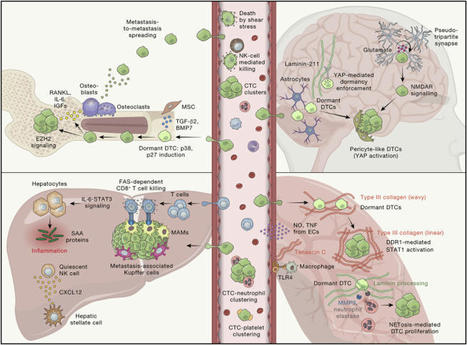
de Visser and Joyce review the complex interplay between the tumor and its microenvironment throughout cancer evolution and discuss the tumor cell-intrinsic, cell-extrinsic, and systemic factors that may be exploited for rational design of anti-cancer treatments.
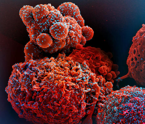
NIH-funded study suggests need to boost CD8+ T cell response after infection. The magnitude and quality of a key immune cell’s response to vaccination with two doses of the Pfizer-BioNTech COVID-19 vaccine were considerably lower in people with prior SARS-CoV-2 infection compared to people without prior infection, a study has found. In addition, the level of this key immune cell that targets the SARS-CoV-2 spike protein was substantially lower in unvaccinated people with COVID-19 than in vaccinated people who had never been infected. Importantly, people who recover from SARS-CoV-2 infection and then get vaccinated are more protected than people who are unvaccinated. These findings, which suggest that the virus damages an important immune-cell response, were published today in the journal Immunity. The study was co-funded by the National Institute of Allergy and Infectious Diseases (NIAID), part of the National Institutes of Health, and led by Mark M. Davis, Ph.D. Dr. Davis is the director of the Stanford Institute for Immunity, Transplantation and Infection and a professor of microbiology and immunology at Stanford University School of Medicine in Palo Alto, California. He is also a Howard Hughes Medical Institute Investigator. Dr. Davis and colleagues designed a very sensitive tool to analyze how immune cells called CD4+ T cells and CD8+ T cells respond to SARS-CoV-2 infection and vaccination. These cells coordinate the immune system’s response to the virus and kill other cells that have been infected, helping prevent COVID-19.
The tool was designed to identify T cells that target any of dozens of specific regions on the virus’s spike protein as well as some other viral regions. The Pfizer-BioNTech vaccine uses parts of the SARS-CoV-2 spike protein to elicit an immune response without causing infection. The investigators studied CD4+ and CD8+ T-cell responses in blood samples from three groups of volunteers. One group had never been infected with SARS-CoV-2 and received two doses of the Pfizer-BioNTech COVID-19 vaccine. The second group had previously been infected with SARS-CoV-2 and received two doses of the vaccine. The third group had COVID-19 and was unvaccinated. The researchers found that vaccination of people who had never been infected with SARS-CoV-2 induced robust CD4+ and CD8+ T-cell responses to the virus’ spike protein. In addition, these T cells produced multiple types of cell-signaling molecules called cytokines, which recruit other immune cells—including antibody-producing B cells—to fight pathogens. However, people who had been infected with SARS-CoV-2 prior to vaccination produced spike-specific CD8+ T cells at considerably lower levels—and with less functionality—than vaccinated people who had never been infected. Moreover, the researchers observed substantially lower levels of spike-specific CD8+ T cells in unvaccinated people with COVID-19 than in vaccinated people who had never been infected. Taken together, the investigators write, these findings suggest that SARS-CoV-2 infection damages the CD8+ T cell response, an effect akin to that observed in earlier studies showing long-term damage to the immune system after infection with viruses such as hepatitis C or HIV. The new findings highlight the need to develop vaccination strategies to specifically boost antiviral CD8+ T cell responses in people previously infected with SARS-CoV-2, the researchers conclude.
Interesting study by Belgian researchers reveals risk factors associated with fatal COVID-19 cases among outbreaks elderly patients in nursing homes. Among…
Introducing the ultimate medical accuracy in digital 3D modeling: our human lymphatic system model. With over 119 individual meshes, each correctly named and organized on layers, this model covers all lymphatic vessels and nodes from head to toe.
Perfect for animation studios, app developers, illustrators, and universities, this model features non-overlapping and non-intersecting geometry, ensuring a clean and accurate representation of the human lymphatic system. With 99.5% quads and minimal triangles, the model boasts a top-quality topology, and 99% of overlapping and intersecting geometry has been eliminated for optimal precision. Elevate your project’s medical accuracy with our digital 3D lymphatic system model.
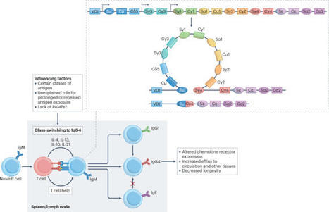
IgG4 is the least abundant subclass of IgG in human serum and has unique functional features. IgG4 is largely unable to activate antibody-dependent immune effector responses and, furthermore, undergoes Fab (fragment antigen binding)-arm exchange, rendering it bispecific for antigen binding and functionally monovalent. These properties of IgG4 have a blocking effect, either on the immune response or on the target protein of IgG4. In this Review, we discuss the unique structural characteristics of IgG4 and how these contribute to its roles in health and disease. We highlight how, depending on the setting, IgG4 responses can be beneficial (for example, in responses to allergens or parasites) or detrimental (for example, in autoimmune diseases, in antitumour responses and in anti-biologic responses). The development of novel models for studying IgG4 (patho)physiology and understanding how IgG4 responses are regulated could offer insights into novel treatment strategies for these IgG4-associated disease settings. IgG4 has unique functional characteristics that are increasingly recognized to have a pathogenic role in autoimmunity, tumour immunology, IgG4-related diseases and anti-biologic responses, as well as a beneficial role in allergy and parasitic infections.
Immunotherapies targeting conventional T cells have revolutionized systemic treatment for many cancers, yet only a subset of patients benefit from these approaches. A better understanding of the complex immune microenvironment of tumours is needed to design the next generation of immunotherapeutics. Innate lymphoid cells (ILCs) and innate-like T cells (ILTCs) are abundant, tissue-resident lymphocytes that have recently been shown to have critical roles in many types of cancers. ILCs and ILTCs rapidly respond to changes in their surrounding environment and act as the first responders to bridge innate and adaptive immunity. This places ILCs and ILTCs as pivotal orchestrators of the final antitumour immune response. In this Review, we outline hallmarks of ILCs and ILTCs and discuss their emerging role in antitumour immunity, as well as the pathophysiological adaptations leading to their pro-tumorigenic function. We explore the pleiotropic, in parts redundant and sometimes opposing, mechanisms that underlie the delicate interplay between the different subsets of ILCs and ILTCs. Finally, we highlight their role in amplifying and complementing conventional T cell functions and summarize immunotherapeutic strategies for targeting ILCs and ILTCs in cancer. Ruf et al. discuss the emerging roles of innate lymphoid cells and innate-like T cells in cancer immunity. The authors highlight their role in bridging adaptive and innate immunity, as well as their potential as immunotherapeutic targets.
Summary. Antimicrobial host defence peptides (HDP) are critical for the first line of defence against bacterial, viral, and fungal pathogens. Over the past deca
Immunoglobulin G (IgG) is the most abundant antibody in the blood and plays a critical role in host immune defense against infectious agents. Glycosylation is known to modulate the effector functions of IgG and is involved in disease development and progression. It is no surprise that the N-glycome of IgG from plasma has already been proposed as a biomarker for various physiological and pathological conditions. However, because saliva is easy to collect, it could be useful for exploring the functional role of salivary IgG N-glycosylation and its potential as a diagnostic biomarker. Therefore, in this study, we described a method for N-glycome analysis of IgG from saliva samples. Salivary IgG N-glycans were analyzed by ultra-high-performance liquid chromatography based on hydrophilic interactions with fluorescence detection (HILIC-UHPLC-FLR). In addition, we compared IgG N-glycan profiles from saliva with those from plasma, assessed the stability of salivary IgG N-glycan profiles under different storage conditions, and evaluated the effects of using a saliva preservation medium. This study provides an ultrasensitive UHPLC method for the analysis of total IgG N-glycosylation from saliva, gives insight into storage stability of salivary IgG, and highlights its (dis)advantages for further biomarker-related research. Graphical Abstract
Our review of 44 studies on the relation between immune biomarkers and frailty identified IL-6 and CRP as the biomarkers that were most consistently associated with frailty.T-lymphocyte subpopulations were investigated but too infrequently to draw strong conclusions yet, although initial results ...
In recent years, the published literature has suggested the key involvement of the cytokine interleukin-31 (IL-31) in the symptomatology of pruritus, and both IL-31 and its receptor have become potential therapeutic targets for a range of pruritic diseases. Elevated levels of IL-31 or its receptor have been reported in the tissue or serum of patients with pruritic skin diseases, such as atopic dermatitis, prurigo nodularis, and psoriasis. Pruritus places a heavy burden on patients, and can have a negative impact on daily life, sleep, and mental health. Since current anti-pruritic treatments are often ineffective, affected patients are in urgent need of new therapies. As a result, drug development targeting the IL-31 pathway is evolving rapidly. To date, only nemolizumab, a humanized monoclonal antibody targeting the IL-31 receptor, has successfully completed late-stage clinical studies. This article will highlight our current clinical understanding of the role of IL-31 in pruritic disease, and explore recent progress in drug development as well as the anticipated future advances in this field.
In humans, senescent fibroblasts do not seem to increase with advancing age in the elderly because their populations are controlled by cytotoxic CD4+ T cells. The expression of T cell-activating ligands and the human cytomegalovirus glycoprotein B marks the senescent fibroblasts for elimination.
To get content containing either thought or leadership enter:
To get content containing both thought and leadership enter:
To get content containing the expression thought leadership enter:
You can enter several keywords and you can refine them whenever you want. Our suggestion engine uses more signals but entering a few keywords here will rapidly give you great content to curate.


 Your new post is loading...
 Scoop.it!












location focus is interesting