Your new post is loading...

|
Scooped by
Gilbert C FAURE
July 29, 2019 4:22 AM
|
PubMed comprises more than 29 million citations for biomedical literature from MEDLINE, life science journals, and online books. Citations may include links to full-text content from PubMed Central and publisher web sites.

|
Suggested by
Société Francaise d'Immunologie
July 26, 2019 2:14 AM
|
Abstract Inhibition of immune cell trafficking to the pancreatic islets during type 1 diabetes (T1D) has therapeutic potential since targeting of T cell and B cell trafficking has been clinically e...
|
|
Suggested by
Société Francaise d'Immunologie
July 25, 2019 2:25 AM
|
Significance B cells have important antibody-independent functions and are now recognized as key players in autoimmune diseases traditionally thought to be T cell-mediated. The role of B cells as antigen-presenting cells, however, is not well understood. By studying the autoantibody response against the enzyme transglutaminase 2 in celiac disease, we have shown that B cells targeting particular epitopes are selectively activated and that this epitope bias reflects efficient presentation of gluten antigen to T cells. Production of antibodies against the preferred epitope coincided with clinical onset of disease, suggesting that B cells with this specificity can be main antigen-presenting cells for pathogenic gluten-specific T cells. Our study thus provides insight into the mechanisms controlling initiation of a T cell-mediated autoimmune condition. Abstract B cells play important roles in autoimmune diseases through autoantibody production, cytokine secretion, or antigen presentation to T cells. In most cases, the contribution of B cells as antigen-presenting cells is not well understood. We have studied the autoantibody response against the enzyme transglutaminase 2 (TG2) in celiac disease patients by generating recombinant antibodies from single gut plasma cells reactive with discrete antigen domains and by undertaking proteomic analysis of anti-TG2 serum antibodies. The majority of the cells recognized epitopes in the N-terminal domain of TG2. Antibodies recognizing C-terminal epitopes interfered with TG2 cross-linking activity, and B cells specific for C-terminal epitopes were inefficient at taking up TG2-gluten complexes for presentation to gluten-specific T cells. The bias toward N-terminal epitopes hence reflects efficient T-B collaboration. Production of antibodies against N-terminal epitopes coincided with clinical onset of disease, suggesting that TG2-reactive B cells with certain epitope specificities could be the main antigen-presenting cells for pathogenic, gluten-specific T cells. The link between B cell epitopes, antigen presentation, and disease onset provides insight into the pathogenic mechanisms of a T cell-mediated autoimmune condition. Footnotes ↵1To whom correspondence may be addressed. Email: rasmus.iversen{at}medisin.uio.no or l.m.sollid{at}medisin.uio.no. ↵2Present address: Symbiosis School of Biological Sciences, Symbiosis International (Deemed University), Lavale, Mulshi, 412115 Pune, India. Author contributions: R.I., B.R., and L.M.S. designed research; R.I., B.R., J.S., L.S.H., and K.H. performed research; I.R.K.-S. and K.E.A.L. contributed new reagents/analytic tools; R.I., B.R., J.S., L.S.H., K.H., and R.S.N. analyzed data; and R.I. and L.M.S. wrote the paper. The authors declare no conflict of interest. This article is a PNAS Direct Submission. Data deposition: Sequence data have been deposited at the European Genome-phenome Archive, https://www.ebi.ac.uk/ega/home (accession no. EGAS00001003658). Proteomics data have been deposited to the ProteomeXchange Consortium, http://www.proteomexchange.org/ (data set identifier PXD013777). This article contains supporting information online at www.pnas.org/lookup/suppl/doi:10.1073/pnas.1901561116/-/DCSupplemental. Published under the PNAS license. References ↵ O. Chan, M. J. Shlomchik, A new role for B cells in systemic autoimmunity: B cells promote spontaneous T cell activation in MRL-lpr/lpr mice. J. Immunol. 160, 51–59 (1998). ↵ N. Molnarfi et al., MHC class II-dependent B cell APC function is required for induction of CNS autoimmunity independent of myelin-specific antibodies. J. Exp. Med. 210, 2921–2937 (2013). ↵ I. Jelcic et al., Memory B cells activate brain-homing, autoreactive CD4+ T cells in multiple sclerosis. Cell 175, 85–100.e23 (2018).OpenUrlCrossRef ↵ S. Hong et al., B cells are the dominant antigen-presenting cells that activate naive CD4+ T cells upon immunization with a virus-derived nanoparticle antigen. Immunity 49, 695–708.e4 (2018).OpenUrl ↵ L. S. Hoydahl et al., Plasma cells are the most abundant gluten peptide MHC-expressing cells in inflamed intestinal tissues from patients with celiac disease. Gastroenterology 156, 1428–1439.e10 (2019).OpenUrl ↵ W. Dieterich et al., Identification of tissue transglutaminase as the autoantigen of celiac disease. Nat. Med. 3, 797–801 (1997).OpenUrlCrossRefPubMed ↵ L. M. Sollid, O. Molberg, S. McAdam, K. E. Lundin, Autoantibodies in coeliac disease: Tissue transglutaminase: Guilt by association? Gut 41, 851–852 (1997).OpenUrlFREE Full Text ↵ L. W. Vader et al., Specificity of tissue transglutaminase explains cereal toxicity in celiac disease. J. Exp. Med. 195, 643–649 (2002). ↵ B. Fleckenstein et al., Gliadin T cell epitope selection by tissue transglutaminase in celiac disease. Role of enzyme specificity and pH influence on the transamidation versus deamidation process. J. Biol. Chem. 277, 34109–34116 (2002). ↵ O. Molberg et al., Tissue transglutaminase selectively modifies gliadin peptides that are recognized by gut-derived T cells in celiac disease. Nat. Med. 4, 713–717 (1998).OpenUrlCrossRefPubMed ↵ Y. van de Wal et al., Selective deamidation by tissue transglutaminase strongly enhances gliadin-specific T cell reactivity. J. Immunol. 161, 1585–1588 (1998). ↵ L. M. Sollid, S. W. Qiao, R. P. Anderson, C. Gianfrani, F. Koning, Nomenclature and listing of celiac disease relevant gluten T-cell epitopes restricted by HLA-DQ molecules. Immunogenetics 64, 455–460 (2012).OpenUrlCrossRefPubMed ↵ D. M. Pinkas, P. Strop, A. T. Brunger, C. Khosla, Transglutaminase 2 undergoes a large conformational change upon activation. PLoS Biol. 5, e327 (2007).OpenUrlCrossRefPubMed ↵ S. Liu, R. A. Cerione, J. Clardy, Structural basis for the guanine nucleotide-binding activity of tissue transglutaminase and its regulation of transamidation activity. Proc. Natl. Acad. Sci. U.S.A. 99, 2743–2747 (2002). ↵ R. Iversen et al., Transglutaminase 2-specific autoantibodies in celiac disease target clustered, N-terminal epitopes not displayed on the surface of cells. J. Immunol. 190, 5981–5991 (2013). ↵ R. Iversen et al., Strong clonal relatedness between serum and gut IgA despite different plasma cell origins. Cell Rep. 20, 2357–2367 (2017).OpenUrl ↵ R. Iversen, S. Mysling, K. Hnida, T. J. Jørgensen, L. M. Sollid, Activity-regulating structural changes and autoantibody epitopes in transglutaminase 2 assessed by hydrogen/deuterium exchange. Proc. Natl. Acad. Sci. U.S.A. 111, 17146–17151 (2014). ↵ R. Di Niro et al., High abundance of plasma cells secreting transglutaminase 2-specific IgA autoantibodies with limited somatic hypermutation in celiac disease intestinal lesions. Nat. Med. 18, 441–445 (2012).OpenUrlCrossRefPubMed ↵ J. Stamnaes, I. Cardoso, R. Iversen, L. M. Sollid, Transglutaminase 2 strongly binds to an extracellular matrix component other than fibronectin via its second C-terminal beta-barrel domain. FEBS J. 283, 3994–4010 (2016).OpenUrlCrossRef ↵ J. Stamnaes, R. Iversen, M. F. du Pré, X. Chen, L. M. Sollid, Enhanced B-cell receptor recognition of the autoantigen transglutaminase 2 by efficient catalytic self-multimerization. PLoS One 10, e0134922 (2015).OpenUrlCrossRefPubMed ↵ K. Hnida et al., Epitope-dependent functional effects of celiac disease autoantibodies on transglutaminase 2. J. Biol. Chem. 291, 25542–25552 (2016). ↵ M. R. Arbuckle et al., Development of autoantibodies before the clinical onset of systemic lupus erythematosus. N. Engl. J. Med. 349, 1526–1533 (2003).OpenUrlCrossRefPubMed ↵ S. Rantapää-Dahlqvist et al., Antibodies against cyclic citrullinated peptide and IgA rheumatoid factor predict the development of rheumatoid arthritis. Arthritis Rheum. 48, 2741–2749 (2003).OpenUrlCrossRefPubMed ↵ T. T. Salmi et al., Immunoglobulin A autoantibodies against transglutaminase 2 in the small intestinal mucosa predict forthcoming coeliac disease. Aliment. Pharmacol. Ther. 24, 541–552 (2006).OpenUrlCrossRefPubMed ↵ G. Oberhuber, G. Granditsch, H. Vogelsang, The histopathology of coeliac disease: Time for a standardized report scheme for pathologists. Eur. J. Gastroenterol. Hepatol. 11, 1185–1194 (1999).OpenUrlPubMed ↵ O. Snir et al., Analysis of celiac disease autoreactive gut plasma cells and their corresponding memory compartment in peripheral blood using high-throughput sequencing. J. Immunol. 194, 5703–5712 (2015). ↵ R. Li, K. R. Patterson, A. Bar-Or, Reassessing B cell contributions in multiple sclerosis. Nat. Immunol. 19, 696–707 (2018).OpenUrl ↵ J. R. Giles, M. Kashgarian, P. A. Koni, M. J. Shlomchik, B cell-specific MHC class II deletion reveals multiple nonredundant roles for B cell antigen presentation in murine lupus. J. Immunol. 195, 2571–2579 (2015). ↵ J. William, C. Euler, S. Christensen, M. J. Shlomchik, Evolution of autoantibody responses via somatic hypermutation outside of germinal centers. Science 297, 2066–2070 (2002). ↵ M. J. Shlomchik, F. Weisel, Germinal center selection and the development of memory B and plasma cells. Immunol. Rev. 247, 52–63 (2012).OpenUrlCrossRefPubMed ↵ S. Sulkanen et al., Tissue transglutaminase autoantibody enzyme-linked immunosorbent assay in detecting celiac disease. Gastroenterology 115, 1322–1328 (1998).OpenUrlCrossRefPubMed ↵ B. Roy et al., High-throughput single-cell analysis of B cell receptor usage among autoantigen-specific plasma cells in celiac disease. J. Immunol. 199, 782–791 (2017). ↵ D. A. Leffler, D. Schuppan, Update on serologic testing in celiac disease. Am. J. Gastroenterol. 105, 2520–2524 (2010).OpenUrlCrossRefPubMed ↵ K. Lindfors, M. Mäki, K. Kaukinen, Transglutaminase 2-targeted autoantibodies in celiac disease: Pathogenetic players in addition to diagnostic tools? Autoimmun. Rev. 9, 744–749 (2010).OpenUrlCrossRefPubMed ↵ B. Jabri, L. M. Sollid, T cells in celiac disease. J. Immunol. 198, 3005–3014 (2017). ↵BSG Coeliac Disease Guidelines Development Group; British Society of Gastroenterology J. F. Ludvigsson et al.; BSG Coeliac Disease Guidelines Development Group; British Society of Gastroenterology, Diagnosis and management of adult coeliac disease: Guidelines from the British Society of Gastroenterology. Gut 63, 1210–1228 (2014). ↵ M. P. Lefranc et al., IMGT, the international ImMunoGeneTics database. Nucleic Acids Res. 27, 209–212 (1999).OpenUrlCrossRefPubMed ↵ J. Cox, M. Mann, MaxQuant enables high peptide identification rates, individualized p.p.b.-range mass accuracies and proteome-wide protein quantification. Nat. Biotechnol. 26, 1367–1372 (2008).OpenUrlCrossRefPubMed ↵ Y. Perez-Riverol et al., The PRIDE database and related tools and resources in 2019: Improving support for quantification data. Nucleic Acids Res. 47, D442–D450 (2019).OpenUrlCrossRefPubMed
|
|
Scooped by
Gilbert C FAURE
July 24, 2019 7:44 AM
|
PubMed comprises more than 29 million citations for biomedical literature from MEDLINE, life science journals, and online books. Citations may include links to full-text content from PubMed Central and publisher web sites.
|
|
Scooped by
Gilbert C FAURE
July 22, 2019 1:52 PM
|
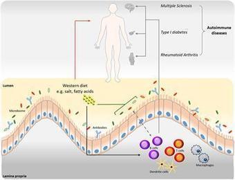
B cells possess a predominant role in adaptive immune responses via antibody-dependent and -independent functions. The microbiome of the gastrointestinal tract is currently being intensively investigated due to its profound impact on various immune responses, including B cell maturation, activation and IgA antibody responses. Recent findings have demonstrated the interplay between dietary components, gut microbiome and autoantibody production. ‘Western’ dietary patterns, such as high fat and high salt diets, can induce alterations in the gut microbiome that in turn affects IgA responses and the production of autoantibodies. This could contribute to multiple pathologies including autoimmune and inflammatory diseases. Here, we summarize current knowledge on the influence of various dietary components on B cell function and (auto)antibody production in relation to the gut microbiota, with a particular focus on the gut-brain axis in the pathogenesis of multiple sclerosis (MS).
|
|
Scooped by
Gilbert C FAURE
July 17, 2019 9:54 AM
|
Objectives Different studies have demonstrated that neutrophil extracellular traps (NETs) may be involved in the pathophysiology of both ANCA‐associated vasculitis (AAV) and systemic lupus erythematosus (SLE). AAV and SLE are clinically and pathologically divergent autoimmune diseases with different autoantibodies. However, the respective autoantigens recognized in AAV and SLE have been shown to be an intricate part of NETs. Therefore, we hypothesized that the mechanisms of NET formation and the composition of NETs might be distinct between AAV and SLE. Methods To investigate this hypothesis, we directly compared AAV‐ and SLE‐induced NET formation, by stimulating healthy neutrophils with AAV (n=80) and SLE (n=59) sera. Results Both AAV and SLE patients had excessive NET formation which correlated with disease activity(r=0.5;p<0.0001 resp. r=0.35;p<0.01). We observed lytic NET formation in AAV after hours versus rapid non‐lytic NET formation coinciding with clustering of neutrophils in SLE within minutes. AAV‐induced NET formation was triggered independent of (ANCA)‐IgG whereas SLE‐immune complexes (ICx) induced NET formation through FcγR‐signaling. AAV‐induced NET formation was dependent of reactive oxygen species and peptidyl‐arginine‐deaminases and was enriched for citrullinated histones(23±2%), all in contrast to SLE‐induced NETs. SLE‐induced NETs had immunogenic properties including NET‐bound HMGB1(30±3%), enrichment for oxidized mtDNA, and were involved in ICx formation. Conclusion In conclusion, morphology, kinetics, induction pathways and composition of excessive NET formation are all intrinsically distinct in AAV compared to SLE. Recognizing the diversity of NET formation between AAV and SLE provides a better understanding of the pathophysiological role of NETs in these different autoimmune diseases. This article is protected by copyright. All rights reserved.
|
|
Scooped by
Gilbert C FAURE
July 11, 2019 8:39 AM
|
New research on the autoimmune disease systemic lupus erythematosus provides hints to the origins of the puzzling disorder. The results were published in Nature Immunology.
|
|
Scooped by
Gilbert C FAURE
July 3, 2019 5:04 AM
|
Sci Rep. 2017 Oct 31;7(1):14426. doi: 10.1038/s41598-017-14350-8. Research Support, Non-U.S.Gov't...
Abstract Type 1 diabetes (T1D) results from the progressive destruction of pancreatic β-cells in a process mediated primarily by T lymphocytes. The T1D research community has made dramatic progress in understanding the genetic basis of the disease as well as in the development of standardized autoantibody assays that inform both disease risk and progression. Despite these advances, there remains a paucity of robust and accepted biomarkers that can effectively inform on the activity of T cells during the natural history of the disease or in response to treatment. In this article, we discuss biomarker development and validation efforts for evaluation of T-cell responses in patients with and at risk for T1D as well as emerging technologies. It is expected that with systematic planning and execution of a well-conceived biomarker development pipeline, T-cell–related biomarkers would rapidly accelerate disease progression monitoring efforts and the evaluation of intervention therapies in T1D. Footnotes This article contains Supplementary Data online at http://diabetes.diabetesjournals.org/lookup/suppl/doi:10.2337/db19-0119/-/DC1. Received February 2, 2019. Accepted April 20, 2019. © 2019 by the American Diabetes Association. http://www.diabetesjournals.org/content/license Readers may use this article as long as the work is properly cited, the use is educational and not for profit, and the work is not altered. More information is available at http://www.diabetesjournals.org/content/license.
Via Krishan Maggon
|
|
Scooped by
Gilbert C FAURE
June 24, 2019 5:07 AM
|
Objectives. Considering the critical role of microRNAs (miRNAs) in regulation of cell activation, we investigated their role in circulating type-2 conventional dendritic cells (cDC2s) of patients with primary Sjögren’s syndrome (pSS) compared to healthy controls (HC).Methods. CD1c-expressing cDC2s were isolated from peripheral blood. A discovery cohort (15 pSS, 6 HC) was used to screen the expression of 758 miRNAs and a replication cohort (15 pSS, 11 HC) was used to confirm differential expression of 18 identified targets. Novel targets for two replicated miRNAs were identified by SILAC in HEK-293T cells and validated in primary cDC2s. Differences in cytokine production between pSS and HC cDC2s were evaluated by intracellular flow-cytometry. cDC2s were cultured in the presence of MSK1-inhibitors to investigate their effect on cytokine production. Results. Expression of miR-130a and miR-708 was significantly decreased in cDC2s from pSS patients compared to HC in both cohorts, and both miRNAs were downregulated upon stimulation via endosomal TLRs. Upstream mediator of cytokine production MSK1 was identified as a novel target of miR-130a and overexpression of miR-130a reduced MSK1 expression in cDC2s. pSS cDC2s showed higher MSK1 expression and an increased fraction of IL-12 and TNF-α-producing cells. MSK1-inhibition reduced cDC2 activation and production of IL-12, TNF-α and IL-6. Conclusions. The decreased expression of miR-130a and miR-708 in pSS cDC2s seems to reflec
|
|
Scooped by
Gilbert C FAURE
June 22, 2019 5:29 AM
|
Autoantibodies are produced when tolerance to self-antigens is broken and they can be mediators of tissue injury and systemic inflammation. They are excellent biomarkers because they are minimally invasive to screen and are highly abundant in serum due to limited proteolysis and slow clearance.
|
|
Scooped by
Gilbert C FAURE
June 17, 2019 1:57 PM
|
Summary CD4+ regulatory T cells (Treg) expressing the forkhead box protein 3 (FOXP3) transcription factor (Tregs) are instrumental for the prevention of autoimmune diseases. There is increasing evidence that the human T regulatory population is highly heterogeneous in phenotype and function. Numerous studies conducted in human autoimmune diseases have shown that Treg cells are impaired either in their suppressive function, in number, or both. However, the contribution of the FOXP3+ Treg subpopulations to the development of autoimmunity has not been delineated in detail. Rare genetic disorders that involve deficits in Treg function can be studied to develop a global idea of the impact of partial or complete deficiency in a specific molecular mechanism involved in Treg function. In patients with reduced Treg numbers (but no functional deficiency), the expansion of autologous Treg cells could be a suitable therapeutic approach: either infusion of in‐vitro autologous expanded cells, infusion of interleukin (IL)‐2/anti‐IL‐2 complex, or both. Treg biology‐based therapies may not be suitable in patients with deficits of Treg function, unless their deficit can be corrected in vivo/in vitro. Finally, it is critical to consider the appropriate stage of autoimmune diseases at which administration of Treg cellular therapy can be most effective. We discuss conflicting data regarding whether Treg cells are more effectual at preventing the initiation of autoimmunity, ameliorating disease progression or curing autoimmunity itself. Introduction CD4+ T regulatory cells (Treg) expressing the forkhead box protein 3 (FOXP3) transcription factor (Treg cells) are capable of suppressing immune responses, especially their initiation, by preventing the activation and proliferation of T and B cells 1. The importance of Treg cells has been demonstrated in animal models whereby their depletion is associated with loss of self‐tolerance and development of severe autoimmunity [e.g. immunodysregulation, polyendocrinopathy enteropathy X‐linked (IPEX) syndrome] 2-5. Further data in murine models have also demonstrated their ability to prevent progression of and even cure established autoimmune/inflammatory disease 6, 7. Overall, these cells are also considered to play a role in allergy prevention 8, gestational tolerance 9, the promotion of graft tolerance post‐transplantation 10 and the prevention of tumour immune responses 11. Human Treg cells were initially characterized as CD4+ T cells co‐expressing the interleukin (IL)‐2 receptor alpha chain 12-18. This was based on murine data demonstrating that depletion of CD4+CD25+ T cells led to the development of severe autoimmunity 2. Since then, Treg cells have been more precisely described as CD4+ T cells expressing the FOXP3 transcription factor in mice 3-5 and in humans 19. However, while the CD25+FOXP3+ phenotype defines Treg cells in mice, both CD25 and FOXP3 can also be induced upon activation in naive CD4+ T cells (of mice and humans) 20. Intriguingly, CD25 and FOXP3 can also be induced in conventional T cells, although this does not equate to them adopting ‘regulatory’ function 21, 22. All this indicates that the mere combination of CD25 and FOXP3 expression is insufficient to define human Treg cells phenotypically in health and diseases 23. In recent years, we have progressed to further categorize human CD4+FOXP3+ T cells into three distinct subpopulations based upon their phenotypical and functional differences. These subpopulations are (a) Fraction I: CD45RA+FOXP3lo naive Treg cells, considered equivalent to natural Treg cells arising from the thymus (tTreg cells) and demonstrating immunosuppressive properties in vitro; (b) Fraction II: CD45RA–FOXP3hi activated effector Treg cells, also immunosuppressive in vitro; and (c) Fraction III: CD45RA–FOXP3lo cytokine‐secreting but not immunosuppressive cells 23. We were also able to classify these populations similarly using CD25 (instead of FOXP3) and CD45RO (instead of CD45RA). Since then, further elegant work has demonstrated that the Fraction III population could be further subdivided on the basis of CD127 to identify two subpopulations. The proteomic analyses performed identified these subpopulations to closely resemble memory conventional T cells or effector Treg cells, respectively 24. Importantly, these aforementioned subpopulations can be distinctly identified in healthy and diseased states. For example, CD45RA+FOXP3lo Treg cells are the main Treg population identified in cord blood 23, whereas effector Treg cells are highly prevalent in tumours or in peripheral blood of patients with sarcoidosis 25, 26 or mycosis fungoides 27. Interestingly, a small proportion of CD45RA–FOXP3lo cells have been identified in the peripheral blood of patients with active systemic lupus erythematosus 27 or in some tumours 28. The impact of these cell subpopulations on their respective pathologies is not yet known. Numerous studies conducted in human autoimmune diseases have shown that Treg cells were impaired either in their suppressive function, in number, or both. A further mechanism involves resistance of conventional T cells to Treg‐mediated suppression via the presence of certain cytokines [tumour necrosis factor (TNF) and IL‐6] in the microenvironment and over‐activated phosphatidylinositol 3‐kinase/protein kinase B (PI3K/Akt) signalling 29. Of note, these findings have been demonstrated in a range of autoimmune diseases 29-31. The conclusions of those studies have been drawn following different phenotypical definitions of human Treg cells, mainly based on the assumption the either that FOXP3‐expressing and/or CD25high CD4+T cells constitute a single homogeneous population of Tregs 19. Hence, the contribution of the heterogeneous FOXP3+ Treg subpopulations to the development of autoimmunity has not been delineated in detail. In this review, we discuss several unresolved questions 32, 33 and emerging issues regarding the role of Treg cells in human autoimmune and inflammatory diseases. Heterogeneity of human FOXP3‐expressing CD4+ T cell subsets There is increasing evidence that the human T regulatory population is highly heterogeneous in phenotype and function 34. While FOXP3‐expressing cells can be roughly separated into three subsets (naive Treg cells, effector Treg cells and FOXP3lo‐activated T cells) 23, there are novel data indicating that these subpopulations can be subdivided phenotypically even further (Fig. 1). CD45RA+ naive Treg cells can be separated into the CD31+ recent thymic emigrant (RTE) and the CD31– naive Treg cell population 35 (Fig. 1). Recent data obtained via cytometry by time of flight (CyTOF) has demonstrated that naive Treg cells can be subdivided based on their expression of CD49b, CD62L and certain chemokine receptors. This has led to their subcategorization as CD49b+, CD49b+CXCR3+CCR4+CCR6+ and CXCR3+RORC2+CD62L+ subsets 36. It is currently unknown how CD31+ RTE Treg cells differentiate into each of these subsets; the prevalence of each subset in human health and disease and how each of those subsets differentiates into other FOXP33+ T cells have also to be determined. Interestingly, CCR4 can also delineate six different subsets among the Fraction III FOXP3lo non‐Treg cell population when combined with CD127 and CD49d. (Fig. 1). Among these subsets, the CD127+CD49d+CCR4– population contains most of the cytokine‐producing cells (IL‐2, IL‐17 and IFN‐γ), while the CD127–CD49d–CCR4+ contains the lowest number of cytokine‐producing cells. This latter subpopulation is, therefore, functionally and phenotypically most similar to the effector Treg subpopulation, as most effector Treg cells are CD127–CD49d–CCR4+ 24. The origin of Fraction III FOXP3lo cells has not yet been completely elucidated: these cells can be derived from naive Treg cells that fail to up‐regulate high levels of FOXP3 upon activation, for instance, because of weak signal transducer and activator of transcription (STAT)‐5 signalling; they may also be derived from some FOXP3hi effector Treg cells through reduced expression of FOXP3, and they can also be derived from conventional CD4+ T cells that transiently up‐regulate FOXP3 upon activation. Of note, all of these phenotypical changes have been observed in vitro, generally in the presence of low‐dose IL‐2 37. Effector Treg cells constitute a functionally homogeneous suppressive subset that is highly proliferative in vivo but poorly proliferative in vitro, and prone to apoptosis in the absence of IL‐2 1, 19. However, from a phenotypical perspective, these cells can be subcategorized based on their co‐expression of effector T cell transcription factors such as T‐bet [T helper type 1 (Th1), GATA binding protein 3 (GATA‐3) (Th2) and retinoid‐related orphan receptor γt (RORC)] (Th17) 32. This is supported by murine data indicating that Th1‐, Th2‐ and Th17‐like Treg cells suppress their related effector Th cell counterparts, respectively 38-40. These Treg subsets also express their effector Th cell counterparts’ chemokine receptors. Hence, Th1‐like Treg cells are CXCR3+, Th2‐like Treg cells are CCR6–CCR4+ and Th17‐like Treg cells are CCR6+CCR4+ 41. A further Treg subset comprise T follicular regulatory (Tfr) cells, which are found in germinal centres and can directly influence B cells. 42 These cells interfere with the interaction between T follicular helper (Tfh) cells and B cells to alter subsequent B cell differentiation towards antibody‐producing plasma cells or a memory phenotype. This interaction requires further study, particularly to improve our understanding of antibody‐mediated autoimmune disease [e.g. thyroiditis, type 1 diabetes (T1D)]. Additionally, recent studies have demonstrated phenotypical heterogeneity between Treg cells in the peripheral circulation and those present in the healthy/diseased tissue 43. For instance, IL‐1 receptor type II (IL‐1R2), a decoy receptor for IL‐1, is highly expressed on breast and colonic Treg cells but not on their peripheral circulation counterparts 44, 45. Similarly, CD15s (sialyl Lewis X) is present on peripherally circulating effector Treg cells but is absent on their pulmonary Treg counterparts 46. Overall, it is clear that historical studies in human autoimmune and/or inflammatory diseases have been conducted on the basis that circulating FOXP3+ T cells were a homogeneous population 30. On the basis of the data discussed above and access to novel technologies, we anticipate the future determination of the distinct contributions of FOXP3+ Treg subsets to human health and disease (Fig. 1). The significance of FOXP3+ Treg cells abnormalities in autoimmune diseases: from pernicious anaemia to IPEX In their seminal publication demonstrating that murine Treg cells displayed the CD4+CD25+ phenotype, Sakaguchi et al. showed that the canonical autoimmune abnormality observed in sick mice depleted of Treg cells was the occurrence of autoimmune gastritis with circulating anti‐parietal autoantibodies 2, a condition that is reminiscent of pernicious anaemia in humans 47. However, the role of Treg cells in pernicious anaemia in humans has not yet been delineated. This knowledge gap can also be extrapolated to other autoimmune diseases whereby the role of Treg cells in their development has been studied using animal or human culture systems that are not necessarily reflective of true human pathology 33. On one hand, upon review of published literature into human autoimmunity, one may be tempted to conclude that all autoimmune diseases could be characterized by either a deficit in Treg number and/or function or resistance of conventional T cells to Treg‐mediated suppression 29-31, 48, 49. On the other hand, the only known condition with clear evidence for total depletion of Treg cells is IPEX 3-5. This condition is provoked by different genetic defects in the FOXP3 gene and is characterized by the occurrence of enteropathy, eczema, T1D, thyroiditis, cytopenia, hepatitis, nephritis and gastritis 50-52. Indeed, the scurfy mouse model is widely utilized for the study of Treg cells, as equivalent defects in the FOXP3 gene lead to a pathologically similar autoimmune disease. Scurfy mice die within a few weeks after birth, while untreated newborns with IPEX die rapidly, both of severe inflammation, allergy and autoimmunity 3-5. Hence, it is clear that a complete defect in Treg cells leads to the development of this lethal systemic autoimmune and inflammatory disease. Due to the rapid progression of IPEX in murine and human newborns (fortunately, a rare condition), the detailed study of Treg cell deficiency in adults with autoimmune disease has remained a challenge. Rudensky et al. and Sparwasser et al. attempted to address this by developing mice with Treg cells bearing the diphtheria toxin receptor 53. Thus, the injection of diphtheria toxin would provoke the complete depletion of Treg cells within days, and thereby lead to a rapidly lethal systemic autoimmune disease that is similar to that observed in IPEX 53, 54. Overall, it is clear from the experimental observations discussed above that a complete deficiency of Treg cells at any stage of life rapidly leads to a fatal systemic disease. However, it is also clear from the clinical literature that human autoimmune diseases tend not to evolve towards an IPEX‐like condition. Indeed, we would reconcile these findings by hypothesizing that Treg number deficiency and dysfunction in human autoimmune diseases is incomplete at any stage of life and is probably mild or minimal. For example, pernicious anaemia may be associated with some organ‐specific endocrine autoimmune diseases such as Graves’ disease, but rarely to other systemic autoimmune diseases 55. Importantly, neither of these conditions evolve towards an IPEX‐like condition. Although not yet proven, if there is indeed a Treg deficit in pernicious anaemia it is likely to be mild or minimal. FOXP3+ Treg cell function deficiencies in human autoimmune diseases: lessons from genetic diseases The lack of validated experimental assays reflective of in‐vivo human Treg biology remains a major limitation in the field. To what extent in‐vitro Treg suppressive activity correlates with in‐vivo Treg function has not yet been established in humans 48. This limitation is important to overcome, as Treg cells have numerous mechanisms of action which require different experimental design and reagents to reliably elicit 56. It is indeed plausible that observed in‐vitro Treg functional deficiency in human autoimmune diseases may be explained by the partial deficiency of one or several mechanisms of suppression. One must also not discount the potential for effector T cells to be resistant to Treg‐mediated suppression mechanisms 29. While the specific roles of these mechanisms can be studied in mice (via different conditional knock‐out models), their corresponding contributions in humans have been mainly elicited using in‐vitro models instead 57. For this reason, patients with rare genetic disorders that involve deficits in Treg function can be studied to develop a global idea of the impact of partial or complete deficiency in a specific molecular mechanism involved in Treg function. One of the most well‐established mechanisms of action of Treg cells is through their cytotoxic T lymphocyte‐associated protein 4 (CTLA‐4) receptor. Indeed, constitutive expression of CTLA‐4 by Treg cells is instrumental for their in‐vivo suppressive capacity 58. Interestingly, CTLA‐4 haploinsufficiency has been described (albeit rarely) in certain families 59, 60. It is therefore noteworthy that patients with heterozygous non‐sense mutations of CTLA‐4 genes develop a systemic autoimmune disease manifesting as diarrhoea, granulomatous interstitial lung disease, autoimmune cytopaenia, thyroiditis, arthritis and skin disease—all of which are reminiscent of IPEX (but with less severity). Of note, none of these patients studied developed autoimmunity in early infancy, but a significant proportion had their first autoimmune abnormality diagnosed in adulthood. From a cellular perspective, although this mutation could have impacted on the CTLA4‐induction properties and function of all activated T cells, the impact on Treg cells specifically is important. This is because normal Treg cells express disproportionally higher surface and intracellular CTLA4 61. Interestingly, in patients with CTLA‐4 haploinsuffiency, they had higher numbers of Treg cells but their individual expression of CTLA‐4 was reduced, especially after activation 59, 60. Hence, CTLA‐4 haploinsufficiency could be considered as a partial CTLA‐4‐related Treg functional deficiency. Additionally, the unintended manifestations of blocking CTLA‐4 have recently been demonstrated in humans with cancer who are receiving anti‐CTLA‐4 checkpoint blockade therapy 62. These therapies work by boosting effector T cell activity and inhibiting Treg cells; however, pharmacovigilance data suggest that some patients develop enteropathy and colitis similar to that of inflammatory bowel disease. It is important to understand why these patients specifically have effector T cells targeting and infiltrating the gut, as the anti‐CTLA‐4 antibody itself is systemically administered. There is also another rare genetic disorder, which leads instead to a complete CTLA‐4‐related functional deficiency. This involves a deficiency in lipopolysaccharide‐responsive and beige‐like anchor protein (LRBA), which is an intracellular protein involved in the membrane expression of CTLA‐4 63. This condition is clinically characterized by a systemic autoimmune syndrome that resembles CTLA‐4 haploinsufficiency as well as IPEX (as some patients develop T1D). However, LRBA deficiency onsets in early infancy and, indeed, in a few patients shortly after birth 64. Similarly, mild reduction in CTLA‐4 expression has also been observed in Treg cells isolated from patients with rheumatoid arthritis (RA). While the CTLA‐4 gene is highly demethylated in normal Treg cells (thus indicating stable CTLA‐4 expression) 65, the CTLA‐4 promoter was instead methylated in Treg cells from RA patients 66. All the above findings are also important from an age‐related perspective; in comparison to LRBA deficiency, which onsets in early infancy, the onset of RA is usually in adults (older than 45–50 years) 67. Hence, by comparing patients with RA, CTLA‐4 haploinsufficiency, LRBA insufficiency and IPEX we can study the relationship between the intensity of CTLA‐4‐related functional deficiency on Treg cells, the effect on Treg biology and the extent of any clinical presentation (including its severity and time of onset) (Fig. 2). Finally, although Treg functional deficiencies have been described in numerous other autoimmune diseases, the detailed molecular mechanism(s) responsible for these deficiencies is/are currently unknown 68. For example, impaired Treg suppression has been described in multiple sclerosis (MS) 69. It is indeed noteworthy that a key mechanism of suppression by Treg cells is related to their high expression of CD25, as Treg cells act as a sink for IL‐2 2, 70, 71. Interestingly, a polymorphism in CD25 has been associated with a high risk for developing MS through a genome‐wide association study (GWAS) 72. Therefore, it is plausible that a CD25‐related mechanism of suppression may be altered in the Treg cells of MS patients. These Treg cells may also be unstable, as loss of CD25 on Treg cells is known to alter FOXP3 expression, Treg cell function and precipitate Th17 effector cell differentiation 73. In parallel with RA patients who have a mild deficiency in CTLA‐4, the extent of the defect in Treg suppression is probably mild or moderate. This is further supported by clinical observations that both RA and MS never evolve toward an IPEX‐like syndrome and rarely occur in early infancy. Targeting FOXP3+ Treg cells for the control of autoimmune responses As deficiencies in Treg number/function or resistance of T conventional cells from Treg‐mediated suppression are observed in most human autoimmune diseases, it seems logical to propose stable and functionally superior Treg‐based immunotherapies as a new therapeutic strategy in order to reinstate immune homeostasis. The two Treg‐based therapeutic strategies currently being clinically evaluated in autoimmunity are in‐vivo Treg expansion and infusion of in‐vitro expanded Treg cells 19, 74, 75. From the perspective of in‐vivo expansion, low‐dose IL‐2 has been evaluated in Phases I and II trials as a therapeutic targeting human Treg cells in order to expand them in vivo within the context of autoimmunity (e.g. T1D, alopecia areata and systemic lupus) or inflammatory conditions (hepatitis C‐related cryoglobulinaemic vasculitis) 76. However, although injection of IL‐2 expands the circulating Treg cell population, it also expands effector cells such as natural killer (NK) cells or eosinophils—thus indicating the lack of Treg specificity 77, 78. This has been overcome through the development of an IL‐2/anti‐IL‐2 complex that can specifically promote the binding of IL‐2 to the high‐affinity receptor of IL‐2 that is expressed by activated Treg cells and promote Treg cell expansion in vivo without modifying other effector cells 79-81. A second approach is the autologous expansion of Treg cells in vitro in order to reinfuse a large number of Treg cells into patients. Due to the recognized potential for reduced FOXP3 expression or reduced immunosuppressive capacity upon in‐vitro expansion, it is important to culture the right cell subpopulation in conditions that favour maintenance of Treg phenotype and function 82. This will help to optimize any expansion protocols and more reliably predict the phenotype of the end product 83. The addition of rapamycin to the culture conditions is important to eliminate contaminating conventional effector cells. Finally, we also consider it important to utilize molecules capable of modifying Treg epigenetics (e.g. DNA methyltransferase inhibitors or vitamin C) 37, 84, 85. These molecules work to maintain FOXP3 expression as well as various other Treg‐specific demethylation patterns, which consequently lead to more stable and functional Treg cells 37, 65, 85. Strategies aiming at increasing the number of autologous Treg cells are suitable for diseases with reduced but fully functional Treg cells. However, it is currently unknown in diseases with deficiencies in Treg function whether the deficiency is present within the entire Treg population (i.e. all Treg cells are impaired) or specific to a distinct subset (indicating that some Treg cells would be functionally impaired while others would be fully functional). In the first case, all expanded Treg cells would be functionally impaired with no beneficial therapeutic effect. In the second case the expansion, either in vitro or in vivo, of the global pool of Treg cells would lead to the expansion of deficient Treg cells and of fully functional Treg cells. It could, therefore, be speculated that the number of expanded fully functional Treg cells would be sufficient to overcome and compensate the functional deficiency of the expanded deficient subset. However, it is important not to overlook the possibility that expanded dysfunctional Treg cells could convert into pathogenic cells when in a proinflammatory environment and also exacerbate disease 86. There are data demonstrating that FOXP3+ human Treg cells can start secreting IL‐17 when exposed to cytokines such as IL‐1β, ‐2, ‐6, ‐15, ‐21 and ‐23 87, 88. These IL‐17‐secreting Treg cells can subsequently lose their anti‐inflammatory function despite continuous FOXP3 expression 88. A third approach is to utilize peripherally induced Treg cells (pTreg cells or iTreg cells if in vitro), which differentiate from naive CD4+ T cells in the presence of transforming growth factor (TGF)‐β and IL‐2 89. One advantage of these cells is the potential to generate antigen‐specific subsets corresponding to the antigens key to the immunopathogenesis of different autoimmune diseases. However, the partially demethylated nature of FOXP3 gives rise to the instability of FOXP3 expression and subsequent loss of suppressive function 89. The in‐vivo stability of human iTreg cells within a proinflammatory microenvironment needs to be optimized if they are to be considered as a safe and non‐pathogenic clinical product. In diseases with functional Treg deficiencies, infusion of autologous Treg cells would be feasible if the impaired molecular mechanism of suppression is identified and corrected by the use of small molecules in vitro or via genetic modifications. An alternative strategy could be the infusion of allogeneic expanded Treg cells sourced from cord blood or other healthy donors 90. Another strategy is to give those patients therapeutics that can compensate for the impaired Treg mechanism; e.g. in patients with CTLA‐4 deficiency, the use of CTLA‐4‐Ig has proved effective in preventing autoimmune events 63. As discussed above, Treg‐based strategies are already being clinically evaluated in some human autoimmune diseases with reported deficiencies in Treg cell numbers 78, 91 (and even in Treg functions) 92, with the assumption that increasing their number would ameliorate or even cure the diseases. The first in‐human trial evaluating Treg cells in autoimmunity was conducted in the setting of T1D 92. Fourteen patients received expanded autologous polyclonal Treg cells (CD25+CD127lo) in a dose‐escalation study (from 0·5 to 26 × 108 cells). The reliability of the expansion process was demonstrated by the purity of the final product, 76–96·9% FOXP3+. Although two patients had serious adverse events of severe hypoglycaemia and ketoacidosis, no directly Treg‐related adverse events were reported. This study was not powered for disease‐specific outcomes, as it was a Phase 1 study—hence, results of future Phase 2 studies are awaited. Further novel work using Treg cells is also ongoing in the contexts of graft‐versus‐host disease and solid organ transplantation 93-96. Treg cells are capable of inhibiting the initiation of immune responses, although the evidence regarding their ability to control active autoimmune/inflammatory disease is more controversial 2, 6, 7, 74. There are in‐vitro data demonstrating that Treg cells cannot inhibit the proliferation of preactivated effector T cells 97, and when transferred into mice after pathogenic cells the Treg cells are incapable of preventing the onset of autoimmunity 2 (Fig. 3). Indeed, this resistance of effector T cells to Treg‐mediated suppression is another key mechanism in autoimmunity. The effector T cells are supported by a signalling pleiotrophic microenvironment consisting of TNF, IL‐6, IL‐1β as well as over‐activated intracellular signalling via the PI3K/Akt pathways 29. Additionally, in another murine model of severe colitis, the progression of this disease was ameliorated and reversed when the mice underwent adoptive transfer of Treg cells 7. This effect was inhibited when mice were administered antibodies to IL‐10, CTLA‐4 or TGF‐β. Together, these data suggest that it is not only Treg cells but also the presence of particular cytokines in the microenvironment that can modulate disease progression. Hence, there was little surprise that only modest benefits are observed in trials evaluating Treg‐based therapies for ongoing autoimmune or inflammatory diseases 78, 98. However, while they are inefficient at controlling activated pathogenic cells, the expanded Treg cells could theoretically prevent the activation of resting pathogenic cells 74, 75. Therefore, some modest beneficial effect can be expected, as Treg cells would inhibit the activation of dormant pathogenic cells. This would indeed be a viable therapeutic approach, as clinical observations have identified the role of steroids and/or immunosuppressants in controlling active autoimmune diseases as they target activated effector cells first. Secondly, when the disease is considered in remission, another line of immunosuppressants and/or of immunomodulatory drugs are given to patients in order to prevent relapses or disease flare‐up 75. As Treg cells are professionally involved in the prevention of the initiation of pathogenic autoimmune responses, we believe that Treg cell‐based treatments should only be considered as a maintenance therapy for the prevention of flares or relapses after the elimination of pathogenic cells. Such strategies would, therefore, be suitable for remitting and relapsing diseases such as MS, RA or anti‐neutrophil cytoplasmic antibodies (ANCA)‐associated vasculitis only during the remission period to prevent relapses. These trials have also not addressed concerning whether or not it is necessary for infused Treg cells in humans to migrate to the diseased tissue in order to prevent further flares/relapses. This is important in the context of autoimmunity, as although patients have a focal site of inflammation (e.g. joints in RA, gut in inflammatory bowel disease), they also have pathology elsewhere (e.g. extra‐articular/intestinal manifestations) 99, 100. Thus, in order to optimize the Treg therapeutic effect and dose, it may be necessary to culture Treg cells that pre‐emptively express tissue‐specific homing markers (if not, induce this phenotype genetically or pharmacologically). This is also important to minimize any off‐target effects such as the risk of malignancy from interference with anti‐tumour immunity. Interestingly, the Treg‐based therapeutic strategies available in humans aim at expanding polyclonal Treg cells, without considering their antigen specificity 74, 75. Studies in mice indicate that antigen‐specific Treg cells are more efficient that polyclonal Treg cells in preventing autoimmune diseases. However, if the activation of Treg cells is dependent on the target antigen of their T cell receptors (TCRs), the suppressive function itself is not only antigen‐specific. Once activated, Treg cells can suppress effector T cells with the same or any other antigen specificity (via bystander suppression) 48. However, as Treg cells expanded in vitro for cell therapy are highly activated, it may not be necessary to take into account antigen‐specificity of Treg cells to obtain beneficial results in the settings of human autoimmune diseases. Conclusions From the data discussed so far, it is clear that abnormalities in the quantity or function of Treg cells are observed in most, if not all, human autoimmune and/or inflammatory diseases. Although numerous Treg subsets have been defined, their individual contributions to human autoimmune and/or inflammatory diseases are largely unknown. The clinical observations of genetic diseases involving deficiencies in Treg function indicate that the severity and the age of disease onset correlate to the depth of Treg functional impairment. Treg biology‐based therapies may not be suitable in patients with deficits of Treg function, unless their deficit can be corrected in vivo/in vitro. It is also critical to consider the appropriate stage of autoimmune diseases whereby administration of Treg cellular therapy can be effective. As highlighted, there are conflicting data regarding whether Treg cells are more effectual at preventing the initiation of autoimmunity, ameliorating disease progression or curing autoimmunity itself. This is because, although Treg cells can prevent the initiation of autoimmune responses, they cannot terminate ongoing responses. We therefore propose here a global sequential therapeutic strategy for autoimmune diseases that includes (1) induction treatments for diseases flares or chronic disease with chronic activity and (2) maintenance treatments that are suitable for diseases during remission phases by utilizing Treg cell biology‐derived therapies to prevent relapses or subsequent flares (Fig. 4). In patients with reduced Treg numbers (but no functional deficiency), the expansion of autologous Treg cells could be a suitable therapeutic approach (either infusion of in‐vitro‐expanded autologous cells, infusion of IL‐2/anti‐IL‐2 complex, or both). In contrast, patients with diseases involving deficiencies of Treg function would benefit from a detailed understanding of the impaired mechanisms of action of their Treg cells. We anticipate the development of suitable therapeutics to correct/reduce the severity of their Treg deficiencies and thereby reduce their disease burden. Another feasible therapeutic option would be to administer functional allogeneic expanded Treg cells. Acknowledgements M. A. is funded by the EASL Juan Rodes PhD Fellowship. The work is supported by the PHRC programme (AOR17082) and by AFPCA (Association Française de la PolyChondrite Atrophiante). Disclosures None declared. References
|
|
Scooped by
Gilbert C FAURE
June 17, 2019 2:24 AM
|
Case Reports Clinical Chemistry 2018 Exercise 4: Autoimmune Encephalitis...
|
|
|
Scooped by
Gilbert C FAURE
July 26, 2019 8:36 AM
|
ABSTRACT An increasing number of movement disorders are associated with autoantibodies. Many of these autoantibodies target the extracellular domain of neuronal surface proteins and associate with ...
|
|
Scooped by
Gilbert C FAURE
July 25, 2019 2:45 PM
|
In this Perspectives article, the authors present how various types of anti-citrullinated protein antibodies (ACPAs) differ based on the structural interactions of these antibodies with their citrullinated antigens, and examine the potential consequences for ACPA-mediated effector functions.
|
|
Suggested by
Société Francaise d'Immunologie
July 25, 2019 2:25 AM
|
Significance Functional loss of gut barrier integrity with subsequent increased antigen trafficking and occurrence of low-grade intestinal inflammation precede the onset of type 1 diabetes (T1D) in patients and preclinical models, thus suggesting that these events are mechanistically linked to the autoimmune pathogenesis of the disease. However, a causal relationship between increased intestinal permeability and autoimmune diabetes was never demonstrated. Our data show that breakage of gut barrier continuity leads to activation of islet-reactive T cells in the intestine, thus triggering autoimmune diabetes. An important implication of our findings is that restoration of a healthy gut barrier through microbiota and diet modulation in diabetes-prone individuals could reduce intestinal activation of islet-reactive T cells and prevent T1D occurrence. Abstract Low-grade intestinal inflammation and alterations of gut barrier integrity are found in patients affected by extraintestinal autoimmune diseases such as type 1 diabetes (T1D), but a direct causal link between enteropathy and triggering of autoimmunity is yet to be established. Here, we found that onset of autoimmunity in preclinical models of T1D is associated with alterations of the mucus layer structure and loss of gut barrier integrity. Importantly, we showed that breakage of the gut barrier integrity in BDC2.5XNOD mice carrying a transgenic T cell receptor (TCR) specific for a beta cell autoantigen leads to activation of islet-reactive T cells within the gut mucosa and onset of T1D. The intestinal activation of islet-reactive T cells requires the presence of gut microbiota and is abolished when mice are depleted of endogenous commensal microbiota by antibiotic treatment. Our results indicate that loss of gut barrier continuity can lead to activation of islet-specific T cells within the intestinal mucosa and to autoimmune diabetes and provide a strong rationale to design innovative therapeutic interventions in “at-risk” individuals aimed at restoring gut barrier integrity to prevent T1D occurrence. Footnotes ↵1To whom correspondence may be addressed. Email: falcone.marika{at}hsr.it. Author contributions: M.F. designed research; C.S., I.C., M.L.C., L.D.G., F.F., R.L., M.R., and R.F. performed research; C.S., R.F., F.S., F.C., and M.F. analyzed data; and C.S. and M.F. wrote the paper. The authors declare no conflict of interest. This article is a PNAS Direct Submission. See Commentary on page 14788. This article contains supporting information online at www.pnas.org/lookup/suppl/doi:10.1073/pnas.1814558116/-/DCSupplemental. Copyright © 2019 the Author(s). Published by PNAS. This open access article is distributed under Creative Commons Attribution-NonCommercial-NoDerivatives License 4.0 (CC BY-NC-ND). References ↵ J. A. Todd, Etiology of type 1 diabetes. Immunity 32, 457–467 (2010).OpenUrlCrossRefPubMed ↵ F. Dotta et al., Coxsackie B4 virus infection of beta cells and natural killer cell insulitis in recent-onset type 1 diabetic patients. Proc. Natl. Acad. Sci. U.S.A. 104, 5115–5120 (2007). ↵ M. C. Honeyman et al., Association between rotavirus infection and pancreatic islet autoimmunity in children at risk of developing type 1 diabetes. Diabetes 49, 1319–1324 (2000).OpenUrlAbstract ↵ O. Vaarala et al., Cow’s milk formula feeding induces primary immunization to insulin in infants at genetic risk for type 1 diabetes. Diabetes 48, 1389–1394 (1999).OpenUrlAbstract ↵ J. M. Norris et al., Timing of initial cereal exposure in infancy and risk of islet autoimmunity. JAMA 290, 1713–1720 (2003).OpenUrlCrossRefPubMed ↵ L. Wen et al., Innate immunity and intestinal microbiota in the development of type 1 diabetes. Nature 455, 1109–1113 (2008).OpenUrlCrossRefPubMed ↵ A. Giongo et al., Toward defining the autoimmune microbiome for type 1 diabetes. ISME J. 5, 82–91 (2011).OpenUrlCrossRefPubMed ↵ C. T. Brown et al., Gut microbiome metagenomics analysis suggests a functional model for the development of autoimmunity for type 1 diabetes. PLoS One 6, e25792 (2011).OpenUrlCrossRefPubMed ↵DIABIMMUNE Study Group T. Vatanen et al.; DIABIMMUNE Study Group, Variation in microbiome LPS immunogenicity contributes to autoimmunity in humans. Cell 165, 842–853 (2016).OpenUrlCrossRefPubMed ↵ E. Badami et al., Defective differentiation of regulatory FoxP3+ T cells by small-intestinal dendritic cells in patients with type 1 diabetes. Diabetes 60, 2120–2124 (2011). ↵ T. Vatanen et al., The human gut microbiome in early-onset type 1 diabetes from the TEDDY study. Nature 562, 589–594 (2018).OpenUrlCrossRef ↵ J. B. Meddings, J. Jarand, S. J. Urbanski, J. Hardin, D. G. Gall, Increased gastrointestinal permeability is an early lesion in the spontaneously diabetic BB rat. Am. J. Physiol. 276, G951–G957 (1999).OpenUrlPubMed ↵ S. Graham et al., Enteropathy precedes type 1 diabetes in the BB rat. Gut 53, 1437–1444 (2004). ↵ F. Maurano et al., Small intestinal enteropathy in non-obese diabetic mice fed a diet containing wheat. Diabetologia 48, 931–937 (2005).OpenUrlCrossRefPubMed ↵ A. D. Mooradian, J. E. Morley, A. S. Levine, W. F. Prigge, R. L. Gebhard, Abnormal intestinal permeability to sugars in diabetes mellitus. Diabetologia 29, 221–224 (1986).OpenUrlCrossRefPubMed ↵ R. Carratù et al., Altered intestinal permeability to mannitol in diabetes mellitus type I. J. Pediatr. Gastroenterol. Nutr. 28, 264–269 (1999).OpenUrlCrossRefPubMed ↵ M. Westerholm-Ormio, O. Vaarala, P. Pihkala, J. Ilonen, E. Savilahti, Immunologic activity in the small intestinal mucosa of pediatric patients with type 1 diabetes. Diabetes 52, 2287–2295 (2003). ↵ M. Secondulfo et al., Ultrastructural mucosal alterations and increased intestinal permeability in non-celiac, type I diabetic patients. Dig. Liver Dis. 36, 35–45 (2004).OpenUrlCrossRefPubMed ↵ E. Bosi et al., Increased intestinal permeability precedes clinical onset of type 1 diabetes. Diabetologia 49, 2824–2827 (2006).OpenUrlCrossRefPubMed ↵ J. Neu et al., Changes in intestinal morphology and permeability in the biobreeding rat before the onset of type 1 diabetes. J. Pediatr. Gastroenterol. Nutr. 40, 589–595 (2005).OpenUrlCrossRefPubMed ↵ O. Vaarala, M. A. Atkinson, J. Neu, The “perfect storm” for type 1 diabetes: The complex interplay between intestinal microbiota, gut permeability, and mucosal immunity. Diabetes 57, 2555–2562 (2008). ↵ J. Sun et al., Pancreatic β-cells limit autoimmune diabetes via an immunoregulatory antimicrobial peptide expressed under the influence of the gut microbiota. Immunity 43, 304–317 (2015).OpenUrlCrossRefPubMed ↵ S. J. Turley, J. W. Lee, N. Dutton-Swain, D. Mathis, C. Benoist, Endocrine self and gut non-self intersect in the pancreatic lymph nodes. Proc. Natl. Acad. Sci. U.S.A. 102, 17729–17733 (2005). ↵ A. Sapone et al., Zonulin upregulation is associated with increased gut permeability in subjects with type 1 diabetes and their relatives. Diabetes 55, 1443–1449 (2006). ↵ T. Pelaseyed et al., The mucus and mucins of the goblet cells and enterocytes provide the first defense line of the gastrointestinal tract and interact with the immune system. Immunol. Rev. 260, 8–20 (2014).OpenUrlCrossRefPubMed ↵ M. E. Johansson, H. Sjövall, G. C. Hansson, The gastrointestinal mucus system in health and disease. Nat. Rev. Gastroenterol. Hepatol. 10, 352–361 (2013).OpenUrlCrossRefPubMed ↵ P. M. Smith et al., The microbial metabolites, short-chain fatty acids, regulate colonic Treg cell homeostasis. Science 341, 569–573 (2013). ↵ N. Arpaia et al., Metabolites produced by commensal bacteria promote peripheral regulatory T-cell generation. Nature 504, 451–455 (2013).OpenUrlCrossRefPubMed ↵ R. L. Gallo, L. V. Hooper, Epithelial antimicrobial defence of the skin and intestine. Nat. Rev. Immunol. 12, 503–516 (2012).OpenUrlCrossRefPubMed ↵ P. Chairatana, E. M. Nolan, Defensins, lectins, mucins, and secretory immunoglobulin A: Microbe-binding biomolecules that contribute to mucosal immunity in the human gut. Crit. Rev. Biochem. Mol. Biol. 52, 45–56 (2017).OpenUrl ↵ M. Shan et al., Mucus enhances gut homeostasis and oral tolerance by delivering immunoregulatory signals. Science 342, 447–453 (2013). ↵ Y. H. Sheng et al., MUC1 and MUC13 differentially regulate epithelial inflammation in response to inflammatory and infectious stimuli. Mucosal Immunol. 6, 557–568 (2013).OpenUrlCrossRefPubMed ↵ P. Courtois, C. Jurysta, A. Sener, F. W. Scott, W. J. Malaisse, Quantitative and qualitative alterations of intestinal mucins in BioBreeding rats. Int. J. Mol. Med. 15, 105–108 (2005).OpenUrlPubMed ↵ I. Jaakkola, S. Jalkanen, A. Hänninen, Diabetogenic T cells are primed both in pancreatic and gut-associated lymph nodes in NOD mice. Eur. J. Immunol. 33, 3255–3264 (2003).OpenUrlCrossRefPubMed ↵ J. D. Katz, B. Wang, K. Haskins, C. Benoist, D. Mathis, Following a diabetogenic T cell from genesis through pathogenesis. Cell 74, 1089–1100 (1993).OpenUrlCrossRefPubMed ↵ K. Haskins, M. Portas, B. Bradley, D. Wegmann, K. Lafferty, T-lymphocyte clone specific for pancreatic islet antigen. Diabetes 37, 1444–1448 (1988). ↵ R. Mueller, L. M. Bradley, T. Krahl, N. Sarvetnick, Mechanism underlying counterregulation of autoimmune diabetes by IL-4. Immunity 7, 411–418 (1997).OpenUrlCrossRefPubMed ↵ M. S. Horwitz et al., Diabetes induced by Coxsackie virus: Initiation by bystander damage and not molecular mimicry. Nat. Med. 4, 781–785 (1998).OpenUrlCrossRefPubMed ↵ M. E. Johansson et al., Bacteria penetrate the inner mucus layer before inflammation in the dextran sulfate colitis model. PLoS One 5, e12238 (2010).OpenUrlCrossRefPubMed ↵ C. Hernández-Chirlaque et al., Germ-free and antibiotic-treated mice are highly susceptible to epithelial injury in DSS colitis. J. Crohns Colitis 10, 1324–1335 (2016).OpenUrlCrossRefPubMed ↵ N. Tai et al., Microbial antigen mimics activate diabetogenic CD8 T cells in NOD mice. J. Exp. Med. 213, 2129–2146 (2016). ↵ F. R. Costa et al., Gut microbiota translocation to the pancreatic lymph nodes triggers NOD2 activation and contributes to T1D onset. J. Exp. Med. 213, 1223–1239 (2016). ↵ X. Li, M. A. Atkinson, The role for gut permeability in the pathogenesis of type 1 diabetes—a solid or leaky concept? Pediatr. Diabetes 16, 485–492 (2015).OpenUrlPubMed ↵ O. Vaarala, Leaking gut in type 1 diabetes. Curr. Opin. Gastroenterol. 24, 701–706 (2008).OpenUrlCrossRefPubMed ↵ O. Vaarala, Is the origin of type 1 diabetes in the gut? Immunol. Cell Biol. 90, 271–276 (2012).OpenUrlCrossRefPubMed ↵ A. Hänninen et al., Akkermansia muciniphila induces gut microbiota remodelling and controls islet autoimmunity in NOD mice. Gut 67, 1445–1453 (2018). ↵ H. Brauner et al., Markers of innate immune activity in patients with type 1 and type 2 diabetes mellitus and the effect of the anti-oxidant coenzyme Q10 on inflammatory activity. Clin. Exp. Immunol. 177, 478–482 (2014).OpenUrlCrossRefPubMed ↵ D. L. Kaufman et al., Spontaneous loss of T-cell tolerance to glutamic acid decarboxylase in murine insulin-dependent diabetes. Nature 366, 69–72 (1993).OpenUrlCrossRefPubMed ↵ K. Robinson, Z. Deng, Y. Hou, G. Zhang, Regulation of the intestinal barrier function by host defense peptides. Front. Vet. Sci. 2, 57 (2015).OpenUrl ↵ L. M. Loonen et al., REG3γ-deficient mice have altered mucus distribution and increased mucosal inflammatory responses to the microbiota and enteric pathogens in the ileum. Mucosal Immunol. 7, 939–947 (2014).OpenUrlCrossRefPubMed ↵ H. W. Koon et al., Cathelicidin signaling via the Toll-like receptor protects against colitis in mice. Gastroenterology 141, 1852–1863.e1-3 (2011).OpenUrlCrossRefPubMed ↵ A. S. Lee et al., Gut barrier disruption by an enteric bacterial pathogen accelerates insulitis in NOD mice. Diabetologia 53, 741–748 (2010).OpenUrlCrossRefPubMed ↵ K. Berer et al., Commensal microbiota and myelin autoantigen cooperate to trigger autoimmune demyelination. Nature 479, 538–541 (2011).OpenUrlCrossRefPubMed ↵ B. D. Stadinski et al., Chromogranin A is an autoantigen in type 1 diabetes. Nat. Immunol. 11, 225–231 (2010).OpenUrlCrossRefPubMed ↵ C. M. Gleeson, W. J. Curry, C. F. Johnston, K. D. Buchanan, Occurrence of WE-14 and chromogranin A-derived peptides in tissues of the human and bovine gastro-entero-pancreatic system and in human neuroendocrine neoplasia. J. Endocrinol. 151, 409–420 (1996). ↵ W. J. Curry et al., Chromogranin A and its derived peptides in the rat and porcine gastro-entero-pancreatic system. Expression, localization, and characterization. Adv. Exp. Med. Biol. 482, 205–213 (2000).OpenUrlPubMed ↵ C. Sorini, M. Falcone, Shaping the (auto)immune response in the gut: The role of intestinal immune regulation in the prevention of type 1 diabetes. Am. J. Clin. Exp. Immunol. 2, 156–171 (2013).OpenUrlPubMed ↵ M. Knip, H. Siljander, The role of the intestinal microbiota in type 1 diabetes mellitus. Nat. Rev. Endocrinol. 12, 154–167 (2016).OpenUrl ↵ J. D. Taurog et al., The germfree state prevents development of gut and joint inflammatory disease in HLA-B27 transgenic rats. J. Exp. Med. 180, 2359–2364 (1994). ↵ C. King, N. Sarvetnick, The incidence of type-1 diabetes in NOD mice is modulated by restricted flora not germ-free conditions. PLoS One 6, e17049 (2011).OpenUrlCrossRefPubMed ↵ M. W. Sadelain, H. Y. Qin, J. Lauzon, B. Singh, Prevention of type I diabetes in NOD mice by adjuvant immunotherapy. Diabetes 39, 583–589 (1990). ↵ T. C. Martins, A. P. Aguas, Changes in B and T lymphocytes associated with mycobacteria-induced protection of NOD mice from diabetes. J. Autoimmun. 9, 501–507 (1996).OpenUrlPubMed ↵ D. Elias, D. Markovits, T. Reshef, R. van der Zee, I. R. Cohen, Induction and therapy of autoimmune diabetes in the non-obese diabetic (NOD/Lt) mouse by a 65-kDa heat shock protein. Proc. Natl. Acad. Sci. U.S.A. 87, 1576–1580 (1990). ↵ P. Saï, A. S. Rivereau, Prevention of diabetes in the nonobese diabetic mouse by oral immunological treatments. Comparative efficiency of human insulin and two bacterial antigens, lipopolysacharide from Escherichia coli and glycoprotein extract from Klebsiella pneumoniae. Diabetes Metab. 22, 341–348 (1996).OpenUrlPubMed ↵ R. Horai et al., Microbiota-dependent activation of an autoreactive T cell receptor provokes autoimmunity in an immunologically privileged site. Immunity 43, 343–353 (2015).OpenUrlCrossRefPubMed ↵ T. C. Fung et al., Lymphoid-tissue-resident commensal bacteria promote members of the IL-10 cytokine family to establish mutualism. Immunity 44, 634–646 (2016).OpenUrlCrossRefPubMed ↵ D. K. Cole et al., Hotspot autoimmune T cell receptor binding underlies pathogen and insulin peptide cross-reactivity. J. Clin. Invest. 126, 3626 (2016).OpenUrl ↵ S. Wirtz, C. Neufert, B. Weigmann, M. F. Neurath, Chemically induced mouse models of intestinal inflammation. Nat. Protoc. 2, 541–546 (2007).OpenUrlCrossRefPubMed ↵ R. L. Brown, R. P. Sequeira, T. B. Clarke, The microbiota protects against respiratory infection via GM-CSF signaling. Nat. Commun. 8, 1512 (2017).OpenUrl ↵ K. Atarashi et al., Induction of colonic regulatory T cells by indigenous Clostridium species. Science 331, 337–341 (2011). ↵ B. Weigmann et al., Isolation and subsequent analysis of murine lamina propria mononuclear cells from colonic tissue. Nat. Protoc. 2, 2307–2311 (2007).OpenUrlCrossRefPubMed ↵ L. Lefrancois, N. Lycke, Isolation of mouse small intestinal intraepithelial lymphocytes, Peyer’s patch, and lamina propria cells. Curr. Protoc. Immunol. Chapter 3, Unit 3.19 (2001).OpenUrl ↵ S. Ghosh et al., Colonic microbiota alters host susceptibility to infectious colitis by modulating inflammation, redox status, and ion transporter gene expression. Am. J. Physiol. Gastrointest. Liver Physiol. 301, G39–G49 (2011).OpenUrlCrossRefPubMed
|
|
Scooped by
Gilbert C FAURE
July 22, 2019 2:26 PM
|
Autoimmune bullous diseases are a group of chronic inflammatory disorders caused by autoantibodies targeted against structural proteins of the desmosomal and hemidesmosomal plaques in the skin and...

|
Suggested by
LIGHTING
July 18, 2019 4:42 AM
|
Objective: To investigate the effectiveness and safety of belimumab in patients with active systemic lupus erythematosus (SLE) in a real-life setting.Methods: All SLE patients treated wit
|
|
Suggested by
Société Francaise d'Immunologie
July 15, 2019 2:31 AM
|
PubMed comprises more than 29 million citations for biomedical literature from MEDLINE, life science journals, and online books. Citations may include links to full-text content from PubMed Central and publisher web sites.
|
|
Scooped by
Gilbert C FAURE
July 11, 2019 6:48 AM
|
PubMed comprises more than 29 million citations for biomedical literature from MEDLINE, life science journals, and online books. Citations may include links to full-text content from PubMed Central and publisher web sites.
|
|
Scooped by
Gilbert C FAURE
July 1, 2019 7:35 AM
|
Renal transplant recipients (RTRs) are known to have a high cardio-vascular disease (CVD) burden only partly explained by traditional CVD risk factors. The aim of this paper was therefore to determine: i) the prognostic value of autoantibodies against apoA-1 (anti-apoA-1 IgG) for incidence of CVD...
|
|
Scooped by
Gilbert C FAURE
June 24, 2019 6:24 AM
|
“We now have a much better understanding of the immunological pathways involved in vitiligo, which is providing insight on how to target and treat the disease better,” an expert says.
|
|
Suggested by
Société Francaise d'Immunologie
June 22, 2019 2:20 PM
|
This proof of concept study was aimed at characterizing novel salivary biomarkers specific for different subsets in primary Sjögren’s syndrome (pSS) in order to improve patients’ profiling. pSS patients were stratified in three subgroups according to both (a) focus score in the minor salivary...
|
|
Scooped by
Gilbert C FAURE
June 20, 2019 9:39 AM
|
Abstract CD4+Foxp3+ regulatory T cells (Tregs) are suppressors of immune activation and play a crucial role in the maintenance of peripheral tolerance. Mutations of Foxp3 result in fatal autoimmunity in multiple organs, including the skin, in both humans and mice. Many studies have demonstrated the altered frequency and functions of Tregs, changes in cytokine and chemokine levels related to Tregs and the differences in genetic background regarding Tregs in autoimmune skin disorders. Recent studies have extended our knowledge of certain properties of Tregs, especially skin‐resident Tregs. In addition, some novel therapies have been performed by modulating the number and the function of Tregs. This review focuses on the role of Tregs in some autoimmune skin disorders, including alopecia areata, vitiligo, pemphigoid and pemphigus, and systemic sclerosis, and discusses questions that remain to be addressed. 1 INTRODUCTION Foxp3+ regulatory T cells (Tregs) are potent suppressors of immune activation and play a crucial role in the maintenance of self‐tolerance.1 Treg cells prevent autoreactive T cells that escape clonal deletion in the thymus from activating and expanding at the periphery. Therefore, a balance between Tregs and effector T cells is a key factor for maintaining appropriate peripheral tolerance. Mutations of Foxp3 result in fatal immune dysregulation, polyendocrinopathy, enteropathy, X‐linked (IPEX) syndrome in humans,2 whereas a mutation in Foxp3 leads to the lymphoproliferative syndrome of scurfy mice.3, 4 Tregs are known to highly express CCR45 and CCR7;6 thus, their ligands CCL22 and CCL21 promote Treg homing to tissues. Various mechanisms by which Tregs directly suppress responder T cells have been proposed: the production of suppressor cytokines IL‐10 and TGF‐β; IL‐2 consumption though CD25 (IL‐2 receptor α), which is highly expressed on Tregs; and granzyme‐mediated cytolysis.7 Tregs also express cytotoxic T lymphocyte‐associated antigen‐4 (CTLA‐4), CD39, CD73, programmed cell death protein 1 (PD‐1) and inducible T‐cell co‐stimulator (ICOS), and these suppress T cells and antigen‐presenting cells.8 In terms of humoral immunity, B cells from patients with IPEX syndrome produce large amounts of autoreactive antibodies and cause hyper‐IgE,9 showing that Tregs play an important role in the maintenance of peripheral anergy in autoreactive B cells. Tregs have specific roles in skin, such as the acceleration of wound healing,10 the promotion of immune tolerance to skin commensal microbes11 and the orchestration of stem cell‐mediated hair follicle regeneration.12 Using an ovalbumin‐expressing mouse model, Rosenblum et al13 demonstrated the existence of skin‐resident memory Tregs, which contribute to mitigating skin inflammation upon repeated antigen exposure. Thus, the function of Tregs in maintaining skin homoeostasis is beginning to be elucidated. This review focuses on some representative autoimmune skin disorders including alopecia areata and vitiligo, which are skin‐specific T cell–mediated autoimmunity; pemphigoid and pemphigus, which are skin‐specific autoantibody‐mediated autoimmunity; and systemic sclerosis, which is a systemic autoimmunity, and discusses the role of Tregs in the pathogenesis of those disorders and questions that should be addressed. 2 ALOPECIA AREATA Alopecia areata (AA) is a site‐specific, T cell–mediated autoimmune disease that affects the hair follicles. The loss of immune privilege in hair follicles associated with an attack by CD8+ T cells on the bulb of the follicle may contribute to the pathogenesis. A study has reported reduced serum TGF‐β levels in patients with AA.14 Meanwhile, no significant difference has been reported in serum IL‐10 levels14-16 and in lesional skin IL‐10 levels17 between AA and healthy control. Thus, the results on Treg‐related cytokines in AA are conflicting. The decreased frequency of Foxp3+ lymphocytes in the scalp lesions of patients with AA has also been reported, whereas circulating CD4+CD25+Foxp3+ Treg levels were marginally higher in severe AA than in mild AA.18 The immunosuppressive function of CD4+CD25+ Tregs is impaired in patients with AA, especially in the ophiasis type of AA, as shown by in vitro assay using TCR stimulation.19 Notably, genomewide association studies (GWAS) have demonstrated single nucleotide polymorphisms in genes that are related to the activation and proliferation of Tregs, such as CTLA‐4, IL‐2, CD25 and Eos (IKZF4, a mediator of Foxp3‐dependent gene silencing in Tregs) in patients with AA.20 These findings suggest that Treg dysfunction is relevant to the development of AA. Interestingly, a recent study using a mouse model demonstrated that Tregs promote hair follicle regeneration by augmenting hair follicle stem cell proliferation and differentiation.12 Thus, skin‐resident Tregs play a major role in hair follicle biology. Castela et al21 reported that the subcutaneous injection of low‐dose recombinant IL‐2 increased the number of Tregs in skin specimens from scalp lesions, resulting in partial hair regrowth, suggesting the possibility that the modulation of Treg number could be a therapeutic strategy for AA. 3 VITILIGO Vitiligo is an autoimmune skin disease of depigmentation that results from the loss of functional melanocytes in the skin. Although the exact pathogenesis remains to be elucidated, studies have demonstrated that CD8+ cytotoxic T cells play a crucial role in inducing the destruction of melanocytes in vitiligo.22 The cytotoxic T cells that produce IL‐6 and IL‐13 induce apoptosis in the melanocytes.23 Furthermore, patients with active vitiligo show greater numbers of CD8+ T cells and lower CD4+/CD8+ ratios than patients with stable vitiligo show.24 Therefore, the imbalance between melanocytes‐reactive CD8+ cytotoxic T cells and Tregs has been suspected as a potential pathogenesis of vitiligo. Many studies have addressed the role of Tregs in vitiligo. The serum level of TGF‐β is significantly lower in patients with vitiligo than in controls, suggesting the dysfunction of Tregs in vitiligo.25-27 The number of circulating Tregs is lower in patients with vitiligo than in controls.24, 27, 28 Furthermore, the frequency and counts of Treg were significantly decreased in the peripheral blood of active patients than in stable patients.24 Significant defects in the immunosuppressive function of CD4+CD25+ Tregs in patients with vitiligo on CD4+CD25− T cells or CD8+ T cells have been shown by in vitro assays with TCR stimulation.27, 28 In vitiligo skin, mRNA levels of molecules related to Treg function were decreased in Foxp3,29, 30 CTLA‐4,31 TGF‐β,30 CCL21 (a ligand for chemokine receptor CCR7)30 and CCL22 (a ligand for CCR4).30 Notably, GWAS using 4680 cases of vitiligo and 39 586 controls identified some associated loci related to immunoregulation, such as CTLA‐4, Eos (IKZF4) and CD80 (a binding partner of CTLA‐4 to inhibit T‐cell activation),32 indicating that a genetic background associated with Treg is involved in the pathogenesis of vitiligo. Some therapeutic approaches involving the manipulation of Tregs have been attempted for the treatment of vitiligo. Chatterjee et al33 examined the immunoregulatory mechanism in vitiligo using transgenic mice that carry T cells with HLA‐A2‐restricted human tyrosinase peptide‐reactive TCR and spontaneously develop vitiligo from an early age. The adoptive transfer of CD4+CD25+Foxp3+ Tregs or treatment with rapamycin, which was reported to be an enhancer of Tregs,34 induces increases in the number of Tregs in skin lesions, resulting in a lasting remission of vitiligo.33 Eby et al35 reported that gene gun treatment whereby CCL22 is introduced into vitiliginous skin lesions on a mouse model promotes Treg recruitment to the lesions, decreases melanocyte‐reactive T cells and leads to significantly reduced depigmentation. These findings show that increasing the number of Tregs at an affected site can be a potential therapeutic for vitiligo. 4 PEMPHIGOID AND PEMPHIGUS Autoantibodies in autoimmune bullous diseases target structural proteins of the epidermis and the epidermal basement membrane zone (BMZ), which cause the loss of cell‐cell adhesion or cell‐matrix adhesion and blistering in the skin. Autoantibodies against BP180, a structural protein at the BMZ, induce bullous pemphigoid (BP). Autoantibodies against desmogleins (Dsg) 1 and 3, desmosomal proteins that are expressed on stratified squamous epithelium including the skin and/or oral mucosa, cause pemphigus foliaceus (PF) and pemphigus vulgaris (PV), respectively. Previous studies reported that peripheral T cells from healthy human donors who carry the susceptible HLA class II alleles of pemphigus vulgaris (PV) or bullous pemphigoid (BP) react to autoantigens.36, 37 Peripheral tolerance may prevent those T cells from activating. However, the number and function of circulating CD4+ CD25+ Foxp3+ Tregs from active patients with BP are similar to those from healthy controls.38 The number of Foxp3+ cells in BP lesions is higher than in control skin.39 A recent study also demonstrated the increased frequency of CD4+CD25++CD127− Tregs in the peripheral blood of patients with BP relative to that in healthy controls.40 In contrast, a significant reduction in circulating CD4+CD25brightFoxp3+ Treg frequency was reported in patients with BP.41 The frequencies of Foxp3+ cells and the numbers of IL‐10+ were significantly lower in skin lesions from patients with BP than in skin lesions from psoriasis or patients with AD, suggesting a possible role of the reduction of Tregs in the pathogenesis of BP.42 Most of these studies did not have functional analyses of Tregs. Rensing‐Ehl et al38 compared the immunosuppressive function of sorted CD4+CD25+ Tregs by co‐culturing them with CD4+CD25− responder T cells at an equal ratio and stimulating them with irradiated allogeneic PBMC or tetanus toxoid. Notably, the immunosuppressive function of Tregs from patients with BP was similar to that of Tregs from healthy controls. Thus, the role of Tregs in the pathogenesis of BP remains controversial. Monoclonal antibodies to PD‐1 or CTLA‐4 have recently been applied for the treatment of melanoma and other neoplasms as immune checkpoint inhibitors, and they are known to suppress Tregs.43, 44 Interestingly, several cases of BP after the administration of those antibodies have been reported.45, 46 In addition, a case of pemphigoid nodularis, a BP subtype, was reported in an IPEX patient.47 These findings suggest that, in some individuals, Tregs suppress T cells that are reactive to BP180 and/or BP230, and that the dysfunction of Tregs may induce the production of autoantibodies to those antigens. In PV, desmoglein (Dsg3)‐reactive type 1 regulatory T cells (Tr1) secreting IL‐10 and TGF‐β are more frequently detected in healthy carriers of PV‐associated HLA class II alleles (DRB1*0402 and DQB1*0503) than in patients with PV, and those Tr1 cells suppress the Dsg3‐reactive T cells in healthy carriers.48 The suppressive function of Tregs has also been demonstrated by a study using an active PV mouse model.49 A recent study demonstrated that the induction of Tregs by the superagonistic anti‐CD28 antibody decreases pathogenic IgG autoantibodies against Dsg3 in a HLA‐DRB1*04:02‐transgenic PV mouse model.50 This study suggests that the modulation of the number of Tregs may provide a novel therapeutic approach against PV. 5 SYSTEMIC SCLEROSIS Systemic sclerosis (SSc) is a systemic autoimmune disorder that is characterized by thickened and sclerotic skin associated with specific autoantibodies. The involvement of internal organs such as the lungs, the gastrointestinal tract, the heart and the kidney is occasionally observed. The pathology of SSc involves three pathways: vasculopathy, fibrosis and aberrant immune activation.51 Many studies have investigated the frequency of Tregs in patients with SSc, and both increases and decreases in their frequency have been reported in peripheral blood52, 53 and in skin lesions.54, 55 The compromised function of CD4+CD25highCD127−/low Tregs in active SSc was also reported.52, 53 The frequency of CD4+CD25brightFoxp3+ Tregs correlates with disease activity and severity in SSc.56 These findings suggest that Tregs play a role in the pathogenesis of SSc, although the significance of that role remains largely unclear. It is known that TGF‐β has potent profibrotic activity, and the expression of TGF‐β‐regulated genes is increased in the skin of patients with SSc.57 Interestingly, a treatment with fresolimumab, a high‐affinity neutralizing antibody that targets TGF‐β, decreases TGF‐β‐regulated gene expression and improves clinical symptoms in patients with SSc,57 suggesting the possible harmful effects of TGF‐β‐producing Tregs in the pathogenesis of SSc. 6 FUTURE DIRECTIONS There are still contradictions in the results regarding the numbers and functions of Tregs, as mentioned above. The variations in methodologies for detecting human Tregs, the age of the studied subjects, and the stages and severities of disease may relate to the inconsistent results in previous reports. Miyara et al58 demonstrated that human CD4+Foxp3+ T cells are divisible into three phenotypically and functionally distinct subpopulations: CD4+CD45RA+Foxp3lo resting Tregs (rTregs), CD4+CD45RA−Foxp3hi activated Tregs (aTregs) and cytokine‐secreting CD4+CD45RA−Foxp3lo non‐suppressive T cells (non‐Tregs). Based on this study, the balance of the three Treg subpopulations has been investigated in some skin diseases.59, 60 Of note, although “non‐Tregs” express Foxp3, they have no immunosuppressive function, suggesting that we should carefully choose the method for detecting or isolating Tregs when we analyse the frequency and function of Tregs in patients. Additionally, the study also demonstrated significant differences in the proportion of the three subpopulations between healthy young donors (between 18 and 40 years old) and aged individuals (between 79 and 90 years old).58 Therefore, we must evaluate the frequency of human Tregs using age‐matched controls. It is also important to notice that Tregs in skin are different from those in blood and secondary lymphoid organs. Fox example, skin‐resident Tregs show activated and memory phenotypes including higher levels of CTLA‐4, CD25, ICOS and CD27 compared to Tregs in blood.61 It is known that conventional CD4+ T cells are mostly activated in human tissues and that activated CD4+ T cells also express Foxp3. Therefore, we should be skeptical of the studies that define Tregs in human skin based solely on expression of CD4 and Foxp3. More sophisticated approaches such as multiparameter flow cytometry or multicolour immunofluorescent microscopy are required to differentiate Tregs from conventional T cells in human skin. In previous studies, functional analyses of Tregs have been commonly performed using TCR stimulation with anti‐CD3 and/or anti‐CD28 antibodies or by co‐culturing with irradiated allogeneic PBMCs.27, 28, 38, 60 If the immunosuppressive function of Tregs is broadly (polyclonally) impaired, why do individuals develop single specific autoimmune diseases but not multiple autoimmunity, such as IPEX syndrome? One possibility is that a portion of the individuals have a genetic background for increased susceptibility to certain autoimmune disorders and carry causative autoreactive T cells that are continuously suppressed by corresponding Tregs in steady state, so that when the immunosuppressive function of Tregs broadly decreases for unknown reasons, the harmful T cells are activated and induce autoimmunity (Figure 1). Maeda et al62 demonstrated that healthy individuals carrying HLA‐A2*0201 possess CD8+ T cells specific to Melan‐A, a self‐antigen expressed by normal melanocytes and targeted in vitiligo. Those CD8+ T cells are anergic and highly express CTLA‐4 and CCR7. In vitro experiments demonstrate that those anergic CD8+ T cells are induced by co‐culturing with CD25highCD4+ Tregs upon antigen stimulation. Melan‐A‐specific CD8+ T cells from healthy individuals express CTLA‐4 at higher levels than those from patients with vitiligo. These findings strongly suggest that the Treg‐mediated induction of anergy in autoreactive T cells is crucial for maintaining self‐tolerance.62 Therefore, we should ideally perform suppression assays using isolated antigen‐specific Tregs and by stimulating them with specific autoantigens, although this is still technically challenging. To find candidates for predisposing factors for autoimmune diseases, comprehensive gene analyses, including GWAS, whole‐exome sequencing or whole‐genome sequencing, may be useful. Most previous studies examined the function of patients’ Tregs using Tregs derived from peripheral blood, because such blood is accessible. However, as previously reported,61 skin‐resident memory Tregs may play an important role in suppressing T cell–mediated inflammation in skin. By contrast, in humoral immunity, CD4+ T follicular helper (Tfh) cells have a particular function in providing T cell help to follicular B cells,63 and they are responsible for the production of autoantibodies;64 also, a subset of Tregs, T follicular regulatory cells (Tfregs), control the expansion of Tfh cells in the germinal centres in secondary lymphoid organs.65 Thus, the appropriate tissue or cell subset for analysing the function of Tregs depends on the type of disease. We should pay attention to this when we plan experiments and interpret the data. In summary, Treg dysfunction is known to be associated with the pathogenesis of certain autoimmune disorders in dermatology, and Treg biology has started to be elucidated. Various approaches to regulate the autoimmunity through the manipulation of Tregs have been tried. A further understanding of Treg properties by investigations with optimal experimental methods promises to lead to novel therapeutic strategies for autoimmune skin diseases. ACKNOWLEDGEMENT None. CONFLICT OF INTEREST The authors have no conflict of interests to declare. AUTHOR CONTRIBUTIONS HU designed the study and wrote the manuscript. REFERENCES Citing Literature
|
|
Suggested by
Société Francaise d'Immunologie
June 17, 2019 6:56 AM
|
Summary There is currently scarce knowledge of the immunological profile of patients with latent autoimmune diabetes mellitus in the adult (LADA) when compared with healthy controls (HC) and patients with classical type 1 diabetes (T1D) and type 2 diabetes (T2D). The objective of this study was to investigate the cellular immunological profile of LADA patients and compare to HC and patients with T1D and T2D. All patients and age‐matched HC were recruited from Uppsala County. Peripheral blood mononuclear cells were isolated from freshly collected blood to determine the proportions of immune cells by flow cytometry. Plasma concentrations of the cytokine interleukin (IL)‐35 were measured by enzyme‐linked immunosorbent assay (ELISA). The proportion of CD11c+CD123– antigen‐presenting cells (APCs) was lower, while the proportions of CD11c+CD123+ APCs and IL‐35+ tolerogenic APCs were higher in LADA patients than in T1D patients. The proportion of CD3–CD56highCD16+ natural killer (NK) cells was higher in LADA patients than in both HC and T2D patients. The frequency of IL‐35+ regulatory T cells and plasma IL‐35 concentrations in LADA patients were similar to those in T1D and T2D patients, but lower than in HC. The proportion of regulatory B cells in LADA patients was higher than in healthy controls, T1D and T2D patients, and the frequency of IL‐35+ regulatory B cells was higher than in T1D patients. LADA presents a mixed cellular immunological pattern with features overlapping with both T1D and T2D. Introduction Latent autoimmune diabetes mellitus in the adult (LADA) 1 accounts for 9% of all adult onset diabetes 1. Phenotypically, LADA patients share characteristics of both type 1 diabetes (T1D) (antibodies against the insulin‐producing β cells) and type 2 diabetes (T2D) patients (aged >35 years and non‐insulin‐dependent at onset) 2, 3. LADA also shares genetic features of both T1D and T2D 4. Despite the presence of autoantibodies, predominantly against glutamic acid decarboxylase (GAD), the disease progress is much slower than in T1D. At present, the focus on the immunological characterization of LADA has concerned the presence of autoantibodies, with only a few studies of other components of the immune system, such as low‐grade proinflammatory markers or cellular immunology 4-6. So far there are no published reports describing the cellular immunological profile that characterizes LADA patients in comparison to healthy individuals, T1D as well as T2D patients, simultaneously. Innate immune cells, such as dendritic cells (DCs), are the main players during early development of autoimmune diabetes 7-10. Allen et al. have reported that plasmacytoid dendritic cells (pDCs) are expanded during the early development of T1D and enhance the islet autoantigen presentation to T cells through an immune complex capture 11. In line with this, it has been shown that interferon (IFN)‐α‐producing pDCs augment the T helper type 1 (Th1) response in T1D and the number of IFN‐α‐producing pDCs are increased in T1D patients compared to healthy controls 12. Therefore, it is of interest to investigate CD11c+CD123–human leucocyte antigen (HLA)‐DR+Lin– (CD11c+CD123–) antigen‐presenting cells (APCs), CD11c+CD123+HLA‐DR+Lin– (CD11c+CD123+) APCs and CD11c–CD123+HLA‐DR+Lin– (CD11c–CD123+) pDCs in LADA patients. Herein, these subsets of DCs were analysed using flow cytometry, as described previously 13. Neutrophils have been found to be reduced in recent‐onset T1D 14, 15, and CD15low neutrophils (low‐density granular neutrophils) have also been shown to have a pathogenic role in systemic autoimmunity 16. In accordance with this, Neuwrith et al. have reported a role of CD15low cells in T1D development 17. Alterations in the frequency of CD16 monocytes has been associated with diabetes complications 18. Natural killer (NK) cells participate in innate immune responses and in animal models of T1D, it has been reported that depletion of NK cells prevents the development of T1D 19, 20. An altered frequency of total number of NK cells has been found in LADA, but not in T2D, patients 21, 22. Furthermore, it has been reported in children with T1D that NK cells have defects in signalling and function that might contribute to the pathogenesis of T1D 23. NK cell subsets could be defined based on the relative expression of CD16 and CD56, i.e. CD56dim (CD56lowCD16+ and CD56lowCD16–) and CD56bright (CD56highCD16+ and CD56highCD16–), as reviewed by Poli et al. 24. CD56dim NK cells are considered to be mature NK cells and CD56bright cells are immature cells. CD56dim NK cells have cytotoxic properties while CD56bright NK cells are proinflammatory, as they produce IFN‐γ 25. In the present study we determined the proportion of both CD56dim and CD56bright NK cell subpopulations using flow cytometry. Adaptive immune cells such as T (CD4+ and CD8+) and B (CD19+) cells also play a major role in autoimmunity and metabolic disorders 26-30. An altered phenotype of CD4+, CD8+ and CD19+ cells has been reported in LADA patients 5, 31-33. Therefore, in this study we determined the proportions of these cells in LADA patients when compared to T1D and T2D patients. Immune cells that produce anti‐inflammatory cytokines, e.g. interleukin (IL)‐10, transforming growth factor (TGF)‐β and IL‐35, are known as regulatory immune cells, i.e. regulatory T (Treg) cells, regulatory B (Breg) cells and tolerogenic APCs 34-36. Treg cells have received a great deal of attention, as these cells play a major role in maintaining the homeostasis of the immune system and preventing the development of autoimmune diseases 37, 38. A reduced number of Treg cells has been reported in different autoimmune diseases, including T1D 39. Treg cells can be identified by using the cell surface markers CD4, CD25 and CD127 and the intracellular forkhead box (Fox) transcription factor FoxP3 40. Breg cells can be identified by using the cell surface markers CD19, CD24, CD38 and CD40 41. Treg cells, Breg cells and APCs produce IL‐35 34-36, 42-44. This cytokine is a novel anti‐inflammatory cytokine, which belongs to the IL‐12 cytokine family, and has two subunits, Ebi3 and IL‐12p35 42, 43. It has previously been reported that IL‐35 suppresses autoimmunity and plays a putative role in tumour biology 35, 36, 42, 45-47. We have found that the systemic administration of IL‐35 prevented the development of autoimmune diabetes in the multiple low‐dose streptozotocin (MLDSTZ) mouse model, and effectively reversed established disease in both the MLDSTZ and non‐obese diabetic mouse models of T1D by maintaining the normal suppressive phenotype of Treg cells 46. We also found that IL‐35 levels were higher in T1D patients with residual C‐peptide compared to T1D patients with no measurable residual C‐peptide 48. Thus, our previous findings indicate that IL‐35 may prevent the loss of β cell mass. The possible alteration in the frequencies of IL‐35‐producing cells in LADA patients has not yet been investigated. In this study we aimed to determine the proportions of different innate, adaptive and regulatory immune cells in order to elucidate the immunological profile that characterizes LADA in comparison to healthy controls, T1D and T2D patients. Materials and methods Informed consent The Uppsala County regional ethics board approved this study, and the reported investigations were carried out in accordance with the principles of the Declaration of Helsinki, as revised in 2000. All participants were provided with oral and written information about the study, and signed a written consent. Inclusion The All New Diabetes in Uppsala (ANDiU) study (www.andiu.se) is an ongoing study with the aim of describing the incidence of diabetes subgroups in Uppsala County, Sweden (population approximately 300 000). Patients newly diagnosed with diabetes (ADA criteria) are included. Upon registration patients are classified into diabetes subtypes according to criteria shared with the All New Diabetics in Scania (ANDIS) study (http://andis.ludc.med.lu.se; Principle Investigator: Leif Groop) in southern Sweden. Criteria for LADA diagnosis are age ≥35 years, GAD antibody (GADA) positivity (>20 IE/ml) and fasting (f)C‐peptide levels ≥0·3 nmol/l. T1D criteria are GADA positivity and fC‐peptide <0·3 nmol/l, whereas for T2D age ≥35 years, GADA negativity and fC‐peptide >0·72 nmol/l are used as diagnostic criteria. For all three diagnoses there should be no history of pancreatitis, pancreatic cancer or other causes of secondary diabetes. There are no unified criteria for LADA diagnosis and classification, but the criteria used in ANDiU and ANDIS are in line with previous literature 3. The exception is fC‐peptide, which is used in ANDiU and ANDIS as an indicator of remaining insulin production to separate LADA from T1D 49, 50. It replaces the insulin therapy criterion (i.e. no insulin therapy within 6–12 months of diagnosis) that is often used, which is open to subjectivity as it reflects the assessment made by the treating physician. From ANDiU we recruited patients classified as LADA and T2D. T1D patients and healthy controls were recruited at Uppsala University Hospital and Eriksberg Primary Health Care Clinic, Uppsala, respectively. T1D and T2D patients, as well as healthy controls, were age‐, sex‐ and body mass index (BMI)‐matched to the LADA patients. Exclusion criteria were ongoing hormonal treatment (ATC code H01‐03), use of non‐steroid anti‐inflammatory drugs (NSAID, ATC code M01), ongoing chemotherapy treatment (ATC code L01‐04) and other autoimmune disease (e.g. hypo/hyperthyroidism, rheumatic disease, vitiligo, psoriasis, coeliac disease, Addison’s disease and inflammatory bowel disease). Table 1 provides characteristics of the included patients and Supporting information, Fig. S1 shows a flow‐chart of the inclusion process of the study. Controls (n = 13) T1D (n = 16) LADA (n = 14) T2D (n = 16) Age (years) 61·6 ± 2·9 66·0 ± 1·4 65·7 ± 2·4 64·0 ± 2·0 Sex (M%/F%) 62%/38% 73%/27% 57%/43% 70%/30% BMI (kg/m2) 28·9 ± 4·0 26·5 ± 0·7 27·9 ± 0·5 29·1 ± 1·0 Age at onset n.a. 19·7 ± 1·8 63·6 ± 1·5b 60·0 ± 2·0b Disease duration n.a. 46·1 ± 2·2 4·9 ± 0·3b 5·3 ± 0·3a, b HbA1c (mmol/mol) 35·8 ± 0·7 58·9 ± 3·2a 45·8 ± 2·3b 52·27 ± 4·0a fC‐peptide (nmol/l) 1·10 ± 0·11 0·22 ± 0·13a 0·95 ± 0·14b 1·30 ± 0·12b, c fp‐glucose (mmol/l) 5·9 ± 0·1 11·7 ± 1·0a 8·0 ± 0·5b 9·1 ± 0·6a, b GAD antibody IgG (IE/ml) 0 ± 0 508 ± 205 337 ± 190 1 ± 0 IA‐2 antibody IgG (IE/ml) 1 ± 0 25 ± 7 387 ± 295 1 ± 0 Hb (g/l) 145 ± 2 142 ± 3 145 ± 3 141 ± 4 Lkc (109/l) 6·0 ± 0·4 5·8 ± 0·4 6·6 ± 0·8 5·4 ± 0·2 CRP (mg/l) 1·2 ± 0·4 1·6 ± 0·3 1·3 ± 0·2 2·4 ± 0·8 All values are given as mean ± standard error of the mean (s.e.m.). Multiple comparison of means by analysis of variance (anova), Tukey’s post‐hoc test. a P‐value versus controls < 0·05; b P‐value versus T1D < 0·05; c P‐value versus LADA < 0·05. GADA = glutamate decarboxylase 65 antibody; eGFR = creatinine estimated glomerular filtration rate; TSH = thyroid‐stimulating hormone; LADA = latent autoimmune diabetes mellitus in the adult (LADA); Ig = immunoglobulin; BMI = body mass index; n.a. = not available; HbAlc = haemoglobinA1c; CRP = C‐reactive peptide; fC‐peptide = fasting C‐peptide; fp‐glucose = fasting plasma glucose; Lkc = leucocyte count. Routine laboratory parameters Blood samples were collected after overnight fasting. All routine laboratory parameters were analysed at the Central Clinical Chemistry Laboratory at Uppsala University Hospital, which is certified by the External Quality Assessment Service (EQUALIS) (equalis.se/en). On inclusion, blood haemoglobin 51, blood leucocyte count (Lkc), plasma C‐reactive protein (CRP), serum transglutaminase [immunoglobulin (Ig)A] antibodies, serum thyroxine (T4), serum triiodothyronine (T3), serum thyroid‐stimulating hormone (TSH), serum TSH‐receptor 18 antibody, serum thyroid peroxidase antibody, plasma IgA antibody, serum cortisol, 21‐hydroxylase 18 antibody, serum GADA, fasting serum C‐peptide, fasting plasma glucose and blood haemoglobinA1c (HbA1c) were analysed. Glomerular filtration rate (GFR) was estimated based on creatinine‐levels calculated with the MDRD formula 52. Flow cytometry Freshly isolated peripheral blood mononuclear cells (PBMCs) were prepared using Histopaque‐1077 (Sigma, St Louis, MO, USA), as described earlier 48. PBMCs were stained with antibodies for surface markers (Supporting information, Table S1). The cells were thereafter fixed and permeabilized with fixation permeabilization buffer (eBioscience, San Diego, CA, USA) for intracellular markers (Supporting information, Table S1). The samples were run on LSR II Fortesa (Becton Dickinson, Franklin Lakes, NJ, USA) using DivaDacker software 53 and 1 million events were counted for analysis. Fluorescence minus one (FMO), isotype and single‐stained controls were used for gating strategies. The flow cytometry standard (FCS) files were analysed on FlowLogic software (Inivai Technologies, Mentone, Australia). Representative gating strategies are shown in Supporting information, Fig. S2–S5. IL‐35 measurements in plasma Plasma IL‐35 concentrations were determined by using an enzyme‐linked immunosorbent assay (ELISA) kit (Cloud‐Clone Corporation, Wuhan, China). Statistics Differences in means between healthy controls, T1D and T2D patients compared to LADA patients were calculated by one‐way analysis of variance (anova) with Dunnett’s post‐hoc test for immunological profiles. Comparisons between all groups were performed by anova with Tukey’s post‐hoc test for research subjects’ characteristics (Table 1). A P‐value < 0·05 was considered as a significant difference between groups. Statistical analyses were carried out in GraphPad Prism version 6.0TM. Results Innate immune cell responses We observed a lower proportion of CD11c+CD123– APCs in LADA patients than in healthy controls and T1D patients (Fig. 1a). Furthermore, the proportion of CD11c+CD123– APCs among HLA‐DR+Lin– cells was lower in LADA patients than in T1D patients (Fig. 1b), whereas the proportions of pDCs and pDCs among HLA‐DR+Lin– were not altered in LADA patients (Fig. 1c,d). We did not observe any difference in the proportion of CD11c+CD123+ APCs (Fig. 1e), but the proportion of CD11c+CD123+ cells among HLA‐DR+Lin– was higher in patients with LADA than in patients with T1D (Fig. 1f). We did not find any alteration in the proportions of CD15low neutrophils and CD16+ monocytes in LADA patients (Supporting information, Fig. S6a,b). We also examined different subsets of NK cells and did not find any alteration in LADA patients compared to the other groups regarding the proportions of CD3–CD56lowCD16+, CD3–CD56lowCD16– and CD3–CD56highCD16– NK cells (Fig. 2a–c). However, the proportion of CD3–CD56highCD16+ NK cells was higher in LADA patients compared to both healthy controls and T2D patients (Fig. 2d). We did not observe any alteration in the proportions of CD3+CD56+ NK T cells between the groups (Supporting information, Fig. S6c). Adaptive immune cell responses The proportions of CD3+, CD4+CD25– and CD8+ T cells were not altered in LADA patients when compared to the other groups (Supporting information, Fig. S7a–c). Similarly, the proportion of CD19+ cells was not altered (Supporting information, Fig. S7d). Regulatory immune cell responses The proportions of CD4+CD25+CD127–FoxP3+ cells (Treg cells) and CD4+CD25+CD127–FoxP3+Helios+thymic‐derived T cells (tTreg) were similar in all four groups (Supporting information, Fig. S8a,b). Similarly, the proportions of Treg cells as well as the tTreg cells among CD4+ T cells were similar in all four groups (Supporting information, Fig. S8c,d). However, the proportions of IL‐35+ cells among Treg and tTreg cells in patients with LADA were similar in patients with T1D and T2D, but lower when compared to healthy controls (Fig. 3a,b). We also analysed the proportion of CD19+CD24+CD40+CD38+ cells (Breg cells), and observed a higher proportion of these cells in LADA patients when compared to healthy controls and patients with T1D and T2D (Fig. 3c). The proportion of IL‐35+ cells among Breg cells was higher in LADA patients when compared to T1D patients (Fig. 3d). Subsequently, we analysed the proportion of IL‐35+ tolerogenic APCs. Interestingly, the proportion of IL‐35+ cells among APCs was higher in patients with LADA compared to patients with T1D (Fig. 3e). Patients with LADA had IL‐35 concentrations in blood plasma that were similar to those in patients with T1D and T2D, but lower compared to those in healthy controls (Fig. 3f). Discussion We determined the proportions of different immune cells to elucidate the immunological patterns that distinguish patients with LADA compared not only to healthy individuals, but also to patients with T1D and T2D. With regard to the innate and adaptive immune cells, we found that LADA shares common features with both T1D and T2D. The proportion of CD11c+CD123– APCs in LADA patients was between that observed in T1D and T2D patients. Similarly, the proportion of CD11c+CD123+ cells among the HLA‐DR+Lin– was highest in T2D patients and lowest in T1D patients, whereas the proportion in LADA patients was between that of T1D and T2D patients. Neutrophils have previously been claimed to play a role in the early development of both T1D 14, 15, 54 and T2D 55. In the present study, we did not find any alteration in the proportion of CD15low neutrophils in LADA patients when compared to the other study groups. It cannot be excluded that early transient alterations during disease development as described for T1D 14, 15 had occurred, as our study subjects with LADA had a mean disease duration of almost 5 years. It has been reported previously that the proportion of CD3–CD56+CD16+ NK cells is decreased in LADA patients 22. Instead, we observed an increased proportion of CD3–CD56highCD16+ NK cells in LADA patients compared to both healthy controls and T2D patients. The reason for the discrepancy between the studies is unknown, but it could be because we used freshly prepared PBMCs instead of cryopreserved cell preparations. CD56high or CD56bright NK cells produce IFN‐γ 25. The proportions of these NK cells were similar in LADA and T1D patients. This result illustrates that, like T1D, LADA is also a Th1 response disorder. The frequency of FoxP3+ Treg cells was similar in all groups. However, the proportion of IL‐35+ cells among Treg cells in patients with LADA was similar to those in patients with T1D and T2D, but lower when compared to healthy controls. This may suggest a common functional defect of Treg cells in LADA, T1D and T2D patients. This idea was further supported because the circulating concentrations of IL‐35 in patients with LADA were similar to patients with T1D and T2D, but lower than in than healthy controls. We have previously shown that decreased IL‐35 production by Treg cells is associated with a functional defect 46. Also, previous findings of a decreased frequency of FoxP3+ cells among CD4+CD25intermediate cells in LADA patients when compared to healthy controls 5, and a decreased Foxp3 gene expression in CD4+ T cells in LADA patients, are considered with defective Treg cell functionality 56. In patients with T1D, it has been reported that Treg cells lose their FoxP3 expression due to DNA methylation in the Foxp3 gene, which may cause a defective function in Treg cells and result in a decreased number of Treg cells 57. Interestingly, Li et al. also reported an abnormal DNA methylation in peripheral CD4+ T cells of LADA patients 33, which indicates a dysfunction of Treg cells 58. Breg cells play an important role in maintaining the autoimmune and/or inflammatory response 35, 36. We found that the frequency of Breg cells was higher in LADA patients compared to all other groups. These results are in agreement with a previous report, where Deng et al. found the lowest frequency of IL‐10‐producing B cells in T1D patients when compared to LADA and T2D patients 32. Our observed lower proportion of IL‐35+ cells among Breg cells in T1D than LADA patients suggests that the response of IL‐35+ Breg cells in LADA patients is similar to that of T2D patients, which might protect against β cell destruction by immunological insults in LADA patients. Although the proportions of IL‐35+ Breg cells and IL‐35+ tolerogenic APCs were higher in LADA than in T1D patients, the circulating concentrations of IL‐35 were similar in LADA and T1D patients. This could be due to other IL‐35‐producing immune cells, such as iTr35 59 and macrophages 60. These particular populations have not been investigated in the present study. APCs (CD123– APCs) and pDCs (CD123+ APCs) prime T cells to react against the insulin‐producing β cells. In the present study, we found that the numbers of APCs were lower in LADA than in T1D patients. This indicates that the initial immune response is lower in LADA patients, which might cause a delay in the development of an autoimmune response. Tolerogenic APCs protect β cells in animal models of T1D 44, and Dixon et al. reported that tolerogenic APCs produce IL‐35 in human peripheral blood 34. We analysed the frequency of IL‐35+ cells among APCs in our study and found that IL‐35+ cells were more frequent in LADA than in T1D patients, which may contribute to a milder disease development. In a previous study, we found that systemic treatment with IL‐35 in two different animal models of T1D prevented destruction of β cells 46. However, as this is an association study it is impossible to determine whether or not the immunological differences are causal. We observed changes in the proportions of different immune cells in LADA patients compared to healthy controls and other diabetes patients. Our conclusion is that the changes in APC and Breg cell numbers in LADA patients are more similar to those also observed in T2D patients, whereas the changes in NK cell numbers are more similar to those found in T1D patients. There are no differences in IL‐35 concentrations and IL‐35+ Treg cell numbers in between LADA, T1D and T2D patients, but in all three conditions these are lower than the healthy individuals. Thus, our analysis of several subsets of immune cells reveals that LADA patients present a mixture of cellular immunological changes with features overlapping with both T1D and T2D. Acknowledgements The technical assistance from research nurses Violeta Armijo Del Valle, Rebecka Hilmius and Karin Kjellström is gratefully acknowledged. The present study was supported financially by the Swedish Research Council (2017‐01343, 921‐2014‐7054), EXODIAB, the Swedish Diabetes Foundation, Diabetes Wellness Sverige, the Swedish Child Diabetes Fund, SEB Diabetesfonden, O.E. och Edla Johanssons vetenskapliga stiftelse and the Novo Nordisk Foundation. Disclosures The authors declare that there are no conflicts of interest associated with this paper. Author contributions M. M. and K. S. contributed to the design of the study, conducted experiments, acquired and analysed data, and participated in writing the manuscript. Z. L., D. E., J. S. and S. S. contributed to the design of the study, analysed data and participated in the writing of the manuscript. P. O. C. designed the study, analysed data and wrote the manuscript. All authors read and approved the final version. K. S. is the guarantor of this work and, as such, had access to all the data in the study and takes responsibility for the integrity of the data and the accuracy of the data analysis. Supporting Information References
|




 Scoop.it!