Your new post is loading...

|
Rescooped by
Dr. Stefan Gruenwald
from DNA and RNA research
March 3, 2018 5:40 PM
|
Huge crowdsourced genealogy databases are inspiring new genetics research. Yaniv Erlich has been a white-hat hacker and a geneticist at Columbia University, and now he works for a genealogy company. This unusual career trajectory has led, most recently, to a 13-million-person family tree unveiled recently in Science. The massive trove of data comes from public profiles on the crowdsourced genealogy website Geni.com, and it sheds light on human longevity and dispersal over time. (I wrote about a preprint of this paper last year.) But most of all, Erlich is excited about overlaying DNA information on top of family trees to study genes implicated in disease. MyHeritage, the company behind Geni.com, also sells DNA ancestry tests. And since 2017, Erlich has been on leave from Columbia working as MyHeritage’s chief scientific officer to develop those DNA tests. If that sounds like a lot of data going into the hands of one company, well, it is. Erlich has very much been in discussions about DNA research and privacy. In 2013, he showed it was possible, using only public information in places like consumer genealogy databases, to identify certain study participants who had contributed their DNA to research projects. For this feat, Nature dubbed him the “genome hacker.”
Via Integrated DNA Technologies
|
|
Rescooped by
Dr. Stefan Gruenwald
from Papers
March 1, 2018 1:09 PM
|
It’s hard to pinpoint the dawn of AI gaming supremacy. You could choose Kasparov’s loss in chess, or Lee Sedol’s defeat at the virtual hands of AlphaGo. Another popular option would be when legendaryJeopardy! champion Ken Jennings lost to IBM’s Watson in 2011. Watson could parse the game’s clues and handle wordplay. The two-day match wasn’t close. “I for one welcome our new computer overlords,” Jennings wrote under his final answer. Watson seemed to be endowed with the kind of clerical skills humans use on a host of real-world problems. It could take a prompt in English, rummage through relevant documents at lightning speed, come up with the relevant snippets of information, and settle on a single best answer. But seven years later, the real world continues to present stubborn challenges for AI. A September report by the health publication Stat found that researching and designing personalized cancer treatments, as Watson’s descendant Watson for Oncology aims to do, is proving difficult. “The questions in Jeopardy! are easier in the sense that they don’t need much common sense,” wrote Bengio, who has collaborated with the Watson team, when asked to compare the two cases from the AI perspective. “Understanding a medical article is much harder. Again, much basic research is needed.” As special as games are, there are still a few real-world problems they resemble. Researchers from DeepMind declined to be interviewed for this article, citing the fact that their AlphaZero work is currently under peer review. But the team has suggested that its techniques may soon help biomedical researchers, who would like to understand protein folding. To do this, they need to figure out how the various amino acids that make up a protein kink and fold into a little three-dimensional machine with a function that depends on its shape. That’s tricky in the same ways chess is tricky: Chemists know the rules roughly well enough to calculate specific scenarios, but there are still so many possible configurations, it’s a hopeless task to search through them all. But what if protein folding could be configured as a game? In fact, it already has been. Since 2008, hundreds of thousands of human players have attempted Foldit, an online game where users are scored on the stability and feasibility of the protein structures they fold. A machine could train itself in a similar manner, perhaps by trying to beat its previous best score with general reinforcement learning.
Via Complexity Digest
|
|
Scooped by
Dr. Stefan Gruenwald
March 1, 2018 12:57 PM
|
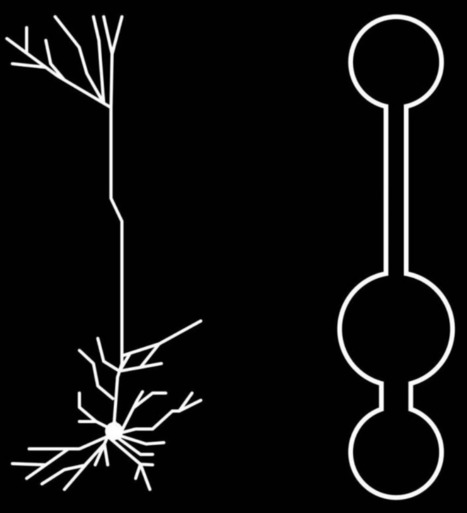
Deep-learning researchers have found that certain neurons in the brain have shape and electrical properties that appear to be well-suited for “deep learning” — the kind of machine-intelligence used in beating humans at Go and Chess. Canadian Institute For Advanced Research (CIFAR) Fellow Blake Richards and his colleagues — Jordan Guerguiev at the University of Toronto, Scarborough, and Timothy Lillicrap at Google DeepMind — developed an algorithm that simulates how a deep-learning network could work in our brains. It represents a biologically realistic way by which real brains could do deep learning. The finding is detailed in a study published December 5th in the open-access journal eLife. (The paper is highly technical; Adam Shai of Stanford University and Matthew E. Larkum of Humboldt University, Germany wrote a more accessible paper summarizing the ideas, published in the same eLife issue.)
|
|
Rescooped by
Dr. Stefan Gruenwald
from DNA and RNA research
March 1, 2018 12:53 PM
|
Analyses across dozens of cancers reveal new potential drug targets. Researchers have combed through the genomes of more than 1,700 tumors, representing more than 20 different kinds of childhood cancer, to unearth potential drug targets and a better understanding of how cancer arises in the youngest patients. The work, published by two teams on 28 February in Nature1,2, is part of a growing movement to harness genomics against childhood cancers. Around the world, researchers are banding together to share genomics data, techniques and cell lines that can be used to study these diseases. The efforts are generating a long list of genetic alterations that might be important in triggering cancer. But it will take considerable work to determine which of these genetic clues are pointing to the best drug targets. “It’s not just a matter of picking through genomics data,” says Susan Weiner, founder of the Children’s Cause for Cancer Advocacy in Washington DC. “There’s also the question of the validity of the targets.”
Via Integrated DNA Technologies
|
|
Rescooped by
Dr. Stefan Gruenwald
from Geology
March 1, 2018 12:49 PM
|
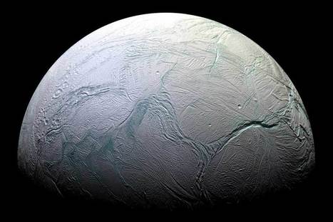
Microbes that produce methane may already be living on Enceladus, a moon of Saturn which is tipped to host life because it boasts a liquid water ocean beneath a crust of solid ice, and strange atmospheric plumes of water. That’s the implication of research showing that an earthbound organism which also produces methane can happily survive in conditions known to exist on Enceladus, from observations by the Cassini space probe before its mission ended last year. Isolated from deep sea vents almost 1000 metres deep in the Okinawa Trough off Japan, Methanothermococcus okinawensis was subjected to gruelling physical and chemical conditions found on Enceladus for more than five years. This microbe, called a methanogenic archaeon, survives without oxygen by combining hydrogen and carbon dioxide – both observed in Enceladus’s atmosphere – to make the energy it needs, emitting methane as a waste product. Cassini detected traces of methane in Enceladus’s plumes, and there’s a chance that some of it may have come from this kind of microbe. “The conditions we mimicked in the lab are as close as possible to those inferred from Cassini on Enceladus,” saysSimon Rittmann at the University of Vienna in Austria, who led the investigations. Rittmann subjected the microbe to various combinations of gases found on Enceladus, and found that it was always able to survive when provided with the moon’s levels of hydrogen and carbon dioxide. It was still able to thrive at temperatures and pressures likely found in Enceladus’s oceans, ranging respectively from 0 to 90 degrees Celsius, and up to 50 Earth atmospheres.
Via Dr. Catherine Russell
|
|
Scooped by
Dr. Stefan Gruenwald
February 22, 2018 6:45 PM
|
Cloud quantum computing has been used to calculate the binding energy of the deuterium nucleus – the first-ever such calculation done using quantum processors at remote locations. Nuclear physicists led by Eugene Dumitrescu at Oak Ridge National Laboratory in the US used publicly available software to achieve the remote operation of two distant quantum computers. Their work could lead to new opportunities for scientists in many fields who want to use quantum simulations to calculate properties of matter. In previous research, scientists have worked alongside quantum computer hardware developers to create quantum simulations. These typically use between two and six qubits to calculate a quantum property of matter – calculations that can be extremely time-consuming to do with a conventional computer. As the number of qubits available in quantum computers increase, the hope is that quantum simulations will be able to do calculations well beyond the reach of even the most powerful conventional computers. However, doing simulations alongside quantum computer specialists can be an inefficient process and the research would be much more streamlined if scientists were able to operate quantum computers themselves.
|
|
Scooped by
Dr. Stefan Gruenwald
February 22, 2018 6:31 PM
|
Meet Todai Robot, an AI project that performed in the top 20 percent of students on the entrance exam for the University of Tokyo -- without actually understanding a thing. While it's not matriculating anytime soon, Todai Robot's success raises alarming questions for the future of human education. How can we help kids excel at the things that humans will always do better than AI?
|
|
Scooped by
Dr. Stefan Gruenwald
February 21, 2018 2:33 PM
|
Aging well is a topic most people have a personal interest in—science certainly does. And it’s revealed some interesting findings in recent years, as long-term studies on “super agers” from across the globe have come in. Of the general population, about a third of people above the age of 90 have dementia, and another third have cognitive decline. But it’s the remaining group of healthy agers that’s so intriguing to researchers.
A couple of new studies presented at a recent American Association for the Advancement of Science meeting looked at people who live well as they age—often into their 90s or beyond. What’s peculiar, and encouraging, is that a lot of how we age has to do not with genetics but with our choices—how we live, physically and socially. And this means that more may be in our control than we think.
One of the new studies, “The 90+ Study,” as the name suggests, has tracked people in their 90s in just about every way possible for 15 years—physical exams, detailed analysis of their social lives and lifestyle habits, and multiple brain scans, before and (if the person died during the study) after death. The other study, on “Super Agers,” looked at people in their 80s, whose cognition and memory matches that of people decades younger.
One factor that played a big role in how a person aged was social interaction: People who lived longer had very close relationships over the years. This connection has been found in many studies on long-term health, the most famous of which was the 80-year Harvard study that found relationships were a key predictor of longevity. “There are brain benefits of having good friends,” said Super Ager study author Emily Rogalski at a press conference.
Another important factor in aging well was, interestingly, drinking alcohol: Those who drank a couple of glasses of wine or beer per day were more likely to live longer, compared to abstainers. “That’s been shown all over the world,” said 90+ Study author Claudia Kawas at the conference. ”I have no explanation for it, but I do firmly believe that modest drinking is associated with longevity.”
Happily, “modest” caffeine intake was also associated with living longer. “The sweet spot for caffeine was 200-400 milligrams a day,” said Kawas.”which, depending on whether you’re a Starbucks fan or an old-fashioned drinker, is about two cups of coffee probably.” People who took in this much from coffee or tea lived longer than people who consumed more or less caffeine.
Another factor was exercising regularly, which isn’t so surprising: People who got as little as 15 minutes per day had an advantage when it came to longevity, and the effect rose with 30 and 45 minutes/day. There was no huge benefit above that, so people who exercised for hours a day had no advantage over those who exercised for 45 minutes.
|
|
Rescooped by
Dr. Stefan Gruenwald
from DNA and RNA research
February 21, 2018 1:52 PM
|
New finding suggests differences in how humans and bacteria control production of DNA’s building blocks. Using a state-of-the-art type of electron microscopy, an MIT-led team has discovered the structure of an enzyme that is crucial for maintaining an adequate supply of DNA building blocks in human cells. Their new structure also reveals the likely mechanism for how cells regulate the enzyme, known as ribonucleotide reductase (RNR). Significantly, the mechanism appears to differ from that of the bacterial version of the enzyme, suggesting that it could be possible to design antibiotics that selectively block the bacterial enzyme. “People have been trying to figure out whether there is something different enough that you could inhibit bacterial enzymes and not the human version,” says Catherine Drennan, an MIT professor of chemistry and biology and a Howard Hughes Medical Institute Investigator. “By considering these key enzymes and figuring out what are the differences and similarities, we can see if there’s anything in the bacterial enzyme that could be targeted with small-molecule drugs.” Drennan is one of the senior authors of the study, which appears in the Feb. 20 issue of the journal eLife. JoAnne Stubbe, the Novartis Professor of Chemistry Emerita at MIT, and Francisco Asturias, an associate professor of biochemistry at the University of Colorado School of Medicine, are also senior authors. The paper’s lead authors are MIT research scientist Edward Brignole and former Scripps Research Institute postdoc Kuang-Lei Tsai, who is now an assistant professor at the University of Texas Houston Medical Center. The RNR enzyme, which is found in all living cells, converts ribonucleotides (the building blocks of RNA) to deoxyribonucleotides (the building blocks of DNA). Cells must keep a sufficient stockpile of these building blocks, but when they accumulate too many, RNR is shut off by a deoxynucleotide molecule known as dATP. When more deoxynucleotides are needed, a related molecule called ATP binds to RNR and turns it back on. An unusual feature of RNR is that it can catalyze the production of four different products: the nucleotide bases often abbreviated as A, G, C, and T. In 2016, Drennan discovered that the enzyme achieves this by changing its shape in response to regulatory molecules. Most of the researchers’ previous work on RNR structure has focused on the version found inE. coli. For those studies, they used X-ray crystallography, a technique that can reveal the atomic and molecular structure of a protein after it has been crystallized.
Via Integrated DNA Technologies
Targeted motor and sensory reinnervation (TMSR) is a surgical procedure on patients with amputations that reroutes residual limb nerves towards intact muscles and skin in order to fit them with a limb prosthesis allowing unprecedented control. By its nature, TMSR changes the way the brain processes motor control and somatosensory input; however the detailed brain mechanisms have never been investigated before and the success of TMSR prostheses will depend on our ability to understand the ways the brain re-maps these pathways. Now, EPFL scientists have used ultra-high field 7 Tesla fMRI to show how TMSR affects upper-limb representations in the brains of patients with amputations, in particular in primary motor cortex and the somatosensory cortex and regions processing more complex brain functions. The findings are published in Brain. Targeted muscle and sensory reinnervation (TMSR) is used to improve the control of upper limb prostheses. Residual nerves from the amputated limb are transferred to reinnervate and activate new muscle targets. This way, a patient fitted with a TMSR prosthetic "sends" motor commands to the re-innervated muscles, where his or her movement intentions are decoded and sent to the prosthetic limb. On the other hand, direct stimulation of the skin over the re-innervated muscles is sent back to the brain, inducing touch perception on the missing limb. But how does the brain encode and integrate such artificial touch and movements of the prosthetic limb? How does this impact our ability to better integrate and control prosthetics? Achieving and fine-tuning such control depends on knowing how the patient's brain re-maps various motor and somatosensory pathways in the motor cortex and the somatosensory cortex.
Via Daniel Perez-Marcos
|
|
Scooped by
Dr. Stefan Gruenwald
February 21, 2018 1:38 PM
|
Lead magnesium niobate (PMN) is a prototypical "relaxor" material, used in a wide variety of applications, from ultrasound to sonar. Researchers have now used state-of-the-art microscopy techniques to see exactly how atoms are arranged in PMN - and it's not what anyone expected. "This work gives us information we can use to better understand how and why PMN behaves the way it does - and possibly other relaxor materials as well," says James LeBeau, an associate professor of materials science and engineering at North Carolina State University and corresponding author of a paper on the work. "What we've found is that the arrangement of atoms in PMN gradually shift along a gradient, from areas of high order to areas of low order; this happens throughout the material," LeBeau says. "That's substantially different than what conventional wisdom predicted, which was there would be alternating areas of high order and no order, right next to each other." This information can be fed into computational models to provide new insights into how PMN's atomic structure influences its characteristics. "This won't happen overnight, but we're optimistic that this may be a step toward the development of processes that create PMN materials with microstructures tailored to emphasize the most desirable characteristics for ultrasound, sonar or other applications," LeBeau says. "It could also potentially offer insights into the role of atomic structure in other relaxor materials, providing similar long-term benefits for the entire class of materials."
|
|
Scooped by
Dr. Stefan Gruenwald
February 16, 2018 4:22 PM
|
An astonishing number of viruses are circulating around the Earth's atmosphere -- and falling from it -- according to new research from scientists in Canada, Spain and the U.S. The study marks the first time scientists have quantified the viruses being swept up from the Earth's surface into the free troposphere, that layer of atmosphere beyond Earth's weather systems but below the stratosphere where jet airplanes fly. The viruses can be carried thousands of kilometers there before being deposited back onto the Earth's surface. "Every day, more than 800 million viruses are deposited per square metre above the planetary boundary layer -- that's 25 viruses for each person in Canada," said University of British Columbia virologist Curtis Suttle, one of the senior authors of a paper in the International Society for Microbial Ecology Journal that outlines the findings. "Roughly 20 years ago we began finding genetically similar viruses occurring in very different environments around the globe," says Suttle. "This preponderance of long-residence viruses traveling the atmosphere likely explains why -- it's quite conceivable to have a virus swept up into the atmosphere on one continent and deposited on another." Bacteria and viruses are swept up in the atmosphere in small particles from soil-dust and sea spray. Suttle and colleagues at the University of Granada and San Diego State University wanted to know how much of that material is carried up above the atmospheric boundary layer above 2,500 to 3,000 meters. At that altitude, particles are subject to long-range transport unlike particles lower in the atmosphere. Using platform sites high in Spain's Sierra Nevada Mountains, the researchers found billions of viruses and tens of millions of bacteria are being deposited per square meter per day. The deposition rates for viruses were nine to 461 times greater than the rates for bacteria.
|
|
Scooped by
Dr. Stefan Gruenwald
February 16, 2018 4:01 PM
|
Ecological scaling laws are intensively studied for their predictive power and universal nature but often fail to unify biodiversity across domains of life. Using a global-scale compilation of microbial and macrobial data, we uncover relationships of commonness and rarity that scale with abundance at similar rates for microorganisms and macroscopic plants and animals. We then show a unified scaling law that predicts the abundance of dominant species across 30 orders of magnitude to the scale of all microorganisms on Earth. Using this scaling law combined with the lognormal model of biodiversity, we predict that Earth is home to as many as 1 trillion (10^12) microbial species.
|
|
|
Scooped by
Dr. Stefan Gruenwald
March 3, 2018 5:38 PM
|
A major trend at NIPS this year is the rise of unsupervised learning and generative modeling. A groundbreaking example of this is the paper “Unsupervised Image-to-Image Translation Networks,” led by NVIDIA researcher Ming-Yu Liu. To date, much of deep learning has used supervised learning to provide machines a human-like object recognition capability. For example, supervised learning can do a good job telling the difference between a Corgi and a German Shepherd, and labeled images of both breeds are readily available for training. To give machines a more “imaginative” capability, such as imagining how a wintery scene would look like in the summer, Liu and team used unsupervised learning and generative modeling. An example of their work is shown below, where the winter and sunny scenes on the left are the inputs and the imagined corresponding summer and rainy scenes are on the right. The NVIDIA Research team’s work uses a pair of generative adversarial networks (GANs) with a shared latent space assumption to obtain these stunning results. Considering the top two images above, the first GAN is trained on the winter scene — overcast skies, bare trees, snow covering just about everything but the cars sailing down the frozen road. The second GAN is trained to understand generally what summer looks like, but hasn’t been trained on the same specific scene as its counterpart. And how could it? You’d need the same footage recorded from the same vantage point, with the same perspective and with all the oncoming traffic and other details in exact same location — for both summer and winter. The unsupervised learning developed by the team removes the need for this capture and labeling, which would otherwise take extensive time and manpower. This unsupervised translation is enabled through a shared latent space assumption, which associates the GANs with each other by tying some of their parameters together. A summery translation of the wintery scene can be generated by transferring the representation from the first GAN to the second. The use of GANs isn’t novel in unsupervised learning, but the NVIDIA research produced results — with shadows peeking through thick foliage under partly cloudy skies — far ahead of anything seen before. The potential benefits of this technique are widespread. In addition to needing less labeled data and the associated time and effort to create and ingest it, deep learning experts can apply the technique across domains. For self-driving cars alone, training data could be captured once and then simulated across a variety of virtual conditions: sunny, cloudy, snowy, rainy, nighttime, etc.
|
|
Rescooped by
Dr. Stefan Gruenwald
from DNA and RNA research
March 1, 2018 1:02 PM
|
A team of scientists has deployed a form of DNA to create a circuit element capable of splitting and combining current—much like an adapter that can connect multiple appliances to a wall outlet. In a new study, a special form, known as G-quadruplex (G4) DNA, is used to improve charge transport properties. G4 DNA is composed of four rather than two strands of DNA that contain only the base guanine. “DNA is capable of conducting charge, but to be useful for nanoelectronics, it must be able to direct charge along more than one path by splitting or combining it,” says Peng Zhang, an assistant research professor of chemistry at Duke University and a co-author of the study. “We have solved this problem by using the guanine quadruplex (G4) in which a charge can arrive on a duplex on one side of this unit and go out either of two duplexes on the other side.” “Various device-like functions have been reported for two-terminal single molecular junctions, but a practical circuit-like element requires three or more terminals for control and for electrical networking,” notes Seeman. “This work demonstrates that a strategically designed G-quadruplex motif can fulfill the critical requirements for moving beyond two-terminal functionality.” “This is the first step needed to transport charge through a branching structure made exclusively of DNA,” Zhang adds. “It is likely that further steps will result in successful DNA-based nanoelectronics that include transistor-like devices in self-assembling ‘pre-programmed’ materials.”
Via Integrated DNA Technologies
|
|
Scooped by
Dr. Stefan Gruenwald
March 1, 2018 12:56 PM
|
Technology that mimics the functions and sensations of human skin may sound futuristic, but it isn’t exactly new. In 2010, scientists at Stanford University and at University of California, Berkeley, developed artificial skin that could feel even a gentle touch. Dubbed e-skin, the device holds great potential to help future robots more delicately interact with humans. But now, researchers at the University of Colorado Boulder are giving e-skin an environmentally friendly upgrade. As Adam Epstein writes for Quartz, the latest iteration of the device can re-heal itself and be recycled to make new layers of skin. The recyclable e-skin, which was described last week in the journal Science Advances, looks like a thin, yellow adhesive sticker. Embedded in this thin layer are sensors that measure pressure, temperature, humidity and air flow. It’s made out of a polymer and silver nanoparticles, which allows it to bend and move with curved spaces like fingers or toes. It’s also soft, but not quite as stretchy as human skin, The Verge’s Alessandra Potenza reports. Many labs around the world are creating e-skin. But what makes this e-skin special, Potenza points out, is that it’s virtually waste-free. It heals itself through a chemical reaction between the compounds that make up the bandage-like film. If it is sliced, the researchers can simply just add more of the original three active compounds that make up the film and the ensuing chemical reaction will knit the e-skin back together, “healing” the wound, Potenza reports. If ever damaged beyond repair, it can also be soaked in a solution that separates out the silver nanoparticles and then be recycled into new e-skin. The healing process happens fast, too. It only takes a few minutes at 140 degrees Fahrenheit or 30 min at room temperature, while the recycling process takes about 30 minutes at 140 degrees or 10 hours at room temperature.
|
|
Scooped by
Dr. Stefan Gruenwald
March 1, 2018 12:51 PM
|
Researchers at VIB, KU Leuven and UZ Leuven have devised a novel approach to develop antibacterial drugs. With antibiotic resistance on the rise worldwide, such new drugs are urgently needed. The Flemish biotech spin-off Aelin Therapeutics will exploit the technology to produce new antibiotics for the clinic. Around the world bacteria are developing new strategies that render them resistant to antibiotics, resulting in an increasing number of patients dying from untreatable infections. Experts now agree that unless we discover new medicines soon, the post-antibiotic era may be upon us, effectively pushing human healthcare back to the 1940s. In particular, a class of bacteria called 'Gram-negative bacteria', which include the well-known species E. coli, are causing major problems in our hospitals. The problem is that most antibiotics that we know today work according to only a few mechanisms of action and so when a bacterium becomes tolerant to one drug, it often becomes tolerant to the whole family. What we need to solve this situation is an entirely new class of drugs that shares no structural or mechanistic similarities with the existing antibiotics. Prof. Joost Schymkowitz and Prof. Frederic Rousseau of VIB-KU Leuven in collaboration with Prof. Johan Van Eldere of University Hospitals Leuven have gone one step further: they have developed a new way of designing antibiotic drugs that can give rise to many new antibacterial molecules. These drugs penetrate bacterial cells where they induce a process called protein aggregation. This process resembles what happens when boiling an egg, but now without heat: proteins that normally need to carry out essential functions for the bacteria—such as digesting their food—clump together and can no longer carry out their work. As this affects many proteins in the bacterial cell all at once, the bacteria rapidly succumb and die.
|
|
Scooped by
Dr. Stefan Gruenwald
February 23, 2018 2:29 PM
|
Astronomers have found one of the most distant exploding stars ever seen — the light from the colossal event took a staggering 10 billion years to travel across the Universe to reach us! And, like so many of its brethren, it will help us understand why these stars explode, what kind of environment they grew up in, and maybe even how the Universe itself has changed over the eons. The supernova is called DEC16C2nm, and was seen in images taken for the Dark Energy Survey, a huge project that maps millions of distant galaxies over 5,000 square degrees (about 1/8th of the whole sky!) to better understand the structure of the Universe. Because it takes such deep images, it will catch lots of stars exploding over time … and over space. This supernova was first seen in images taken in August 2016. Once it was discovered, astronomers looked in earlier data to see if they could spot it, and it might have been seen in observations taken as early as March, but definitely not in February. It reached peak brightness in September of 2016 and has been fading since.
|
|
Scooped by
Dr. Stefan Gruenwald
February 22, 2018 6:43 PM
|
Technique could be used to create quantum-information systems Molecules” made from three photons have been created by physicists in the US. The photon triplets were made by firing laser light into an atomic gas and the researchers believe that their technique could be useful for creating entangled photons for quantum-information systems. Normally photons do not interact with each other and therefore will not bind together to form molecule-like structures. But in 2013 Mikhail Lukin at Harvard University and Vladan Vuletić at the Massachusetts Institute of Technology managed to get pairs of photons to stick together by firing a weak laser beam through an ultracold gas of atoms. Now, they have repeated the feat for three photons. Their technique involves the light creating a “Rydberg polariton” in the atomic gas. This is a particle-like collective state in which a highly-excited electron is shared by several atoms. This polariton propagates through the gas like a slow-moving photon with non-zero mass. When it reaches the opposite edge of the gas, the polariton is converted back to light. The presence of a Rydberg polariton prevents nearby photons from creating their own excitations – a phenomenon called Rydberg blockade. Instead, and under certain conditions, the index of refraction of the gas near the Rydberg polariton is modified such that other photons will bunch-up with the polariton. This is effectively an attractive interaction between photons that creates molecules of light. In this latest experiment a team led by Vuletić and Lukin observed both pairs and triplets of photons emerging from the atomic gas, rather than the random emission of single photons that would occur if molecules were not forming. They also measured the phase of the photon pairs and triplets and found this to be consistent with an attractive interaction. Indeed, the phase measurements revealed that the photon triplets were more strongly bound than the photon pairs.
|
|
Scooped by
Dr. Stefan Gruenwald
February 21, 2018 2:47 PM
|
Researchers have created a tiny robot, small enough to navigate a stomach or urinary system, that one day may be used to deliver drugs inside the body. Researchers in Germany have developed a robot that is about a seventh of an inch long and looks at first like no more than a tiny strip of something rubbery. Then it starts moving. The robot walks, jumps, crawls, rolls and swims. It even climbs out of the pool, moving from a watery environment into a dry one. The robot prototype is small enough to move around in a stomach or urinary system, said Metin Sitti, head of the physical intelligence department at the Max Planck Institute for Intelligent Systems in Stuttgart, Germany, who led the research team. The robot hasn’t been tested in humans yet, but the goal is to improve it for medical use — for instance, delivering drugs to a target within the body. What is most unusual about the research, Dr. Sitti said, is that such a “minimalist robot” can achieve “all different type of motion possibilities to navigate in complex environments.” Leif Ristroph, a mathematician at New York University’s Courant Institute who developed a small flying robot that mimics the motion of jellyfish, wrote in an email: “The array of behaviors and capabilities is certainly impressive and sets this robot apart from most others.” “These critters are very cute!” he said. “Love how the authors put the little guy through mini-obstacle courses.” “My other thought is that the pilot, who we don’t see, is also quite impressive,” added Dr. Ristroph, who was not involved in the research. “Clearly whoever is controlling the magnetic fields has gained some hard-earned intuition and fine skills based on a lot of experience and trial-and-error.” The research was reported Wednesday in the journal Nature. Below are excerpts from a telephone conversation with Dr. Sitti. They have been edited for length and clarity.
|
|
Scooped by
Dr. Stefan Gruenwald
February 21, 2018 2:27 PM
|
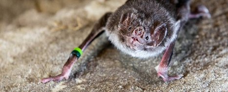
Vampire bats are probably not the cuddliest of critters, considering nearly their entire food source consists of blood. There are three types of bats who solely drink blood, the common vampire bat (Desmodus rotundus), the hairy-legged vampire bat (Diphylla ecaudata), and the white-winged vampire bat (Diaemus youngi). All three of these animals evolved from bats who eat fruit, but we still aren't sure how they managed to make the huge leap to the minimally nutritious, and relatively dangerous activity of drinking blood. This is because subsisting on just blood, called hematophagy, is very uncommon. The amount of liquid can overwhelm the kidneys, too much can cause iron poisoning, and excessive protein isn't good for the body either. And blood is super high in protein (93 percent), but extremely low in carbohydrates (1 percent) and vitamins. Plus there are a bunch of blood-borne diseases. The international group of researchers took samples of the bats' droppings to look at something called the 'hologenome' – the entire set of genes of an organism, including all the bacteria and other microbes that make that creature their home. They analyzed the common vampire bat's hologenome against a number of insect-, fruit-, or meat-eating bats to try and determine what makes the vampire bat so weird. What they found is that the gut microbes in the bat were an especially unique combination, which most other bats (or other mammals) wouldn't be able to stomach. In fact, more than 280 of the bacterial species found in the droppings are known to cause disease in other mammals. "The data suggests that there is a close evolutionary relationship between the gut microbiome and the genome of the vampire bat for adaptation to sanguivory (feeding exclusively on blood)," biologist Marie Zepeda Mendoza of the University of Copenhagen in Denmark explains. The researchers also found that the vampire bat's genome had more transposons, also known as 'jumping genes' - genes in the DNA that are able to multiply and move around the genome. There was as much as a 2.2 fold increase in the amount of one particular transposon, called MULE-MuDR in the vampire bat, compared to other types of bats the researchers looked at. The extra MULE-MuDR copies were mostly found in areas involved in immune response, viral defence and metabolism. The researchers think this helps the bat better process the huge amount of blood it can ingest per day, without getting sick. "It is clear from our results that the common vampire bat has adapted to sanguivory through a close relationship between its genome and gut microbiome," the researchers write in the paper.
|
|
Rescooped by
Dr. Stefan Gruenwald
from Spaceport UK
February 21, 2018 1:47 PM
|
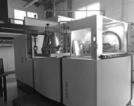
Edinburgh-based Skyrora, a company with partners in Ukraine, will launch a partially 3D printed suborbital vehicle from the north of Scotland later this year. Skyrora’s rocket engines run on hydrogen peroxide and kerosene. America, Russia, China, and… Scotland? Yes, the newest contender in the space race is that tiny northern country of the United Kingdom, Scotland, whose very own Skyrora has developed a suborbital launch vehicle that will take off from northern Scotland in the last three months of 2018. It’s a great achievement for the company, which also operates from Ukraine and which has used 3D printing to develop parts for its spacecraft. Potential launch locations include Shetland, where the Shetland Space Centre is bidding for a license from the UK Space Agency. If the company can secure a launch location, its partly 3D printed suborbital launch vehicle could be taking off within the year.
Via Aso Galicia
|
|
Scooped by
Dr. Stefan Gruenwald
February 21, 2018 1:42 PM
|
When we talk about the dangers posed by artificial intelligence, the emphasis is usually on the unintended side effects. We worry that we might accidentally create a super-intelligent AI and forget to program it with a conscience; or that we’ll deploy criminal sentencing algorithms that have soaked up the racist biases of their training data. But this is just half the story. What about the people who actively want to use AI for immoral, criminal, or malicious purposes? Aren’t they more likely to cause trouble — and sooner? The answer is yes, according to more than two dozen experts from institutes including the Future of Humanity Institute, the Centre for the Study of Existential Risk, and the Elon Musk-backed non-profit OpenAI. Very much yes. In a report published today titled “The Malicious Use of Artificial Intelligence: Forecasting, Prevention, and Mitigation,” these academics and researchers lay out some of the ways AI might be used to sting us in the next five years, and what we can do to stop it. Because while AI can enable some pretty nasty new attacks, the paper’s co-author, Miles Brundage of the Future of Humanity Institute, tells The Verge, we certainly shouldn’t panic or abandon hope. “I like to take the optimistic framing, which is that we could do more,” says Brundage. “The point here is not to paint a doom-and-gloom picture — there are many defenses that can be developed and there’s much for us to learn. I don’t think it’s hopeless at all, but I do see this paper as a call to action.” The report is expansive, but focuses on a few key ways AI is going to exacerbate threats for both digital and physical security systems, as well as create completely new dangers. It also makes five recommendations on how to combat these problems — including getting AI engineers to be more upfront about the possible malicious uses of their research; and starting new dialogues between policymakers and academics so that governments and law enforcement aren’t caught unawares.
|
|
Rescooped by
Dr. Stefan Gruenwald
from DNA and RNA research
February 21, 2018 1:37 PM
|
University of Washington and Microsoft researchers revealed today that they have taken a significant step forward in their quest to develop a DNA-based storage system for digital data. In a paper published in Nature Biotechnology, the members of the Molecular Information Systems Laboratory (MISL) describe the science behind their world record-setting achievement of 200 megabytes stored in synthetic DNA. They also present their system for random access — that is, the selective retrieval of individual data files encoded in more than 13 million DNA oligonucleotides. While this is not the first time researchers have achieved random access in DNA, the UW and Microsoft team have produced the first demonstration of random access at such a large scale. One of the big advantages to DNA as a digital storage medium is its ability to store vast quantities of information, with a raw limit of one exabyte — equivalent to one billion gigabytes — per cubic millimeter. The data must be converted from digital 0s and 1s to the molecules of DNA: adenine, thymine, cytosine, and guanine. To restore the data to its digital form, the DNA is sequenced and the files decoded back to 0s and 1s. This process becomes more daunting as the amount of data increases — without the ability to perform random access, the entire dataset would have to be sequenced and decoded in bulk in order to find and retrieve specific files. In addition, the DNA synthesis and sequencing processes are error-prone, which can result in data loss. MISL researchers addressed these problems by designing and validating an extensive library of primers for use in conjunction with polymerase chain reaction (PCR) to achieve random access. Before synthesizing the DNA containing data from a file, the researchers appended both ends of each DNA sequence with PCR primer targets from the primer library. They then used these primers later to select the desired strands through random access, and used a new algorithm designed to more efficiently decode and restore the data to its original, digital state. “Our work reduces the effort, both in sequencing capacity and in processing, to completely recover information stored in DNA,” explained Microsoft Senior Researcher Sergey Yekhanin, who was instrumental in creating the codec and algorithms used to achieve the team’s results. “For the latter, we have devised new algorithms that are more tolerant to errors in writing and reading DNA sequences to minimize the effort in recovering this information.” Using synthetic DNA supplied by Twist Bioscience, the MISL team encoded and successfully retrieved 35 distinct files ranging in size from 29 kilobytes to over 44 megabytes — amounting to a record-setting 200 megabytes of high-definition video, audio, images, and text. This represents a significant increase over the previous record of 22 megabytes set by researchers from Harvard Medical School and Technicolor Research & Innovation in Germany. “The intersection of biotech and computer architecture is incredibly promising and we are excited to detail our results to the community,” said Allen School professor Luis Ceze, who co-leads the MISL. “Since this paper was submitted for publication we have reached over 400 megabytes, and we are still growing and learning more about large-scale DNA data storage.”
Via Integrated DNA Technologies
|
|
Scooped by
Dr. Stefan Gruenwald
February 16, 2018 4:13 PM
|
Deaf mice have been able to hear a tiny whisper after being given a "landmark" gene therapy by US scientists. They say restoring near-normal hearing in the animals paves the way for similar treatments for people "in the near future".
Studies, published in Nature Biotechnology, corrected errors that led to the sound-sensing hairs in the ear becoming defective. The researchers used a synthetic virus to nip in and correct the defect.
"It's unprecedented, this is the first time we've seen this level of hearing restoration," said researcher Dr Jeffrey Holt, from Boston Children's Hospital.
About half of all forms of deafness are due to an error in the instructions for life - DNA. In the experiments at Boston Children's Hospital, Massachusetts Eye and Ear and Harvard Medical School, the mice had a genetic disorder called Usher syndrome. It means there are inaccurate instructions for building microscopic hairs inside the ear.
In healthy ears, sets of outer hair cells magnify sound waves and inner hair cells then convert sounds to electrical signals that go to the brain. The hairs normally form these neat V-shaped rows. Sound waves produce the sensation of hearing by vibrating hair-like structures on the inner ear’s sensory hair cells. But how this mechanical motion gets converted into electrical signals that go to our brains has long been a mystery. Scientists have believed some undiscovered protein is involved. Such proteins have been identified for taste, smell and sight, but the protein required for hearing has been elusive. In part, that’s because it’s hard to get enough cells from the inner ear to study – they’re embedded deep in the cochlea. “People have been looking for more than 30 years,” says Jeffrey Holt of the department of otolaryngology at Children’s Hospital Boston. “Five or six possibilities have come up, but didn’t pan out.” Recently, in the Journal of Clinical Investigation, team led by Holt and Andrew Griffith, of the National Institute on Deafness and Other Communication Disorders (NIDCD), demonstrated that two related proteins, TMC1 and TMC2, are essential for normal hearing – paving the way for a test of gene therapy to reverse a type of genetic deafness. The two proteins make up gateways known as ion channels, which sit atop the hair-like structures (a.k.a. stereocilia) and let electrically charged molecules move in to the cell, generating an electrical signal that ultimately travels to the brain. When both the TMC1 and the TMC2 genes are mutated, sound waves can’t be converted to electrical signals – they literally fall on deaf ears.
|