 Your new post is loading...

|
Scooped by
Gilbert C FAURE
August 14, 2019 8:29 AM
|
Abstract Regulatory T cells (Tregs) are specialized in immune suppression and play a dominant role in peripheral immune tolerance. Treg cell lineage development and function maintenance is determined by the Foxp3 transcriptional factor, whose activity is fine-tuned by its post-translational modifications (PTMs) and interaction partners. In this review, we summarize current studies in the crystal structures, the PTMs and interaction partners of Foxp3 protein, and discuss how these insights may provide a roadmap for new approaches to modulate Treg suppression, and new therapies to enhance immune tolerance in autoimmune diseases. This article is protected by copyright. All rights reserved.
|
|
Scooped by
Gilbert C FAURE
August 14, 2019 4:24 AM
|
In the past 50 years, technological developments and intensive basic research have enabled substantial progress in our understanding of how the normal immune system works, how it participates in the pathophysiology of human diseases such as autoimmunity and immunodeficiencies, and how it can be pharmacologically influenced to combat diseases such as cancer, autoimmunity, inflammation, and infections. Arguably, the most detailed comprehension of the immune system concerns the activation and development of T cells and B cells, which collectively stipulate antigen‐specific immunity, by way of antigen‐specific helper or cytotoxic T cells and antigen‐specific antibody‐producing B cells. Beginning with the identification and gene cloning of the antigen receptors on T cells and B cells, research in this area has led to the characterization of multiple other receptors that diligently fine‐tune the activation and development of T cells and B cells, including co‐receptors, co‐stimulatory receptors, and co‐inhibitory receptors (Figure 1). Similarly, the intracellular signal transduction machineries coupling these receptors to cellular responses have been significantly elucidated. Mechanisms enabling diversification and specification of intracellular signals have been identified. More recent work, which has been facilitated by developments in microscopy technology and biophysical assays, has yielded an increasingly more detailed appreciation of the organization of signaling mechanisms at the molecular level, both in space and in time. A clearer picture of how alterations in these molecular pathways are linked to human diseases has ensued. In this issue of Immunological Reviews, leaders in the field review the literature and provide insights regarding various aspects of signal transduction and diversification in T cells and B cells. First, three reviews deal with the mechanisms by which engagement of the T‐cell antigen receptor (TCR) and other immune receptors leads to signal initiation. Wolgang Schamel (Freiburg, Germany) and collaborators review the main models of TCR conformational regulation. In essence, they converse on the current hypotheses regarding how ligand binding to the TCR changes the structure of the associated CD3‐ζ subunits. A unifying model is proposed. Etienne Gagnon (Montréal, Québec, Canada) and his colleague review the importance of electrostatic interactions in assembly, maintenance of inactive state, triggering and signaling by immune receptors, with a special emphasis on T cells. They discuss how the cytoplasmic domains of TCR subunits interact with charged lipids in the plasma membrane, and how these interactions are regulated in order to influence TCR signal initiation and strength. Furthermore, Enfui Hui (San Diego, CA, USA) and his collaborators review recent inroads into T‐cell signaling that have been provided by membrane reconstitution analyses. Notably, their own work has led to key new insights into how the inhibitory immune checkpoint receptor programmed death‐1 controls T‐cell activation. Next, two reviews focus on the mechanisms involved in amplification of antigen receptor‐driven signals. Claire Hivroz (Paris, France) and her colleagues review how TCR signaling is amplified and diversified in the endocytic compartment. They present data showing that the transmembrane adapter linker for activation of T cells (LAT) is shuttled between endocytic compartments and the immune synapse during T‐cell activation to provide signal amplification. Likewise, Wanli Liu (Beijing, China) and collaborators examine how the phospholipid phosphatidylinositol (PI) 4,5‐bisphosphate (PIP2) amplifies B‐cell antigen receptor (BCR)‐driven signals in B cells. Based on the studies discussed, the authors propose a “gasoline engine model” for amplification of B‐cell activation. They also review data showing the potential broad applicability of this model to other receptor signaling systems. In addition to antigen receptors, multiple other receptors and intracellular signaling molecules play essential roles in T‐cell and B‐cell activation and development. Four reviews cover specific aspects of this topic. Inducible costimulator (ICOS) is a receptor expressed on T cells that plays a critical role in co‐stimulation. Woong‐Kyung Suh (Montréal, Québec, Canada) and his team discuss in detail the roles, mechanisms of action, and signaling mechanisms of ICOS in immune cells, not only in follicular T‐helper (TFH) cells but also in other cell types. The authors also compare the functions and signaling mechanisms of ICOS with those of the related receptor CD28. Ion channels, including the calcium release‐activated channel (CRAC), have been broadly implicated in lymphocyte functions. Bebhinn Treanor (Toronto, Ontario, Canada) and colleagues review how these channels regulate B‐cell activation and development. They discuss not only calcium channels such as CRAC, but also chloride, potassium, and other types of ion channels. The Cbl proteins are ubiquitin ligases implicated in ubiquitination and degradation of several components of the antigen receptor signaling machinery in T cells and B cells. Hua Gu (Montréal, Québec, Canada) and his colleagues review accumulating data showing the broad range of molecular targets for the Cbl proteins in T cells and B cells, allowing the Cbl proteins to be gatekeepers of T‐cell activation, B‐cell development, and the germinal center reaction. Lastly, Jeroen Roose (San Francisco, CA, USA) and his collaborators review recent data showing how more downstream effectors of T‐cell activation, such as mammalian target of rapamycin and other kinases, are regulated to control T‐cell activation. The involvement of these kinases in mRNA translation, metabolic adaptation, and tonic signaling is also discussed. Last, three reviews deal with how alterations in components of the antigen receptor signaling machinery in T cells, B cells, or both have been broadly implicated in the pathophysiology of human diseases. Pamela Schwartzberg (Bethesda, MA, USA) and her team show that activating mutations in PI 3′ kinase in humans result in altered T‐cell and B‐cell functions, thereby causing a range of clinical manifestations including immunodeficiency and autoimmunity. In a related way, Sylvain Latour (Paris, France) and his collaborators examine how mutations in a broad array of components of the T‐cell signaling apparatus can cause immunodeficiencies in humans, especially upon infection by Epstein‐Barr virus. Some of these mutations affect proteins required for T‐cell cytotoxicity, while others involve polypeptides needed for T‐cell proliferation. Finally, Louis Staudt (Bethesda, MD, USA) and colleagues underscore how activating mutations in the BCR signaling machinery are implicated in human B‐cell malignancies. They discuss compelling clinical data showing that inhibitors of the mutated molecules can be used to treat effectively these diseases. Altogether, this monograph by leading scientists underscores the extensive progress that has been made through basic research in understanding how activation and development of T cells and B cells is controlled at the molecular level. It also clearly shows that alterations in the T‐cell or B‐cell signaling apparatus are implicated in human diseases. Furthermore, it indicates that dysregulated components of this machinery can be targeted to treat human pathologies. Nonetheless, while progress in this research area has been enormous over the past 50 years, it is clear that much more work in the future is needed to comprehend fully the processes involved, in order to use best the information provided for understanding and treating human diseases.
|
|
Scooped by
Gilbert C FAURE
August 9, 2019 1:23 PM
|
Immunotherapy with immune checkpoint blockers (ICBs) has revolutionized cancer treatment. Unfortunately, ICB therapy usually benefits <15% of patients and causes immune-related adverse events in a substantial number of patients. Another immunotherapy—engineered chimeric antigen receptor (CAR) T cells that specifically target tumor-associated antigens—has transformed the treatment of multiple hematological cancers and exhibited therapeutic potential in solid tumors. Emerging data show that the function of blood vessels associated with tumors is critical in the response to these immunotherapies (1, 2). The vasculature in tumors is abnormal, which impairs blood flow and limits the delivery of oxygen, nutrients, and therapeutics, including antibodies and immune cells (3) (see the figure). The resulting hypoxia (low oxygen concentration) and low pH can in turn induce the production of immunosuppressive molecules, such as transforming growth factor–β (TGFβ), vascular endothelial growth factor (VEGF), and adenosine in the tumor microenvironment (TME). Some of these molecules lower the expression of adhesion molecules on tumor vascular endothelium, the lining of vessel walls that normally supports immune cell adhesion and transmigration into tissues. Reducing the expression of adhesion molecules therefore interferes with the ability of immune cells to adhere to and migrate across the vessel wall, preventing their entry into the tumor. In addition, pro-angiogenic molecules produced by cancer, stromal, and immune cells up-regulate Fas ligand on endothelial cells and thereby induce the apoptosis of infiltrating immune cells. VEGF can hinder maturation of dendritic cells (DCs), which are needed to present tumor-associated antigens and activate effector immune cells. Furthermore, hypoxia directly up-regulates the expression of immune checkpoint molecules [e.g., programmed cell death protein ligand 1 (PD-L1)], which suppress effector T cell activation, on various cells in the TME, including myeloid-derived suppressor cells (MDSCs) and DCs. Hypoxia also promotes the recruitment of immunosuppressive regulatory T lymphocytes (Treg cells) and reprograms tumor-associated macrophages (TAMs) from an antitumor to a protumor phenotype. Notably, the immunosuppressive molecules generated in the TME can enter the circulation and cause systemic immunosuppression. Given the consequences of malfunctioning blood vessels, multiple therapeutic strategies to improve the function of the tumor vasculature have been developed (4). Because excess VEGF contributes to vascular abnormalities, it was proposed that the judicious use of anti-VEGF agents could normalize tumor vessels and thereby improve oxygenation and drug delivery. This was supported by experiments showing that low-dose anti–VEGF receptor 2 (VEGFR2) treatment enhances trafficking of immune cells to tumors, reprograms TAMs to an antitumor phenotype, and improves outcomes of vaccine therapy in mouse breast cancer models (5). A number of preclinical studies have now shown the benefit of combining anti-VEGF agents with various immunotherapies, including ICBs and adoptively transferred engineered CAR T cells (3). Anti-VEGF–induced vascular normalization is transient, which limits efficacy. Thus, a number of strategies are emerging to extend the window of normalization. One such strategy is to target angiopoietin-2 (ANG-2), which begins to increase toward the end of the normalization window. Importantly, dual blockade of VEGF and ANG-2 prolonged the duration of normalization and survival in a number of mouse tumor models, compared with inhibiting either molecule alone. Relevant for immunotherapies, VEGF and ANG-2 dual targeting reprogrammed TAMs to an antitumor phenotype (6) and enhanced tumor response to ICBs (7). Furthermore, circulating ANG-2 concentrations are initially elevated or increase in melanoma patients with an unfavorable response to ICBs. Moreover, blood ANG-2 concentrations inversely correlate with response to ICBs with or without anti-VEGF therapy in melanoma patients (3), providing additional rationale for VEGF and ANG-2 dual inhibition. A number of other agents that target endothelial cells and/or pericytes, which support the structure and function of blood vessels, have been shown to transiently normalize tumor vessels. These targets include the receptor tyrosine kinase TIE-2, Notch signaling, endothelial cell metabolism, and oxygen sensors (4). Other strategies that can induce vessel normalization—either directly or indirectly—include restoring perivascular nitric oxide gradients, some chemotherapeutics such as eribulin, and aerobic exercise. Notably, two recent studies showed that ICBs can normalize tumor vessels in breast cancer models in mice (8, 9), which is intriguing because it implies that T cells can directly affect vascular function. These studies showed that the increased vessel perfusion by ICB is positively correlated with its efficacy (9). Future efforts will determine whether these agents can be combined with particular ICBs in specific cancer types. Physical forces generated by cells and the extracellular matrix (ECM) in tumors can also impair the function of blood and lymphatic vessels through compression (4). This is particularly the case in desmoplastic tumors, which are characterized by excessive fibrosis and activated stroma [e.g., pancreatic ductal adenocarcinoma (PDAC) and triple-negative breast cancer] and which generally have abundant carcinoma-associated fibroblasts (CAFs) and ECM. Killing cells with cytotoxic agents or depleting specific ECM components by using enzyme-based drugs can reopen these vessels. However, as cancer cells become resistant to cytotoxic agents and begin to proliferate, vessels collapse again. Similarly, enzymes that target a single ECM component (e.g., hyaluronan), become ineffective when other ECM components (e.g., collagen I) begin to contribute to compressive forces. Agents that reprogram CAFs so that they stop producing multiple ECM components can overcome this compression. For example, angiotensin system inhibitors (ASIs) are a class of molecules commonly used to treat hypertension that also reprogram CAFs and have been shown to improve the function of tumor vessels as well as the delivery and efficacy of various therapeutics (10). Moreover, these widely used drugs can activate both innate and adaptive antitumor immune responses in mouse models and patients (10). On the basis of a successful phase II clinical trial, this approach is now being tested in a randomized clinical trial in patients with locally advanced PDAC (NCT03563248). Because ASIs can cause hypotension, new formulations are needed to lower the systemic exposure of these drugs. Recently, valsartan—an ASI linked to pH-sensitive polymers—was shown to reprogram CAFs in the acidic tumor environment to alleviate antitumor immunosuppression and improve T lymphocyte activity without adverse hypotension. This approach improved the response to ICBs in mice bearing primary and metastatic breast tumors (2). Other strategies are also emerging for targeting CAF-mediated desmoplasia and vessel compression. For example, blocking C-X-C motif chemokine 12 (CXCL12)-C-X-C chemokine receptor 4 (CXCR4) signaling in mouse models of primary and metastatic breast cancer reduces fibrosis, vessel compression, and hypoxia while alleviating immunosuppression, which enhances the efficacy of ICBs (1). Similarly, the activity of tumor-associated immunosuppressive cells and the highly desmoplastic stroma in PDACs are fueled by cancer cell focal adhesion kinase (FAK) activity. Selective FAK inhibitors normalized the fibrotic TME, improved T cell infiltration of the tumor, and increased survival of tumor-bearing mice (11). Endothelial cells present in tertiary lymphoid organs (organized structures consisting of lymphocytes and stromal cells that form outside of the spleen or lymph nodes) within tumors can also regulate antitumor immunity. By combining VEGFR2 and PD-L1 antibodies, it is possible to induce the formation of specialized vessels called high endothelial venules (HEVs) in some tumor models, which improved survival (12). HEVs facilitate the entry of immune cells into lymph nodes and might serve a similar function in tumors. Moreover, depletion of Treg cells can induce the formation of HEVs outside of lymph nodes and the spleen, where they are normally found. When HEVs form in tumors, they can support lymphocyte infiltration and subsequent tumor cell killing by cytotoxic T lymphocytes (13). Lymphatic vessels are the routes from tissue to lymph nodes and back to the blood circulation. Therefore, lymphatic endothelial cells (LECs) are among the first cells to contact antigens that drain from tissue. LECs can control DC maturation and migration as well as directly present antigens on major histocompatibility complex class I (MHC I) and MHC II molecules to T cells (14). They also produce cytokines that modulate the immune response. Numerous lines of evidence demonstrate that LECs help limit and resolve effector T lymphocyte responses, modulate leukocyte function, and maintain peripheral tolerance, which prevents autoimmune responses (14). In cancer, LECs also suppress immune cell antitumor responses (14). Paradoxically, tumors that contain LECs (even in the absence of functional lymphatic vessels) show better responses to ICBs. Tumors with more intratumoral LECs have more T cells, but they become quiescent. These dysfunctional yet antigen-educated T cells can be reawakened by ICBs to produce antitumor immunity. Immune-related toxicities are a major problem with ICB and CAR–T cell immunotherapies. However, discontinuing or reducing the dose of these agents can reduce or even abrogate these toxicities. Improving the function of tumor blood vessels can improve the delivery of antibodies and cells and thus may lower the required immunotherapy dose. Such approaches hold promise for reducing toxicities while improving efficacy. However, one of the biggest challenges in realizing this goal is the lack of validated biomarkers for guiding the dose and schedule of drugs that directly or indirectly target blood vessels. This is further complicated by the recently described “paradoxical effects of obesity.” Obesity is known to fuel tumor progression, desmoplasia, hypoxia, immunosuppression, and resistance to various therapies, including chemotherapies and anti-VEGF therapies (3). Yet in mouse models of melanoma and lung cancer, and in various human cancers, obesity is associated with improved response to ICBs (15). Given the epidemic of obesity, a better understanding of the mechanisms underlying these paradoxical effects are likely to yield strategies to improve immunotherapy further. Combining strategies that improve function of tumor vessels with ICBs holds promise. For example, three recent phase III trials have shown the benefit of combining PD1 or PD-L1 antibodies with anti-VEGF or anti-VEGFR agents, leading to U.S. Food and Drug Administration approvals for the combination in lung and kidney cancers (NCT02684006, NCT02853331, and NCT02366143). However, the relative contribution to patient survival from immunomodulatory versus nonimmunomodulatory effects of VEGF blockade with ICBs in these trials is not known. The ongoing trial that aims to test the role of adding losartan—a safe, inexpensive, and widely prescribed antihypertensive ASI shown to decompress tumor blood vessels—to chemo-radiation and PD1 antibody therapy will reveal the potential of this approach in improving the treatment outcome in patients with PDAC (NCT03563248). http://www.sciencemag.org/about/science-licenses-journal-article-reuse This is an article distributed under the terms of the Science Journals Default License. References and Notes ↵ I. X. Chen et al., Proc. Natl. Acad. Sci. U.S.A. 116, 4558 (2019). ↵ V. P. Chauhan et al., Proc. Natl. Acad. Sci. U.S.A. 116, 10674 (2019). ↵ D. Fukumura et al., Nat. Rev. Clin. Oncol. 15, 325 (2018).OpenUrlCrossRefPubMed ↵ J. D. Martin et al., Annu. Rev. Physiol. 81, 505 (2019).OpenUrl ↵ Y. Huang et al., Proc. Natl. Acad. Sci. U.S.A. 109, 17561 (2012). ↵ J. Kloepper et al., Proc. Natl. Acad. Sci. U.S.A. 113, 4476 (2016). ↵ M. Schmittnaegel et al., Sci. Transl. Med. 9, eaak9670 (2017). ↵ L. Tian et al., Nature 544, 250 (2017).OpenUrlCrossRef ↵ X. Zheng et al., J. Clin. Invest. 128, 2104 (2018).OpenUrl ↵ M. Pinter, R. K. Jain, Sci. Transl. Med. 9, eaan5616 (2017).OpenUrl ↵ H. Jiang et al., Nat. Med. 22, 851 (2016).OpenUrlCrossRefPubMed ↵ E. Allen et al., Sci. Transl. Med. 9, eaak9679 (2017). ↵ C. F. Sautès-Fridman et al., Nat. Rev. Cancer 19, 307 (2019).OpenUrl ↵ K. Maisel et al., Adv. Drug Deliv. Rev. 114, 43 (2017).OpenUrl ↵ Z. Wang et al., Nat. Med. 25, 141 (2019).OpenUrl Acknowledgments: L.L.M. and R.K.J. thank Z. Amoozgar, D. G. Duda, D. Fukumura, Y. Huang, and T. Padera for helpful comments. R.K.J. received honorarium from Amgen; consultant fees from Chugai, Enlight, Merck, Ophthotech, Pfizer, SPARC, and SynDevRx; owns equity in Enlight, Ophthotech, and SynDevRx; and serves on the Boards of Trustees of Tekla Healthcare Investors, Tekla Life Sciences Investors, Tekla Healthcare Opportunities Fund, and Tekla World Healthcare Fund. The Massachusetts General Hospital has applied for patents related to concepts discussed in this article. L.L.M. owns equity in Bayer AG.
|
|
Scooped by
Gilbert C FAURE
August 9, 2019 6:44 AM
|
One of the mechanisms by which tumors evade immune surveillance is through shedding of the major histocompatibility complex (MHC) class I chain-related protein A and B (MICA/B) from their cell surface. MICA/B are ligands for the activating receptor NKG2D on NK and CD8 T cells.
|
|
Scooped by
Gilbert C FAURE
August 7, 2019 2:40 PM
|
Germinal centers (GCs) have long been considered sites in which Ig class-switch recombination
(CSR) is favored. Roco et al. show that CSR occurs during the initial T cell:B cell
interaction prior to GC formation and rapidly declines as B cells differentiate into
GC cells and somatic hypermutation commences.
|
|
Scooped by
Gilbert C FAURE
August 6, 2019 5:13 AM
|
Research ArticleImmunologyInflammation Free access | 10.1172/JCI127542 TLR9 signaling in fibroblastic reticular cells regulates peritoneal immunity Li Xu,1,2 Yiming Li,1,3 Chenxuan Yang,1,4 Patricia Loughran,1,5 Hong Liao,1 Rosemary Hoffman,1 Timothy R. Billiar,1 and Meihong Deng1 First published August 5, 2019 - More info Abstract Fibroblastic reticular cells (FRCs), a subpopulation of stromal cells in lymphoid organs and fat-associated lymphoid clusters (FALCs) in adipose tissue, play immune-regulatory roles in the host response to infection and may be useful as a form of cell therapy in sepsis. Here, we found an unexpected major role of TLR9 in controlling peritoneal immune cell recruitment and FALC formation at baseline and after sepsis induced by cecal ligation and puncture (CLP). TLR9 regulated peritoneal immunity via suppression of chemokine production by FRCs. Adoptive transfer of TLR9-deficient FRCs more effectively decreased mortality, bacterial load, and systemic inflammation after CLP than WT FRCs. Importantly, we found that activation of TLR9 signaling suppressed chemokine production by human adipose tissue–derived FRCs. Together, our results indicate that TLR9 plays critical roles in regulating peritoneal immunity via suppression of chemokine production by FRCs. These data form a knowledge basis upon which to design new therapeutic strategies to improve the therapeutic efficacy of FRC-based treatments for sepsis and immune dysregulation diseases. Graphical Abstract Introduction Sepsis is now the leading cause of death in US hospitals and is projected to account for more than 5 million deaths globally each year (1–3). Currently, no effective treatments for sepsis are available. With an improved understanding of the overall disease process, scientists have developed single molecule–targeted therapies that modulate the activity of a specific pathway of the innate immune system, such as TLRs and proinflammatory cytokines. Clinical trials testing these agents have yielded little or no success, most likely due to the complex, multifaceted, and redundant nature of the host response to sepsis (4). Recently, this has led to a focus on cell-based therapies, founded on the notion that cells with immune-regulatory properties would improve outcomes in sepsis by influencing many different cells and molecular events (5, 6). Fibroblastic reticular cells (FRCs), characterized as CD45–, CD31–, and podoplanin+ (PDPN+), are a unique subpopulation of stromal cells in lymphoid organs, such as lymph nodes and fat-associated lymphoid clusters (FALCs) (7, 8). FRCs are essential for the formation of lymphoid organs, as they fashion the scaffolding that recruits and supports immune cells (9–16). Besides their structural functions, FRCs have been recognized as major regulators of both innate and adaptive immunity in response to microbial pathogen invasion through interactions with neighboring immune cells within lymphoid tissues (9, 10, 14, 17). A single injection of ex vivo–expanded allogeneic lymph node–derived FRCs reduced mortality in cecal ligation and puncture–induced (CLP-induced) sepsis (17). FRCs are therefore attractive candidates for use as cell-based therapy in sepsis. A greater understanding of the cellular and molecular mechanisms of the effects of FRC in both health and disease may lead to more effective therapies employing modified FRCs. Previous studies have shown that genetically and pharmacologically blocking TLR9 alleviates inflammation, organ damage, and mortality in mouse polymicrobial sepsis models (18–20). However, the mechanisms underlying the protective roles of TLR9 inhibition are unclear. A recent study in rodents demonstrated that FRCs in FALCs drive protective immunity in a myeloid differentiation primary response 88–dependent (MyD88-dependent) manner (14). TLR2 and TLR4 signaling via MyD88 play roles in the regulation of immunomodulatory functions of FRCs and enable them to orchestrate peritoneal immunity in mouse Salmonella infection (14). MyD88 is an important downstream adapter protein for many TLR- and cytokine-signaling pathways (e.g., TNF and IL-1) (21, 22). TLR9 is known to signal through MyD88 (21, 22); however, the role of TLR9 in regulating FRC function remains unknown. In this study, we show that blocking TLR9 signaling increases peritoneal immune cell recruitment and FALC formation. TLR9 regulation of peritoneal immunity occurs via suppression of chemokine expression in FRCs. These findings not only address our knowledge gaps regarding the detrimental roles of TLR9 in sepsis, but also identify an unsuspected role for TLR9 in baseline FRC function and in peritoneal responses to bacterial infection. Modulating TLR9 signaling could improve the therapeutic efficacy of FRC-based sepsis therapies. Results TLR9 controls peritoneal immunity at baseline and during sepsis. We first confirmed the findings of others that genetically or pharmacologically blocking TLR9 reduces bacterial load, inflammation, and mortality in mouse polymicrobial sepsis (Figure 1, A–E; supplemental material available online with this article; https://doi.org/10.1172/JCI127542DS1; refs. 18–20). However, the mechanisms underlying the protection resulting from TLR9 suppression in sepsis are unclear. Recruitment of immune cells to infectious foci is an essential mechanism for peritoneal immunity and is critical for early bacterial clearance as well as prevention of an excessive inflammatory response (23, 24). Previous studies have reported that blocking TLR9 signaling increases immune cell recruitment into the peritoneal cavity after CLP (20). Here, we observed that total peritoneal cell counts were significantly higher in Tlr9CpG1/CpG1 mutant mice and global Tlr9–/– mice compared with WT mice at baseline and after CLP (Supplemental Figure 1F). These data suggest that TLR9 may regulate host defense via modulation of peritoneal immunity. To understand the basis of TLR9 in the regulation of peritoneal immunity, we analyzed the cellular composition and TLR9 expression patterns in cells of the peritoneum. Peritoneal cell components were heterogenous at baseline and after CLP and included B cells, T cells, neutrophils, macrophages, and dendritic cells (DCs) (Figure 1A). At baseline, B cells were the dominant cell type, while neutrophils were recruited in large numbers after CLP and became the dominant cell type in the peritoneal cavity (Figure 1A). B cells, DCs, and macrophages expressed TLR9 (Figure 1B). However, the expression levels of TLR9 in DCs and macrophages significantly decreased after CLP compared with control levels (Figure 1B). The expression of TLR9 in T cells and neutrophils was undetectable by flow cytometry (Supplemental Figure 2). We next assessed the number of each cell type in peritoneal cavities of WT and Tlr9–/– mice using flow cytometry. Unexpectedly, numbers of all cell types were significantly higher in Tlr9–/– mice compared with WT mice before and after CLP (Figure 1, C–G). These data indicate that TLR9 controls the recruitment of not just one specific cell type, but all types of immune cells, into the peritoneal cavity at baseline and during sepsis. Figure 1 TLR9 controls peritoneal immunity at baseline and during sepsis. WT and Tlr9–/– mice were subjected to CLP. PLF was collected at 18 hours after CLP. (A) Percentages of indicated immune cells were measured by flow cytometry. (B) TLR9 expression in indicated peritoneal cells. MFI for TLR9 expression was measured by flow cytometry. (C–G) Peritoneal cell counts of (C) B cells, (D) macrophages, (E) DCs, (F) neutrophils, and (G) T cells in control and after CLP. Data are shown as mean ± SD from 2 separate experiments. Symbols represent individual mice. *P < 0.05; **P < 0.01; ***P < 0.001, unpaired, 2-tailed Student’s t tests. B cells are required for the protective effects of Tlr9–/– mice during sepsis. The above data indicate that B cells are the dominant cell type in the peritoneal cavity at baseline. Furthermore, peritoneal B cells highly express TLR9 before and after CLP. B-1 cells, an innate-like B cell population, predominantly reside in body cavities and spontaneously secrete IgM as part of first-line host defenses against invading microorganisms (25–28). Consistent with the increased peritoneal B cell numbers in Tlr9–/– mice, peritoneal B-1 cell numbers and peritoneal IgM levels were significantly higher in Tlr9–/– mice compared with WT mice at baseline and after CLP (Figure 2, A–C). Upon activation, B-1 cells increase the production of cytokines such as GM-CSF to modulate macrophage and neutrophil functions (29). We observed that circulating and peritoneal GM-CSF levels were not detectable in control mice. However, peritoneal GM-CSF levels were significantly higher in Tlr9–/– mice compared with WT mice after CLP (Figure 2D). Circulating IgM and GM-CSF levels did not significantly differ between WT and Tlr9–/– mice at baseline or after CLP, suggesting that TLR9 had a local regulatory effect on peritoneal B-1 cells (Figure 2, B–D). To determine whether peritoneal B cells are required for the protective effects of TLR9 inhibition in sepsis, peritoneal B cells were depleted with CD19-neutralizing antibodies (Supplemental Figure 3). CD19-neutralizing antibodies significantly increased mortality and peritoneal bacterial load after CLP in Tlr9–/– mice compared with control IgG pretreatment (Figure 2, E and F). Peritoneal IgM levels were inversely correlated to bacterial load (Figure 2G). These results indicate that peritoneal B cells are required for the protective effects resulting from TLR9 deletion in sepsis. Figure 2 B cells are required for the protective effects of Tlr9–/– mice during sepsis. (A–D) WT and Tlr9–/– mice were subjected to CLP. Plasma and PLF were collected at 18 hours after CLP. (A) Peritoneal B-1 cell numbers. (B) Plasma IgM levels. (C) Peritoneal IgM levels. (D) Peritoneal GM-CSF levels. (E–G) Tlr9–/– mice were treated with CD19 neutralizing antibodies (10 mg/mouse) or control IgG (10 mg/mouse) at 24 hours before CLP. PLF was collected at 18 hours after CLP. (E) Seven-day survival after CLP. Data are from 2 separate experiments. n = 10. *P < 0.05, log-rank test. (F) Bacterial clearance in PLF. Data are from 2 separate experiments. Symbols represent individual mice. *P < 0.05, nonparametric Mann-Whitney U test. (G) Peritoneal IgM levels. For A–D and G, data are shown as mean ± SD from 2 separate experiments. Symbols represent individual mice. *P < 0.05; **P < 0.01; ***P < 0.001, unpaired, 2-tailed Student’s t test. TLR9 inhibits peritoneal B cell recruitment via suppressing CXCL13 production. We next tested whether TLR9 regulates B cell recruitment into the peritoneal cavity. CXCL13 is a selective B cell–attracting chemokine (30, 31). Notably, peritoneal CXCL13 levels in Tlr9–/– mice were significantly higher than in WT mice at baseline and after CLP (Figure 3A), suggesting that TLR9 may regulate peritoneal B cell recruitment via modulation of CXCL13 production. To determine whether CXCL13 is necessary for the protective effects of TLR9 in sepsis, WT and Tlr9–/– mice were treated with CXCL13-neutralizing antibodies and subjected to CLP. As expected, treatment with CXCL13-neutralizing antibodies significantly decreased peritoneal B-1 cell numbers and IgM levels in both WT and Tlr9–/– mice (Figure 3, B and C). Consistently, treatment with CXCL13-neutralizing antibodies also significantly impaired bacterial clearance and increased circulating IL-6 levels compared with control IgG treatment (Figure 3, D and E). Notably, treatment with CXCL13-neutralizing antibodies significantly increased mortality in Tlr9–/– mice compared with IgG treatment (Figure 3F). Furthermore, the addition of recombinant CXCL13 significantly increased peritoneal B-1 cell numbers and IgM levels in WT mice after CLP (Figure 3, G and H). Importantly, the addition of CXCL13 in WT mice significantly reduced bacterial load, circulating IL-6 levels, and mortality compared with PBS control treatment (Figure 3, I–K). These results indicate that TLR9 inhibits peritoneal B cell recruitment via the suppression of CXCL13 production during sepsis. Figure 3 TLR9 inhibits peritoneal B cell recruitment via suppressing CXCL13 production. (A) WT and Tlr9–/– mice were subjected to CLP. PLF was collected at 18 hours after CLP. Peritoneal CXCL13 levels were assessed using ELISA. (B–F) WT and Tlr9–/– mice were treated with CXCL13 neutralizing antibodies (10 mg/mouse) or control IgG (10 mg/mouse) immediately after CLP. PLF and plasma were collected at 18 hours after CLP. (B) Peritoneal B-1 cell numbers. (C) Peritoneal IgM levels. (D) Bacterial load in PLF. (E) Plasma IL-6 levels. (F) Seven-day survival. For A–C and E, data are shown as mean ± SD. Symbols represent individual mice. *P < 0.05; **P < 0.01, unpaired, 2-tailed Student’s t tests. For D, symbols represent individual mice. *P < 0.05, nonparametric Mann-Whitney U test. For F, n = 13–19/group as indicated. *P < 0.05 versus Tlr9–/– IgG, log-rank test. (G–K) WT mice were treated with recombinant CXCL13 (10 mg/mouse) or PBS immediately after CLP. (G) Peritoneal B-1 cell number. (H) Peritoneal IgM levels. (I) Bacterial load in PLF. (J) Plasma IL-6 levels. (K) Seven-day survival. For G, H, and J, data are shown as mean ± SD. Symbols represent individual mice. *P < 0.05, unpaired, 2-tailed Student’s t test. For I, symbols represent individual mice. *P < 0.05, nonparametric Mann-Whitney U test. For K, n = 10/group. *P < 0.05, log-rank test. TLR9 in B cells does not account for the detrimental effects of TLR9 observed in sepsis. The above results indicate that B cells are required for the more efficient bacterial clearance observed in Tlr9–/– mice during sepsis. To determine whether TLR9 expressed by B cells is required for either B cell recruitment or function, we generated B cell–specific Tlr9–/– (Cd19-Tlr9–/–) mice. Deletion of Tlr9 in B cells was confirmed using flow cytometry (Supplemental Figure 4). There were no significant differences between Cd19-Tlr9–/– mice and the control Tlr9loxp/loxp (flox) mice in bacterial clearance, peritoneal total cell count, B cell counts, and B-1 cell counts, or systemic and peritoneal IgM levels following CLP (Supplemental Figure 5, A–F). Importantly, Tlr9 deletion in B cells did not affect CLP-induced increases in Cxcl13 production compared with WT mice (Supplemental Figure 5G). To further test the role of TLR9 in B-1 cell function, peritoneal B-1 cells were sorted from WT and Tlr9–/– mice and treated with or without a TLR9 agonist (ODN1585) and/or the TLR4 agonist LPS for 18 hours. We did not observe a significant difference in media IgM levels between WT and Tlr9–/– B-1 cell cultures (Supplemental Figure 5H), suggesting that TLR9 did not regulate IgM secretion from B-1 cells. Together, these data indicate that, while B-1 cells are required for the improved bacterial clearance observed in Tlr9-deficient mice, TLR9 in B cells does not contribute the antimicrobial functions of B-1 cells during peritoneal sepsis. TLR9 inhibits CXCL13 expression in FRCs in mesenteric adipose tissues. The above results indicate that TLR9 inhibits peritoneal B cell recruitment via suppression of CXCL13 production; however, how TLR9 regulates peritoneal CXCL13 levels is unclear. CXCL13 is expressed in multiple cell types other than B cells (32), including DCs (33), macrophages (34), and FRCs in FALCs (9) and lymph nodes (35). We first tested to determine whether TLR9 regulated CXCL13 production from cells in the peritoneal cavity or cells from FALCs in mesenteric adipose tissues. Peritoneal cells and mesenteric adipose tissues were isolated from WT and Tlr9–/– mice before and after CLP. Cxcl13 expression levels, assessed by PCR, in peritoneal cells did not significantly differ between WT and Tlr9–/– mice before or after CLP (Figure 4A). However, the expression of Cxcl13 in mesenteric adipose tissues was significantly higher in Tlr9–/– mice than in WT mice before and after CLP (Figure 4B). These data suggest that TLR9 may regulate CXCL13 production from FALCs in mesenteric adipose tissues. To further identify the cell type in which TLR9 regulated CXCL13 production, mesenteric adipose tissues from WT and Tlr9–/– mice were isolated and subjected to immunofluorescence staining for CXCL13 as well as cell-specific markers (CD45 for hematopoietic cells including macrophages, T cells, B cells, and DCs; CD11c for DCs). CXCL13 rarely colocalized with CD45+ immune cells or CD11+ DCs (Figure 4C), suggesting that nonimmune cells in FALCs were a major source of CXCL13. FRCs, a subpopulation of stromal cells in FALCs, have been shown to express CXCL13 (9). To determine the role of TLR9 in the regulation of CXCL13 expression in FRCs, WT and Tlr9–/– FRCs were isolated from mesenteric adipose tissues and expanded ex vivo (Supplemental Figure 6). We found that FRCs constitutively expressed TLR9 (Supplemental Figure 6). Notably, Cxcl13 expression was significantly downregulated in FRCs after stimulation with all 3 classes of TLR9 agonists (class A, ODN1585; class B, ODN1826; class C, ODN2395) compared with control (Figure 4D), while Cxcl13 expression was not significantly downregulated after LPS (a TLR4 ligand) or Poly I:C (PIC) (a TLR3 ligand) stimulation (Figure 4D). Deletion of Tlr9 in FRCs abrogated the suppressive effects of CpG1585 in Cxcl13 expression (Figure 4E). These data reveal an unrecognized role of TLR9 in the regulation of FRC chemokine production. Figure 4 TLR9 inhibits CXCL13 expression in FRCs in mesenteric adipose tissues. (A–C) WT and Tlr9–/– mice were subjected to CLP. PLF and mesenteric adipose tissue were collected at18 hours after CLP. (A and B) Cxcl13 expression in peritoneal cells (A) and mesenteric adipose tissue (B) were assessed using quantitative PCR. Data are shown as mean ± SD from 2 separate experiments. Symbols represent individual mice. *P < 0.05, 2-tailed Student’s t test. (C) Immunofluorescence staining of CXCL13 (red), CD45 (green), CD11c (white), and nucleus (blue) in mesenteric adipose tissue. Scale bar: 5 μm. (D and E) Cxcl13 expression in mouse FRCs. WT and Tlr9–/– FRCs were isolated from mesenteric adipose tissues and expanded ex vivo. FRCs were stimulated with indicated TLR ligands (ODN1585, 5 μM; ODN1826, 5 μM; ODN2395, 5 μM, LPS, 1 μg/mL; PIC, 20 μg/mL) for 18 hours. Cxcl13 expression was assessed using quantitative PCR. Data are shown as mean ± SD from 1 representative experiment. Experiments were performed 3 times. *P < 0.05; **P < 0.01; ***P < 0.001, 1-way ANOVA with Bonferroni’s post hoc analysis. TLR9 regulates peritoneal immunity via modulation of chemokine expression in FRCs. We next tested whether TLR9 also regulated chemokine expression in FRCs for the recruitment of other cell types in the peritoneal cavity. Cultured WT FRCs were stimulated with ODN1585, LPS, PIC, and ODN1585+LPS for 18 hours. The expression of chemokines in FRCs was assessed using quantitative PCR. Notably, activation of TLR9 signaling in FRCs with ODN1585 significantly reduced the expression of chemokines known to attract neutrophils (Cxcl2 and Cxcl5), monocytes (Cxcl3 and Ccl2), DCs (Ccl19 and Ccl2), and lymphocytes (Cxcl13, Ccl19, and Ccl21) (Figure 5A). The expression of Cxcl13, Cxcl3, and Ccl19 decreased dramatically after the addition of TLR9 agonists (Figure 5A). In contrast, stimulation with LPS increased the expression of Cxcl13, Cxcl5, Cxcl2, Cxcl3, Ccl19, and Ccl2 in FRCs (Figure 5A), while stimulation with PIC increased the expression of Cxcl2, Cxcl3, Ccl19, Ccl21 and Ccl2 in FRCs (Figure 5A). These data suggest that individual TLRs play unique roles in regulating FRC responses to immune stimulators. Surprisingly, addition of ODN1585 suppressed LPS-induced Cxcl5 expression in FRCs in a dose-dependent manner (Figure 5B). Furthermore, addition of ODN1585 suppressed the LPS-induced upregulation of Cxcl13, Cxcl5, Cxcl2, Cxcl3, and Ccl2 expression in FRCs (Figure 5C). These data suggest a dominant role for TLR9 in FRCs that reside within the FALCs in the peritoneal cavity. Figure 5 TLR9 regulates peritoneal immunity via modulation of chemokine expression in FRCs. (A–C) Chemokine expression in mouse FRCs. WT FRCs were isolated from mesenteric adipose tissues and expanded ex vivo. FRCs were stimulated with indicated TLR ligands (ODN: ODN1585, 5 μM; LPS, 1 μg/mL; PIC, 20 μg/mL; ODN+LPS: ODN1585, 5 μM +LPS, 1 μg/mL) for 18 hours. Indicated chemokine expression was assessed using quantitative PCR. Data are shown as mean ± SD from 1 representative experiment. Experiments were performed 3 times. *P < 0.05; **P < 0.01; ***P < 0.001, 1-way ANOVA with Bonferroni’s post hoc analysis. #, not detectable. (D) FALCs in mesenteric adipose tissue were stained with 0.05% Toluidine Blue. Arrows indicate representative FALCs. Scale bar: 1 mm. (E) Total cell counts in mesenteric FALCs were measured using Cellometer. Data are shown as mean ± SD from 2 separate experiments. Symbols represent individual mice. *P < 0.05 vs. WT, 2-tailed Student’s t test. (F) B-1 cell counts in mesenteric FALCs were measured using flow cytometry. Data are shown as mean ± SD from 2 separate experiments. Symbols represent individual mice. *P < 0.05, 1-way ANOVA with Bonferroni’s post hoc analysis. As FRCs have been shown to play essential roles in the formation of FALCs during the activation of host defenses, we next tested to determine whether TLR9 regulated FALC formation. Interestingly, the formation of FALCs as well as FALC total cell count in mesenteric adipose tissue was significantly higher in Tlr9–/– mice compared with WT mice at baseline and after CLP (Figure 5, D and E). It is known that B-1 cells migrate to FALC via CXCL13 during inflammation and thus are retained in the peritoneal cavity (9). Consistently, B-1 cell numbers in FALC increased significantly in both mouse strains after CLP compared with control (Figure 5F). Furthermore, B-1 cell numbers in FALC were significantly higher in Tlr9–/– mice compared with WT mice at baseline and after CLP (Figure 5F). These findings indicate that TLR9 plays critical roles in controlling peritoneal immunity and that the mechanisms are mediated via suppression of chemokine expression in FRCs for immune cell recruitment and FALC formation. Blocking TLR9 signaling in FRCs increases the efficiency of FRC-based therapy for sepsis. A recent study showed that a single adoptive transfer of FRCs (106/mouse) improved survival after CLP (17). Our data indicate that TLR9 negatively regulates the establishment of peritoneal immunity via suppressing chemokine production in FRCs. Therefore, we hypothesized that blocking TLR9 signaling in FRCs may increase the efficiency of FRC-based therapy for sepsis. To test our hypothesis, WT mice were injected i.p. with WT or Tlr9–/– FRCs (2 × 10⁵/ mouse) at an early time point (1 hour) or a late time point (12 hours) after CLP (Figure 6A). Adoptive transfer of this low number of WT FRCs at 1 hour after CLP only slightly reduced peritoneal bacterial load and IL-6 levels compared with PBS control (Figure 6, B and C). Furthermore, there was no significant difference in mortality between mice receiving adoptive transfer of WT FRCs and mice injected with PBS (Figure 6, D and E). Surprisingly, adoptive transfer of the same low number of Tlr9–/– FRCs at 1 hour after CLP significantly reduced peritoneal bacterial load and circulating IL-6 levels compared with PBS control (Figure 6, B and C). Notably, both early (1 hour after CLP) and late (12 hours after CLP) treatment with Tlr9–/– FRCs significantly reduced mortality compared with PBS control (Figure 6, D and E). These results support the idea that blocking TLR9 signaling in FRCs improves the efficiency of FRC-based therapy for sepsis. Figure 6 Blocking TLR9 signaling in FRCs increases the efficiency of FRC-based therapy for sepsis. (A) Schematic timeline of experimental set and analysis for adoptive transfer studies. (B and C) Mice were subjected to CLP. WT or Tlr9–/– FRCs (2 × 10⁵/ mouse) were injected i.p. at 1 hour after CLP. (B) Bacterial load in PLF at 18 hours after CLP. (C) Circulating IL-6 levels at 18 hours after CLP. Data are shown as mean ± SD from 2 separate experiments. *P < 0.05, unpaired, 2-tailed Student’s t test. (D and E) Seven-day survival. (D) Mice were subjected to CLP. PBS, WT, or Tlr9–/– FRCs (2 × 10⁵/ mouse) were injected i.p. 1 hour after CLP. n = 13 in PBS group; n = 19 in WT FRC group; n = 22 in Tlr9–/– FRC group. (E) Mice were subjected to CLP. PBS, WT, or Tlr9–/– FRCs (2 × 10⁵/ mouse) were injected i.p. at 12 hours after CLP. n = 10 in PBS group; n = 14 in WT FRC group; n = 15 in Tlr9–/– FRC group. *P < 0.05 vs. PBS, log-rank test. Data are from 3 separate experiments for D and 2 separate experiments for E. Statistical differences were determined using the log-rank test. TLR9 signaling suppresses chemokine production in human adipose tissue–derived FRCs. The above results indicate that TLR9 signaling regulates peritoneal immunity via suppression of chemokine production in FRCs in a mouse model. To determine whether TLR9 signaling also regulates human FRC functions, we first tested whether FRCs are present in human adipose tissue. We successfully isolated FRCs from lipoaspirates human adipose tissue and expanded these ex vivo (Figure 7A). Human adipose tissue–derived FRCs also expressed TLR9 (Figure 7A). Importantly, we found that chemokine expression in human FRCs was significantly downregulated after class A TLR9 agonist (ODN2216) stimulation (Figure 7B). Therefore, our findings may provide a knowledge foundation for the FRC-based treatment of human sepsis. Figure 7 TLR9 signaling suppresses chemokine production in human adipose tissue–derived FRCs. Human FRCs were isolated from adipose tissue and expanded ex vivo. (A) TLR9 expression in human FRCs. TLR9 expression was assessed using flow cytometry. Numbers indicate percentage of FRCs (CD45–CD31–PDPN+). (B) Chemokine expression in human FRCs. FRCs were stimulated with indicated TLR ligands (ODN2216, 5 μM; LPS, 1 μg/mL; PIC, 20 μg/mL) for 18 hours. Indicated chemokine expression was assessed using quantitative PCR. Data are shown as mean ± SD from 1 representative experiment. Experiments were performed 3 times. *P < 0.05; **P < 0.01; ***P < 0.001, 1-way ANOVA with Bonferroni’s post hoc analysis. Discussion In studies aimed at understanding the detrimental roles of TLR9 in the host response to i.p. sepsis, we uncovered the finding that TLR9 plays critical roles in regulating peritoneal immunity via suppression of chemokine production in FRCs at baseline and during polymicrobial sepsis (Figure 8). Specifically, we discovered that FRCs constitutively express TLR9 and that activation of TLR9 signaling in FRCs suppresses the expression of chemokines, such as Cxcl13, Ccl19, Ccl21, Cxcl2, Cxcl5, Cxcl3, and Ccl2, which are essential for recruiting B cells, T cells, monocytes, and neutrophils into the peritoneal cavity as well as driving the formation of FALCs (Figure 8). The important regulatory role of TLR9 in FRCs was confirmed in FRCs derived from human adipose tissue. Furthermore, our data indicate that adoptive transfer of low numbers of Tlr9–/– FRCs protects mice from sepsis lethality. These findings unravel the mechanisms of protection against sepsis lethality previously observed with TLR9 inhibition and strongly suggest that DNA sensing through TLR9 by FRCs is an important regulatory step in controlling FALC formation and the trafficking of cells into the peritoneal cavity. Figure 8 Schematic representation of TLR9 regulates peritoneal immunity via suppression of chemokine production by FRCs during polymicrobial sepsis. Activation of TLR9 signaling in FRCs suppresses the production of chemokines, which are essential for recruiting B cells, T cells, macrophages, and neutrophils into the peritoneal cavity as well as driving the formation of FALCs. While it has been suspected that one possible mechanism underlying the detrimental roles of TLR9 in sepsis is impaired leukocyte recruitment into the peritoneal cavity (19, 20), we show here that deficiency of TLR9 generally increases the numbers of all peritoneal cell types, including B cells, T cells, neutrophils, macrophages, and DCs, as well as the formation of FALCs before and after CLP. Neutrophils are responsible for early bacterial clearance and, not surprisingly, have been shown to be necessary for the protective effects of TLR9 inhibition in CLP-induced sepsis (20). We show that peritoneal neutrophils express very low levels of TLR9 before and after CLP, which excludes the possibility that TLR9 in neutrophils directly regulates neutrophil function or recruitment. While B cells express abundant TLR9, deletion of Tlr9 specifically in B cells did not mimic the effects of global Tlr9 deletion at baseline or after CLP. DCs have been shown to produce chemokines that regulate neutrophil influx into tissues, including the peritoneum, and are known to express TLR9 (36). A previous study showed that deletion of TLR9 results in a higher number of peritoneal DCs after CLP (20). However, we found no role for TLR9 in the regulation of chemokine production by immune cells inside the peritoneal cavity and showed that DCs were not a major source of CXCL13 in FALC. In the setting of massive infections, as seen during the tissue necrosis and fecal contamination that occurs in the CLP model (as would be seen in gastrointestinal perforations in humans), it is likely that many pathogen-associated molecular patterns (PAMPs) are released, and these should activate other pattern recognition receptors that can drive immune responses in the peritoneum. For example, we have shown that TLR4 activation on DCs induces a robust IL-10 response (37). Our findings point to a dominant role for TLR9 in FRCs that reside with the FALCs in the peritoneal cavity. While TLR9 stimulation suppresses chemokine production by FRC, agonists that activate TLR3 or -4 enhanced chemokine production in these same cells. Notably, activation of the TLR9 signaling pathway markedly suppressed LPS-induced chemokine production by FRCs. Why TLR9 responses take priority within the FRC TLR signaling hierarchy at baseline and during CLP is not clear. Peritoneal B cells, including B-1 cells, express abundant TLR9 and were the dominant cell type in the peritoneal cavity at baseline. B-1 cells are an innate-like B cell population predominantly residing in body cavities. These cells spontaneously produce most of the natural IgM antibodies required for pathogen opsonization and clearance (25–28). Our data indicate that peritoneal B cell and B-1 cell numbers, as well as associated peritoneal IgM levels, were markedly higher in Tlr9–/– mice compared with WT mice, and deletion of B cells reversed the protective effects of TLR9 deletion in CLP sepsis. The impact of TLR9 on B cell trafficking and B-1 cell function was not due to the activation of TLR9 in B cells, but instead mediated through the negative regulation of the critical B cell chemokine CXCL13 in FRCs. This was surprising considering the known roles of TLR9 in the regulation of B cell responses in other disease states, such as autoimmunity (38, 39), but here again, the CLP model results in the massive and acute release of many immune activators locally and systemically that may directly or indirectly regulate B cell responses. Although the numbers of peritoneal B-1 cells in Tlr9–/– mice decreased after CLP, peritoneal IgM levels markedly increased in Tlr9–/– mice after CLP compared with in controls. The decrease of B-1 cell numbers in the peritoneal cavity may be the result of B-1 cell migration to FALCs. B-1 cells are known to migrate to FALCs through CXCL13 and therefore are retained in the peritoneal cavity during inflammation (9). Consistent with this possibility, our data indicate CXCL13 expression in Tlr9–/– mesenteric adipose tissue was markedly higher compared with that in adipose tissue from WT mice after CLP. This was associated with enhanced FALC formation as well as markedly higher total cell numbers and B-1 cell numbers in FALC. The higher B-1 cell numbers in Tlr9–/– FALCs correlate with the higher levels of IgM in peritoneal cavities in Tlr9–/– mice compared with WT mice after CLP. The highly selective and major role for TLR9 on the FRCs in regulating the trafficking of peritoneal cells including B cells is surprising. FRCs are a unique subpopulation of stromal cells that build the scaffolding for lymphoid organs, including lymph nodes and FALCs (7, 8, 13). Recently, FRCs have been recognized as major regulators of both innate and adaptive immunity in response to microbial pathogen invasion through interactions with neighboring immune cells within lymphoid tissues (9, 10, 14) as well as through the production of high levels of inflammatory cytokines and chemokines (7, 8). Mesenteric adipose tissue–derived FRCs have been shown to regulate inflammatory monocyte recruitment as well as the number of antibody-secreting B cells in the peritoneal cavity in a MyD88-dependent manner (14). TLR4 and TLR2 have been implicated in the regulation of immunomodulating functions of FRCs (14). However, the roles of individual TLR signaling in regulating FRC functions are unclear, and differential TLR/MyD88 signaling may have varying and even antagonistic effects (38–41). We observed marked increases in chemokine expression in FRCs after treatment with a TLR4 agonist, which can signal through MyD88 and Trif. Furthermore, we also found that stimulation with a TLR3 agonist (PIC), which signals through Trif, could increase chemokine expression in FRCs. However, activation of TLR9/MyD88 signaling with ODN1585 markedly suppressed chemokine expression in FRCs. The divergence of chemokine expression in response to TLR4 and TLR9 may be due to different signaling pathways downstream of MyD88. Furthermore, our data suggest that TLR9 signaling interacts with TLR4 signaling to control the chemokine production by FRCs. However, details on the TLR4 and TLR9 signaling pathways in FRCs require further study. The benefits to the host for TLR9 signaling to impair chemokine production through endosomal DNA sensing is unclear. However, the strong evidence that this inhibition takes place at baseline and in the absence of infections suggests that the role of TLR9 may be more important to suppressing chemokine production at baseline to prevent an overproduction of chemokines by the stroma in the resting state. The source of the DNA that drives this signaling is not revealed by our work, but could include dying cells or microbes. Stromal cell–based therapy has been shown to have beneficial effects on various immune dysregulation diseases experimentally and clinically (6). However, the quantity of stromal cells required for therapeutic efficacy ranges from 2.5 × 10⁵ cells/mouse to 40 × 106 cells/mouse in mouse sepsis models (6). The large numbers of stromal cells required for achieving therapeutic efficacy are not always available clinically. Therefore, new strategies are needed to improve the efficacy of stromal cell–based therapy. Lymph node–derived FRC transplant (1 × 10⁶/mouse) reduced circulating bacterial load and mortality in murine sepsis, which promises a possible FRC-based therapy for sepsis (17). However, the source of lymph node–derived FRCs is limited. Here we have shown that survival and bacterial clearance are improved in mice receiving adoptive transfer of relatively low numbers of Tlr9–/– FRCs (2 × 10⁵ cells/mouse). Importantly, we identified FRCs in human adipose tissue from lipoaspirates. TLR9 also negatively regulates chemokine expression in human adipose tissue–derived FRCs. Large numbers of adipose FRCs can be easily obtained from lipoaspirates or during abdominal operations on healthy donors and rapidly expanded in vitro to generate a clinically effective number of FRCs. Therefore, adipose-derived FRCs are a potential source of cells for cell-based treatments. In conclusion, we have provided compelling evidence showing that TLR9 plays critical roles in the regulation of peritoneal immunity for host defense. The mechanism underlying the regulation of TLR9 in peritoneal immunity occurs via suppression of chemokine expression in FRCs. These data address knowledge gaps on the mechanisms of TLR9 regulation of FRC pathobiology for host defense during sepsis. Finally, identification of human adipose tissue–derived FRCs and recognition of TLR9 in the regulation of chemokine expression in human FRCs provides a knowledge foundation for translating our findings into therapies for human sepsis. Methods Reagents. LPS-EK (LPS from E. coli K12), PIC low molecular weight (LMW), Mouse TLR9 Agonist Kit, and Human TLR9 Agonist Kit were purchased from InvivoGen. Mouse CXCL13/BLC/BCA-1 antibody, recombinant mouse CXCL13/BLC/BCA-1 protein, and normal goat IgG control were from R&D Systems. Anti-mouse CD19 neutralizing antibody (clone 1D3) and rat IgG2a isotype control (clone 2A3) were obtained from Bio X Cell. Mice. WT C57BL/6 mice and Cd19tm1(cre)Cgn/J mice were purchased from the Jackson Laboratory. Tlr9–/– mice and Tlr9loxp/loxp (Flox) mice (42) on a C57BL/6 background were a gift from Mark J. Shlomchik (Department of Immunology, University of Pittsburgh). Tlr9CpG1/CpG1 mutant mice (43) on a C57BL/6 background were a gift from Bruce Beutler (Center for the Genetics of Host Defense, UT Southwestern Medical Center, Dallas, Texas, USA). Tlr9–/– mice, Tlr9fl/fl mice, and Tlr9CpG1/CpG1 mutant mice were bred in our animal facility. Tlr9fl/fl mice were interbred with heterozygous stud males to generate B cell–specific TLR9-deficient mice (Tlr9loxP/Cd19–cre, Cd19-Tlr9–/–). Transgenic mice used for experiments were confirmed to be the desired genotype via standard genotyping techniques. These animals were bred in our facility. CLP procedure. Sepsis was induced by CLP. Both male and female mice that were 25 to 30 g in weight were used. The skin was disinfected with a 2% iodine tincture. Laparotomy was performed under 2% isoflurane (Piramal Critical Care) with oxygen. For the sublethal model, 50% of the cecum was ligated and punctured twice with a 22-gauge needle. Saline (1 mL) was given subcutaneously for resuscitation immediately after operation. Mice were sacrificed at 18 hours after CLP. For the lethal model, 75% of the cecum was ligated and punctured twice with an 18-gauge needle. Mice were monitored twice daily by personnel experienced in recognizing signs of a moribund state. Mice were euthanized with CO2 when they became moribund or at observation end point (7 days). We used moribundity as the end point for our survival study following the Animal Research Advisory Committee Guidelines from NIH. Isolation and ex vivo expansion of mouse FRCs. Mesenteric adipose tissue was carefully excised from the small intestine, large intestine, and cecum using scissors without rupture of intestines and lymph nodes. Lymph nodes were removed. Adipose tissue was minced in digestion media (low glucose DMEM [Corning, Cellgro], 50 mM HEPES [Corning, Cellgro], 1% fatty acid free BSA [MilliporeSigma], Liberase TL [0.2 mg/mL, MilliporeSigma], DNase I [0.25 mg/mL, MilliporeSigma]) and agitated for 30 minutes at 37°C using magnetic stir bars and a multistirrer platform. Adipose tissue lysate was filtered through a 70 μM filter and spun down at 400 g. The pellet was resuspended in 100 μL of MACS buffer (Miltenyi Biotec). CD45+ immune cells were depleted using mouse CD45 MicroBeads (Miltenyi Biotec) according to the manufacturer’s instructions. The negatively isolated cells were centrifuged at 400 g for 5 minutes. The pellet was resuspended in MesenCult Expansion full media (STEMCELL Technology) and cultured at 37°C with 5% CO2 for 7 days. The purity of FRCs (CD45–CD31–PDPN+) was assessed using flow cytometry. Culture of human FRCs. Human adipose tissue–derived stromal cells were obtained from the adipose stem cell center in the Department of Plastic Surgery at the University of Pittsburgh. Human adipose tissue–derived stromal cells were cultured and expanded in MesenCult Expansion full media (STEMCELL Technology) at 37°C with 5% CO2 for 7 days. The purity of FRCs (CD45–CD31–PDPN+) was assessed using flow cytometry. Adoptive transfer of FRCs. Mesenteric adipose tissue-derived FRCs from WT or Tlr9–/– mice were generated in vitro as described above, and 2 × 10⁵ FRCs were resuspended in PBS and injected i.p. into WT mice at 1 hour or 12 hours after CLP. Bacterial culture. Peritoneal lavage fluid (PLF) and blood for bacterial culture were collected as mice were euthanized at 18 hours after CLP. PLF and blood were subjected to serial 10-fold dilutions and cultured at 37°C overnight in 5% sheep blood agar (Teknova). CFUs were quantified by manual counting. Assessment of cytokine levels. Plasma and PLF samples were analyzed using IL-6, IL-1β, and CXCL13 ELISA kits from R&D Systems as well as an IgM ELISA kit from Invitrogen. Immunofluorescence microscopy. For immunofluorescence staining of mesenteric adipose tissue, the whole animal was perfused with PBS and fixed in 2% paraformaldehyde. Tissue was then placed in 2% paraformaldehyde for 2 hours and then switched to 30% sucrose in distilled water solution for 12 hours. Whole-mount mesenteric adipose tissue was incubated with 2% BSA in PBS for 1 hour, followed by 5 washes with PBS containing 0.5% BSA (PBB). The samples were then incubated overnight with primary antibodies (rat anti-CD45 antibody, catalog 553076, BD Pharmingen; goat anti-CXCL13, catalog AF470, R&D Systems; hamster anti-CD11c, catalog MA11C5, BD Pharmingen). Samples were washed with PBS prior to incubation with secondary antibodies. Imaging conditions were maintained at identical settings within each antibody-labeling experiment, with original gating performed using the primary depletion control. Imaging was performed using a Nikon A1 confocal microscope. Quantification was performed using NIS Elements Software (Nikon). Toluidine blue staining. Mesenteric adipose tissue was placed in 2% paraformaldehyde for 2 hours and then switched to 30% sucrose in distilled water solution for 12 hours. Whole-mount mesenteric adipose tissue was incubated with 0.1% Toluidine Blue (MilliporeSigma) in 1% sodium chloride (MilliporeSigma, pH 2.3) for 5 minutes, followed by 10 washes with PBS. Mesenteric adipose tissue was imaged using an Olympus MVX10 Macroscope stand equipped with a Hamamatsu camera and an Olympus MV PLAPO 2XC objective. Data were acquired with NIS Elements (Nikon) Flow cytometry. Cells were blocked for Fc receptors with anti-mouse CD16/32 (BD Bioscience) for 5 minutes and then were stained with fluorochrome-conjugated antibody (Supplemental Table 1) for 30 minutes, at 4°C in darkness. Data were acquired with a BD FACS LSR Fortessa Flow cytometer (BD Bioscience) and analyzed with FlowJo analytical software (TreeStar). Each experiment was repeated 3 times. Comparative PCR analysis. Total RNA was extracted with the RNeasy Mini Extraction Kit (QIAGEN) according to the manufacturer’s instructions. Two-step, real-time reverse transcription PCR (RT-PCR) was performed as previously described (44) with forward and reverse primer pairs prevalidated and specific for indicated target genes (Supplemental Table 2). All samples were assayed in duplicate and normalized to actin mRNA abundance. Statistics. All data were analyzed using GraphPad Prism software (version 8.11). Unpaired, 2-tailed Student’s t tests were used for comparisons between 2 groups. For multiple comparisons, 1-way ANOVA with Bonferroni’s post hoc test was applied. For measurements of bacterial CFU, groups were compared using nonparametric Mann-Whitney U test. Survival data were analyzed using the log-rank test. A P value of less than 0.05 was considered statistically significant for all experiments. All values are presented as mean ± SD. Study approval. All animal studies were approved by the Institutional Animal Care and Use Committees of the University of Pittsburgh. Author contributions MD and TRB conceived the project and wrote the manuscript. MD supervised the study, designed experiments, performed experiments, and analyzed the data. LX and YL performed experiments and analyzed the data. CY, PL, HL, and RH performed experiments. LX and YL contributed equally to this study. LX initiated this study and therefore is in the first position in the author list. MD and TB contributed equally to supervising this study. MD initiated and led this study and therefore is in the last position in the author list. Supplemental material View Supplemental data Acknowledgments We thank Christine H. Burr for editing the manuscript. This work was supported by NIH grant R35-GM127027 (to TRB). Confocal microscopy performed at the University of Pittsburgh was made possible though NIH grant 1S10OD019973-01 (awarded to Simon C. Watkins). Footnotes Conflict of interest: The authors have declared that no conflict of interest exists. Copyright: © 2019, American Society for Clinical Investigation. Reference information: J Clin Invest. https://doi.org/10.1172/JCI127542. References Carney DE, Matsushima K, Frankel HL. Treatment of sepsis in the surgical intensive care unit. Isr Med Assoc J. 2011;13(11):694–699. View this article via: PubMed Google Scholar Daniels R. Surviving the first hours in sepsis: getting the basics right (an intensivist’s perspective). J Antimicrob Chemother. 2011;66 Suppl 2:ii11–ii23. View this article via: PubMed Google Scholar Rudd KE, et al. The global burden of sepsis: barriers and potential solutions. Crit Care. 2018;22(1):232. View this article via: PubMed CrossRef Google Scholar Vincent JL. Acute kidney injury, acute lung injury and septic shock: how does mortality compare? Contrib Nephrol. 2011;174:71–77. View this article via: PubMed CrossRef Google Scholar Keane C, Jerkic M, Laffey JG. Stem Cell-based Therapies for Sepsis. Anesthesiology. 2017;127(6):1017–1034. View this article via: PubMed CrossRef Google Scholar Laroye C, Gibot S, Reppel L, Bensoussan D. Concise Review: Mesenchymal Stromal/Stem Cells: A New Treatment for Sepsis and Septic Shock? Stem Cells. 2017;35(12):2331–2339. View this article via: PubMed CrossRef Google Scholar Valencia J, et al. Characterization of human fibroblastic reticular cells as potential immunotherapeutic tools. Cytotherapy. 2017;19(5):640–653. View this article via: PubMed CrossRef Google Scholar Malhotra D, et al. Transcriptional profiling of stroma from inflamed and resting lymph nodes defines immunological hallmarks. Nat Immunol. 2012;13(5):499–510. View this article via: PubMed CrossRef Google Scholar Bénézech C, et al. Inflammation-induced formation of fat-associated lymphoid clusters. Nat Immunol. 2015;16(8):819–828. View this article via: PubMed CrossRef Google Scholar Fletcher AL, et al. Lymph node fibroblastic reticular cells directly present peripheral tissue antigen under steady-state and inflammatory conditions. J Exp Med. 2010;207(4):689–697. View this article via: PubMed CrossRef Google Scholar Cremasco V, et al. B cell homeostasis and follicle confines are governed by fibroblastic reticular cells. Nat Immunol. 2014;15(10):973–981. View this article via: PubMed CrossRef Google Scholar Astarita JL, et al. The CLEC-2-podoplanin axis controls the contractility of fibroblastic reticular cells and lymph node microarchitecture. Nat Immunol. 2015;16(1):75–84. View this article via: PubMed CrossRef Google Scholar Fletcher AL, Acton SE, Knoblich K. Lymph node fibroblastic reticular cells in health and disease. Nat Rev Immunol. 2015;15(6):350–361. View this article via: PubMed CrossRef Google Scholar Perez-Shibayama C, et al. Fibroblastic reticular cells initiate immune responses in visceral adipose tissues and secure peritoneal immunity. Sci Immunol. 2018;3(26):eaar4539. View this article via: PubMed CrossRef Google Scholar Brown FD, Turley SJ. Fibroblastic reticular cells: organization and regulation of the T lymphocyte life cycle. J Immunol. 2015;194(4):1389–1394. View this article via: PubMed CrossRef Google Scholar Acton SE, et al. Dendritic cells control fibroblastic reticular network tension and lymph node expansion. Nature. 2014;514(7523):498–502. View this article via: PubMed CrossRef Google Scholar Fletcher AL, et al. Lymph node fibroblastic reticular cell transplants show robust therapeutic efficacy in high-mortality murine sepsis. Sci Transl Med. 2014;6(249):249ra109. View this article via: PubMed CrossRef Google Scholar Hu D, et al. Inhibition of Toll-like receptor 9 attenuates sepsis-induced mortality through suppressing excessive inflammatory response. Cell Immunol. 2015;295(2):92–98. View this article via: PubMed CrossRef Google Scholar Tsuji N, Tsuji T, Ohashi N, Kato A, Fujigaki Y, Yasuda H. Role of mitochondrial DNA in septic AKI via Toll-like receptor 9. J Am Soc Nephrol. 2016;27(7):2009–2020. View this article via: PubMed CrossRef Google Scholar Plitas G, Burt BM, Nguyen HM, Bamboat ZM, DeMatteo RP. Toll-like receptor 9 inhibition reduces mortality in polymicrobial sepsis. J Exp Med. 2008;205(6):1277–1283. View this article via: PubMed CrossRef Google Scholar Balka KR, De Nardo D. Understanding early TLR signaling through the Myddosome. J Leukoc Biol. 2019;105(2):339–351. View this article via: PubMed CrossRef Google Scholar Deguine J, Barton GM. MyD88: a central player in innate immune signaling. F1000Prime Rep. 2014;6:97. View this article via: PubMed Google Scholar Nathan C. Neutrophils and immunity: challenges and opportunities. Nat Rev Immunol. 2006;6(3):173–182. View this article via: PubMed CrossRef Google Scholar Deng M, et al. Lipopolysaccharide clearance, bacterial clearance, and systemic inflammatory responses are regulated by cell type-specific functions of TLR4 during sepsis. J Immunol. 2013;190(10):5152–5160. View this article via: PubMed CrossRef Google Scholar Baumgarth N, Herman OC, Jager GC, Brown LE, Herzenberg LA, Chen J. B-1 and B-2 cell-derived immunoglobulin M antibodies are nonredundant components of the protective response to influenza virus infection. J Exp Med. 2000;192(2):271–280. View this article via: PubMed CrossRef Google Scholar Ochsenbein AF, et al. Control of early viral and bacterial distribution and disease by natural antibodies. Science. 1999;286(5447):2156–2159. View this article via: PubMed CrossRef Google Scholar Haas KM, Poe JC, Steeber DA, Tedder TF. B-1a and B-1b cells exhibit distinct developmental requirements and have unique functional roles in innate and adaptive immunity to S. pneumoniae. Immunity. 2005;23(1):7–18. View this article via: PubMed CrossRef Google Scholar Wang H, Coligan JE, Morse HC. Emerging functions of natural IgM and its Fc receptor FCMR in immune homeostasis. Front Immunol. 2016;7:99. View this article via: PubMed Google Scholar Cook AD, Braine EL, Hamilton JA. Stimulus-dependent requirement for granulocyte-macrophage colony-stimulating factor in inflammation. J Immunol. 2004;173(7):4643–4651. View this article via: PubMed CrossRef Google Scholar Gunn MD, Ngo VN, Ansel KM, Ekland EH, Cyster JG, Williams LT. A B-cell-homing chemokine made in lymphoid follicles activates Burkitt’s lymphoma receptor-1. Nature. 1998;391(6669):799–803. View this article via: PubMed CrossRef Google Scholar Legler DF, Loetscher M, Roos RS, Clark-Lewis I, Baggiolini M, Moser B. B cell-attracting chemokine 1, a human CXC chemokine expressed in lymphoid tissues, selectively attracts B lymphocytes via BLR1/CXCR5. J Exp Med. 1998;187(4):655–660. View this article via: PubMed CrossRef Google Scholar Litsiou E, et al. CXCL13 production in B cells via Toll-like receptor/lymphotoxin receptor signaling is involved in lymphoid neogenesis in chronic obstructive pulmonary disease. Am J Respir Crit Care Med. 2013;187(11):1194–1202. View this article via: PubMed CrossRef Google Scholar Vissers JL, Hartgers FC, Lindhout E, Figdor CG, Adema GJ. BLC (CXCL13) is expressed by different dendritic cell subsets in vitro and in vivo. Eur J Immunol. 2001;31(5):1544–1549. View this article via: PubMed CrossRef Google Scholar Bandow K, Kusuyama J, Shamoto M, Kakimoto K, Ohnishi T, Matsuguchi T. LPS-induced chemokine expression in both MyD88-dependent and -independent manners is regulated by Cot/Tpl2-ERK axis in macrophages. FEBS Lett. 2012;586(10):1540–1546. View this article via: PubMed CrossRef Google Scholar Hähnlein JS, et al. Impaired lymph node stromal cell function during the earliest phases of rheumatoid arthritis. Arthritis Res Ther. 2018;20(1):35. View this article via: PubMed CrossRef Google Scholar Guo Z, Zhang M, Tang H, Cao X. Fas signal links innate and adaptive immunity by promoting dendritic-cell secretion of CC and CXC chemokines. Blood. 2005;106(6):2033–2041. View this article via: PubMed CrossRef Google Scholar Deng M, et al. Toll-like receptor 4 signaling on dendritic cells suppresses polymorphonuclear leukocyte CXCR2 expression and trafficking via interleukin 10 during intra-abdominal sepsis. J Infect Dis. 2016;213(8):1280–1288. View this article via: PubMed CrossRef Google Scholar Christensen SR, Shupe J, Nickerson K, Kashgarian M, Flavell RA, Shlomchik MJ. Toll-like receptor 7 and TLR9 dictate autoantibody specificity and have opposing inflammatory and regulatory roles in a murine model of lupus. Immunity. 2006;25(3):417–428. View this article via: PubMed CrossRef Google Scholar Nickerson KM, et al. TLR9 regulates TLR7- and MyD88-dependent autoantibody production and disease in a murine model of lupus. J Immunol. 2010;184(4):1840–1848. View this article via: PubMed CrossRef Google Scholar Kfoury A, Virard F, Renno T, Coste I. Dual function of MyD88 in inflammation and oncogenesis: implications for therapeutic intervention. Curr Opin Oncol. 2014;26(1):86–91. View this article via: PubMed CrossRef Google Scholar Salcedo R, Cataisson C, Hasan U, Yuspa SH, Trinchieri G. MyD88 and its divergent toll in carcinogenesis. Trends Immunol. 2013;34(8):379–389. View this article via: PubMed CrossRef Google Scholar Garcia-Martinez I, et al. Hepatocyte mitochondrial DNA drives nonalcoholic steatohepatitis by activation of TLR9. J Clin Invest. 2016;126(3):859–864. View this article via: JCI PubMed CrossRef Google Scholar Tabeta K, et al. Toll-like receptors 9 and 3 as essential components of innate immune defense against mouse cytomegalovirus infection. Proc Natl Acad Sci U S A. 2004;101(10):3516–3521. View this article via: PubMed CrossRef Google Scholar Deng M, Loughran PA, Zhang L, Scott MJ, Billiar TR. Shedding of the tumor necrosis factor (TNF) receptor from the surface of hepatocytes during sepsis limits inflammation through cGMP signaling. Sci Signal. 2015;8(361):ra11. View this article via: PubMed CrossRef Google Scholar Version history Version 1 (August 5, 2019): Electronic publication
|
|
Scooped by
Gilbert C FAURE
August 4, 2019 5:30 AM
|
Summary Plants are able to effectively cope with invading pathogens by activating an immune response based on the detection of invasion patterns (IPs) originating from the pathogen or released by t...
|
|
Scooped by
Gilbert C FAURE
August 2, 2019 1:02 PM
|
A rare, short-lived population of immune cells in the bloodstream may serve as 'periscopes' to monitor immune status via lymph nodes deep inside the body, according to researchers in the Perelman School of Medicine at the University of Pennsylvania.
|
|
Scooped by
Gilbert C FAURE
July 31, 2019 2:28 PM
|
|
|
Scooped by
Gilbert C FAURE
July 31, 2019 9:15 AM
|
|
|
Scooped by
Gilbert C FAURE
July 30, 2019 2:38 PM
|
Bhandoola and colleagues show that the transcription factor TCF-1 restrains the dendritic cell lineage potential in innate lymphoid cell progenitors.
|
|
Scooped by
Gilbert C FAURE
July 30, 2019 8:24 AM
|
Sleep enhances immune defences, and afferent signals from immune cells promote sleep. However, in response to chronic stressors, the normally adaptive function of sleep can become dysregulated, with implications for inflammatory and antiviral responses.
|
|
Scooped by
Gilbert C FAURE
July 30, 2019 8:12 AM
|
Abstract The peripheral maturation of human CD1d‐restricted natural killer T (NKT) cells has not been well described. In this study, we identified four major subsets of NKT cells in adults, disting...
|
|
|
Scooped by
Gilbert C FAURE
August 14, 2019 8:26 AM
|
Factor XII (FXII) is the zymogen of serine protease, factor XIIa (FXIIa). FXIIa enzymatic activities have been extensively studied and FXIIa inhibition is emerging as a promising target to treat or prevent thrombosis without creating a hemostatic defect.
|
|
Scooped by
Gilbert C FAURE
August 13, 2019 1:59 PM
|
The inflammation is the protective response of the body against various harmful stimuli, however, the aberrant and inappropriate activation tends to become harmful. The acute inflammatory response tends to resolved once the offending agent is subside but this acute response becomes chronic in nature when the body is unable to successfully neutralized the noxious stimuli. This chronic inflammatory microenvironment is associated with the release of various pro-inflammatory and oncogenic mediators such as nitric oxide (NO), cytokines (IL-1β, IL-2, IL-6 and TNF-α), growth factor and chemokines. These mediators make the inflammatory microenvironment more vulnerable towards tumorigenesis. The pro-inflammatory mediators released during the chronic inflammation tends to induce several molecular signaling cascades such as NF-κB, MAPKinase, Nrf2, Pi3K, JAK/STAT, Wnt/B-catenin, and CREB. The immune system and its components have a pleiotropic effect on inflammation and cancer progression. Immune components such as T cells, NK cells, macrophages, and neutrophils either inhibit or enhance tumor initiation depending upon the type of tumor and immune cells involved. Tumor-associated macrophages (TAMs) and tumor-associated neutrophils (TANs) are pro-tumorigenic cells highly prevalent in inflammation-mediated tumors. Similarly, presence of T regulatory (Treg) cells in an inflammatory and tumor setting suppresses the immune system, thus paving the way for oncogenesis. However, Treg cell
|
|
Scooped by
Gilbert C FAURE
August 9, 2019 6:44 AM
|
KEY POINTS IL-33 is expressed during early B cell development in both mice and humans. Deficiency of IL-33 promotes B cell fitness via a cell-endogenous mechanism. IL-33 expression is modulated in B cell malignancies. Abstract IL-33 is an IL-1 family member protein that is a potent driver of inflammatory responses in both allergic and nonallergic disease. This proinflammatory effect is mediated primarily by extracellular release of IL-33 from stromal cells and binding of the C-terminal domain of IL-33 to its receptor ST2 on targets such as CD4+ Th2 cells, ILC2, and mast cells. Notably, IL-33 has a distinct N-terminal domain that mediates nuclear localization and chromatin binding. However, a defined in vivo cell-intrinsic role for IL-33 has not been established. We identified IL-33 expression in the nucleus of progenitor B (pro-B) and large precursor B cells in the bone marrow, an expression pattern unique to B cells among developing lymphocytes. The IL-33 receptor ST2 was not expressed within the developing B cell lineage at either the transcript or protein level. RNA sequencing analysis of wild-type and IL-33–deficient pro-B and large precursor B cells revealed a unique, IL-33–dependent transcriptional profile wherein IL-33 deficiency led to an increase in E2F targets, cell cycle genes, and DNA replication and a decrease in the p53 pathway. Using mixed bone marrow chimeric mice, we demonstrated that IL-33 deficiency resulted in an increased frequency of developing B cells via a cell-intrinsic mechanism starting at the pro-B cell stage paralleling IL-33 expression. Finally, IL-33 was detectable during early B cell development in humans and IL33 mRNA expression was decreased in B cell chronic lymphocytic leukemia samples compared with healthy controls. Collectively, these data establish a cell-intrinsic, ST2-independent role for IL-33 in early B cell development. Footnotes This work was supported by National Institutes of Health Grants R01AI145265, R01AI124456, R01AI111820, R21AI145397 and U19AI095227 (all to R.S.P.), F30 AI114262 (to M.T.S.), R01CA226432 (to C.M.E.), and R01DK084246 (to P.L.K.); U.S. Department of Veterans Affairs Grant I01 BX004299 (to R.S.P.); Vanderbilt Medical Scientist Training Program Grant T32 GM007347; National Center for Research Resources Grant 1S10RR029557-01, National Center for Advancing Translational Sciences Grant UL1TR000445; and U.S. Department of Veterans Affairs Grant I01 BX 002882 (to P.L.K.). The RNA sequencing data presented in this article have been submitted to the Gene Expression Omnibus database (https://www.ncbi.nlm.nih.gov/geo/query/acc.cgi?acc=GSE132123) under accession number GSE132123. The online version of this article contains supplemental material. Received April 10, 2019. Accepted July 3, 2019. Copyright © 2019 by The American Association of Immunologists, Inc.
|
|
Scooped by
Gilbert C FAURE
August 8, 2019 2:08 PM
|
T follicular helper (Tfh) cells are a specialized subset of CD4+ T cells that collaborate with B cells to promote and regulate humoral responses. Unlike other CD4+ effector lineages, Tfhs require interactions with both dendritic cells (DCs) and B cells to complete their differentiation. While numerous studies have assessed the potential of different DC subsets to support Tfh priming, the conclusions of these studies depend heavily on the model and method of immunization used. We propose that the location of different DCs subsets within the lymph node (LN) and their access to antigen determine their potency in Tfh priming. Finally, we provide a three-step model that accounts for the ability for multiple DCs subsets and related lineages to support the Tfh differentiation program.
|
|
Scooped by
Gilbert C FAURE
August 6, 2019 12:33 PM
|
Abstract Among the family of regulatory B cells, the subset able to produce interleukin‐10 (IL‐10) is the most studied, yet its biology is still a matter of investigation. The DNA methylatio
|
|
Scooped by
Gilbert C FAURE
August 5, 2019 11:26 AM
|
T regulatory (Treg) cells were discovered more than 20 years ago and have remained a topic of intense investigation by immunologists. The initial doubts about their existence were dissipated by the discovery in 2003 of the lineage specific transcription factor Foxp3. In this article, I will discuss some of the questions that I believe still need to be answered before we will be able to fully apply Treg therapy to the clinic. The major issue that remains to be resolved is how they mediate their suppressive functions. In order to correct defective suppression in autoimmune disease (assuming it is a causative factor) or to augment suppression in graft versus host disease (GVHD) or during organ transplantation, we still need to fully understand the biochemical nature of suppressor mechanisms. Similarly, in cancer, it is now widely accepted that reversal of Treg suppression would be highly desirable, yet which of the many purported pathways of suppression are operative in different tumors in different anatomic sites. Many of the concepts we have developed are based on in vitro studies, and it remains unclear if these concepts can readily be applied to Treg function in vivo. Our lack of a specific cell surface marker that readily allows us to identify and target Treg in vivo, particularly in man, remains a major stumbling block. Lastly, I will review in some detail controversies regarding the origin of Treg, thymus versus periphery, and attempts to reverse Treg suppression b
|
|
Scooped by
Gilbert C FAURE
August 2, 2019 1:04 PM
|
Germinal centers (GCs) have long been considered sites in which Ig class-switch recombination
(CSR) is favored. Roco et al. show that CSR occurs during the initial T cell:B cell interaction prior to GC formation and rapidly declines as B cells differentiate into GC cells and somatic hypermutation...
|
|
Scooped by
Gilbert C FAURE
August 1, 2019 2:44 AM
|
|
|
Scooped by
Gilbert C FAURE
July 31, 2019 9:16 AM
|
Heterogeneous populations of myeloid regulatory cells (MRC), including monocytes, macrophages, dendritic cells and neutrophils, are found in cancer and infectious diseases. The inflammatory environment in solid tumors as well as infectious foci with persistent pathogens promotes the development and recruitment of MRC. These cells help to resolve inflammation and establish host immune homeostasis by restricting T lymphocyte function, inducing regulatory T-cells and releasing immune suppressive cytokines and enzyme products. Monocytic MRC, also termed monocytic myeloid-derived suppressor cells (M-MDSC), are bona fide phagocytes, capable of pathogen internalization and persistence, while exerting localized suppressive activity. Here we summarize molecular pathways controlling M-MDSC genesis and functions in microbial-induced non-resolved inflammation and immunopathology. We focus on the roles of M-MDSC in infections, including opportunistic extracellular bacteria and fungi as well as persistent intracellular pathogens, such as mycobacteria and certain viruses. Better understanding of M-MDSC biology in chronic infections and their role in antimicrobial immunity, will advance development of novel, more effective and broad-range anti-infective therapies.
The tumor necrosis factor receptor superfamily member HVEM is one of the most frequently mutated surface proteins in germinal center (GC)-derived B ce…...
Via Krishan Maggon
|
|
Scooped by
Gilbert C FAURE
July 30, 2019 2:31 PM
|
NK cells recognize class I and stress-associated ligands. Altfeld and colleagues identify the NK cell receptor NKp44 as directly interacting with the class II HLA-DP in a partially peptide-dependent manner and with functional consequences for NK cell activity.
|
|
Scooped by
Gilbert C FAURE
July 30, 2019 8:12 AM
|
This Review focuses on evidence implicating innate lymphoid cells (ILCs) as previously unappreciated regulators of the adaptive immune system. Reciprocal interactions between ILCs and adaptive immune cells are a crucial determinant of tissue immune responses during homeostasis and disease.
|





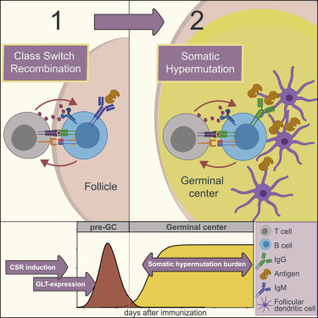


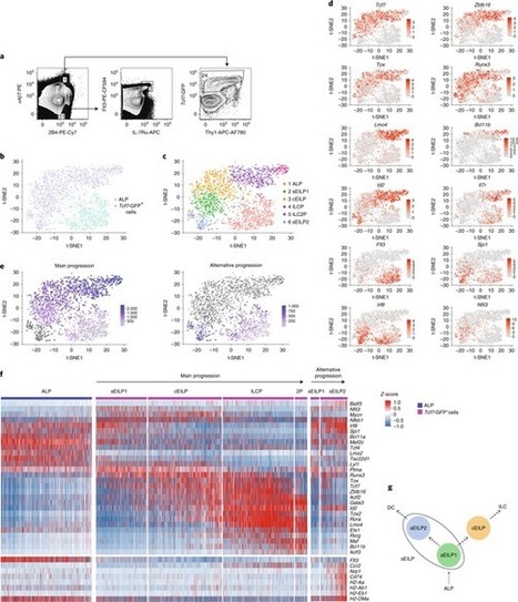





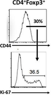







more on tregs
https://www.scoop.it/topic/immunology?q=treg