Your new post is loading...

|
Scooped by
Gilbert C FAURE
February 25, 3:56 AM
|
By screening viral proteins’ effects on human cells, researchers in Science discover that immune cells can detect infections not only by recognizing pathogens directly, but also by sensing the damage caused by viral attack.
The findings show that this plant-like immune defense strategy—known as effector-triggered immunity—also operates in mammals.
Learn more: https://scim.ag/4aqbirX
|
|
Scooped by
Gilbert C FAURE
February 24, 9:47 AM
|
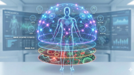
The Real Question Is: Who Builds the Immune Intelligence Layer? The race to build the Immunological Digital Twin (IDT) has begun. Pharma wants it to de-risk billion-dollar trials.
|
|
Scooped by
Gilbert C FAURE
February 20, 3:17 AM
|
|
|
Scooped by
Gilbert C FAURE
February 19, 2:57 AM
|
The success of modern vaccines depends not only on antibodies — but on T-helper cell polarization.
Understanding Th1, Th2, and Th17 differentiation is key to designing targeted and durable immunity.
🔬 T-Helper Cell Differentiation: A Central Player in Vaccine Efficacy
The type of T-helper (Th) cell activated during an immune response determines the quality and direction of protection generated after infection or vaccination. Understanding these differentiation pathways is critical for designing next-generation vaccines that induce long-lasting and targeted immunity.
🧬 Naïve CD4+ T cells (Th0) differentiate into distinct subsets depending on the cytokine environment:
🔴 Th1 Cells
Activated in response to intracellular pathogens such as viruses and certain bacteria.
• Driven by IL-12 and IFN-γ
• Characterized by T-bet transcription factor
• Promote macrophage activation and cell-mediated immunity
🟠 Th2 Cells
Activated during extracellular parasitic infections (e.g., helminths).
• Driven by IL-4
• Characterized by GATA3
• Support B-cell activation and antibody production (humoral immunity)
🟣 Th17 Cells
Important in defense against extracellular bacteria and fungi.
• Driven by TGF-β and IL-6
• Characterized by RORγt
• Recruit neutrophils and promote inflammation
🔵 Treg Cells
Maintain immune balance and prevent excessive inflammation.
• Driven by TGF-β and IL-2
• Express Foxp3
• Promote immune regulation and tolerance
💡 Why does this matter for vaccines?
Modern vaccine design aims to direct the immune response toward the most protective Th pathway. For example:
• Viral vaccines often require a strong Th1 response.
• Parasitic infections benefit from Th2-mediated antibody responses.
• Mucosal vaccines may require balanced Th17 activity.
A precise understanding of T-helper cell polarization enables scientists to design smarter adjuvants and achieve durable immunological memory.
#Immunology #Vaccinology #TCells #AdaptiveImmunity #ScientificResearch
|
|
Scooped by
Gilbert C FAURE
February 11, 9:22 AM
|
The Immune System’s “Hidden Organs”: Why Serous Cavities Matter More Than We Thought
This recent review frames the peritoneal, pleural, and pericardial cavities as active immune organs rather than passive fluid spaces. The authors synthesize a decade of work showing that serous cavities are dynamic immune reservoirs, metabolic niches, and communication hubs that shape infection control, fibrosis, cancer spread, and even cardiac and pulmonary disease. For drug developers and translational scientists, this challenges long-standing tissue-centric thinking and opens a largely untapped therapeutic and diagnostic frontier.
Key insights from the review:
◾ Serous cavities host specialized immune ecosystems, including cavity-resident macrophages, B1 cells, innate lymphoid cells, and fat-associated lymphoid clusters (FALCs).
◾ Immune responses in these cavities can rapidly shift from a “fluid phase” to a “solid phase,” with macrophage aggregation, clotting, and tissue tethering acting as first-line containment mechanisms.
◾ Stromal cells (mesothelium, fibroblasts) actively instruct immune identity via retinoic acid, cytokines, and metabolic cues, tightly coupling immunity to tissue repair and fibrosis.
◾ Immune cells traffic bidirectionally between cavities and organs (lung, heart, liver, intestine), blurring classical boundaries between local and systemic immunity.
◾ Sex, age, and developmental origin of immune cells materially influence disease outcomes, with direct implications for translational relevance.
From a therapeutic standpoint, serous cavities represent both a target and a delivery opportunity. These compartments concentrate immune cells, metabolites, and antigens in ways that are distinct from blood or solid tissue. This has implications for immunomodulators, biologics, cell therapies, and even local delivery strategies for inflammatory disease, cancer metastasis, fibrosis, and post-surgical adhesions.
Diagnostics may also evolve: serous fluids could serve as rich, dynamic biomarkers reflecting organ-adjacent immune states more sensitively than peripheral blood.
Most importantly, this work argues for designing drugs and trials that account for cavity-specific immunology rather than assuming uniform tissue behavior.
Diseases like peritonitis, pleural infection, pericarditis, fibrosis, endometriosis, and metastatic cancer all intersect with serous cavity biology. Understanding and therapeutically steering these immune niches could mean earlier intervention, more precise targeting, and fewer off-target effects.
For those in immunology, drug development, or translational medicine, it may be time to start viewing them as actionable immune organs.
Read the full review here: https://lnkd.in/ens2rZhj
#Immunology #DrugDevelopment #TranslationalScience #TissueImmunity #Biotech
|
|
Scooped by
Gilbert C FAURE
February 10, 10:50 AM
|
Within the IL-12 superfamily of heterodimeric cytokines, IL-12 and IL-23, which share a subunit, are among the most pro-inflammatory members. Both are primarily produced by phagocytes and have key roles in activating and regulating T lymphocytes, natural killer cells and innate lymphoid cells. IL-12 predominantly promotes type 1 immune responses, whereas IL-23 is closely associated with type 3 immunity. Their receptors are also heterodimeric and, upon engagement, they trigger ‘cytokine polarization’ (the imprinting of functional identities on immune cells by activating lineage-defining transcription factors), which contributes to inflammation and immunopathology. IL-12 has a key role in various inflammatory conditions and is a potent driver of antitumour immunity, and IL-12 delivery is being explored in several clinical trials in cancer. By contrast, IL-23 is essential for maintaining barrier tissue integrity, yet its dysregulation is a central driver of autoimmune diseases such as psoriasis. Beyond their well-established pro-inflammatory roles, studies of both cytokines have also yielded paradoxical findings. Emerging evidence suggests that both IL-12 and IL-23 can also attenuate immune responses. In this Review, we explore the discovery of IL-12 and IL-23, their canonical pro-inflammatory functions, and recent insights into their immunoregulatory roles in inflammation, cancer and autoimmunity. Here, Becher and colleagues examine the paradoxical roles of IL-12 and IL-23, two IL-12 family cytokines that drive type 1 and type 3 immune responses, respectively. Both promote inflammation by activating T cells, natural cells and innate lymphoid cells through cytokine polarization, yet IL-12 also supports antitumour immunity and IL-23 maintains barrier integrity. This Review highlights emerging evidence that both cytokines can also dampen immune responses, revealing unexpected regulatory roles in cancer, autoimmunity and tissue homeostasis.
|
|
Scooped by
Gilbert C FAURE
January 29, 1:27 PM
|
Platelets are cell fragments that plug leaks in blood vessels, preventing bleeding. However, they also form clots that impede blood flow (thrombosis) and increase inflammation.
Activated platelets, which can bind to each other, and hyporesponsive platelets, which do not form clots, are detected in patients with acute severe infections, such as COVID-19 and sepsis. How to uncouple the protective and harmful proinflammatory functions of platelets is a therapeutic dilemma.
In Science, researchers report the formation of platelet-derived integrin-and tetraspanin-enriched tethers (PITTs) in patients with severe infections.
PITTs, which are enriched in specific platelet proteins, remain anchored to the blood vessel wall (endothelium) and promote neutrophil recruitment, inflammation, and tissue damage, whereas the main platelet body shears off and reenters circulation in a hyporesponsive state. PITTs may contribute to blood clotting, immune dysregulation, and bleeding complications that characterize severe infections.
Learn more in a new #SciencePerspective: https://scim.ag/4pYNOyA
|
|
Scooped by
Gilbert C FAURE
January 28, 4:12 AM
|
The immune system has evolved intricate and sophisticated mechanisms to recognize and eliminate tumours. Among these, T helper 17 (TH17) cells seem to have a complex role in tumour immunity. Substantial evidence indicates that TH17 cells can impair the ability of immune cells to kill tumours, but growing research suggests that TH17 cells may also be crucial for facilitating effective antitumour immune responses. This apparent paradox is likely rooted, at least in part, in the remarkable plasticity of TH17 cells, which can adopt a range of effector functions depending on the environmental cues present in distinct tissue niches. Understanding the spectrum of functional adaptations that TH17 cells can undergo is especially important in cancer, where tumours exhibit substantial tissue heterogeneity. Here, we examine the context-dependent roles of TH17 cells in cancer, with a focus on the environmental signals and regulatory networks that may shape their phenotypes during cancer development and progression. Finally, we discuss how these insights could inform strategies to manipulate TH17 cells for the development of next-generation cancer immunotherapies. T helper 17 (TH17) cells can have both pro-tumour and antitumour effects. Understanding how they are regulated by environmental and regulatory cues according to context will inform strategies to harness their activity for the development of next-generation cancer immunotherapies.
|
|
Scooped by
Gilbert C FAURE
January 20, 4:41 AM
|
Foundations of the Human Immune System
Immunity is defined as the body’s ability to resist infection through specialized cells and molecules.
Types of Immunity:
Innate (Natural) Immunity:
This is the first line of defense. It is non-specific, meaning it reacts the same way to all pathogens. It does not require prior exposure and does not have "memory."
• External Defenses: The first line of defense includes physical, chemical, and biological barriers. The skin serves as a structural barrier, while secretions like lactic acid in sweat and hydrochloric acid in the stomach maintain acidic environments to halt microbial growth.
• Pattern Recognition: Phagocytic cells like macrophages and dendritic cells identify unique microbial substances called pathogen-associated molecular patterns (PAMPs) using pattern recognition receptors (PRRs), such as Toll-like receptors (TLRs).
• Phagocytosis: Once a pathogen is recognized, specialized white blood cells (neutrophils, monocytes, and macrophages) engulf and destroy the particle. This process involves seven steps, including physical contact, the formation of a phagosome, and the fusion of lysosomal granules to create a phagolysosome where digestion occurs.
• The Oxidative Burst: To kill ingested microbes, phagocytes increase oxygen consumption to generate toxic oxygen radicals, such as superoxide and hydrogen peroxide.
• Natural Killer (NK) Cells: These cells represent the first line of defense against virally infected and tumor cells. They function through a balance of inhibitory signals (recognizing MHC class I molecules) and activating signals. If activated, they release perforins and granzymes to induce apoptosis in the target cell.
• Adaptive (Acquired) Immunity:
Adaptive immunity is characterized by specificity for individual pathogens and the ability to remember prior exposures (immunologic memory).
• Antigen Presentation (MHC): For T cells to recognize an antigen, it must be "presented" by Major Histocompatibility Complex (MHC) molecules. MHC Class I molecules present endogenous antigens (produced inside the cell, like viruses) to CD8+ cytotoxic T cells. MHC Class II molecules present exogenous antigens (taken in from outside) to CD4+ helper T cells.
• T-Cell Effector Actions:
◦ Helper T (Th) cells produce cytokines that orchestrate the activity of other cells, such as B cells and macrophages.
◦ Cytotoxic T (Tc) cells directly kill infected or malignant cells by releasing cytotoxic granules or ligating death receptors.
• B-Cell and Antibody Responses: B cells differentiate into plasma cells to secrete antibodies.
◦ T-dependent antigens require help from T cells to stimulate B-cell proliferation and isotype switching (e.g., from IgM to IgG).
◦ Antibody functions include opsonization (coating pathogens to enhance phagocytosis), neutralization of toxins, and antibody-dependent cellular cytotoxicity (ADCC).
|
|
Scooped by
Gilbert C FAURE
January 14, 4:23 AM
|
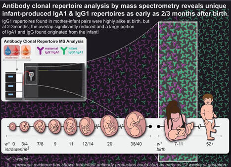
Infant immunity is not just passively transferred from the mother—it's built from scratch. FAB clonal profiling reveals newborns rapidly forge their own antibody repertoires, rewriting the textbook on early-life immune development.
Which begs the question, how quickly do newborns begin building their own antibody defenses? In our new multi-center study with colleagues from Utrecht, Shenzhen, Beijing, and Wuhan, we show that infants start generating unique IgA1 and IgG1 antibody repertoires within 7–11 weeks after birth, distinct from maternal serum and breast milk.
What we found: By 7-11 weeks after birth (T2), infant IgA1 clones in serum are new and do not overlap with maternal milk or serum, indicating they are produced by the infant. Infant IgG1 repertoires at T2 contain many “new” clones, while maternal IgG1 signatures faded as expected—strong evidence that newborns begin producing IgG1 antibodies early on. From this small cohort, we found no convincing evidence of milk maternal antibody transfer into infant circulation, prompting a re-examination of some long-held assumptions.
How we did it: We used intact LC–MS–based FAB clonal profiling to resolve antibody repertoires with clonal precision across maternal serum, maternal milk, and infant serum. This approach quantitatively tracks overlapping versus unique clones, pinpointing the emergence of infant-origin antibodies.
Why it matters: These findings shift our understanding of early-life humoral immunity—from how maternal antibodies shape neonatal responses, to when and how we time early-life vaccinations for durable protection. This method also opens a path to study neonatal vaccine responses and maternal–infant immune interactions with unprecedented resolution.
To read the full publication follow this link: https://lnkd.in/eu7xfaiR
Grateful to work with this outstanding international team: Albert Bondt, Minjie Tan, Danique van Rijswijck, Yuexiao Chen, Shuai Zhu, Ye Tian Tianhui Lin, Yuanzhen Zhang, Yanyi Huang, Guanbo Wang, Jing Zhu, Juanjuan Guo, Albert Heck
#UtrechtUniversity #BeijingAcademyOfScienceAndTechnology #ZhongnanHospitalOfWuhanUniversity #PekingUniversityBeijing
#InfantImmunity #IgA1 #IgG1 #AntibodyRepertoires #MassSpectrometry #ClonalProfiling #Proteomics #Neonatology #Vaccines | 10 comments on LinkedIn
|
|
Scooped by
Gilbert C FAURE
January 13, 3:42 AM
|
I love how this newly discovered immune signaling in plants looks kind of like fireworks. It's a celebration of botany! That and more of the best from Science Magazine and science in this edition of #ScienceAdviser: https://lnkd.in/gaedvdfa
|
|
Scooped by
Gilbert C FAURE
January 12, 6:44 AM
|
Deuxième poster de la série NK !
Ce poster explore le développement des cellules Natural Killer, depuis leur origine jusqu’à leur activation.
Les cellules Natural Killer sont des cellules de l’immunité innée dont le développement s’inscrit dans la lignée lymphoïde. Elles se développent principalement dans la moelle osseuse, à partir des cellules souches hématopoïétiques (CSH), via le progéniteur lymphoïde commun (CLP). Ce progéniteur est également à l’origine des lymphocytes B et T, ainsi que des cellules lymphoïdes innées, incluant les cellules NK.
#Immunologie #CellulesNK #Biologie #RechercheScientifique #VulgarisationScientifique
|
|
Scooped by
Gilbert C FAURE
January 6, 1:02 PM
|
Long COVID (LC) involves a spectrum of chronic symptoms after acute severe acute respiratory syndrome coronavirus 2 infection. Current hypotheses for the pathogenesis of LC include persistent virus, tissue damage, autoimmunity, endocrine insufficiency, immune dysfunction and complement activation. We performed immunological, virological, transcriptomic and proteomic analyses from a cohort of 142 individuals between 2020 and 2021, including uninfected controls (n = 35), acutely infected individuals (n = 54), convalescent controls (n = 24) and patients with LC (n = 28). The LC group was characterized by persistent immune activation and proinflammatory responses for more than 180 days after initial infection compared with convalescent controls, including upregulation of JAK-STAT, interleukin-6, complement, metabolism and T cell exhaustion pathways. Similar findings were observed in a second cohort enrolled between 2023 and 2024, including convalescent controls (n = 20) and patients with LC (n = 18). These data suggest that LC is characterized by persistent activation of chronic inflammatory pathways, suggesting new therapeutic targets and potential biomarkers of disease. Long COVID (LC) involves a spectrum of chronic symptoms after resolution of acute severe acute respiratory syndrome coronavirus 2 infection. Barouch and colleagues show that LC is characterized by persistent activation of chronic inflammatory pathways and T cell exhaustion.
|
|
|
Scooped by
Gilbert C FAURE
February 25, 3:28 AM
|
Interleukin 4 (IL-4) shares a common receptor subunit, IL-4Rα, with IL-13. These two cytokines play an important role in anti-parasitic immune responses. Dysregulated IL-4 /IL-13 expression contributes to Th2-mediated diseases, including asthma and atopic dermatitis.
InvivoGens Recombinant human IL-4 is a high-quality biologically active cytokine, validated using proprietary IL-4/IL-13 reporter cells. This member of the IL-2/γc superfamily is produced in CHO cells to ensure protein glycosylation and bona fide 3D structure (https://lnkd.in/dnemrMTj).
Applications:
- Standard for IL-4 detection and quantification assays
- Screening and release assays for antibodies blocking IL-4 signaling
- Screening and release assays for engineered IL-4
Recombinant human IL-4 can be used together with HEK-Blue™ IL-4/IL-13 cells for the screening of inhibitory molecules, such as Dupilumab, a therapeutic monoclonal antibody targeting the IL-4Rα subunit of the IL-4 receptor (see figures 📊).
#InvivoGen #DrugDiscovery #AsthmaResearch #AtopicDermatisis #Biopharma #Biotech #Lablife
|
|
Scooped by
Gilbert C FAURE
February 21, 4:10 AM
|
Superbe collaboration pour le diagnostic d’un déficit rare en complément chez une jeune fille de 14 ans qui avait été hospitalisée dans le service et qui avait présenté une infection invasive à méningocoque Y
https://lnkd.in/eTZygZmp
💡 toujours faire une évaluation du complément chez les enfants présentant une infection invasive à meningocoque en dosant le C3, C4, CH50 et AP50 et ce, dès le 1er épisode!
💡 recommandation HAS de vaccination par le tétravalent A, C, Y, W à 6 mois et rappel à 12 mois chez tous les enfants
Camille Bougeard Eléonore Eskander @paula viera martins Joan Bitan Julia ROQUIGNY Christèle Kyheng @Veronique Fremeaux-Bacchi, Cécile Gonnin carine el sissy
AP-HP, Assistance Publique - Hôpitaux de Paris
CHU du Kremlin-Bicêtre
#méningocoque #déficitencomplément
#pédiatriegénérale
|
|
Scooped by
Gilbert C FAURE
February 19, 3:18 AM
|
The Trigger: DAMPs and Immunogenic Cell Death
The process begins with tumor cell stress or death (often induced by therapies like radiation or chemotherapy). When these cells die, they release Damage-Associated Molecular Patterns (DAMPs). As shown in the "DAMP signaling" box, this includes:
• Calreticulin: Translocates to the cell surface as an "eat-me" signal.
• HMGB1 & DNA fragments: Passively released from the nucleus/cytoplasm.
• ATP: Actively released to act as a "find-me" signal for immune cells.
These signals activate Dendritic Cells (DCs), which perform cross-presentation—the vital step of taking tumor antigens and presenting them to CD8^+ T cells to "prime" the immune system to recognize the cancer.
2. The cGAS-STING Signaling Pathway
The lower-left portion of the diagram details the intracellular machinery of the cGAS-STING pathway, a primary sensor of cytosolic DNA.
• Detection: In a dying tumor cell or an immune cell that has engulfed tumor DNA, the enzyme cGAS (cyclic GMP-AMP synthase) detects double-stranded DNA in the cytosol.
• Synthesis: cGAS uses ATP and GTP to catalyze the production of cGAMP, a secondary messenger.
• Activation: cGAMP binds to STING (Stimulator of Interferon Genes) located on the endoplasmic reticulum/Golgi apparatus.
• The Signaling Cascade: Activated STING recruits kinases like TBK1 and IKK.
• TBK1 phosphorylates IRF3, which moves into the nucleus.
• IKK leads to the activation of P65 (NF-κB).
• Outcome: These transcription factors trigger the production of Type I Interferons (IFN-I) and pro-inflammatory cytokines such as IL-1\beta, IL-6, and TNF-\alpha.
3. Reprogramming the Microenvironment
The diagram shows that cGAS-STING activation doesn't just kill cells; it reprograms the TME. The "pro-inflammatory TME" at the bottom right is the result of shifting the environment from "cold" (immunosuppressive) to "hot" (immune-active).
• Cellular Reprogramming: The signaling induces the conversion of:
• TAMs (Tumor-Associated Macrophages): Shifting them toward the M1 (anti-tumor) phenotype.
• CAFs (Cancer-Associated Fibroblasts): Modifying their activity to reduce physical barriers to immune entry.
• Tregs & MDSCs: Inhibiting these immunosuppressive cells (Regulatory T cells and Myeloid-Derived Suppressor Cells) that usually help the tumor hide.
|
|
Scooped by
Gilbert C FAURE
February 18, 7:34 AM
|
🔷 Le vieillissement du système immunitaire, est un défi crucial pour la santé des seniors. Des chercheurs se sont penchés sur cette thématique dans un article "Immunofitness in the elderly: The role of vaccination in promoting healthy aging".
Leur objectif, mettre en lumière le rôle capital de la vaccination chez les personnes âgées pour préserver et restaurer l’"immunofitness" sur laquelle nous reviendrons demain.
🔹Le vieillissement induit une transformation profonde du système immunitaire, appelée immunosénescence. Ce processus se caractérise par une diminution progressive de la capacité à générer des réponses immunitaires efficaces face à de nouveaux agents infectieux.
🔹En parallèle, les personnes âgées développent une inflammation chronique de bas grade, qualifiée d’« inflammaging ».
🔹Cette inflammation persistante, alimentée par des cellules immunitaires sénescentes adoptant un profil pro-inflammatoire, ainsi que par des dysfonctionnements au niveau des macrophages et des cellules dendritiques, perturbe l’équilibre immunitaire et favorise l’apparition ou l’aggravation de maladies chroniques.
🔹C'est la combinaison de ces mécanismes qui explique la vulnérabilité accrue des seniors aux infections sévères, notamment respiratoires, leur moindre réponse aux vaccins, ainsi que leur susceptibilité aux complications associées.
🔹Au niveau cellulaire, on a une accumulation de cellules immunitaires qui vieillissent donc, avec des capacités réduites de prolifération, une altération de la présentation antigénique par les cellules dendritiques et une production excessive de cytokines pro-inflammatoires.
🔹Prendre en compte ces altérations immunitaires liées à l’âge est essentiel pour concevoir des interventions ciblées, comme des vaccins adaptés incluant des adjuvants puissants par exemple, et pour développer des stratégies de prévention vaccinale.
Sans oublier des mesures de promotion de la santé comme la nutrition, l'activité physique, afin d’améliorer la qualité de vie de nos seniors !
#santé #prévention #seniors #vaccination #vaccins #santépublique
|
|
Scooped by
Gilbert C FAURE
February 11, 4:15 AM
|
Immunoglobulins Roles In The Immune System
This post explains the roles of all five immunoglobulins in a clear and visual way. IgG provides long term protection, IgA protects mucosal surfaces, IgM signals early infection, IgE mediates allergy and parasite defense, and IgD supports B cell activation. Designed for quick learning, exams, and daily lab reference.
#Immunoglobulins #Antibodies #fblifestyle #IgG #IgA #IgM #IgE #IgD #Immunology #ClinicalImmunology #MLS #MedicalLaboratoryScientist
|
|
Scooped by
Gilbert C FAURE
January 31, 1:50 PM
|
Natural killer (NK) cells are cytotoxic lymphocytes of the innate immune system with essential roles in immune surveillance, tissue homeostasis and inflammation. In the kidney, NK cells comprise a heterogeneous population that includes both circulating and tissue-resident subsets, each shaped by environmental cues and genetic factors through interactions with major histocompatibility complex class I molecules. The latest research data highlight multifaceted NK cell contributions to kidney physiology and pathology. In steady state, NK cells support kidney immune surveillance and crosstalk with epithelial, myeloid and lymphoid cells. In disease, NK cells can promote injury through direct cytotoxicity and pro-inflammatory cytokine release. Experimental models demonstrate pathogenic roles for NK cells in ischaemia–reperfusion injury and chronic kidney disease, with emerging evidence implicating NK cell-derived mediators in fibrogenesis. In kidney transplantation, NK cells are effectors of antibody-dependent and antibody-independent allograft injury. Educated NK cells expressing CD16a (also known as FcγRIIIa) mediate antibody-dependent cellular cytotoxicity, whereas loss of inhibitory receptor–ligand interactions (for example, due to killer immunoglobulin-like receptor–HLA mismatch) can trigger NK cell activation independently of donor-specific antibodies. Advances in high-resolution profiling have deepened mechanistic insights and uncovered novel therapeutic targets. Here, we provide a comprehensive overview of NK cell biology in the kidney, highlighting roles in health, disease and transplantation, and we consider its translational implications for diagnosis and therapy. This Review discusses the latest advances in natural killer cell biology, including insights into population heterogeneity, and examines the roles of these innate lymphocytes in kidney health and disease, as well as their contribution to kidney allograft rejection and its therapeutic implications.
|
|
Scooped by
Gilbert C FAURE
January 28, 1:13 PM
|
🟥 Bringing the Human Immune System into Mice: The Latest Breakthroughs in Humanized Mouse Models
Scientists have long faced a challenge in studying the human immune system: many immune responses cannot be accurately replicated in ordinary experimental animals. To address this problem, humanized mouse models have emerged and have made significant progress in recent years, gradually becoming a "key bridge" in immunology and translational medicine.
A humanized mouse refers to a mouse in which a functional human immune system is reconstructed through genetic engineering and cell transplantation techniques in an immunodeficient mouse. Early models primarily relied on injecting human peripheral blood immune cells or hematopoietic stem cells. While this could temporarily create human immune cells, it suffered from limitations such as incomplete immune composition and poor stability. In recent years, a new generation of humanized models has significantly improved the development and functional maturity of human T cells, B cells, and innate immune cells by optimizing the immunodeficient background, introducing human cytokine genes, and combining the transplantation of human hematopoietic stem cells with tissues (such as the thymus and liver).
These improvements have enabled humanized mice to demonstrate unique value in several fields. In tumor immunology research, they can be used to evaluate the efficacy and safety of therapies such as immune checkpoint inhibitors and CAR-T cells; in infection and vaccine research, the models can more accurately simulate the human immune response process; and in autoimmune disease and transplant rejection research, humanized mice also provide an important platform for mechanism analysis and drug screening.
Of course, these models still face challenges, such as high construction costs and differences between the human immune system and the mouse microenvironment. However, with the development of gene editing, stem cell, and microenvironment engineering technologies, humanized mice are constantly evolving towards being "closer to humans." It is foreseeable that they will play an increasingly central role in the development of precision medicine and novel immunotherapies. Follow us (www.csteamus.com) to learn more about the biological model products and related services provided by CSTEAM biotechnology.
Keywords:
Humanized mice, immune system reconstruction, tumor immunology, hematopoietic stem cells, translational medicine
|
|
Scooped by
Gilbert C FAURE
January 23, 9:54 AM
|
|
|
Scooped by
Gilbert C FAURE
January 14, 11:17 AM
|
The Chinese Immune Multi-Omics Atlas
🧠 𝗔 𝗽𝗮𝗽𝗲𝗿 𝗮 𝗱𝗮𝘆 𝗸𝗲𝗲𝗽𝘀 𝗯𝗿𝗮𝗶𝗻 𝗱𝗲𝗰𝗮𝘆 𝗮𝘄𝗮𝘆 🧠
Wednesday, 14 January 2026
𝘛𝘩𝘪𝘴 𝘴𝘵𝘶𝘥𝘺 𝘱𝘳𝘦𝘴𝘦𝘯𝘵𝘴 𝘊𝘐𝘔𝘈 (𝘊𝘩𝘪𝘯𝘦𝘴𝘦 𝘐𝘮𝘮𝘶𝘯𝘦 𝘔𝘶𝘭𝘵𝘪-𝘖𝘮𝘪𝘤𝘴 𝘈𝘵𝘭𝘢𝘴), 𝘪𝘯𝘵𝘦𝘨𝘳𝘢𝘵𝘪𝘯𝘨 𝘱𝘰𝘱𝘶𝘭𝘢𝘵𝘪𝘰𝘯-𝘴𝘤𝘢𝘭𝘦 𝘴𝘪𝘯𝘨𝘭𝘦-𝘤𝘦𝘭𝘭 𝘵𝘳𝘢𝘯𝘴𝘤𝘳𝘪𝘱𝘵𝘰𝘮𝘪𝘤𝘴, 𝘤𝘩𝘳𝘰𝘮𝘢𝘵𝘪𝘯 𝘢𝘤𝘤𝘦𝘴𝘴𝘪𝘣𝘪𝘭𝘪𝘵𝘺, 𝘢𝘯𝘥 𝘨𝘦𝘯𝘦𝘵𝘪𝘤𝘴 𝘵𝘰 𝘭𝘪𝘯𝘬 𝘯𝘰𝘯𝘤𝘰𝘥𝘪𝘯𝘨 𝘥𝘪𝘴𝘦𝘢𝘴𝘦 𝘷𝘢𝘳𝘪𝘢𝘯𝘵𝘴 𝘵𝘰 𝘪𝘮𝘮𝘶𝘯𝘦 𝘤𝘦𝘭𝘭 𝘴𝘵𝘢𝘵𝘦𝘴, 𝘳𝘦𝘨𝘶𝘭𝘢𝘵𝘰𝘳𝘺 𝘦𝘭𝘦𝘮𝘦𝘯𝘵𝘴, 𝘢𝘯𝘥 𝘨𝘦𝘯𝘦 𝘯𝘦𝘵𝘸𝘰𝘳𝘬𝘴, 𝘸𝘩𝘪𝘭𝘦 𝘳𝘦𝘷𝘦𝘢𝘭𝘪𝘯𝘨 𝘴𝘦𝘹- 𝘢𝘯𝘥 𝘢𝘯𝘤𝘦𝘴𝘵𝘳𝘺-𝘥𝘦𝘱𝘦𝘯𝘥𝘦𝘯𝘵 𝘪𝘮𝘮𝘶𝘯𝘦 𝘳𝘦𝘨𝘶𝘭𝘢𝘵𝘪𝘰𝘯 𝘸𝘪𝘵𝘩 𝘪𝘮𝘱𝘭𝘪𝘤𝘢𝘵𝘪𝘰𝘯𝘴 𝘧𝘰𝘳 𝘵𝘩𝘦 𝘵𝘳𝘢𝘯𝘴𝘭𝘢𝘵𝘢𝘣𝘪𝘭𝘪𝘵𝘺 𝘰𝘧 𝘌𝘶𝘳𝘰𝘱𝘦𝘢𝘯-𝘥𝘦𝘳𝘪𝘷𝘦𝘥 𝘣𝘪𝘰𝘮𝘢𝘳𝘬𝘦𝘳𝘴 𝘢𝘯𝘥 𝘵𝘩𝘦𝘳𝘢𝘱𝘪𝘦𝘴.
💡 𝗧𝗮𝗸𝗲 𝗵𝗼𝗺𝗲 𝗺𝗲𝘀𝘀𝗮𝗴𝗲
• CIMA profiles 10.25 million peripheral blood mononuclear cells from 428 healthy Chinese adults using joint scRNA-seq and scATAC-seq, resolving 73 immune cell states and directly linking transcriptional programs to regulatory chromatin landscapes at scale.
• A deep-learning chromatin language model (CIMA-CLM), combined with cell-type quantitative trait locus mapping, enables inference of expression- and chromatin-associated variants beyond experimentally profiled cells, revealing that most disease-associated variants act through highly restricted immune cell types.
• The asthma-associated variant rs34415530 selectively regulates IKZF4 expression in FOXP3 regulatory T cells, highlighting cell-type-specific genetic control of immune tolerance pathways.
• The East Asian-enriched variant rs11886530 regulates the circadian genes NPAS2 and NR1D1 specifically in T cells and is largely absent from European datasets, illustrating ancestry-dependent regulatory mechanisms.
🔥 𝗜𝗺𝗽𝗮𝗰𝘁
• By showing that immune regulatory wiring differs between Chinese, European and Japanese populations, this work challenges the assumption that targets, biomarkers, and drug-response models derived from European cohorts are universally valid, with direct implications for precision medicine and immunotherapy development across ancestries.
❓ 𝗢𝗽𝗲𝗻 𝗾𝘂𝗲𝘀𝘁𝗶𝗼𝗻𝘀
• Which population-specific variants drive clinically relevant immune phenotypes in East Asian cohorts?
• How can Chinese, Japanese, and European immune atlases be integrated to define shared versus ancestry-specific causal mechanisms?
• Which sex-associated immune differences translate into divergent responses to vaccines, infections, or immunotherapies?
𝗖𝗵𝗶𝗻𝗲𝘀𝗲 𝗜𝗺𝗺𝘂𝗻𝗲 𝗠𝘂𝗹𝘁𝗶-𝗢𝗺𝗶𝗰𝘀 𝗔𝘁𝗹𝗮𝘀
Jianhua Yin, et al.
Science, January 2026
Corresponding authors: Yanan Cao, Xun Xu, Longqi Liu, Xin Jin, Chuanyu Liu
𝘐𝘭𝘭𝘶𝘴𝘵𝘳𝘢𝘵𝘪𝘰𝘯 𝘢𝘥𝘢𝘱𝘵𝘦𝘥 𝘧𝘳𝘰𝘮 𝘵𝘩𝘦 𝘢𝘳𝘵𝘪𝘤𝘭𝘦
|
|
Scooped by
Gilbert C FAURE
January 13, 4:24 AM
|
Thanks for highlighting our piece
|
|
Scooped by
Gilbert C FAURE
January 12, 9:05 AM
|
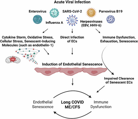
Myalgic encephalomyelitis/chronic fatigue syndrome (ME/CFS) and long COVID are two post-viral diseases, which share many common symptoms and pathophysiological alterations. Yet a mechanistic explanation of disease induction and maintenance is lacking. This hinders the discovery and implementation of biomarkers and treatment options, and ultimately the establishment of effective clinical resolution. Here, we propose that acute viral infection results in (in)direct endothelial dysfunction and senescence, which at the blood-brain barrier, cerebral arteries, gastrointestinal tract, and skeletal muscle can explain symptoms. The endothelial senescence-associated secretory phenotype (SASP) is proinflammatory, pro-oxidative, procoagulant, primed for vasoconstriction, and characterized by impaired regulation of tissue repair, but also leads to dysregulated inflammatory processes. Immune abnormalities in ME/CFS and long COVID can account for the persistence of endothelial senescence long past the acute infection by preventing their clearance, thereby providing a mechanism for the chronic nature of ME/CFS and long COVID. The systemic and tissue-specific effects of endothelial senescence can thus explain the multisystem involvement in and subtypes of ME/CFS and long COVID, including dysregulated blood flow and perfusion deficits. This can occur in all tissues, but especially the brain as evidenced by findings of reduced cerebral blood flow and impaired perfusion of various brain regions, post-exertional malaise (PEM), gastrointestinal disturbances, and fatigue. Paramount to this theory is the affected endothelium, and the bidirectional sustainment of immune abnormalities and endothelial senescence. The recognition of endothelial cell dysfunction and senescence as a core element in the aetiology of both ME/CFS and Long COVID should aid in the establishment of effective biomarkers and treatment regimens.
|
|
Scooped by
Gilbert C FAURE
January 12, 6:40 AM
|
Un DIU de grande qualité permettant aux internes d’approfondir leurs connaissances en immunologie, enseignement auquel participent bien évidemment les immunologistes mais également tous nos autres collègues notre discipline étant éminemment transversale
|