Leonard Calabrese, DO, discusses his findings entitled, “Applied Immunology to Autoimmune Diseases: COVID-19 as Immunology,” which was presented at the Rheumatology Nurses Society Conference....
Abstract Lymphadenopathies can be part of the clinical spectrum of several primary immunodeficiencies, including diseases with immune dysregulation and autoinflammatory disorders, as the clinical e...
The phenotype of autoreactive T cells in type 1 diabetes is described as Th1, Th17 and/or Th21, but is largely uncharacterized.We combined multi-para…...
In the ANCA-associated vasculitides (AAV), Li et al review the genetic associations including both the MHC and non-MHC regions associated with AAV. They demonstrate that the MHC associations provide an additional basis for further dividing the AAV into three subtypes, while specific mutations in...
Abstract The classification of idiopathic inflammatory myopathies (IIM) is based on clinical, serological and histological criteria. The identification of myositis-specific antibodies has helped to...
Using ERVmap, the authors determined that ERV-K102 expression was elevated in SLE patients’ peripheral blood cells and correlated with the interferon signature.
Professor Laurent ARNAUD from the National Reference Center for Autoimmune diseases of Strasbourg France (CRMR RESO) tells the amazing story of the term lupus, from the early middle age to the most contemporary treatments, including the differential diagnoses between cutaneous lupus, systemic lupus and other types of lupus such as lupus vulgaris (skin tuberculosis). AN AMAZING STORY over 10 centuries ------------------------------------------------------------------------------------------------ Twitter: @Lupusreference & @CRMR_RESO Website of RESO: https://maladie-autoimmune.fr Check our orignal paper about the history of lupus in JAAD: https://doi.org/10.1016/j.jaad.2020.04.150
Patients with autoimmune polyendocrine syndrome type-1 (APS-1) have circulating auto-Abs neutralizing most type I interferons. These auto-Abs can underlie life-
Ann Arthritis Clin Rheumatol | Volume 2, Issue 2 | Research Article | Open Access Autoantibodies against Autonomic Nerve Receptors in Adolescent Japanese Girls after Immunization with Human Papillomavirus Vaccine Akiyo Hineno1,2, Shu-ichi Ikeda1*, Carmen Scheibenbogen3,4, Harald Heidecke5, Kai Schulze- Forster5, Juliane Junker5, Gabriela Riemekasten6, Ralf Dechend8, Duska Dragun9 and Yehuda Shoenfeld10 1Intractable Disease Care Center, Shinshu University Hospital, Japan 2Department of Medicine (Neurology and Rheumatology), Shinshu University School of Medicine, Japan 3Department of Medicine, Institute for Medical Immunology, Germany 4Berlin-Brandenburg Center for Regenerative Therapies (BCRT), Germany 5Cell Trend GmbH, Germany 6Department of Rheumatology, Universitätsklinikum Schleswig-Holstein, Germany 7Experimental and Clinical Research Center, Charité-Universitätsmedizin Berlin Germany 8Department of Cardiology and Nephrology, HELIOS-Klinikum, Germany 9Department of Nephrology and Intensive Care Medicine, Charité-Universitätsmedizin Berlin, Germany 10Zabludowicz Center for Autoimmune Diseases, Sheba Medical Center, Israel *Correspondance to: Shu-ichi Ikeda Fulltext PDF Abstract In Japan a significant number of adolescent girls complain of unusual symptoms after human papillomavirus (HPV) vaccination, and these symptoms, composed of orthostatic dysregulation, chronic regional pain syndrome (CRPS) and cognitive dysfunction are considered adverse effects of HPV vaccination. However, a causal link between HPV vaccination and these adverse effects has not been demonstrated. In the present study, we investigated autoantibodies against diverse Gprotein coupled receptors in the serum of girls who complained of possible adverse effects after HPV vaccination. Fifty five girls with HPV vaccination and 57 girls without HPV immunization were enrolled in the study. The serum levels of autoantibodies against the adrenergic receptors α1, α2, β1and β2, muscarinic acetylcholine receptors 1, 2, 3, 4, 5; and endothelin receptor A was significantly elevated in girls with HPV vaccination, compared with those in the controls. The serum levels of these autoantibodies tended to decrease with the time course of the illness, but there was no statistically meaningful association between the clinical symptoms and elevated serum levels of these autoantibodies. This preliminary study provides evidence that post-vaccination abnormal autoimmunity plays an important role in the development of unique symptoms after HPV vaccination. Keywords: Autonomic nerve dysfunction; Autoantibody; Autoimmune disorder; Chronic regional pain syndrome; HPV; Human papilloma vaccination; Chronic regional pain syndrome Citation: Hineno A, Shu-ichi Ikeda, Scheibenbogen C, Heidecke H, Schulze- Forster K, Junker, et al. Autoantibodies against Autonomic Nerve Receptors in Adolescent Japanese Girls after Immunization with Human Papillomavirus Vaccine. Ann Arthritis Clin Rheumatol. 2019; 2(2): 1014.
Plusieurs personnes vaccinées à l’AstraZeneca ont été victimes d’accidents vasculaires parfois mortels, causés par la formation de caillots sanguins survenus peu de temps après son administration.
A new study has shown that treating type 1 diabetes-prone mice with the small protein MOTS-c prevented the immune system from destroying insulin-producing pancreatic cells, effectively preventing the onset of the autoimmune disease.
Immunology of Diabetes Society Review Series: Insights into Pathogenesis of Type 1 Diabetes Series Editors: F. Susan Wong and Timothy I. Tree A wave of exciting research in recent years has given us greater insight into the immunological processes involved in the pathogenesis of type-1 diabetes. In this issue of Clinical & Experimental Immunology, Guest Editors F. Susan Wong, of Cardiff University, and Timothy I. Tree, of King’s College London, present a new Review Series exploring key focus areas in this developing field of immunology. The series papers were commissioned in collaboration with the Immunology of Diabetes Society, and encompass new findings presented at their 2018 IDS conference in London. The role of autoantibodies in the early diagnosis of type-1 diabetes. The discovery of a new CD4 T cell autoantigenic target that facilitates this. The mechanisms by which immune cells subsequently infiltrate the pancreas. Finally, the destructive interactions of immune cells with islets in diagnosed patients. These four reviews provide a detailed coverage from the original to latest discoveries into these areas of the immunology behind type-1 diabetes. We invite you to read the freely accessible series papers below. Part One Editorial: Historical and New Insights into Pathogenesis of Type 1 Diabetes F.S. Wong, T.I. Tree Series papers: Birth and coming of age of islet autoantibodies Bonifacio, P. Achenbach Islet-Immune Interactions in Type 1 Diabetes: The Nexus of Beta Cell Destruction Peters, A. Posgai, T. Brusko Immune cell trafficking to the islets during type 1 diabetes A.M. Sandor, J. Jacobelli, R.S. Friedman HIPs and HIP-reactive T cells T.A. Wiles, T. Delong Part Two Editorial: Historical and New Insights into Pathogenesis of Type 1 Diabetes (2) F.S. Wong, T.I. Tree Series papers: Identifying the ‘Achilles heel’ of type 1 diabetes M. Battaglia, J. H. Buckner, M. K. Levings, S. J. Richardson, F. S. Wong and T. I. Tree Spliced HLA‐bound peptides: a Black Swan event in immunology P. Faridi , M. Dorvash, A. W. Purcell
Abstract Different lymphocyte subsets are involved in autoimmune pathogenesis of Type 1 Diabetes (T1D). Previous studies suggested a role of CD5 expressing T and B cells including rare unconvention...
ObjectivesThe aim of this study was to evaluate the clinical validity of early Sjögren’s syndrome (SS) autoantibodies (eSjA), which were originally marketed for early diagnosis of SS, for juvenile SS (JSS) in a recently identified pediatric cohort.MethodsA total of 105 symptomatic subjects with eSjA results available were evaluated at the Center for Orphaned Autoimmune Disorders at the University of Florida and enrolled for this study. JSS diagnosis was based on the 2016 ACR/EULAR SS criteria. Demographic/clinical/laboratory parameters were compared between JSS (n = 27) and non-JSS (n = 78) for % positivity, sensitivity, and specificity of eSjA (SP1, anti-salivary protein; CA6, anti-carbonic anhydrase VI; PSP, anti-parotid secretory protein) and classic SS-autoantibodies (cSjA; ANA, SSA/SSB, RF, and others) either alone or in combination. Associations between eSjA and diagnostic/glandular parameters were also determined by Fisher’s exact test.ResultsCompared to non-JSS, JSS patients exhibited sicca symptoms demonstrating reduced unstimulated salivary flow rate (USFR) and abnormal glandular features revealed by salivary gland ultrasound (SGUS). Among cSjA, ANA demonstrated the highest sensitivity of 69.2%, while SSA, SSB, and RF showed around 95% specificities for JSS diagnosis. The % positive-SSA was notably higher in JSS than non-JSS (56% vs. 5%). Of eSjA, anti-CA6 IgG was the most prevalent without differentiating JSS (37%) from non-JSS (32%). Sensitivity an
COVID-19 has been associated with both transient and persistent systemic symptoms that do not appear to be a direct consequence of viral infection. The generation of autoantibodies has been propose
The realization that autoantibodies can contribute to dysfunction of the brain has brought about a paradigm shift in neurological diseases over the past decade, offering up important novel diagnostic and therapeutic opportunities. Detection of specific autoantibodies to neuronal or glial targets has resulted in a better understanding of central nervous system autoimmunity and in the reclassification of some diseases previously thought to result from infectious, ‘idiopathic’ or psychogenic causes. The most prominent examples, such as aquaporin 4 autoantibodies in neuromyelitis optica or NMDAR autoantibodies in encephalitis, have stimulated an entire field of clinical and experimental studies on disease mechanisms and immunological abnormalities. Also, these findings inspired the search for additional autoantibodies, which has been very successful to date and has not yet reached its peak. This Review summarizes this rapid development at a point in time where preclinical studies have started delivering fundamental new data for mechanistic understanding, where new technologies are being introduced into this field, and — most importantly — where the first specifically tailored immunotherapeutic approaches are emerging. Here, Harald Prüss discusses how autoantibodies can contribute to neurological diseases. The identification of specific autoantibodies to neuronal and glial targets has increased our understanding of autoimmunity in the central nervous system and led to the reclassification of some diseases previously thought to result from infection or ‘idiopathic’ causes.
COVID-19 manifests with a wide spectrum of clinical phenotypes that are characterized by exaggerated and misdirected host immune responses1–6. While pathological innate immune activation is well documented in severe disease1, the impact of autoantibodies on disease progression is less defined. Here, we used a high-throughput autoantibody (AAb) discovery technique called Rapid Extracellular Antigen Profiling (REAP)7 to screen a cohort of 194 SARS-CoV-2 infected COVID-19 patients and healthcare workers for autoantibodies against 2,770 extracellular and secreted proteins (the “exoproteome”). We found that COVID-19 patients exhibit dramatic increases in autoantibody reactivities compared to uninfected controls, with a high prevalence of autoantibodies against immunomodulatory proteins including cytokines, chemokines, complement components, and cell surface proteins. We established that these autoantibodies perturb immune function and impair virological control by inhibiting immunoreceptor signaling and by altering peripheral immune cell composition, and found that murine surrogates of these autoantibodies exacerbate disease severity in a mouse model of SARS-CoV-2 infection. Analysis of autoantibodies against tissue-associated antigens revealed associations with specific clinical characteristics and disease severity. In summary, these findings implicate a pathological role for exoproteome-directed autoantibodies in COVID-19 with diverse impacts on immune functionality and associations with clinical outcomes.
Understanding the pathogenesis of type 1 diabetes (T1D), in order to direct therapy for prevention and treatment is a major goal of research into T1D. At the International Congress of the Immunolog
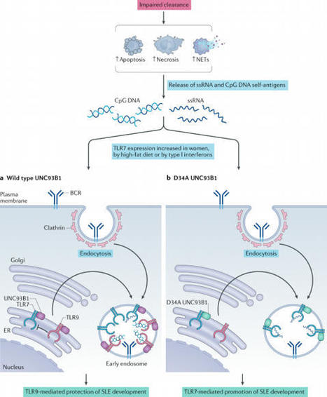
B lymphocytes have a central role in autoimmune diseases, which are often defined by specific autoantibody patterns and feature a loss of B cell tolerance. A prototypic disease associated with B cell hyperactivity is systemic lupus erythematosus (SLE). In patients with SLE, the loss of B cell tolerance to autoantigens is controlled in a cell-intrinsic manner by Toll-like receptors (TLRs), which sense nucleic acids in endosomes. TLR7 drives the extrafollicular B cell response and the germinal centre reaction that are involved in autoantibody production and disease pathogenesis. Surprisingly, TLR9 seems to protect against SLE, even though it is required for the production of autoantibodies recognizing double-stranded DNA-associated antigens, which are abundant in SLE and are a hallmark of this disease. The protective function of TLR9 is at least partly mediated by its capacity to limit the stimulatory activity of TLR7. The roles of TLR7 and TLR9 in the effector function of B cells in lupus-like disease and in patients with SLE, and the unique features of TLR signalling in B cells, suggest that targeting TLR signalling in SLE might be therapeutically beneficial. Loss of B cell tolerance to autoantigens in systemic lupus erythematosus (SLE) is driven by TLR7, whereas TLR9 appears to protect against SLE by limiting the stimulatory activity of TLR7. The unique features of Toll-like receptor signalling in B cells implicate it as a therapeutic target in SLE.
As part of the centennial celebration of insulin’s discovery, this review summarizes the current understanding of the genetics, pathogenesis, treatment, and outcomes in type 1 diabetes (T1D). T1D results from an autoimmune response that leads to destruction of the β cells in the pancreatic islet and requires lifelong insulin therapy. While much has been learned about T1D, it is now clear that there is considerable heterogeneity in T1D with regard to genetics, pathology, response to immune-based therapies, clinical course, and susceptibility to diabetes-related complications. This Review highlights knowledge gaps and opportunities to improve the understanding of T1D pathogenesis and outlines emerging therapies to treat or prevent T1D and reduce the burden of T1D.
To get content containing either thought or leadership enter:
To get content containing both thought and leadership enter:
To get content containing the expression thought leadership enter:
You can enter several keywords and you can refine them whenever you want. Our suggestion engine uses more signals but entering a few keywords here will rapidly give you great content to curate.


 Your new post is loading...
 Scoop.it!