Your new post is loading...

|
Scooped by
Dr. Stefan Gruenwald
June 16, 2016 7:08 PM
|
Neanderthals and Denisovans would have been a good source of helpful DNA for our ancestors. They had lived in Europe and Asia for hundreds of thousands of years — enough time to adjust to the cold climate, weak sun and local microbes. “What better way to quickly adapt than to pick up a gene variant from a population that had probably already been there for 300,000 years?” Akey said. Indeed, the Neanderthal and Denisovan genes with the greatest signs of selection in the modern human genome “largely have to do with how humans interact with the environment,” he said. To find these adaptive segments, scientists search the genomes of contemporary humans for regions of archaic DNA that are either more common or longer than expected. Over time, useless pieces of Neanderthal DNA — those that don’t help the carrier — are likely to be lost. And long sections of archaic DNA are likely to be split into smaller segments unless there is selective pressure to keep them intact. In 2014, two groups, one led by Akey and the other by David Reich, a geneticist at Harvard Medical School, independently published genetic maps that charted where in our genomes Neanderthal DNA is most likely to be found. To Akey’s surprise, both maps found that the most common adaptive Neanderthal-derived genes are those linked to skin and hair growth. One of the most striking examples is a gene called BNC2, which is linked to skin pigmentation and freckling in Europeans. Nearly 70 percent of Europeans carry the Neanderthal version. Scientists surmise that BNC2 and other skin genes helped modern humans adapt to northern climates, but it’s not clear exactly how. Skin can have many functions, any one of which might have been helpful. “Maybe skin pigmentation, or wound healing, or pathogen defense, or how much water loss you have in an environment, making you more or less susceptible to dehydration,” Akey said. “So many potential things could be driving this — we don’t know what differences were most important.”
|
|
Scooped by
Dr. Stefan Gruenwald
June 16, 2016 7:01 PM
|
Scientists at Towson University and the Johns Hopkins University are reporting a new way to peer through the event horizons around black holes and visualize what lies beneath. This computer-simulated image shows a supermassive black hole at the core of a galaxy. The black region in the center represents the black hole's event horizon, where no light can escape the massive object's gravitational grip. The black hole's powerful gravity distorts space around it like a funhouse mirror. Light from background stars is stretched and smeared as the stars skim by the black hole.
|
|
Scooped by
Dr. Stefan Gruenwald
June 16, 2016 6:53 PM
|
Bioengineers at the University of California, San Diego have developed an electrical graphene chip capable of detecting mutations in DNA. Researchers say the technology could one day be used in various medical applications such as blood-based tests for early cancer screening, monitoring disease biomarkers and real-time detection of viral and microbial sequences. The advance was published June 13 in the online early edition of Proceedings of the National Academy of Sciences. The chip essentially works by performing DNA strand displacement, the process in which a DNA double helix exchanges one strand for another complementary strand. The new complementary strand—which, in this case, contains the single nucleotide mutation—binds more strongly to one of the strands in the double helix and displaces the other strand. In this study, the DNA probe is a double helix containing two complementary DNA strands that are engineered to bind weakly to each other: a “normal” strand, which is attached to the graphene transistor, and a “weak” strand, in which four the G’s in the sequence were replaced with inosines to weaken its bond to the normal strand. DNA strands that have the perfectly matching complementary sequence to the normal strand—in other words, strands that contain the SNP—will bind to the normal strand and knock off the weak strand. Researchers engineered the chip to generate an electrical signal when an SNP-containing strand binds to the probe, allowing for quick and easy SNP detection in a DNA sample.
|
|
Scooped by
Dr. Stefan Gruenwald
June 15, 2016 12:19 PM
|
Pundits often describe deep learning as an imitation of the human brain. But it's really just simple math executed on an enormous scale. Deep learning is a subfield of machine learning, which is a vibrant research area in artificial intelligence, or AI. Abstractly, machine learning is an approach to approximating functions based on a collection of data points. For example, given the sequence “2, 4, 6,…” a machine might predict that the 4th element of the sequence is 8, and that the 5th is 10, by hypothesizing that the sequence is capturing the behavior of the function 2 times X, where X is the position of the element in the sequence. This paradigm is quite general. It has been highly successful in applications ranging from self-driving cars and speech recognition to anticipating airfare fluctuations and much more. In a sense, deep learning is not unique. Any machine learning system—deep or not—consists of the following fundamental components: - Performance element: the component of the system that takes some action in the world (e.g., making moves in the game of Go).
- Target function: the function being learned (e.g., a mapping from board positions to move choices in Go).
- Training data: the set of labeled data points used to approximate the target function (e.g., a set of Go board positions, each labeled with the move chosen by a human expert in that position).
- Data representation: each data point is typically represented as a vector of pre-determined variables (e.g., the position of a piece on the Go board).
- Learning algorithm: the algorithm that computes an approximation of the target function based on the training data.
- Hypothesis space: the space of possible functions the learning algorithm can consider.
This architecture captures the full gamut of machine learning methods from simple linear regression methods to complex deep-learning algorithms. Technically, we are referring to supervised learning where each data point is labeled, typically by humans. When the data isn’t labeled, we have unsupervised learning or clustering, and that’s much harder to pull off. When some of the data is labeled, we have semi-supervised learning. Statisticians refer to estimating the value of an independent variable based on dependent variables as regression. It’s important to realize that the first five components of a machine learning architecture are manually crafted inputs; the human programmer constructs each of these elements, and they are outside of the control of the learning program. In fact, the programmer typically analyzes the behavior of the learning program, realizes that it is unsatisfactory, and manually modifies one or more of these elements. This laborious process is often repeated many times over the course of a year or more before the desired performance level is achieved.
Cell phones and Wi-Fi devices typically transmit data using radio waves, but as the demand for wireless data transfer increases, congestion in the radio spectrum is expected to become more of a problem. One way to solve this problem is with visible light communication (VLC), a technology that uses visible light rather than radio waves for data transmission.
VLC uses lasers or LEDs that look just like traditional lights, but by rapidly switching on and off faster than the eye can see, the light transmits data in binary code to a receiver. Besides expanding the spectrum of data transfer, VLC is expected to have other advantages over wireless radio communication, including faster speeds, higher security, and better energy efficiency.
Currently, however, one of the biggest challenges facing VLC is a very small bandwidth, which greatly limits the speed of data transmission. The main reason for this problem is the small bandwidth of the color converter—a component that converts blue LED light into the different colors needed to make the white light that is ultimately used to transmit data.
In a new paper published in ACS Photonics, a team of researchers led by Osman Bakr and Boon Ooi at the King Abdullah University of Science and Technology (KAUST) in Saudi Arabia has developed a new VLC color converter that has a bandwidth that is 40 times greater than that of commercial converters, and more than twice as large as that of any potential candidate converter proposed to date.
"In this work we break the record for data communication using visible light, and more impressively produce white light with a very high color rendering index of 89, by designing a special color converter based on hybrid perovskite nanocrystals," Bakr told Phys.org. "Our work demonstrates white light as both a lighting source and a system for ultra-high-speed data communications."
Via Mariaschnee
|
|
Scooped by
Dr. Stefan Gruenwald
June 15, 2016 11:46 AM
|
Scientists from ITMO University in St. Petersburg, Russia, the Federal Research and Clinical Centre of Physical-Chemical Medicine and MIPT have developed a software program enabling them to quickly compare sets of DNA of microorganisms living in different environments. The researchers have already suggested exactly how the new program could be applied in practice. Using the algorithm to compare the microflora of a healthy person with the microflora of a patient, specialists would be able to detect previously unknown pathogens and their strains, which can aid the development of personalized medicine. The results of the study have been published in Bioinformatics. Every person has a genome - a specific sequence of genes according to which an individual develops. However, any living organism contains another gene sequence that is called the metagenome.It is the total DNA content of the many different microorganisms that inhabit the same environment - bacteria, fungi, and viruses. The metagenome is often indicative of various diseases or predispositions to such diseases. Studying microbiota, i.e. the full range of microorganisms inhabiting different parts of the human body, has therefore a critical role in metagenomic research. The software tool developed by the scientists and called MetaFast is able to conduct a rapid comparative analysis of large amounts of metagenomes. "In studying the intestinal microflora of patients, we may be able to detect microorganisms associated with a particular disease, such as diabetes, or a predisposition to the disease. This already forms a basis for applying personalized medicine techniques and developing new drugs. Using the results obtained with the software, biologists will be able to draw conclusions on how to further develop their research, because the algorithm enables them to study environments that we currently know nothing about," says Vladimir Ulyantsev, lead developer of the algorithm and researcher at the Computer Technologies Laboratory at ITMO University.
|
|
Scooped by
Dr. Stefan Gruenwald
June 15, 2016 11:39 AM
|
For the first time, an international team of scientists from the European Society for Translational Antiviral Research (ESAR) has mapped the spread of the HIV virus around the globe after it reached the United States in the early 1970s. The major study, published in the Journal of Molecular Epidemiology and Evolutionary Genetics of Infectious Diseases (MEEGID), finds that HIV travelled from the US to Western Europe on a number of occasions, whereas Central and Eastern Europe remained isolated for the most part of the early epidemic. Analysis of thousands of genome sequences shows that geopolitical events such as the fall of the Iron Curtain had a big impact on human migration patterns within Europe and thus the spread of the virus through the continent. Co-lead author Dr Gkikas Magiorkinis, of the Department of Zoology at Oxford University, said: 'The story of HIV up to its arrival in the US is already known. What happened after that, however, has been unclear. We wanted to see how HIV spread in the Western world. 'One of our main findings is that North America was spreading the virus much more than importing it, whereas Europe was absorbing the infection.' Co-lead author Assistant Professor Dimitrios Paraskevis, of the Department of Hygiene, Epidemiology and Medical Statistics at the National and Kapodistrian University of Athens, said: 'Another striking finding is the clear segregation between Eastern and Western Europe in the early days of the virus, which probably has to do with the political situation on the continent. These distinct strains in Eastern and Western Europe were able to connect again in the 1990s once movement became less curtailed.' Human immunodeficiency virus type 1 (HIV-1) was discovered in the early 1980s, when the virus had already established a pandemic. For at least three decades the epidemic in the Western world has been dominated by subtype B infections as part of a sub-epidemic that travelled from Africa through Haiti to the US. However, the pattern of the subsequent spread still remains poorly understood. The researchers analysed almost 9,000 genomes of globally representative HIV-1 subtype B strains to map their spread around the world over the past 50 years and to highlight any significant spread patterns. They show that subtype B travelled from North America to Western Europe on different occasions, while Central and Eastern Europe remained isolated for the most part of the early epidemic. Looking at Europe in more detail, the study shows that the UK, France and Switzerland all exchanged viral isolates more often with non-European countries than with European ones. The observed pattern is likely to mirror geopolitical landmarks in the post-World War II era – namely, the rise and fall of the Iron Curtain – and traditional links between countries as a result of European colonialism. HIV-1 therefore spread along specific migration routes that are consistent with geopolitical factors that affected human activities during the past 50 years, such as migration, tourism and trade. The researchers say their findings support the argument that epidemic control policies should be global and incorporate political and socioeconomic factors.
|
|
Scooped by
Dr. Stefan Gruenwald
June 14, 2016 8:53 PM
|
A team of researchers working at Monash University in Australia has found an example of a rodent that has a human-like menstrual cycle. Mice, as most people are aware, are very commonly used to study diseases in humans, and more importantly, to test treatments. Unfortunately, this has not included human female reproductive ailments, such as endometriosis, because until now, it was believe that there were no mice that had a similar reproductive system, which would of course include a menstrual cycle. The rodents, a type of spiny mice, were discovered by the team to have not only a menstrual cycle, but one that was similar to human females—they had on average a nine day cycle which included bleeding on average for three days—which works out to bleeding for approximately 20 to 40 percent of their cycle. The human cycle is longer, averaging 28 days, but because the bleeding is approximately 15 to 35 percent of the days of the cycle, the range between the two species is similar. Because it has been widely believed that mice do not menstruate, the researchers conducted a very careful study. In addition to simply watching and recording females, the team also flushed the vaginas of some of them to make sure they were getting the right counts. And then to make sure the rinsing was not the cause of the bleeding, they did the same to another species of mice that do not menstruate, and found no bleeding. They also killed several specimens and dissected their uteri at different stages of their observed cycles. Notably, the genome of spiny mice was just recently sequenced by another team, which means that studies can begin immediately to ascertain which genes are responsible for regulating the menstruation cycle.
|
|
Scooped by
Dr. Stefan Gruenwald
June 14, 2016 2:53 PM
|
University of Maryland researchers have developed a method to quickly and inexpensively assemble diamond-based hybrid nanoparticles from the ground up in large quantities while avoiding many of the problems with current methods. These hybrid nanoparticles could speed the design of room-temperature qubits for quantum computers and create brighter dyes for biomedical imaging or highly sensitive magnetic and temperature sensors, for example. The basic trick in creating a interesting or useful diamond is, ironically: Add a defect in the diamond’s crystal lattice. It’s similar to doping silicon to give it special electronic properties (such as making it work as a transistor). Pure diamonds consist of an orderly lattice of carbon atoms and are completely transparent. However, pure diamonds are quite rare in natural diamond deposits; most have defects resulting from non-carbon impurities such as nitrogen, boron and phosphorus. Such defects create the subtle and desirable color variations seen in gemstone diamonds. This altered bond is also the source of the optical, electromagnetic, and quantum physical properties that will make a nanodiamond useful when paired with other nanomaterials. The most useful impurity — and used in the Maryland study — is the famous “nitrogen vacancy” defect: Sticking in a single nitrogen atom where a carbon atom should be, with an empty space right next to it. A nitrogen vacancy in a diamond (or other crystalline materials) can lead to a variety of interesting new properties, such as a highly sensitive way to detect neural signals, an ultrasensitive real-time magnetic field detector, and importantly, making a nanodiamond behave as a quantum bit (qubit) for use in quantum computing and other applications. Nearly all qubits studied to date require ultra-cold temperatures to function properly. A qubit that works at room temperature would represent a significant step forward, helping use quantum circuits in industrial, commercial and consumer-level electronics. That’s of special interest to Ougang’s team.
|
|
Scooped by
Dr. Stefan Gruenwald
June 14, 2016 2:50 PM
|
After axonal injury, nearby mitochondria become incapable of producing ATP, as indicated by their change in color from yellow (healthy) to green (damaged) Although neuronal regeneration is a highly energy-demanding process, axonal mitochondrial transport progressively declines with maturation. Mature neurons typically fail to regenerate after injury, thus raising a fundamental question as to whether mitochondrial transport is necessary to meet enhanced metabolic requirements during regeneration. Now, scientists reveal that reduced mitochondrial motility and energy deficits in injured axons are intrinsic mechanisms controlling regrowth in mature neurons. Axotomy induces acute mitochondrial depolarization and ATP depletion in injured axons. Thus, mature neuron-associated increases in mitochondria-anchoring protein syntaphilin (SNPH) and decreases in mitochondrial transport cause local energy deficits. Strikingly, enhancing mitochondrial transport via genetic manipulation facilitates regenerative capacity by replenishing healthy mitochondria in injured axons, thereby rescuing energy deficits. An in vivo sciatic nerve crush study further shows that enhanced mitochondrial transport in snphknockout mice accelerates axon regeneration. Understanding deficits in mitochondrial trafficking and energy supply in injured axons of mature neurons benefits development of new strategies to stimulate axon regeneration.
|
|
Scooped by
Dr. Stefan Gruenwald
June 14, 2016 2:42 PM
|
Researchers at MIT and elsewhere have found a new way of generating light and other electromagnetic radiation, using a sheet of graphene, a pure two-dimensional form of carbon. When an airplane begins to move faster than the speed of sound, it creates a shockwave that produces a well-known “boom” of sound. Now, researchers at MIT and elsewhere have discovered a similar process in a sheet of graphene, in which a flow of electric current can, under certain circumstances, exceed the speed of slowed-down light and produce a kind of optical “boom”: an intense, focused beam of light. This entirely new way of converting electricity into visible radiation is highly controllable, fast, and efficient, the researchers say, and could lead to a wide variety of new applications. The work is reported today in the journal Nature Communications, in a paper by two MIT professors — Marin Soljačić, professor of physics; and John Joannopoulos, the Francis Wright Davis Professor of physics — as well as postdoc Ido Kaminer, and six others in Israel, Croatia, and Singapore. The new finding started from an intriguing observation. The researchers found that when light strikes a sheet of graphene, which is a two-dimensional form of the element carbon, it can slow down by a factor of a few hundred. That dramatic slowdown, they noticed, presented an interesting coincidence. The reduced speed of photons (particles of light) moving through the sheet of graphene happened to be very close to the speed of electrons as they moved through the same material. “Graphene has this ability to trap light, in modes we call surface plasmons,” explains Kaminer, who is the paper’s lead author. Plasmons are a kind of virtual particle that represents the oscillations of electrons on the surface. The speed of these plasmons through the graphene is “a few hundred times slower than light in free space,” he says. This effect dovetailed with another of graphene’s exceptional characteristics: Electrons pass through it at very high speeds, up to a million meters per second, or about 1/300 the speed of light in a vacuum. That meant that the two speeds were similar enough that significant interactions might occur between the two kinds of particles, if the material could be tuned to get the velocities to match. That combination of properties — slowing down light and allowing electrons to move very fast — is “one of the unusual properties of graphene,” says Soljačić. That suggested the possibility of using graphene to produce the opposite effect: to produce light instead of trapping it. “Our theoretical work shows that this can lead to a new way of generating light,” he says. Specifically, he explains, “This conversion is made possible because the electronic speed can approach the light speed in graphene, breaking the ‘light barrier.’” Just as breaking the sound barrier generates a shockwave of sound, he says, “In the case of graphene, this leads to the emission of a shockwave of light, trapped in two dimensions.”
|
|
Scooped by
Dr. Stefan Gruenwald
June 12, 2016 10:38 AM
|
MIT researchers have developed a technique to integrate both analogue and digital computation in living cells, allowing them to form gene circuits capable of carrying out complex processing operations. Living cells are capable of performing complex computations on the environmental signals they encounter. These computations can be continuous, or analogue, in nature — the way eyes adjust to gradual changes in the light levels. They can also be digital, involving simple on or off processes, such as a cell’s initiation of its own death. Synthetic biological systems, in contrast, have tended to focus on either analogue or digital processing, limiting the range of applications for which they can be used. But now a team of researchers at MIT has developed a technique to integrate both analogue and digital computation in living cells, allowing them to form gene circuits capable of carrying out complex processing operations. The synthetic circuits, presented in a paper published today in the journal Nature Communications, are capable of measuring the level of an analogue input, such as a particular chemical relevant to a disease, and deciding whether the level is in the right range to turn on an output, such as a drug that treats the disease. In this way they act like electronic devices known as comparators, which take analogue input signals and convert them into a digital output, according to Timothy Lu, an associate professor of electrical engineering and computer science and of biological engineering, and head of the Synthetic Biology Group at MIT’s Research Laboratory of Electronics, who led the research alongside former microbiology PhD student Jacob Rubens. “Most of the work in synthetic biology has focused on the digital approach, because digital systems are much easier to program,” Lu says. However, since digital systems are based on a simple binary output such as 0 or 1, performing complex computational operations requires the use of a large number of parts, which is difficult to achieve in synthetic biological systems. “Digital is basically a way of computing in which you get intelligence out of very simple parts, because each part only does a very simple thing, but when you put them all together you get something that is very smart,” Lu says. “But that requires you to be able to put many of these parts together, and the challenge in biology, at least currently, is that you can’t assemble billions of transistors like you can on a piece of silicon,” he says. The mixed signal device the researchers have developed is based on multiple elements. A threshold module consists of a sensor that detects analogue levels of a particular chemical. This threshold module controls the expression of the second component, a recombinase gene, which can in turn switch on or off a segment of DNA by inverting it, thereby converting it into a digital output. If the concentration of the chemical reaches a certain level, the threshold module expresses the recombinase gene, causing it to flip the DNA segment. This DNA segment itself contains a gene or gene-regulatory element that then alters the expression of a desired output. “So this is how we take an analogue input, such as a concentration of a chemical, and convert it into a 0 or 1 signal,” Lu says. “And once that is done, and you have a piece of DNA that can be flipped upside down, then you can put together any of those pieces of DNA to perform digital computing,” he says. The team has already built an analogue-to-digital converter circuit that implements ternary logic, a device that will only switch on in response to either a high or low concentration range of an input, and which is capable of producing two different outputs.
|
|
Scooped by
Dr. Stefan Gruenwald
June 11, 2016 12:27 PM
|
King Tutankhamun, the boy king of Egypt, was buried with a dagger made of space from a meteorite, a new study has found. The weapon, placed on the right thigh of the mummified body, came from iron of meteoric origins, a team of Italian and Egyptians researchers has confirmed. The team used a non-invasive X-ray technique to confirm the composition of the iron without damaging it, according to the study published in the journal of Meteoritics and Planetary Science. "Meteoritic iron is clearly indicated by the presence of a high percentage of nickel," the study's main author Daniela Comelli told Discovery News. Iron meteorites are mostly made of iron and nickel, with small quantities of cobalt, phosphorus, sulfur and carbon. Artefacts produced with iron ore quarrying will show 4 per cent nickel at most, however the dagger found in the tomb was composed of nearly 11 per cent nickel. The cobalt traces found in the iron dagger further confirmed the meteoric origin, Associate Professor Comelli said. "The nickel and cobalt ratio in the dagger blade is consistent with that of iron meteorites that have preserved the primitive chondritic ratio during planetary differentiation in the early solar system," she said.
|
|
|
Scooped by
Dr. Stefan Gruenwald
June 16, 2016 7:06 PM
|
A new technique for phase-locking arrays of terahertz lasers using mutual admittance borrows techniques from antenna engineering. Terahertz radiation — the band of electromagnetic radiation between microwaves and visible light — has promising applications in security and medical diagnostics, but such devices will require the development of compact, low-power, high-quality terahertz lasers. In a recent issue of Nature Photonics, researchers at MIT and Sandia National Laboratories describe a new way to build terahertz lasers that could significantly reduce their power consumption and size, while also enabling them to emit tighter beams, a crucial requirement for most practical applications. The work also represents a fundamentally new approach to laser design, which could have ramifications for visible-light lasers as well. The researchers’ device is an array of 37 microfabricated lasers on a single chip. Its power requirements are so low because the radiation emitted by all of the lasers is “phase locked,” meaning that the troughs and crests of its waves are perfectly aligned. The device represents a fundamentally new way to phase-lock arrays of lasers. In their paper, the researchers identified four previous phase-locking techniques, but all have drawbacks at the microscale. Some require positioning photonic components so closely together that they’d be difficult to manufacture. Others require additional off-chip photonic components that would have to be precisely positioned relative to the lasers. Hu and his colleagues’ arrays, by contrast, are monolithic, meaning they’re etched entirely from a single block of material. “This whole work is inspired by antenna engineering technology,” says Qing Hu, a distinguished professor of electrical engineering and computer science at MIT, whose group led the new work. “We’re working on lasers, and usually people compartmentalize that as photonics. And microwave engineering is really a different community, and they have a very different mindset. We really were inspired by microwave-engineer technology in a very thoughtful way and achieved something that is totally conceptually new.”
|
|
Scooped by
Dr. Stefan Gruenwald
June 16, 2016 6:57 PM
|
Lawrence Livermore National Laboratory scientists have combined biology and 3-D printing to create the first reactor that can continuously produce methanol from methane at room temperature and pressure. The team removed enzymes from methanotrophs, bacteria that eat methane, and mixed them with polymers that they printed or molded into innovative reactors. The research, which could lead to more efficient conversion of methane to energy production, appears in the June 15 edition of Nature Communications. "Remarkably, the enzymes retain up to 100 percent activity in the polymer," said Sarah Baker, LLNL chemist and project lead. "The printed enzyme-embedded polymer is highly flexible for future development and should be useful in a wide range of applications, especially those involving gas-liquid reactions." Advances in oil and gas extraction techniques have made vast new stores of natural gas, composed primarily of methane, available. However, a large volume of methane is leaked, vented or flared during these operations, partly because the gas is difficult to store and transport compared to more-valuable liquid fuels. Methane emissions also contribute about one-third of current net global warming potential, primarily from these and other distributed sources such as agriculture and landfills.
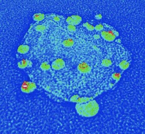
When the body is fighting an invading pathogen, white blood cells--including T cells--must respond. Now, Salk Institute researchers have imaged how vital receptors on the surface of T cells bundle together when activated. This study, the first to visualize this process in lymph nodes, could help scientists better understand how to turn up or down the immune system's activity to treat autoimmune diseases, infections or even cancer. The results were published this week in the Proceedings of the National Academy of Sciences. "We had seen these receptors cluster and reposition in cultured cells that were artificially stimulated in the lab, but we've never seen their natural arrangements in lymph nodes until now," says senior author Björn Lillemeier, an associate professor in Salk's Nomis Laboratories for Immunobiology and Microbial Pathogenesis, and the Waitt Advanced Biophotonics Center. T cells are activated when receptors embedded in their outer membrane bind to other immune cells that have digested an antigen, such as a virus, bacteria or cancer cell. In turn, the activated T cells switch on cellular pathways that help the body both actively seek out and destroy the antigen and remember it for the future. In the past, by looking at T-cell receptors embedded in isolated cells under the microscope, researchers discovered that the receptors are arranged in clusters--called protein islands--that merge when the cells are activated. Lillemeier wanted more detail on how the receptors are arranged in tissue and how that arrangement might change when the T cells are activated in living hosts. The team used a super-resolution microscope developed in the laboratory of co-senior author Hu Cang, assistant professor at Salk's Waitt Advanced Biophotonics Center and holder of the Frederick B. Rentschler Developmental Chair. This microscopy approach, called light-sheet direct stochastic optical reconstruction microscopy (dSTORM), let the researchers watch T cell receptors in the membranes of T cells in mouse lymph nodes at a resolution of approximately 50 nanometers. The new imagery confirmed the previous observation that protein islands of T-cell receptors merge into larger "microclusters" when T cells are activated. But it also showed that, before cells are activated, the protein islands are already arranged in groups--dubbed "territories" by Lillemeier's team. "The pre-organization on the molecular level basically turns the T cell into a loaded gun," says Lillemeier.
Via Mariaschnee
Astronomers are beginning to find strong links between dark matter and primordial black holes. Could dark matter — the elusive substance that composes most of the material universe — be made of black holes? Some astronomers are beginning to think this tantalizing possibility is more and more likely. Alexander Kashlinsky, an astronomer at the NASA Goddard Space Flight Center in Maryland, thinks that black holes that formed soon after the Big Bang can perfectly explain the observations of gravitational waves, or ripples in space-time, made by the Laser Interferometer Gravitational-Wave Observatory (LIGO) last year, as well as previous observations of the early universe. If Kashlinsky is correct, then dark matter might be composed of these primordial black holes, all galaxies might be embedded within a vast sphere of black holes, and the early universe might have evolved differently than scientists had thought.
Via Mariaschnee
The organic molecule methyl alcohol (methanol) has been found by the Atacama Large Millimeter/Submillimeter Array (ALMA) in the TW Hydrae protoplanetary disc. This is the first such detection of the compound in a young planet-forming disc. Its detection helps astronomers understand the chemical processes that occur during the formation of planetary systems and that ultimately lead to the creation of the ingredients for life.
The protoplanetary disc around the young star TW Hydrae is the closest known example to Earth, at a distance of only about 170 light-years. As such it is an ideal target for astronomers to study discs. This system closely resembles what astronomers think the Solar System looked like during its formation more than four billion years ago.
The Atacama Large Millimeter/Submillimeter Array (ALMA) is the most powerful observatory in existence for mapping the chemical composition and the distribution of cold gas in nearby discs. These unique capabilities have now been exploited by a group of astronomers led by Catherine Walsh (Leiden Observatory, the Netherlands) to investigate the chemistry of the TW Hydrae protoplanetary disc.
The ALMA observations have revealed the fingerprint of gaseous methyl alcohol, or methanol (CH3OH), in a protoplanetary disc for the first time. Methanol, a derivative of methane, is one of the largest complex organic molecules detected in discs to date. Identifying its presence in pre-planetary objects represents a milestone for understanding how organic molecules are incorporated into nascent planets.
Furthermore, methanol is itself a building block for more complex species of fundamental prebiotic importance, like amino acid compounds. As a result, methanol plays a vital role in the creation of the rich organic chemistry needed for life.
Catherine Walsh, lead author of the study, explains: "Finding methanol in a protoplanetary disc shows the unique capability of ALMA to probe the complex organic ice reservoir in discs and so, for the first time, allows us to look back in time to the origin of chemical complexity in a planet nursery around a young Sun-like star."
Via Mariaschnee
|
|
Scooped by
Dr. Stefan Gruenwald
June 15, 2016 11:41 AM
|
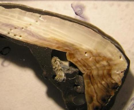
Shells of California mussels collected from the Pacific Ocean off the coast of Washington in the 1970s are on average 32 percent thicker than modern specimens, according to a new study published by University of Chicago biologists. Shells collected by Native Americans 1,000 to 1,300 years ago were also 27 percent thicker than modern shells, on average. The decreasing thickness over time, in particular the last few decades, is likely due to ocean acidification as a result of increased carbon in the atmosphere. "Archival material provided by past researchers, the Makah Tribal Nation, and the Olympic National Park allowed us to document this intriguing and concerning pattern in shell thickness," said Cathy Pfister, PhD, professor of ecology and evolution at the University of Chicago and lead author. The study was published June 15, 2016, in theProceedings of the Royal Society B. As humans burn fossils fuels, the oceans absorb a large portion of the additional carbon released into the atmosphere. This in turn causes pH levels of ocean water to drop, making it more acidic. Mussels, oysters, and certain species of algae have difficulty producing their calcium carbonate shells and skeletons in such an environment, and can provide an early indicator of how increasing ocean acidification affects marine life. In previous studies, Pfister and her colleagues documented declining pH levels in the waters surrounding Tatoosh Island off the coast of Washington state. In 2011 they further analyzed carbon and oxygen isotopes taken from modern mussel shells, shells collected by the local Makah tribe between AD 668 and 1008, and shells collected by biologists in the 1970s.
|
|
Rescooped by
Dr. Stefan Gruenwald
from Geology
June 15, 2016 11:35 AM
|
Pottery from China has revealed the secrets of some of the oldest known beer makers, who may be linked to the rise of hierarchical societies in East Asia. We now have some idea of how the beer would have been made 5000 years ago, thanks to the residue left on an apparent beer-making toolkit uncovered in Shaanxi, northern China. Jiajing Wang at the University of Stanford and colleagues found remnants of wide-mouthed pots, funnels and amphorae that would have been used for beer brewing, filtration and storage. They analysed traces of the pottery’s former contents – microscopic starch fragments and phytoliths, silica structures found in cereal husks. They identified these deposits as having come from broomcorn millet, Job’s tears, barley and tubers such as snake gourd root. “Many of the starch grains were damaged, and the damage patterns precisely match the morphological changes developed during malting and mashing,” says Wang. What’s more, the team found oxalates, organic compounds associated with the mashing and fermentation of cereals. The oldest chemically confirmed alcoholic beverage in the world, dating to 7000-6600 BC at nearby Jiahu in the Yellow River valley, was found by Pat McGovern at the University of Pennsylvania Museum of Archaeology and Anthropology.
Via Dr. Catherine Russell
|
|
Scooped by
Dr. Stefan Gruenwald
June 14, 2016 2:55 PM
|
A new material that can kill E. coli bacteria within 30 seconds has been developed by researchers at the Institute of Bioengineering and Nanotechnology (IBN) of A*STAR in Singapore. Triclosan, a common antibacterial ingredient found in many products such as toothpastes, soaps, and detergents to reduce or prevent bacterial infections, has been linked to making bacteria resistant to antibiotics, with adverse health effects. The European Union has restricted the use of triclosan in cosmetics, and the U.S. FDA is conducting an ongoing review of this ingredient. To find a more suitable alternative, IBN Group Leader Yugen Zhang, PhD, and his team synthesized a chemical compound made up of molecules linked together in a chain (“imidazolium oligomers”), which they found can kill 99.7% of the E. coli bacteria within 30 seconds. The chain-like structure helps to penetrate the cell membrane and destroy the bacteria.
|
|
Scooped by
Dr. Stefan Gruenwald
June 14, 2016 2:51 PM
|
Finnish researchers have developed a method for building highly efficient miniaturized micro-supercapacitor energy storage directly inside a silicon microcircuit chip, making it possible to power autonomous sensor networks, wearable electronics, and mobile internet-of-things (IoT) devices. Supercapacitors function similar to standard batteries, but store electrostatic energy instead of chemical energy. The researchers at VTT Technical Research Centre of Finland have developed a hybrid nano-electrode that’s only a few nanometers thick. It consists of porous silicon coated with a titanium nitride layer formed by atomic layer deposition. The nano-electrode design features the highest-ever conductive surface-to-volume ratio. That combined with an ionic liquid (in a microchannel formed in between two electrodes), results in an extremely small form factor and efficient energy storage. That design makes it possible for a silicon-based micro-supercapacitor to achieve higher energy storage (energy density) and faster charge/discharge (power density) than the leading carbon- and graphene-based supercapacitors, according to the researchers. The micro-supercapacitor can store 0.2 joule (55 microwatts of power for one hour) on a one-square-centimeter silicon chip. This design also leaves the surface of the chip available for active integrated microcircuits and sensors. Micro-supercapacitors can also be integrated directly with active microelectronic devices to store electrical energy generated by thermal, light, and vibration energy harvesters to supply electrical energy (see, for example, Wireless device converts ‘lost’ microwave energy into electric power). An open-access paper on the research has been published in Nano Energy journal.
|
|
Scooped by
Dr. Stefan Gruenwald
June 14, 2016 2:48 PM
|
The first study to systematically measure the number of neurons in the brains of birds has found that they have significantly more neurons packed into their small brains than are stuffed into mammalian and even primate brains of the same mass. Birds are remarkably intelligent, although their brains are small. Corvids and some parrots are capable of cognitive feats comparable to those of great apes. How do birds achieve impressive cognitive prowess with walnut-sized brains? We investigated the cellular composition of the brains of 28 avian species, uncovering a straightforward solution to the puzzle: brains of songbirds and parrots contain very large numbers of neurons, at neuronal densities considerably exceeding those found in mammals. Because these “extra” neurons are predominantly located in the forebrain, large parrots and corvids have the same or greater forebrain neuron counts as monkeys with much larger brains. Avian brains thus have the potential to provide much higher “cognitive power” per unit mass than do mammalian brains.
|
|
Scooped by
Dr. Stefan Gruenwald
June 14, 2016 2:38 PM
|
It's unlikely to contain life, but the Jupiter-sized planet is the largest real-life version of Tatooine, the fictional world that boasts two suns from 'Star Wars.' Astronomers say they’ve found another real-life version of Tatooine, the fictional world from “Star Wars” that features two suns, and it’s the largest one yet. The new planet is indeed far, far away – 3,700 light-years – and approximately 4.4 billion years old, about the same age as Earth. It’s about the same size as Jupiter, making it the largest planet orbiting two suns ever identified. The newly found discovery, uncovered with the help of the Kepler telescope and known as Kepler-1647b, was identified by a team led by researchers at NASA and San Diego State University. It was presented Monday during a meeting of the American Astronomical Society in San Diego. “It’s a bit curious that this biggest planet took so long to confirm, since it is easier to find big planets than small ones,” San Diego State astronomer Jerome Orosz, who worked on the study, said in a statement. The researchers say the planet takes 1,107 days, or just over three years, to orbit its two suns, the longest period discovered so far. It is also located much further away from its host stars than other circumbinary planets, as those with two suns are called. The unusual orbit also puts it in what’s known as the habitable zone, where life-sustaining water can be in liquid form. But its large size makes it an unlikely candidate to support life, the researchers say. But binary star systems could theoretically play host to Earth-like, rocky planets as well, a pair of astrophysicists argued in a paper last year
|
|
Scooped by
Dr. Stefan Gruenwald
June 11, 2016 12:32 PM
|
Thanks to her work on a class project, a University of British Columbia science student has discovered four new planet candidates, including one, located 3,200 light years away from Earth, that could potentially support life-sustaining liquid water.
It was a needle in a haystack search for newly-graduated physics and astronomy student Michelle Kunimoto. She spent months of her final year at school poring over data collected from NASA's Kepler satellite as part of a class assignment aimed at giving students real-life exposure to a career in astronomy. Eventually she found four tiny specks that all the other scientists who examined the data had likely missed.
|