Your new post is loading...

|
Scooped by
Dr. Stefan Gruenwald
November 2, 2016 8:28 PM
|
Researchers at the University of Houston have reported a new method for inducing superconductivity in non-superconducting materials, demonstrating a concept proposed decades ago but never proven. The technique can also be used to boost the efficiency of known superconducting materials, suggesting a new way to advance the commercial viability of superconductors, said Paul C.W. Chu, chief scientist at the Texas Center for Superconductivity at UH (TcSUH) and corresponding author of a paper describing the work, published Oct. 31 in the Proceedings of the National Academy of Sciences. "Superconductivity is used in many things, of which MRI (magnetic resonance imaging) is perhaps the best known," said Chu, the physicist who holds the TLL Temple Chair of Science at UH. But the technology used in health care, utilities and other fields remains expensive, in part because it requires expensive cooling, which has limited widespread adoption, he said. The research, demonstrating a new method to take advantage of assembled interfaces to induce superconductivity in the non-superconducting compound calcium iron arsenide, offers a new approach to finding superconductors that work at higher temperatures. Superconducting materials conduct electric current without resistance, while traditional transmission materials lose as much as 10 percent of energy between the generating source and the end user. That means superconductors could allow utility companies to provide more electricity without increasing the amount of fuel used to generate electricity.
|
|
Scooped by
Dr. Stefan Gruenwald
November 2, 2016 8:24 PM
|
Modern experiment methods in biology can generate overwhelming amounts of raw data. To manage this, scientists must create entirely new workflows and systems capable of merging large, disparate data sets and presenting them intuitively. Arrowland is a combined -omics visualization tool that runs on a web browser, tablet, or cellphone. It shows functional genomics data (fluxomics, metabolomics, proteomics and transcriptomics) together on a zoomable, searchable map, similar to the street maps used for navigation. In addition to providing a coherent layout for -omics measurements, the maps in Arrowland are also an easy-to-use reference for the relationships between the reactions, genes, and proteins involved in metabolism. “At JBEI we have leveraged the insights provided by metabolic fluxes measured through 13C Metabolic Flux Analysis to make quantitative predictions for E. coli, and we shared these fluxes on Arrowland” said Hector Garcia Martin, Director of Quantitative Metabolic Modeling at JBEI. “By making this cloud-based interactive tool publicly available, our hope is to enable application of this tool in a wide range of fields, not only bioenergy. We are open to collaborations with the broader scientific community and industry.” Setting itself apart from other available tools, Arrowland is beneficial for its clarity and ease of use. With its unique interface and presentation method, Arrowland makes the exploration of -omics data as intuitive as possible, on a platform that does not require special hardware or configuration. Four different types of -omics data are integrated into the same map – an ability that no competing visualization platforms share – making important correlations and progressions easy to recognize and examine. Arrowland is open-source and currently under active development, with plans to add more maps, editing and curation features, and is integrated with an open-source modeling package that JBEI plans to release separately, called the JBEI Quantitative Metabolic Modeling Library. For the time being, a sample data set from the flux profiles published in “A Method to Constrain Genome-Scale Models with 13C Labeling Data”, PLOS Computational Biology (Garcia Martin et al, 2015) can be viewed at http://public-arrowland.jbei.org.
|
|
Rescooped by
Dr. Stefan Gruenwald
from DNA and RNA research
November 2, 2016 8:18 PM
|
Colliding tectonic plates pushed up a strip of land from the watery abyss that once divided North and South America, forming the isthmus of Panama. But a study now hints that this happened millions of years earlier than scientists had thought. Evolutionary and population-genetics data from Eciton army ants, which can only travel on dry ground, suggest that the isthmus formed 4–8 million years ago. The research, published on 25 October in Molecular Ecology1, challenges the long-held idea that the link between continents emerged no more than 3 million years ago. “Our genomic data is very strong evidence that the army ants crossed this region much earlier in time than the model of the simple closure of the isthmus suggests,” says Corrie Moreau, associate curator at the Field Museum in Chicago, Illinois, and co-author of the study. She notes that recent geological studies have also hinted that the isthmus may have emerged earlier than 3 million years ago.
Via Integrated DNA Technologies
|
|
Scooped by
Dr. Stefan Gruenwald
November 2, 2016 4:00 PM
|
Implanted medical devices like catheters, surgical mesh and dialysis systems are ideal surfaces on which bacteria can colonize and form hard-to-kill sheets called biofilms. Known as biofouling, this contamination of devices is responsible for more than half of the 1.7 million hospital-acquired infections in the United States each year. In a report published in Biomaterials today, a team of scientists at Beth Israel Deaconess Medical Center (BIDMC), the Wyss Institute for Biologically Inspired Engineering and the John A. Paulson School of Engineering and Applied Sciences (SEAS) at Harvard University has demonstrated that an innovative, ultra-low adhesive coating prevented bacteria from attaching to surfaces treated with it, reducing bacterial adhesion by more than 98 percent in laboratory tests. “Device related infections remain a significant problem in medicine, burdening society with millions of dollars in health care costs,” said Elliot Chaikof, MD, PhD, chair of the Roberta and Stephen R. Weiner Department of Surgery and Surgeon-in-Chief at BIDMC and an associate faculty member at the Wyss Institute. “Antibiotics alone will not solve this problem. We need to use new approaches to minimize the risk of infection, and this strategy is a very important step in that direction.” The self-healing slippery surface coatings – known as ‘slippery liquid-infused porous surfaces’ (SLIPS) – were developed by Joanna Aizenberg, PhD, a Wyss Institute core faculty member, Professor of Chemistry and Chemical Biology and the Amy Smith Berylson Professor of Materials Science at SEAS at Harvard University. Inspired by the carnivorous Nepenthes pitcher plant that uses the slippery surface of its leaves to trap insects, Aizenberg engineered surface coatings that work to repel a variety of substances across a broad range of temperature, pressure and other environmental conditions. They are stable when exposed to UV light, and are low-cost and simple to manufacture. The current study is the first to demonstrate that SLIPS not only limit the ability of bacteria to adhere to surfaces, but also impede infection in an animal model. “We are developing SLIPS recipes for a variety of medical applications by working with different medical-grade materials, ensuring the stability of the coating, and carefully pairing the non-fouling properties of the SLIPS materials to specific contaminates, environments and performance requirements,” said Aizenberg. “Here we have extended our repertoire and applied the SLIPS concept very convincingly to medical-grade lubricants, demonstrating its enormous potential in implanted devices prone to bacterial fouling and infection.”
|
|
Scooped by
Dr. Stefan Gruenwald
November 2, 2016 3:52 PM
|
Humans started brewing beer thousands of years ago, probably in ancient Egypt. Without any knowledge of the microorganisms powering their breweries, it's no surprise that the first tipplers worked blind, brewing with whatever wild yeasts found their way into the brewing process. But according to genetic analysis of over 100 yeasts used by industrial brewers today, brewers started unknowingly breeding the best yeasts sometime around the 1500s. "Brewers were smart enough to realize that if they used the sediment found at the end of the fermentation process, if you used it to inoculate a new batch, the fermentation will go much quicker," explained study author Kevin Verstrepen, a yeast geneticist at the University of Leuven and VIB in Belgium. "And when you take the sediment from a really nice beer, the new batch will for the most part turn out pretty nice." They didn't know it, but these early brewers were selecting the fittest beer yeasts, which settle down into sediment after they stop producing gas. By putting them into a new batch of beer (a process known as back-slopping), the brewers were keeping their favorite strains of yeast warm and well-fed, encouraging them to reproduce. "It was something between an art and a science, because they didn't know what microbes were but knew the sediment, whatever it was, was important," Verstrepen said. "So then you had this Darwinian selection, with the gut feeling of the brewers working hand in hand with nature." According to the study's analysis of 157 different strains of yeast used to make beer, wine, spirits, sake, bread and bioethanol, this process has caused the yeast used in beer brewing to grow more distinct from feral cousins than microbes used for other commercial fermentation.
|
|
Scooped by
Dr. Stefan Gruenwald
November 1, 2016 8:24 PM
|
According to a new study, a tiny skin patch may help treat peanut allergies. Peanut allergies are among the most serious food allergies, requiring constant vigilance. In severe cases, exposure to even trace amounts of the nut can trigger a deadly reaction. But according to a new study published last week in the Journal of Allergy and Clinical Immunology, a tiny skin patch may help treat peanut allergies. Over the course of a year, researchers from the Consortium of Food Allergy Research tested 74 peanut-allergic volunteers, ages 4 through 20, to see whether a daily Viaskin peanut patch could help raise their peanut threshold. The patch, called epicutaneous immunotherapy, released peanut proteins into the participants' skin, building cellular tolerance to the nuts. The results showed that participants who received higher doses of peanut protein in the patch were able to consume more peanuts after a year. The patch was the most effective on children ages 4 to 11 and significantly less effective on older participants, according to a statement from the National Institutes of Health, which funded the study through its National Institute of Allergy and Infectious Diseases. The peanut patch trial was conducted at five research sites: Arkansas Children's Hospital, the National Jewish Health Center in Denver, Johns Hopkins University, the Icahn School of Medicine at Mount Sinai and the University of North Carolina at Chapel Hill School of Medicine. The newly published results were from the first year of the trial, but the researchers will continue monitoring the participants for a total of two and a half years. To conduct the yearlong study, researchers divided participants into three groups: a placebo group that received patches with no peanut protein, a group given patches with 100 micrograms of peanut protein and one given patches with 250 micrograms of peanut protein. The 74 participants were randomly assigned to each group. Before the trial began, researchers assessed each volunteer's peanut allergy via an oral food challenge with food containing peanuts. The median amount of nuts tolerated, before having allergic symptoms was about one-seventh of a peanut, according to Dr. Stacie M. Jones, chief of allergy and immunology at Arkansas Children's Hospital and the study lead.
|
|
Scooped by
Dr. Stefan Gruenwald
October 31, 2016 1:20 AM
|
Discovery of icy worlds in the distant solar system offer new clues in searches for hypothetical giant. New solar system objects used to be a distraction for Konstantin Batygin, a planetary scientist and theorist at the California Institute of Technology (Caltech) in Pasadena. Each discovery added another complication to his computer models of the solar system, which twirl planetoids around the sun. But now, Batygin is eager to find more of the objects himself, and missed opportunities pain him. In late September and early October, cloudy skies foiled a 6-night run at the Subaru Telescope atop Mauna Kea in Hawaii. All you can do, he says, is “just sit quietly and wait for things to get slightly better.” What drew Batygin into the hunt is the ultimate prize: a new planet, the first to be added to our solar system in more than a century. Colloquially called Planet Nine, this distant hypothetical world could have 10 times the mass of Earth and take 15,000 years to go around the sun. This past January, Batygin and Mike Brown, a Caltech astronomer, proposed that the giant could explain the peculiarly clustered orbits of six icy bodies beyond Neptune. Now, several teams, including Batygin and Brown’s, are racing to spot Planet Nine directly.
|
|
Scooped by
Dr. Stefan Gruenwald
October 31, 2016 1:16 AM
|
There is an ongoing 20,000-mile storm at Saturn's North Pole and it has intrigued scientists ever since it was discovered. But what's causing more confusion is that the 'hexagon' changed colors upon comparing images taken of Saturn's North Pole from 2012 to 2016. From the images released by NASA, it is evident the once blue color turned into yellowish-gold. The Cassini spacecraft observing the planet since 2004 captured the images. The change appeared to happen gradually based on the observation launched since Nov. 2012 up to Sept. 2016. The mystery hexagon is a cloud pattern in Saturn's northern hemisphere where an evident weather activity is taking place. Experts believe that the shape could have been caused by the varying wind speed in Saturn. Saturn, the second largest planet in the Solar System next to Jupiter, is also a gas planet. It is composed mainly of hydrogen, methane, and helium. But despite the surprising changes, astronomers and scientists already offered some potential causes of the unusual phenomenon. "In particular the change from a bluish color to a more golden hue may be due to the increased production of photochemical hazes in the atmosphere as the north pole approaches summer solstice in May 2017," a NASA official said in a statement.
|
|
Scooped by
Dr. Stefan Gruenwald
October 31, 2016 1:11 AM
|
Astronomers are adamant in trying to find signs of alien life if there's any in Tabby's star. The Breakthrough Listen initiative will reportedly spend $100 million to search for any signs of alien life in Tabby's star for the next 10 years. The speculations of an alien civilization in Tabby's star are what make the mission enticing. Tabby's star will be investigated using the Green Bank Telescope located in West Virginia as announced last Tuesday, Oct. 25th, 2016. "The Green Bank Telescope is the largest fully steerable radio telescope on the planet, and it's the largest, most sensitive telescope that's capable of looking at Tabby's star given its position in the sky," Breakthrough Listen co-director Andrew Siemion, also a director at Berkeley Search for Extraterrestrial Intelligence, said in a statement. "We've deployed a fantastic new SETI instrument that connects to that telescope, that can look at many gigahertz of bandwidth simultaneously and many, many billions of different radio channels all at the same time so we can explore the radio spectrum very, very quickly," Siemion added. The mission will conduct eight hours per nights for the next two months starting this Wednesday, Oct. 26. Astronomers are hopeful that with funding, there will be more data gathered about Tabby's star. Astronomers are also hopeful to find evidence of an advanced alien civilization in Tabby's star. The interest in the region sparked when astronomers detected the unusual light behavior, something that no one has ever seen before. The star dims in such a way that no other star does, according to a report.
|
|
Scooped by
Dr. Stefan Gruenwald
October 31, 2016 1:07 AM
|
Hookworms are among the oldest life forms on Earth, and when they sink their teeth into our blood vessels, they cause nausea, anemia, cramping, and more. Ancient Egyptians referred to the symptoms in a 3500-year-old papyrus, and Hippocrates may have described miners infected by them. In the early 1900s, an intensive campaign eliminated the worms from the intestines of U.S. schoolchildren and spawned the formation of the U.S. Public Health Service. Now, after years of viewing hookworms as the enemy, scientists are beginning to wonder whether these tiny critters, shorter than the width of a human fingernail, could also be an ally. Buried in the creatures, many believe, are molecules with healing powers. Scattered studies in diseases like Crohn’s and celiac, as well as self-treatment by patients, suggest the worms might alleviate symptoms of autoimmune conditions—the exact ailments that have become more and more prevalent as hookworms have faded from our lives and general cleanliness has prevailed. Severine Navarro, an immunologist at James Cook University in Cairns, Australia, is among those on the hunt for these magic molecules, and she believes she has found one: anti-inflammatory protein-2 (AIP-2). In mice with asthma, AIP-2 suppresses airway inflammation, Navarro and her colleagues report, and the protein also appears to damp down the proliferation of some immune cells in the blood of people with allergies. “It’s a clear demonstration that a single product from a parasite can have sweeping systemic changes that are very favorable,” says Rick Maizels, a parasite immunologist at the University of Glasgow in the United Kingdom, who wasn’t involved in the research. The work, published in Science Translational Medicine, reflects a shift in the field, from examining hookworms’ immune effects to identifying the molecules that drive them. The ultimate goal is new drugs based on these prized molecules.
|
|
Scooped by
Dr. Stefan Gruenwald
October 31, 2016 1:03 AM
|
IBM recently announced a new IBM Watson Data Platform that combines the world’s fastest data ingestion engine touting speeds up to 100+GB/second with cloud data source, data science, and cognitive API services. IBM is also making IBM Watson Machine Learning Service more intuitive with a self-service interface. According to Bob Picciano, Senior Vice President of IBM Analytics “Watson Data Platform applies cognitive assistance for creating machine learning models, making it far faster to get from data to insight. It also, provides one place to access machine learning services and languages, so that anyone, from an app developer to the Chief Data Officer, can collaborate seamlessly to make sense of data, ask better questions, and more effectively operationalize insight.” For more information or a free trial of IBM Watson Data Platform, Data Science Experience, Watson APIs, or Bluemix, the following resources are useful:
|
|
Scooped by
Dr. Stefan Gruenwald
October 30, 2016 2:43 PM
|
Scientists have developed a new compound that's effective at inhibiting the growth of multiple types of cancer models in the lab – slowing as many as a quarter of all known types of cancer. The molecule, called S63845, works by blocking the protein MCL1, which many different types of cancer cells rely on to grow. And without access to MCL1, the cancer cells die off. The discovery could help researchers fight blood cancers such as acute myeloid leukemia, lymphoma, and multiple myeloma. Solid tumors – including melanoma and cancers of the lung and breast – could also see new treatments. "MCL1 (a bcl2 family member) is important for many cancers because it is a pro-survival protein that allows the cancerous cells to evade the process of programmed cell death that normally removes cancer cells from the body," says one of the team, Guillaume Lessene from the Walter and Eliza Hall Institute in Australia. "Extensive studies performed in a variety of cancer models have shown that S63845 potently targets cancer cells dependent on MCL1 for their survival." Cancerous cells are difficult for the body's immune system to kill, because they can quickly evolve and spread faster than our defences can keep up, and can also evade what's called apoptosis – a form of programmed cell death. Blasting them with chemotherapy drugs or radiation can be an effective way to combat cancer cells, but these techniques usually take healthy cells along with them, and cause debilitating side effects. What makes S63845 so promising is that, in addition to cutting off cancer's life support system, it can be given at doses that don't harm normal cells, the team suggests. The compound is the latest development in a new class of anti-cancer drugs called BH3 mimetics, which target a family of proteins that cancers rely on to sidestep and resist programmed cell death. "BH3 mimetics inhibit a group of proteins known as the 'pro-survival BCL-2 proteins'," says Lessene. "MCL1 is a member of this protein family, and inhibiting it activates the process of programmed cell death." It's worth pointing out that research is still at a pre-clinical stage, and the researchers need to run further tests on the compound before turning it into a drug that can be taken safely by human patients. But the early signs are very encouraging.
|
|
Scooped by
Dr. Stefan Gruenwald
October 30, 2016 2:31 PM
|
Application of a femtosecond spectroscopy technique to a copper surface has allowed the desorption of carbon monoxide molecules to be tracked with unprecedented detail. It has long been a chemist’s dream to catch a chemical reaction in the act. In a chemical reaction, reactant atoms or molecules pass over energy barriers, through transition states, and eventually convert into new chemical products. Chemists would like to observe and interfere with this process in mid-course and thereby control its outcome. The development of lasers that produce pulses of light just a few femtoseconds long has allowed researchers to initiate and track chemical reactions in the gas phase. Typically, one femtosecond pulse initiates the reaction through excitation of the reactant to a new electronic state, which drives the reactant’s nuclear motion. Another pulse, time-delayed from the first, then monitors the nuclear motion as the excited reactant decays or a new product forms. This approach gave rise to the field of femtochemistry and earned Ahmed Zewail (1946–2016) the 1999 Nobel Prize in Chemistry [1]. Many important chemical reactions, however, occur in more complex environments where molecules experience interactions, such as in condensed-matter systems. Moreover, there has been a strong impetus to extend methods of femtochemistry to surfaces because they can serve as models for catalysis. Catalytic substrates are used to lower energy barriers for chemical reactions of adsorbed molecules and thereby reduce the cost of producing economically valuable products, such as fertilizers, fuels, and pharmaceuticals. Yoshiyasu Matsumoto from Kyoto University, Japan, and co-workers now report [2] on one of the most basic chemical processes on a surface—the desorption of carbon monoxide (CO) molecules from a copper surface, driven by femtosecond-laser heating of its electrons. When a femtosecond “pump” laser pulse interacts with a surface, an adsorbed molecule can be energized in two ways: directly, by light-induced charge transfer from the surface to the molecule; or indirectly, through the heating of surface electrons [3]. The latter method, which Matsumoto and colleagues employ, is more general because hot electrons can transfer their energy to any adsorbed molecule, regardless of its electronic structure, through a process called electronic friction [4]. Electrons in a semiconductor or metal have a much smaller heat capacity than their host lattice. This means that, when a femtosecond laser pulse excites these materials, electrons can quickly reach temperatures exceeding 5000 K. These extremely hot electrons subsequently transfer their energy to the vibrational modes of the adsorbed molecule and the lattice, allowing chemical reactions between the two to be initiated [5]. To track the dynamics provoked by the transfer of energy from hot electrons, such as the breaking or rearrangement of the chemical bond between the molecule and the surface, one needs a “meter stick” to “report on” the process. This can be accomplished by probing the change in the molecule-surface electronic structure, using methods such as photoemission spectroscopy [6] and x-ray absorption spectroscopy [7]. But these methods are demanding: the first requires relatively long-lived (tens of femtoseconds) electronically excited states, which are rarely found, whereas the second entails using ultrafast x-ray laser sources that exist only in a few specialized user facilities. The approach taken by Matsumoto and co-workers has none of these drawbacks, relying on a broadly applicable and accessible time-dependent measurement of the vibrational spectrum of the excited complex. The approach incorporates phase-sensitive detection—to precisely follow the minute frequency changes of a molecular vibration—into a time-resolved sum-frequency generation (SFG) spectroscopy experiment. In this type of experiment, two femtosecond pulses of photons with different frequency interact with the nonlinear electrical polarizability of the surface and combine to form a third pulse whose photon frequency is the sum of the first two. With one incident pulse tuned to the desired molecular vibration, which in Matsumoto and co-workers’ case was the internal stretching vibration of the adsorbed CO molecules, the SFG signal gains resonance enhancement and simultaneously probes the frequency of the vibration. Similar SFG experiments have been used previously to study the desorption of CO from a metal surface [8]. But the combination of phase-sensitive detection with SFG has allowed Matsumoto and colleagues to garner information on how hot electrons interact with molecular vibrations to affect the desorption process. In other words, the researchers used the phase of the CO stretching vibration as a “reporter” [9], with a period of about 13 fs, of the kicks and shoves delivered by hot electrons to the external motions of the CO molecule during desorption.
|
|
|
Rescooped by
Dr. Stefan Gruenwald
from DNA and RNA research
November 2, 2016 8:25 PM
|
Berkeley Lab researchers found that aberrant strands of genetic code have telltale signs that enable gateway proteins to recognize and block them from exiting the nucleus. They found that RNA-binding proteins are regulated such that gateway proteins can recognize and block aberrant strands of genetic code from exiting the nucleus. Unused messenger RNA (mRNA) strands that cannot exit the nucleus would eventually disintegrate. Their findings, published today in the journal Scientific Reports, shed light on a complex system of cell regulation that acts as a form of quality control for the transport of genetic information out of the nucleus. Getting a more complete picture of how genetic information gets expressed in cells is important in disease research, the researchers said. “Some components of this machinery are dysregulated in various types of cancers,” said study principal investigator Mohammad Mofrad, faculty scientist at Berkeley Lab’s Molecular Biophysics and Integrated Bioimaging Division. “Understanding the molecular mechanism of genetic information transport and quality control would substantially improve the current knowledge about various types of cancers and other human diseases.” Biology textbooks already describe how strands of mRNA copy sections of DNA inside a cell’s nucleus and then exit to the cell’s cytoplasm. It is in the cytoplasm where the genetic code is used to synthesize proteins, so ensuring that only the correct mRNA strands get used is critical to the formation of properly functioning proteins. “Just like all production lines, the process of genetic information transfer and protein production is quality controlled at different stages,” said Mofrad. “To date, the exact mechanism of this quality control step has remained unclear.” Previous studies have looked at specific steps in this process, but the complex system of sorting out RNA that is ready to leave the nucleus has not been well understood.
Via Integrated DNA Technologies
|
|
Scooped by
Dr. Stefan Gruenwald
November 2, 2016 8:22 PM
|
Living Planet Report predicts that by 2020, populations will have declined by two-thirds from 1970. The populations of Earth’s wild mammals, birds, amphibians, fish and other vertebrates declined by more than half between 1970 and 2012, according to a report from environmental charity WWF and the Zoological Society of London (ZSL). Activities such as deforestation, poaching and human-induced climate change are in large part to blame for the decline. If the trend continues, then by 2020 the world will have lost two-thirds of its vertebrate biodiversity, according to the Living Planet Report 2016. “There is no sign yet that this rate will decrease,” the report says. “Across land, freshwater and the oceans, human activities are forcing species populations and natural systems to the edge,” says Marco Lambertini, director-general of WWF International.
|
|
Scooped by
Dr. Stefan Gruenwald
November 2, 2016 7:01 PM
|
The researchers, from Bristol University, found the leaves only develop this color when put in almost dark conditions - and in bright light the sheen slowly disappears. Begonias with striking blue leaves may represent a next step in plant evolution that puts them ahead of their ordinary green neighbors, research suggests. The blue color results from a super-efficient form of photosynthesis that allows them to use light other plants reject. These findings could help scientists develop light-harvesting devices to make better electronics. The researchers looked at Begonia pavonina - a flower found under the thick canopies of tropical forests in Malaysia. It has iridescent blue leaves because of its unusual chloroplasts, known as iridoplasts, in its surface layers. These contain regularly spaced stacks of three to four 'thylakoids' - which resemble a photonic crystal and strongly reflect wavelengths of light between 430 and 560 nanometers. This is what causes the leaves' blue iridescence. The thylakoids look very similar to the artificial structures commonly used to make miniature lasers that control the flow of light. In the Malaysian forests, the small amount of light that reaches the plant's leaves is mainly at the green-red end of the spectrum. The iridoplasts concentrate these specific wavelengths onto the plant's photosynthetic apparatus - increasing the efficiency of its photosynthesis by five to ten per cent. The study, published in Nature Plants, rules out a previous popular hypothesis that it was to to deter predators. 'The real novelty of what the begonia is doing is combining the light harvesting mechanism with the photonic structure - light is being both structurally manipulated and harvested with high levels of quantum efficiency within the same material,' Dr Heather Whitney, from Bristol University, said. The plants - including species commonly grown as houseplants or in gardens - have long been using a special form of nanotechnology called photonics to create structures in their leaves to help them harvest light. 'We discovered under the microscope, individual chloroplasts in these leaves reflected blue light brightly, almost like a mirror,' said Matt Jacobs, a PhD student at Bristol University. 'Looking in more detail by using a technique known as electron microscopy, we found a striking difference between the 'blue' chloroplasts found in the begonias, also known as "iridoplasts" due to their brilliant blue iridescent coloration, and those found in other plants.
|
|
Scooped by
Dr. Stefan Gruenwald
November 2, 2016 3:57 PM
|
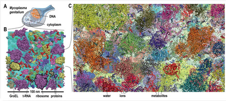
Biological macromolecules function in highly crowded cellular environments. The structure and dynamics of proteins and nucleic acids are well characterized in vitro, but in vivo crowding effects remain unclear. Using molecular dynamics simulations of a comprehensive atomistic model cytoplasm scientists found that protein-protein interactions may destabilize native protein structures, whereas metabolite interactions may induce more compact states due to electrostatic screening. Protein-protein interactions also resulted in significant variations in reduced macromolecular diffusion under crowded conditions, while metabolites exhibited significant two-dimensional surface diffusion and altered protein-ligand binding that may reduce the effective concentration of metabolites and ligands in vivo. Metabolic enzymes showed weak non-specific association in cellular environments attributed to solvation and entropic effects. These effects are expected to have broad implications for the in vivo functioning of biomolecules. This work is a first step towards physically realistic in silico whole-cell models that connect molecular with cellular biology. DOI: http://dx.doi.org/10.7554/eLife.19274.001
|
|
Scooped by
Dr. Stefan Gruenwald
November 2, 2016 3:45 PM
|
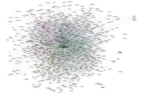
In 2014 at the Museums and the Web conferences in Baltimore, Maryland, and Florence, Italy, a group of data scientists presented a report on how two thousand museums were relating on Twitter. After our presentation, they decided to continue their investigation in two directions: The first are the case studies. They wanted to analyze some museums' Twitter environments, e.g., how museums are relating to their followers, how they form different communities defined by underlying relationships, the growth of the network around a museum, detection of influencers, etc. They also wanted to study how to use this information in order to design better social-media strategies for an improved and more realistic assessment of the work and actions’ results on Twitter. Case studies from London's Victoria & Albert, Turin's Palazzo Madama, and Barcelona's Center for Contemporary Culture have chosen museums with different Twitter strategies, countries, languages, and follower bases ranging from 7,000 at the time of writing this abstract (Palazzo Madama) to the 450,000 from V&A. The goal was to have different situations represented and run a useful research for museums of different sizes. The second direction nowadays is how museum professionals are interacting with each other on Twitter thanks to the use of hashtags, like #musetech, in order to share information, questions, and experiences.
|
|
Scooped by
Dr. Stefan Gruenwald
October 31, 2016 2:14 AM
|
Axel Visel, a genomicist at the Lawrence Berkeley National Laboratory in Berkeley, California, used python and boa genomes to track down the cause of leglessness. He and his colleagues compared the genomes of those snakes with the genomes of snakes such as vipers and cobras, which evolved more recently and have no leg remnants at all. They noticed that the Sonic hedgehog (SHH) enhancer had many deletions and mutations. They then showed that a mutated SHH enhancer does indeed affect leg growth by testing its influence on mouse limbs. They replaced the rodent’s own version of the enhancer with the snake version using the CRISPR-Cas9 gene-editing technique. They also repeated the experiment, subbing in the same enhancer from a fish and, later, from a human. Even with the fish and human enhancers, the mouse legs developed normally, demonstrating that enhancers from other species still work in the mice. But with the snake enhancer, the legs turned into little nubs, Visel and his colleagues report their whole study in a current issue of Cell. Finally, when researchers put the missing DNA into the snake enhancer and then put the modified enhancer back into mice, their legs grew just fine. “The Visel paper is a beautiful study; a tour de force that takes functional genomics using CRISPR-Cas9 to a new level,” Cohn says. Visel says that Cohn’s experiments with snake embryos “independently confirm what we saw in our mouse models. It's very elegant work.” But Cohn, Visel, and Hanken caution that changes in this enhancer are not the whole story behind the evolution of the serpentine form. Recently, for example, other researchers discovered that another enhancer was responsible for snakes’ extra ribs and long backbones. And it’s possible that the Sonic hedgehog enhancer was not the first step in leg loss, Hanken says. “But it’s certainly a major player.”
|
|
Scooped by
Dr. Stefan Gruenwald
October 31, 2016 1:18 AM
|
First fossil evidence of a dinosaur brain doesn’t suggest they were particularly smart. A decade ago, a fossil hunter was combing the beach in southeastern England when he found a strange, brown pebble. The surface of it caught his eye: It was smooth and strangely undulating, and also slightly crinkly in some places. That oddly textured pebble, scientists report here today at the Society for Vertebrate Paleontology meeting, is actually an endocast—an impression preserved in the rock—that represents the first known evidence of fossilized brain tissue of a dinosaur (likely a close relative of Iguanodon, a large, herbivorous type of dinosaur that lived about 133 million years ago). Human brains and bird brains are packed tightly into the brain case, so that their convolutions leave an impression of the inside of the case. But dinosaur (and reptile) brains are more loosely fitted; they are surrounded within the brain case by membranes called meninges, tough sheaths that protect and support the brain. So an endocast of a dinosaur brain might be expected to show those structures—and it did (structures above the green line, above). But beneath them, remineralized in calcium phosphate, the researchers also spied a pattern of tiny capillaries and other cortical tissues—the sort of fabric you’d expect for the cortex of a brain. That those textures were pressed up against the brain case doesn’t necessarily mean that dinosaurs were bigger-brained and smarter than we thought, however: Instead, the dinosaur had likely simply toppled over and been preserved upside down, its brain tissue preserved by surrounding acidic, low-oxygen waters that pickled and hardened the membranes and tissues, providing a template for mineralization. The structure of the brain, studied with scanning electron microscopes, reveal similarities to both birds and crocodiles. It’s the first known evidence of such a dinosaur brain—but now that they know to look for it, the researchers say, they might go back and look at other endocranial casts of dinosaurs to see if they might contain traces of other such structures. The team also reports its findings today in a Special Publication of the Geological Society of London.
|
|
Scooped by
Dr. Stefan Gruenwald
October 31, 2016 1:14 AM
|
They could do wonders for medicine, chemistry, banking and information-gathering. There's just one problem... Quantum mechanics, Carl Sagan once observed, is so strange that "common sense is almost useless in approaching it." Scientists still don't understand exactly why matter behaves as it does at the quantum level. Yet they're getting better at exploiting its peculiar dynamics -- in ways that may soon upend the technology business. One of the most interesting applications is in computing. In theory, quantum computers could take advantage of odd subatomic interactions to solve certain problems far faster than a conventional machine could. Although a full-scale quantum computer is still years off, scientists have lately made a lot of progress on the materials, designs and methods needed to make one. And that could have some striking benefits. Quantum computers could simulate how atoms and molecules behave, to the great advantage of chemists and drug designers. They could solve optimization problems -- say, how to efficiently route airplane traffic -- far faster than current technology can. They could speed advances in artificial intelligence, improve sensors, and lead to the design of stronger and lighter industrial materials.
|
|
Rescooped by
Dr. Stefan Gruenwald
from DNA and RNA research
October 31, 2016 1:10 AM
|
A group coordinated by the International School for Advanced Studies (SISSA) in Trieste has built a three-dimensional computer model of the human genome. The shape of DNA (as well as its sequence) significantly affects biological processes and is therefore crucial for understanding its function. This new study has provided a first three-dimensional, approximate but realistic, identikit of the human genome. Thanks to the characteristics of the new method, the structural reconstruction based on both experimental information and statistical methods will be refined as new experimental data become available. The study, carried out in collaboration with the University of Oslo, has just been published in Scientific Reports (a journal of the Nature group). Genome sequencing is a milestone in modern biology as it allows access to the entire “list of instructions” (the chemical sequence of genetic makeup) for the development and function of organisms. Sequencing the genome is a bit like writing down the exact order of the color of beads in a necklace: knowing how they are arranged along the thread gives us no indication as to the shape of the necklace. The shape of the DNA strand can be highly complex, given that the chromosomes are loosely arranged in an apparently chaotic tangle in the cell nucleus. Since the shape of chromosomes may have a decisive effect on their function, it is important that it should be characterized, in part because scientists think the DNA tangle in the nucleus is only apparently chaotic and that it has instead a specific “geography” for each tissue and stage of cell life. “Arriving at a precise description of the shape of the DNA tangle is unfortunately incredibly complicated”, explains Cristian Micheletti, SISSA professor and coordinator of the new study. “In our case, we used experimental data on ‘proximity pairs’”. “Imagine having to create a map of a city”, he explains, “based only on information like ‘the post office is opposite the station’, ‘the chemist is close to the gym’, ‘the fruit and vegetable market is near the football field’ and so on. If you have only a small number of such statements to go by, your map will be approximate and in some cases indeterminate. But if you have hundreds, thousands or even more, then your map will become increasingly precise and accurate. This is the logic we followed”. “Proximity pairs” therefore refers to information on the closeness of two points on the map. In the case of nuclear DNA, this information was provided by a technique (which Micheletti defines as “brilliant”) known as Hi-C, developed by North American research groups in 2010. In this chemical-physical technique, bits of genome located close to each another in the nucleus are tied together and then identified by their sequence. By collecting large numbers of these proximity pairs scientists discovered which points of the chromosomes lie close to each other in the nucleus. While this is today the most powerful technique for investigating DNA organisation in the nucleus, it is still inadequate for inferring its overall shape. “For this reason, we thought we would try to go ‘further’”, comments Micheletti.
Via Integrated DNA Technologies
|
|
Scooped by
Dr. Stefan Gruenwald
October 31, 2016 1:06 AM
|
New method grants unprecedented flexibility for creating organs-on-chips. Scientists have a long way to go before they can match the self-building powers of the human body. But now, a new technique for assembling flexible polymers and living cells may get them one step closer. Previously, researchers investigating how tissues and organs function (or don’t) had few options for creating artificial tissues—so-called organs-on-chips—that they could use to avoid experimenting on animals and people. They typically built their tiny organ models one piece at a time from individual gel bricks filled with suspended cells, a time-intensive process that limited what the bricks could be made of and how they could be shaped. Now, scientists say they have solved those problems with microfluidics and electricity. Using electric fields, they can push and pull tiny, particle-filled droplets between two glass plates, assemble them into a larger configuration, and treat the droplets to form adhesive gels. This lets them control the droplets’ movement and manipulate the particles, like cells, suspended within. As a result, they can assemble gels from a wide variety of materials and control the configuration of both the bricks and the cells inside them. Already, the researchers have used the technique to create a heart-like tissue whose cells beat in time, they report today in Science Advances. If this technique can be refined to provide perfect control of cells, it can be used to build precise architectures of living cells, paving the way for artificial tissues, cell-based robots, and more sophisticated organ models.
It’s not a bad day at work. Five of the biggest fundamental problems in physics seem sorted in one go. The model that can do this, formulated by Guillermo Ballesteros at the University of Paris-Saclay in France and his colleagues, may explain dark matter, neutrino oscillations, baryogenesis, inflation and the strong CP problem. Dubbed SMASH, the model is based on the standard model of particle physics, but has a few bits tacked on. The standard model is a collection of particles and forces that describes the building blocks of the universe. Although it has passed every test thrown at it, it can’t explain some phenomena. For example, we don’t understand dark matter, the mysterious substance that makes up 84 per cent of the universe’s mass. Nor why there is more matter than antimatter. Nor why the universe grew so rapidly in its youth during a period known as inflation. The list continues. So something is still missing from the standard model. “Presumably we need some new particles,” says Mikhail Shaposhnikov at the Swiss Federal Institute of Technology in Lausanne. “The question is, how many new particles do we need? Some models, like supersymmetry, add hundreds of particles – none of which have been spotted at colliders like the LHC. But SMASH adds only six: three neutrinos, a fermion and a field that includes two particles. That’s a reasonable approach, Shaposhnikov says. “I would start by assuming that the number of new particles is very small,” he says. “And then add new particles only if you really need them.” SMASH is several theories smashed together, says co-author Andreas Ringwald at the German Electron Synchrotron, DESY, in Hamburg. It builds on Shaposhnikov’s model from 2005, which added three neutrinos to the three already known in order to solve four fundamental problems in physics: dark matter, inflation, some questions about thenature of neutrinos, and the origins of matter. SMASH adds a new field to explain some of those problems a little differently. This field includes two particles: the axion, a dark horse candidate for dark matter, and the inflaton, the particle behind inflation. As a final flourish, SMASH uses the field to introduce the solution to a fifth puzzle: the strong CP problem, which helps explain why there is more matter than antimatter in the universe. “The best thing about the theory is that it can be tested or checked within the next 10 years or so,” Ringwald says. “You can always invent new theories, but if they can only be tested in 100 years, or never, then this is not real science but meta-science.” SMASH predicts that the axion should be about ten billion times lighter than the electron. Particles this small could be probed by the CULTASK experiment running in South Korea, or the proposed ORPHEUS experiment in the US and the planned MADMAX experiment in Germany. This doesn’t mean it’s game over. It’s more like game on. Physicists will continue to compete to find experimental evidence or a better model. “The battle is open,” Ringwald says Reference: https://arxiv.org/pdf/1608.05414v1.pdf
Via Levin Chin
|
|
Scooped by
Dr. Stefan Gruenwald
October 30, 2016 2:36 PM
|
Google Brain has created two artificial intelligences that evolved their own cryptographic algorithm to protect their messages from a third AI, which was trying to evolve its own method to crack the AI-generated crypto. The study was a success: the first two AIs learnt how to communicate securely from scratch. The Google Brain team (which is based out in Mountain View and is separate from Deep Mind in London) started with three fairly vanilla neural networks called Alice, Bob, and Eve. Each neural network was given a very specific goal: Alice had to send a secure message to Bob; Bob had to try and decrypt the message; and Eve had to try and eavesdrop on the message and try to decrypt it. Alice and Bob have one advantage over Eve: they start with a shared secret key (i.e. this is symmetric encryption). Importantly, the AIs were not told how to encrypt stuff, or what crypto techniques to use: they were just given a loss function (a failure condition), and then they got on with it. In Eve's case, the loss function was very simple: the distance, measured in correct and incorrect bits, between Alice's original input plaintext and its guess. For Alice and Bob the loss function was a bit more complex: if Bob's guess (again measured in bits) was too far from the original input plaintext, it was a loss; for Alice, if Eve's guesses are better than random guessing, it's a loss. And thus an adversarial generative network (GAN) was created.
|