Your new post is loading...

|
Suggested by
LIGHTING
September 20, 2018 5:18 AM
|

|
Suggested by
Société Francaise d'Immunologie
September 20, 2018 4:40 AM
|
When microbes invade, they are taken up by body cells or immune cells such as monocytes, which digest them into protein fragments, or antigens, that are then presented on their cell surface. These antigens are recognized by a specialized receptor on the surface of T cells (the T cell receptor, or TCR), whose signaling prompts secretion of proteins called cytokines that coordinate the immune system's efforts to fight off the pathogen. Download: PPT PowerPoint slide PNG larger image TIFF original image In the colonic mucosa, antigens of Faecalibacterium prausnitzii (a clostridium bacteria from the gut microbiota) are presented by dendritic cells. Recognition of these antigens by the CD4 T cells equipped with a specific TCR contributes to their differentiation into FoxP3-lacking regulatory T cells (Treg) characterized by the co-expression of CD4 and CD8α. Most of these Treg stay in the colonic mucosa where they prevent excessive inflammatory responses. A fraction of them migrate into the blood, likely contributing to the immune tolerance outside the gut. https://doi.org/10.1371/journal.pbio.1001834.g001 Each individual T cell expresses just one of myriad different versions of the TCR, each of which responds to a particular antigen. This wide diversity allows the recognition of a large repertoire of threats, but also carries the risk of inappropriate reactivity against normal tissues. While T cells that may mount such attacks are usually weeded out during their development, autoreactive cells do occasionally appear. In addition, changes to body homeostasis may sometimes provoke T cell autoimmunity. As a failsafe against these problems, a specialized type of T cell called the “T regulatory” cell (Treg) reins in inappropriate immune responses by secreting immunosuppressive cytokines such as IL-10. Tregs can be distinguished from other types of T cells primarily by their expression of the transcription factor FoxP3, together with a characteristic set of cell surface proteins. The ability to restrict immune responses is delicately balanced. For example, the immune system normally tolerates several species of commensal gut bacteria, and does not mount immune responses against them. However, perturbances to the mixture of commensal bacteria communities can lead—through mechanisms that remain unclear—to bowel inflammation, and sometimes serious autoimmune diseases such as inflammatory bowel disease (IBD) or ulcerative colitis. In their paper published this month in PLOS Biology, Guillaume Sarrabayrouse, Frédéric Altare, Francine Jotereau, and colleagues describe a new type of T cell that mediates immune tolerance, which may be important for preventing bowel inflammation. While examining the lymphocyte population in the lamina propria of normal human gut mucosa, Sarrabayrouse et al. noticed a group of T cells that simultaneously expressed two different types of T cell–specific surface proteins, CD4 and CD8αα. The researchers were intrigued by these double-positive CD4CD8αα lamina propria lymphocytes (DP8α LPL) because most T cells outside the thymus (where developing T cells reside) express only one of either CD4 or CD8. The authors decided to investigate the function of these cells and found that, compared with CD4 single-positive T cells derived from the same site, DP8α LPL expressed surface markers classically associated with Treg. Curiously, however, DP8α LPL did not express FoxP3. Despite their lack of FoxP3, DP8α LPL secreted a high amount of IL-10 and they were potently immunosuppressive, able to prevent the maturation and proliferation of other types of immune cells in vitro. Previous studies had indicated that certain commensal bacteria, particularly those of the Clostridium IV group, can induce the appearance of FoxP3 regulatory T cells in mice. The authors therefore studied whether DP8α LPL responded to these commensals. In fact, although DP8α LPL expressed a widely heterogeneic population of TCRs, when presented with human monocytes pre-incubated with a Clostridium IV group bacterium, Faecalibacterium prausnitzii (F prau), a large portion of DP8α LPL responded by proliferating and secreting cytokines. Other commensals, even closely related bacteria of the Clostridium IV group, failed to elicit any response from DP8α LPL. Occasionally, bacteria can activate T cells nonspecifically by secreting proteins called “superantigens,” which activate TCR signaling regardless of whether pathogen-derived antigens are being presented to the T cell. But Sarrabayrouse et al. showed that DP8α LPL proliferative and cytokine responses to F prau did not depend on superantigens, and that DP8α LPL were specifically reactive to F prau–derived antigens. Further studies showed that DP8α T cells could be detected not only in the gut but also in the peripheral blood (such cells were dubbed DP8α PBL). DP8α PBL differed from their LPL counterparts in that they lacked expression of cell surface markers associated with regulatory T cells, and a lower fraction of DP8α PBL displayed reactivity to F prau. The authors suspect DP8α PBL may be derived from DP8α LPL, although what role they play in the body remains unclear. Sarrabayrouse et al. next investigated what happens to DP8α T cells during gut inflammation. They observed that both DP8α LPL and PBL populations were smaller in patients with active IBD than in healthy donors, which correlates with the fact that F prau levels drop precipitously during IBD flare-ups. Furthermore, the authors observed that DP8α PBL and LPL from IBD patients sometimes failed to proliferate or secrete cytokines in response to F prau. Collectively, these data suggest that DP8α T cells are a novel type of regulatory T cell that respond specifically to F prau, and whose loss or dysfunction may affect immune homeostasis in the gut. Sarrabayrouse G, Bossard C, Chauvin J-M, Jarry A, Meurette G, et al. (2014) CD4CD8αα Lymphocytes, a Novel Human Regulatory T Cell Subset Induced by Colonic Bacteria and Deficient in Inflammatory Bowel Disease Patients. doi:10.1371/journal.pbio.1001833

|
Scooped by
Gilbert C FAURE
September 19, 2018 2:22 PM
|
|
|
Scooped by
Gilbert C FAURE
September 12, 2018 2:46 PM
|
Citation: Sullender ME, Baldridge MT (2018) Norovirus interactions with the commensal microbiota. PLoS Pathog 14(9): e1007183. https://doi.org/10.1371/journal.ppat.1007183 Editor: Richard C. Condit, University of Florida, UNITED STATES Published: September 6, 2018 Copyright: © 2018 Sullender, Baldridge. This is an open access article distributed under the terms of the Creative Commons Attribution License, which permits unrestricted use, distribution, and reproduction in any medium, provided the original author and source are credited. Funding: MTB was supported by NIH grant K22 AI127846-01, DDRCC grant P30 DK052574, and the Global Probiotics Council’s Young Investigator Grant for Probiotics Research. The funders had no role in study design, data collection and analysis, decision to publish, or preparation of the manuscript. Competing interests: The authors have declared that no competing interests exist. Introduction Human norovirus (HNoV) is the leading cause of epidemic nonbacterial gastroenteritis worldwide, causing an acute diarrheal infection and occasionally chronic infection in immunocompromised individuals. Mouse and tissue culture models utilizing murine norovirus (MNoV) have allowed for interrogation of viral mechanisms of infection and pathogenesis. Here, we outline the interactions between the commensal microbiota of the intestine and norovirus and their implications (Fig 1). Download: PPT PowerPoint slide PNG larger image TIFF original image Fig 1. Norovirus pathogenesis is affected by many factors in the enteric environment. a) The presence of commensal bacteria allows for efficient MNoV infection, with tuft cells being one rare cell population infected. b) Absence of commensal bacteria reduces MNoV titers by depleting tuft cell populations and potentially altering innate immune responses during persistent MNoV infection. c) Binding of HNoV to HBGA-positive enteric bacteria has been found to facilitate infection of B cells. d) Sialic acid moieties on the cell surface have been found to act as coreceptors for MNoV infection of macrophages, while bile acids have been found to be important for the establishment of HNoV infection of enterocytes. e) MNoV infection has been found to trigger the expression of IFN-λ in infected cells, up-regulating interferon simulated genes that restrict viral replication and subsequent spread of infection—the mechanism of this process has yet to be characterized, but commensals are believed to play a major regulatory role. HBGA, histo-blood group antigen; HNoV, human norovirus; IFN-λ, interferon lambda; MNoV, murine norovirus. https://doi.org/10.1371/journal.ppat.1007183.g001 Question 1: Is norovirus infection in mice a good model for norovirus infection in humans? Due to the fact that HNoV cannot readily grow in mice and, until recently, has not been culturable in vitro, the use of MNoV has provided robust animal and tissue culture model systems, which allow for mechanistic studies of an orthologous pathogen [1–4]. The MNoV model system allows for the merging of basic mechanistic principles of infection and replication from cell culture systems to pathogenesis in a host system that is both genetically malleable and affordable. Thus far, MNoV studies have allowed for the elucidation of a species-specific proteinaceous receptor (CD300LF) and viral tropism for a rare intestinal epithelial cell population called tuft cells during persistent infection as well as macrophages, dendritic cells, and lymphocytes during acute infection in vivo [5–7]. Many external factors outside of the narrow window of host cell–virus interactions that affect NoV pathogenicity have also been identified, such as bile, sialic acid, and intestinal bacteria [2,5,6,8]. Additionally, host mucosal cytokine interferon lambda (IFN-λ) has recently been identified as a potent anti-NoV molecule, opening the possibility of its therapeutic use for HNoV in the future [9]. Multiple strains of MNoV allow for the study of both chronic (MNV.CR6, MNV-3) and acute (MNV-1, MNV-1.CW3) infection, further adding to the strengths and complexity of this model system [10]. Despite some differences in symptom presentation and species-specific receptors, HNoV and MNoV exhibit many similarities in cellular tropism, requirement of carbohydrate attachment factors, and potential for persistent viral shedding after symptom resolution [3,10]. However, some clinical symptoms do differ between MNoV and HNoV infections, most notably the lack of vomiting and inability to produce more than statistically significant mild diarrhea in mice [11]. Question 2: Do all strains of norovirus depend on the commensal microbiota for infection? A large and diverse population of commensal microbes, consisting of bacteria, viruses, fungi, and parasites, reside within the intestinal lumen. NoV, being an enteric pathogen, encounters and interacts with members of this community, resulting in outcomes beneficial or detrimental to the host. HNoV has been found to interact directly with commensal (Enterobacter cloacae) and pathogenic (Clostridium difficile) bacterial species via the viral capsid and histo-blood group antigen (HGBA)-like carbohydrates expressed on bacterial surface membranes [12,13]. In addition, both HNoV and MNoV have been reported to bind sialic acid residues, which can be expressed on bacteria, suggesting that MNoV could also interact directly with the enteric microbiota [4,14]. Experimental alteration of the enteric microbiota with oral antibiotics drastically depletes the intestinal bacterial population. This in turn reduces the severity of acute MNoV infection (reduced MNV-1 titers) and also prevents or reduces persistent infections (drastically reduced MNV.CR6 and MNV-3 titers) in the ileum and colon [15]. Additionally, infection by MNV.CR6 can be rescued by fecal microbiota transplant (FMT) from nonantibiotic-treated to antibiotic-treated mice, highlighting the importance of commensal bacteria for MNoV infection [15]. While all murine NoV strains tested and reported thus far exhibit a dependence upon commensal bacteria for infection, further studies will be needed to determine whether this is a phenomenon affecting all HNoV and MNoV strains and to define strain-specific mechanisms. One putative explanation by which bacteria can promote MNoV infection comes from the recent description of tuft cells as the physiologic target cell of persistent MNoV infection and propagation. Within the mouse intestine, tuft cells are regulated by commensal bacteria such that antibiotic treatment correlates with reduced numbers of tuft cells and leads to reduced viral titers [6]. In addition to commensal bacteria, parasitic worms (such as Trichinella spiralis) also exhibit a proviral effect in the context of MNoV infection [16,17] via induction of tuft cells by type 2 immune responses (IL-4, IL-25 cytokines) [6]. Thus, both bacteria and enteric metazoans can regulate NoV infection. In addition to microbe–NoV and microbe–host interactions impacting viral pathogenesis, NoV infection itself can also alter the enteric microbial communities of the host. This virus-induced dysbiosis is characterized by an enhanced Firmicutes to Bacteroidetes ratio. This alteration is seen both in a subset of HNoV infections and early acute MNoV infections (MNV-1) [18,19]. However, this effect was not detected in longitudinal studies of both acute and persistent strains of MNoV (MNV-1, MNV.CR6, and MNV-4), suggesting potential temporal or facility-based effects [20]. Question 3: How do commensal bacteria regulate enteric virus infections via immune skewing? The host intestinal immune system is highly regulated by a complex interplay of various lymphoid tissues, immune cells, cytokines, and their receptors [21–23] and possesses three distinct layers: mucus, epithelia, and lamina propria. In the small intestine, mucus-secreting goblet cells and antimicrobial peptide-secreting Paneth cells form the mucosal barrier that segregates commensal bacteria from the intestinal epithelia [24]. Intestinal epithelial cells directly interact with and survey the gut environment in coordination with innate lymphoid cells, which communicate with the immune system via secretion of cytokines and chemokines [24–26]. Dendritic cells ferry antigen from the lumen across the epithelial barrier to draining lymph nodes and mucosal lymphoid tissues in the lamina propria [27], and innate inflammatory signals and other luminal signals activate T- and B-cell responses [26]. These interacting layers play a large role in maintaining the microbiota and host immune system in homeostasis as well as regulating infection, inflammation, and autoimmunity. While the interactions between commensal bacteria, enteric viruses, and the intestinal immune system are still poorly understood, several recent studies have suggested important interplay between these factors. Innate immune responses are primed via commensal bacterial recognition by the enteric epithelium, which activates antiviral intestinal responses after a secondary viral-induced signal within the gut [22]. In contrast, mouse mammary tumor virus (MMTV) has evolved to evade innate immune responses by binding bacterial lipopolysaccharide, inducing the immunosuppressive cytokine IL-10 via Toll-like receptor signaling pathways [21]. This effect is entirely dependent upon the microbiota; mice receiving parenteral administration of MMTV do not experience a suppressed immune response, and antibiotic-treated or germ-free mice receiving MMTV orally fail to pass MMTV to their offspring. Microbial modulation of innate immunity also offers a second explanation of the preventive effect of antibiotics on MNoV infection: it is the result of the bacterial microbiota hindering a yet-to-be-identified immune pathway, which limits the antiviral efficacy of IFN-λ during persistent MNoV infection [23]. Evidence for this comes from experiments demonstrating that mice lacking IFN-λ signaling no longer require commensal bacteria for successful MNoV infection [23]. This triangle of interactions—commensals, viral pathogen, and host—produce anti- or proviral environments through many disparate and yet-to-be-characterized mechanisms. Question 4: Does a dependence on the commensal microbiota apply to other viruses? Other enteric viruses, including rotavirus and poliovirus, have been found to depend on enteric bacteria to infect, similar to MNoV [28,29]. Commensal bacteria act as a proviral factor during poliovirus infection, as antibiotic treatment results in mice being less susceptible to infection and a reduced viral load in the intestine [28]. The mechanism underlying this involves viral particles binding to bacterial lipopolysaccharide, causing enhanced host cell receptor binding and virion stability [28,30]. Paradoxically, microbial depletion was found to increase antibody responses against rotavirus, which may contribute to enhanced viral clearance during antibiotic treatment [29]. In contrast, nonenteric viral infections are enhanced in mice depleted of commensal bacteria. For neurotropic flavivirus (West Nile, dengue, Zika) infections, depletion of the enteric microbiota significantly increased viral susceptibility, viral burden, disease severity, and lethal outcomes in mice [31]. Additionally, respiratory influenza A virus (IAV) and lymphocytic choriomeningitis virus (LCMV) infection have been found to be intensified (sustained, high viral titers in lung tissue and serum, respectively) due to impaired immune responses secondary to depletion of gram-positive bacteria in the gut [32,33]. In these cases, antibiotic treatment reduced virus-specific cluster of differentiation 8+ (CD8+) T-cell responses, though there is apparent variation in the manifestation of defects. Antibiotic treatment resulted in decreased numbers of dendritic cells (DCs) for antigen presentation in the case of flavivirus infection, whereas in the case of IAV and LCMV, there was a defect in DC migration to lymph nodes attributed to reduced inflammasome activation upon infection [31,32]. The bacterial metabolite desminotyrosine was also found to regulate type I IFN signaling in the lung to control IAV infection [32,34]. These findings suggest that bacteria interact with both innate and adaptive immune systems to control both local and systemic antiviral responses, leading to distinct outcomes for enteric and nonenteric viruses. Question 5: Does microbiome modulation have therapeutic potential for infectious diseases in humans? While it is clear that the microbiome plays a significant role in both infectious and noninfectious diseases alike, much remains unknown about the exact mechanisms of action. FMTs have proven to be an effective treatment for Clostridium difficile (C. diff) infection and treatment-resistant irritable bowel syndrome (IBS) and may have potential in inflammatory bowel diseases [35]. It is likely that different underlying mechanisms contribute to the efficacy of these treatments; for example, specific bile acids regulated by intestinal bacteria are critical for resistance to C. diff infection [36,37]. Targeted administration of efficacious microbes would be ideal to prevent disease, and we are just beginning to identify the specific bacterial species that may regulate diseases from multiple sclerosis to diabetes to norovirus. While it may appear that treating HNoV patients with antibiotics would prove beneficial since antibiotic treatment reduces MNoV titers in mice, it may actually cause more harm than good due to the overall beneficial impact of the microbiome on human health and the potential for increased susceptibility to other viral, fungal, or bacterial infections. Probiotics may be a better therapeutic option, such as adding helpful microbes to a patient’s microbiome to fight infection or as a form of biological vaccine adjuvant. And in the case of NoV, development of drugs that temporarily mitigate the effect of commensal microbes and their metabolites without clearing bacterial populations could prove to be a viable treatment option in the future. Acknowledgments We apologize to all colleagues whose work could not be cited due to length restrictions. We also wish to thank Mudra Hegde, Dylan Lawrence, and Elizabeth Kennedy for their helpful critiques of this review. References 1. Jones MK, Grau KR, Costantini V, Kolawole AO, de Graaf M, Freiden P, et al. Human norovirus culture in B cells. Nat Protoc [Internet]. 2015 Dec [cited 2018 Apr 30];10(12):1939–47. Available from: http://www.ncbi.nlm.nih.gov/pubmed/26513671 pmid:26513671 View Article PubMed/NCBI Google Scholar 2. Ettayebi K, Crawford SE, Murakami K, Broughman JR, Karandikar U, Tenge VR, et al. Replication of human noroviruses in stem cell-derived human enteroids. Science (80-) [Internet]. 2016 Sep 23 [cited 2018 Apr 30];353(6306):1387–93. Available from: http://www.ncbi.nlm.nih.gov/pubmed/27562956 pmid:27562956 View Article PubMed/NCBI Google Scholar 3. Wobus CE, Thackray LB, Virgin HW. Murine norovirus: a model system to study norovirus biology and pathogenesis. J Virol [Internet]. 2006 Jun 1 [cited 2018 Apr 30];80(11):5104–12. Available from: http://www.ncbi.nlm.nih.gov/pubmed/16698991 pmid:16698991 View Article PubMed/NCBI Google Scholar 4. Bartnicki E, Cunha JB, Kolawole AO, Wobus CE. Recent advances in understanding noroviruses. F1000Research [Internet]. 2017 [cited 2018 May 1];6:79. Available from: http://www.ncbi.nlm.nih.gov/pubmed/28163914 pmid:28163914 View Article PubMed/NCBI Google Scholar 5. Orchard RC, Wilen CB, Doench JG, Baldridge MT, McCune BT, Lee Y-CJ, et al. Discovery of a proteinaceous cellular receptor for a norovirus. Science [Internet]. 2016 [cited 2018 Apr 30];353(6302):933–6. Available from: http://www.ncbi.nlm.nih.gov/pubmed/27540007 pmid:27540007 View Article PubMed/NCBI Google Scholar 6. Wilen CB, Lee S, Hsieh LL, Orchard RC, Desai C, Hykes BL, et al. Tropism for tuft cells determines immune promotion of norovirus pathogenesis. Science [Internet]. 2018 Apr 13 [cited 2018 Apr 30];360(6385):204–8. Available from: http://www.ncbi.nlm.nih.gov/pubmed/29650672 pmid:29650672 View Article PubMed/NCBI Google Scholar 7. Grau KR, Roth AN, Zhu S, Hernandez A, Colliou N, DiVita BB, et al. The major targets of acute norovirus infection are immune cells in the gut-associated lymphoid tissue. Nat Microbiol [Internet]. 2017 Dec 6 [cited 2018 Jun 19];2(12):1586–91. Available from: http://www.ncbi.nlm.nih.gov/pubmed/29109476 8. Taube S, Perry JW, Yetming K, Patel SP, Auble H, Shu L, et al. Ganglioside-linked terminal sialic acid moieties on murine macrophages function as attachment receptors for murine noroviruses. J Virol [Internet]. 2009 May [cited 2018 May 1];83(9):4092–101. Available from: http://www.ncbi.nlm.nih.gov/pubmed/19244326 pmid:19244326 View Article PubMed/NCBI Google Scholar 9. Lee S, Wilen CB, Orvedahl A, McCune BT, Kim K-W, Orchard RC, et al. Norovirus Cell Tropism Is Determined by Combinatorial Action of a Viral Non-structural Protein and Host Cytokine. Cell Host Microbe [Internet]. 2017 Oct 11 [cited 2018 May 9];22(4):449–459.e4. Available from: https://www.sciencedirect.com/science/article/pii/S1931312817303542?via%3Dihub pmid:28966054 View Article PubMed/NCBI Google Scholar 10. Baldridge MT, Turula H, Wobus CE. Norovirus Regulation by Host and Microbe. Trends Mol Med [Internet]. 2016 [cited 2018 Apr 30];22(12):1047–59. Available from: http://www.ncbi.nlm.nih.gov/pubmed/27887808 pmid:27887808 View Article PubMed/NCBI Google Scholar 11. Liu G, Kahan SM, Jia Y, Karst SM. Primary high-dose murine norovirus 1 infection fails to protect from secondary challenge with homologous virus. J Virol [Internet]. 2009 Jul [cited 2018 Jun 19];83(13):6963–8. Available from: http://www.ncbi.nlm.nih.gov/pubmed/19403675 pmid:19403675 View Article PubMed/NCBI Google Scholar 12. Li D, Breiman A, le Pendu J, Uyttendaele M. Binding to histo-blood group antigen-expressing bacteria protects human norovirus from acute heat stress. Front Microbiol [Internet]. 2015 Jul 1 [cited 2018 May 1];6:659. Available from: http://journal.frontiersin.org/article/10.3389/fmicb.2015.00659 13. Miura T, Sano D, Suenaga A, Yoshimura T, Fuzawa M, Nakagomi T, et al. Histo-blood group antigen-like substances of human enteric bacteria as specific adsorbents for human noroviruses. J Virol [Internet]. 2013 Sep 1 [cited 2018 May 1];87(17):9441–51. Available from: http://www.ncbi.nlm.nih.gov/pubmed/23804639 pmid:23804639 View Article PubMed/NCBI Google Scholar 14. Almagro-Moreno S, Boyd EF. Bacterial catabolism of nonulosonic (sialic) acid and fitness in the gut. Gut Microbes [Internet]. 2010 Jan [cited 2018 May 1];1(1):45–50. Available from: http://www.ncbi.nlm.nih.gov/pubmed/21327116 pmid:21327116 View Article PubMed/NCBI Google Scholar 15. Baldridge MT, Nice TJ, McCune BT, Yokoyama CC, Kambal A, Wheadon M, et al. Commensal microbes and interferon-λ determine persistence of enteric murine norovirus infection. Science [Internet]. 2015 Jan 16 [cited 2018 Apr 30];347(6219):266–9. Available from: http://www.ncbi.nlm.nih.gov/pubmed/25431490 pmid:25431490 View Article PubMed/NCBI Google Scholar 16. Hayes KS, Bancroft AJ, Goldrick M, Portsmouth C, Roberts IS, Grencis RK, et al. Exploitation of the intestinal microflora by the parasitic nematode Trichuris muris. Science [Internet]. 2010 Jun 11 [cited 2018 Apr 30];328(5984):1391–4. Available from: http://www.ncbi.nlm.nih.gov/pubmed/20538949 pmid:20538949 View Article PubMed/NCBI Google Scholar 17. Osborne LC, Monticelli LA, Nice TJ, Sutherland TE, Siracusa MC, Hepworth MR, et al. Coinfection. Virus-helminth coinfection reveals a microbiota-independent mechanism of immunomodulation. Science [Internet]. 2014 Aug 1 [cited 2018 May 9];345(6196):578–82. Available from: http://www.ncbi.nlm.nih.gov/pubmed/25082704 pmid:25082704 View Article PubMed/NCBI Google Scholar 18. Nelson AM, Walk ST, Taube S, Taniuchi M, Houpt ER, Wobus CE, et al. Disruption of the human gut microbiota following Norovirus infection. PLoS ONE [Internet]. 2012 [cited 2018 May 1];7(10):e48224. Available from: http://www.ncbi.nlm.nih.gov/pubmed/23118957 pmid:23118957 View Article PubMed/NCBI Google Scholar 19. Hickman D, Jones MK, Zhu S, Kirkpatrick E, Ostrov DA, Wang X, et al. The effect of malnutrition on norovirus infection. MBio [Internet]. 2014 Mar 4 [cited 2018 May 1];5(2):e01032–13. Available from: http://www.ncbi.nlm.nih.gov/pubmed/24595373 pmid:24595373 View Article PubMed/NCBI Google Scholar 20. Nelson AM, Elftman MD, Pinto AK, Baldridge M, Hooper P, Kuczynski J, et al. Murine norovirus infection does not cause major disruptions in the murine intestinal microbiota. Microbiome [Internet]. 2013 Feb 18 [cited 2018 Apr 30];1(1):7. Available from: http://www.ncbi.nlm.nih.gov/pubmed/24451302 pmid:24451302 View Article PubMed/NCBI Google Scholar 21. Kane M, Case LK, Kopaskie K, Kozlova A, MacDearmid C, Chervonsky A V, et al. Successful transmission of a retrovirus depends on the commensal microbiota. Science [Internet]. 2011 Oct 14 [cited 2018 May 9];334(6053):245–9. Available from: http://www.ncbi.nlm.nih.gov/pubmed/21998394 pmid:21998394 View Article PubMed/NCBI Google Scholar 22. Sansone CL, Cohen J, Yasunaga A, Xu J, Osborn G, Subramanian H, et al. Microbiota-Dependent Priming of Antiviral Intestinal Immunity in Drosophila. Cell Host Microbe [Internet]. 2015 Nov 11 [cited 2018 May 9];18(5):571–81. Available from: http://www.ncbi.nlm.nih.gov/pubmed/26567510 pmid:26567510 View Article PubMed/NCBI Google Scholar 23. Baldridge MT, Nice TJ, McCune BT, Yokoyama CC, Kambal A, Wheadon M, et al. Commensal microbes and interferon-λ determine persistence of enteric murine norovirus infection. Science [Internet]. 2015 Jan 16 [cited 2018 May 9];347(6219):266–9. Available from: http://www.ncbi.nlm.nih.gov/pubmed/25431490 pmid:25431490 View Article PubMed/NCBI Google Scholar 24. Shi N, Li N, Duan X, Niu H. Interaction between the gut microbiome and mucosal immune system. Mil Med Res [Internet]. 2017 [cited 2018 Apr 30];4:14. Available from: http://www.ncbi.nlm.nih.gov/pubmed/28465831 pmid:28465831 View Article PubMed/NCBI Google Scholar 25. Ivanov II, Honda K. Intestinal commensal microbes as immune modulators. Cell Host Microbe [Internet]. 2012 Oct 18 [cited 2018 May 9];12(4):496–508. Available from: http://www.ncbi.nlm.nih.gov/pubmed/23084918 pmid:23084918 View Article PubMed/NCBI Google Scholar 26. Hepworth MR, Monticelli LA, Fung TC, Ziegler CGK, Grunberg S, Sinha R, et al. Innate lymphoid cells regulate CD4+ T-cell responses to intestinal commensal bacteria. Nature [Internet]. 2013 Jun 6 [cited 2018 May 9];498(7452):113–7. Available from: http://www.ncbi.nlm.nih.gov/pubmed/23698371 pmid:23698371 View Article PubMed/NCBI Google Scholar 27. Karst SM. The influence of commensal bacteria on infection with enteric viruses. Nat Rev Microbiol [Internet]. 2016 Apr [cited 2018 May 9];14(4):197–204. Available from: http://www.ncbi.nlm.nih.gov/pubmed/26853118 pmid:26853118 View Article PubMed/NCBI Google Scholar 28. Kuss SK, Best GT, Etheredge CA, Pruijssers AJ, Frierson JM, Hooper L V, et al. Intestinal microbiota promote enteric virus replication and systemic pathogenesis. Science [Internet]. 2011 Oct 14 [cited 2018 May 9];334(6053):249–52. Available from: http://www.ncbi.nlm.nih.gov/pubmed/21998395 pmid:21998395 View Article PubMed/NCBI Google Scholar 29. Uchiyama R, Chassaing B, Zhang B, Gewirtz AT. Antibiotic treatment suppresses rotavirus infection and enhances specific humoral immunity. J Infect Dis [Internet]. 2014 Jul 15 [cited 2018 May 9];210(2):171–82. Available from: http://www.ncbi.nlm.nih.gov/pubmed/24436449 pmid:24436449 View Article PubMed/NCBI Google Scholar 30. Robinson CM, Jesudhasan PR, Pfeiffer JK. Bacterial lipopolysaccharide binding enhances virion stability and promotes environmental fitness of an enteric virus. Cell Host Microbe [Internet]. 2014 Jan 15 [cited 2018 Jun 22];15(1):36–46. Available from: http://www.ncbi.nlm.nih.gov/pubmed/24439896 pmid:24439896 View Article PubMed/NCBI Google Scholar 31. Thackray LB, Handley SA, Gorman MJ, Murphy KM, Virgin HW, Correspondence MSD, et al. Oral Antibiotic Treatment of Mice Exacerbates the Disease Severity of Multiple Flavivirus Infections. CellReports [Internet]. 2018 [cited 2018 Apr 30];22:3440–3453.e6. Available from: pmid:29590614 View Article PubMed/NCBI Google Scholar 32. Ichinohe T, Pang IK, Kumamoto Y, Peaper DR, Ho JH, Murray TS, et al. Microbiota regulates immune defense against respiratory tract influenza A virus infection. Proc Natl Acad Sci U S A [Internet]. 2011 Mar 29 [cited 2018 Apr 30];108(13):5354–9. Available from: http://www.ncbi.nlm.nih.gov/pubmed/21402903 pmid:21402903 View Article PubMed/NCBI Google Scholar 33. Abt MC, Osborne LC, Monticelli LA, Doering TA, Alenghat T, Sonnenberg GF, et al. Commensal Bacteria Calibrate the Activation Threshold of Innate Antiviral Immunity. Immunity [Internet]. 2012 Jul 27 [cited 2018 Apr 30];37(1):158–70. Available from: http://www.ncbi.nlm.nih.gov/pubmed/22705104 pmid:22705104 View Article PubMed/NCBI Google Scholar 34. Steed AL, Christophi GP, Kaiko GE, Sun L, Goodwin VM, Jain U, et al. The microbial metabolite desaminotyrosine protects from influenza through type I interferon. Science [Internet]. 2017 [cited 2018 May 9];357(6350):498–502. Available from: http://www.ncbi.nlm.nih.gov/pubmed/28774928 pmid:28774928 View Article PubMed/NCBI Google Scholar 35. Holleran G, Scaldaferri F, Ianiro G, Lopetuso L, Mc Namara D, Mele MC, et al. Fecal microbiota transplantation for the treatment of patients with ulcerative colitis and other gastrointestinal conditions beyond Clostridium difficile infection: an update. Drugs Today (Barc) [Internet]. 2018 Feb [cited 2018 May 9];54(2):123–36. Available from: http://www.ncbi.nlm.nih.gov/pubmed/29637938 36. Theriot CM, Bowman AA, Young VB. Antibiotic-Induced Alterations of the Gut Microbiota Alter Secondary Bile Acid Production and Allow for Clostridium difficile Spore Germination and Outgrowth in the Large Intestine. mSphere [Internet]. 2016 [cited 2018 May 9];1(1). Available from: http://www.ncbi.nlm.nih.gov/pubmed/27239562 37. Buffie CG, Bucci V, Stein RR, McKenney PT, Ling L, Gobourne A, et al. Precision microbiome reconstitution restores bile acid mediated resistance to Clostridium difficile. Nature [Internet]. 2015 Jan 8 [cited 2018 May 9];517(7533):205–8. Available from: http://www.ncbi.nlm.nih.gov/pubmed/25337874 pmid:25337874 View Article PubMed/NCBI Google Scholar
|
|
Suggested by
Société Francaise d'Immunologie
September 12, 2018 2:23 PM
|
IL-1β in gingival crevicular fluid (GCF) is a marker of inflammation in periodontal disease. Here, Offenbacher et al. identify genetic variants in the IL37 locus associated with GCF-IL-1β and show that the IL-1β-increasing allele at rs3811046 leads to an enhanced inflammatory response in vitro and...
|
|
Rescooped by
Gilbert C FAURE
from Immunology
September 12, 2018 2:15 PM
|
Group 2 innate lymphoid cells (ILC2s) that produce the cytokine IL-5 are found in lung, gut, fat and skin tissues. New findings indicate that ILC2s in different tissues selectively express distinct functional cytokine receptors for cell activation in response to the local environment.
|
|
Scooped by
Gilbert C FAURE
September 11, 2018 3:42 AM
|
Antibodies binding to their cognate cellular receptors can trigger important downstream immune responses. We mapped out critical amino acids on the IgA1 antibody that govern binding to its specific receptor, FcαRI.
|
|
Scooped by
Gilbert C FAURE
September 11, 2018 3:41 AM
|
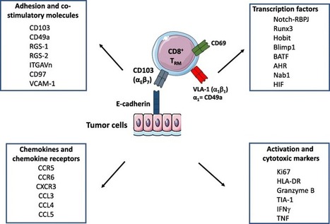
CD8+ T lymphocytes are the major anti-tumor effector cells. Most cancer immunotherapeutic approaches seek to amplify cytotoxic T lymphocytes (CTL) specific to malignant cells. A recently identified subpopulation of memory CD8+ T cells, named tissue-resident memory T (TRM) cells, persists in...
|
|
Suggested by
LIGHTING
September 9, 2018 10:46 AM
|
A study suggests "good bacteria" have little or no effect inside the body.
|
|
Scooped by
Gilbert C FAURE
September 8, 2018 2:50 PM
|
Abstract: The palatine tonsil is the portal of entry for food and air, and is continuously subjected to environmental challenges including pathogens which use the tonsil and pharynx as a primary site of replication. In pigs, this includes the viruses causing porcine respiratory and reproductive syndrome, and classical and African swine fever; diseases which have impacted the pig production industry globally. Despite the importance of tonsils in host defence, little is known regarding the phenotype of the myeloid cells resident in the porcine tonsil. Here, we have characterised five myeloid cell populations that align to orthologous populations defined in other mammalian species: a CD4+ plasmacytoid DC (pDC) defined by expression of the conserved markers E2.2 and IRF-7, a conventional dendritic cell (cDC1) population expressing CADM1highCD172alow and high levels of XCR1 able to activate allogeneic CD4 and CD8 T cells; a cDC2 population of CADM1dim cells expressing FLT3, IRF4 and CSF1R with an ability to activate allogeneic CD4 T cells; CD163+ macrophages (MƟs) defined by high levels of endocytosis and responsiveness to LPS and finally a CD14+ population likely derived from a myelo-monocytic lineage, which showed the highest levels of endocytosis, a capacity for activation of CD4+ memory cells, combined with lower relative expression of FLT3. Increased knowledge regarding the phenotypic and functional properties of myeloid cells resident in porcine tonsil, wil
|
|
Scooped by
Gilbert C FAURE
September 6, 2018 12:17 PM
|
Probiotics perturb rather than aid in microbiota recovery back to baseline after antibiotic
treatment in humans.
|
|
Scooped by
Gilbert C FAURE
August 29, 2018 3:47 PM
|
Single-cell RNA sequencing is used to characterize cell types in nasal tissues from human patients with chronic rhinosinusitis, revealing a role for tissue stem cells in allergic inflammatory memory.
|
|
Suggested by
Société Francaise d'Immunologie
August 17, 2018 12:09 PM
|
Human airway organoid cultures provided results that were comparable to those observed in human ex-vivo bronchus cultures, and thus provide an alternative physiologically relevant experimental model for investigating virus tropism and replication competence that could be used to assess the...
|
|
|
Scooped by
Gilbert C FAURE
September 20, 2018 4:42 AM
|
IFN-λ is a cytokine expressed in epithelial tissues and plays a central role in antiviral mucosal immune response. The expression of IFN-λ in the airway is impaired in chronic airway diseases (e.g., asthma, chronic obstructive pulmonary disease), which renders patients susceptible to viral...
|
|
Scooped by
Gilbert C FAURE
September 20, 2018 2:11 AM
|
|
|
Scooped by
Gilbert C FAURE
September 12, 2018 2:47 PM
|
Inflammasome signalling has a central role in the regulation of gastrointestinal health and disease.Here, an overview of inflammasome biology in relation to the gastrointestinal tract is presented, with insights into how targeted interventions might be useful to treat inflammasome-mediated gastrointestinal...
|
|
Scooped by
Gilbert C FAURE
September 12, 2018 2:45 PM
|
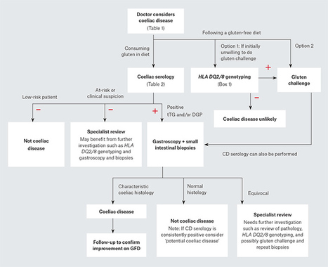
This article is the second in a series on pathology testing. Articles in this series aim to provide information about emerging laboratory tests that general practitioners may encounter. Coeliac disease is an immune illness, triggered by dietary gluten, that causes a broad range of gastrointestinal and extra-intestinal manifestations.1 Untreated disease reduces quality of life, increases healthcare use and is associated with substantial morbidity.2–4 Mortality is increased because of lymphoproliferative malignancy, sepsis and refractory disease.3 As 1.5% of Australians have coeliac disease, it is one of the most common autoimmune illnesses that general practitioners (GPs) will encounter. However, its broad and often subtle presentation makes detection challenging, and means 80% of Australians with coeliac disease remain undetected.5 As expeditious diagnosis and treatment with a strict, lifelong gluten-free diet (GFD) minimises long-term complications,3 application of the appropriate tests to ensure accurate diagnosis and follow-up is crucial. Making the diagnosis In clinical practice, suspected patients are generally screened with coeliac disease serology. In patients with positive coeliac disease serology, the diagnosis is confirmed by the presence of characteristic small intestinal mucosal changes. The key diagnostic features are: intestinal histology showing raised intraepithelial lymphocytes (>25 per 100 enterocytes), crypt hyperplasia and villous atrophy (Figure 1) disease remission confirmed by symptom resolution, normalised coeliac disease serology and, most reliably and importantly, mucosal healing following treatment with a GFD.3,6 Correlation of histology and serology with clinical history is important. Coeliac disease can be present despite negative coeliac disease serology, but this is uncommon and excluding other causes of villous atrophy (see below) is important. Testing at-risk individuals is strongly recommended to detect cases before substantial morbidity develops.3,6 An active case-finding approach can improve detection of coeliac disease by more than 40-fold,7 but this only works when doctors are mindful of the disease. Approximately 30 at-risk individuals need to be tested to find a positive case of coeliac disease.7 There is insufficient evidence to support population screening.8 Figure 2 provides an outline of a recommended diagnostic pathway. Figure 1. Healthy small intestine, compared with villous atrophy in coeliac disease. A. Normal small intestinal mucosa in adequately treated coeliac disease. B. Untreated coeliac disease showing the classic triad of infiltration of the epithelium with lymphocytes, crypt hyperplasia and villous atrophy. Magnification ×100, haematoxylin and eosin stain. Figure 2. Recommended diagnostic pathway for coeliac disease CD, coeliac disease; DGP, deamidated gliadin peptide antibody; tTG, tissue transglutaminase antibody When to test for coeliac disease Symptoms and clinical features that identify patients who might benefit from testing are shown in Table 1.9 ‘Classical’ symptoms caused by intestinal inflammation, such as diarrhoea and weight loss, are frequent, but the ‘non-classical’, extra-intestinal manifestations are even more common. These non-classical features include lethargy, headaches, osteoporosis, iron deficiency, transaminase elevation, infertility, other autoimmune disease and dermatitis herpetiformis. A positive family history of coeliac disease carries the strongest predictive value for the disease. Tips and pitfalls Coeliac disease can develop at any age. The median age of diagnosis is 40 years, but do not discount coeliac disease in the young and elderly. Coeliac disease affects both sexes, with a modest female predominance. Men with coeliac disease are often overlooked.5 Coeliac disease is a global disorder that is common in Western populations, North Africa, the Middle East, India and Pakistan.10 Reports from Asian countries, such as China, are on the rise. Coeliac disease should not be excluded on the basis of a patient’s ethnicity or appearance. Clinical heterogeneity is substantial. Some patients have minimal or no obvious symptoms, or only extra‑intestinal issues. One-third of patients with coeliac disease are overweight or obese at diagnosis.11 Coeliac serology Currently, serologic testing for coeliac disease consists of the transglutaminase (tTG) and deamidated gliadin peptide (DGP) antibody tests. In practice, both tests have >85% sensitivity and >90% specificity.12 The DGP assay has replaced the whole-protein anti-gliadin antibody (AGA) assay because of improved specificity; however, many labs will report the DGP result as the ‘anti-gliadin antibody’. The anti-endomysial antibody (EMA) test measures tTG antibodies, but is labour-intensive, user-dependent and less widely performed. Testing approaches are shown in Table 2. Tips and pitfalls Positive coeliac disease serology in isolation is insufficient for the diagnosis of coeliac disease. The higher the titre of serology, the greater the positive predictive value for coeliac disease.13 Coeliac disease serology has a false negative rate of 10–15%.14 Check if your patient is on a GFD or taking immunosuppressants. The tTG normal range varies by manufacturer as there is no international standard. Comparing titres is not possible if different labs or tTG assays are used. In patients with risk factors for coeliac disease, negative coeliac disease serology has lower negative predictive value, so further work-up should be considered.15 Point-of-care tests to detect coeliac disease antibodies have not been validated in primary practice, so cannot currently be recommended.11 Patients with persistently positive coeliac disease serology but normal small intestinal histology may have ‘potential’ (or ‘latent’) coeliac disease, and follow-up is recommended.3,6 Gastroscopy and small bowel biopsies Histological evaluation of biopsies from the small intestine is the cornerstone of coeliac disease diagnosis.3,6 Gastroscopy is typically performed with intravenous sedation, is simple and safe, and takes as little as 10 minutes. As coeliac disease causes patchy involvement of the proximal small intestine, multiple biopsies are recommended (eg two from the first and four from the second part of the duodenum).3,6 Endoscopic changes of coeliac disease, such as mucosal scalloping, are occasionally seen, but diagnosis rests on the microscopic appearance. Tips and pitfalls Villous atrophy is suggestive but not pathognomonic of coeliac disease. Other causes to consider, especially if coeliac disease serology is negative, include Giardia, common variable immunodeficiency, Crohn’s disease, tropical sprue, autoimmune enteropathy, cow’s milk protein intolerance and some medications (eg olmesartan). A GFD or immunosuppression can obscure changes of villous atrophy. Correct biopsy processing and interpretation by a skilled pathologist is vital. When there is diagnostic uncertainty, review of the pathology can be informative. Human leukocyte antigen DQ2/8 genotyping The strong association between coeliac disease and specific human leukocyte antigen (HLA) genes makes HLA genotyping a useful tool in specific situations (Table 3).16 The main susceptibility genes are HLA-DQ2 (specifically HLA-DQ2.5 and HLA-DQ2.2) and HLA-DQ8, which are collectively seen in almost all (99%) patients with coeliac disease, compared with 40–50% of the Australian community.5 Although these genes, especially HLA-DQ2.5, impart substantial relative risk for coeliac disease, the absolute risk is low, and most patients with one or more of these genes will not develop coeliac disease. HLA-DQ7 (composed of half the DQ2.5 allele, DQA1*05) may impart a very low risk for coeliac disease but this remains unclear.16 The main benefit of HLA typing is its ability to exclude coeliac disease diagnosis when the susceptibility genotypes are absent (likelihood of coeliac disease <1%). A positive HLA test does not diagnose coeliac disease, but indicates that further investigation may be warranted. Genotyping is widely available through commercial labs in Australia with a request for ‘HLA-DQ2/8 genotyping’. It is performed on a blood sample, but can be done on a buccal scrape through some collection centres (patients can check in advance). HLA typing reports can be difficult to interpret, so Australasian guidelines have been developed to simplify and standardise reporting.16 Tips and pitfalls HLA typing is expensive (Medicare Benefits Schedule item number 71151; $118.85), so it is important to use it prudently (Table 3).16 HLA typing is a ‘once only’ test as a person’s genotype does not change. HLA typing results are not adversely affected by a GFD. Most patients with HLA susceptibility for coeliac disease will not have the disease or ever develop it. Table 1. When to test for coeliac disease9 Offer serological testing for coeliac disease to people with any of the following: Persistent unexplained abdominal or gastrointestinal symptoms Faltering growth Prolonged fatigue Unexpected weight loss Severe or persistent mouth ulcers Unexplained iron, vitamin B12 or folate deficiency Type 1 diabetes, at diagnosis Autoimmune thyroid disease, at diagnosis Irritable bowel syndrome (in adults) First-degree relatives of people with coeliac disease Consider serological testing for coeliac disease in people with any of the following: Metabolic bone disorder (reduced bone mineral density or osteomalacia) Unexplained neurological symptoms (particularly peripheral neuropathy or ataxia Unexplained subfertility or recurrent miscarriage Persistently raised liver enzymes with unknown cause Dental enamel defects Down syndrome Turner syndrome Reproduced with permission from the National Institute for Health and Care Excellence. Coeliac disease: Recognition, assessment and management. London: NICE, 2015. Available at www.nice.org.uk/guidance/ng20 Gluten-sensitive or wheat‑sensitive patients Many Australians adopt a GFD without assessment for coeliac disease. This poses a diagnostic dilemma as coeliac disease serology and intestinal histology can become falsely negative if the patient has been on a GFD for more than a few months. Many Australians remove gluten from their diet because they feel it helps improve gastrointestinal or other symptoms.17 For these people, a definitive diagnosis is desirable as: a formal diagnosis of coeliac disease will ensure strict treatment and follow-up of a serious medical illness many people who self-report ‘gluten sensitivity’ are not actually sensitive to gluten. These patients, instead of excluding gluten, may benefit more from excluding other symptom-inducing wheat components, such as fermentable carbohydrates (FODMAPs).18 There are two diagnostic approaches – option 1 may be appropriate if the patient is unwilling to undertake the gluten challenge; however, option 2 is definitive. Option 1: HLA-DQ2/8 genotyping The absence of HLA susceptibility means that coeliac disease is unlikely and further investigations can focus on other diagnoses. The presence of HLA susceptibility genes is not diagnostic of coeliac disease, so option 2 is required. Option 2: Gluten challenge, then testing The amount and duration of gluten required to consistently trigger diagnostic changes of coeliac disease appears highly variable and more research is required. Approximately 3–6 g of gluten consumed daily for two weeks will cause intestinal changes of coeliac disease in 50–70% of affected adults, with the development of positive serology after four weeks in 10–55%.19,20 To optimise the diagnostic yield, patients should be encouraged to return to consuming 3–6 g or more of gluten each day for, ideally, six or more weeks. This daily amount of gluten can be found in two to four slices of wheat bread, two to four Weet-Bix or 0.5–1 cup of cooked pasta. Tips and pitfalls Symptomatic relapse with a gluten challenge is common, but has poor predictive value for coeliac disease. Gluten challenge is informative only if accompanied by objective testing. The tolerability of a gluten challenge may be improved by commencing with a small amount of gluten and slowly increasing over subsequent days, and consuming it in divided doses over the course of the day (eg breakfast and lunch). Fermented breads with lower FODMAP content (that still contain gluten) are commercially available and may also improve challenge tolerability. Family screening The risk of coeliac disease in patients who have an affected family member with the disease is 10%, but increases up to 20% if multiple family members are affected. Screening patients with a family history of coeliac disease is important and strongly indicated when there are suggestive symptoms or signs.3,6 As it is increasingly recognised that many ‘asymptomatic’ patients with coeliac disease have underlying nutrient deficiencies, reduced bone density, or have symptom improvement following a GFD (indicating they were never asymptomatic), this means all relatives, irrespective of symptom status, should be considered for screening.21 First-degree relatives should be screened, and if there are several affected family members second-degree relatives should also be tested. Family screening using HLA DQ2/8 genotyping with coeliac disease serology is more informative than serology alone.16 A relative without HLA susceptibility does not require monitoring for coeliac disease. If HLA susceptibility is present but coeliac disease serology is normal, the individual is at risk for future development of the disease. Repeat coeliac disease serology would be recommended if they develop suggestive symptoms. If asymptomatic, some experts recommend screening every two to three years during childhood to avoid the detrimental effects of unrecognised coeliac disease on growth and bone health.13 Tips and pitfalls Screening children with a family history of coeliac disease can be delayed until the age of four years if they are well and symptom-free. Remind your patients with coeliac disease that their relatives are at increased risk of the disease and should be considered for testing. Paediatric testing Although similar to adults, there are additional considerations when assessing children for coeliac disease.22 New European guidelines, based on evidence that high-titre tTG is strongly predictive of coeliac disease in children, suggest small intestinal biopsies can be avoided if children meet the following criteria:13 characteristic symptoms of coeliac disease tTG-IgA levels >10× upper limit of normal a positive endomysial antibody (EMA) on a different blood sample positive HLA susceptibility for coeliac disease. The utility of this approach in Australia is uncertain because of the limited availability of the EMA test, as well as intra-lab variation and lack of standardisation of the tTG assay. Further validation is warranted. The decision to make a non-biopsy diagnosis should only be made with specialist paediatric input. Tips and pitfalls The tTG assay has lower sensitivity in children under three years of age. Ensure DGP-IgG testing is performed alongside tTG-IgA to overcome this issue. Children with coeliac disease may present with more ‘classical’ complaints than adults,22 but be mindful of extra-intestinal issues. Anxiety, depression, aggressive behaviour and sleep problems can be a presenting feature.23 Table 2. How to test for coeliac disease 1. Confirm your patient is consuming a normal, gluten-containing diet 2. Request coeliac serology as follows: Option 1: Transglutaminase-IgA (tTG-IgA) + Deamidated gliadin peptide-IgG (DGP-IgG) – Medicare Benefits Schedule (MBS) item number 71164, double antibody test ($39.90) is the preferred* one-step approach. Or Option 2: Transglutaminase-IgA (tTG-IgA) + Total IgA level† – If the IgA level is low, perform the deamidated gliadin peptide-IgG (DGP-IgG). MBS item number 71163, single antibody test ($24.75). 3. If tTG-IgA and/or DGP-IgG is positive, irrespective of titre, refer for confirmatory small intestinal biopsy *Option 1 overcomes the need to assess the total IgA level by performing DGP-IgG, which is not adversely affected by IgA deficiency. Further, DGP-IgG enhances the pick-up of coeliac disease by 15% compared to tTG-IgA alone.25 Positive tTG-IgA and DGP-IgG together provides greater predictive value for coeliac disease than either alone.26 †Total IgA level detects the 3% of people with coeliac disease with selective IgA deficiency that can cause false negative results. Clinical follow-up Confirming successful treatment of coeliac disease with the GFD is important as the risk of complications is higher in patients not achieving mucosal remission.3 Yearly follow-up to review medical and dietary progress is recommended.9 A repeat gastroscopy may be considered in adults after two years of starting a GFD to assess for mucosal healing.3 Coeliac disease serology is frequently used as a surrogate marker of intestinal healing. In adults, values correlate poorly with the state of the intestinal mucosa.24 A trend for normalisation is reassuring (titres generally normalise on a GFD in 12 months), and persistently positive titres suggest ongoing gluten exposure. In children, resolving tTG titres correlate better with mucosal healing.25 A child who improves clinically and normalises their serology on a GFD does not require follow-up endoscopy. Coeliac disease serology cannot detect small dietary indiscretions, and intestinal biopsies are recommended when disease activity needs to be accurately assessed. Conclusion Coeliac disease is highly prevalent in general practice. Good patient care depends on knowing when and how to test for it and how to monitor progress. Awareness of the strengths and limitations of each testing approach is vital for optimal diagnosis and follow-up. Box 1. Clinical scenarios when HLA DQ2/8 genotyping can be useful16 When coeliac disease serology and/or small bowel examination is inconclusive or equivocal When there has been failure to improve on a gluten-free diet When a person has commenced a gluten-free diet prior to assessment by serology or small bowel examination and are unwilling or unable to undertake a gluten challenge prior to investigation In patients clinically assessed to be at higher risk of coeliac disease in order to exclude those where further testing for coeliac disease is not required Key points Recognition and testing at-risk patients are keys to expediting coeliac disease diagnosis. Coeliac disease serology and histology are not accurate in people following a GFD. Ask about diet when testing. Positive coeliac disease serology does not diagnose coeliac disease in isolation. The diagnosis depends on showing the characteristic intestinal changes and improvement on the GFD. Diagnosing coeliac disease without intestinal biopsies has been considered for children, but is contentious and requires specialist input. HLA genotyping can exclude a coeliac disease diagnosis, but has poor positive predictive value. A positive result does not diagnose coeliac disease. Distinguishing coeliac disease from ‘gluten sensitivity’ has important implications for the patient and their family’s medical care. Do not forget family screening given the insidious nature and adverse outcomes of undiagnosed coeliac disease.26,27
|
|
Suggested by
Société Francaise d'Immunologie
September 12, 2018 2:23 PM
|
|
|
Scooped by
Gilbert C FAURE
September 11, 2018 3:43 AM
|
Objective Initiation of a gluten-free diet without proper diagnostic work-up of coeliac disease is a frequent and demanding problem. Recent diagnostic guidelines suggest a gluten challenge of at least 14 days followed by duodenal biopsy in such patients.
|
|
Scooped by
Gilbert C FAURE
September 11, 2018 3:42 AM
|
Scientists have created a new method of particle tracking based on machine learning that is far more accurate and provides better automation than techniques currently in use.
|
|
Scooped by
Gilbert C FAURE
September 9, 2018 1:31 PM
|
The composition of the human microbiome is considered a major source of interindividual variation in immunity and, by extension, susceptibility to diseases. Intestinal bacteria have been the major focus of research. However, diverse communities of viruses that infect microbes and the animal host cohabitate the gastrointestinal tract and collectively constitute the gut virome. Although viruses are typically investigated as pathogens, recent studies highlight a relationship between the host and animal viruses in the gut that is more akin to host–microbiome interactions and includes both beneficial and detrimental outcomes for the host. These viruses are likely sources of immune variation, both locally and extraintestinally. In this review, we describe the components of the gut virome, in particular mammalian viruses, and their ability to modulate host responses during homeostasis and disease.
|
|
Scooped by
Gilbert C FAURE
September 8, 2018 3:16 PM
|
Probiotics transiently colonize the human gut mucosa in highly individualized patterns,
thereby differentially impacting the indigenous microbiome and host gene-expression
profile, a trait which is predictable by baseline host and microbiome features, but
not by stool shedding.
|
|
Scooped by
Gilbert C FAURE
September 8, 2018 2:39 AM
|
Tissue-resident memory T (TRM) cells provide first-line defense against invading pathogens encountered at barrier sites. In the lungs, TRM cells protect against respiratory infections, but wane more quickly than TRM cells in other tissues. This lack of a sustained TRM population in the lung parenchyma explains, at least in part, why infections with some pathogens, such as influenza virus and respiratory syncytial virus (RSV), recur throughout life. Intranasal vaccination with a murine cytomegalovirus (MCMV) vector expressing the M protein of RSV (MCMV-M) has been shown to elicit robust populations of CD8+ TRM cells that accumulate over time and mediate early viral clearance. To extend this finding, we compared the inflationary CD8+ T cell population elicited by MCMV-M vaccination with a conventional CD8+ T cell population elicited by an MCMV vector expressing the M2 protein of RSV (MCMV-M2). Vaccination with MCMV-M2 induced a population of M2-specific CD8+ TRM cells that waned rapidly, akin to the M2-specific CD8+ TRM cell population elicited by infection with RSV. In contrast to the natural immundominance profile, however, coadminstration of MCMV-M and MCMV-M2 did not suppress the M-specific CD8+ T cell response, suggesting that progressive expansion was driven by continuous antigen presentation, irrespective of the competitive or regulatory effects of M2-specific CD8+ T cells. Moreover, effective viral clearance mediated by M-specific CD8+ TRM cells was not affected b
|
|
Scooped by
Gilbert C FAURE
September 5, 2018 2:29 PM
|
|
|
Scooped by
Gilbert C FAURE
August 21, 2018 2:25 AM
|
|


 Scoop.it!