Your new post is loading...

|
Scooped by
Gilbert C FAURE
October 15, 2018 1:26 PM
|
In recent years, innate lymphoid cells (ILCs) have emerged as key mediators of protection and repair of mucosal surfaces during infection. The lung, a dynamic mucosal tissue that is exposed to a plethora of microbes, is a playground for respiratory infection‐causing pathogens which are not only a major cause of fatalities worldwide, but are also associated with comorbidities and decreased quality of life. The lung provides a rich microenvironment to study ILCs in the context of innate protection mechanisms within the airways, unraveling their distinct functions not only in health but also in disease. In this review, we discuss how pulmonary ILCs play a role in protection against viral, parasitic, bacterial, and fungal challenge, along with the mechanisms underlying this ILC‐mediated immunity.
|
|
Scooped by
Gilbert C FAURE
October 15, 2018 1:24 PM
|
IFN-λ is a cytokine expressed in epithelial tissues and plays a central role in antiviral mucosal immune response. The expression of IFN-λ in the airway is impaired in chronic airway diseases (e.g., asthma, chronic obstructive pulmonary disease), which renders patients susceptible to viral...
|
|
Scooped by
Gilbert C FAURE
October 12, 2018 2:25 PM
|
The role of the spleen in the induction of an immune response to orally administered antigens is still under discussion. Although it is well known that after oral antigen administration specific germinal centres are not only formed in the Peyers patches (PP) and the mesenteric lymph nodes (mLN)...
|
|
Scooped by
Gilbert C FAURE
October 12, 2018 6:29 AM
|
|
|
Scooped by
Gilbert C FAURE
October 9, 2018 12:27 PM
|
Escherichia coli is a facultative anaerobic symbiont found widely among mammalian gastrointestinal tracts. Several human studies have reported increased commensal E. coli abundance in the intestine during inflammation; however, host immunological responses towards commensal E. coli during inflammation are not well-defined. Here, we show that colonization of gnotobiotic mice with different genotypes of commensal E. coli isolated from healthy conventional microbiota mice and representing distinct populations of E. coli elicited strain-specific disease phenotypes and immunopathological changes following treatment with the inflammatory stimulus, dextran sulfate sodium (DSS). Production of the inflammatory cytokines GM-CSF, IL-6 and IFN-γ was a hallmark of the severe inflammation induced by E. coli strains of Sequence Type 129 (ST129) and ST375 following DSS administration. In contrast, colonization with E. coli strains ST150 and ST468 caused mild intestinal inflammation and triggered only low levels of pro-inflammatory cytokines, a response indistinguishable from that of E. coli-free control mice treated with DSS. The disease development observed with ST129 and ST375 colonization was not directly associated with their abundance in the GI tract as their levels did not change throughout DSS treatment, and no major differences in bacterial burden in the gut were observed among the strains tested. Data mining and in vivo neutralization identified IL-6 as a key cytokin
|
|
Scooped by
Gilbert C FAURE
October 9, 2018 2:28 AM
|
|
|
Scooped by
Gilbert C FAURE
October 8, 2018 8:18 AM
|
|
|
Scooped by
Gilbert C FAURE
October 4, 2018 4:55 AM
|
Eradicating HIV in infected patients likely requires disrupting the reservoir of infected T cells in the gastrointestinal tract. One way to accomplish this is by targeting cells expressing the α4β7, which has been tested in SIV models and is an approved therapy for inflammatory bowel disease. Uzzan and colleagues studied a small cohort of HIV-infected individuals on antiretroviral therapy that began anti-α4β7 treatment for their mild inflammatory bowel disease. The investigators performed colonoscopies before and several months after the anti-integrin treatment, allowing them to assess lymphoid populations and HIV prevalence before and after treatment. They saw that treatment disrupted local lymphoid aggregates and also affected peripheral immune populations. As with non–HIV-infected IBD patients, treatment was well tolerated. Their results are sure to draw interest from scientists and clinicians who believe targeting this integrin could possibly help cure HIV.
Gut homing CD4+ T cells expressing the integrin α4β7 are early viral targets and contribute to HIV-1 pathogenesis, likely by seeding the gastrointestinal (GI) tract with HIV. Although simianized anti-α4β7 monoclonal antibodies have shown promise in preventing or attenuating the disease course of simian immunodeficiency virus in nonhuman primate studies, the mechanisms of drug action remain elusive. We present a cohort of individuals with mild inflammatory bowel disease and concomitant HIV-1 infection receiving anti-α4β7 treatment. By sampling the immune inductive and effector sites of the GI tract, we have discovered that anti-α4β7 therapy led to a significant and unexpected attenuation of lymphoid aggregates, most notably in the terminal ileum. Given that lymphoid aggregates serve as important sanctuary sites for maintaining viral reservoirs, their attrition by anti-α4β7 therapy has important implications for HIV-1 therapeutics and eradication efforts and defines a rational basis for the use of anti-α4β7 therapy in HIV-1 infection.
|
|
Scooped by
Gilbert C FAURE
October 4, 2018 2:30 AM
|
In a study published today in Science Translational Medicine, Mount Sinai researchers describe for the first time a mechanism that may shrink collections of immune cells in the gastrointestinal (GI) tract, called lymphoid ...

|
Suggested by
Société Francaise d'Immunologie
October 1, 2018 9:13 AM
|
The inflammasome: a well‐regulated immune platform also present in non‐myeloid cells The canonical assembly of the inflammasome is a well‐regulated process that starts after the recognition of PAMPs or DAMPs by cytosolic receptors. The cytosolic receptor acts as a sensor protein and typically belongs to the NLR family but the activation of other receptors such as AIM2 also leads to the formation of an inflammasome5 (Table 1). The most studied inflammasome is NLRP3 because of its ability to respond not only to pathogens but also to sterile stimuli.7 Activation of the cytosolic receptor leads to the recruitment of the effector enzyme capase‐1. Depending on which cytosolic receptor is activated, the recruitment of the effector enzyme, caspase‐1, will require an adaptor molecule known as apoptosis‐associated speck‐like protein containing a caspase activation and recruitment domain (ASC) or not (Table 1). Upon oligomerization of the inflammasome, the effector enzyme caspase‐1 is activated and leads to the cleavage, maturation and release of IL‐1β and IL‐18, and to a form of cell death known as pyroptosis8 (Fig. 1). It is important to mention that, although most of the inflammasome‐forming receptors need ASC for their function, NLRP1b and NLRC4 have been shown to have both ASC‐dependent and ASC‐independent functions. Although ASC is dispensable (but significantly increases) NLRP1b function, NLRC4‐mediated pyroptosis (but not cytokine release) is ASC‐independent.9-11 Subfamily Receptor Requirement for ASC Main activating signal Main epithelia involved NLRP NLRP1 Yesa Anthrax toxin and ATP Oral, airway22-24 NLRP3 Yes Ionophores, crystals, ATP, bacterial toxins Oral, airway, intestinal, skin22, 23, 25, 26 NLRP6b Yes Microbiota‐modulated metabolites (taurine) Intestinal27 NLRP9b Yes Rotavirus dsRNA Intestinal28 NLRC NLRC4/IPAF Yesa Flagellin (in mouse)/Type III secretion system (in human) Intestinal29 Other inflammasome‐forming sensors AIM2 Yes dsDNA Oral, skin23, 30 Not all the inflammasome‐forming receptor proteins require the adaptor protein apoptosis‐associated speck‐like protein containing a caspase activation and recruitment domain (ASC) to activate caspases‐1, however, when present, ASC potentiates inflammasome activity. Also shown the main activating signals and epithelial distribution. a Both nucleotide NACHT, LRR and PYD domains‐containing protein 1 (NLRP1) and NLRC4 protein have a caspase recruitment domain and have shown activity in the presence or absence of ASC.9, 31 b The ability of NLRP6 to form inflammasomes has only been suggested but not directly shown. Although canonical inflammasomes rely on the activation of caspase‐1, cells can also form ‘non‐canonical’ inflammasomes, which lead to the activation of caspase‐11 (caspase‐4 and caspase‐5 in humans) or caspase‐8.13, 14 Caspase‐11 acts as an intracellular lipopolysaccharide (LPS) sensor by direct binding using its caspase activation and recruitment domain (CARD). Interestingly, this process is widely extended into myeloid and non‐myeloid cells.15 Therefore, caspase‐11 could be a widespread mechanism to detect intracellular Gram‐negative bacteria in cells, including infected epithelial cells. Upon LPS recognition, caspase‐11 is activated and causes K+ efflux, which induces NLRP3 canonical inflammasome formation and IL‐1β and IL‐18 release.16 Both IL‐1β and IL‐18 are potent pro‐inflammatory cytokines with a pivotal role during the first steps of inflammation and their deregulation is extremely detrimental to health. Hence, these are tightly regulated proteins, not only at transcriptional level, but also post‐transcriptionally by their activation within the inflammasome. Although IL‐1β and IL‐18 are activated by the inflammasome in a similar manner, their contribution to the inflammatory response is very different. Interleukin‐1β drives inflammation by controlling the recruitment of neutrophils to the site of infection, the induction of IL‐8 by epithelial cells and the release of IL‐17 from T cells.17 On the other hand, the main role of IL‐18 is to foster the activation of natural killer and T cells and the release of interferon‐γ.18-20 Although most studies focus on the release of IL‐1β and IL‐18 by immune cells such as macrophages, epithelial cells are also able to release these pro‐inflammatory cytokines.19, 21 However, the mechanistic insights by which this occurs remain unclear. As a result of their essential role as triggers of innate immunity, and in order to tightly control their release, IL‐1β and IL‐18 are synthesized as pro‐forms lacking a signal peptide. The maturation and release of these pro‐inflammatory cytokines are regulated by the assembly of a multiprotein complex known as the inflammasome, also present in epithelial cells (Table 1; Fig. 2).22 Despite caspase‐1 being the main caspase involved in this process, others such as caspase‐4/5 (human orthologues of murine caspase‐11) and caspase‐8 can also play an important role.13, 32 Recently, a new protein‐mediating pyroptosis has been described: gasdermin‐D.33, 34 Gasdermin‐D is activated by caspase‐1 and forms pores in the membrane, which leads to cell death.35-38 It has been recently proposed that IL‐1β and IL‐18 release happens through gasdermin‐D pores;39 even so, further research is needed. Gasdermin‐D is also expressed in epithelial cells, and its involvement on inflammasome activity has been reported. Although no direct cleavage of gasdermin‐D has been shown in epithelial cells, the pore‐forming protein has been shown to be necessary for NLRC4‐mediated and NLRP9b‐mediated pyroptosis.28, 40, 41 This would show that gasdermin‐D is functional and important for epithelial cell inflammasome function. Interestingly, in recent years, it has been reported that under certain circumstances the release of pro‐inflammatory cytokines and cell death are uncoupled upon inflammasome activation in several cell types such as neutrophils, monocytes and macrophages.39, 42, 43 This is particularly important for epithelial cells where the conservation of the integrity of the epithelial layer is key to maintain barrier homeostasis44 (Fig. 2). Whether the release of IL‐1β and IL‐18 is uncoupled from cell death in epithelial cells remains unknown. Despite increasing research in this area, there are still unanswered questions regarding inflammasome activation in the epithelium (Fig. 2). The epithelium is exposed to mechanical and chemical damage associated with the exterior milieu as well as to a wide range of microorganisms colonizing our body. Hence, the inflammasome activity has to be tightly regulated in epithelial cells, not only to prevent an inflammasome response to healthy microbiota but also to potentiate the response to an altered microbiome. In this review, we will summarize the current knowledge on inflammasome activation at different barrier sites, and how these molecular complexes contribute not only to maintenance of barrier homeostasis but also to disease. Gut epithelial cells The most studied inflammasome‐forming epithelium is the gut epithelium. In this barrier, the heavy colonization of commensal microbiota makes the tight regulation of the inflammatory cascade crucial. How gut epithelial cells respond to invasive pathogens and not to commensal bacteria remains only partially understood. It has been shown that differential distribution of TLRs on the membrane of gut epithelial cells allows the response to invasive microorganisms not just after apical contact but after the disruption of the epithelium47 (Fig. 2). The requirement of invasion or damage of epithelium integrity to activate the immune function of epithelial cells guarantees that repair mechanisms and inflammatory processes are triggered only when needed. In addition to TLRs, the cytosolic localization of the inflammasome‐forming sensors NLRs and AIM2, and their ability to respond to damage signals as well as to pathogens, places them as ideal platforms to detect pathogen invasion. The role of inflammasomes at this barrier became apparent after early reports showed that intestinal epithelial cells (IEC) were able to up‐regulate IL‐1β in response to infection48 as well as being the principal contributors to the total levels of IL‐18 in the gut under physiological conditions.49 Since then, an increasing amount of evidence has shown an involvement of the inflammasome in maintaining homeostasis in this barrier as well as responding to pathogens. The gut epithelium has been shown to rely on the NLRC4 inflammasome to respond to Citrobacter rodentium, a mouse model of enteropathogenic Escherichia coli infection.29, 50 Interestingly, epithelial NLRP3 is also involved in C. rodentium clearance as Nlrp3−/− mice show higher bacterial burden and higher epithelium invasion.51 This effect is likely to be mediated by both IL‐18 and IL‐1β as Il‐18−/− and Il‐1β−/− mice present a similar phenotype to the Nlrp3−/− mouse when challenged with C. rodentium.50 These findings have also been confirmed in human cells after E. coli infection of the epithelial cell line Caco‐2 induced increased levels of IL‐18 release.52 Apart from this role in infection, caspase‐1 and NLRC4 have also been reported as essential in preventing colonic inflammation‐induced tumorigenesis by regulating the response of epithelial cells to injury, highlighting the role of NLRC4 in epithelial cells.53, 54 In another model of infection, inflammasome‐dependent release of IL‐18 has been shown to mediate natural killer cell recruitment after Salmonella typhimurium infection.55 Although authors do not investigate the origin of gut IL‐18 in this paper, the above‐mentioned studies suggest that this IL‐18 is likely to be of epithelial cell origin. Using the same infection model, it has been shown that inflammasome‐forming sensors are also involved in the extrusion and expulsion of intestinal epithelial cells. During S. typhimurium infection, caspase‐4 mediates epithelial cell expulsion through a process dependent on pyroptosis.56 This process is epithelial cell intrinsic and independent of cytokine signalling, showing a specific role for pyroptosis and for non‐canonical inflammasomes.57 However, this is not the only inflammasome‐dependent mechanisms for epithelial cell expulsion. In another study exploring the role of NLRC4 in gut epithelial cells, authors showed that caspase‐1 and gasdermin‐D are dispensable for IEC expulsion from the epithelial layer upon treatment with the NLRC4 activator FlaTox (Legionella pneumophila flagellin fused to Bacillus anthracis lethal factor), as cells were still expelled from the epithelium in the absence of these mediators. In contrast, the expelled cells were not dead, implying that both caspase‐1 and gasdermin‐D are required for IEC pyroptosis upon NLRC4 activation.41 How the two mechanisms can act together is still not understood. Although this is an important mechanism necessary for the maintenance of normal microbiota and pathogen clearance, all these studies highlight the danger of excessive inflammasome response and gasdermin‐D‐mediated pyroptosis in gut epithelial cells. An excessive activation of pyroptosis could eventually lead to loss of membrane integrity and systemic infection. Another NLR involved in the relationship between gut epithelium and bacteria is NLRP6. Recent findings emphasize the importance of having proper controls to elucidate the role of different inflammasomes in immunity, especially in maintaining commensal bacteria. Although the role of NLRP6 in gut epithelial cells is still not totally clear, NLRP6 is highly expressed and functional in gut epithelial cells.27 Mamantopoulos et al.,58 in two different animal facilities, dismissed a role of NLRP6 in maintaining homeostasis in the gut microbiota at steady state. However, it has been proposed that NLRP6 does contribute to microbiota balance, but this contribution is dependent on microbiota community structure (and especially in the presence of pathobionts such as Helicobacter spp.). Both of these studies point to familial transmission of microbiota rather than NLRP6 impairment for the observed changes in previous studies.59, 60 How the cells activate NLRP6 and whether NLRP6 forms an active inflammasome are still not fully answered questions. No direct evidence shows NLRP6 forming an inflammasome but Nlrp6−/− studies mimic the results of Caspase‐1−/− and Asc−/−, suggesting that this might be the case.61, 62 Recently, taurine, an amino acid present in bile acid and shown to regulate gut microbiota, has been proposed to be the activating signal for NLRP6 as it induces IL‐18 secretion in wild‐type mice but not in Nlrp6−/− mice.62, 63 Although this finding needs to be further tested and investigated, it would be highly interesting as the gut microbiota in return has also been shown to regulate bile acid metabolism and especially taurine.64 Further research is needed to elucidate the molecular events leading to NLRP6 activation. A highly prevalent condition of the gut is inflammatory bowel disease (IBD, including Crohn's disease and ulcerative colitis). IBD is a chronic inflammation of the digestive system associated with the alterations of the gut microbiota.65 The most common mouse model of IBD is to induce colitis using dextran sodium sulphate (DSS), which damages the epithelium, increasing gut permeability.66 Inflammasome responses in experimental models of colitis are well characterized, but mainly in monocytes and macrophages.67 In such models, NLRP3 inhibition ameliorates DSS‐induced colitis.68-70 However, these studies fail to show a specific role for epithelial cells, despite these being the main cell type affected by IBD. In fact, recent studies suggest that NLRP3 is not expressed by epithelial cells in the colon of mice, placing tissue‐resident macrophages as the principal contributors to this NLRP3‐dependent response.71 These studies suggest that although NLRP3 is a main actor in IBD, the epithelial cell layer's contribution to IBD might be NLRP3 independent. Surprisingly, epithelial cell‐specific depletion of capase‐1 induces decreased IL‐18 levels and decreased pathology in a DSS‐induced colitis model, pointing to the involvement of another inflammasome‐forming receptor.72 This is consistent with the finding that during colitis, IL‐18 produced by epithelial cells exacerbates pathology. Interestingly, epithelial IL‐18 signalling during colitis targets epithelial cells themselves as Il‐18R−/− in haematopoietic cells fails to rescue mice from colitis, whereas specific Il‐18R−/− in epithelial cells protects mice from DSS‐induced colitis.73 Recent studies also point to a role of AIM2 in DSS‐induced colitis. AIM2 knockout mice are more severely affected by colitis and present a higher bacterial burden; however, how AIM2 is activated or how DNA is delivered in the epithelial cells during colitis is unknown. In fact, authors suggest that the role of AIM2 in colitis is to control the anti‐microbial peptide production (such as α‐ and β‐defensins) in IEC, and to regulate tissue repair, activating an IL‐18‐dependent pathway.74, 75 All these reports together show an increasingly recognized role of inflammasomes in innate immunity driven by gut epithelial cells and places the inflammasome response as a coordinated process with several outcomes. However, a more profound study using IEC or myeloid‐cell‐specific knockout mice would be useful to better understand the role of epithelial cells in barrier defence. Lung epithelium Due to the extended area of contact between internal and external stimuli, airway epithelium is a primary target for pathogens and damaging agents. Lung epithelial cells have been shown to activate the inflammasome and release IL‐1β and IL‐18 upon a diverse range of stimuli such as influenza virus, ozone, particulate matter, or asbestos.76-79 However, despite the importance of the release of IL‐1β and IL‐18 in this organ, the events leading to their release from epithelial cells are still not well understood and are the subject of controversy, probably partly due to different cell types used in different studies. For instance, the involvement of caspase‐1 is not clear because some studies show that epithelial cell lines (Beas2Bs, 16HBE, Calu‐3) and also primary cells (human bronchial epithelial cells) express little to no caspase‐1 in an infection model, whereas others show a role for caspase‐1 in epithelial cells (for instance in Beas2Bs, mouse tracheobronchial epithelial cells and A549).80-83 It has been suggested that as some of these cells do not express the inflammasome component, caspase‐11 is responsible for the inflammasome response to infections, probably by direct biding of LPS.84 These differential results highlight the importance of analysing the expression of inflammasome components in the lung epithelial cell lines before conducting inflammasome experiments and the need to work in mouse and human primary cells. As in the gut, the lungs constitute an area that is heavily colonized by commensal bacteria. However, in contrast to the gut, the lung epithelium is also heavily exposed to agents causing sterile inflammation such as particulate matter present in tobacco smoke or urban pollution. Despite the fact that airway macrophages respond strongly to particulate matter due to their increased ability to phagocytose, epithelial cells also respond to air pollutants.85 Human bronchial epithelial cells (Beas2B) have been shown to up‐regulate, process and release IL‐1β in response to crystal silica exposure, which is found in the air and taken up by epithelial cells.83 Interestingly, in the same study, they also detect release of High mobility group box 1 protein (HMGB1) from epithelial cells after silica exposure. HMGB1 is a potent danger signal also released by A549 cells under infection.82 These observations are relevant because the crosstalk among epithelial cells and between epithelial cells and immune cells is important for tissue repair. Other urban particulate matter (particulate matter < 10 µm) can also activate the inflammasome in an NLRP3‐dependent manner.79 This could be particularly important in urban or industrial areas. This particulate matter uptake by epithelial cells and inflammasome activation could worsen already existing conditions or cause new ones. In fact, it has been suggested that following periods of poor air quality (characterized by higher levels of particulate matter), inflammasome activity could be contributing to the exaggerated response to viruses such as influenza and allergy.21, 86 Another major agent for lung dysfunction, cigarette smoke, increases the release of IL‐1β in human lung epithelial cells, and this increase is even higher in patients with chronic obstructive pulmonary disease (COPD).87 Indeed, IL‐1β has been shown to be a marker of COPD severity as individuals with COPD present higher levels of IL‐1β in serum.88 In contrast, another study shows that cigarette smoke reduces the inflammatory response in mice treated with another NLRP3‐activating crystal: asbestos.89 Although contradictory, the reduction of the inflammatory response could be explained by the promotion of the proteasomal degradation of NLRP3 mediated by its ubiquitination.90 In addition to sterile agents, the lung is also exposed to pathogens. A condition where the lung epithelium integrity is compromised is during aspergillosis. Being inhaled every day, the spores of Aspergillus fumigatus invade the lung of not only immunocompromised patients but also patients with COPD.91 Inflammasomes have long been involved with the innate immune response to A. fumigatus, and recent studies show that inflammasome activation occurs early during infection and is key for macrophage anti‐fungal activity.92, 93 Interestingly, β‐glucan, a component of the A. fumigatus cell wall, upon dectin‐1‐binding activates NLRP3 inflammasome in bronchoepithelial cells.94 In fact, Syk tyrosine kinase, a kinase activated through dectin‐1, has been shown to be involved in inflammasome activation by A. fumigatus in monocytes, reinforcing the role of β‐glucan in inflammasome activation in lung epithelial cells.95 Both studies point at reactive oxygen species, a major mediator of inflammasome activation, as a key factor during aspergillosis. The complexity of the lung together with the distinct environments and stimuli that lung epithelial cells face makes the study of the epithelial inflammasome in this organ quite challenging. However, these studies show the inflammasome in the lung as a clear therapeutic target to ameliorate a broad range of pathologies with an inflammatory component. Oral epithelium An entry gate for nutrients, the oral mucosa is a heavily exposed environment where particulate matter, and bacterial and fungal agents constantly challenge the homeostasis of the epithelium. Gingivitis is inflammation of the gums, where the gum pulls away from the bone, creating cavities that are colonized by pathogens, which if left untreated can lead to periodontitis. One of the main pathogens colonizing these cavities and causing the disease is Porphyromonas gingivalis. Periodontitis has been linked to increased expression of inflammasome‐sensing proteins NLRP3 and AIM2, mainly in the epithelial layer.22, 96 Moreover, a polymorphism in other NLR family member, NLRC5, has been linked to increased susceptibility to periodontitis,97 although how it contributes to this pathology has not been investigated. NLRC5 has a caspase recruitment domain but it has not been shown to form inflammasomes on its own. In fact, NLRC5 can associate with NLRP3 in response to different PAMPs and DAMPs to cooperatively activate the inflammasome.98 This would suggest that the polymorphisms of NLRC5 induced to increased susceptibility to periodontitis are the consequence of an aberrant assistance to NLRP3 to form inflammasomes. Other roles for NLRC5 have been described including negative regulation of nuclear factor‐κB,99 interferon signalling99, 100 and regulation of major histocompatibility complex class I genes,101 all of which could also explain its contribution to periodontitis. As expected, infection of gingival epithelial cells with P. gingivalis induced an increase in Il‐1β gene expression. However, IL‐1β was left unprocessed and accumulated intracellularly. Interleukin‐1β was only processed and released after the activation of the ATP‐gated receptor P2X7, which led to NLRP3 inflammasome formation. The P2X7R is also involved in NLRP3 inflammasome formation and release of mature IL‐1β from salivary epithelial cells, as the release of this pro‐inflammatory cytokine is dampened in P2X7R knockout mice.102, 103 Why IL‐1β remains uncleaved and is intracellular during P. gingivalis infection remains an interesting question. We could speculate that the immune machinery of epithelial cells might reduce the activation of caspase‐1 during chronic infection. This mechanism could potentially dampen immune reactions to commensal bacteria and prevent pyroptosis to maintain the integrity of the epithelial layer. In fact, in macrophages, it has been shown that IL‐1β can be released via exosomes in a mechanism dependent on NLRP3 and ASC but independent of caspase‐1.104, 105 Potentially, IL‐1β produced by epithelial cells could be stored and released on exosomes to have two potential effects. (i) Be processed by other cells (potentially immune cells) to avoid activating caspase‐1 and compromising the epithelial layer in epithelial cells. (ii) Have an effect in areas distant to the site of infection. Although epithelial cells have been shown to release cytokines trapped in exosomes, it is unknown whether IL‐1β could follow the same pathway.106 Interestingly, another NLR family member, NLRX1, has recently been shown to potentiate the inflammasome response to ATP in gingival epithelial cells through the generation of mitochondrial reactive oxygen species. However, how NLRX1 (not predicted to form inflammasomes) potentiates the response to ATP is not well understood and might assist NLRP3 in its function in a similar manner to NLRC5.107 Although inflammasome activity is required for optimal control and clearance of pathogens, over‐activation could lead to unwanted outcomes. Excessive levels of IL‐1β and of pyroptosis have been linked to bone loss during periodontitis and to malignancy during oral carcinogenesis.108, 109 Epithelial cells can avoid this and reduce the extent of inflammasome activity during acute infection through the stress‐induced enzyme haem oxygenase‐1. This enzyme has anti‐oxidant and anti‐inflammatory effects, and can dampen LPS signalling and inflammasome activity in oral epithelial cells, although the exact mechanism by which this occurs is not clear.110 Skin epithelial cells The skin is the main physical barrier that protects our body from the external environment, therefore it is active in the immune control of pathogen invasion. Immune homeostasis in the skin is essential to maintain skin integrity and its role as a barrier. Although the role of the inflammasome in skin is not well characterized, mutations in different genes coding for NLR‐forming inflammasomes have been associated with skin conditions. Even before the discovery of the inflammasome, mutations in the NLRP3 gene were found to cause a spectrum of diseases known as cryopyrin‐associated periodic syndromes (or CAPS), which are associated with skin inflammation and rashes.111 Rashes typically disappear, or are controlled, after anti‐IL‐1β therapy, showing a role for IL‐1β signalling in skin rashes and in CAPS in general.112 Recently the first genetic evidence connecting inflammasome signalling to non‐fever skin diseases has been found. Gain of function mutations in the NLRP1 sensor protein cause two overlapping skin disorders: multiple self‐healing palmoplantar carcinoma and familial keratosis lichenoides chronica. These mutations lead to spontaneous inflammasome activation and paracrine IL‐1 signalling in keratinocytes from these patients.24 Polymorphisms of NLRP1 have also been associated with the autoimmune disease vitiligo, a condition characterized by the development of white patches in the skin due to lack of melanin.113 This is believed to occur through higher IL‐1β production and NLRP1 activation, although the specific mechanism by which the NLRP1 inflammasome shows greater functional activity is not known.114 Inflammasome has also been involved in another chronic inflammatory syndrome affecting the skin. Deregulated levels of IL‐1β and AIM2 inflammasome activity by keratinocytes have been linked to psoriatic disease. Abnormally high levels of IL‐1β could potentiate a T helper type 17 response with up‐regulation of IL‐17 and excessive infiltration of neutrophils, characteristics of psoriatic diseases.115 It is not surprising that AIM2, a receptor involved in the recognition of cytosolic DNA, is involved in psoriatic diseases as cytosolic DNA has been shown to be responsible for inflammasome activation in keratinocytes during psoriasis.116 Interestingly, a recent study points to a role in AIM2 in immune memory of epithelial cells.30 Using epithelial cell stem cells they determined that these cells possess immune memory, enabling a rapid second response after a previous exposure to inflammation. This striking capacity of skin epithelial stem cells is dependent on AIM2 and downstream of IL‐1β as Il‐1β−/− and Il‐1r1−/− but not Il‐18−/− failed to induce immune memory. Given the above‐mentioned role of AIM2 in recurrent skin diseases such as psoriasis, we could speculate that both mechanisms might be linked, suggesting major implications for immune memory of epithelial cells in chronic inflammatory diseases. Finally, the role of IL‐18 in skin has been widely studied. In fact, IL‐18 has been linked with psoriatic disease, where increased caspase‐1 activity has been reported.117 Variations in the IL‐18 gene have also been associated with increased risk of atopic eczema, a chronic inflammatory condition of the skin.118 Final remarks In this review we have presented the involvement of inflammasome activation in the role of epithelial cells as mediators of the immune response. We have reviewed how epithelial cells are able to release IL‐1β and IL‐18. However, despite the relatively low concentration of these cytokines released by epithelial cells compared with macrophages, it is important to mention that in healthy conditions epithelial cells are relatively more abundant in barriers than immune cells. Therefore, the epithelial compartment could contribute greatly to the inflammatory response to pathogens, at least in early infection when not many immune cells have been recruited. We have highlighted the diversity of responses and of sensor proteins involved in the inflammasome response. The differential expression of sensor molecules and inflammasome components and mediators in the different types of epithelial cells will shape the immune response according to the needs and threats of each specific site. Similarly, the types of pathogens and threats that each barrier will face vary from one epithelium to another, raising the question of whether there is a general mechanism leading to inflammasome activation in epithelial cells or the response is specific to each epithelium and threat. The increasing body of publications in the field points in the direction of several types of inflammasomes involved in different epithelia, making the response tissue‐dependent. We conclude that, in order to better understand this question, the field would benefit from a broad study comparing the involvement of different inflammasomes in the different epithelia. Even more, it is not always clear and differs from one cell line model to another what inflammasome components are present in each epithelium. In our review we have highlighted several examples of differential expression of inflammasome components or pro‐inflammatory cytokines in different cell types or tissues. We could hypothesize that within one organ, different types of epithelial cells will express different inflammasome components, including cytosolic sensors and effector caspases. That would ensure a diverse, adapted and targeted response and prevent a much feared loss of epithelial integrity by massive activation of inflammasomes. It is also important to highlight that most of the available data on the role of inflammasomes in epithelial cell immunity has been obtained using either human cell lines, or primary epithelial cells from mouse. Key differences between human and murine inflammasomes have already been described (for instance NLRC4 is not activated by the same components of bacterial flagellum in mouse and humans and DNA inflammasome activation in human monocytes depends on NLRP3 instead of AIM2119, 120). Therefore, more studies are needed in human lung epithelial cells to confirm the findings mentioned in this review. Gasdermin‐D, as mentioned before, is the executor of pyroptosis. However, its role and function in non‐haematopoietic cells is still not well characterized. We could speculate that after inflammasome activation in epithelial cells, the triggering of pyroptosis could have two possible outcomes, both of them highlighting the need for a tight control of pyroptosis in epithelial cells. Pyroptosis during infection could be detrimental (and would be somehow silenced or dampened in epithelial cells) by causing membrane integrity loss. This result is in line with the previously mentioned report showing accumulation of pro‐IL‐1β during P. gingivalis infection of the oral mucosa. Nevertheless, pyroptosis could also be beneficial during epithelium colonization, as could a mechanism of containing the infection and avoiding further invasion of internal layers of the epithelium. To reinforce this idea, during Salmonella infection caspase‐1 and gasdermin‐D cleavage are required for IEC pyroptosis but not for IEC expulsion.41 In fact, in that study, the authors demonstrate that during the death and expulsion of IEC, the epithelial integrity is maintained through the rearrangement of neighbouring cells. This result also links with the discovery that small amounts of caspases‐1 are required for gasdermin‐D cleavage (meaning that even a weak activation of inflammasome produces cell death) while detecting active IL‐1β requires higher levels of processed caspases‐1.121 In such a scenario, weak invasion of the epithelial layer would lead to epithelial cell death and expulsion but still no immune response as the threshold to release IL‐1β is much higher than for cell death. This mechanism, potentially occurring with low infection rates where few cells are being infected, could be important to maintain the equilibrium with normal microbiota. The recent discovery of inflammasome inhibitors such as MCC950 or boron‐based compounds is opening the path to the future treatment of inflammasome‐related pathologies with specific drugs.122, 123 However, while the field is moving towards better drugs, there is also an increasing body of evidence that inflammasome activity is key in the immune response of many cell types. The potential specific targeting of either immune cells or epithelial cells in different inflammasome‐related pathologies would be beneficial. Such a specificity would allow the targeting of the over‐activation of inflammasome in cells where its activity is detrimental while allowing the normal inflammasome function in other cells. In conclusion, the role of the inflammasomes in epithelium has been extensively reported. We hypothesize that their contribution to the triggering of inflammation, in opposition to the common belief, is far from being negligible. This combination of findings provides some support for the conceptual premise that targeting inflammasome activity in epithelial cells might be essential for further treatment of a wide range of inflammatory disorders. There is then, abundant room for further progress in elucidating the mechanism of inflammasome activation in epithelial cells and investigating whether it is similar to that in macrophages or not.
|
|
Suggested by
Société Francaise d'Immunologie
September 28, 2018 1:51 PM
|
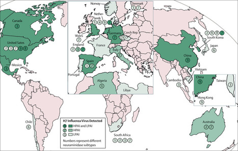
Avian and human influenza A viruses alike have shown a capacity to use the eye as
a portal of entry and cause ocular disease in human beings. However, whereas influenza viruses generally represent a respiratory pathogen and only occasionally cause ocular complications, the H7 virus subtype stands...
|
|
Scooped by
Gilbert C FAURE
September 24, 2018 2:25 PM
|
|
|
Scooped by
Gilbert C FAURE
September 20, 2018 2:42 PM
|
Find could lead to new treatments for obesity, depression
|
|
|
Scooped by
Gilbert C FAURE
October 15, 2018 1:25 PM
|
|
|
Suggested by
Société Francaise d'Immunologie
October 13, 2018 5:35 AM
|
The adult lung consists of highly specialized cell types that are protected by a variety of immune cells. How these immune cells migrate to the lungs during development and after birth and influence each other is poorly understood.
|
|
Scooped by
Gilbert C FAURE
October 12, 2018 1:39 PM
|
Browse the archive of articles on Mucosal Immunology
|
|
Scooped by
Gilbert C FAURE
October 10, 2018 2:30 PM
|
The leukotriene E4 receptor CysLT3R regulates expansion of chemosensory brush cells and production of interleukin-25 in the airways.
|
|
Scooped by
Gilbert C FAURE
October 9, 2018 8:58 AM
|
The dynamic and polymicrobial oral microbiota is a direct precursor of diseases such as dental caries and periodontitis. In this Review, Lamont, Koo and Hajishengallis discuss the mechanisms by which oral microbial communities develop and become functionally specialized. They also examine the progression of polymicrobial communities towards pathogenicity.
|
|
Scooped by
Gilbert C FAURE
October 8, 2018 11:48 AM
|
|
|
Scooped by
Gilbert C FAURE
October 4, 2018 3:07 PM
|
Generation of protective immune responses requires coordinated stimulation of innate and adaptive immune responses. An important mediator of innate immunity is stimulator of IFN genes (STING, MPYS, MITA), a ubiquitously but differentially expressed adaptor molecule that functions in the relay of...
|
|
Suggested by
Société Francaise d'Immunologie
October 4, 2018 3:46 AM
|
|
|
Suggested by
Société Francaise d'Immunologie
October 1, 2018 9:13 AM
|
Recent evidence has demonstrated an expanding role of respiratory epithelial cells in immune surveillance and modulation. Studies have been focusing on the earl...
|
|
Suggested by
Société Francaise d'Immunologie
September 30, 2018 4:26 AM
|
Research ArticleGastroenterologyInflammation Free access | 10.1172/jci.insight.121886 Essential role of IFN-γ in T cell–associated intestinal inflammation Yoshihiro Eriguchi,1 Kiminori Nakamura,2 Yuki Yokoi,2 Rina Sugimoto,2 Shuichiro Takahashi,3 Daigo Hashimoto,3 Takanori Teshima,3 Tokiyoshi Ayabe,2 Michael E. Selsted,1 and André J. Ouellette1 First published September 20, 2018 - More info Abstract Paneth cells contribute to small intestinal homeostasis by secreting antimicrobial peptides and constituting the intestinal stem cell (ISC) niche. Certain T cell–mediated enteropathies are characterized by extensive Paneth cell depletion coincident with mucosal destruction and dysbiosis. In this study, mechanisms of intestinal crypt injury have been investigated by characterizing responses of mouse intestinal organoids (enteroids) in coculture with mouse T lymphocytes. Activated T cells induced enteroid damage, reduced Paneth cell and Lgr5+ ISC mRNA levels, and induced Paneth cell death through a caspase-3/7–dependent mechanism. IFN-γ mediated these effects, because IFN-γ receptor–null enteroids were unaffected by activated T cells. In mice, administration of IFN-γ induced enteropathy with crypt hyperplasia, villus shortening, Paneth cell depletion, and modified ISC marker expression. IFN-γ exacerbated radiation enteritis, which was ameliorated by treatment with a selective JAK1/2 inhibitor. Thus, IFN-γ induced Paneth cell death and impaired regeneration of small intestinal epithelium in vivo, suggesting that IFN-γ may be a useful target for treating defective mucosal regeneration in enteric inflammation. Graphical Abstract Introduction Paneth cells of the small intestinal epithelium contribute to small bowel homeostasis (1). First, they secrete α-defensins (2), which both confer immunity to Salmonella enterica serovar Typhimurium infection (3) and select the composition of the ileal microbiota (4–6). Also, they contribute to crypt stability by releasing niche signals for maintenance of Lgr5+ crypt intestinal stem cells (ISCs), which position themselves to optimize surface contact with Paneth cells (7), and Paneth cells stimulate ISC numbers to expand during caloric restriction via mTORC1 signaling (8). Thus, physiologic events that impair Paneth cell health and viability jeopardize enteric homeostasis, representing risk factors associated with inflammatory bowel diseases. Paneth cell homeostasis is sensitive to proinflammatory conditions that induce Paneth cell depletion and may impair the ability of crypt ISCs to proliferate and regenerate the epithelial barrier. For example, loss of Paneth cells is associated with the onset of inflammation in graft-versus-host disease (GVHD) (9–11), during Toxoplasma gondii infection (12, 13), and in autoimmune enteropathy (AIE) (14–17). The subsequent reductions in mucosal defense mechanisms and the resulting dysbiosis exacerbate inflammation and may compromise tissue repair by disturbing the ISC crypt microenvironment. In GVHD, loss of Paneth cells is associated with gut dysbiosis and bacterial translocation across the epithelial barrier (9, 10), which positively correlates with mortality (9–11). Similarly, in mice infected with T. gondii, activated CD4+ Th1 cells and intestinal dysbiosis both induce transient, IFN-γ–dependent elimination of Paneth cells (12, 13). Immune responses to the parasite are complex, in that germ-free mice do not develop intestinal pathology during T. gondii infection (12). The microbiota activated Paneth cell–specific autophagy via induction of IFN-γ, and deletion of Atg5 in Paneth cells exacerbated intestinal immunopathology in response to T. gondii infection (13). In ex vivo studies investigating Paneth cell degranulation, IFN-γ was identified as mediating release of host defense molecules into the lumen of cultured enteroids (18). Enteroids, small intestinal crypt organoids, consist of a 3D epithelial monolayer that maintains crypt-villus architecture with replicating ISCs that differentiate into the major small intestinal epithelial lineages (19). Exposure of enteroids to IFN-γ induced Paneth cells to extrude their secretory granules and nuclei. Also, IFN-γ increased the number of enteroid cells positive for cleaved caspase-3, although FACS analyses did not identify which cells were positive for activated caspase (18). In addition, exposure of enteroids to IFN-γ reduced Paneth cell and ISC marker expression, and induced MHC class II expression in all enteroid cells (18), suggesting that IFN-γ exerts direct adverse effects on all intestinal epithelial populations. To improve understanding of effector mechanisms that mediate crypt injury by activated T cells, we investigated mouse enteroids in coculture with activated T cells to identify mediators of inflammation-induced Paneth cell loss. Using this ex vivo system, we identified proinflammatory mediators released by activated T cells and characterized enteroid responses to specific cytokines, demonstrating that IFN-γ targets Paneth cells and Lgr5+ ISCs. Subsequently, we showed that systemic administration of IFN-γ to mice disturbed ileal crypt homeostasis in vivo, and crypts depleted of Paneth cells by IFN-γ were highly sensitive to injury by sublethal irradiation and failed to recover. Results Activated T cells induce enteroid damage associated with Paneth cell and Lgr5+ ISC loss ex vivo. Because activated donor T cells are primarily responsible for GVHD etiology (9, 10), and parasite-induced Th1 cells are associated with Paneth cell elimination (12), we tested whether activated T cells secrete factors that disrupt the intestinal epithelium. Studying mouse enteroids cultured ex vivo allowed epithelial responses to T cell activation to be isolated from possible humoral or paracrine factors released by gut stromal cells, distal tissues, or the gut microbiota. Activation of mouse splenic CD4+ or CD8+ T cells with anti-CD3/28 in coculture with enteroids (Figure 1, A and B) induced enteroid damage within 2 days, with increasing severity over 3–4 days (Figure 1, B–E); but in the absence of T cells, anti-CD3/28 had no effect on enteroid integrity (data not shown). The lack of damage in cocultures with resting T cells demonstrated the requirement for T cell activation (Figure 1, B, D, and E). Figure 1 Activated T cells induce enteroid damage. (A) Overall design of enteroid and T cell coculture experiments. (B) Enteroids cocultured with CD4+ or CD8+ T cells (5 × 10⁴ cells per well) with (activated) or without (resting) anti-CD3/anti-CD28 beads. (C) Enteroid damage scoring system. Damage scores of enteroids cocultured with CD4+ (D) or CD8+ (E) T cells. Data are representative of 2 independent experiments and are shown as mean ± SEM (n = 100 enteroids per group); gray bars, enteroids alone; blue bars, enteroids plus resting T cells; red bars, enteroids plus activated T cells. Dunnett’s multiple comparisons test was used to compare each group with the control group. ***P < 0.001. Scale bars: 200 μm. Because the number of recognizable Paneth cells is reduced in GVHD (9) and T. gondii infection (12), lineage-specific marker mRNAs in enteroids incubated with activated or resting T cells were quantified to test for selective reductions in Paneth cells and Lgr5+ ISCs. Quantitative reverse transcription PCR (qRT-PCR) analyses showed that levels of Paneth cell α-defensin-22 (Defa22) and ISC Lgr5 mRNAs were reduced in enteroids incubated for 1–2 days with activated but not resting T cells (Figure 2, A–D). Enteroids embedded in Matrigel did not contact T cells directly (Figure 1B), suggesting that soluble factors released by activated T cells affected Paneth cell and ISC marker mRNA expression. Exposure to activated CD8+ T cells caused a greater reduction in Paneth cell and ISC marker mRNA levels than did CD4+ T cells, implying that CD8+ T cells release higher levels of, or additional, enteroid-damaging factors. Figure 2 Activated T cells reduce Paneth cell and Lgr5+ ISC marker mRNA levels. (A–D) Enteroids cocultured with CD4+ or CD8+ T cells (5 × 10⁴ cells per well) with (activated) or without (resting) anti-CD3/anti-CD28 beads. After removal of CD4+ (A and C) and CD8+ (B and D) T cells, enteroid RNAs were analyzed by qRT-PCR to measure levels of Defa22 (A and B) and Lgr5 (C and D) mRNAs. Data are representative of 2 independent experiments and shown as mean ± SEM (enteroids alone, n = 3 independent wells, gray bars; enteroids + resting T cells, n = 4, blue bars; enteroids + activated T cells, n = 4, red bars). Dunnett’s multiple comparisons test was used to compare each group with the control group. *P < 0.05, **P < 0.01, ***P < 0.001. IFN-γ induces enteroid damage and Paneth cell and Lgr5+ ISC loss. To identify T cell mediators of enteroid damage, ELISAs were performed to measure cytokines in mixed culture media from 1 to 4 days after T cell activation. Levels of IFN-γ, TNF-α, IL-2, and IL-17A secreted by activated CD4+ and CD8+ T cells increased relative to unactivated T cells (Supplemental Figure 1, A–J; supplemental material available online with this article; https://doi.org/10.1172/jci.insight.121886DS1). Only low levels of IL-6 secretion were detected, and none of the cytokines were released by unactivated T cells. Accordingly, enteroids exposed to secreted cytokines at the highest levels detected (Supplemental Figure 1, A–J) were evaluated for loss of integrity. At 2 ng/ml, IFN-γ damaged enteroids within 2 days, but no other cytokine tested affected enteroid integrity (Figure 3, A and B). The effects of IFN-γ on enteroid epithelial lineages were tested by quantifying lineage marker mRNAs, including Defa22 (Paneth cells), Lgr5 (rapidly cycling ISCs), Bmi1 (quiescent ISCs), Muc2 (goblet cells), Dclk1 (tuft cells), Chga (enteroendocrine cells), and Alpi (enterocytes) in enteroids treated with IFN-γ for 3 days. Relative to control enteroids not exposed to cytokines, IFN-γ reduced Defa22 mRNA 450-fold and Lgr5 mRNA 24-fold (Figure 3C), but effects of IFN-γ on Dclk1, Muc2, and Chga mRNAs were modest, and Bmi1 and Alpi mRNA levels were not affected significantly (Figure 3C). All lineage marker mRNAs remained at normal levels in enteroids exposed to all other cytokines tested (Figure 3C). Figure 3 IFN-γ induces enteroid damage. (A) Enteroids were exposed to IFN-γ, TNF-α, IL-2, and IL-17A, the highest levels released into coculture media, or with 100 pg/ml in the case of IL-6 to determine which cytokine(s) mediate enteroid damage. Only IFN-γ treatment caused damage. (B) Damage scores of enteroids exposed for 3 days to the proinflammatory cytokines shown. Data are from 2 independent experiments and shown as mean ± SEM (n = 100 enteroids per group). (C) qRT-PCR analyses of lineage markers in enteroids exposed to cytokines for 3 days. Data are representative of 2 independent experiments and shown as mean ± SEM (n = 4 independent wells). Dunnett’s multiple comparisons test was used to compare each group with the control group. *P < 0.05, **P < 0.01, ***P < 0.001. Scale bar: 200 μm. IFN-γ induces dose-dependent loss of Paneth cells and rapidly cycling ISCs. To assess the relative contribution of activated CD4+ and CD8+ T cell–derived IFN-γ to the induction of Paneth cell and Lgr5+ ISC loss, enteroids were cultured for 3 days with IFN-γ at levels released by both activated T cell populations. At 500 pg/ml, the maximum level of IFN-γ released by CD4+ T cells (Supplemental Figure 1A), enteroid damage was extensive (Figure 4), and mRNA levels for Paneth cell markers Defa22, Defa5, pan-cryptdins, and lysozyme were reduced significantly by IFN-γ in a dose-dependent manner (Figure 5A and Supplemental Figure 2, A–C). Also, levels of Lgr5 and Olfm4 mRNAs, markers of rapidly cycling ISCs downstream of Wnt and Notch signaling (20, 21), decreased (Figure 5, C and D), as did mRNAs for Paneth cell niche factors Wnt3 and Notch ligand Dll4 (Figure 5, E and F). Bmi1 mRNA levels were similar to controls (Figure 5B), as were mRNAs that mark the other major epithelial lineages (Supplemental Figure 2, D–G). We conclude that the quantity of IFN-γ secreted by CD4+ T cells is sufficient to induce loss of enteroid integrity, but CD8+ cell activation is judged to have a greater impact on enteroid Paneth cells and Lgr5+ ISCs, because they release 4 times more IFN-γ than CD4+ T cells. Figure 4 IFN-γ induces dose-dependent enteroid damage. Enteroids were exposed to IFN-γ for 3 days as shown. Representative images from 2 experiments are shown. Scale bar: 200 μm. Figure 5 IFN-γ induces dose-dependent loss of Paneth cells and rapidly cycling stem cells. Enteroids were exposed to IFN-γ for 3 days. (A–G) qRT-PCR analyses to quantify lineage-specific mRNAs, niche signal mRNAs of Paneth cell origin, and apoptosis mRNA levels in enteroids exposed to IFN-γ for 1, 2, and 3 days. Data are from 2 independent experiments and are shown as mean ± SEM (n = 4 independent wells). Dunnett’s multiple comparisons test was used to compare each group with the control group at each time point. *P < 0.05, **P < 0.01, ***P < 0.001. IFN-γ induces Paneth cell death through a caspase-3/7–dependent pathway. In a previous study, the overall fraction of enteroid cells positive for cleaved caspase-3 increased with IFN-γ exposure, although the identities of the positive cells were not reported (18). To identify enteroid cells that have apoptotic responses to IFN-γ exposure (18), we tested whether IFN-γ specifically induces Paneth cell death by quantifying Casp3 mRNA. Over 3 days, IFN-γ increased enteroid Casp3 mRNA dose-dependently (Figure 5G), and its sustained expression contrasted with the reduced levels of Paneth cell marker mRNAs (Figure 5A), suggesting that IFN-γ was inducing Casp3-dependent Paneth cell death. To test for cleaved caspase-3 in Paneth cells, caspase-3/7 activity was visualized by live confocal microscopy of enteroids exposed to 2 ng/ml IFN-γ (Figure 6 and Supplemental Videos 1 and 2), which showed that caspase-3/7 activation occurred in Paneth cells within 13–16 hours of cytokine exposure (Figure 6 and Supplemental Videos 1 and 2). Concurrent with caspase-3/7 activation, Paneth cells extruded their cell contents into the crypt lumen, as previously reported (18). We conclude that reduction of Paneth cell marker mRNAs by IFN-γ occurs by selective induction of cell death by caspase-3/7 activation. Figure 6 IFN-γ induces Paneth cell death through a caspase-3/7–dependent pathway. Still images from time-lapse analysis of caspase-3/7 activity in an enteroid exposed to IFN-γ (from Supplemental Videos 1 and 2). Paneth cells (white arrowheads) are granule-containing and appear white in the differential interference contrast images (top row). Cells containing activated caspase-3/7 have bright green nuclei (red arrowheads), which were detected only in Paneth cells and extruded into the crypt lumen after caspase-3/7 activation (bottom row). Time after addition of 2 ng/ml IFN-γ is indicated in the upper left corner of each image. Representative images from 2 experiments are shown. Scale bars: 20 μm. IFN-γ is required for induction of enteroid damage by activated splenocytes. To test whether Paneth cell and ISC depletion depended exclusively on IFN-γ, we measured IFN-γ receptor–null (IFN-γRKO) enteroid responses to coculture with activated splenic T cells. As before (Figure 1B), activated splenocytes from C57BL/6 (WT) mice induced damage in WT enteroids within 2–4 days, but IFN-γRKO enteroids grew and differentiated normally under the same coculture conditions (Figure 7, A and B). WT enteroids cocultured with activated T cells for 4 days had markedly reduced levels of Paneth cell Defa22 and ISC Lgr5 mRNAs. In contrast, Defa22 mRNA levels remained normal, and Lgr5 mRNA was only slightly lower in IFN-γRKO enteroids under the same conditions (Figure 7C). Whether the 2- to 4-fold changes in other lineage markers are meaningful relative to the magnitude of effects on WT Paneth cells and Lgr5+ ISCs is unclear (Figure 7C). The moderate reduction in the ISC, enteroendocrine cell, and enterocyte markers in cocultured IFN-γRKO enteroids (Figure 7C) suggests that cytokines other than IFN-γ from activated T cells also may induce damage, but to a lesser extent. Activated T cells increased Casp3 mRNA ~6-fold in WT enteroids but not in IFN-γR knockouts (Figure 7C). We conclude that IFN-γ signaling mediates selective Paneth cell and Lgr5+ ISC depletion under induced proinflammatory conditions. Figure 7 IFN-γ mediates enteroid damage induced by splenocyte activation. (A) WT or IFN-γ receptor–KO (IFN-γRKO, KO) enteroids in coculture with WT splenocytes with or without activation using anti-CD3/anti-CD28 beads. (B) Damage scores of WT or KO enteroids in coculture with control or activated WT splenocytes. Data are representative of 2 independent experiments and shown as mean ± SEM, with n = 100 enteroids per group. (C) qRT-PCR analyses to quantify lineage-specific and apoptosis marker mRNAs in WT or IFN-γRKO enteroids in coculture with control or activated WT splenocytes for 4 days. Data are representative of 2 independent experiments and shown as mean ± SEM (n = 5 independent wells). Dunnett’s multiple comparisons test was used to compare each group with the WT control group. *P < 0.05, **P < 0.01, ***P < 0.001. Scale bar: 200 μm. IFN-γ receptor 1 expression on both Paneth cells and Lgr5+ ISCs. To test for the lineage-specific expression of the IFN-γ receptor, single-cell suspensions of CD24hiLgr5-EGFPlo Paneth cells and Lgr5-EGFPhi ISCs isolated from Lgr5-GFP-CreERT2 mouse small intestinal crypts were analyzed via flow cytometry for IFN-γ receptor 1 (Ifngr1) expression (Supplemental Figure 3, A–D). The data show that Paneth cell and Lgr5+ ISC levels of Ifngr1 were equivalent (Supplemental Figure 3, E and F), and a priori both crypt subpopulations appear to be equally capable of responding to IFN-γ. Accordingly, determining whether IFN-γ exerts direct or indirect effects on Paneth cells or Lgr5+ ISCs will require analyses of lineage-specific conditional knockouts. Ruxolitinib prevents IFN-γ–induced enteroid damage. To test whether the effects of IFN-γ could be prevented with targeted therapy, WT enteroids treated with IFN-γ were cultured with 0.1–10 μM ruxolitinib, a selective inhibitor of JAK1/2 in IFN-γ signaling (22). Ruxolitinib inhibited IFN-γ signaling in enteroids, as confirmed by suppression of Socs1 gene expression (Supplemental Figure 4C), and it diminished IFN-γ–induced enteroid damage dose-dependently without affecting enteroid integrity, growth, or differentiation (Supplemental Figure 4, A and B). Ruxolitinib blocked IFN-γ–induced Casp3 expression (Supplemental Figure 4D) and prevented reductions in Defa22, Lgr5, and Olfm4 mRNAs, which remained at normal levels (Supplemental Figure 4, E, G, and H). Ruxolitinib did not alter enteroid Bmi1 mRNA levels regardless of IFN-γ challenge (Supplemental Figure 4F). Thus, IFN-γ induces selective effects on Paneth cells and ISCs in enteroids that are prevented by ruxolitinib. Exogenous IFN-γ depletes Paneth cells and disrupts the ileal crypt niche in mice. IFN-γ released by activated T cells disrupts enteroid integrity and crypt homeostasis by selective reduction of Paneth cells and Lgr5+ ISCs, and T cell activation also induces IFN-γ–dependent Paneth cell loss in vivo (12, 18). The effects of IFN-γ on rapidly cycling ISCs in vivo were tested by i.p. administration to mice, followed by immunofluorescence and histochemical staining of terminal ileal samples (Figure 8A). Paneth cell numbers and levels of the Paneth cell α-defensin Defa1 (cryptdin-1) were reduced in ileum by IFN-γ treatment (Figure 8, C, F, and I, and Figure 9E), and crypts that were positive for Lgr5 and Olfm4 decreased with exogenous IFN-γ administration (Figure 9, F and G). Although IFN-γ reduced Olfm4 staining in ileal crypts (Figure 8I), diffuse Lgr5 immunofluorescence still was detected in crypts that lacked apparent Paneth cells (Figure 8F). These findings suggest that exogenous IFN-γ resulted in replacement of Lgr5+ and Olfm4+ rapidly cycling ISCs with Lgr5lo and Olfm4–negative (Lgr5loOlfm4–) cells in association with apparent loss of Paneth cells. In vivo IFN-γ treatment reduced ileal Defa22 mRNA ~300-fold and Olfm4 mRNA ~30-fold, but Lgr5 mRNA levels were nearly normal in ileum (Figure 9H), consistent with the immunohistological findings (Figure 8F). Exogenous IFN-γ modified ileal morphology, inducing villus shortening and crypt elongation (Figure 8C and Figure 10B). These defects resemble a phenotype common in GVHD (9), T. gondii infection (12), and AIE (14–16, 23), suggesting that IFN-γ contributes to disease pathogenesis. Compared with the ~300-fold reduction in ileal Defa22 mRNA, IFN-γ treatment had modest effects on other ileal lineage markers (Figure 9H), except for enterocyte Alpi mRNA, which was reduced as a possible consequence of the shortened ileal villi (Figure 8C). As in enteroids, inhibition of IFN-γ signaling by ruxolitinib in vivo protected Paneth cells and rapidly cycling ISCs in ileum (Figure 8, D, G, and J, and Figure 9, E–H). Thus, exogenous IFN-γ caused ileal Paneth cell depletion, with Lgr5loOlfm4– cell replacement of Lgr5+Olfm4+ ISCs (Figure 8, F and I) and disruption of normal crypt-villus architecture (Figure 8C and Figure 10B). These findings demonstrate that exogenous IFN-γ alters the ileal niche in vivo in a manner resembling enteroid exposure to IFN-γ ex vivo. Figure 8 Exogenous IFN-γ depletes Paneth cells and modifies the expression of rapidly cycling ISC markers in vivo. (A) Overall experimental scheme for IFN-γ and ruxolitinib administration in mice. Ileum samples were collected 72 hours after the first dose of IFN-γ. Histochemical staining (H&E) of ileum sections from control (B), IFN-γ–treated (C), or IFN-γ + ruxolitinib–treated (D) C57BL/6 mice. (E–J) Confocal image for cryptdin-1 (red) of Lgr5-EGFP-ires-creERT2 mouse ileum sections. (E–G) Lgr5-GFP+ ISCs are green (white arrows). (H–J) Sections stained for Olfm4 (green) with DAPI (blue). Control (E and H), IFN-γ–treated (F and I), and IFN-γ + ruxolitinib–treated (G and J) ileum sections are shown. The results are representative of 2 experiments. Note that some ileal crypts of IFN-γ–treated mice lack Paneth cells and Olfm4+ ISCs (I), but crypts that are faintly positive for Lgr5-GFP cells are still evident (F). Scale bars: 100 μm. Figure 9 Aggravated radiation enteropathy and impaired epithelial regeneration induced by IFN-γ. (A) Schematics of the study to determine effects of TBI, IFN-γ, and ruxolitinib on intestinal epithelium in mice. Ileum tissue was collected 48 hours after TBI and analyzed. (B) Representative small intestines of control (C), IFN-γ (I), IFN-γ + ruxolitinib (IR), TBI (T), TBI + IFN-γ (TI), and TBI + IFN-γ + ruxolitinib (TIR) mice are shown. Scale bars: 8 cm. (C) Lengths of small bowels in cohort. Measurements from 2 experiments were combined and presented as mean ± SD; control group, n = 4; TIR, n = 7; remaining groups, n = 6. (D–G) Quantification of crypt numbers per ileum circumference section at 48 hours after TBI. Whole (D), cryptdin-1+ (E), Lgr5-GFP+ (F), and Olfm4+ (G) crypts were counted in 2 ileum circumference sections of 3 different Lgr5-EGFP-ires-creERT2 mice from each group. Data are from 2 separate experiments and show mean ± SD. (H and I) qRT-PCR analyses were performed to quantify mRNAs coding for lineage-specific markers, niche factors, and requisite lineage-determining transcription factors in whole terminal ileum tissue. Data from 2 separate experiments were combined and are shown as mean ± SEM; TIR group, n = 7; all other groups, n = 6 biological replicates. Tukey’s multiple comparisons test was used to compare each cohort. Mean values of data in C–G and statistical results in C–I are presented in Supplemental Tables 2–4. Figure 10 IFN-γ modulation of ileal crypt cell proliferation in response to irradiation injury. (A) Schematics of the study to determine effects of TBI, IFN-γ, and ruxolitinib on intestinal epithelium in mice. Ileum tissue was collected 48 hours after TBI and analyzed. (B) IHC staining of Ki-67 in mouse ileal sections. Control (C), IFN-γ (I), IFN-γ + ruxolitinib (IR), TBI (T), TBI + IFN-γ (TI), and TBI + IFN-γ + ruxolitinib (TIR) are shown. Scale bars: 100 μm. (C) Ki-67+ cell numbers per longitudinal crypt section were counted in 30 ileal crypts per mouse. Data from 5 different C57BL/6 mice in each group were combined from 2 separate experiments and are shown as mean ± SD. Tukey’s multiple comparisons test was used to compare the each cohort. Statistical results of data in C are presented in Supplemental Table 5. IFN-γ increases intestinal radiosensitivity and associated mortality. Because IFN-γ disrupted rapidly cycling ISCs in vivo (Figure 8, C, F, and I), we tested its effects on the regenerative response to radiation injury. Mice that received IFN-γ were subjected to sublethal total body irradiation (TBI), followed by reconstitution with syngeneic bone marrow cells 24 hours after IFN-γ administration (Figure 11A). All TBI mice treated with IFN-γ (TI) developed profuse diarrhea beginning 32 hours after TBI and died within 2 days. In contrast, cohorts treated only with IFN-γ (I), receiving only TBI (T), or TI mice receiving ruxolitinib (TIR) did not develop diarrhea, recovered from body weight loss and clinical manifestations within 7 days of TBI, and all survived (Figure 11, B–D). Figure 11 IFN-γ increases the radiosensitivity of intestinal epithelial tissue. (A) Schematic depiction of TBI, and IFN-γ and ruxolitinib administration in mice. After TBI, mice received syngeneic bone marrow transplantation (BMT) to rescue from bone marrow death on day 0. Survival (B), body weight as a percentage of initial weight (C), and clinical severity scores (D) from 2 independent experiments were combined, and data are shown as mean values ± SEM (IFN-γ alone group, n = 6; TBI alone group, n = 6; TBI + IFN-γ group, n = 10; TBI + IFN-γ + ruxolitinib group, n= 10). (E–M) Histology of mouse ileal sections 48 hours after TBI. H&E staining of ileum sections from mice receiving TBI (E), TBI + IFN-γ (F), or TBI + IFN-γ + ruxolitinib (G). (H–M) Confocal imaging of Lgr5-EGFP-ires-creERT2 mouse ileum sections. Immunofluorescence staining of cryptdin-1 (red, H-M), Lgr5-GFP+ (green, white arrows, H-J), Olfm4 (green) and DAPI (blue) (K-M) for mice that received TBI (H and K), TBI + IFN-γ (I and L), or TBI + IFN-γ + ruxolitinib (J and M). The results are representative of 2 separate experiments and illustrate the reduction in crypt numbers and the loss and absence of Paneth cells and rapidly cycling ISCs in ileum of TBI + IFN-γ–treated mice (F, I, and L). Scale bars: 100 μm. To evaluate the combined effects of IFN-γ and irradiation on ileal homeostasis and pathology, we collected full-length intestines from T, TI, and TIR mice 2 days after TBI; from I mice and mice treated with IFN-γ + ruxolitinib without TBI (IR); and from control cohorts (C), which received only vehicle (Figure 9, A and B). In TI mice, the small intestinal lumen contained watery fluid (Figure 9B), and the intestine had contracted significantly in length compared with all cohorts except I mice (Figure 9, B and C, and Supplemental Tables 2 and 3). The terminal ilea of TI mice were depleted of Paneth cells and had disrupted crypt-villus architecture, with thinning of the epithelial lining (Figure 11F). TI induced differential effects on expression of ISC markers. For example, TI ileal Olfm4 mRNA was markedly reduced relative to control ileum, but Lgr5 mRNA levels were only ~3-fold lower (Figure 9H). The modest effects on Lgr5 mRNA levels appear to be inconsistent with the severity of crypt-villus destruction and the reduced numbers of Lgr5+ crypts in TI ileum (Figure 9F and Figure 11, F and I). Their weak and diffuse immune staining suggested that Lgr5+ ISCs had been depleted. Also, Olfm4 and Defa1 staining both were diminished (Figure 9, E and G, and Figure 11, I and L), and, as observed in I mice, TI reduced ileal Defa22 mRNA levels markedly (Figure 9H). To evaluate ileal disruption further, crypts in ileal cross sections were counted, which showed that IFN-γ reduced the number of crypts per circumference to 83% of the number in control ileum, and TI further reduced the number to 65% of control values (Figure 9D and Supplemental Tables 2 and 3). Thus, the loss of Paneth cells and Lgr5+Olfm4+ ISCs induced by TI appears to impair epithelial regeneration secondary to depletion of functional rapidly cycling ISCs. Consequently, the total number of functioning crypts decreased with concomitant bowel shortening. Consistent with villus shortening and crypt loss (Figure 11F), TI also reduced enterocyte and goblet cell numbers, as evidenced by the respective ~450-fold and 26-fold decreases in Alpi and Muc2 mRNAs (Figure 9H). Reductions in Defa22 and Paneth cell niche factor mRNA levels in TI and I cohorts were not significantly different (Figure 9, H and I), showing that the additive effects of irradiation on Paneth cells in IFN-γ–treated mice were modest. Similarly, levels of mRNAs marking other ileal epithelial lineages were not significantly different between the TI and I groups (Figure 9H). As observed in enteroids (Supplemental Figure 4) and in ileum of IFN-γ–treated mice (Figure 8, D, G, and J, and Figure 9, E–H), ruxolitinib partially prevented the loss of ileal Paneth cells and rapidly cycling ISCs of TI mice, as well as IFN-γ–induced alterations to the crypt niche environment and differentiation factors (Figure 9, E–I, and Figure 11, G, J, and M). Also, ruxolitinib restored the number of crypts per ileum cross section, reduced contraction of the intestine (Figure 9, B–D), and conferred 100% survival on TIR mice (Figure 11B). Thus, ruxolitinib treatment may restore homeostasis from intestinal inflammation secondary to Paneth cell depletion mediated by IFN-γ. IFN-γ modulation of ileal crypt cell proliferation in response to irradiation injury. To assess the combined effects of IFN-γ and irradiation on the proliferation status of cells in ileal crypts, the number and distribution of Ki-67+ cells were quantified on a per-crypt basis. With IFN-γ administration, the number of crypt Ki-67+ cells increased, and crypt Ki-67+ cells became abundant at the crypt base in place of Ki-67– Paneth cells and slender Ki-67+ rapidly cycling cells (Figure 10, B and C). In contrast, in irradiated mice, IFN-γ reduced Ki-67+ crypt cell numbers and increased radiosensitivity (Figure 10, B and C). These data are consistent with disruption of rapidly cycling ISCs in vivo by IFN-γ, although whether IFN-γ effects are mediated directly or indirectly by the Paneth cell niche remains unknown. Ruxolitinib ameliorated the effect of IFN-γ on crypt cell proliferation regardless of irradiation injury (Figure 10, B and C). Discussion We have investigated molecular mechanisms by which activated CD4+ and CD8+ T cells mediate small intestinal injury. These studies were prompted, in part, by prior findings that GVHD is mediated by activated donor T cells after allogeneic hematopoietic stem cell transplantation in which Paneth cells are primary targets, and their loss correlates positively with GVHD mortality (9–11). In mouse crypt enteroids cocultured with activated T cells, IFN-γ from activated T cells mediated loss of epithelial enteroid integrity and induced Paneth cell loss via caspase-3/7 activation. In addition, IFN-γRKO enteroids were unresponsive to activated splenic T cells, demonstrating that IFN-γ was the soluble mediator of Paneth cell and rapidly cycling ISC loss. The rescue of enteroid integrity and prevention of Paneth cell death by ruxolitinib further confirmed the role of IFN-γ in targeting the intestinal epithelium. Because Paneth cells and Lgr5+ ISCs both express Ifngr1 (Supplemental Figure 3), whether IFN-γ acted directly or indirectly on Paneth cells and Lgr5+ ISCs remains unknown. Recently, the gut microbiota was shown to activate Paneth cell–specific autophagy by inducing IFN-γ, and the Paneth cell–restricted deficiency in autophagy led to intestinal pathology mediated by TNF-α and IFN-γ (13), providing insight into the basis of Paneth cell sensitivity to IFN-γ. To test whether enteroid responses to IFN-γ predict its effects in vivo, we administered IFN-γ systemically and determined its consequences on mouse ileum integrity and on recovery of mice from irradiation injury. Consistent with ex vivo findings, i.p. administration of IFN-γ in vivo depleted ileal Paneth cells, impaired recovery, and exacerbated morbidity and mortality in radiation-induced enteritis. IFN-γ greatly reduced expression of the rapidly cycling ISC marker Olfm4 but had only a negligible effect on Lgr5 expression in ileum (Figure 9H). In vivo in Atoh1-null mice, crypts are Paneth cell deficient but still maintain normal crypt architecture and Lgr5+ ISC function (24, 25). Ex vivo, however, Atoh1-null crypts fail to grow and cannot differentiate into enteroids without Wnt supplementation (24). Furthermore, Wnt3 is not required in vivo for maintenance of ISCs in mice, but crypt organoids in culture require Paneth cell–derived Wnt3 in order to develop (20). These findings imply that a Wnt source other than epithelial cells exists in vivo, perhaps deriving from stromal telocytes (26–28). Accordingly, the persistence of Lgr5 expression and its modified distribution pattern in ileum after IFN-γ administration (Figure 8, F and I) may be due to alternative Wnt signals from mesenchymal stromal cells following induced Paneth cell loss. Although IFN-γ–treated mice survived without TBI exposure, IFN-γ administration enhanced sensitivity to sublethal irradiation injury (Figure 11, B–D). The ileal pathology observed in mice after IFN-γ administration is consistent with reported IFN-γ–induced enteropathy with crypt hyperplasia (29). The results suggest that depletion of Paneth cells removes niche factors that they contribute normally to the crypt environment (30), perhaps inducing conversion of Lgr5+ ISCs to the transit-amplifying (TA) cell compartment, where Lgr5+Olfm4+ ISCs may differentiate or be replaced by Lgr5loOlfm4– cells. IFN-γ administration resulted in loss of Ki-67– Paneth cells from the crypt base and replacement with Ki-67+ rapidly cycling cells and increased numbers of Ki-67+ cells in crypts, possibly contributing to crypt hyperplasia (Figure 8, C and F, Figure 10, and Figure 12). We speculate that the restructured crypt cell subpopulations may be deficient in responding to irradiation-induced injury (Figure 11), perhaps because quiescent ISCs cannot differentiate into adequate numbers of functional rapidly cycling ISCs and TA cells that were depleted by radiation injury (Figure 10, B and C, and Figure 12). Of potential clinical relevance, patients with chronic inflammation and elevated levels of IFN-γ may have increased risk of enteropathy after abdominal irradiation or systemic chemotherapy. Figure 12 Schematic representation of hypothetical IFN-γ etiology augmenting irradiation injury. Upper images depict the course of crypt recovery from irradiation injury. Rapidly cycling ISCs that converted from quiescent ISCs after irradiation (center image, blue arrows) restore damaged crypts promptly (33, 34). Lower panel shows the hypothetical IFN-γ etiology. In the absence of niche signals previously derived from Paneth cells, Lgr5loOlfm4– cells, and TA cells become prevalent, and Lgr5loOlfm4– cells replace positions the base of the crypts that formerly were occupied by the depleted Paneth cells. The increase in Lgr5loOlfm4– cells relative to Lgr5+Olfm4+ ISCs may contribute to crypt hyperplasia. However, in crypts depleted of Paneth cells by IFN-γ exposure, Bmi1+ quiescent ISCs cannot convert to rapidly cycling ISCs in sufficient numbers to respond to irradiation, resulting in failure to repair and restore epithelial integrity. Blue arrows indicate ISC division. Our findings suggest that ruxolitinib may be a candidate for treating IFN-γ–induced Paneth cell loss. In the severe and prolonged inflammation of GVHD (9–11) driven by IFN-γ from activated T cells, the clinical outcome is made worse by systemic infections coincident with Paneth cell loss, dysbiosis, and lipopolysaccharide translocation across the damaged gut barrier. Those conditions stimulate donor T cells further and accelerate disease (9), which ruxolitinib ameliorates by increasing regulatory T cell numbers (31) and altering T cell trafficking (22) in GVHD mice. Also, in 2 patients with corticosteroid-refractory intestinal GVHD, ruxolitinib treatment reduced disease severity (31) and resulted in improvement of gut GVHD. We have focused in this report on direct adverse effects on small intestinal epithelial lineages and ISCs mediated by IFN-γ. The efficacy of ruxolitinib in preventing IFN-γ–induced effects and damage both ex vivo and in vivo identifies Paneth cells and the epithelium as potentially new cell targets for this therapeutic agent. Methods Mice. Female C57BL/6, B6.1297S7-Ifngr1tm1.Agt/J, and Lgr5-EGFP-ires-creERT2 mice were purchased from the Jackson Laboratory or Japan SLC, Hamamatsu. Enteroid culture. Crypt isolation and enteroid culture were performed as previously described (7). Crypts were isolated from mouse ilea by incubation for 30 minutes at 4°C in PBS containing 2 mM EDTA. A total of 50 crypts were mixed with 10 μl Matrigel Growth Factor Reduced (Corning) and plated in 96-well plates. After the Matrigel polymerized, culture medium (Advanced DMEM/F12 supplemented with 100 U/ml penicillin, 100 μg/ml streptomycin, 10 mM HEPES, 2 mM Glutamax, 1× N2 supplement, 1× B27 supplement [all from Thermo Fisher Scientific], 1 mM N-acetylcysteine [Sigma-Aldrich], 50 ng/ml mouse EGF [PeproTech], 100 ng/ml mouse Noggin [PeproTech], and 1 μg/ml human R-spondin 1 [R&D Systems]) was added. Culture media were replaced every 4 days. For passage, enteroids were removed from Matrigel and mechanically dissociated into single-crypt domains, and then transferred to fresh Matrigel. Passage was performed every 1–2 weeks, with a 1:5 split ratio. Every experiment was started on day4 after enteroid passage. For T cell coculture experiments, CD4+ and CD8+ T cells were isolated with CD4 and CD8a MicroBeads (Miltenyi Biotec) from C57BL/6 mouse splenocytes. T cells or splenocytes were cocultured at a ratio of 50,000 cells per 40–60 enteroids, which were embedded in Matrigel. T cells were activated by addition of Dynabeads Mouse T-Activator CD3/CD28 (Thermo Fisher Scientific) to cultures. For experiments evaluating the effects of cytokines on enteroids, rmIL-2, rmIL-6, rmIL-17A, rmIFN-γ, rmTNF-α were added to culture wells (BioLegend). In some experiments, enteroids were cultured in the presence of ruxolitinib (SelleckChem). To evaluate disruption of enteroid integrity, an enteroid damage scoring system was established as follows: normal condition, 1; partial disruption or loss of crypt domains, 2; enteroid dissociation >50%, 3; and complete enteroid dissociation, 4. Cytokine ELISA assay. Conditioned media were collected from enteroid cultures and stored at −20°C. Cytokine ELISA assays were performed using mouse IL-2, IL-6, IL-17A, IFN-γ, and TNF-α ELISA MAX Deluxe (BioLegend) and performed according to the manufacturer’s protocol. qRT-PCR. Total RNA was extracted using TRIzol Reagent (Thermo Fisher Scientific). cDNA was synthesized using a PrimeScript RT Reagent kit with gDNA Eraser (TaKaRa Bio). Quantitative RT-PCR was performed using a LightCycler 480 Instrument II (Roche) with LightCycler 480 SYBR Green I Master (Roche) and gene-specific primers (Supplemental Table 1). The relative amount of each mRNA was calculated by the cycling threshold (Ct) method as 2–ΔΔCt normalized to Gapdh of control samples at the same time point. Live imaging. For phase contrast microscopy, enteroids were cultured as described above. Images were captured using an Eclipse TE2000-U microscope (Nikon), SPOT RT-KE Camera, and SPOT 5.1 Advanced software (Diagnostic Instruments). For confocal time-lapse analysis, enteroids were exposed to 2 ng/ml IFN-γ with CellEvent caspase-3/7 Green Detection Reagent (Thermo Fisher Scientific) for 18 hours. Live imaging was acquired by confocal microscopy (A1, Nikon) at 37°C with 5% CO2. A Z-stack was acquired every 10 minutes for 10–16 hours. Reconstruction of images was done using NIS-Element C version 4.40 software. Histological and immunofluorescence analysis. For pathological analysis, samples of the ileum were fixed in 4% paraformaldehyde, embedded in paraffin, and examined by H&E staining and a spectrum of immunofluorescence reactions. The primary antibodies were chicken anti-GFP (1:500, ab13970, Abcam), rabbit anti-Olfm4 (1:200, D6Y5A, Cell Signaling Technology), rabbit anti–Ki-67 mAb (1:400, D3B5, Cell Signaling Technology), and rat anti-Crp1 mAb (1 μg/ml, clone 77-R63) generated following immunization of rats with synthetic mouse cryptdin-1 (32). The secondary antibodies were Alexa Fluor 488–conjugated anti-chicken IgG (A-11039), Alexa Flour 488–conjugated anti-rabbit IgG (A-11034), and Alexa Fluor 594–conjugated anti-rat IgG (A-11007) purchased from Life Technologies. Immunohistochemistry Application Solutions Kit (Rabbit) (Cell Signaling Technology) was used for IHC staining. DNA was stained by DAPI (Molecular Probes). Pictures from tissue sections were taken at room temperature using a digital camera (DS-Ri2, Nikon) mounted on a microscope (Eclipse TE2000 or A1, Nikon) and NIS-Element Br version 4.40 software. All crypts present within 50 μm of the muscularis externa were counted per ileum circumference section after staining for cryptdin-1, GFP, and Olfm4 of Lgr5-EGFP-ires-creERT2 ileum. Drug administration. Mice were injected i.p. with 50 μg mouse IFN-γ (GenScript) dissolved in 1.5 ml saline or vehicle daily in 3 equal doses every 8-hour interval for 3 days. Ruxolitinib (LC Laboratories) was dissolved in ethanol and mixed with PBS with 0.1% Tween-20. Mice received 30 mg/kg ruxolitinib p.o. in 200 μl vehicle by oral gavage 3 times per day for 3 days. The control group received vehicle alone. Irradiation and BMT. The mice were given sublethal TBI in a single 5-Gy dose using an X-RAD 320ix instrument (Precision X-Ray) 24 hours after the first administration of IFN-γ or vehicle. Then, mice were injected i.v. with 1 × 10⁷ syngeneic bone marrow cells to rescue from bone marrow death. Mice were maintained under specific pathogen–free conditions and received normal chow and filtered water. Survival, body weight, and severity of clinical manifestations were monitored daily until 28 days after TBI. Clinical severity scores were assessed by the sum of changes in 5 clinical parameters: weight loss, posture, activity, fur texture, and skin integrity (maximum index, 10) as previously described (9). Small intestinal crypt cell preparation and flow cytometry. For crypt isolation, the villi of Lgr5-EGFP-ires-CreERT2 mouse small intestine were scraped off using a scalpel blade, and remaining tissues were washed with cold PBS. After incubation with 30 mM EDTA in HBSS for 10 minutes at room temperature, the tissue fragments were shaken vigorously in fresh HBSS to exfoliate crypts. The dissociated crypts were further digested with shaking at 180 rpm in HBSS supplemented with 300 U/ml collagenase (Sigma-Aldrich), 10 μM Y-27632 (Sigma-Aldrich), and 1 mM N-acetylcysteine for 5 minutes at 37°C on a horizontal shaker (TAITEC). Crypt cells were then briefly treated with 50 μg/ml DNase I (Roche), filtered with a 40-μm cell strainer (Corning), and washed with DMEM/F12 supplemented with 10 μM Y27632 and 1 mM N-acetyl-l-cysteine. 5 × 10⁶ crypt cells were incubated with primary antibodies: Brilliant Violet 421–conjugated rat anti-mouse CD24 mAb (2.5 μg/ml, M1/69, BioLegend) and biotinylated hamster anti–mouse CD119 (Ifngr1) mAb (5 μg/ml, 2E2, BD Biosciences); or stained with matched isotype control IgG: Brilliant Violet 421–conjugated rat IgG Ctrl antibody (2.5 μg/ml, RTK4530, BioLegend) and hamster IgG monoclonal isotype control (5 μg/ml, 299Arm, Abcam) biotinylated with Biotin Labeling kit-NH2 (Dojindo) for 10 minutes at room temperature. After washing with sorting buffer (2% FBS in PBS), crypt cells were incubated with biotinylated goat anti-hamster IgG Ab (5 μg/ml, Poly4055, BioLegend) for 10 minutes at room temperature, washed with sorting buffer, and subsequently incubated with APC Streptavidin (5 μg/ml, BioLegend) for 10 minutes at 4°C. Cells were washed with sorting buffer, filtered with a 35-μm cell strainer (Corning), and subjected to Paneth cell and Lgr5+ ISC subpopulation analysis using a JSAN cell sorter (Bay Bioscience) and FlowJo software version 10.4.2 (TreeStar Inc.). The sorted Paneth cell subpopulation was incubated with 10 μM Zinpyr-1 (Santa Cruz Biotechnology Inc.) for 10 minutes at 37°C to stain secretory granules and identified as Paneth cells using a confocal microscope (A1, Nikon). Statistics. Groups were compared using 1-way ANOVA with Dunnett’s multiple comparisons test, 1-way ANOVA with Tukey’s multiple comparisons test, or 2-tailed unpaired t test, as indicated in the figure legends. All tests were performed with GraphPad Prism version 7 software. Statistical significance for comparisons was assigned at *P < 0.05, **P < 0.01, ***P < 0.001, and ****P < 0.0001. Study approval. All animal experiments were performed with approval of the Institutional Animal Care and Use Committee of University of Southern California and under the auspices of the Institutional Animal Care and Research Advisory Committee of Hokkaido University. Author contributions YE and AJO developed the conceptual framework of the study; YE designed and performed experiments and analyzed data; YE and AJO wrote the manuscript; YY, KN, RS, and ST conducted experiments; KN, TA, DH, and TT supervised experiments. MES analyzed data and edited the manuscript. Supplemental material Acknowledgments This study was supported by NIH grants R21AI105057 (AJO) and R01AI22931 (MES); a fellowship (2013-2014) from the Uehara Memorial Foundation, Tokyo, Japan (YE); Grants-in-Aid for Scientific Research from the Japan Society for the Promotion of Science 26462831 (KN) and 17H04206 (TT); the Ministry of Education, Culture, Sports, Science, and Technology of Japan (TA); and the Center of Innovation Program from the Japan Science and Technology Agency (KN). This work was also supported in part by USC Norris Cancer Center Support Grant P30CA014089 from the National Cancer Institute. Footnotes Conflict of interest: The authors have declared that no conflict of interest exists. Reference information: JCI Insight. 2018;3(18):e121886. https://doi.org/10.1172/jci.insight.121886. References Clevers HC, Bevins CL. Paneth cells: maestros of the small intestinal crypts. Annu Rev Physiol. 2013;75:289–311. View this article via: PubMed CrossRef Google Scholar Ayabe T, Satchell DP, Wilson CL, Parks WC, Selsted ME, Ouellette AJ. Secretion of microbicidal alpha-defensins by intestinal Paneth cells in response to bacteria. Nat Immunol. 2000;1(2):113–118. View this article via: PubMed CrossRef Google Scholar Salzman NH, Ghosh D, Huttner KM, Paterson Y, Bevins CL. Protection against enteric salmonellosis in transgenic mice expressing a human intestinal defensin. Nature. 2003;422(6931):522–526. View this article via: PubMed CrossRef Google Scholar Salzman NH, et al. Enteric defensins are essential regulators of intestinal microbial ecology. Nat Immunol. 2010;11(1):76–83. View this article via: PubMed CrossRef Google Scholar Masuda K, Sakai N, Nakamura K, Yoshioka S, Ayabe T. Bactericidal activity of mouse α-defensin cryptdin-4 predominantly affects noncommensal bacteria. J Innate Immun. 2011;3(3):315–326. View this article via: PubMed CrossRef Google Scholar Salzman NH, Bevins CL. Dysbiosis — a consequence of Paneth cell dysfunction. Semin Immunol. 2013;25(5):334–341. View this article via: PubMed CrossRef Google Scholar Sato T, et al. Paneth cells constitute the niche for Lgr5 stem cells in intestinal crypts. Nature. 2011;469(7330):415–418. View this article via: PubMed CrossRef Google Scholar Yilmaz ÖH, et al. mTORC1 in the Paneth cell niche couples intestinal stem-cell function to calorie intake. Nature. 2012;486(7404):490–495. View this article via: PubMed CrossRef Google Scholar Eriguchi Y, et al. Graft-versus-host disease disrupts intestinal microbial ecology by inhibiting Paneth cell production of α-defensins. Blood. 2012;120(1):223–231. View this article via: PubMed CrossRef Google Scholar Eriguchi Y, et al. Decreased secretion of Paneth cell α-defensins in graft-versus-host disease. Transpl Infect Dis. 2015;17(5):702–706. View this article via: PubMed CrossRef Google Scholar Levine JE, et al. Low Paneth cell numbers at onset of gastrointestinal graft-versus-host disease identify patients at high risk for nonrelapse mortality. Blood. 2013;122(8):1505–1509. View this article via: PubMed CrossRef Google Scholar Raetz M, et al. Parasite-induced TH1 cells and intestinal dysbiosis cooperate in IFN-γ-dependent elimination of Paneth cells. Nat Immunol. 2013;14(2):136–142. View this article via: PubMed CrossRef Google Scholar Burger E, et al. Loss of Paneth cell autophagy causes acute susceptibility to Toxoplasma gondii-mediated inflammation. Cell Host Microbe. 2018;23(2):177–190.e4. View this article via: PubMed CrossRef Google Scholar Al Khalidi H, Kandel G, Streutker CJ. Enteropathy with loss of enteroendocrine and paneth cells in a patient with immune dysregulation: a case of adult autoimmune enteropathy. Hum Pathol. 2006;37(3):373–376. View this article via: PubMed CrossRef Google Scholar Singhi AD, Goyal A, Davison JM, Regueiro MD, Roche RL, Ranganathan S. Pediatric autoimmune enteropathy: an entity frequently associated with immunodeficiency disorders. Mod Pathol. 2014;27(4):543–553. View this article via: PubMed CrossRef Google Scholar Masia R, Peyton S, Lauwers GY, Brown I. Gastrointestinal biopsy findings of autoimmune enteropathy: a review of 25 cases. Am J Surg Pathol. 2014;38(10):1319–1329. View this article via: PubMed CrossRef Google Scholar Greenson JK. The biopsy pathology of non-coeliac enteropathy. Histopathology. 2015;66(1):29–36. View this article via: PubMed CrossRef Google Scholar Farin HF, et al. Paneth cell extrusion and release of antimicrobial products is directly controlled by immune cell-derived IFN-γ. J Exp Med. 2014;211(7):1393–1405. View this article via: PubMed CrossRef Google Scholar Sato T, et al. Single Lgr5 stem cells build crypt-villus structures in vitro without a mesenchymal niche. Nature. 2009;459(7244):262–265. View this article via: PubMed CrossRef Google Scholar Farin HF, Van Es JH, Clevers H. Redundant sources of Wnt regulate intestinal stem cells and promote formation of Paneth cells. Gastroenterology. 2012;143(6):1518–1529.e7. View this article via: PubMed CrossRef Google Scholar VanDussen KL, et al. Notch signaling modulates proliferation and differentiation of intestinal crypt base columnar stem cells. Development. 2012;139(3):488–497. View this article via: PubMed CrossRef Google Scholar Choi J, et al. IFNγR signaling mediates alloreactive T-cell trafficking and GVHD. Blood. 2012;120(19):4093–4103. View this article via: PubMed CrossRef Google Scholar Serra S, Jani PA. An approach to duodenal biopsies. J Clin Pathol. 2006;59(11):1133–1150. View this article via: PubMed CrossRef Google Scholar Durand A, et al. Functional intestinal stem cells after Paneth cell ablation induced by the loss of transcription factor Math1 (Atoh1). Proc Natl Acad Sci USA. 2012;109(23):8965–8970. View this article via: PubMed CrossRef Google Scholar Kim TH, Escudero S, Shivdasani RA. Intact function of Lgr5 receptor-expressing intestinal stem cells in the absence of Paneth cells. Proc Natl Acad Sci USA. 2012;109(10):3932–3937. View this article via: PubMed CrossRef Google Scholar Kabiri Z, et al. Stroma provides an intestinal stem cell niche in the absence of epithelial Wnts. Development. 2014;141(11):2206–2215. View this article via: PubMed CrossRef Google Scholar Shoshkes-Carmel M, et al. Subepithelial telocytes are an important source of Wnts that supports intestinal crypts. Nature. 2018;557(7704):242–246. View this article via: PubMed CrossRef Google Scholar Samuelson LC. Debate over the identity of an intestinal niche-cell population settled. Nature. 2018;558(7710):380–381. View this article via: PubMed CrossRef Google Scholar Guy-Grand D, DiSanto JP, Henchoz P, Malassis-Séris M, Vassalli P. Small bowel enteropathy: role of intraepithelial lymphocytes and of cytokines (IL-12, IFN-gamma, TNF) in the induction of epithelial cell death and renewal. Eur J Immunol. 1998;28(2):730–744. View this article via: PubMed CrossRef Google Scholar Cheng H, Merzel J, Leblond CP. Renewal of Paneth cells in the small intestine of the mouse. Am J Anat. 1969;126(4):507–525. View this article via: PubMed CrossRef Google Scholar Spoerl S, et al. Activity of therapeutic JAK 1/2 blockade in graft-versus-host disease. Blood. 2014;123(24):3832–3842. View this article via: PubMed CrossRef Google Scholar Hayase E, et al. R-Spondin1 expands Paneth cells and prevents dysbiosis induced by graft-versus-host disease. J Exp Med. 2017;214(12):3507–3518. View this article via: PubMed CrossRef Google Scholar Tian H, et al. A reserve stem cell population in small intestine renders Lgr5-positive cells dispensable. Nature. 2011;478(7368):255–259. View this article via: PubMed CrossRef Google Scholar Metcalfe C, Kljavin NM, Ybarra R, de Sauvage FJ. Lgr5+ stem cells are indispensable for radiation-induced intestinal regeneration. Cell Stem Cell. 2014;14(2):149–159. View this article via: PubMed CrossRef Google Scholar Version history Version 1 (September 20, 2018): Electronic publication
|
|
Scooped by
Gilbert C FAURE
September 28, 2018 4:59 AM
|
Author summary Candida albicans is an opportunistic fungal pathogen that can infect many parts of the body, including the oral cavity, where saliva continuously flows across exposed surfaces. However, only a handful of studies have looked at growth under flow, and many questions about how C.
|
|
Scooped by
Gilbert C FAURE
September 22, 2018 2:31 AM
|
Human memory and marginal zone B cells share some features including CD27 expression and somatic hypermutation, but their lineage relationship is still unclear. Here the authors use mass cytometry and sequential clustering methods to show that, despite their shared features, memory and marginal zone B cells represent distinct lineage choices.
|


 Scoop.it!