Your new post is loading...

|
Suggested by
LIGHTING
June 26, 2019 7:04 AM
|
New findings indicate that IFN-λ (type III IFN) has a non-redundant role in antiviral, antifungal and antiprotozoal defences of mucosal barriers that differs in several aspects from the functions of IFN-α and IFN-β (type I IFNs).

|
Suggested by
Société Francaise d'Immunologie
June 26, 2019 7:04 AM
|
Host microbiota interactions regulate many aspects of immunity. Kim et al. demonstrate
that microbial adhesion to intestinal epithelium helps regulate the balance between
pro- and anti-inflammatory T cell responses through induction of IL-10 by a subset
of intestinal antigen-presenting cells.

|
Scooped by
Gilbert C FAURE
June 24, 2019 12:55 PM
|
A closer look at the gut microbiome of elite marathon runners unveils a microbe-encoded enzymatic process that contributes to enhanced athletic performance.
|
|
Scooped by
Gilbert C FAURE
June 24, 2019 9:24 AM
|
|
|
Scooped by
Gilbert C FAURE
June 23, 2019 4:06 AM
|
Swimming in the ocean alters the skin microbiome and may increase the likelihood of infection, according to research presented at ASM Microbe 2019, the annual meeting of the American Society for Microbiology.
|
|
Scooped by
Gilbert C FAURE
June 20, 2019 3:06 PM
|
The distinctions between inflammatory Th17 cells elicited by pathogens and tissue-resident
homeostatic Th17 cells elicited by commensals are unclear. Omenetti et al. show that
tissue-resident Th17 cells, in contrast to pathogen-elicited Th17 cells, exhibit little
plasticity towards inflammatory cytokines, show muted metabolism, and do not participate
in inflammatory reactions. These findings highlight the link between metabolic fitness
and functional state.
|
|
Scooped by
Gilbert C FAURE
June 19, 2019 1:44 PM
|
This chart shows the share of the population practicing open defecation in 2017.
|
|
Suggested by
Société Francaise d'Immunologie
June 16, 2019 5:45 AM
|
Abstract The understanding of the immune mechanisms of vitiligo has profoundly improved over the past years. The recent discovery of a new population of antigen‐experienced memory T cells called resident memory T cells (TRM) has changed the concept of immune surveillance in peripheral tissue as skin, and the presence of melanocyte‐specific TRM is clearly demonstrated in vitiligo, a disease that could be now seen such as a memory skin disease. This review summarizes the recent knowledge on skin TRM and their role in vitiligo. Future management or therapies for this disease will have the goal to block their migration/differentiation, to dampen their activation and/or their accumulation in the vitiligo skin to prevent flare‐up or to promote repigmentation. 1 INTRODUCTION Vitiligo is a complex disease that combines genetic predisposition, environmental triggers (eg, friction, chemicals) together with melanocyte intrinsic abnormalities, metabolism alteration, and deregulation of the inflammatory and immune response. Epidermal cells, including melanocytes, can release danger‐associated molecular patterns (DAMPs), as HSP70 for example, that will contribute to the activation of innate and adaptive resident immune cells.1-3 A T‐cell infiltrate is consistently found in vitiligo patients skin, in vicinity to melanocytes, and the involvement of T cells as well as cytokines produced by T cells, such as interferon (IFN)‐γ, tumor necrosis factor (TNF)‐α or interleukin (IL)‐17, in vitiligo pathogenesis has been emphasized by several studies ex vivo, in vitro, and in vivo.4 Over the past decade, a new population of antigen‐experienced memory T cells called resident memory T cells (TRM) has been described in tissues and has been involved in the development, maintenance and flares of chronic inflammatory disorders, such as psoriasis.5 This review aims to depict and expend our current knowledge on the potential role of TRM in vitiligo pathogenesis and during flare‐up of the disease. 2 VITILIGO: A SKIN MEMORY DISEASE 2.1 Defining TRM Under physiological conditions, TRM reside in epithelial barrier tissues, as skin, at the interface between the environment and the host. It has now been clearly demonstrated that TRM can respond rapidly to pathogen challenge, proliferate locally and produce high amounts of pro‐inflammatory cytokines, leading to rapid protective immunity that is the hallmark of adaptive immune memory.6 However, inadvertent activation of these TRM must be tightly controlled and we have previously shown that a skin resident population of regulatory T cells can locally proliferate and dampen skin TRM response.7 In addition, in patients predisposed to develop an autoimmune disease, these TRM can dysfunction and generate a dramatic local auto‐reactive responses leading to the emergence of skin immune diseases, such as vitiligo. The lack of recirculation is a characteristic feature of TRM. This T‐cell subset displays a memory phenotype, has a distinct transcriptional programme and expresses characteristic cell surface markers, as CD69, CD103, CD49a and CD44.8, 9 CD69, known to be a T‐cell activation marker, is the hallmark to define TRM in tissues and is involved in tissue retention, through downregulation of surface expression of sphingosine 1 phosphate receptor 1 (S1PR1), required for T‐cell egress from tissues.10, 11 CD103, aka αE integrin, is another TRM marker. In the skin, expression of CD103 rather defines CD69+ TRM found within the epidermis, likely because of the expression of its ligand E‐cadherin by epidermal cells, while CD69+ TRM lacking CD103 are mainly found in the dermis. Lastly, it was recently suggested CD49a, also known as α1 integrin, could define a subset of functional TRM that produces high levels of IFNγ together with cytotoxic molecules that is mainly localized in the epidermis.11 It is now clear that the microenvironment plays a crucial role during TRM formation and regulation. Indeed, expression of CD103 depends on transforming growth factor (TGF)‐β,12-15 and an increasing number of studies are reporting the involvement of several cytokines involved in T‐cell homeostasis (IL‐15) or inflammation (IL‐12, IL‐18, IL‐33, IFN‐β, TNFα) during TRM differentiation or regulation.16-19 In addition, chemokines/chemokine receptors signalling appears important to drive TRM into the appropriate tissue environment for their formation/maintenance, as shown for the chemokine receptors CXCR3 and CXCR6.17, 20, 21 TRM in the skin emerge from T‐cell precursors that lack effector cell marker KLRG1 in response to antigen challenge, as shown in vivo in mice following HSV infection for example.17 Furthermore, these cells can be repeatedly stimulated, accumulate overtime and remain in the skin to provide protection, as shown in mouse model of skin infection with vaccinia virus,22 showing that TRM can persist for a long period of time in the skin. In addition, TRM have the propensity to release inflammatory cytokines that promote an immune defense against recurrent infections. It could then be foreseen that TRM could also be harmful to our organism and be central players during chronic inflammatory disorders, as shown in psoriasis.5 Indeed, the development of psoriasis lesions from non‐lesional psoriasis skin grafted onto immunodeficient mice 23 highlighted the pivotal role of skin T resident cells to promote inflammation and their potential involvement during flares of the disease. 2.2 Translating TRM knowledge in vitiligo pathogenesis Several studies pointed out the involvement of T cells in vitiligo pathogenesis, and a CD8 T‐cell infiltrate is consistently found in vitiligo skin.4 Furthermore, after repigmentation, vitiligo lesions often recur on similar anomic sites, suggesting the involvement of TRM. We recently identified subsets of CD69+ CD103+ and CD69+ CD103‐ TRM that accumulate both in the epidermis and the dermis of perilesional skin of vitiligo patients with stable or active disease, CD103+ TRM being more abundant within the epidermis. Melanocyte‐specific TRM are characterized by the expression of CXCR3 and mount under activation high levels of the pro‐inflammatory cytokines TNFα and interferon IFNγ.24 TRM expressing CD49a were also observed both in the dermis and epidermis of vitiligo lesional skin and were characterized by production of IFNγ, perforin and granzyme B,11 notably when TRM cells were cultured in the presence of IL‐15, a cytokine that has been shown to be involved in TRM differentiation and maintenance. In line with the involvement of TRM in vitiligo, melanoma‐associated vitiligo supports TRM that can provide anti‐tumor immunity in vivo.25 Soluble factors produced by TRM will be further involved in melanocyte loss observed in vitiligo. Indeed, TNFα or IFNγ have been previously shown to inhibit melanocyte function, among other inflammatory cytokines.4 Regulatory T cells (Treg) dysfunction has also been previously reported in vitiligo skin and their replenishment could be of interest to limit depigmentation.23, 24 Interestingly, skin resident regulatory T cells have been shown to be critical for hair follicle stem cell regeneration.26 As hair follicles could represent an important melanocyte reservoir for epidermal melanocyte replenishment of depigmented lesions, it could be hypothesized that Treg cells could support melanocyte stem cells regeneration. Therefore, inhibition of Treg cells by TRM and pro‐inflammatory cytokines produced (such as IFNγ and TNFα) could have an indirect effect on the inhibition of melanocyte regeneration. As shown in models of murine skin infections, TRM remain in skin tissue long after clearance of the pathogens. These sequences of events can play out again and again over the lifetime of a human being, and the consequence is the accumulation of diverse TRM cell populations in skin. Therefore, it could be hypothesized that similar mechanisms could be seen in the context of auto‐immunity such as vitiligo. Indeed, increased oxidative stress, characterizing the disease, could lead to apoptosis of epidermal cells (keratinocytes and/or melanocytes) and subsequently to the release of self‐antigens that could induce in a predisposed patient response of the immune system with accumulation overtime of melanocytes specific TRM cells (Figure 1). Accumulation of TRM in perilesional skin of patients with stable disease24 emphasize this concept and their involvement during reactivation of the disease. This could occur during repetitive flares up of the disease, leading to the recruitment of new populations of skin TRM not only at site of the flare‐up but all over the rest of the body. 3 LOOKING FOR EARLY INTERVENTION IN VITILIGO These recent findings regarding the role of skin TRM in the mechanism of vitiligo suggest the need to better target this memory T‐cell subset in order to prevent the depigmentation and maybe provide insights to allow regeneration of melanocytes. Different strategies could be discussed: 1/ to prevent their generation/differentiation into the skin by targeting the cytokines important for their development/survival, 2/ to prevent their accumulation overtime occurring during repetitive flare‐up of the disease and 3/ to dampen their activation through the use of immunomodulating agents. 3.1 To prevent skin TRM differentiation and maintenance in the skin in vitiligo As already discussed, despite not being completely understood, the molecular factors IL‐15, TGF‐β, TNF‐α and IL‐33 have been implicated for TRM generation/maintenance in peripheral tissues as skin.6, 9 TGF‐β, TNF‐α and IL‐33 have all been shown to have a role in the induction of CD103 expression and acquisition of a TRM cell phenotype. Factors that induce upregulation of CD69 expression include TNF‐α and type I interferons.27 As an example, strategies targeting IL‐15 could be promising to inhibit the generation of TRM; however, it is still unknown whether a targeted antibody can efficiently deplete TRM cells located into the skin, as recently shown with the use of an antibody directed against CD122, the β subunit of the IL‐15 receptor expressed on TRM cells.28 However, human data are still lacking to confirm these results obtained in a mouse model of depigmentation. Another strategy will be the use of small molecules as inhibitor of JAK3, blocking the IL‐15 signalling pathway.29 Finally, it was recently demonstrated that TRM survival is dependent on exogenous fatty acids uptake and metabolism, suggesting that modulating the lipid metabolic programme could better lead to TRM elimination from peripheral tissues to treat chronic inflammatory disorders.30, 31 3.2 To prevent skin TRM accumulation overtime in vitiligo As previously discussed, TRM could accumulate into the skin after repetitive flare‐up of the disease.22 Therefore, vitiligo could be seen as an emergency disease needing an early intervention during the initiation of the disease to prevent this inadvertent accumulation of resident T cells. This is a goal to reach in order to prevent depigmentation or to induce the capacity of melanocyte regeneration. Therefore, a maintenance therapy remains important to consider in vitiligo management to prevent new episodes of flare‐up as well as accumulation of TRM cells. In this line, it has been clearly demonstrated that the use of topical calcineurin inhibitors as a maintenance therapy twice a week, in patients with repigmented lesions, can reduce the recurrences of previously repigmented vitiligo lesions.32 These results are consistent with the potential role of the maintenance therapy to dampen the local activation/proliferation of skin resident TRM important for flare‐up of the disease. However, application of topical treatments as a maintenance therapy could be possible for patients with a limited number of lesions and low body surface area involved or for specific affected sites, as head and neck. Therefore, systemic immunomodulating agents could be another strategy for patients with higher body surface area involved for whom topical therapies remain difficult to apply or in case of very progressive disease to avoid rapid spreading of the depigmentation. To date, only mini‐pulse of systemic steroids is being used for the management of vitiligo, mainly for patients with progressive disease.33 The duration of the systemic steroids used is often limited (around 3 months) because of the potential side effects due to the long‐term use of systemic steroids. Steroid‐sparing immunomodulating agents that could be used for mid and/or long‐term period, such as methotrexate or JAK inhibitors, have to be considered in the future, to avoid steroid systemic side effects. 3.3 To dampen skin TRM activation/proliferation through the use of immunomodulating agents Removing TRM cell subsets from peripheral tissues such as skin remains to be clearly demonstrated. However, as mentioned above, immunomodulating agents could at least dampen their activation, proliferation and accumulation into the skin. Local immunomodulating agents (eg, topical steroids, topical calcineurin inhibitors or topical JAK inhibitors) should be used for patients with limited body surface area involved (as face and neck). Systemic agents would be rather efficient for patients with high body surface area involved (over 10%). In line with the discussion made before, it could be reasonable to decide starting immunomodulating agents quickly during the initiation of the disease or during disease flare‐up. Therefore, it could be important to start the treatment as early as possible during the first initiation of the disease or during a recent flare‐up in patients with active disease. Of course, compared to other chronic inflammatory skin disorders such as psoriasis or atopic dermatitis, immunomodulating agents alone might be sufficient to dampen skin inflammation but not to induce repigmentation. Indeed, UV‐light therapies inducing melanocyte activation and differentiation together with modulation of the immune response should be always considered to achieve repigmentation.34 As an example, in our department seven vitiligo patients initiated a treatment with methotrexate (15 mg/wk) that was first indicated for another chronic inflammatory condition associated with vitiligo (Table 1). UVB‐light therapy was systematically associated with methotrexate (Figure 2). As shown in Figure 2, patients with a progressive disease at time of the initiation of the combination therapy presented greater efficacy in terms of body surface area involved and spreading of the disease after 6 months of treatment compared to patients with stable disease who showed no or limited response. These results provide incentive to test the efficacy and tolerance of the association of methotrexate and phototherapy vs phototherapy in adults with progressive vitiligo during a randomized double‐blind prospective study that will start in 2019. This study will include patients with a progressive vitiligo involving more than 10% of the body surface area characterized by the presence of new patches or extension of old lesions during the last 6 months and the presence of hypochromic aspect and/or perifollicular hypopigmentation under Wood's lamp examination.35, 36 We believe that these criteria should be applied for studies evaluating the use of systemic immunomodulating agents associated with UV‐light in vitiligo in the future to maximize the chance of dampening skin inflammation and to allow rapid repigmentation. Vitiligo Stable Active (n = 3) (n = 4) Sex (n) Male 2 0 Female 1 4 Age (mean) 47.6 48 Duration of the disease (mean, y) 13 12 Type of vitiligo (n) Acrofacial 1 0 Generalized 2 4 Concomitant autoimmune disease (n) Autoimmune thyroiditis 0 1 Alopecia areata 1 0 Psoriasis 2 2 Rheumatoid arthritis 0 2 Body surface area (%, mean) 6.6 22 In conclusion, vitiligo should be viewed as a skin memory disorder with a likely prominent role of TRM during disease development and flare‐up, and targeting these cells as early as possible during disease initiation, appears an attractive therapeutic strategy for patients. CONFLICT OF INTEREST The authors have declared no conflicting interests. REFERENCES Citing Literature
|
|
Scooped by
Gilbert C FAURE
June 15, 2019 2:30 PM
|
|
|
Scooped by
Gilbert C FAURE
June 14, 2019 2:41 PM
|
Regulatory B cells (Bregs) are immunosuppressive cells that modulate immune responses through multiple mechanisms. The signals required for the differentiation and activation of these cells remain still poorly understood. We have already shown that overexpression of A PRoliferation-Inducing Ligand (APRIL) reduces the incidence and severity of collagen-induced arthritis (CIA) in mice. Furthermore, we have described that APRIL, but not BAFF, promoted IL-10 production and regulatory functions in human B cells. Therefore, we hypothesized that APRIL, but not BAFF, may be involved in the induction and/or activation of IL-10 producing Bregs that suppress inflammatory responses in vitro and in vivo. Here, we describe that APRIL promotes the differentiation of naïve human B cells to IL-10-producing IgA+ B cells. These APRIL-induced IgA+ B cells display a Breg phenotype and inhibit T cell and macrophage responses through IL-10 and PD-L1. Moreover, APRIL-induced IL-10 producing Bregs suppress inflammation in vivo in experimental autoimmune encephalitis (EAE) and contact hypersensitivity (CHS) models. Finally, we showed a strong correlation between APRIL and IL-10 in the inflamed synovial tissue of inflammatory arthritis patients. Collectively, these observations indicate the potential relevance of this novel APRIL-induced IgA+ Breg population for immune homeostasis and immunopathology.
|
|
Suggested by
Société Francaise d'Immunologie
June 13, 2019 2:29 PM
|
Abstract Candida vaginitis is a frequent clinical diagnosis with up to 8% of women experiencing recurrent vulvovaginal candidiasis (RVVC) globally. RVVC is characterized by at least three episodes per year. Most patients with RVVC lack known risk factors, suggesting a role for genetic risk factors in this condition. Through integration of genomic approaches and immunological studies in two independent cohorts of patients with RVVC and healthy individuals, we identified genes and cellular processes that contribute to the pathogenesis of RVVC, including cellular morphogenesis and metabolism, and cellular adhesion. We further identified SIGLEC15, a lectin expressed by various immune cells that binds sialic acid–containing structures, as a candidate gene involved in RVVC susceptibility. Candida stimulation induced SIGLEC15 expression in human peripheral blood mononuclear cells (PBMCs) and a polymorphism in the SIGLEC15 gene that was associated with RVVC in the patient cohorts led to an altered cytokine profile after PBMC stimulation. The same polymorphism led to an increase in IL1B and NLRP3 expression after Candida stimulation in HeLa cells in vitro. Last, Siglec15 expression was induced by Candida at the vaginal surface of mice, where in vivo silencing of Siglec15 led to an increase in the fungal burden. Siglec15 silencing was additionally accompanied by an increase in polymorphonuclear leukocytes during the course of infection. Identification of these pathways and cellular processes contributes to a better understanding of RVVC and may open new therapeutic avenues.
|
|
Scooped by
Gilbert C FAURE
June 11, 2019 8:56 AM
|
|
|
Scooped by
Gilbert C FAURE
June 11, 2019 2:26 AM
|
Functional metagenomic screening identified a pair of enzymes from the gut bacterium Flavonifractor plautii that can efficiently convert A antigen to the H antigen of universal O type blood.
|
|
|
Suggested by
Société Francaise d'Immunologie
June 26, 2019 7:04 AM
|
Antibodies with a specific glycan modification and with the ability to activate NK
cells are selectively transferred across the placenta to the neonate.
|
|
Scooped by
Gilbert C FAURE
June 24, 2019 2:00 PM
|
Immune functions in the mouse intestine differ by segment.
|
|
Rescooped by
Gilbert C FAURE
from Immunology and Biotherapies
June 24, 2019 10:45 AM
|
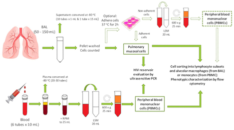
We describe a method for processing bronchoalveolar lavage fluid and matched peripheral blood from chronically HIV-infected individuals ...
Via Krishan Maggon
|
|
Scooped by
Gilbert C FAURE
June 24, 2019 8:58 AM
|
Just like the gut, the skin and the mouth, the eye also has a collection of microbes that keep it healthy. Understanding the eye microbiome may lead to new probiotic therapies.
|
|
Suggested by
Société Francaise d'Immunologie
June 22, 2019 2:20 PM
|
The Nutritional Immunology and Molecular Medicine Laboratory (NIMML) has developed a high-resolution model of the gut immune system by using advanced computational systems. The new high-performance computing (HPC)-driven ...
|
|
Scooped by
Gilbert C FAURE
June 20, 2019 9:09 AM
|
Abstract In addition to their critical role in allergic disorders, mast cells (MCs) are well recognized for their protective effector functions during bacteria and parasite infections. This review describes recent advancements of our understanding of the complex role of MCs in fungal infections. Specifically, we outline key features of the contribution of MCs to infections with six fungal pathogens, namely Sporothrix, Paracoccidioides, Aspergillus, Malassezia, Candida and Dermatophytes. Evidence from studies of these pathogens suggests that MCs can function as positive regulators that detect and contain fungi at the site of infection. However, it appears that the inflammation induced by MCs following fungal infections may not always and only be beneficial to the host. MC responses during fungal infections may primarily benefit the pathogen by facilitating its spreading and contributing to a greater severity of fungal infections. This review also highlights key drivers of MCs activation and effector mechanisms that have been identified for the multidimensional function of MCs in fungal diseases and in allergic diseases combined with fungal infection. 1 INTRODUCTION Fungi are a very diverse group of eukaryotic organisms that separate from plants and animals in their own kingdom. Today, fungal infections represent a serious public health problem around the world with more than 300 million people suffering from serious fungal infections and an estimated 1.6 million deaths annually.1 Fungi can cause a broad range of mycoses in humans: (a) Superficial fungal infections of the skin, nails and hair are commonly due to dermatophytes, for example Trichophyton rubrum or Trichophyton mentagrophytes, or yeasts, for example Candida or Malassezia furfur.2-5 (b) Subcutaneous fungal infections include sporotrichosis, chromoblastomycosis, mycetoma, phaeohyphomycosis, hyalohyphomycosis and lacaziosis.6, 7 (c) Paracoccidioidomycosis, coccidioidomycosis, histoplasmosis, mucormycosis and cryptococcosis are common systemic fungal infections.8 Subcutaneous and systemic mycoses occur mainly in immunosuppressed patients.7 Systemic fungal infections are linked to significant morbidity and mortality.8 In addition, fungal spores, for example, Aspergillus spores, are important allergenic agents and can cause persistent allergic inflammation of the airways.9 The immune mechanisms of defense against fungal infections are numerous, ranging from protective mechanisms that were present early in evolution (innate immunity) to sophisticated adaptive mechanisms that are induced specifically during infection and disease (adaptive immunity).10-12 Traditionally, mast cells (MCs) are seen as key effector cells in the elicitation of allergic signs and symptoms.13-15 However, there is an increasing number of studies that indicate that MCs are also essential players in innate immune responses.16-18 MCs are strategically located in tissues that are borders to the external environment such as the lung, the gut and the skin. Therefore, MCs are perfectly positioned to orchestrate protective immune responses against invading pathogens including fungi. Most studies of anti‐fungal immune responses are focused on the effects of epithelial cells, neutrophils, macrophages/monocytes and dendritic cells (DCs).19-23 As of now, only a few publications and reviews have addressed the role of MCs in the immune response to fungi infections. In the innate immune system, MCs act as sentinels for invading pathogens, as initiators of inflammatory responses and as regulatory cells in the coordination of raising and limiting innate immune responses.18, 24, 25 Recent findings suggest that, during innate immune responses against invading pathogens, MCs function as positive or negative immunoregulatory cells depending on the situation. In this review, we focus on the recent advancements of our understanding of the complex role of MCs in fungal infections—as positive regulators that detect and contain fungi at the site of infection and as negative regulators that promote a wider dissemination and a greater severity of fungal infections. Specifically, we highlight key features of the contribution of MCs to infections with six fungal pathogens, namely Sporothrix, Paracoccidioides, Aspergillus, Malassezia, Candida and Dermatophytes. 2 WHAT IS THE ROLE AND RELEVANCE OF MCs IN SPOROTHRIX SCHENCKII INFECTION? Sporothrix schenckii (S. schenckii) is an opportunistic dimorphic fungus with a global distribution.26, 27 S. schenckii is the cause of sporotrichosis, which usually affects the skin, less commonly the lungs, joints, bones, and brain of humans and other mammals.28 Veterinarians, animal technicians and caretakers, and owners of cats with sporotrichosis are regarded to be at increased risk for the acquisition of the disease.27 Sporotrichosis is transferred via the inoculation of S. schenckii (conidia or hyphae) from contaminated soil, plants or organic matter into sites of tissue injury such as wounds (traumatic inoculation).27 During interactions of S. schenckii with the host, the conidia‐phase of S. schenckii will transform into the yeast‐phase of S. schenckii in response to the host's temperature. Yeast is the dominant form of S. schenckii in sporotrichosis.29 In vitro studies showed that IgG‐seroreactive proteins from Sporothrix yeast cell extracts such as Gp 70 may be important for the pathogenesis and invasion of S. schenckii in human.30 In 2012, Romo‐Lozano et al. first reported that S. schenckii conidia can activate mouse peritoneal MCs to release early response cytokines such as TNF‐α and IL‐6 without degranulation.31, 32 By using an experimental murine model of systemic S. schenckii infection, which used intraperitoneal inoculation with S. schenckii conidia, they demonstrated that mice that had been depleted of peritoneal MCs presented with a decreased fungal load in organs and reduced severity of clinical manifestations. Their results suggest that mediators released by MCs may be implicated in promoting a wider dissemination of S. schenckii and a greater severity of the infection, indicating that MCs might act as negative immunoregulators in S. schenckii infection. However, this study used a functional MCs depletion model where MCs were depleted by injection of compound 48/80 into the peritoneal cavity. Moreover, their experimental model investigated systemic S. schenckii infection following intraperitoneal injection, whereas subcutaneous infection is the more common clinical manifestation of sporotrichosis. In summary, the in vivo evidence for a relevant role of MCs in S. schenckii infection is still limited. Based on the limited evidence available from mouse studies so far, MCs appear to promote S. schenkii, rather than protect from them. How MCs are involved in human sporotrichosis still needs to be clarified. 3 DO MCs PROMOTE PARACOCCIDIOIDES BRASILIENSIS INFECTION? The fungus Paracoccidioides brasiliensis (Pb) causes paracoccidioidomycosis (PCM), an autochthonous systemic mycosis found primarily in Central and South America.33 PCM is characterized by mucous membrane ulcerations of the mouth and nose with spreading through the lymphatic system. The route of infection is assumed to be inhalation. The lungs, lymph nodes and mucous membrane of the mouth are the most frequently infected tissues.34 That MCs interact with Pb has been shown by Valim et al., who demonstrated that co‐culturing of Pb yeast cells with MC lines rat basophilic leukaemia (RBL)‐2H3 MCs induced morphological changes of MCs and their degranulation.35 Furthermore, endogenous PbPga1, which is an immunoregulatory protein on the surface of Pb yeast cells, is able to activate rodent MCs through the NF‐κB pathway, resulting in the release of the inflammatory interleukin IL‐6. IL‐6 is involved in the induction of inflammatory responses that can lead to tissue destruction. Recently, a study of skin biopsies from patients with paracoccidioidomycosis suggested that MCs contribute to skin responses characterized by loose granuloma formation. This type of cutaneous response, which does not effectively contain or clear the infection, features markedly increased numbers of MCs and a Th2 cytokine profile.36 Interestingly, most MCs in these lesions were positive for IL‐10 suggesting that MCs are an important source of this immunosuppressive cytokine. Moreover, Batista et al. observed a downregulation of the inducible nitric oxide synthase (iNOS) in the tissues of oral paracoccidioidomycosis (OP). This results in reduced production of fungicidal nitric oxide (NO) in multinucleated giant cells and most mononuclear cells.37 MCs have been shown to be one of the major sources of NO in humans with fungus infections.38 These findings suggest that a low expression of iNOS in OP may represent a possible MC‐mediated mechanism that permits local fungal multiplication and maintenance of active oral lesions. Taken together, MCs appear to contribute to the morbidity of PCM, by several mechanisms. However, most of the above findings are from ex vivo or in vitro experiments, and proof that MCs play a harmful role in Pb infection needs to come from future in vivo studies. 4 MCs CONTRIBUTE TO THE MORBIDITY OF ASPERGILLUS FUMIGATUS INFECTION Aspergillus fumigatus (A. fumigatus) is a ubiquitous, spore‐forming fungus that has been associated with multiple pulmonary disorders. Inhaled conidia of A. fumigatus adhere to pulmonary epithelial cells, causing opportunistic infection followed by inflammatory responses to A. fumigatus antigens.39 Most studies on the role of MCs in chronic lung disease due to A. fumigatus focus on IgE‐mediated allergic bronchopulmonary aspergillosis (ABPA).40-42 ABPA is associated with early allergic and late‐phase inflammatory responses of the lungs to A. fumigatus antigens that occur in a minority (less than 1%) of patients with asthma and, more often (7%‐10%), in patients with cystic fibrosis.43 ABPA is characterized by elevated total and A. fumigatus‐specific immunoglobulin E (IgE) levels accompanied by eosinophilia, pulmonary infiltrates, bronchiectasis and fibrosis leading to asthmatic attacks and wheezing in affected patients. A. fumigatus‐specific IgE has been shown to activate MCs by cross‐linking of the high‐affinity receptor for IgE(FcεRI) leading to the immediate release of preformed mediators from intracellular granules and the de novo synthesis of lipid mediators and cytokines, all of which contribute to the subsequent development of inflammatory responses and obstruction of the airways.44-46 Proteases released from MCs can promote the release of growth factors from airway epithelial cells driving strong and irreversible remodelling reactions in airways of patients with ABPA, resulting in bronchiectasis lesions and fibrosis.46 Treatment with omalizumab, a humanized mAb that binds to IgE, has been shown to be an effective therapy for patients with severe allergic asthma and ABPA as shown by attenuated asthma symptoms and improved pulmonary function parameters in patients with ABPA.47 Recently, clinical trials demonstrated that omalizumab treatment not only provided a clinically important reduction in serum IgE, exacerbation rates and steroid requirement but also showed attenuated asthma symptoms and improved pulmonary function parameters in patients with ABPA.47 Interestingly, however, the majority of patients with airway colonization by A. fumigatus do not develop ABPA, yet they still exhibit a progressive decline in lung function.48 IgE‐independent MC degranulation in response to A. fumigates may be involved in this response. Urb and co‐workers found that direct contact with mature A. fumigatus hyphae can induce the degranulation of bone marrow‐derived mast cells (BMCMCs) in the absence of IgE, whereas no significant degranulation is induced by either conidia or immature hyphae.49 Although the exact mechanism of this is still unknown, the fungal proteins StuA and MedA, which control the composition of the cell surface molecules of mature A. fumigatus hyphae, were shown to induce BMCMC degranulation in the absence of IgE.49 Therefore, the authors speculated that A. fumigatus may directly worsen pulmonary disorders, by inducing degranulation in MCs that are in contact to hyphae, which may aggravate the inflammation and obstruction of airway rather than eliminate the fungus. Recent data shows that similar to ABPA in man, MCs may be implicated in recurrent airway obstruction (RAO), a type I hypersensitivity response to A. fumigatus in horses.49, 50 As in ABPA, specific IgE‐mediated cross‐linking of FcεRI results in MC degranulation and the release of histamine, serotonin, leukotrienes and eicosanoids such as thromboxane and 15‐hydroxyeicosatetraenoic acid. In addition, in experimental eosinophilic esophagitis induced by A. fumigatus, Niranjan et al. reported that MCs numbers are markedly increased and play a significant role in promoting oesophageal functional abnormalities, contributing to muscle cell hyperplasia and hypertrophy.51 Finally, a recent report suggests that MCs also contribute to A. fumigatus‐induced inflammation in acute invasive fungal rhinosinusitis (AIFR), an aggressive fungal infection with high morbidity and mortality rates in immunocompromised patients.52 Yan et al. created a rat model of AIFR based on A. fumigatus infection and investigated the role of MCs.53 Compared to control rats, the total number of MCs was unchanged, but MC degranulation was only found in the infected nasal cavities of AIFR rats. In summary, these findings suggest that MC responses to A. fumigatus appear to help the pathogen rather than the host. Further studies will be required to determine the underlying mechanisms of the contribution of MCs to A. fumigatus infection by valuable mice models.54 5 EXACERBATION OF MALASSEZIA‐INDUCED SKIN INFLAMMATION BY MCs IN PATIENTS WITH ATOPIC DERMATITIS Malassezia comprises a family of opportunistic pathogenic fungi, which includes 19 species found on the skin of humans and animals. These Malassezia species have been associated with dermatological diseases such as seborrhoeic dermatitis, pityriasis versicolor, atopic dermatitis (AD) and folliculitis.55 AD is one of the most common chronic inflammatory diseases of the skin.56 Malassezia sympodialis (M. sympodialis) is frequently isolated from AD patients and healthy individuals.57 Recently, it was demonstrated that mouse and human MCs respond to extracts from M. sympodialis.58-60 For example, M. sympodialis can enhance IgE‐mediated MC degranulation by signalling through the TLR2/MyD88 pathway. Interestingly, Ribbing et al. further observed that MCs derived from peripheral blood CD34+ progenitor cells of Malassezia‐infected patients contain more preformed granule mediators and exhibit enhanced IL‐6 secretion in response to M. sympodialis, as compared to MCs from healthy controls.60 In addition, Malassezia also can elicit specific IgE and T‐cell reactivity in AD patients.61, 62 Hiragun and colleagues identified the fungal protein, MGL_1304, derived from Malassezia globosa (M. globosa) in purified sweat, as an allergen in AD patients.63-65 They confirmed its ability to induce IgE‐dependent degranulation in human basophils and RBL cells expressing the human FcεRI receptor. Their results suggest that MGL_1304 is a major allergen in human sweat and could cause type I allergy in patients with AD. These findings indicate that M. sympodialis can activate MCs and thereby exacerbate inflammatory responses in AD patients. 6 DO MCs CONTRIBUTE TO THE PATHOGENIC EFFECTS OF CANDIDA ALBICANS? Candida albicans (C. albicans) is an opportunistic fungal pathogen, and they are normally grown as a harmless commensal on the skin and mucosal surfaces of healthy individuals.66 However, it can also cause superficial and systemic infections, especially in immunosuppression condition. C. albicans initially interacts with epithelial cells, resulting in fungal recognition and the formation of hyphae.67 The ability to undergo the morphological transition from yeast to hyphal growth is critical for its pathogenesis. Epithelial activation leads to the production of inflammatory mediators that recruit innate immune cells, which together work with epithelial cells to clear the fungal infection. The main immune cell in the defense against these candida infections is assigned to professional phagocytes, neutrophils and macrophages. In addition to the above cells, MCs also have been identified as the main cell types involved in the defense against this pathogen.68, 69 Yamaguchi et al. have shown that ovalbumin‐sensitized mice colonized with C. albicans show higher specific IgG and IgE titres than control mice, and colonized tissues promoted infiltration and degranulation of MCs.70 Their results suggest that gastrointestinal C. albicans colonization may promote sensitization against food antigens, at least partly due to MC‐mediated hyperpermeability in the gastrointestinal mucosa of mice. Furthermore, Takenaka and colleagues reported that increased levels of Candida‐specific IgE were linked to MC hyperplasia in lymph nodes in Kimura's disease.71 In addition, Segal and co‐workers observed that the injection of C. albicans together with Complete Freund's Adjuvant into the foot‐pads of guinea‐pigs provoked a state of hypersensitivity, characterized by direct degranulation of mouse peritoneal MCs when exposed in vitro to the antigen.72 Noverr and co‐workers reported that overgrowth of the yeast C. albicans following antibiotic treatment can promote the development of allergic airway disease characterized by increased levels of MCs.73 Taken together, these findings suggest that C. albicans may contribute to atopic diseases, at least in part by effects on MCs. Interestingly, MCs may also protect from C. albicans infection. Recent evidence shows that rat peritoneal MCs can kill C. albicans in the extracellular environment, very likely through secreted granular mediators.74 That C. albicans can degranulate rat peritoneal MCs to release mediators has been shown as early as in 1974 when Nosál et al. reported that glycoprotein from C. albicans can induce histamine release in isolated rat MCs, which was confirmed by later in vivo studies showing C. albicans‐induced MCs degranulation in the gut of mice.75 More recently, Nieto‐Patlán et al. have shown that both yeasts and hyphae of C. albicans can induce BMCMCs degranulation, production of TNF‐α, IL‐6, IL‐10, CCL3, CCL4, and ROS as well as NO involving TLR2 and dectin‐1.76 Moreover, MC seems to be able to phagocytose C. albicans. According to Lopes JP et al., the protective response of primary human MCs in C. albicans infections has three stages: Initially, during the first 3 hours, MCs reduce C. albicans viability by degranulation. During the intermediate stage of the response, 3‐12 hours after infection, MC cytokine secretion and the recruitment of neutrophils by releasing chemoattractants such as CXCL1/CXCL2 help to contain the infections by phagocytosis, the release of proinflammatory mediators and by recruiting other leucocytes. Later responses, that is after 12 hours, are characterized by the release of anti‐inflammatory cytokines and the recruitment of effector cells of adaptive immunity.77 Taking together, these results suggest that MCs can help the infected host by killing C. albicans and by containing the infection by recruiting neutrophils and macrophages to the sites of infection. As for the role of MCs in C. albicans infection, there are still many questions to be answered, and further evidence from in vivo studies is needed to arrive at robust conclusions.78 7 WHAT IS THE RELEVANCE OF MCs IN DERMATOPHYTE INFECTIONS? Dermatophytes, a group of keratinophilic fungi with approximately 40 species, are grouped into three genera: Trichophyton, Microsporum and Epidermophyton.79 Dermatophytes can invade human skin, hair, and nails and cause dermatophytosis (tinea).80 The role and relevance of MCs in dermatophyte infections are ill characterized. MCs have been observed to accumulate at sites of dermatophyte infections. For example, in Microsporum canis‐infected cats, the histopathological features of lesional skin include MC infiltration of the superficial dermis.81 Moskalewski et al. reported that the number of skin MCs is higher in mice infected with dermatophytes.82 In a Trichophyton quinckeanum dermatophytosis mouse model,83 MCs also accumulated in the dermis at the site of infection. This may or may not be relevant for the course of dermatophyte infections, and we will need further studies to clarify this. 8 WHAT CAN SPOROTHRIX, PARACOCCIDIOIDES, ASPERGILLUS, MALASSEZIA, CANDIDA AND DERMATOPHYTES TEACH US ABOUT THE EFFECTS OF MCs IN FUNGAL INFECTIONS? It appears that the inflammation induced by MCs following fungal infections may not always and only be beneficial to the host, but may primarily benefit the fungal pathogen by facilitating spreading (Figure 1 and Table 1). Additional support for this comes from studies with the fungal cell wall product zymosan.84-86 Zymosan‐induced peritonitis was found to be suppressed in MC‐deficient mice (WBB6F1‐W/Wv), and this was restored by adoptively transferred BMCMCs.87 MCs also appear to be involved in the morphine‐mediated inhibition of zymosan‐induced peritonitis in mice.88, 89 Gelatinase B/MMP‐9 as an inflammatory marker enzyme in mouse zymosan peritonitis was constitutively present in unstimulated MCs,90 and zymosan‐activated peritoneal MCs reportedly contain and produce more MMP‐9 after zymosan administration. Fungal species (family) Main colonized tissues Study host Study type IgE/non–IgE‐mediated response Reference Sporothrix schenckii (Ophiostomataceae) Skin, lungs, joints, bones, brain Mouse In vivo Non–IgE‐mediated response 31, 32 Paracoccidioides brasiliensis (Ajellomycetaceae) Lymph nodes, mucous membrane of the mouth Mouse In vivo Non–IgE‐mediated response 35 Human In vitro Non–IgE‐mediated response 36, 37 Aspergillus fumigatus (Trichocomaceae) Lung Mouse In vivo IgE‐mediated response 40 Human In vivo IgE‐mediated response 41, 44, 47 Mouse In vivo Non–IgE‐mediated response 45 Mouse In vitro Non–IgE‐mediated response 49 Horse In vivo IgE‐mediated response 50 Mouse In vivo, In vitro Non–IgE‐mediated response 51 Malassezia (Malasseziaceae) Skin Human In vitro IgE‐mediated response 59-61 Candida albicans (Saccharomycetaceae) Skin, mucosal surfaces Mouse In vivo IgE‐mediated response 69, 72 Guinea‐pig In vitro IgE‐mediated response 71 Rat In vitro Non–IgE‐mediated response 73 Mouse In vivo Non–IgE‐mediated response 74 Mouse In vitro Non–IgE‐mediated response 75 Human In vitro Non–IgE‐mediated response 76 Dermatophyte Skin Mouse In vivo Unclear 87, 88 While the published findings from studies on MCs in fungal infections point to a role of MCs that is different from that in bacteria and parasite infection, they do not provide the underlying mechanisms. In Sporotrichosis, for example, MCs promote a wider dissemination of S. schenckii and a greater severity of the infection in mice, but it remains unclear how MCs can mediate these negative effects. Also, most of the investigations performed so far, as described above, have been conducted with mouse models, and it has to be stressed that care should be taken in the translation of findings from the murine to the human system. MCs in mice and humans share many characteristic features, but also differ significantly, for example, in the expression of certain mediators such as proteases.91 In addition, the characterization of the role of MCs in fungal infections, as of now, has not made use of novel models and insights recently introduced to MC research, a limitation that will need to be addressed by future studies. Further evidence from ongoing and forthcoming research will need to provide a better picture of the mechanisms and the relevance of MC responses in fungal infections in humans. Finally, more focus has to come to the clinical relevance of characterizing the role of MCs in fungal infections. Several MC inhibitors are currently under development for the treatment of MC‐driven diseases. These inhibitors should be explored for their effects on fungal diseases. They may be especially helpful (and instructive) in the treatment of patients with allergic diseases and fungal infections. On the other hand, MC inhibitors may carry a risk of exacerbating some fungal infections, candidiasis for example, as MCs can reportedly contribute significantly to the clearance of C. albicans. In conclusion, further studies are needed to characterize the complex function of MCs in fungal diseases and, especially, in allergic diseases exacerbated by fungal infections. In the long run, this will lead to better strategies for preventing and treating fungal infections. ACKNOWLEDGEMENTS This work was supported by grants from A Joint Sino‐German Research project (no. GZ901) and Jiangsu Provincial Medical Youth Talent (no. QNRC2016736). CONFLICT OF INTEREST The authors confirm that there are no conflicts of interest. REFERENCES
|
|
Scooped by
Gilbert C FAURE
June 18, 2019 2:42 PM
|
Iliev and colleagues review the impact of the gut mycobiota on the regulation of host
immunity in homeostasis and in human disease and discuss the current understanding
of the mechanisms that underlie these effects.
|
|
Scooped by
Gilbert C FAURE
June 16, 2019 5:33 AM
|
mSystems. 2019 Jun 4;4(3). pii: e00129-19. doi: 10.1128/mSystems.00129-19.
|
|
Suggested by
Société Francaise d'Immunologie
June 15, 2019 4:42 AM
|
In the first study of its kind, LSU Health New Orleans researchers report that women's pre-pregnancy overweight or obesity produces changes in breast milk, which can affect infant growth.
|
|
Suggested by
LIGHTING
June 13, 2019 2:31 PM
|
In a mouse model of herpes simplex 2 virus infection, circulating memory B cells—rather than plasma cells—are shown to be the source of antibody secretion in the vaginal lumen following secondary infection.
|
|
Scooped by
Gilbert C FAURE
June 12, 2019 9:29 AM
|
Significance Functional loss of gut barrier integrity with subsequent increased antigen trafficking and occurrence of low-grade intestinal inflammation precede the onset of type 1 diabetes (T1D) in patients and preclinical models, thus suggesting that these events are mechanistically linked to the autoimmune pathogenesis of the disease. However, a causal relationship between increased intestinal permeability and autoimmune diabetes was never demonstrated. Our data show that breakage of gut barrier continuity leads to activation of islet-reactive T cells in the intestine, thus triggering autoimmune diabetes. An important implication of our findings is that restoration of a healthy gut barrier through microbiota and diet modulation in diabetes-prone individuals could reduce intestinal activation of islet-reactive T cells and prevent T1D occurrence. Abstract Low-grade intestinal inflammation and alterations of gut barrier integrity are found in patients affected by extraintestinal autoimmune diseases such as type 1 diabetes (T1D), but a direct causal link between enteropathy and triggering of autoimmunity is yet to be established. Here, we found that onset of autoimmunity in preclinical models of T1D is associated with alterations of the mucus layer structure and loss of gut barrier integrity. Importantly, we showed that breakage of the gut barrier integrity in BDC2.5XNOD mice carrying a transgenic T cell receptor (TCR) specific for a beta cell autoantigen leads to activation of islet-reactive T cells within the gut mucosa and onset of T1D. The intestinal activation of islet-reactive T cells requires the presence of gut microbiota and is abolished when mice are depleted of endogenous commensal microbiota by antibiotic treatment. Our results indicate that loss of gut barrier continuity can lead to activation of islet-specific T cells within the intestinal mucosa and to autoimmune diabetes and provide a strong rationale to design innovative therapeutic interventions in “at-risk” individuals aimed at restoring gut barrier integrity to prevent T1D occurrence. Footnotes ↵1To whom correspondence may be addressed. Email: falcone.marika{at}hsr.it. Author contributions: M.F. designed research; C.S., I.C., M.L.C., L.D.G., F.F., R.L., M.R., and R.F. performed research; C.S., R.F., F.S., F.C., and M.F. analyzed data; and C.S. and M.F. wrote the paper. The authors declare no conflict of interest. This article is a PNAS Direct Submission. This article contains supporting information online at www.pnas.org/lookup/suppl/doi:10.1073/pnas.1814558116/-/DCSupplemental. Copyright © 2019 the Author(s). Published by PNAS. This open access article is distributed under Creative Commons Attribution-NonCommercial-NoDerivatives License 4.0 (CC BY-NC-ND). References ↵ J. A. Todd, Etiology of type 1 diabetes. Immunity 32, 457–467 (2010).OpenUrlCrossRefPubMed ↵ F. Dotta et al., Coxsackie B4 virus infection of beta cells and natural killer cell insulitis in recent-onset type 1 diabetic patients. Proc. Natl. Acad. Sci. U.S.A. 104, 5115–5120 (2007). ↵ M. C. Honeyman et al., Association between rotavirus infection and pancreatic islet autoimmunity in children at risk of developing type 1 diabetes. Diabetes 49, 1319–1324 (2000).OpenUrlAbstract ↵ O. Vaarala et al., Cow’s milk formula feeding induces primary immunization to insulin in infants at genetic risk for type 1 diabetes. Diabetes 48, 1389–1394 (1999).OpenUrlAbstract ↵ J. M. Norris et al., Timing of initial cereal exposure in infancy and risk of islet autoimmunity. JAMA 290, 1713–1720 (2003).OpenUrlCrossRefPubMed ↵ L. Wen et al., Innate immunity and intestinal microbiota in the development of type 1 diabetes. Nature 455, 1109–1113 (2008).OpenUrlCrossRefPubMed ↵ A. Giongo et al., Toward defining the autoimmune microbiome for type 1 diabetes. ISME J. 5, 82–91 (2011).OpenUrlCrossRefPubMed ↵ C. T. Brown et al., Gut microbiome metagenomics analysis suggests a functional model for the development of autoimmunity for type 1 diabetes. PLoS One 6, e25792 (2011).OpenUrlCrossRefPubMed ↵DIABIMMUNE Study Group T. Vatanen et al.; DIABIMMUNE Study Group, Variation in microbiome LPS immunogenicity contributes to autoimmunity in humans. Cell 165, 842–853 (2016).OpenUrlCrossRefPubMed ↵ E. Badami et al., Defective differentiation of regulatory FoxP3+ T cells by small-intestinal dendritic cells in patients with type 1 diabetes. Diabetes 60, 2120–2124 (2011). ↵ T. Vatanen et al., The human gut microbiome in early-onset type 1 diabetes from the TEDDY study. Nature 562, 589–594 (2018).OpenUrlCrossRef ↵ J. B. Meddings, J. Jarand, S. J. Urbanski, J. Hardin, D. G. Gall, Increased gastrointestinal permeability is an early lesion in the spontaneously diabetic BB rat. Am. J. Physiol. 276, G951–G957 (1999).OpenUrlPubMed ↵ S. Graham et al., Enteropathy precedes type 1 diabetes in the BB rat. Gut 53, 1437–1444 (2004). ↵ F. Maurano et al., Small intestinal enteropathy in non-obese diabetic mice fed a diet containing wheat. Diabetologia 48, 931–937 (2005).OpenUrlCrossRefPubMed ↵ A. D. Mooradian, J. E. Morley, A. S. Levine, W. F. Prigge, R. L. Gebhard, Abnormal intestinal permeability to sugars in diabetes mellitus. Diabetologia 29, 221–224 (1986).OpenUrlCrossRefPubMed ↵ R. Carratù et al., Altered intestinal permeability to mannitol in diabetes mellitus type I. J. Pediatr. Gastroenterol. Nutr. 28, 264–269 (1999).OpenUrlCrossRefPubMed ↵ M. Westerholm-Ormio, O. Vaarala, P. Pihkala, J. Ilonen, E. Savilahti, Immunologic activity in the small intestinal mucosa of pediatric patients with type 1 diabetes. Diabetes 52, 2287–2295 (2003). ↵ M. Secondulfo et al., Ultrastructural mucosal alterations and increased intestinal permeability in non-celiac, type I diabetic patients. Dig. Liver Dis. 36, 35–45 (2004).OpenUrlCrossRefPubMed ↵ E. Bosi et al., Increased intestinal permeability precedes clinical onset of type 1 diabetes. Diabetologia 49, 2824–2827 (2006).OpenUrlCrossRefPubMed ↵ J. Neu et al., Changes in intestinal morphology and permeability in the biobreeding rat before the onset of type 1 diabetes. J. Pediatr. Gastroenterol. Nutr. 40, 589–595 (2005).OpenUrlCrossRefPubMed ↵ O. Vaarala, M. A. Atkinson, J. Neu, The “perfect storm” for type 1 diabetes: The complex interplay between intestinal microbiota, gut permeability, and mucosal immunity. Diabetes 57, 2555–2562 (2008). ↵ J. Sun et al., Pancreatic β-cells limit autoimmune diabetes via an immunoregulatory antimicrobial peptide expressed under the influence of the gut microbiota. Immunity 43, 304–317 (2015).OpenUrlCrossRefPubMed ↵ S. J. Turley, J. W. Lee, N. Dutton-Swain, D. Mathis, C. Benoist, Endocrine self and gut non-self intersect in the pancreatic lymph nodes. Proc. Natl. Acad. Sci. U.S.A. 102, 17729–17733 (2005). ↵ A. Sapone et al., Zonulin upregulation is associated with increased gut permeability in subjects with type 1 diabetes and their relatives. Diabetes 55, 1443–1449 (2006). ↵ T. Pelaseyed et al., The mucus and mucins of the goblet cells and enterocytes provide the first defense line of the gastrointestinal tract and interact with the immune system. Immunol. Rev. 260, 8–20 (2014).OpenUrlCrossRefPubMed ↵ M. E. Johansson, H. Sjövall, G. C. Hansson, The gastrointestinal mucus system in health and disease. Nat. Rev. Gastroenterol. Hepatol. 10, 352–361 (2013).OpenUrlCrossRefPubMed ↵ P. M. Smith et al., The microbial metabolites, short-chain fatty acids, regulate colonic Treg cell homeostasis. Science 341, 569–573 (2013). ↵ N. Arpaia et al., Metabolites produced by commensal bacteria promote peripheral regulatory T-cell generation. Nature 504, 451–455 (2013).OpenUrlCrossRefPubMed ↵ R. L. Gallo, L. V. Hooper, Epithelial antimicrobial defence of the skin and intestine. Nat. Rev. Immunol. 12, 503–516 (2012).OpenUrlCrossRefPubMed ↵ P. Chairatana, E. M. Nolan, Defensins, lectins, mucins, and secretory immunoglobulin A: Microbe-binding biomolecules that contribute to mucosal immunity in the human gut. Crit. Rev. Biochem. Mol. Biol. 52, 45–56 (2017).OpenUrl ↵ M. Shan et al., Mucus enhances gut homeostasis and oral tolerance by delivering immunoregulatory signals. Science 342, 447–453 (2013). ↵ Y. H. Sheng et al., MUC1 and MUC13 differentially regulate epithelial inflammation in response to inflammatory and infectious stimuli. Mucosal Immunol. 6, 557–568 (2013).OpenUrlCrossRefPubMed ↵ P. Courtois, C. Jurysta, A. Sener, F. W. Scott, W. J. Malaisse, Quantitative and qualitative alterations of intestinal mucins in BioBreeding rats. Int. J. Mol. Med. 15, 105–108 (2005).OpenUrlPubMed ↵ I. Jaakkola, S. Jalkanen, A. Hänninen, Diabetogenic T cells are primed both in pancreatic and gut-associated lymph nodes in NOD mice. Eur. J. Immunol. 33, 3255–3264 (2003).OpenUrlCrossRefPubMed ↵ J. D. Katz, B. Wang, K. Haskins, C. Benoist, D. Mathis, Following a diabetogenic T cell from genesis through pathogenesis. Cell 74, 1089–1100 (1993).OpenUrlCrossRefPubMed ↵ K. Haskins, M. Portas, B. Bradley, D. Wegmann, K. Lafferty, T-lymphocyte clone specific for pancreatic islet antigen. Diabetes 37, 1444–1448 (1988). ↵ R. Mueller, L. M. Bradley, T. Krahl, N. Sarvetnick, Mechanism underlying counterregulation of autoimmune diabetes by IL-4. Immunity 7, 411–418 (1997).OpenUrlCrossRefPubMed ↵ M. S. Horwitz et al., Diabetes induced by Coxsackie virus: Initiation by bystander damage and not molecular mimicry. Nat. Med. 4, 781–785 (1998).OpenUrlCrossRefPubMed ↵ M. E. Johansson et al., Bacteria penetrate the inner mucus layer before inflammation in the dextran sulfate colitis model. PLoS One 5, e12238 (2010).OpenUrlCrossRefPubMed ↵ C. Hernández-Chirlaque et al., Germ-free and antibiotic-treated mice are highly susceptible to epithelial injury in DSS colitis. J. Crohns Colitis 10, 1324–1335 (2016).OpenUrlCrossRefPubMed ↵ N. Tai et al., Microbial antigen mimics activate diabetogenic CD8 T cells in NOD mice. J. Exp. Med. 213, 2129–2146 (2016). ↵ F. R. Costa et al., Gut microbiota translocation to the pancreatic lymph nodes triggers NOD2 activation and contributes to T1D onset. J. Exp. Med. 213, 1223–1239 (2016). ↵ X. Li, M. A. Atkinson, The role for gut permeability in the pathogenesis of type 1 diabetes—a solid or leaky concept? Pediatr. Diabetes 16, 485–492 (2015).OpenUrlPubMed ↵ O. Vaarala, Leaking gut in type 1 diabetes. Curr. Opin. Gastroenterol. 24, 701–706 (2008).OpenUrlCrossRefPubMed ↵ O. Vaarala, Is the origin of type 1 diabetes in the gut? Immunol. Cell Biol. 90, 271–276 (2012).OpenUrlCrossRefPubMed ↵ A. Hänninen et al., Akkermansia muciniphila induces gut microbiota remodelling and controls islet autoimmunity in NOD mice. Gut 67, 1445–1453 (2018). ↵ H. Brauner et al., Markers of innate immune activity in patients with type 1 and type 2 diabetes mellitus and the effect of the anti-oxidant coenzyme Q10 on inflammatory activity. Clin. Exp. Immunol. 177, 478–482 (2014).OpenUrlCrossRefPubMed ↵ D. L. Kaufman et al., Spontaneous loss of T-cell tolerance to glutamic acid decarboxylase in murine insulin-dependent diabetes. Nature 366, 69–72 (1993).OpenUrlCrossRefPubMed ↵ K. Robinson, Z. Deng, Y. Hou, G. Zhang, Regulation of the intestinal barrier function by host defense peptides. Front. Vet. Sci. 2, 57 (2015).OpenUrl ↵ L. M. Loonen et al., REG3γ-deficient mice have altered mucus distribution and increased mucosal inflammatory responses to the microbiota and enteric pathogens in the ileum. Mucosal Immunol. 7, 939–947 (2014).OpenUrlCrossRefPubMed ↵ H. W. Koon et al., Cathelicidin signaling via the Toll-like receptor protects against colitis in mice. Gastroenterology 141, 1852–1863.e1-3 (2011).OpenUrlCrossRefPubMed ↵ A. S. Lee et al., Gut barrier disruption by an enteric bacterial pathogen accelerates insulitis in NOD mice. Diabetologia 53, 741–748 (2010).OpenUrlCrossRefPubMed ↵ K. Berer et al., Commensal microbiota and myelin autoantigen cooperate to trigger autoimmune demyelination. Nature 479, 538–541 (2011).OpenUrlCrossRefPubMed ↵ B. D. Stadinski et al., Chromogranin A is an autoantigen in type 1 diabetes. Nat. Immunol. 11, 225–231 (2010).OpenUrlCrossRefPubMed ↵ C. M. Gleeson, W. J. Curry, C. F. Johnston, K. D. Buchanan, Occurrence of WE-14 and chromogranin A-derived peptides in tissues of the human and bovine gastro-entero-pancreatic system and in human neuroendocrine neoplasia. J. Endocrinol. 151, 409–420 (1996). ↵ W. J. Curry et al., Chromogranin A and its derived peptides in the rat and porcine gastro-entero-pancreatic system. Expression, localization, and characterization. Adv. Exp. Med. Biol. 482, 205–213 (2000).OpenUrlPubMed ↵ C. Sorini, M. Falcone, Shaping the (auto)immune response in the gut: The role of intestinal immune regulation in the prevention of type 1 diabetes. Am. J. Clin. Exp. Immunol. 2, 156–171 (2013).OpenUrlPubMed ↵ M. Knip, H. Siljander, The role of the intestinal microbiota in type 1 diabetes mellitus. Nat. Rev. Endocrinol. 12, 154–167 (2016).OpenUrl ↵ J. D. Taurog et al., The germfree state prevents development of gut and joint inflammatory disease in HLA-B27 transgenic rats. J. Exp. Med. 180, 2359–2364 (1994). ↵ C. King, N. Sarvetnick, The incidence of type-1 diabetes in NOD mice is modulated by restricted flora not germ-free conditions. PLoS One 6, e17049 (2011).OpenUrlCrossRefPubMed ↵ M. W. Sadelain, H. Y. Qin, J. Lauzon, B. Singh, Prevention of type I diabetes in NOD mice by adjuvant immunotherapy. Diabetes 39, 583–589 (1990). ↵ T. C. Martins, A. P. Aguas, Changes in B and T lymphocytes associated with mycobacteria-induced protection of NOD mice from diabetes. J. Autoimmun. 9, 501–507 (1996).OpenUrlPubMed ↵ D. Elias, D. Markovits, T. Reshef, R. van der Zee, I. R. Cohen, Induction and therapy of autoimmune diabetes in the non-obese diabetic (NOD/Lt) mouse by a 65-kDa heat shock protein. Proc. Natl. Acad. Sci. U.S.A. 87, 1576–1580 (1990). ↵ P. Saï, A. S. Rivereau, Prevention of diabetes in the nonobese diabetic mouse by oral immunological treatments. Comparative efficiency of human insulin and two bacterial antigens, lipopolysacharide from Escherichia coli and glycoprotein extract from Klebsiella pneumoniae. Diabetes Metab. 22, 341–348 (1996).OpenUrlPubMed ↵ R. Horai et al., Microbiota-dependent activation of an autoreactive T cell receptor provokes autoimmunity in an immunologically privileged site. Immunity 43, 343–353 (2015).OpenUrlCrossRefPubMed ↵ T. C. Fung et al., Lymphoid-tissue-resident commensal bacteria promote members of the IL-10 cytokine family to establish mutualism. Immunity 44, 634–646 (2016).OpenUrlCrossRefPubMed ↵ D. K. Cole et al., Hotspot autoimmune T cell receptor binding underlies pathogen and insulin peptide cross-reactivity. J. Clin. Invest. 126, 3626 (2016).OpenUrl ↵ S. Wirtz, C. Neufert, B. Weigmann, M. F. Neurath, Chemically induced mouse models of intestinal inflammation. Nat. Protoc. 2, 541–546 (2007).OpenUrlCrossRefPubMed ↵ R. L. Brown, R. P. Sequeira, T. B. Clarke, The microbiota protects against respiratory infection via GM-CSF signaling. Nat. Commun. 8, 1512 (2017).OpenUrl ↵ K. Atarashi et al., Induction of colonic regulatory T cells by indigenous Clostridium species. Science 331, 337–341 (2011). ↵ B. Weigmann et al., Isolation and subsequent analysis of murine lamina propria mononuclear cells from colonic tissue. Nat. Protoc. 2, 2307–2311 (2007).OpenUrlCrossRefPubMed ↵ L. Lefrancois, N. Lycke, Isolation of mouse small intestinal intraepithelial lymphocytes, Peyer’s patch, and lamina propria cells. Curr. Protoc. Immunol. Chapter 3, Unit 3.19 (2001).OpenUrl ↵ S. Ghosh et al., Colonic microbiota alters host susceptibility to infectious colitis by modulating inflammation, redox status, and ion transporter gene expression. Am. J. Physiol. Gastrointest. Liver Physiol. 301, G39–G49 (2011).OpenUrlCrossRefPubMed
|
|
Scooped by
Gilbert C FAURE
June 11, 2019 3:52 AM
|
bioRxiv - the preprint server for biology, operated by Cold Spring Harbor Laboratory, a research and educational institution
|


 Scoop.it!