Your new post is loading...

|
Scooped by
Gilbert C FAURE
June 10, 2019 1:38 PM
|
Colorectal cancer stages are associated with distinct microbial and metabolomic profiles that could shed light on cancer progression.

|
Suggested by
LIGHTING
June 7, 2019 2:02 PM
|
The intervillous space of the placenta is a part of the fetal-maternal interface, where maternal blood enters to provide nutrients and gas exchange. Little is known about the maternal immune cells at this site, which are in direct contact with fetal tissues. We have characterized the T cell composition and chemokine profile in paired intervillous and peripheral blood samples from healthy mothers giving birth following term pregnancies. Mucosal-associated invariant T (MAIT) cells and effector memory (EM) T cells were enriched in the intervillous blood compared to peripheral blood, suggesting that MAIT cells and other EM T cells home to the placenta during pregnancy. Furthermore, pregnant women had lower proportions of peripheral blood MAIT cells compared to non-pregnant women. The levels of several chemokines were significantly higher in intervillous compared to peripheral blood, including macrophage migration inhibitory factor (MIF), CXCL10, and CCL25, whereas CCL21, CCL27 and CXCL12 were lower. Migration assays showed that MAIT cells and EM T cells migrated towards conditioned medium from placental explants. A multivariate factor analysis indicated that high levels of MIF and CCL25 were associated with high proportions of MAIT cells in intervillous blood. Blocking of MIF or a combination of MIF, CCL25, and CCL20 in migration assays inhibited MAIT cell migration towards placenta conditioned medium. Finally, MAIT cells showed migratory capacities towards recombinant MIF
|
|
Scooped by
Gilbert C FAURE
May 29, 2019 2:42 PM
|
Vaxart, Inc.(Nasdaq: VXRT), a clinical-stage biotechnology company developing oral recombinant vaccines that are administered by tablet rather than by inj...

|
Suggested by
Société Francaise d'Immunologie
May 24, 2019 8:10 AM
|
An intricate network of innate and immune cells and their derived mediators function in unison to protect us from toxic elements and infectious microbial diseases that are encountered in our environment.
|
|
Scooped by
Gilbert C FAURE
May 22, 2019 2:16 PM
|
|
|
Scooped by
Gilbert C FAURE
May 21, 2019 1:40 PM
|
Tissue-resident memory T (TRM) cells have been studied mainly in mouse models. Schumacher and colleagues have developed an imaging technology to track in real time skin-resident human CD8+ TRM cells in situ.
|
|
Scooped by
Gilbert C FAURE
May 20, 2019 2:45 PM
|
One of the fundamental issues in the microbiome research is characterization of the healthy human microbiota. Recent studies have elucidated substantial divergences in the microbiome structure between healthy individuals from different race and ethnicity. This review provides a comprehensive account of such geography, ethnicity or life-style-specific variations in healthy microbiome at five major body habitats – Gut, Oral-cavity, Respiratory Tract, Skin and Urogenital Tract (UGT). The review focuses on the general trend in the human microbiome evolution - a gradual transition in the gross compositional structure along with a continual decrease in diversity of the microbiome, especially of the gut microbiome, as the human populations passed through three stages of subsistence like foraging, rural farming and industrialized urban western life. In general, gut microbiome of the hunter-gatherer populations is highly abundant with Prevotella, Proteobacteria, Spirochaetes, Clostridiales, Ruminobacter etc., while those of the urban communities are often enriched in Bacteroides, Bifidobacterium and Firmicutes. The oral and skin microbiome are the next most diverse among different populations, while respiratory tract and UGT microbiome show lesser variations. Higher microbiome diversity is observed for oral-cavity in hunter-gatherer group with higher prevalence of Haemophilus than agricultural group. In case of skin microbiome, rural and urban Chinese populations show variation i
|
|
Scooped by
Gilbert C FAURE
May 16, 2019 1:35 PM
|
J Comp Pathol. 2019 Jan;166:69-86. doi: 10.1016/j.jcpa.2018.11.001. Epub 2018 Dec 17.
|
|
Suggested by
Société Francaise d'Immunologie
May 14, 2019 2:31 PM
|
It's well-known that breastfed babies tend to be healthier than babies given a bottle filled with formula, especially in their first year of life, and the benefits are numerous. What's more, the longer a baby is breastfed, the more far-reaching...
|
|
Scooped by
Gilbert C FAURE
May 14, 2019 5:45 AM
|
The human microbiome, the huge collection of microbes that live inside and on our body, profoundly affects human health and disease. The human gut flora in particular, which harbor the densest number of microbes, not only ...
|
|
Scooped by
Gilbert C FAURE
May 12, 2019 9:36 AM
|
While the vertebrate microbiota is critical to the normal function of many host traits, hosts may expend a large amount of energy to constrain and interface with their microbiota via their immune system to avoid the high fitness costs associated with gut dysbiosis, pathobionts, and opportunistic...
|
|
Scooped by
Gilbert C FAURE
May 11, 2019 10:39 AM
|
High-resolution imaging of bacterial populations in whole infected organs reveals a novel mechanism of mucosal colonization, where the occupation of a specialized microenvironment, or "microniche," in the gastric glands allows Helicobacter pylori to establish a stable bacterial reservoir in the...
|
|
Suggested by
Société Francaise d'Immunologie
May 11, 2019 7:05 AM
|
|
|
|
Scooped by
Gilbert C FAURE
June 8, 2019 2:50 AM
|
Methods Mol Biol. 2019;1884:189-202. doi: 10.1007/978-1-4939-8885-3_13. Research Support, Non-U.S.Gov't...
|
|
Suggested by
Société Francaise d'Immunologie
June 7, 2019 2:02 PM
|
Group 3 innate lymphoid cells are recruited to the lung and involved in the immune response to infection with tuberculosis in humans and mice, as a reduction in group 3 innate lymphoid cells in the lung impaired early immune control of Mycobacterium tuberculosis in mice.
|
|
Scooped by
Gilbert C FAURE
May 29, 2019 2:40 PM
|
MAbs. 2019 May 24. doi: 10.1080/19420862.2019.1622940.[Epub ahead of print]...
|
|
Suggested by
Société Francaise d'Immunologie
May 24, 2019 3:49 AM
|
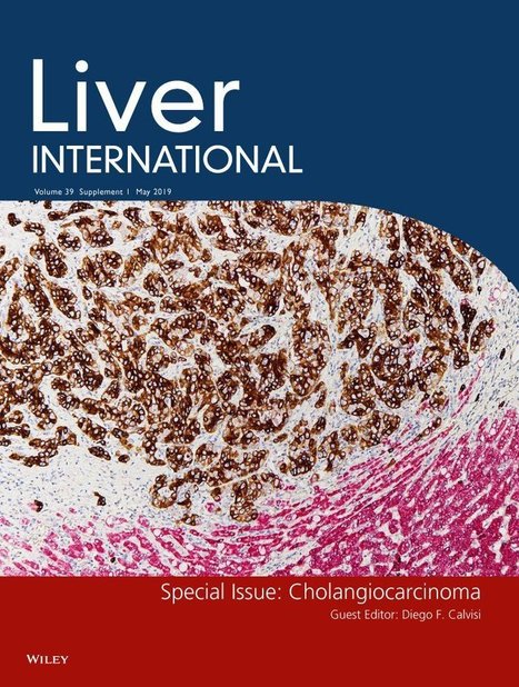
Abstract The high mortality rate of cholangiocarcinoma (CCA) is due, in part, to the lack of non‐invasive approaches able to accurately detect this silent tumour at early stages, when therapeutic options can be potentially curative or may at least increase the overall survival of patients. The fact that the majority of CCA tumours are not linked to any known aetiological factor highly compromises the monitoring of patients at risk for tumour development and also their early diagnosis. Combination of clinical/biochemical features, imaging techniques and analysis of non‐specific tumour biomarkers in serum are commonly used to help in the diagnosis of CCA, but tumour biopsy is usually required to confirm the diagnosis. Moreover, no prognostic biomarkers are currently used in the clinical setting, deserving more innovative research, and international validation and consensus. Important efforts have been made in the last few years to identify accurate non‐invasive biomarkers, by using innovative techniques and high‐throughput omics technologies. This review summarizes and discusses the advances in the investigation of novel diagnostic and prognostic biomarkers in CCA and envisions the future directions in this field of research. Abbreviations Abbreviations AUC area under the ROC curve CA19‐9 carbohydrate antigen 19‐9 CCA cholangiocarcinoma CEA carcinoembryonic antigen cfDNA cell‐free DNA cfRNA cell‐free RNA CTCs circulating tumour cells dCCA distal cholangiocarcinoma eCCA extrahepatic cholangiocarcinoma EVs extracellular vesicles HCC hepatocellular carcinoma iCCA intrahepatic cholangiocarcinoma IL‐6 interleukin 6 lncRNA long non‐coding RNAs miR microRNA pCCA perihilar cholangiocarcinoma PSC primary sclerosing cholangitis Key Points Currently available diagnostic biomarkers for CCA are inaccurate. Potential new biomarkers in liquid biopsies may include nucleic acids, proteins and metabolites, extracellular vesicles and circulating tumour cells. New omics technologies facilitate the profiling and analysis of disease‐specific signatures and are powerful sources of biomarkers. Novel promising biomarkers for the diagnosis of CCA need to be validated in large international cohorts of patients, including appropriate control groups of individuals/patients. 1 INTRODUCTION Cholangiocarcinoma (CCA) comprises a highly aggressive and heterogeneous group of biliary malignancies that can originate at any site of the biliary tree, and account for ~15% of all primary liver cancers.1 Its incidence is increasing worldwide and currently represents ~2% of all cancer‐related deaths per year. According to the anatomical localization, CCAs are classified into intrahepatic (iCCA), perihilar (pCCA) and distal (dCCA).1 The aetiology of the majority (~80%) of CCAs is unknown, but there are several risk factors that may predispose to its development, including age, obesity, diabetes, inflammatory liver diseases (primary sclerosing cholangitis [PSC], hepatolithiasis, cirrhosis), infectious agents (Opisthorchis viverrini, Clonorchis sinensis, hepatitis B [HBV], hepatitis C [HCV], human immunodeficiency virus [HIV]), drugs/toxins (alcohol, smoking, thorotrast, nitrosamines, asbestos, oral contraceptive pills, etc), and congenital disorders (choledochal cysts, Caroli disease, congenital hepatic fibrosis).1 The diagnosis of CCA is usually made throug a combination of clinical, biochemical, radiological and histological information. Different imaging techniques may be used for the diagnosis of each CCA subtype: ultrasound (US), computed tomography (CT), magnetic resonance imaging/magnetic resonance cholangiopancreatography (MRI/MRCP) and positron emission tomography (PET) for iCCA, MRCP for pCCA and dCCA, percutaneous transhepatic cholangiography for pCCA, and endoscopic retrograde cholangiopancreatography or endoscopic ultrasound for dCCA.2 Histological analysis is mandatory to confirm the diagnosis and can provide valuable information for the clinical management of patients,3 but it is not always recommended due to the location of the tumours and the risk of peritoneal seeding. Moreover, the serum levels of non‐specific tumour biomarkers, such as carbohydrate antigen 19‐9 (CA19‐9), are currently measured to help in the diagnosis of CCA, but these are unreliable due to their low sensitivity and specificity, particularly in early stages of the disease.4 As such, most patients with CCA are diagnosed late, when the disease is in an advanced stage, and when therapeutic options are reduced, resulting in dismal prognosis. However, in the small proportion of patients in which tumours are detected in early stages (~35%), surgical resection of the tumour or liver transplantation (in cirrhotic livers) can be potentially curative or, at least, significantly increase the overall survival of patients.1 Therefore, there is an urgent need to identify accurate non‐invasive biomarkers for the diagnosis of these tumours, and thus increase the number of potential resectable cases. In recent years, new innovative studies have been conducted in the quest for accurate biomarkers for the early diagnosis of CCA and also to predict prognosis, risk of relapse after surgery, and select the best therapeutic regimen(s) for patients. These strategies involve “omics” approaches in blood, bile, urine, extracellular vesicles (EVs) and tissues, and have resulted in promising candidates that can change the current paradigm. 2 Non‐INVASIVE BIOMARKERS 2.1 Circulating nucleic acids Circulating nucleic acids can be found in most biofluids and comprise fragments of genomic DNA (cell‐free DNA [cfDNA]) and RNA (cfRNA; typically microRNAs [miRs], but also long non‐coding RNAs [lncRNAs]). Whether actively exported or originated from dying cells, circulating nucleic acids embody potential diagnostic and/or prognostic tools for human disease, including CCA.4-7 2.1.1 Cell‐free DNA cfDNA was first shown to reflect changes in cancer aggressiveness and tumour size in the late 70’s,8 highlighting its potential as a diagnostic and/or prognostic biomarker. The ability to screen for mutations in cfDNA is particularly appealing when compared to the primary tumour, as it more accurately reflects the overall mutational pattern of these heterogeneous tumours. This concept was recently validated in CCA; plasma samples from CCA patients with known tumour genomic background were screened for 31 oncogenic mutations in KRAS, NRAS, BRAF and PIK3CA genes by multiplex digital PCR.9 For each patient, the exact mutations in the tumour were also found in the plasma. Of note, the cfDNA from patients with CCA tumours, but wild type for the 31 studied mutations, was used as negative control and strongly supported the data. These results suggest that the use of cfDNA screening in patients with CCA may be helpful in order to determine the mutational characteristics of the primary tumours and to guide potential mutation‐based therapeutic interventions. In this regard, an integrative genomic characterization of cfDNA, primary tumours, and metastases of iCCA patients with FGFR2 mutations and with acquired resistance to BGJ398, a pan‐FGFR inhibitor, revealed de novo point mutations in the FGFR2 kinase domain that were detected in cfDNA.10 Despite the small sample size, given that FGFR2 mutations are found exclusively in iCCA and account for ~10% of the diagnosed cases,11 screening for these alterations in cfDNA may represent an important approach to guide clinical decisions. 2.1.2 Cell‐free non‐coding RNA Throughout the last decade, cfRNAs, particularly miRs, have been envisioned as promising disease biomarkers due to their abundance and stability in biofluids. In comparison to other putative biomarkers, such as proteins or metabolites, miRs are less resistant to degradation and/or modification and can be easily detected and amplified. In the context of CCA, few studies have investigated the circulating miR profiles in patients (Figure 1), but two meta‐analyses have suggested their overall potential diagnostic value.12, 13 Data analysis on both meta‐analyses pinpointed miRs as promising tools for CCA diagnosis, with pooled sensitivities of 0.83 and 0.76, and specificities of 0.79 and 0.91 respectively. In both studies, pooled area under the ROC curve (AUC) was ~0.9. Further, bile was the biological fluid showcasing the highest diagnostic efficiency (AUC of 0.95), followed by serum (0.913), tissue (0.846) and urine (0.745).12 Circulating miR‐21 is one of the most well‐characterized miRs in terms of its potential as a biomarker for CCA. Increased serum and plasma levels of miR‐21 were already shown to allow for the differential diagnosis between patients with iCCA and healthy controls,5 with an AUC of 0.91 in serum and 0.94 in plasma.14 More recently, serum miR‐21 levels were also found to positively correlate with tumour stage (TNM criteria) and poor survival. Indeed, miR‐21 serum levels decreased after tumour resection, highlighting the value of circulating miR‐21, not only as a diagnostic tool, but also as a putative prognostic biomarker.15 However, the levels of this miR, considered an onco‐miR,16 are also found elevated in serum of patients with hepatocellular carcinoma (HCC)17-20 and other cancers,21-24 probably limiting its specificity to discriminate between tumours, particularly primary liver tumours. Serum miR‐26a levels are also increased in patients with CCA and, similarly to miR‐21, they positively correlate with clinical stage, metastasis, tumour differentiation status and poor survival. In terms of its diagnostic value, serum miR‐26a yielded an AUC value of 0.90 (sensitivity: 84.8%; specificity: 81.8%) in distinguishing CCA from healthy controls.5 Decreased serum levels of miR‐106a in CCA patients appear to also act as a predictor of poor prognosis, while signalling a higher likelihood of lymph node metastasis.25 miR‐150 represents another potential CCA diagnostic biomarker, although apparently contradictory results have been published. A microarray study using plasma from patients with CCA reported reduced miR‐150 levels in CCA compared with healthy individuals and patients with PSC.26 In contrast, a different study highlighted miR‐150 as being upregulated in iCCA compared with controls without cancer (AUC: 0.764; sensitivity: 80.6%; specificity: 58.1%).5 Of note, the above study also showed that the combination of miR‐150 with CA19‐9 improved the diagnostic accuracy of both biomarkers. Indeed, it is now apparent that merging different miRs into a panel of biomarkers can offer greater sensitivity and specificity. For instance, an miR profile of eight plasma miRs (483‐5p, 505‐3p, 874, 885‐5p, 320b, 92b‐3p, 1275, 1307‐3p) was shown to associate with iCCA, regardless of the degree of tumour differentiation.27 The levels of miR‐192 were also shown to be increased in serum from patients with O viverrini‐related CCA compared with healthy controls (AUC: 0.803; sensitivity: 74%; specificity: 71%), positively correlating with lymph node metastasis and poor survival.28 Of note, in this study, the authors also reported increased serum levels of both miR‐21 and miR‐150, further supporting their potential diagnostic value, while miR‐26a was found downregulated, conflicting with previous data and thus rising some questions regarding its diagnostic accuracy for all forms of the disease. Since PSC is a well‐established risk factor for CCA development, some studies have evaluated and compared the serum miR profile between patients with isolated PSC and those with PSC‐derived CCA. For instance, increased serum levels of miR‐222 (AUC: 0.71) and miR‐483‐5p (AUC: 0.70) were found in CCA patients compared with PSC patients. The combination of both miRs increased the AUC to 0.77, suggesting that they could help in the monitoring of PSC patients for early CCA detection.29 In another study, a panel composed of five serum miRs (26a, 30b, 122, 126 and 1281) was markedly different in patients with CCA compared with PSC patients,30 further underlining their potential diagnostic value. Nevertheless, it is worth mentioning that these studies did not specifically include CCA patients with a PSC background, which would be of great importance in order to find accurate diagnostic biomarkers for PSC patients who might be at risk for CCA development. Cholangiocytes have a pivotal role in bile formation, regulation and transport. In turn, circulating bile miRs may also embody a key diagnostic and/or prognostic value in CCA. miRs have already been profiled in bile samples from patients with CCA through high‐throughput PCR miRNA microarray, with miR‐9 being highlighted as a potential diagnostic biomarker (AUC: 0.98; sensitivity: 88.9%; specificity: 100%).31 Furthermore, the levels of miR‐150‐5p were found lower in bile from patients with CCA compared with healthy individuals.26 On the other hand, the levels of miR‐412, −640, −1537 and −3189 were found increased in bile from patients with PSC‐derived CCA compared to those with isolated PSC, allowing the differential diagnosis of these two diseases with an AUC value of ~0.8. Of note, combination of miR‐1537 with CA19‐9 provided a higher diagnostic value when compared solely with CA19‐9.30 Some studies have also explored the potential of urinary miRs as biomarkers for CCA (Figure 1). For instance, urinary levels of miR‐192 and miR‐21 have been found markedly increased in patients with O viverrini‐related CCA compared with healthy individuals, and the combination of these two miRs increased its diagnostic value, when comparing with each one used alone (AUC: 0.85; sensitivity: 81.8%; specificity: 71.4%).32 Overall, since most data on circulating nucleic acids as diagnostic and prognostic biomarkers in CCA have resulted from proof‐of‐concept studies, larger and international evaluations are eagerly awaited to validate their potential clinical value. 2.2 Cytokines/proteins CA19‐9 and carcinoembryonic antigen (CEA) are the most widely clinically used biomarkers to help in the diagnosis and/or monitoring of CCA, but there are large differences in sensitivity and specificity among the different published studies,33-35 limiting their diagnostic and prognostic value. For CA19‐9, the most recent data resulting from a large meta‐analysis described a pooled sensitivity and specificity of 72% and 84%, respectively, regarding the distinction between CCA and healthy controls or patients with benign biliary disease.36 Similarly, the diagnostic sensitivity and specificity of CEA range from 42% to 85% and 70% to 89%37-39 respectively. Increased serum levels of CEA and CA19‐9 have been proposed as an indicator of reduced overall survival in resectable or inoperable CCAs.39 However, while an important number of studies describe CEA and CA19‐9 as independent prognostic markers,39-41 prognostic cut‐off values vary significantly between reports and large meta‐analyses are still lacking. Nonetheless, when elevated in CCA, CA19‐9 was further suggested as biomarker to monitor response to chemotherapy and predict outcome in CCA patients.42 Other promising circulating diagnostic and prognostic biomarkers (Table 1) include cytokeratin‐19 fragment (CYFRA 21‐1), matrix metalloproteinase‐7 (MMP‐7) and osteopontin. CYFRA 21‐1 is elevated in patients with iCCA compared to patients with benign biliary diseases (sensitivity: 75.6%; specificity: 96.2%), presenting superior diagnostic values than CA19‐9 and CEA.43 In addition, serum levels of CYFRA 21‐1 correlate with disease stage, and represent an independent predictor of impaired relapse‐free and overall survival.43, 44 MMP‐7 serum levels are also elevated in patients with CCA compared to patients with benign biliary (sensitivity: 75%; specificity: 78%),45, 46 but its prognostic relevance is still unclear. Circulating osteopontin, a secreted glycol phosphoprotein, is also elevated in CCA patients compared to healthy controls or patients with PSC. It is important to note that high pre‐ and post‐operative osteopontin levels were associated with reduced overall survival after tumour resection.47 Protein/Cytokine Source Levels Comparison SEN (%) SPE (%) AUC Reference MMP7 Serum Up CCA (n = 44) vs benign biliary tract disease (n = 36) 75 78 0.730 46 Osteopontin Serum Up CCA (n = 80) vs healthy controls (n = 42) 88 100 0.964 47 IL‐6 Serum Up CCA (n = 26) vs healthy controls (n = 23) 73 92 0.875 49 S100A6 Serum Up CCA (n = 29) vs healthy controls (n = 22) 86 91 0.909 50 DKK1 Serum Up iCCA (n = 37) vs healthy controls (n = 50) 76 100 0.872 51 SSP411 Serum Up CCA (n = 35) vs “cholangitis (n = 13) and healthy controls (n = 23)” 90 83 0.913 53 SEN, sensitivity; SPE, specificity. Circulating cytokines have also been proposed as diagnostic and prognostic biomarkers in CCA patients. The pro‐inflammatory cytokine interleukin 6 (IL‐6), secreted by CCA cells48 is found elevated in serum of patients with CCA compared to healthy individuals (sensitivity: 73%; specificity: 92%), and was further proposed as marker for therapy monitoring.49 Other potential biomarker candidates reported to diagnose CCA are S100A6,50 DKK1,51 KL‐6‐Mucin52 and SSP411.53 However, larger studies are warranted to investigate/confirm their diagnostic and/or prognostic relevance in CCA. 2.3 Metabolites Metabolomics or metabolic profiling, defined as the analysis of low molecular weight metabolites (<1500 Da) in biological samples, is a promising approach for the identification of potential biomarkers useful in the diagnosis and prognosis of different diseases, including different types of cancers, like CCA.4 Due to the large number of molecules present in biological specimens, powerful bioinformatic tools for data mining and visualization are used to present the results in a comprehensive way (Figure 2). Cancer cells present profound alterations in their metabolism,54 which represent an opportunity for diagnosis and monitoring. The analysis of the metabolome in body fluids (blood/serum/plasma, bile or urine) is emerging as a new diagnostic strategy in cancer, since changes in metabolites may reflect, at least partly, what is happening in tumour cells. However, the identification of specific metabolites is a challenging goal due to the presence of many confounding factors, including age, gender, diet, underlying liver diseases, concomitant diseases, drugs and others. To date, only a reduced number of studies have investigated the usefulness of metabolites in body fluids in the diagnosis of CCA. In bile, the analysis of bile acid concentration and composition in patients with biliary tract cancer (iCCA, pCCA or extrahepatic CCA [eCCA]), biliary tract stones and healthy controls showed a reduction in the proportion of secondary bile acids in patients with CCA compared with those with biliary tract stones and healthy individuals.55 This finding was associated with an alteration of bile acid transport that could explain the exposure of the bile duct epithelium to cocarcinogenic bile acids.56 Another study analysed bile compounds in patients with CCA, PSC and benign biliary diseases and showed that changes in phosphatidylcholines, bile acids and lipids were able to discriminate CCA from other conditions (sensitivity: 88.9%; specificity: 78.1%).57 The analysis of metabolites in bile of patients with CCA, HCC, non‐malignant liver diseases and healthy individuals58 showed a decrease in glycine‐ and taurine‐conjugated bile acids, phospholipids and cholesterol in patients with CCA compared to control groups and, to a certain extent, also to HCC patients. In contrast, another study that analysed the metabolites in bile of patients with inoperable pCCA or dCCA and non‐malignant biliary diseases without cholestasis, including PSC, found increased levels of glycine‐conjugated bile acids and phosphatidylcholines in patients with CCA, and constructed models that were able to discriminate CCA patients from those with non‐malignant biliary diseases (sensitivity: 80%; specificity: 95%).59 From all these studies, bile acid species60 are positioned as some of the metabolites with potential as biomarkers. However, future studies should confirm in larger and international cohorts of patients the usefulness of the determination of bile acids and phospholipids in bile for the diagnosis of biliary tumours. Other studies have demonstrated that the analysis of metabolites in serum could help in the diagnosis of CCA. Serum analysis in two independent Chinese cohorts of patients with CCA identified several metabolites useful in the early diagnosis of this tumour, as well as to distinguish iCCA from eCCA. In particular, an increase in serum 21‐deoxycortisol and bilirubin levels and a decrease of lysophosphatidylcholines LPC(14:0) and LPC(15:0) levels were found in patients with CCA compared with healthy individuals.61 Moreover, the combination of the four candidate biomarkers was useful for distinguishing CCA from healthy controls with high accuracy (99%). A recent study analysing the metabolomics profile in biopsy‐proven patients with iCCA, HCC, PSC and healthy individuals has demonstrated that specific changes in serum concentrations of certain metabolites can help in the early and differential diagnosis of these diseases. Several metabolites presented higher diagnostic values for iCCA vs the other groups under study, with superior AUC than that found for CA19‐9.62 An algorithm combining six metabolites; three sphingomyelins (SMs), two phosphatidylcholines (PCs) and one ceramide (Cer) – SM(42:3), SM(43:2), PC(O‐16:0/20:3), PC(O‐18:0/18:2), SM(d18:2/16:0) and Cer(d18:1/16:0) – accurately differentiated iCCA from HCC (AUC: 0.9; sensitivity: 80%; specificity: 90%). Another algorithm that combined PC(34:3) and histidine accurately differentiated PSC from iCCA (AUC: 0.990; sensitivity: 100%; specificity: 70%). These interesting data, however, should be confirmed in patients with CCA arising from PSC. Remarkably, all of these results were successfully validated in another independent cohort of patients. An integrated analysis of the transcriptome and metabolome in surgically resected tumour tissue of patients with iCCA and HCC showed specific profiles of genes and metabolites that could be useful in the diagnosis of iCCA.63 In addition, the integration of genomics, transcriptomics and metabolomics in tumour tissue has also been proposed for the stratification of molecular subtypes of iCCA and HCC with similar prognosis.64 2.4 Extracellular vesicles In terms of minimally invasive biomarkers, EVs became of particular interest during the last few years. EVs can be found in all body fluids including blood,65 saliva,66 urine67 and bile.68 Commonly, EVs include two main subclasses that can be differentiated according to their size and biogenesis.69 According to the generally accepted nomenclature, larger EVs (also called microvesicles [MVs]) roughly range from 100 to 1000 nm in size and directly bud from the plasma membrane of their parental cell, whereas small EVs (also called exosomes) are considerably smaller (below 100 nm) and originate from accumulated intraluminal vesicles within the endomembranous system, forming so‐called multivesicular bodies (MVBs). The fusion of the MVBs with the plasma membrane results in the release of exosomes into the extracellular space.70, 71 EVs contain a variety of biomolecules including lipids, nucleic acids and proteins/antigens, and act as physiological mediators of cell communication.72 Furthermore, EVs amount and content reflect the pathobiological state of the cells they originate from.69 EVs have been shown to support the generation of tumour stroma during CCA development by inducing the differentiation of mesenchymal stem cells to fibroblasts,73 thus preparing their own tumour niche. Proteomic profiling of EVs derived from human serum has revealed promising candidate proteins (FCN2, ITIH4, FIBG) for the differential diagnosis of early‐stage CCA and PSC patients (AUC: 0.96; sensitivity: 0.88; specificity: 0.88).74 Importantly, some of the identified serum EV protein biomarkers allowed the accurate and early diagnosis of CCA (AMPN, VNN1 and PIGR; AUC: 0.88, 0.88 and 0.84 respectively) and HCC (LG3BP, PIGR and A2MG; AUC: 0.90, 0.84 and 0.80 respectively) compared to healthy individuals, as well as the differential diagnosis of iCCA and HCC (FIBG, A1AG1, VTDB; AUC: 0.89, 0.85 and 0.82 respectively), which currently comprises a major challenge nowadays. Of note, the combinations of some of these biomarkers increased their diagnostic accuracy. Moreover, the analysis of the surface antigen composition of serum EVs allowed to diagnose CCA from healthy individuals and other cancer entities with up to 90% sensitivity, but was unable to differentiate between CCA and HCC.75 Additionally, a correlation between CCA/HCC tumour burden and EV levels specific for those cancer entities was observed, highlighting the prognostic value of EVs, especially in terms of early detection of small tumours. The concentration of EVs per se in bile and serum was found increased in patients with CCA and pancreatic carcinoma, discriminating malignant from non‐malignant pancreatobiliary diseases with 100% sensitivity in bile and 47% in serum.76 Another study used a combined approach comprising EV isolation followed by microRNA content profiling. In particular, a miRNA panel (miRs 191, 486‐3p, 1274b, 16 and 484) showed good diagnostic values for CCA diagnosis compared to non‐malignant biliary diseases (sensitivity: 67%; specificity: 96%).77 An overview of the conducted studies summarizing their diagnostic capability for EV‐based CCA diagnosis can be found in Table 2. Biomarker Source Method Controls SEN SPE AUC Reference FCN2, ITIH4, FIBG Serum EVs Proteomics PSC 92‐100 81 0.88‐0.96 74 EpCAM+ ASGPR1+ CD133+ Serum EVs FACS Healthy 90 50 0.82 75 Total amount Serum EVs NTA Non‐malignant bile duct stenoses 47 80 0.81 76 Total amount Bile EVs NTA Non‐malignant bile duct stenoses 100 100 0.10 77 miR‐191 miR‐486‐3p miR‐1274b miR‐16 miR‐484 Serum EVs miR arrays Non‐malignant bile duct stenoses 67 96 — 78 AUC, area under (ROC) curve; EV, extracellular vesicles; FACS, fluorescence‐activated cell sorting; miR, microRNA; NTA, nanoparticle tracking analysis; SEN, sensitivity; SPE, specificity; PSC, primary sclerosing cholangitis. 2.5 Circulating tumour cells Due to the difficulty in obtaining a histological or cytological diagnosis in biliary‐pancreatic cancers, circulating tumour cells (CTCs) are of great interest. CTCs are released by primary tumours into the bloodstream at a concentration of about 106 CTCs/g of tumour/day78 and, although at very low concentrations, may be a route for metastasis of some solid neoplasms. Few studies have evaluated the presence of CTCs as a diagnostic tool for CCA, although there are emerging data in other cancers such as glioblastoma multiforme,79 hepatocellular carcinoma,80 and pancreatic,81 breast,82 and colorectal83 cancers. A number of technologies have been developed to isolate and identify CTCs from peripheral blood, including enzyme‐linked immunosorbent spot (ELISPOT) assay, real time PCR, flow cytometry, and immunocytochemistry, and automated or semi‐automated systems (CellSearch, CellSpotter, or iChip). To date, the only tool approved by the US Food and Drug Administration for the detection of CTCs is the CellSearch System, a semi‐automated platform for the preparation and subsequent capture of CTCs using epithelial cell‐specific EpCAM antibodies, prior to labelling with immunofluorescent markers. To further strengthen the discriminating capability of this tool, cells are also sorted for their positivity to DAPI, cytokeratins 8/18 (markers of hepatocytes), and 19 (marker of cholangiocytes in the liver) and their negativity for CD45 (marker of leukocytes).84 This system has the disadvantage that, apparently, few CCA tumours (10%‐20%) have significant elevation of EpCAM expression.85 Using this system, the prevalence of CTCs in blood from large cohorts of patients with metastatic carcinomas was 36% (≥2 CTCs per 7.5 mL of blood) compared to 0.3% in healthy and non‐malignant disease subjects.86 A different technology, based on a microfluidic platform capable of separating CTCs from peripheral whole blood samples using EpCAM‐coated microposts to differentiate between epithelial cells and blood leukocytes (the ‘CTC‐chip’), was developed and the efficiency of CTC capture determined in clinical specimens was 65%‐71% and the sensitivity and specificity were close to 100% in the wide variety of solid‐organ cancers tested.87, 88 A further refinement of this technology is the CTC‐iChip, which has been used to sort very rare CTCs from patients suffering from prostate cancer.89 To date, the number of studies suggesting that circulating CTCs are associated with poor prognosis in patients with advanced CCA are scarce.84, 90, 91 However, it remains unclear whether they have any diagnostic role in CCA. The first pioneering study on biliary‐pancreatic cancers study was conducted in patients using a reverse transcriptase‐PCR approach to detect CEA in blood samples; this study outlined a correlation between increased CEA expression and hematogenous dissemination and worst prognosis.92 In addition, it was also reported, using the CellSearch System, that only 25% of patients with biliary tract cancer had elevated CTCs (>2 per 7.5 mL of blood),84 suggesting the possibility of using CTCs as non‐invasive biomarkers to predict the outcome of patients.93 Of note, the presence of CTCs has been shown to correlate with higher tumour extent and lower overall survival in patients with CCA.90 Moreover, an elevation of CTCs count over baseline, together with other circulating markers, predicted a worse overall and disease‐free survival in patients with CCA.94 From a biological point of view, it was hypothesized that CTCs in the bloodstream could be putatively sustained by the interaction of the tumour cells with immune cells and CD105+ CD14+ myeloid fibroblasts, and be responsible for the metastatic spread of CCA.81 CTCs have also been isolated from portal venous blood in patients with hepatobiliary‐pancreatic malignancies.95 Although mainly focusing on patients with pancreatic ductal adenocarcinoma, portal vein derived CTCs were also examined in a small subgroup of CCA patients. Blood samples were obtained by direct intraoperative venipuncture during pancreaticoduodenectomy. CTCs were isolated by fluorescence‐activated cell sorting (CD44+, CD147+, EpCAM+, CD45−) and characterized for mRNA expression and acetylated chromatin encoding K‐RAS exon 12 mutation (K‐RAS mut). K‐RAS mut mRNA was detected at low levels and with high variability in CCA patients. The authors hypothesize that K‐RAS mut gene expression may be a useful indicator for aggressive adenocarcinoma CTCs. These cells might retain malignant potential in portal venous blood even after successful tumour resection with high risk for disease recurrence and metastatic progression.95 In line with this, another study was conducted to examine KRAS mutation‐positive CTCs in the portal venous blood of patients with hepatobiliary‐pancreatic malignancies, including a small subgroup of patients with dCCA.81 The CTCs isolated from the portal circulation were shown to be highly proliferative and resistant to apoptosis. CTCs recruited multiple immune cell types, including myeloid fibroblasts suggesting that CTC survival inside the portal venous circulation is supported by their interactions with immune cells within multi‐cell type clusters that could represent a source of local recurrence and metastatic progression. 3 BIOMARKERS IN TUMOUR TISSUE Biomarkers in tumour tissue may be of particular value for resected CCAs, as they could predict prognosis (ie, overall survival and tumour recurrence) (Table 3) and response to potential adjuvant therapies. In this regard, specific genomic and transcriptomic signatures have already been identified. High genomic heterogeneity was reported in CCA, with the most prevalent alterations related to DNA repair (TP53),96-100 growth pathways (KRAS, BRAF, SMAD4, FGFR2, PTPN3),96, 99-105 chromatin remodelling (KMT2C, ARID1A, PBRM1 and BAP1)97, 98 and developmental pathways that significantly impact the cancer growth, such as Notch and Wnt signalling pathways (NOTCH1, NICD, WNT7B and WNT10A).106, 107 Noteworthy, FGFR2 gene fusions, usually found in 5.5% to 13.6% patients with iCCA, deserve special attention since they are pharmacologically targetable and are specifically found in iCCA tumours, while being absent in any other liver malignancy, harbouring also a diagnostic value.11, 100, 101, 103, 109 IDH1 and IDH2 gene mutations are also frequently found in non‐infectious CCA, mainly in iCCA, accounting for 4.9% to 36% of the cases.97, 98, 100, 102, 110, 111 These alterations were also described in CCA cells and correlated with their methylation status.115 In a high‐throughput screening of several cancer cell lines, including 17 biliary tract cancer cells, IDH‐mutant iCCA cells exhibited good response to dasatinib, a multi‐tyrosine kinase inhibitor, which was also shown to increase apoptosis and tumour regression in IDH‐mutant xenografts.116 Consequently, a clinical trial is now being conducted to evaluate the therapeutic efficacy of dasatinib in IDH‐mutant advanced iCCA (NTC02428855). Other clinical trials are also evaluating the clinical efficacy of IDH inhibitors for CCA (NCT02989857; NCT02381886). Gene Description Expression (high/low) Method Overall survival Recurrence‐free survival Reference KRAS Kirsten rat sarcoma viral oncogene homolog High TES/ WES Decreased Decreased 117 TP53 Tumour protein 53 Low TES/ WES Decreased Decreased PROM 1 Prominin‐1/CD133 High IHC Decreased — 134 CTGF Connective tissue growth factor High IHC Increased — 135 VIM Vimentin High IHC Decreased — 136 DKK1 Dickkopf WNT signalling pathway inhibitor 1 High IHC/ PCR Decreased — 51 SOX 2 SRY‐box 2 High IHC Decreased — 137 SOX17 SRY‐box 17 Low PCR Decreased — 138 MUC1 Mucin 1, cell surface associated High PCR Decreased — 139 PTEN Phosphatase and tensin homologue Low ISH Decreased — 5 PTPN14 Protein tyrosine phosphatase, non‐receptor type 14 Low ISH Decreased — 5 lnc RNA AFAP1‐AS1 AFAP1 antisense RNA 1 High PCR Decreased — 140 lnc RNA PANDAR Promoter Of CDKN1A antisense DNA damage activated RNA High PCR Decreased — 141 CEACAM 6 Carcinoembryonic antigen‐related cell adhesion molecule 6 High PCR/ IHC — Decreased 142 CD151 Cluster of differentiation 151 High PCR/ IHC/ WB Decreased Decreased 143 C‐met MET proto‐oncogene, receptor tyrosine kinase Low PCR/ IHC Increased Increased 143 BECN1 Beclin 1 High PCR Increased Increased 144, 145 STAT3 Signal transducer and activator of transcription 3 High PCR/ IHC Decreased Decreased 146 CAPN4/CAPNS1 Calpain small subunit 1 High PCR/ IHC/ WB Decreased Decreased 147 SOX9 SRY‐box 9 High IHC Decreased — 148 CDH1 E‐cadherin Low IHC Decreased — 136, 149 FASCIN/FSCN1 Fascin actin‐bundling protein 1 High IHC Decreased — 136 S100A4 S100 calcium‐binding protein A4 High IHC Decreased — 150 EGFR Epidermal growth factor receptor High IHC Decreased — 151 VEGF Vascular endothelial growth factor High IHC Decreased — 152 MUC4 Mucin 4, cell surface associated High IHC Decreased — 153 MUC16/CEA 125 Mucin 16, cell surface associated High IHC Decreased — 154 CD44 Cluster of differentiation 44 High IHC Decreased — 155 FBXW7 F‐box and WD repeat domain containing 7 Low IHC Decreased Decreased 156 CDKN1B/p27 Cyclin‐dependent kinase inhibitor 1B Low IHC Decreased Decreased 157 CCND1 Cyclin D1 High IHC Decreased Decreased 158 HDGF Heparin‐binding growth factor High IHC Decreased — 152 KRT103 Keratin103 Low IHC Increased — 159 HDAC1 Histone deacetylase 1 High IHC Decreased Decreased 160 NOTCH4 Notch4 High IHC Decreased — 161 PTP4A3/PRL3 Protein tyrosine phosphatase type IVA, member 3 High IHC Decreased — 162 AKT1 AKT serine/threonine kinase 1 High IHC Increased — 163, 164 MTOR Mechanistic target of rapamycin kinase High IHC Increased — 163, 164 SMAD7 SMAD family member 7 High IHC Decreased Decreased 165 FOXC2 Forkhead box C2 High IHC Decreased Decreased 166 SKP2 S‐phase kinase‐associated protein 2 High IHC Decreased — 167 CTL4 Cytotoxic T‐lymphocyte antigen 4 High mRNA microarray — Decreased 120 IL‐33 Interleukin 33 High PCR/ ISH — Increased 121 MIR21 MicroRNA 21 High ISH/ PCR Decreased Decreased 5, 126 IHC, immunohistochemistry; ISH, in situ hybridization; PCR, polymerase chain reaction; TES, targeted exome sequencing; WB, western blot; WES, whole‐exome sequencing. Particular aetiological and/or risk factors for CCA may indeed select specific mutations that allow cancer development, progression and evolution.98, 102, 104, 106, 108 In this regard, mutations in TP53 seem to be highly frequent (58%) in CCA arising from patients with HBV.117 Therefore, the mutational genomic analysis and gene expression profiles of CCA may allow a precise stratification of patients, paving the path for personalized therapy.101 Mutations in KRAS (12%‐16%) and TP53 (13%‐20%) have been associated with worse prognosis, that is, lower overall survival and higher tumour recurrence than mutations in IDH1/2 or undetermined, in two large and independent cohorts of patients with iCCA undergoing tumour resection.103, 117 The analysis of the transcriptome of iCCA tumours revealed two distinct types of iCCA: the “inflammation type”, which is mainly characterized by the increased expression of inflammatory‐related genes and the “proliferation type”, which shows the worst outcome and is characterized by the activation of oncogenes.104 Further, a panel of 36 biomarkers that corelated with poor survival was identified in an mRNA microarray from patients with surgically resected iCCA.118 Moreover, mutations on KRAS/BRAF genes were directly linked to patients’ poor prognosis, along with an increased expression of human epidermal growth factor receptor 2 (HER2), which was completely absent in CCA with good prognosis. Furthermore, 73 studies based on immunohistochemical analysis of 4126 CCA patients were combined in a meta‐analysis that allowed the identification of 77 prognostic biomarkers in CCA patients that underwent surgical resection.119 The results from this meta‐analysis indicated that fascin, EGFR, mucin 1 (MUC1), MUC4 and p27 are independently associated with overall survival in resected CCA patients. Furthermore, the analysis of 53 patients with biliary tract cancer who underwent tumour resection revealed 39 transcriptomic prognostic biomarkers, all of them related with T‐cell activation and immune response. For instance, the expression levels of cytotoxic T‐lymphocyte antigen 4 (CTL4) and forkhead box P3 (FOXP3) correlated with recurrence‐free survival, suggesting an enrichment in T regulatory cells in the tumour microenvironment.120 Moreover, high IL‐33 tissue expression correlated with favourable prognosis in patients with iCCA or pCCA,121 but data on circulating IL‐33 levels were lacking. In parallel, individual case reports have further described Granulocyte colony‐stimulating factor (G‐CSF)‐expressing CCAs and suggested monitoring circulating G‐CSF to diagnose disease relapse after radical tumour resection.122, 123 Differential miR expression profiles were also identified in CCA tumour tissue compared to non‐tumour liver tissue. Several miRs were already shown to be deregulated in CCA6, 7 and interestingly, the oncomiR miR‐21 has arisen as a promising biomarker in tumour tissue. Remarkably, in two different studies, miR‐21 was found to be overexpressed in CCA tumour tissue, regardless of its aetiology, and provided a 95% sensitivity and 100% specificity in the differential identification of CCA and normal bile duct specimens or non‐tumour liver tissue respectively.124, 125 The expression of miR‐21 in tumour tissue positively correlated with the clinical stage at diagnosis and with the tumour differentiation status and, more importantly, increased levels of miR‐21 in iCCA were directly associated with poor overall and progression‐free survival.5, 126 High miR‐21 expression levels were also evident in CCA cell lines compared with non‐malignant cholangiocytes,127 while experimental inhibition of miR‐21 was shown to dampen CCA growth in vivo.5, 128 It is worth mentioning that miR‐21 expression is also increased in tumour tissue from patients with HCC, compared to healthy individuals.129, 130 Specific tissue miRNA expression profiles may also be used for diagnostic purposes since distinct miR signatures were associated with different subtypes and histological grade of O viverrini‐induced iCCA.132 Furthermore, a panel of seven miRs was found differentially expressed in tumour tissue of patients with CCA and pancreatic adenocarcinoma, further proposing that different miR expression tissue profiles may also allow the differential diagnosis of tumours with similar clinical presentations.133 4 SUMMARY AND FUTURE PERSPECTIVES The early and accurate non‐invasive diagnosis of CCA remains a major challenge. This is particularly important in order to increase the number of patients eligible for surgical tumour resection or liver transplantation, which are the only potential curative options nowadays. For this purpose, it is of pivotal importance to develop novel diagnostic strategies as well as to determine the unknown aetiologies of the majority of CCAs, which is key for monitoring patients at risk and early diagnose tumour development. Several novel approaches have been recently investigated in the search of non‐invasive biomarkers for CCA, including CTCs, EVs, miRNAs and metabolites, and multiple potential biomarkers have been described. However, the most promising biomarkers (single or clusters) need to be internationally validated in large biopsy‐proven cohorts of patients, with appropriate control groups. Moreover, future studies should investigate the accuracy of potential candidate biomarkers for all types of CCA, or for specific subgroups associated with known risk factors. In this regard, the European Network for the Study of Cholangiocarcinoma (ENS‐CCA: www.enscca.org / www.cholangiocarcinoma.eu), a pan‐European and multidisciplinary collaborative group, represents an ideal platform for these types of validation studies, for the generation of consensus statements, and for accelerating the translation of biomarkers into the clinics. ACKNOWLEDGEMENT The authors of this review article are members of the European Network for the Study of Cholangiocarcinoma and participate in the European Cholangiocarcinoma Network Action granted by the European Commission (Horizon 2020; CA18122). CONFLICT OF INTEREST The authors declare no competing interests. REFERENCES
|
|
Suggested by
Société Francaise d'Immunologie
May 22, 2019 2:21 AM
|
BioMed Research International is a peer-reviewed, Open Access journal that publishes original research articles, review articles, and clinical studies covering a wide range of subjects in life sciences and medicine.
|
|
Scooped by
Gilbert C FAURE
May 21, 2019 2:38 AM
|
Infection with helminth parasites poses a significant challenge to the mammalian immune
system. The type 2 immune response to helminth infection is critical in limiting worm-induced
tissue damage and expelling parasites. Conversely, aberrant type 2 inflammation can
cause debilitating allergic disease. Recent studies have revealed that key type 2
inflammation-associated immune and epithelial cell types respond to Notch signaling,
broadly regulating gene expression programs in cell development and function.
|
|
Scooped by
Gilbert C FAURE
May 20, 2019 8:13 AM
|
In this Opinion article, Sonnenburg and Sonnenburg explore whether individuals in the industrialized world may be harbouring a microbial community that is now incompatible with human biology, and they hypothesize that the modern, industrial lifestyle has contributed to alterations in the microbiota that may be linked to the deterioration of human health.
|
|
Scooped by
Gilbert C FAURE
May 15, 2019 8:04 AM
|
Many of the bacterial components of the female urinary tract have been characterized, but the viral fraction of this community is largely unknown. Here, the authors describe the current knowledge of bacteriophages within the urinary tract and their possible contribution to urinary tract health.
|
|
Suggested by
Société Francaise d'Immunologie
May 14, 2019 2:30 PM
|
<strong>Oncogene</strong><br> January 30, 2019...
|
|
Suggested by
Société Francaise d'Immunologie
May 13, 2019 2:43 PM
|
PLoS Pathog. 2012 Apr; 8(4): e1002619. Published online 2012 Apr 26. doi: 10.1371/journal.ppat.1002619 PMCID: PMC3343118 PMID: 22577358 The Role of Mast Cells in the Defence against Pathogens Joseph Heitman, Editor This article has been cited by other articles in PMC. Although mast cells are best known for their role in mediating allergic diseases, recent studies have highlighted the important role that these cells play in the protection against infection with a variety of organisms. What Are Mast Cells? Mast cells are leukocytes that are derived from haematopoietic progenitor cells. They circulate in the blood in an immature form before migrating to vascularised tissues, where they undergo final differentiation and maturation with the help of stem-cell factor and other cytokines secreted by endothelial cells and fibroblasts. Mast cells are found in most tissues of the body, particularly in locations that are in close contact with the external environment, such as skin, airways, and intestines. They are, therefore, ideally placed to participate in the early recognition of pathogens. Activation of mast cells results in the release of a variety of soluble factors. Within seconds of stimulation, mast cells can undergo degranulation, rapidly releasing pre-formed mediators present within cytoplasmic granules, including histamine, the proteases tryptase and chymase, and pre-formed tumour necrosis factor-alpha (TNF-α; reviewed in [1], [2]). Shortly after the initiation of degranulation, mast cells can produce lipid-derived eicosanoids such as prostaglandin D2 and leukotriene C4 (LTC4). Finally, over the course of hours, the transcriptional up-regulation of cytokines and chemokines, including TNF-α and interleukin-4, can be observed. Importantly, each of these responses may occur alone or in combination depending on the stimulus. Because of their location, their plasticity, and the various mediators they produce, mast cells are important immune effector and modulatory cells that help link innate and adaptive immunity in the fight against pathogens. How Do Pathogens Activate Mast Cells? The best studied mechanism for the activation of mast cells is via stimulation of the high-affinity immunoglobulin E (IgE) receptor FcεRI (reviewed in [3]). Binding of an antigen by FcεRI-bound specific IgE leads to FcεRI clustering, which in turn induces downstream signalling events and ultimately the release of mediators. Although initially described in the context of allergy, this response is important in the response to parasites, including nematodes and malaria. Mast cells also express Fc receptors that bind IgG and a variety of complement receptors, and therefore can potentially respond to opsonised organisms. The role of these receptors in mast cell activation during infection remains less well defined. As with other leukocytes, mast cells can also be activated by directly interacting with pathogens through pattern recognition receptors (PRRs), including the Toll-like receptors (TLRs), Nod-like receptors, C-type lectins such as Dectin-1, and the glycosylphosphatidylinositol-anchored protein CD48. Selective engagement of PRRs is also an important mechanism in governing the type of mast cell response. For example, while peptidoglycan stimulation of bone marrow-derived mast cells via TLR2 leads to both cytokine release and degranulation, lipopolysaccharide (LPS) stimulation through TLR4 results in cytokine release alone [4]. Furthermore, Dectin-1 binding of fungal β-glucan induces the release of LTC4 by mast cells [5] while CD48 binds to the Escherichia coli adhesin FimH, and induces the release of TNF-α [6]. How Do Mast Cells Contribute to Host Defence? Mast cells are well placed to serve as immune sentinel cells to both respond directly to pathogens and send signals to other tissues to modulate both innate and adaptive immune responses (Figure 1). Mast cells can participate in direct killing of organisms by phagocytosis and reactive oxygen species production [7], and can produce antimicrobial peptides, such as cathelicidins, both constitutively and in response to LPS or lipoteichoic acid exposure [8]. These peptides were found to mediate killing of Group A streptococci (GAS) in vitro and in vivo [8]. Additionally, similar to neutrophils, mast cells have been found to produce extracellular traps that encompass and kill organisms, such as GAS, in vitro [9]. Although these microbicidal responses may be important in some infections, the relatively small number of mast cells in tissues suggests that indirect effects of mast cells in coordinating host innate and adaptive responses may be more important in the balance of host defence. Future studies defining the relative contributions of direct and indirect antimicrobial effects are required. Mast cells can modulate host innate immune responses through the release of granular and secreted mediators (reviewed in [1], [2]). The release of histamine and other vasoactive mediators increases vascular permeability and local blood flow, and can act on smooth muscle to increase the expulsion of mucosal parasites. In addition, histamine enhances epithelial cell mucus production, which may aid in pathogen immobilisation and cytoprotection. Finally, mast cell production of chemotactic factors can enhance the recruitment of multiple inflammatory cells including eosinophils (eotaxin), natural killer (NK) cells (IL-8), and neutrophils (IL-8 and TNF-α). Mast cell products have also been implicated in the regulation of adaptive immune responses (reviewed in [1], [2]). Mast cell–derived cytokines and chemokines can enhance the migration of dendritic cells (DCs; TNF-α and CCL20) and effector T cells (CXCL10/IP10 and CCL5/RANTES) to the site of infection and to draining lymph nodes. Mast cells can also function directly as antigen-presenting cells, particularly for CD8+ T cells. In addition, mast cell products can enhance the maturation of immature DCs, and up-regulate antigen presentation and the expression of co-stimulatory molecules. Interestingly, while mast cell–derived histamine was observed to favour the polarization of naive T cells towards a Th2 phenotype by reducing DC production of IL-12 and increasing IL-10 secretion in response to LPS [10], direct contact with mast cells can prime DCs to promote Th17 and Th1 polarization in vitro [11]. Although these findings require confirmation in vivo, they suggest that mast cells may act to promote the development of different immune responses depending on environmental and other cues. Importantly, while mast cell responses may act to increase host defence locally at the site of infection, it is also possible that mast cell–mediated enhancement of inflammation could induce damage of host tissues and worsen outcome during some infections. In support of this hypothesis, a recent study observed that while intraperitoneal mast cells were found to be protective in a model of experimental polymicrobial intra-abdominal sepsis, extraperitoneal mast cells in the same system produced pro-inflammatory IL-6, which was associated with increased mortality [12]. This increase in mortality was associated with increased circulating histamine, suggesting that severe sepsis led to the induction of systemic mast cell degranulation distal to the site of infection, and subsequent overproduction of pro-inflammatory mediators. Mast Cells Have Been Implicated in the Defence against Which Pathogens? Recent studies have demonstrated that mast cells play a protective role against many pathogens. Significant advances have been made using mast cell–deficient mice, although the specific mechanisms by which mast cells inhibit most pathogens remain relatively undefined. The first evidence supporting the protective role of mast cells against pathogens came from studies of parasitic infections including helminths, nematodes, and protozoa (reviewed in [1], [2]). Experiments using mast cell–deficient mice have found that mast cells accelerate hookworm expulsion from the gut in association with the production of mast cell protease 2. Similar studies in models of Trichinella spiralis and Strongyloides infection have found that mast cells mediate gut expulsion of nematodes and limit the parasite tissue burden. Moreover, mast cell–deficient mice develop increased parasite burden and larger lesions during infection with Leishmania major in association with a reduction in inflammation and IL-12 production at the site of infection. Finally, a critical role for mast cell–derived TNF in limiting parasitaemia in a murine model of malaria has been demonstrated by reconstituting mast cell–deficient mice with mast cells derived from wild-type and TNF-deficient mice [13]. More recently, the contributions of mast cells to antibacterial immunity have also been established, particularly with respect to gram-negative bacteria (reviewed in [1], [2]). Mast cells have been found to attenuate experimental pulmonary infection with Klebsiella pneumoniae [14] and Mycoplasma pneumoniae; Pseudomonas aeruginosa and GAS skin infection; Haemophilus influenzae otitis media; and E. coli peritoneal and urinary infections, as well as polymicrobial intra-abdominal sepsis [15]. Evidence that mast cells mediate antiviral immunity is more limited. Mast cell activation by synthetic viral dsRNA led to the recruitment of CD8+ T cells to the site of infection that was absent in mast cell–deficient mice [16]. Dengue infected mast cell–deficient mice had an increased viral burden within draining lymph nodes due to the lack of recruitment of NK and NK T cells to the site of infection [17]. Conversely, however, in HIV infection, mast cells may serve as a viral reservoir during latent infection [18]. The role of mast cells in the pathogenesis of fungal infection is even less well understood. In vitro studies have found that mast cells released LTC4 in response to zymosan, a Saccharomyces cerevisiae cell wall preparation [5]. A single study examining the interaction of live fungi and mast cells in vitro found that Aspergillus fumigatus hyphae induced degranulation of mast cells via an IgE-independent mechanism [19]. Extending these studies in vivo will be critical for understanding the role of mast cells in fungal infections, as there may be important differences between the role of mast cells in the defence against fungi and other eukaryotic pathogens such as parasites. For example, while the induction of a mast cell–associated Th2 response is classically protective in parasitic infection, a Th2 response is usually detrimental during fungal infection [20]. Could Enhancing Mast Cell Function Protect against Infection? In allergic diseases, mast cells are seen as harmful triggers of chronic inflammation, and mast cell stabilizing agents and inhibitors are frequently used as treatment. However, emerging data suggest that mast cells are crucial in protecting the host from many infections. Although substantial effort has been directed towards defining and reversing the effects of corticosteroids and other immunosuppressive agents on neutrophil, macrophage, and dendritic cell function, similar studies are lacking for mast cells. Failure of mast cells to function as immune sentinels early in infection may play an important role in mediating susceptibility to infection in patients receiving corticosteroids or other mast cell–suppressing agents. Future studies will be required to understand the effects of these agents on specific aspects of mast cell function and subsequent susceptibility to specific infections. New strategies focused on enhancing the beneficial roles of mast cells may facilitate the early response to pathogens when the microbial burden is low. Footnotes The authors have declared that no competing interests exist. This work was supported by an operating grant from the Canadian Cystic Fibrosis Foundation to DCS and federal funds from the National Institute of Allergy and Infectious Diseases, under contract no. N01-AI-30041 1. DCS is supported by Chercheur-Clinicien Award from the Fonds de Recherche du Québec -Santé. The funders had no role in study design, data collection and analysis, decision to publish, or preparation of the manuscript. References 1. Abraham SN, St John AL. Mast cell-orchestrated immunity to pathogens. Nat Rev Immunol. 2010;10:440–452. [PMC free article] [PubMed] [Google Scholar] 2. Dawicki W, Marshall JS. New and emerging roles for mast cells in host defence. Curr Opin Immunol. 2007;19:31–38. [PubMed] [Google Scholar] 3. Gilfillan AM, Tkaczyk C. Integrated signalling pathways for mast-cell activation. Nat Rev Immunol. 2006;6:218–230. [PubMed] [Google Scholar] 4. Supajatura V, Ushio H, Nakao A, Okumura K, Ra C, et al. Protective roles of mast cells against enterobacterial infection are mediated by Toll-like receptor 4. J Immunol. 2001;167:2250–2256. [PubMed] [Google Scholar] 5. Olynych TJ, Jakeman DL, Marshall JS. Fungal zymosan induces leukotriene production by human mast cells through a dectin-1-dependent mechanism. J Allergy Clin Immunol. 2006;118:837–843. [PubMed] [Google Scholar] 6. Malaviya R, Gao Z, Thankavel K, van der Merwe PA, Abraham SN. The mast cell tumor necrosis factor alpha response to FimH-expressing Escherichia coli is mediated by the glycosylphosphatidylinositol-anchored molecule CD48. Proc Natl Acad Sci U S A. 1999;96:8110–8115. [PMC free article] [PubMed] [Google Scholar] 7. Malaviya R, Ross EA, MacGregor JI, Ikeda T, Little JR, et al. Mast cell phagocytosis of FimH-expressing enterobacteria. J Immunol. 1994;152:1907–1914. [PubMed] [Google Scholar] 8. Di Nardo A, Vitiello A, Gallo RL. Cutting edge: mast cell antimicrobial activity is mediated by expression of cathelicidin antimicrobial peptide. J Immunol. 2003;170:2274–2278. [PubMed] [Google Scholar] 9. von Kockritz-Blickwede M, Goldmann O, Thulin P, Heinemann K, Norrby-Teglund A, et al. Phagocytosis-independent antimicrobial activity of mast cells by means of extracellular trap formation. Blood. 2008;111:3070–3080. [PubMed] [Google Scholar] 10. Mazzoni A, Young HA, Spitzer JH, Visintin A, Segal DM. Histamine regulates cytokine production in maturing dendritic cells, resulting in altered T cell polarization. J Clin Invest. 2001;108:1865–1873. [PMC free article] [PubMed] [Google Scholar] 11. Dudeck A, Suender CA, Kostka SL, von Stebut E, Maurer M. Mast cells promote Th1 and Th17 responses by modulating dendritic cell maturation and function. Eur J Immunol. 2011;41:1883–1893. [PubMed] [Google Scholar] 12. Seeley EJ, Sutherland RE, Kim SS, Wolters PJ. Systemic mast cell degranulation increases mortality during polymicrobial septic peritonitis in mice. J Leukoc Biol. 2011;90:591–597. [PMC free article] [PubMed] [Google Scholar] 13. Furuta T, Kikuchi T, Iwakura Y, Watanabe N. Protective roles of mast cells and mast cell-derived TNF in murine malaria. J Immunol. 2006;177:3294–3302. [PubMed] [Google Scholar] 14. Malaviya R, Ikeda T, Ross E, Abraham SN. Mast cell modulation of neutrophil influx and bacterial clearance at sites of infection through TNF-alpha. Nature. 1996;381:77–80. [PubMed] [Google Scholar] 15. Echtenacher B, Mannel DN, Hultner L. Critical protective role of mast cells in a model of acute septic peritonitis. Nature. 1996;381:75–77. [PubMed] [Google Scholar] 16. Orinska Z, Bulanova E, Budagian V, Metz M, Maurer M, et al. TLR3-induced activation of mast cells modulates CD8+ T-cell recruitment. Blood. 2005;106:978–987. [PubMed] [Google Scholar] 17. St John AL, Rathore AP, Yap H, Ng ML, Metcalfe DD, et al. Immune surveillance by mast cells during dengue infection promotes natural killer (NK) and NKT-cell recruitment and viral clearance. Proc Natl Acad Sci U S A. 2011;108:9190–9195. [PMC free article] [PubMed] [Google Scholar] 18. Sundstrom JB, Ellis JE, Hair GA, Kirshenbaum AS, Metcalfe DD, et al. Human tissue mast cells are an inducible reservoir of persistent HIV infection. Blood. 2007;109:5293–5300. [PMC free article] [PubMed] [Google Scholar] 19. Urb M, Pouliot P, Gravelat FN, Olivier M, Sheppard DC. Aspergillus fumigatus induces immunoglobulin E-independent mast cell degranulation. J Infect Dis. 2009;200:464–472. [PubMed] [Google Scholar] 20. Cenci E, Perito S, Enssle KH, Mosci P, Latge JP, et al. Th1 and Th2 cytokines in mice with invasive aspergillosis. Infect Immun. 1997;65:564–570. [PMC free article] [PubMed] [Google Scholar] Articles from PLoS Pathogens are provided here courtesy of Public Library of Science
|
|
Scooped by
Gilbert C FAURE
May 11, 2019 10:40 AM
|
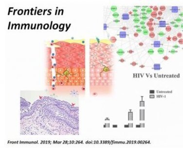
Investigators tested a laboratory-made version of a naturally occurring protein (recombinant fragment of human Surfactant Protein D or rfhSP-D) on bioengineered vaginal tissues, immune cells and microbes to determine if the drug candidate could help prevent HIV transmission safely.
|
|
Scooped by
Gilbert C FAURE
May 11, 2019 10:38 AM
|
This study examines the extent to which memory CD4+ T cells share immunosurveillance strategies with CD8+ resident memory T cells (TRM). After acute viral infection, memory CD4+ T cells predominantly used residence to survey nonlymphoid tissues, albeit not as stringently as observed for CD8+ T...
|


 Scoop.it!