Your new post is loading...

|
Suggested by
Société Francaise d'Immunologie
December 10, 2017 5:14 AM
|
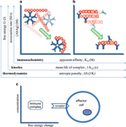
Spatiotemporal compartmentalization of signaling pathways and second messengers is pivotal for cell biology and membrane rafts are therefore required for several lymphocyte functions. On the other hand, T cells have the specific necessity of tuning signaling amplification depending on the context in which the antigen is presented. In this review, we discuss of membrane rafts in the context of T cell signaling, focusing on CD28-mediated costimulation.

|
Scooped by
Gilbert C FAURE
December 6, 2017 2:38 PM
|
There is an exponentially growing interest in targeting immune checkpoint molecules in breast cancer, particularly in the triple-negative subtype where unmet treatment needs remain. This study was designed to analyze the expression, localization and prognostic role of PD-1, PD-L1, PD-L2, CTLA-4, LAG3 and TIM3 in primary breast cancer. Gene expression analysis using the METABRIC microarray dataset found that all six immune checkpoint molecules are highly expressed in basal-like and HER2-enriched compared to the other breast cancer molecular subtypes. Flow cytometric analysis of fresh tissue homogenates from untreated primary tumors show that PD-1 is principally expressed on CD4+ or CD8+ T cells and CTLA-4 on CD4+ T cells. The global proportion of PD-L1+, PD-L2+, LAG3+ and TIM3+ tumor infiltrating lymphocytes was low and detectable in only a small number of tumors. Immunohistochemically staining fixed tissues from the same tumors was employed to score tumor infiltrating lymphocytes and tertiary lymphoid structures. PD-L1+, PD-L2+, LAG3+ and TIM3+ cells were detected in some tertiary lymphoid structures in a pattern that resembles secondary lymphoid organs. This observation suggests that tertiary lymphoid structures are important sites of immune activation and regulation, particularly in tumors with extensive baseline immune infiltration. Significantly improved overall survival was correlated with PD-1 expression in the HER2-enriched and PD-L1 or CTLA-4 expression in basal-like breast cancer. PD-1 and CTLA-4 proteins were most frequently detected on tumor infiltrating lymphocytes, which supports the correlations observed between their gene expression and improved long term outcome in basal-like and HER2-enriched breast cancer. PD-L1 expression by tumor or immune cells is uncommon in breast cancer. Overall, the data presented here distinguish PD-1 as a marker of T cell activity in both the T and B cell areas of breast cancer associated tertiary lymphoid structures. We found that immune checkpoint molecule expression parallels the extent of tumor infiltrating lymphocytes and tertiary lymphoid structures, although there is a noteworthy amount of heterogeneity between tumors even within the same molecular subtype. These data indicate that assessing the levels of immune checkpoint molecule expression in an individual patient has important implications for the success of therapeutically targeting them in breast cancer.
The unfolded protein response (UPR) is a conserved pathway that is stimulated when
endoplasmic reticulum (ER) proteostasis is disturbed or lost. Accumulating evidence
indicates that chronic activation of the UPR supports the main hallmarks of cancer
by favoring cancer cell-autonomous and nonautonomous processes, which ultimately foster
the immunosuppressive and protumorigenic microenvironment. However, certain forms
of therapy-induced ER stress can elicit immunogenic cancer cell death (ICD), which
enables the release of key immunostimulatory or danger signals, eventually driving
efficient antitumor immunity.
Via Krishan Maggon
|
|
Scooped by
Gilbert C FAURE
November 29, 2017 12:59 PM
|
The tumor microenvironment impairs the function of effector and memory T cells, but its effect on naïve T cells is not well understood. Xia et al. show that the autophagy component FAK family–interacting protein of 200 kDa (FIP200) is critical for promoting antitumor responses mediated by T cells. Ovarian cancer patients and mouse tumor models had significantly higher rates of apoptosis and impaired autophagy in naïve T cells, as well as the selective loss of FIP200, which disrupted the balance of pro- and antiapoptotic factors. Tumor-derived lactate was shown to suppress FIP200 expression, thus causing enhanced apoptosis and attenuated antitumor responses.
|
|
Scooped by
Gilbert C FAURE
November 28, 2017 2:32 PM
|
Credit: Charles Evavold, Boston Children's Hospital
Researchers at Boston Children's Hospital have identified, for the first time, the molecule that enables immune cells to release interl..
|
|
Scooped by
Gilbert C FAURE
November 25, 2017 12:51 AM
|
Complement is crucial to the immune response, but dysregulation of the system causes inflammatory disease. Complement is activated by three pathways: classical, lectin, and alternative. The classical and lectin pathways are initiated by the C1r/C1s (classical) and MASP-1/MASP-2 (lectin) proteases. Given the role of complement in disease, there is a requirement for inhibitors to control the initiating proteases. In this article, we show that a novel inhibitor, gigastasin, from the giant Amazon leech, potently inhibits C1s and MASP-2, whereas it is also a good inhibitor of MASP-1. Gigastasin is a poor inhibitor of C1r. The inhibitor blocks the active sites of C1s and MASP-2, as well as the anion-binding exosites of the enzymes via sulfotyrosine residues. Complement deposition assays revealed that gigastasin is an effective inhibitor of complement activation in vivo, especially for activation via the lectin pathway. These data suggest that the cumulative effects of inhibiting both MASP-2 and MASP-1 have a greater effect on the lectin pathway than the more potent inhibition of only C1s of the classical pathway.
The peptide-loading complex is key to the initiation of an immune response that raises killer T cells in vertebrates. Its structure has now been determined, and might provide information that improves immune protection.
Via Krishan Maggon
Our assumptions on the identity and functions of Langerhans cells (LCs) of the epidermis
have undergone considerable changes. Once thought to be prototypic representatives
of the dendritic cell (DC) lineage, they are now considered to be a specialized subset
of tissue-resident macrophages. Despite this, LCs display a remarkable mixture of
properties. Like many tissue macrophages, they self-maintain locally. However, unlike
tissue macrophages and similar to DCs, they homeostatically migrate to lymph nodes
and present antigen to antigen-specific T cells.
Via Krishan Maggon
|
|
Scooped by
Gilbert C FAURE
November 24, 2017 5:27 AM
|
|
|
Suggested by
Société Francaise d'Immunologie
November 21, 2017 1:16 PM
|
Innate defense regulator (IDR) peptide-1002 is a synthetic host defense peptide derivative with strong anti-inflammatory properties. Extending previous data, IDR-1002 suppressed in vitro inflammatory responses in RAW 264.7 murine monocyte/macrophage cells challenged with the TLR4 agonist LPS and TLR2 agonists lipoteichoic acid and zymosan. To investigate the anti-inflammatory mechanisms of IDR-1002 in vivo, the PMA-induced mouse ear inflammation model was used. Topical IDR-1002 treatment successfully dampened PMA-induced ear edema, proinflammatory cytokine production, reactive oxygen and nitrogen species release, and neutrophil recruitment in the ears of CD1 mice. Advanced RNA transcriptomic analysis on the mouse ear transcriptome revealed that IDR-1002 reduced sterile inflammation by suppressing the expression of transmembrane G protein–coupled receptors (class A/1 rhodopsin-like), including receptors for chemokines, PGs, histamine, platelet activating factor, and anaphylatoxin. IDR-1002 also dampened the IFN-γ response and repressed the IFN regulatory factor 8–regulated network that controls central inflammatory pathways. This study demonstrates that IDR-1002 exhibits strong in vitro and in vivo anti-inflammatory activities, informs the underlying anti-inflammatory mechanisms, and reveals its potential as a novel therapeutic for inflammatory diseases.
|
|
Scooped by
Gilbert C FAURE
November 21, 2017 1:14 PM
|
Immune deficiency disorders are a heterogeneous group of diseases of variable genetic aetiology. While the hallmark of immunodeficiency is susceptibility to infection, it is increasingly clear tha
|
|
Scooped by
Gilbert C FAURE
November 21, 2017 11:34 AM
|
|
|
Scooped by
Gilbert C FAURE
November 20, 2017 1:37 AM
|
T follicular helper cells (Tfh) are a specialized subset of CD4+ T cells that were first identified in the human tonsil. They play a critical role in protective immunity helping B cells produce antibody against foreign pathogens. Tfh are located in secondary lymphoid organs (SLOs), including the tonsil, spleen and lymph nodes. These organs contain numerous lymphocytes, separated into defined T and B cell zones.
|
|
|
Scooped by
Gilbert C FAURE
December 8, 2017 11:21 AM
|
|
|
Scooped by
Gilbert C FAURE
December 5, 2017 11:44 AM
|
The expansion of CD8+CD28– T cells, a population of terminally differentiated memory T cells, is one of the most consistent immunological changes in humans during aging. CD8+CD28– T cells are highly cytotoxic, and their frequency is linked to many age-related diseases. As they do not accumulate in mice, many of the molecular mechanisms regulating their fate and function remain unclear. In this paper, we find that human CD8+CD28– T cells, under resting conditions, have an enhanced capacity to use glycolysis, a function linked to decreased expression of the NAD+-dependent protein deacetylase SIRT1. Global gene expression profiling identified the transcription factor FoxO1 as a SIRT1 target involved in transcriptional reprogramming of CD8+CD28– T cells. FoxO1 is proteasomally degraded in SIRT1-deficient CD8+CD28– T cells, and inhibiting its activity in resting CD8+CD28+ T cells enhanced glycolytic capacity and granzyme B production as in CD8+CD28– T cells. These data identify the evolutionarily conserved SIRT1–FoxO1 axis as a regulator of resting CD8+ memory T cell metabolism and activity in humans.
|
|
Scooped by
Gilbert C FAURE
November 30, 2017 2:32 PM
|
|
|
Scooped by
Gilbert C FAURE
November 29, 2017 3:57 AM
|
Bats host a large number of zoonotic viruses, including several viruses that are highly pathogenic to other mammals. The mechanisms underlying this rich viral diversity are unknown, but they may be linked to unique immunological features that allow bats to act as asymptomatic viral reservoirs. Vertebrates respond to viral infection by inducing IFNs, which trigger antiviral defenses through IFN-stimulated gene (ISG) expression. Although the IFN system of several bats is characterized at the genomic level, less is known about bat IFN-mediated transcriptional responses. In this article, we show that IFN signaling in bat cells from the black flying fox ( Pteropus alecto ) consists of conserved and unique ISG expression profiles. In IFN-stimulated cells, bat ISGs comprise two unique temporal subclusters with similar early induction kinetics but distinct late-phase declines. In contrast, human ISGs lack this decline phase and remained elevated for longer periods. Notably, in unstimulated cells, bat ISGs were expressed more highly than their human counterparts. We also found that the antiviral effector 2-5A–dependent endoribonuclease, which is not an ISG in humans, is highly IFN inducible in black flying fox cells and contributes to cell-intrinsic control of viral infection. These studies reveal distinctive innate immune features that may underlie a unique virus–host relationship in bats.
In mammals, two types of major histocompatibility complex (MHC) molecules, MHC I and MHC II, are found on the surface of cells in association with peptides. If these peptides are antigenic—that is, if they induce an immune response—the resulting MHC I–peptide complexes are recognized by CD8+ T cells, which are effector immune cells, whereas MHC II-peptide complexes are recognized by CD4+ T cells, which facilitate both humoral and cellular immune responses. MHC I and MHC II clasp the peptides in a binding groove consisting of two antiparallel α helices that overlay an eight-stranded β sheet. On pages [1064][1] and [1060][2] of this issue, Jiang et al. ( 1 ) and Thomas and Tampé ( 2 ) reveal how peptide binding to MHC I molecules is regulated.
[1]: /lookup/doi/10.1126/science.aao5154
[2]: /lookup/doi/10.1126/science.aao6001
Via Krishan Maggon
Immunotherapy can reawaken T cells to destroy tumour cells. Modelling of tumour and T-cell interactions suggests why certain tumour cells are targeted and improves predictions of immunotherapy outcome.
Via Krishan Maggon
Since the 1990s it has been known that B and T lymphocytes exhibit low-level, constitutive
signaling in the basal state (tonic signaling). These lymphocytes display a range
of affinity for self, which in turn generates a range of tonic signaling. Surprisingly,
what signaling pathways are active in the basal state and the functional relevance
of the observed tonic signaling heterogeneity remain open questions today. Here we
summarize what is known about the mechanistic and functional details of tonic signaling.
Via Krishan Maggon
|
|
Scooped by
Gilbert C FAURE
November 24, 2017 6:47 AM
|
Clinical & Translational Immunology focuses on the general functioning of the immune system in its broadest sense, with a particular emphasis on its cell biology. Areas that are covered include but are not limited to: Cellular immunology, Innate and adaptive immunity, Immune responses to pathogens,Tumour immunology,Immunopathology, Immunotherapy, Immunogenetics, Immunological studies in humans and model organisms (including mouse, rat, Drosophila etc)
|
|
Scooped by
Gilbert C FAURE
November 23, 2017 11:34 AM
|
|
|
Suggested by
Société Francaise d'Immunologie
November 21, 2017 1:16 PM
|
Producing suppressive cytokines is a key molecular mechanism for Tregs to conduct
long-range suppressive functions. Wei et al. demonstrated that effector Tregs can
be diversified into two functionally distinct subsets based on IL-35 and IL-10 expression
that work in a complementary way to maintain immune tolerance.
|
|
Scooped by
Gilbert C FAURE
November 21, 2017 11:44 AM
|
A wealth of in vitro data has demonstrated a central role for receptor ubiquitination in endocytic sorting. However, how receptor ubiquitination functions in vivo is poorly understood. Herein, we report that ablation of B cell antigen receptor ubiquitination in vivo uncouples the receptor from CD19 phosphorylation and phosphatidylinositol 3-kinase (PI3K) signals. These signals are necessary and sufficient for accumulating phosphatidylinositol (3,4,5)-trisphosphate (PIP3) on B cell receptor–containing early endosomes and proper sorting into the MHC class II antigen-presenting compartment (MIIC). Surprisingly, MIIC targeting is dispensable for T cell–dependent immunity. Rather, it is critical for activating endosomal toll-like receptors and antiviral humoral immunity. These findings demonstrate a novel mechanism of receptor endosomal signaling required for specific peripheral immune responses.
T cells undergo positive and negative selection in the thymic cortex and medulla,
respectively. A promiscuous expression of a wide array of self-antigens in the thymus
is essential for the negative selection of self-reactive T cells and the establishment
of central tolerance. Aire was originally thought to be the exclusive factor regulating
the expression of tissue-restricted antigens, but Fezf2 recently emerged as a critical
transcription factor in this regulatory activity. Fezf2 is selectively expressed in
thymic medullary epithelial cells, regulates a large number of tissue-restricted antigens
and suppresses the onset of autoimmune responses.
Via Krishan Maggon
|