Your new post is loading...

|
Scooped by
Gilbert C FAURE
August 8, 2018 1:59 PM
|
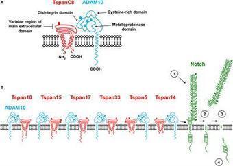
A disintegrin and metalloproteinase 10 (ADAM10) is a ubiquitously-expressed transmembrane protein that functions as a ‘molecular scissor’ to cleave the extracellular regions from its transmembrane target proteins. ADAM10 is well-characterized as the ligand-dependent activator of Notch proteins, which control cell fate decisions. Indeed, conditional knockouts of ADAM10 in mice reveal impaired B-, T- and myeloid cell development and/or function. ADAM10 cleaves many other leukocyte-expressed substrates. On B-cells, ADAM10 cleavage of the low affinity IgE receptor CD23 promotes allergy and asthma, cleavage of ICOS ligand impairs antibody responses, and cleavage of the BAFF-APRIL receptor TACI, and BAFF receptor BAFFR, reduce B-cell survival. On microglia, increased ADAM10 cleavage of a rare variant of the scavenger receptor TREM2 may increase susceptibility to Alzheimer’s disease. We and others recently showed that ADAM10 interacts with one of six different regulatory tetraspanin membrane proteins, which we termed the TspanC8 subgroup, comprising Tspan5, Tspan10, Tspan14, Tspan15, Tspan17 and Tspan33. The TspanC8s are required for ADAM10 exit from the endoplasmic reticulum, and emerging evidence suggests that they dictate ADAM10 subcellular localization and substrate specificity. Therefore, we propose that ADAM10 should not be regarded as a single scissor, but as six different scissors with distinct substrate specificities, depending on the associated TspanC8. I
Summary
Follicular T helper (Tfh) cells highly express the programmed cell death-1 (PD-1) molecule. Whereas inhibition of T cell receptor (TCR) signaling and CD28 co-stimulation is thought to be the primary mode of PD-1 functions, whether and how PD-1 regulates Tfh cell development and function is unclear. Here we showed that, when engaged by the ensemble of bystander B cells constitutively expressing PD-1 ligand 1 (PD-L1), PD-1 inhibited T cell recruitment into the follicle. This inhibition involved suppression of PI3K activities downstream of the follicle-guidance receptor CXCR5, was independent of co-signaling with the TCR, and necessitated ICOS signaling to overcome. PD-1 further restricted CXCR3 upregulation on Tfh cells, serving to concentrate these cells toward the germinal center territory, where PD-L1-PD-1 interactions between individual Tfh and B cells optimized B cell competition and affinity maturation. Therefore, operating in both costimulation-independent and -dependent manners, PD-1 controls tissue positioning and function of Tfh cells.
Via Krishan Maggon
|
|
Scooped by
Gilbert C FAURE
August 7, 2018 4:23 AM
|
J Leukoc Biol. 2018 Aug 1. doi: 10.1002/JLB.MR1117-457R.[Epub ahead of print] Review...

|
Suggested by
LIGHTING
August 4, 2018 11:46 AM
|
|
|
Scooped by
Gilbert C FAURE
August 3, 2018 2:33 PM
|
Directional cell motility toward a chemical gradient, chemotaxis, is critical during inflammation, embryogenesis, and cancer metastasis. In this issue, Kriebel et al. (2018. J. Cell Biol. <https://doi.org/10.1083/jcb.201710170>) demonstrate that the key cAMP chemoattractant for Dictyostelium discoideum amoebas is synthesized within and released from extracellular vesicles to promote chemotaxis.

|
Suggested by
Société Francaise d'Immunologie
August 2, 2018 2:24 AM
|
Macrophages carry out numerous physiological activities that are essential for both systemic and local homeostasis, as well as innate and adaptive immune responses. Their biology is intricately regulated by hormones, neuropeptides and neurotransmitters, establishing distinct neuroendocrine axes. The control is pleiotropic, including maturation of bone marrow-derived myeloid precursors, cell differentiation into functional subpopulations, cytotoxic activity, phagocytosis, production of inflammatory mediators, antigen presentation and activation of effector lymphocytes. Additionally, neuroendocrine components modulate macrophage ability to influence tumor growth and to prevent the spreading of infective agents. Interestingly, macrophage-derived factors enhance glucocorticoid production through the stimulation of the hypothalamic-pituitary-adrenal (HPA) axis. These bidirectional effects highlight a tightly controlled balance between neuroendocrine stimuli and macrophage function in the development of innate and adaptive immune responses. Herein we discuss how components of neuroendocrine axes impact on macrophage development and function, and may ultimately influence inflammation, tissue repair, infection or cancer progression. The knowledge of the crosstalk between macrophages and endocrine or brain-derived components may contribute to improve and create new approaches with clinical relevance in homeostatic or pathological conditions.
|
|
Scooped by
Gilbert C FAURE
July 31, 2018 3:56 AM
|
|
|
Scooped by
Gilbert C FAURE
July 28, 2018 11:04 AM
|
Zaire ebolavirus (EBOV), one of five species in the genus Ebolavirus , is the causative agent of the hemorrhagic fever disease epidemic that claimed more than 11,000 lives from 2014 to 2016 in West Africa. The combination of EBOV’s ability to disseminate broadly and rapidly within the host and its high pathogenicity pose unique challenges to the human immune system postinfection. Potential transmission from apparently healthy EBOV survivors reported in the recent epidemic raises questions about EBOV persistence and immune surveillance mechanisms. Clinical, virological, and immunological data collected since the West Africa epidemic have greatly enhanced our knowledge of host–virus interactions. However, critical knowledge gaps remain in our understanding of what is necessary for an effective host immune response for protection against, or for clearance of, EBOV infection. This review provides an overview of immune responses against EBOV and discusses those associated with the success or failure to control EBOV infection.
|
|
Scooped by
Gilbert C FAURE
July 24, 2018 1:43 AM
|
|
|
Scooped by
Gilbert C FAURE
July 22, 2018 3:20 AM
|
DC-SIGN is an antigen uptake receptor expressed on dendritic cells (DCs) with specificity for glycans present on a broad variety of pathogens and is capable of directing its cargo to MHC-I and MHC-II pathways for the induction of CD8+ and CD4+ T cell responses, respectively. Therefore, DC-SIGN is a very promising target for the delivery of antigen for anti-cancer vaccination. Although the endocytic route leading to MHC-II presentation is characterized to a large extent, the mechanisms controlling DC-SIGN targeted cross-presentation of exogenous peptides on MHC-I, is are not completely resolved yet. In this paper, we used imaging flow cytometry and antigen specific CD8+ T cells to investigate the intracellular fate of DC-SIGN and its cargo in human dendritic cells.Using different techniques w e investigated the intracellular fate of DC-SIGN and its cargo during cross-presentation in DCs. Our data demonstrates that both immature DCs and Toll-like receptor (TLR4) 4 stimulated DCs (TLR4)-stimulated DCs are had similar internalization capacity and were both able to cross-present antigen targeted via DC-SIGN. Interestingly, while the internalization capacity of DC-SIGN was similar in immature and TLR4-stimulated DCs, simultaneous triggering of TLR4 and DC-SIGN in DCs resulted in the translocation of cargo to the cytosol, leading to proteasome-dependent processing and increased CD8+ T cell activation. Understanding the dynamics of DC-SIGN-mediated uptake and processing i
|
|
Scooped by
Gilbert C FAURE
July 20, 2018 1:21 AM
|
Myocarditis is defined by WHO as an inflammatory disease affecting the myocardium, diagnosed by endomyocardial biopsy (EMB) using established histological, immunological and immunohistochemical criteria; it may be idiopathic, infectious or autoimmune[1-4]. A wide variety of histological myocarditis patterns have been described according to the diverse etiologies and to the stage of the disease at the time of EMB ascertainment. Viral infections represent the most common cause in Europe and North America[5]. Different viral genomes can be detected in the myocardium of patients with myocarditis and dilated cardiomyopathy(DCM) using molecular techniques[6–14]. When no infectious agents are identified on EMB and other known causes are excluded, autoimmune myocarditis is the presumed etiology[15]. These so called “autoimmune myocarditis”(AMy) or “virus-negative myocarditis”(VNMy) may occur as a distinct disease with exclusive cardiac involvement, or in the context of systemic autoimmune or inflammatory disorders with extra-cardiac involvement[5,16-23]. It is likely that several etiologic types of myocarditis confluence in a common immune-mediated pathogenic process leading to chronic inflammation and tissue damage. Irrespective of triggering agents, acute inflammation may progress to subacute and chronic stages and eventually result in tissue remodeling, fibrosis, contractile dysfunction, and finally DCM[4,6,8,24][25-29]. Besides contractile dysfunction, both early myocardia
|
|
Scooped by
Gilbert C FAURE
July 19, 2018 4:07 AM
|
Detecting signatures of past pathogen selection on human HLA loci: are there needles in the haystack? - Volume 145 Issue 6 - Bridget S. Penman, Sunetra Gupta
|
|
Suggested by
Société Francaise d'Immunologie
July 18, 2018 8:31 AM
|
A study published by Descours et al., in Nature last year, identified CD32a as marker of the CD4 T cell HIV reservoir. This study showed that CD32a+ CD4 T cells
|
|
|
Suggested by
LIGHTING
August 8, 2018 8:56 AM
|
Nuclear factor-κB (NF-κB) is an important transcription factor that induces the expression of antiviral genes and viral genes. NF-κB activation needs the activation of NF-κB upstream molecules, which include receptors, adaptor proteins, NF-κB (IκB) kinases (IKKs), IκBα, and NF-κB dimer p50/p65. To survive, viruses have evolved the capacity to utilize various strategies that inhibit NF-κB activity, including targeting receptors, adaptor proteins, IKKs, IκBα, and p50/p65. To inhibit NF-κB activation, viruses encode several specific NF-κB inhibitors, including NS3/4, 3C and 3C-like proteases, viral deubiquitinating enzymes (DUBs), phosphodegron-like (PDL) motifs, viral protein phosphatase (PPase)-binding proteins, and small hydrophobic (SH) proteins. Finally, we briefly describe the immune evasion mechanism of human immunodeficiency virus 1 (HIV-1) by inhibiting NF-κB activity in productive and latent infections. This paper reviews a viral mechanism of immune evasion that involves the suppression of NF-κB activation to provide new insights into and references for the control and prevention of viral diseases.
|
|
Scooped by
Gilbert C FAURE
August 7, 2018 1:18 PM
|
Natural killer (NK) cells can acquire ‘memory’ signatures. Sun and colleagues examine the dynamic ATAC-seq and functional RNA-seq changes observed upon generation of NK cell memory and show distinct roles for transcription factors STAT1 and STAT4.
|
|
Scooped by
Gilbert C FAURE
August 5, 2018 10:56 AM
|
Scott et al. demonstrate that ZEB2 is critical for maintaining the tissue identities
of macrophages. Loss of ZEB2 results in tissue-specific changes in different macrophage
populations and their subsequent disappearance. In Kupffer cells, ZEB2 maintains LXRα
expression, loss of which reproduces the change in Kupffer cell identity and their
disappearance.
|
|
Scooped by
Gilbert C FAURE
August 4, 2018 2:17 AM
|
The many functions of extracellular vesicles (EVs) like exosomes and microvesicles released from healthy cells have been well characterised, particularly in relation to their roles in immune modulation. Apoptotic bodies (ApoBDs), a major class of EV released as a product of apoptotic cell disassembly, and other types of EVs released from dying cells are also becoming recognised as key players in this emerging field. There is now increasing evidence to suggest that EVs produced during apoptosis have important immune regulatory roles, a concept relevant across different disease settings including autoimmunity, cancer and infection. Therefore, this review focuses on how the formation of EVs during apoptosis could be a key mechanism of immune modulation by dying cells.
|
|
Scooped by
Gilbert C FAURE
August 3, 2018 9:33 AM
|
A high-throughput screen for high-sensitivity detection of receptor-ligand interactions
is used to identify long-sought cell-surface receptors responsible for human cytomegalovirus
(HCMV) infection.
|
|
Scooped by
Gilbert C FAURE
July 31, 2018 6:21 AM
|
|
|
Scooped by
Gilbert C FAURE
July 29, 2018 2:42 AM
|
Natural Killer (NK) cells play a pivotal role during immunity against viruses and circumstantial evidence also indicate that they can protect the host against developing tumors. Peripheral blood NK cells comprise CD56brightCD16lo/- cells that constitutively express CD25 (IL-2Rα) and CD56dimCD16hi cells that express CD25 upon activation. Using NK cells from two patients, one with a primary immunodeficiency characterized by a homozygous mutation in CD25 (born in year 2007 and studied since she was 3 years old) and one with a homozygous mutation in STAT5b (born in year 1992 and studied since she was 10 years old), we observed that the absence of IL-2 signaling through CD25 promotes the accumulation of CD56brightCD16high NK cells, and that CD56brightCD16lo, CD56brightCD16high and CD56dimCD16high NK cells of this patient exhibited higher content of perforin and granzyme B, and proliferation capacity, compared to healthy donors. Also, CD56bright and CD56dim NK cells of this patient exhibited a reduced IFN- production in response to cytokine stimulation and increased degranulation against K562 cells. Also, the CD25-deficient patient presented a lower frequency of terminally differentiated NK cells in the CD56dimCD16hi NK cells compared to the HD (assessed by CD57 and CD94 expression). Remarkably, CD56dimCD16high NK cells from both patients exhibited notoriously higher expression of CD62L compared a HD, suggesting that in the absence of IL-2 signaling through CD25 and STAT5b, N
|
|
Scooped by
Gilbert C FAURE
July 26, 2018 9:14 AM
|
Under physiological conditions, the distribution/intensity of the surface markers CD56 and CD16 (FcγRIII) defines two subsets of NK lymphocytes: the CD56bright/CD16neg-dim (generally referred to as CD56bright) population that make up about 10% of blood NK cells in adults and the CD56dim/CD16bright (generally referred to as CD56dim) cells that comprise for about 90% of circulating NK cells. CD56bright NK cells exert only marginal cytotoxic capacity but are higly proficient in production of cytokines such as interferon IFN-γ, TNF-α and GM-CSF. Because of this property, CD56bright NK cells have been generally referred to as regulatory NK cells. Conversely, CD56dim NK cells were originally identified as the main subset with a key role in natural and antibody-dependent cellular cytotoxicity (ADCC), although subsequent data indicated that they can also produce relatively high amounts of pro-inflammatory cytokines following the engagement of activating NK receptors. The different functional outcomes of CD56bright and CD56dim NK cells are dependent on the differential expression of a large repertoire of activating and inhibitory receptors, and on the expression of a distinct subset of chemokine receptors that influence NK cell homing. Indeed, CD56dim NK cells preferentially migrate to inflamed peripheral tissues on the basis of their increased expression of CXCR1, CX3CR1 and ChemR23, whereas the CD56bright subset expresses CCR7 and homes mostly to secondary lymphoi
|
|
Scooped by
Gilbert C FAURE
July 24, 2018 1:42 AM
|
Cell death mechanisms have mainly been studied in vitro. Exploring their roles in
immune defense in vivo has been done largely by indirect means. Wallach and Kang provide
a perspective of the limitations of these means in studying the roles of apoptosis
in lymphocyte development and of necroptosis in inflammation.
|
|
Suggested by
Société Francaise d'Immunologie
July 21, 2018 4:06 AM
|
Telomerase activity is required for both initiation and maintenance of tumorigenesis and over 90% cancers overexpress telomerase. Therefore, telomerase targeting has emerged as a potential strategy for cancer treatment.
|
|
Scooped by
Gilbert C FAURE
July 19, 2018 2:41 PM
|
Regulatory T cells (Tregs) are crucial for suppressing autoimmunity and inflammation mediated by conventional T cells. To be useful, some Tregs should have overlapping specificity with relevant self-reactive or pathogen-specific clones. Whether matching recognition between Tregs and non-Tregs might arise through stochastic or deterministic mechanisms has not been addressed. We tested the hypothesis that some Tregs that arise in the thymus or that are induced during Ag-driven expansion of conventional CD4+ T cells might be clonally related to non-Tregs by virtue of asymmetric Foxp3 induction during cell division. We isolated mouse CD4+ thymocytes dividing in vivo, wherein sibling cells exhibited discordant expression of Foxp3 and CD25. Under in vitro conditions that stimulate induced Tregs from conventional mouse CD4+ T cells, we found a requirement for cell cycle progression to achieve Foxp3 induction. Moreover, a substantial fraction of sibling cell pairs arising from induced Treg stimulation also contained discordant expression of Foxp3. Division-linked yet asymmetric induction of Treg fate offers potential mechanisms to anticipate peripheral self-reactivity during thymic selection as well as produce precise, de novo counterregulation during CD4+ T cell–mediated immune responses.
|
|
Scooped by
Gilbert C FAURE
July 18, 2018 11:41 AM
|
Lymphocytes interpret antigenic signals into a functional response. Richard et al. demonstrate that the overall qualitative response by CTLs is independent of TCR ligand strength; instead, ligands determine the rate and uniformity at which CTLs respond.
|