Your new post is loading...

|
Scooped by
Dr. Stefan Gruenwald
May 24, 2016 4:53 PM
|
Whether you have a huge honker, a puny proboscis, or a snubbed schnoz, the shape of your nose is in your genes. Now, researchers have sniffed out five of those stretches of DNA that control nose and chin shape. The team sequenced the genomes of more than 6000 men and women in Central and South America and used photographs of the participants to categorize 14 of their facial features—from cheekbone protrusion to lip shape. Then, the scientists analyzed whether any of the features were associated with certain genes. GLI3 and PAX1, both known to be involved in cartilage growth, were linked to the breadth of a person’s nostrils;DCHS2, also related to cartilage, controlled nose pointiness; RUNX2, which drives bone development, was associated with the width of the nose bridge, the upper area of the nose; and EDAR, which has previously been linked to ear and tooth shape and hair texture, affected chin protrusion. The results, published online today in Nature Communications, may help shed light on how the human face evolved and why different ethnicities have distinct facial features. Moreover, the research could help forensic scientists reconstruct faces based on genetic samples.
A new study by an international team of researchers, affiliated with Ulsan National Institute of Science and Technology (UNIST) has announced that they have succeeded for the first time in observing the structural change. The breakthrough comes from a research, conducted by Professor Chae Un Kim (School of Natural Science) of UNIST in collaboration with researchers from Soongsil University, Cornell University, and University of Florida. Carbonic anhydrase, which is found within red blood cells, is a crucial enzyme that stabilizes carbon dioxide (CO2 ) concentrations. This enzyme catalyzes a reaction converting CO2 and water into carbonic acid, which associates into protons and bicarbonate ions. Moreover, it is also known that carbonic anhydraseis is able to catalyze at a rate of 106 reactions per second. In the absence of this catalyst, the conversion from CO2 to bicarbonate, and vice versa, would be extremely slow and difficult. One of the important functions of the enzyme in humans is to adjust the acidity of the chemical environment to prevent damage to the body, as well as to help transport carbon dioxide out from tissue cells to the lungs. Although carbonic anhydrase performs a lot of beneficial functions, defects in the enzyme are responsible for developing diseases, such as glaucoma, acidemia, or osteopetrosis. Prof. Kim, the lead researcher of the study states, "The reaction rate of carbonic anhydrase is one of the fastest of all enzymes." He continues, "Due to the rapid movement of proteins, direct observation for such movement has been extremely difficult to obtain, protein scientists say." In this study, Prof. Kim's team used their own method of "High-pressure Crycooling" and "X-ray Crystallography" to capture the gaseous carbon dioxide in crystals of carbonic anhydrase and follow the sequential structure changes as the carbon dioxide is released. The results of the study will not only greatly contribute to the future biomedical research and new drug development, but will also help make carbon capture more economic. According to Prof. Kim of UNIST, "This study also shows technical methods that may be applicable to other enzymes that bind and react to low-molecular weight substrates, such as CO2 and NO2 ."
Via Mariaschnee
|
|
Scooped by
Dr. Stefan Gruenwald
May 24, 2016 4:35 PM
|
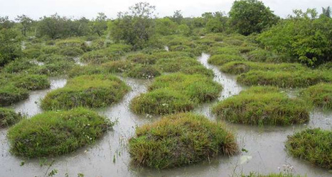
During the rainy season in the Orinoco Llanos of Columbia and Venezuela, an odd landscape feature appears in places: mounds of grassy plants, as big as five meters across and two meters tall, surrounded by water. Traversing this landscape, called surales, requires either hopping from mound to mound or trudging through the boggy bits in between. Locals and scientists have generally agreed that some kind of earthworm creates the mounds, but what species and how it does so has been a mystery. Now Anne Zangerlé of the Braunschweig University of Technology in Germany and colleagues report that they’ve found the culprit — giant Andiorrhinus earthworms, which can grow to a meter in length as juveniles. And the mounds themselves, the team reports May 11 in PLOS ONE, are actually made mostly of earthworm poop. Zangerlé and her colleagues used Google Earth images to locate surales landscapes, finding that they come in the shape of both mounds and labyrinths. Leaving the complex labyrinths for a future study, the team studied the mounds and the lands on which they were found in both the rainy and dry seasons. They characterized the soil and the plants and worms living in and on the mounds. And then they pieced all of that information together to come up with a scenario that they think explains the construction of the mounds.
|
|
Rescooped by
Dr. Stefan Gruenwald
from DNA and RNA research
May 24, 2016 4:30 PM
|
After nearly a half trillion tries, a rare event was seen that might solve an evolutionary puzzle about noncoding sequences of DNA in genomes and address speciation and the cause of diseases like cancer. For a long time, scientists have known that much of the DNA within any given organism’s genome does not code for functional molecules or protein. However, recent research has found that these genetic sequences, misnamed “junk” DNA in the past, often do have functional significance. These introns are no exception. Now known to play a role in gene expression, introns are the portion of gene sequences that are removed or spliced out of RNA before genes are translated into protein. When eukaryotes first diverged from bacteria, there was a massive invasion of introns into the genome. All living eukaryotes — from yeast to mammals — share this common ancestor, and whereas simple organisms such as yeast have eliminated most of their introns, organisms such as mammals have considerably expanded their intron inventory. Humans have more than 200,000 introns that take up about 40 percent of the genome. In a current paper, Stevens and co-author Sujin Lee, a former graduate student in cellular and molecular biology at UT Austin, used a new reporter assay to directly detect the loss and gain of introns in budding yeast (Saccharomyces cerevisiae). The team tested nearly a half trillion yeast and found only two instances in which an intron was added to a new gene. The proposed mechanism for this addition is a reversal of a splicing reaction. Normally, to make proteins, RNA is read from the instructions in DNA, and the introns are spliced out. But in these two instances, the cell allowed the spliced out introns to make it back into a different RNA and was recombined back into the genome, thus creating a permanent genetic change. These are called intron gains, and if these accumulate over time, they can contribute to the development of new species as well as human disease. “We showed in this project that introns continue to be gained, although infrequently at any point in time,” says Stevens. “But can introns drive evolution? If these sequences give organisms a selective advantage and become fixed in a population, others have shown that it can be a major factor in the creation of new species.” These evolutionary advances come at a cost, however, because diseases such as cancer correlate with the improper removal of introns from RNA. Stevens adds, “We are continuing this work to further understand how this process impacts our genetic history, our future, and the prospects of curing disease.”
Via Integrated DNA Technologies
Our bodies are constantly and successfully fighting off the development of cells that lead to tumors - but when there is disruption to this process cancer is free to develop. Walter and Eliza Hall Institute researchers, led by Dr Sandra Nicholson and Dr Nicholas Huntington, together with colleagues from the Queensland Institute of Medical Research, are investigating ways to 'switch on' our Natural Killer (NK) cells. Natural Killer cells exist to detect and then destroy any deviant cells in our bodies before those cells go on to develop into tumors or before infection spreads, Dr Nicholson said. "Natural Killer cells are a key part of our immune system that locate other cells posing a danger to health either because they are infected or because they are becoming a cancer cell," she said. However, it is known that abnormal cells sometimes escape the immune system and develop into a cancer. The researchers identified a protein 'brake' within Natural Killer cells that controls their ability to destroy their target tumor cells. In their paper published today in Nature Immunology, they showed that when the brake was removed in an experimental model, the NK cells were better able to protect the body against metastatic melanoma. Natural Killer cells rely on a growth factor called Interleukin 15 (IL15) to activate. Dr Nicholson and Dr Huntington's research has shown that an inhibitor protein made inside the Natural Killer cells limits the ability of the NK cell to respond to IL15 and therefore kill cancer cells. By identifying for the first time how this protein inhibits NK cell responses, they now hope that a drug can be developed that will improve the NK cells' response to this growth factor and help patients fight cancer with their own immune system. "This is about learning how to activate the NK cells of the individual patient and boost their immune system to tackle the disease," Dr Huntington said.
Via Mariaschnee
|
|
Scooped by
Dr. Stefan Gruenwald
May 24, 2016 2:58 PM
|
Ed Hawkins, a climate scientist at the National Center for Atmospheric Science at the University of Reading, devised the spiral animation to show how global average surface temperatures are increasing relative to the average temperature during preindustrial times.
|
|
Scooped by
Dr. Stefan Gruenwald
May 24, 2016 1:05 PM
|
When a sperm and an egg cell merge a new life begins. This is the case in humans and in animals, but in principle also in plants. A German-Israeli team led by the biologists Professor Ralf Reski from Freiburg and Professor Nir Ohad from Tel-Aviv has discovered a gene trigger in the moss Physcomitrella patens which leads to offspring without fertilization. The researchers assume that this mechanism is conserved in evolution and holds the key to answer fundamental questions in biology. The study is published in the journal “Nature Plants”.
„Just like humans and animals, mosses possess egg cells and motile sperm. That is why they are particularly well-suited to answer fundamental questions in biology”, Reski says. After fusion of sperm and egg cell, a network of genes is activated. That leads to the development of an embryo which grows into a new living being. Until now it was unclear whether a central genetic switch for this gene activation exists. In their latest publication the team describes the gene BELL1 as a master regulator for the formation of embryos and their development in Physcomitrella. After the researchers activated this gene in the plants by genetic engineering, embryos developed spontaneously on a specific cell type. These embryos grew to fully functional moss sporophytes. These spore capsules could even form spores, which grew into new moss plants. Thus, the team identified BELL1 as a master regulator for embryo development in mosses.
The protein encoded by this gene belongs to the class of so-called homeobox transcription factors. Similar homeotic genes are also present in humans and animals, where they also control pivotal developmental processes. Whether a congener of BELL1 is a master regulator of embryo development in humans is not yet known. “Our results are important beyond mosses”, Reski says. “On the one hand they can explain how algae developed into land plants and thus shaped our current ecosystems. Secondly, they may help to revive the concept of genetic master regulators in the development of plants, animals and humans.” Ohad explains, “Moreover, our results may help to modernize agriculture through the creation of genetically identical offspring from high-yielding crop plants. In seed plants such offsprings are formed by parthenogenesis or apomixis.”
Ralf Reski from the University of Freiburg is a specialist in moss research and has helped to develop Physcomitrella as a model organism for biology and biotechnology at a world-wide scale. Nir Ohad from Tel-Aviv University is a specialist in the epigenetic regulation of reproductive development. He helped to identify the first BELL genes in seed plants about 20 years ago as member of a team led by Professor Robert Fischer from UC Berkeley. Research was supported by the German-Israeli Foundation GIF, the Freiburg Excellence Cluster BIOSS and the Freiburg Institute for Advanced Studies.
|
|
Scooped by
Dr. Stefan Gruenwald
May 24, 2016 12:58 PM
|
Clutter is a special challenge for robots, but new Carnegie Mellon University software is helping robots cope, whether they're beating a path across the Moon or grabbing a milk jug from the back of the refrigerator. The software not only helped a robot deal efficiently with clutter, it surprisingly revealed the robot's creativity in solving problems. "It was exploiting sort of superhuman capabilities," Siddhartha Srinivasa, associate professor of robotics, said of his lab's two-armed mobile robot, the Home Exploring Robot Butler, or HERB. "The robot's wrist has a 270-degree range, which led to behaviors we didn't expect. Sometimes, we're blinded by our own anthropomorphism." In one case, the robot used the crook of its arm to cradle an object to be moved. "We never taught it that," Srinivasa added. The rearrangement planner software was developed in Srinivasa's lab by Jennifer King, a Ph.D. student in robotics, and Marco Cognetti, a Ph.D. student at Sapienza University of Rome who spent six months in Srinivasa's lab. They will present their findings May 19 at the IEEE International Conference on Robotics and Automation in Stockholm, Sweden. In addition to HERB, the software was tested on NASA's KRex robot, which is being designed to traverse the lunar surface. While HERB focused on clutter typical of a home, KRex used the software to find traversable paths across an obstacle-filled landscape while pushing an object. Robots are adept at "pick-and-place" (P&P) processes, picking up an object in a specified place and putting it down at another specified place. Srinivasa said this has great applications in places where clutter isn't a problem, such as factory production lines. But that's not what robots encounter when they land on distant planets or, when "helpmate" robots eventually land in people's homes. P&P simply doesn't scale up in a world full of clutter. When a person reaches for a milk carton in a refrigerator, he doesn't necessarily move every other item out of the way. Rather, a person might move an item or two, while shoving others out of the way as the carton is pulled out. The rearrangement planner automatically finds a balance between the two strategies, Srinivasa said, based on the robot's progress on its task. The robot is programmed to understand the basic physics of its world, so it has some idea of what can be pushed, lifted or stepped on. And it can be taught to pay attention to items that might be valuable or delicate, in case it must extricate a bull from a china shop.
|
|
Scooped by
Dr. Stefan Gruenwald
May 24, 2016 12:54 PM
|
An international team of scientists, including two professors and three graduate students from UCLA, has detected and confirmed the faintest early-universe galaxy ever. Using the W. M. Keck Observatory on the summit on Mauna Kea in Hawaii, the researchers detected the galaxy as it was 13 billion years ago. The results were published in the Astrophysical Journal Letters. Tommaso Treu, a professor of physics and astronomy in the UCLA College and a co-author of the research, said the discovery could be a step toward unraveling one of the biggest mysteries in astronomy: how a period known as the "cosmic dark ages" ended. The researchers made the discovery using an effect called gravitational lensing to see the incredibly faint object, which was born just after the Big Bang. Gravitational lensing was first predicted by Albert Einstein almost a century ago; the effect is similar to that of an image behind a glass lens appearing distorted because of how the lens bends light. The detected galaxy was behind a galaxy cluster known as MACS2129.4-0741, which is massive enough to create three different images of the galaxy. According to the Big Bang theory, the universe cooled as it expanded. As that happened, Treu said, protons captured electrons to form hydrogen atoms, which in turn made the universe opaque to radiation -- giving rise to the cosmic dark ages. "At some point, a few hundred million years later, the first stars formed and they started to produce ultraviolet light capable of ionizing hydrogen," Treu said. "Eventually, when there were enough stars, they might have been able to ionize all of the intergalactic hydrogen and create the universe as we see it now." That process, called cosmic reionization, happened about 13 billion years ago, but scientists have so far been unable to determine whether there were enough stars to do it or whether more exotic sources, like gas falling onto supermassive black holes, might have been responsible. "Currently, the most likely suspect is stars within faint galaxies that are too faint to see with our telescopes without gravitational lensing magnification," Treu said. "This study exploits gravitational lensing to demonstrate that such galaxies exist, and is thus an important step toward solving this mystery." The research team was led by Marusa Bradac, a professor at UC Davis. Co-authors include Matthew Malkan, a UCLA professor of physics and astronomy, and UCLA graduate students Charlotte Mason, Takahiro Morishita and Xin Wang. The galaxy's magnified spectra were detected independently by both Keck Observatory and Hubble Space Telescope data.
|
|
Scooped by
Dr. Stefan Gruenwald
May 20, 2016 2:00 PM
|
What used to be dismissed by many as "junk DNA" is back with a vengeance as growing data points to the importance of non-coding RNAs (ncRNAs)—genome's messages that do not code for proteins—in development and disease formation. But our progress in understanding these molecules has been slow because of the lack of technologies that allow the systematic mapping of their functions. Now, Professor Benjamin Blencowe's team at the University of Toronto's Donnelly Centre, including lead authors Eesha Sharma and Tim Sterne-Weiler, have developed a method, described in May 19, 2016 issue of Molecular Cell, that enables scientists to explore in depth what ncRNAs do in human cells. The study is published on the same day with two other papers in Molecular Cell and Cell, respectively, from Dr. Yue Wan's group at the Genome Institute of Singapore and Dr. Howard Chang's group at Stanford University in California, who developed similar methods to study RNAs in different organisms. Of the 3 billion letters in the human genome, only two per cent make up the protein-coding genes. The genes are copied, or transcribed, into messenger RNA (mRNA) molecules, which provide templates for building proteins that do most of the work in the cell. Much of the remaining 98 per cent of the genome was initially considered by some as lacking in functional importance. However, large swaths of the non coding genome—between half and three quarters of it—are also copied into RNA. What the resulting ncRNAs might do depends on whom you ask. Some researchers believe that most ncRNAs have no function, that they are just a by-product of the genome's powerful transcription machinery that makes mRNA. However, it is emerging that many ncRNAs have important roles in gene regulation. This view is supported in that some ncRNAs act as carriages for shuttling the mRNAs around the cell, or provide a scaffold for other proteins and RNAs to attach to and do their jobs. But the majority of available data has trickled in piecemeal or through serendipitous discovery. And with emerging evidence that ncRNAs could drive disease progression, such as cancer metastasis, there was a great need for a technology that would allow a systematic functional analysis of ncRNAs. "Up until now, with existing methods, you had to know what you are looking for because they all require you to have some information about the RNA of interest. The power of our method is that you don't need to preselect your candidates, you can see what's occurring globally in cells, and use that information to look at interesting things we have not seen before and how they are affecting biology," says Eesha Sharma, a PhD candidate in Blencowe's group who, along with postdoctoral fellow Tim Sterne-Weiler, co-developed the method. The new tool, called 'LIGR-Seq', captures interactions between different RNA molecules. When two RNA molecules have matching sequences - strings of letters copied from the DNA blueprint - they will stick together like Velcro. The paired RNA structures are then removed from cells and analyzed by state-of-the-art sequencing methods to precisely identify the RNAs that are stuck together. "Most researchers in the life sciences agree that there's an urgent need to understand what ncRNAs do. This technology will open the door to developing a new understanding of ncRNA function," says Blencowe, who is also a professor in the Department of Molecular Genetics. Not having to rely on pre-existing knowledge is one strength of the method that will boost the discovery of RNA pairs that have never been seen before. The other is that scientists can for the first time look at RNA interactions as they occur in living cells, in all their complexity, unlike in the juices of mashed up cells that they had to rely on before. This is a bit like moving on to explore marine biology from collecting shells on the beach to scuba-diving among the coral reefs where the scope for discovery is so much bigger. ncRNAs come in multiple flavours: there's rRNA, tRNA, snRNA, snoRNA, piRNA, miRNA, and lncRNA, to name a few, where prefixes reflect the RNA's place in the cell or some aspect of its function. But the truth is that no one really knows the extent to which these ncRNAs control what goes on in the cell, nor how they do this. The new technology developed by Blencowe's group has been able to pick up new interactions involving all classes of RNAs and has already revealed some unexpected findings.
Tiny, rolling balls of brain cells knocking around in a lab may one day help keep you from losing your marbles—among other things. The small cellular balls act like mini-brains, mimicking aspects of the real thing, including forming noggin-like structures and pulsing with electrical signals like a thinking mind, researchers reported Friday at the annual meeting of the American Association for the Advancement of Science in Washington. The mini-brains, which can be personalized based on whose cells they’re made from, may soon help scientists study a wide variety of diseases and health problems—from autism and Parkinson’s to multiple sclerosis and Alzheimer’s, as well as stroke, brain trauma, and infections, such as Zika virus. “There are a variety of places where a mini brain could be useful,” said Wayne Drevets of Janssen Pharmaceuticals Inc., who was not involved with the research. In some cases, they may offer a cheaper, more ethical, and more realistic model for human health than mice and other animals, he and other researchers said at the conference. Researchers who developed the wee noodles, led by Thomas Hartung, of Johns Hopkins University Bloomberg School of Public Health, hope to have the mini-brains commercially available this year.
Via Krishan Maggon
The tau protein is a subunit of one of the major hallmarks of Alzheimer disease (AD), the neurofibrillary tangles, and is therefore of major interest as an indicator of disease mechanisms. Many of the unusual properties of Tau can be explained by its nature as a natively unfolded protein. Examples are the large number of structural conformations and biochemical modifications (phosphorylation, proteolysis, glycosylation, and others), the multitude of interaction partners (mainly microtubules, but also other cytoskeletal proteins, kinases, and phosphatases, motor proteins, chaperones, and membrane proteins). The pathological aggregation of Tau is counterintuitive, given its high solubility, but can be rationalized by short hydrophobic motifs forming β structures. The aggregation of Tau is toxic in cell and animal models, but can be reversed by suppressing expression or by aggregation inhibitors. This review summarizes some of the structural, biochemical, and cell biological properties of Tau and Tau fibers. Further aspects of Tau as a diagnostic marker and therapeutic target, its involvement in other Tau-based diseases, and its histopathology are covered by other chapters in this volume.
Via Krishan Maggon
Electronic materials have been a major stumbling block for the advance of flexible electronics because existing materials do not function well after breaking and healing. A new electronic material created by an international team, however, can heal all its functions automatically even after breaking multiple times. This material could improve the durability of wearable electronics. "Wearable and bendable electronics are subject to mechanical deformation over time, which could destroy or break them," said Qing Wang, professor of materials science and engineering, Penn State. "We wanted to find an electronic material that would repair itself to restore all of its functionality, and do so after multiple breaks." Self-healable materials are those that, after withstanding physical deformation such as being cut in half, naturally repair themselves with little to no external influence. In the past, researchers have been able to create self-healable materials that can restore one function after breaking, but restoring a suite of functions is critical for creating effective wearable electronics. For example, if a dielectric material retains its electrical resistivity after self-healing but not its thermal conductivity, that could put electronics at risk of overheating. The material that Wang and his team created restores all properties needed for use as a dielectric in wearable electronics -- mechanical strength, breakdown strength to protect against surges, electrical resistivity, thermal conductivity and dielectric, or insulating, properties. They published their findings online in Advanced Functional Materials. Most self-healable materials are soft or "gum-like," said Wang, but the material he and his colleagues created is very tough in comparison. His team added boron nitride nanosheets to a base material of plastic polymer. Like graphene, boron nitride nanosheets are two dimensional, but instead of conducting electricity like graphene they resist and insulate against it.
Via Mariaschnee
|
Earth’s continuously changing magnetic field is thought to be largely generated by superheated, swirling liquid iron in Earth’s outer core. Other sources of earthly magnetism include minerals in our world’s mantle and crust. Earth’s ionosphere, magnetosphere and oceans also play a role. The European Space Agency (ESA) now has two years of data from a trio of satellites in Earth-orbit, designed to measure magnetism from these various sources. The mission is called Swarm. At last week’s Living Planet Symposium held in Prague in the Czech Republic (May 9-13, 2016), scientists presented new results from the Swarm satellite trio and provided some recent insights about how Earth’s magnetic field is changing at this time. Among other things, they said that the field has weakened by about 3.5% at high latitudes over North America, while it has grown about 2% stronger over Asia. The region where the field is at its weakest field – the South Atlantic Anomaly – has moved steadily westward and further weakened by about 2%. Meanwhile, the magnetic north pole has been wandering east, towards Asia. The animation shown in this article is based partly on results from ESA’s Swarm mission, and partly on information from the CHAMP and Ørsted satellites. It shows how the strength of Earth’s magnetic field changed between 1999 and mid-2016. Blue depicts where the field is weak and red shows regions where the field is strong. As you can see, the changes in field strength are relatively small.
Via Mariaschnee
|
|
Scooped by
Dr. Stefan Gruenwald
May 24, 2016 4:38 PM
|
The CRISPR/Cas9 system is currently the technique in genome editing. Thanks to recent developments in the CRISPR genome editing system, we are able to alter DNA with unprecedented precision and accuracy. Ultimately, this revolutionary genome editing technique allows us to modify any region of the genome of any species—without harming other genes. But more than that, we are able to edit these genes at just a fraction of the cost of previous methods. It has utterly revolutionized gene editing. However, a team of researchers has just developed a new approach that just might prove to be as efficient and effective as the current standard. In a study published in Nature Biotechnology, researchers from Chunyu Han’s lab have developed a novel genome editing technique basically just like CRISPR. According to the report, the method is based on the Natronobacterium gregoryi Argonaute (NgAgo), a DNA-guided endonuclease that’s similar to Cas9, the endonuclease of CRISPR. These are a specific type of protein, otherwise known as restriction enzymes, that are responsible for cutting DNA at specific locations. Remarkably, the study shows that NgAgo is suitable for genome editing in human cells. The team asserts that their study has a number of key differences between NgAgo and Cas9, where the former is at the advantage. They claim that that the method appears to have a low tolerance to guide-target mismatches, leading to a high efficiency in editing: - One of these is that NgAgo does not need to be followed by a protospacer-adjacent motif (PAM), a DNA sequence seen in Cas9. Notably, Cas9 will not successfully bind or cleave a target DNA sequence if this is not followed by the PAM (as it is responsible for the protein to differentiate CRISPR DNA from target DNA).
- Another benefit is that the loading temperature of NgAgo is found to be at 55°C and not at Cas9’s 37°C. This shows that this could be another option for genome editing at conditions such as this. Also, at this temperature, the method follows a “one-guide-faithful” rule, that is, NgAgo cannot swap target DNA with other free DNA, minimizing off-target effects.
Though there has yet to be an extensive side-by-side comparison of the two enzymes, NgAgo and Cas9 appear to have similar efficiencies. The authors report that their tests with 47 guides targeting 8 human genes, the results showed a 21% to 41% efficiency.
Old-fashioned breeding techniques are bearing more fruit than genetic engineering in developing hyper-efficient plants. Big corporations such as DuPont Pioneer in Johnston, Iowa, have spent more than a decadedeveloping improved crops through genetic engineering, and some companies say that their transgenic varieties look promising in field trials. But there are still no fertilizer-frugal transgenic crops on the market, and several agricultural organizations around the globe are reviewing their biotechnology initiatives in this area. Plant biologist Allen Good of the University of Alberta in Edmonton, Canada, spent years working with companies to develop genetically modified (GM) crops that require little fertilizer, but he says that this approach has not been as fruitful as conventional techniques. The problem is that there are so many genes involved in nutrient uptake and use — and environmental variations alter how they are expressed. “Nutrient efficiency was supposed to be one of those traits with broad applicability that could make companies lots of money. But they haven't developed the way we thought,” says Good. Despite the scientific and breeding challenges, some researchers say that all strategies must be explored to develop crops that are less nutrient needy. With the global population heading towards 10 billion people by 2050, frugal crops could be essential to feed the planet. “There is a huge worldwide potential for these traits to help increase food production and sustainable development,” says Matin Qaim, an agricultural economist at the University of Göttingen in Germany.
Via Meristemi
Is dark energy the reason time moves forward? For years, physicists have attempted to explain dark energy - a mysterious influence that pushes space apart faster than gravity can pull the things in it together. But physics isn’t always about figuring out what things are. A lot of it is figuring out what things cause. In a recent paper, a group of physicists asked this very question about dark energy, and found that in some cases, it might cause time to go forward.
When you throw a ball into the air, it starts with some initial speed-up, but then it slows as Earth’s gravity pulls it down. If you throw it fast enough (about 11 km per second, for those who want to try), it’ll never slow down enough to turn around and start falling back towards you, but it’ll still move more slowly as it moves away from you, because of Earth’s gravity.
Physicists and astronomers in the 1990s expected something similar to have occurred after the big bang - an event that threw matter out in all directions. The collective gravity from all that matter should have slowed it all down, just like the Earth slows down the ball. But that’s not what they found.
Instead, everything seems to have sped up. There’s something pervading the Universe that physically spreads space apart faster than gravity can pull things together. The effect is small - it’s only noticeable when you look at far-away galaxies - but it’s there. It’s become known as dark energy - "dark", because no one knows what it is.
Science is nothing if not the process of humans looking for things they can’t explain, so this isn’t the first time the Universe has stumped us. For centuries, one of those stumpers has been time itself: Why does time have an arrow pointing from the past to the present to the future?
It might seem like a silly question - I mean, if time didn’t go forward, then effects would precede causes, and that seems like it should be impossible - but it’s less of one than you might think.
The Universe, as far as we can tell, only operates according to laws of physics. And just about all of the laws of physics that we know are completely time-reversible, meaning that the things they cause look exactly the same whether time runs forward or backward.
One example is the path of a planet going around a star, which is governed by gravity. Whether time runs forward or backward, planetary orbits follow the exact same paths. The only difference is the direction of the orbit.
But one important piece of physics isn’t time-reversible, and that’s the second law of thermodynamics. It states that as time moves forward, the amount of disorder in the Universe will always increase. Just like dark energy, it’s something we’ve noticed about the Universe, and it’s something that we still don’t totally understand - though admittedly we have a better idea of it than we do of dark energy.
Physicists have, for this reason, reluctantly settled on the second law as the source of time’s arrow: disorder always has to increase after something happens, which requires that time can only move in one direction.
So physicists A. E. Allahverdyan from the Yerevan Physics Institute and V. G. Gurzadyan from Yerevan State University, both in Armenia, decided to see if - at least in a limited situation - dark energy and the second law might be related. To test it, they looked at the simple case of something like a planet orbiting a star with a changing mass.
They found that if dark energy either doesn’t exist or if it pulls space together, the planet just dully orbits the star without anything interesting happening. There’s no way to tell an orbit going forward in time from one going backward in time.
But if dark energy pushes space apart, like it does in our Universe, the planet eventually gets thrown away from the star on a path of no return. This gives us a distinction between the past and the future: run time one way, and the planet is flung off, run it the other way, and the planet comes in and gets captured by the star.
Dark energy naturally leads to an arrow of time. The authors stress that this is a really limited situation, and they’re certainly not claiming dark energy is the reason time only ever moves forward. But they’ve shown a possible link between thermodynamics and dark energy that could help us to understand either - or maybe both - better than we ever have.
The research has been published in Physical Review E.
Via Kim Frye, Tania Gammage, ThePlanetaryArchives/BlackHorseMedia - San Francisco
|
|
Scooped by
Dr. Stefan Gruenwald
May 24, 2016 3:05 PM
|
A team of scientists from the Moscow Institute of Physics and Technology (MIPT), the National Research University of Electronic Technology (MIET), and the Prokhorov General Physics Institute have proposed a theoretical model that explains the unexpectedly high values of the linear magnetoelectric effect in BiFeO3 (bismuth ferrite) that have been observed in a number of experiments. The team also suggested a way of further enhancing the effect. The results of the study have been published in the journal Physical Review B. One particular feature of bismuth ferrite is that in bulk samples, spins of Fe3+ iron ions are arranged in the form of a cycloid. This spin structure can be destroyed by a strong magnetic field or mechanical stress. Without a spin cycloid, bismuth ferrite exhibits a large linear magnetoelectric effect, and this effect was the focal point of the study. "The theoretical description presented in the paper may be applicable to other multiferroics similar to BiFeO3. This will help in predicting the value of their magnetoelectric effect, which, in turn, will make it easier to find new and promising materials for industrial applications," says the head of MIPT's Laboratory of physics of magnetic heterostructures and spintronics for energy-saving information technologies, Prof. Anatoly Zvezdin. Multiferroics are materials that simultaneously exhibit different ferroic orders, including magnetic, ferroelectric and/or ferroelastic. If there is an interaction between electric and magnetic subsystems in a material, a magnetoelectric (ME) effect may occur. The magnetoelectric effect is when electric polarization occurs under the influence of an external magnetic field and magnetization occurs under the influence of an electric field. This allows an electric field to be used to control the magnetic properties of a material and a magnetic field to be used to control the electric properties. If the value of the ME effect is high (dozens or hundreds of times higher than normal), it is called a giant ME effect. The main use of the magnetoelectric effect is in variable and static magnetic field sensors. These sensors are used in navigation systems, electric motors, and also in vehicle ignition systems. Compared to similar devices based on the Hall effect or magnetoresistance, sensors based on the ME effect are more sensitive (according to research, up to one million times more sensitive) and they are also relatively cheap to manufacture. The ME effect offers exciting possibilities for the use of multiferroics in new types of magnetic memory, e.g. ROM -- read only memory. The ME effect could also potentially be used to create high-precision equipment for working with radiation in the microwave range, and to wirelessly transmit power to miniaturized electronic devices. The subject of the study was bismuth ferrite (BiFeO3) -- a highly promising multiferroic that is very promising in terms of its practical applications. It is planned to be used to create ultra energy-efficient magnetoelectric memory. In addition, bismuth ferrite exhibits a magnetoelectric effect at room temperature, while in most other magnetoelectrics an ME effect of this magnitude is only observed at extremely low temperatures (below -160 degrees Celsius). Bismuth ferrite is an antiferromagnetic, which means that the magnetic moments of its magnetic sublattices (structures formed by atoms with the same parallel spins) cancel each other out, and the total magnetization of the material is close to zero. However, the spatial arrangement of the spins forms the same cycloidal spin structure.
|
|
Scooped by
Dr. Stefan Gruenwald
May 24, 2016 1:57 PM
|
New system extends the lives of flying microrobots. Call them the RoboBats. In a recent article in Science, Harvard roboticists demonstrate that their flying microrobots, nicknamed the RoboBees, can now perch during flight to save energy -- like bats, birds or butterflies. "Many applications for small drones require them to stay in the air for extended periods," said Moritz Graule, first author of the paper who conducted this research as a student at the Harvard John A. Paulson School of Engineering and Applied Sciences (SEAS) and Wyss Institute for Biologically Inspired Engineering at Harvard University. "Unfortunately, smaller drones run out of energy quickly. We want to keep them aloft longer without requiring too much additional energy." The team found inspiration in nature and simple science. "A lot of different animals use perching to conserve energy," said Kevin Ma, a post-doc at SEAS and the Wyss Institute and coauthor. "But the methods they use to perch, like sticky adhesives or latching with talons, are inappropriate for a paperclip-size microrobot, as they either require intricate systems with moving parts or high forces for detachment." Instead, the team turned to electrostatic adhesion -- the same basic science that causes a static-charged sock to cling to a pants leg or a balloon to stick to a wall. When you rub a balloon on a wool sweater, the balloon becomes negatively charged. If the charged balloon is brought close to a wall, that negative charge forces some of the wall's electrons away, leaving the surface positively charged. The attraction between opposite charges then causes the balloon to stick to the wall. "In the case of the balloon, however, the charges dissipate over time, and the balloon will eventually fall down," said Graule. "In our system, a small amount of energy is constantly supplied to maintain the attraction." The RoboBee, pioneered at the Harvard Microrobotics Lab, uses an electrode patch and a foam mount that absorbs shock. The entire mechanism weighs 13.4 mg, bringing the total weight of the robot to about 100mg -- similar to the weight of a real bee. The robot takes off and flies normally. When the electrode patch is supplied with a charge, it can stick to almost any surface, from glass to wood to a leaf. To detach, the power supply is simply switched off. "One of the biggest advantages of this system is that it doesn't cause destabilizing forces during disengagement, which is crucial for a robot as small and delicate as ours," said Graule.
|
|
Scooped by
Dr. Stefan Gruenwald
May 24, 2016 1:03 PM
|
Icy objects in our solar system have large oceans under their surfaces and here life could evolve and flourish. So says a new thesis by Jesper Lindkvist, PhD student at the Swedish Institute of Space Physics and Umeå University. The thesis will be defended on Tuesday 31 May at the Swedish Institute of Space Physics in Kiruna, Sweden. There has long been speculation as to whether Jupiter's large, icy moon Callisto has an ocean beneath its surface. Observations of Callisto's near-space environment by instruments on board the spacecraft Galileo, which is orbiting Jupiter, lead us to believe that there is such a sub-surface ocean. Computer simulations of the space plasma interactions support this belief. "If you find an ocean beneath the surface of one moon, perhaps the same is true of other icy objects in space," says Jesper Lindkvist. Outflow of water vapor has been detected from the surface of the dwarf planet Ceres, which is in the asteroid belt between Mars and Jupiter. This could indicate a reservoir of water, also related to a large sea beneath the surface. The spacecraft Dawn is at Ceres right now, trying to answer this question. "If someone was planning to build a future space base on one of these Solar System objects, for example to seek after signs of life, I would suggest they take an extra long ice bore and their fishing equipment," jokes Jesper Lindkvist. The outflow of water vapor from moons and dwarf planets is similar to that we see from comets. The icy comet 67/Churyumov-Gerasimenko which rounded the sun in the summer of 2015 has been followed closely by the European spacecraft Rosetta. Measurements of the space environment round it show that the water flowing out from the comet's nucleus forms a prominent atmosphere which interacts with the constant flow of ionized particles from the sun, the so-called solar wind. "Understanding their origin and how icy bodies evolve is one more piece of the puzzle we need to lay in order to explain the origin of our Solar System and its eventual fate," says Jesper Lindkvist. Jesper Lindkvist comes from the town of Vidsel in Northern Sweden. He has a Masters degree in engineering physics from Luleå University of Technology.
|
|
Scooped by
Dr. Stefan Gruenwald
May 24, 2016 12:56 PM
|
A team of scientists working at the U.S. Department of Energy's (DOE) Argonne National Laboratory and led by Northern Illinois University physicist and Argonne materials scientist Zhili Xiao has created a new material, called "rewritable magnetic charge ice," that permits an unprecedented degree of control over local magnetic fields and could pave the way for new computing technologies. The scientists' research report on development of magnetic charge ice is published in the May 20, 2016 issue of the journal Science. With potential applications involving data storage, memory and logic devices, magnetic charge ice could someday lead to smaller and more powerful computers or even play a role in quantum computing, Xiao said. Current magnetic storage and recording devices, such as computer hard disks, contain nanomagnets with two polarities, each of which is used to represent either 0 or 1 -- the binary digits, or bits, used in computers. A magnetic charge ice system could have eight possible configurations instead of two, resulting in denser storage capabilities or added functionality unavailable in current technologies. "Our work is the first success achieving an artificial ice of magnetic charges with controllable energy states," said Xiao, who holds a joint appointment between Argonne and NIU. "Our realization of tunable artificial magnetic charge ices is similar to the synthesis of a dreamed material. It provides versatile platforms to advance our knowledge about artificial spin ices, to discover new physics phenomena and to achieve desired functionalities for applications." Over the past decade, scientists have been highly interested in creating, investigating and attempting to manipulate the unusual properties of "artificial spin ices," so-called because the spins have a lattice structure that follows the proton positioning ordering found in water ice. Scientists consider artificial spin ices to be scientific playgrounds, where the mysteries of magnetism might be explored and revealed. However, in the past, researchers have been frustrated in their attempts to achieve global and local control of spin-ice magnetic charges. To overcome this challenge, Xiao and his colleagues decoupled the lattice structure of magnetic spins and the magnetic charges. The scientists used a bi-axis vector magnet to precisely and conveniently tune the magnetic charge ice to any of eight possible charge configurations. They then used a magnetic force microscope to demonstrate the material's local write-read-erase multi-functionality at room temperature. For example, using a specially developed patterning technique, they wrote the word, "ICE," on the material in a physical space 10 times smaller than the diameter of a human hair. Magnetic charge ice is two-dimensional, meaning it consists of a very thin layer of atoms, and could be applied to other thin materials, such as graphene. Xiao said the material also is environmentally friendly and relatively inexpensive to produce.
|
|
Scooped by
Dr. Stefan Gruenwald
May 24, 2016 12:51 PM
|
Although the technique presently works on just one virus, scientists say it could be adapted to detect a range of viruses that plague humans including Ebola, Zika and HIV. "The ultimate goal is to build a cheap, easy-to-use device to take into the field and measure the presence of a virus like Ebola in people on the spot," says Jeffrey Dick, a chemistry graduate student and co-lead author of the study. "While we are still pretty far from this, this work is a leap in the right direction." The new method is highly specific, meaning it is only sensitive to one type of virus, filtering out possible false negatives caused by other viruses or contaminants. There are two other commonly used methods for detecting viruses in biological samples, but they have drawbacks. One requires a much higher concentration of viruses, and the other requires samples to be purified to remove contaminants. The new method, however, can be used with urine straight from a person or animal. The researchers demonstrated their new technique on a virus that belongs to the herpesvirus family, called murine cytomegalovirus (MCMV). To detect individual viruses, the team places an electrode — a wire that conducts electricity, in this case, one that is thinner than a human cell — in a sample of mouse urine. They then add to the urine some special molecules made up of enzymes and antibodies that naturally stick to the virus of interest. When all three stick together and then bump into the electrode, there's a spike in electric current that can be easily detected. The researchers say their new method still needs refinement. For example, the electrodes become less sensitive over time because a host of other naturally occurring compounds stick to them, leaving less surface area for viruses to interact with them. To be practical, the process will also need to be engineered into a compact and rugged device that can operate in a range of real-world environments.
|
|
Scooped by
Dr. Stefan Gruenwald
May 20, 2016 1:58 PM
|
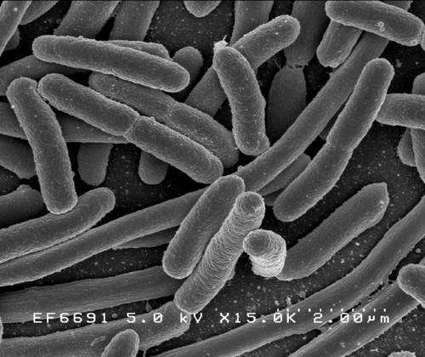
A key biochemical enables bacteria to repair otherwise fatal damage to their DNA, including that caused by antibiotics. That is the finding of a study. Adjusting the action of a molecule called ppGpp (guanosine-3',5'-(bis)pyrophosphate), with future treatments may disable DNA repair in microbes to make them many times more vulnerable to existing antibiotics, say the study authors. Bacteria repeatedly exposed to the same drugs become resistant to treatment, according to the Centers for Disease Control and Prevention, with related infections linked to 23,000 deaths and 2 million illnesses each year in the United States. "Most antibiotics have their effect, directly or indirectly, by causing damage to bacterial DNA, so finding ways to cripple DNA repair would represent a significant advance in the treatment of resistant infections," says senior study author Evgeny Nudler, PhD, the Julie Wilson Anderson Professor of Biochemistry, Department of Biochemistry and Molecular Pharmacology, NYU Langone. "While reducing DNA repair in bacteria could help to overcome antibiotic resistance, we're also excited about the prospect of boosting DNA repair in human cells," says Nudler, also an investigator with the Howard Hughes Medical Institute. "DNA damage accumulates with age and creates risk for degenerative diseases from Alzheimer's to cancer." The study results revolve around the delicacy of DNA molecules, the letters making up the genetic code. Experts estimate that DNA is damaged thousands of times an hour in each bacterial cell, and perhaps a million times a day in a human cell with larger, more complicated DNA chains. Sunlight and toxins do much of the damage, but the biggest culprit may be highly reactive byproducts created as cells use oxygen to turn sugar into energy. Given that damaged DNA can result in lethal mistakes in the building of proteins that comprise vital structures and messages, cells evolved early on to have overlapping, split-second DNA repair mechanisms. In both humans and the bacteria, a key protein complex called RNA polymerase clamps onto and ticks down the DNA chain, reading the code of DNA "letters" as it translates genetic instructions into intermediary RNA molecules on the way to building proteins. Studies in recent years have revealed that the RNA polymerase in bacteria also inspects the DNA chain for damage as it reads. In 1997, Nudler and colleagues published a paper in Cell that found bacterial RNA polymerase, which moves down the DNA chain in one direction during normal reading, instead stops and slips backward in some instances - a process Nudler called backtracking. If RNA polymerase encounters a lesion in DNA, the theory went, backtracking could make room for repair enzymes to fly in, cut out the damaged section, and rebuild a normal chain in a process called nucleotide excision DNA repair (NER). Indeed, in 2014, Nudler's team published work in Nature that found the NER enzyme UvrD causes RNA polymerase to backtrack in the bacterial species E. coli. The newly published paper identifies ppGpp (guanosine-3',5'-(bis)pyrophosphate), a compound related in structure to the guanine building block of DNA, as the central controller of UvrD-driven backtracking in the NER pathway. Levels of ppGpp rise rapidly as bacterial RNA polymerase encounters damage and backtracks, then drop as soon as the chain is repaired to return RNA polymerase to normal transcription. The study authors conclude that ppGpp is the sensor that enables RNA polymerase to shift back and forth between DNA transcription and repair, coupling the two processes in bacteria. Bacteria must be able to repair DNA and preserve their genomic integrity to survive, so targeting this ability is sound strategy for drug development, says Nudler. In seeking to translate this work into new treatments that defeat antibiotic resistance, he says, the field needs to determine whether or not RNA polymerase directly communicates with enzymes that produce ppGpp, and if they do, to design specific inhibitors against them. Researchers also hope to soon confirm that RNA polymerase backtracking enables related forms of DNA repair in human cells as theorized, an important step toward boosting human DNA repair in the future.
The recombinant poliovirus therapy developed at the Preston Robert Tisch Brain Tumor Center at Duke Health has been granted “breakthrough therapy designation” from the U.S. Food and Drug Administration. Duke’s poliovirus therapy is an immunotherapy developed in the laboratory of Matthias Gromeier, M.D., a professor in the departments of Neurosurgery, Molecular Genetics and Microbiology, and Medicine at Duke University School of Medicine. Using a modified form of poliovirus that has been altered to eliminate harm, the therapy preferentially attacks cancer cells, which have an abundance of receptors that work like magnets to attract the poliovirus. The modified poliovirus then kills the infected tumor cells while also igniting an additional immune response. A phase I clinical trial using the therapy was launched in 2012 to determine an optimal dose of the novel treatment among adult patients with glioblastoma whose cancer had returned after receiving traditional therapy. Early testing found that lower doses of the treatment were superior to higher doses. Of 23 glioblastoma patients enrolled at the optimal dose level, 15 are still alive and enrollment is ongoing. Three patients treated early using different dosages are still alive more than 36 months after treatment. With current standard therapy, the median survival time for people with glioblastoma is 14.6 months. The Duke team is moving to expand its work and open a clinical trial for children with brain tumors, which is expected to begin enrollment before year’s end. The researchers have also received federal grants to explore the therapy’s effect on solid tumors. Laboratory studies are already underway in breast cancer models.
Via Krishan Maggon
|
|
Scooped by
Dr. Stefan Gruenwald
May 18, 2016 2:57 PM
|
Scientists spot mutations that could explain how giraffes became the world’s tallest living mammals. Call it a tall task: researchers have decoded the genomes of the giraffe and its closest relative, the okapi. The sequences, published on May 17 in Nature Communications, reveal clues to the age-old mystery of how the giraffe evolved its unusually long neck and legs. Researchers in the United States and Tanzania analyzed the genetic material of two Masai giraffes (Giraffa camelopardalis tippelskirchi) from the Masai Mara National Reserve in Kenya, one at the Nashville Zoo in Tennessee and an okapi fetus (Okapia johnstoni) from the White Oak Conservation Center in Yulee, Florida. “This is one more wonderful demonstration of the power of comparative genomics to connect the evolution of animal species on this planet to molecular events that we know must underpin the extraordinary diversity of life on this planet,” says David Haussler, director of the Genomics Institute at the University of California, Santa Cruz.
|