Your new post is loading...

|
Scooped by
Dr. Stefan Gruenwald
October 17, 2016 5:52 PM
|
A Mars lander is due to leave its mothership and head toward the Red Planet's surface to test technologies for Europe's first planned Mars rover, which will search for signs of past and present life. After a seven-month journey from Earth as part of the European-Russian ExoMars program, the Schiaparelli lander is expected to separate from spacecraft Trace Gas Orbiter (TGO) on October 20 and start a three-day descent to the surface. Schiaparelli represents only the second European attempt to land a craft on Mars, after a failed mission by the British landing craft Beagle 2 in 2003. Landing on Mars, Earth's neighbor some 56 million kilometers away, is a notoriously difficult task that has bedevilled most Russian efforts and given NASA trouble as well. The United States currently has two operational rovers on Mars, Curiosity and Opportunity. But a seemingly hostile environment has not detracted from the allure of Mars, with US President Barack Obama recently highlighting his pledge to send people to the planet by the 2030s. Elon Musk's SpaceX is developing a massive rocket and capsule to transport large numbers of people and cargo to Mars with the ultimate goal of colonising the planet, with Mr Musk saying he would like to launch the first crew as early as 2024.
|
|
Scooped by
Dr. Stefan Gruenwald
October 17, 2016 5:41 PM
|
The motorcycle of the future is so smart that it could eliminate the need for protective gear, according to automaker BMW. To mark its 100th birthday, BMW has unveiled a number of concept vehicles that imagine the future of transportation. Possibly its most daring revelation, the so-called Motorrad Vision Next 100 concept motorcycle is so advanced that BMW claims riders wouldn't need a helmet. The Motorrad Vision Next 100 would have a self-balancing system that keeps the bike upright both in motion and when still. BMW touted the motorbike's futuristic features, saying it would allow for riders of all skill levels to "enjoy the sensation of absolute freedom." According to the automaker, the Motorrad wouldn't require protective gear such as helmets and padded suits. Another traditional feature was also missing from the concept: a control panel. Instead, helmetless riders would wear a visor that acts as a smart display. "Information is exchanged between rider and bike largely via the smart visor," BMW said in a statement. "This spans the rider's entire field of view and provides not only wind protection but also relevant information, which it projects straight into the line of sight as and when it is needed." Such information would not be needed all the time because drivers will be able to hand over active control of the vehicle at points; the Motorrad and other Vision Next 100 vehicles would be equipped with self-driving technology, according to BMW.
|
|
Scooped by
Dr. Stefan Gruenwald
October 17, 2016 5:33 PM
|
The brains of people with autism contain unusual amounts of short regulatory RNAs, according to the largest-yet study of this kind1. Many of these so-called microRNAs control genes linked to autism. The findings suggest that tipping the balance among some of the brain’s hundreds of microRNAs may trigger gene expression changes associated with autism. “There are very widespread changes [in the brain] at multiple levels,” says lead investigator Daniel Geschwind, distinguished professor of neurology, psychiatry and human genetics at the University of California, Los Angeles. The new study represents a comprehensive approach to understanding the molecular changes in the brains of people with autism, Geschwind says. The study appeared 29 August 2016 in Nature Neuroscience. MicroRNAs dampen gene expression by destabilizing messenger RNA, the template for a protein created from a gene. Each microRNA is roughly 20 nucleotides in length, but it can tamp down hundreds of genes. Geschwind’s team sequenced small RNAs in samples of postmortem brain tissue from 55 people with autism and 42 controls. Previous studies of microRNAs in autism have used fewer samples or focused on a subset of microRNAs, says Peng Jin, professor of human genetics at Emory University in Atlanta, who was not involved in the study. The data Geschwind’s team collected likely also include information about several other types of small regulatory RNAs that warrant exploration, Jin says. The new work “really provides a very good resource for people studying autism.” Geschwind and his team focused on small RNAs in samples of the cerebral cortex and cerebellum. They identified 699 microRNAs in the samples, 147 of which had not been seen before. They then homed in on 58 microRNAs whose expression is altered in the cerebral cortex of people with autism. Of these, 17 are scarce in autism brains and 41 are abundant. The researchers identified 11 groups, or ‘modules,’ of microRNA that have similar expression patterns across the brains. The microRNAs in two of these modules are relatively abundant in people with autism, and those in two other modules are unusually scarce. Using an algorithm, the researchers found a large proportion of microRNAs expected to control autism genes in three of the four autism-linked modules.
|
|
Scooped by
Dr. Stefan Gruenwald
October 17, 2016 5:24 PM
|
By forcefully embedding two silicon atoms in a diamond matrix, Sandia researchers have demonstrated for the first time on a single chip all the components needed to create a quantum bridge to link quantum computers together. People have already built small quantum computers," says Sandia researcher Ryan Camacho. "Maybe the first useful one won't be a single giant quantum computer but a connected cluster of small ones." Distributing quantum information on a bridge, or network, could also enable novel forms of quantum sensing, since quantum correlations allow all the atoms in the network to behave as though they were one single atom. The joint work with Harvard University used a focused ion beam implanter at Sandia's Ion Beam Laboratory designed for blasting single ions into precise locations on a diamond substrate. Sandia researchers Ed Bielejec, Jose Pacheco and Daniel Perry used implantation to replace one carbon atom of the diamond with the larger silicon atom, which causes the two carbon atoms on either side of the silicon atom to feel crowded enough to flee. That leaves the silicon atom a kind of large landowner, buffered against stray electrical currents by the neighboring non-conducting vacancies. Though the silicon atoms are embedded in a solid, they behave as though floating in a gas, and therefore their electrons' response to quantum stimuli are not clouded by unwanted interactions with other matter. "What we've done is implant the silicon atoms exactly where we want them," said Camacho. "We can create thousands of implanted locations, which all yield working quantum devices, because we plant the atoms well below the surface of the substrate and anneal them in place. Before this, researchers had to search for emitter atoms among about 1,000 randomly occurring defects -- that is, non-carbon atoms -- in a diamond substrate of a few microns to find even one that emitted strongly enough to be useful at the single photon level."
Researchers at the University of Waterloo's Institute for Quantum Computing (IQC) recorded an interaction between light and matter 10 times larger than previously seen. The strength of the interaction between photons and a qubit was so large that it opens the door to a realm of physics and applications unattainable until now.
The results appear in the paper, "Ultrastrong coupling of a single artificial atom to an electromagnetic continuum in the nonperturbative regime," published in Nature Physics.
"We are enabling the investigation of light-matter interactions in a new domain in quantum optics," said Pol Forn-Diaz, a postdoctoral fellow at IQC and lead author of the paper. "The possibilities are exciting because our circuit could potentially act as a quantum simulator to study other interesting quantum systems in nature."
The ultrastrong coupling between photons and qubits may lead to the exploration of new physics related to biological processes, exotic materials such as high-temperature superconductors, and even relativistic physics.
Via Mariaschnee
|
|
Scooped by
Dr. Stefan Gruenwald
October 14, 2016 7:37 PM
|
In a study using Real-time intraoperative magnetic resonance imaging (RT-IMRI) to guide the transplantation of induced pluripotent stem cell (iPSC)-derived neurons into the brains of non-human primates modeled with Parkinson's disease, researchers found that RT-IMRI guidance not only allows for better visualization and monitoring of the procedure, but also helps cell survival. Induced pluripotent stem cells, a type of stem cell that can be generated directly from adult cells, offer great benefits for regenerative medicine as they propagate indefinitely and can differentiate into a variety of cell types, such as neurons, heart, pancreatic, and liver cells. In previous studies, the researchers have found that while iPSC-derived neurons provide great opportunities for cell replacement they also present challenges. "Our team developed an MRI-compatible trajectory guidance system that has been successful for intraoperative MRI," said study lead author Dr. Marina E. Emborg, Preclinical Parkinson's Research Program Center, Wisconsin National Primate Research Center University of Wisconsin-Madison. "We recently upgraded the system for real-time targeting and guidance and, as a result of the improvements, the procedure provides several advances for cell delivery."
|
|
Scooped by
Dr. Stefan Gruenwald
October 14, 2016 7:32 PM
|
By recoding bacterial genomes, it is possible to create organisms that can potentially synthesize products not commonly found in nature. By systematic replacement of seven codons with synonymous alternatives for all protein-coding genes, Ostrov et al. recoded the Escherichia coli genome. The number of codons in the E. coli genetic code was reduced from 64 to 57 by removing instances of the UAG stop codon and excising two arginine codons, two leucine codons, and two serine codons. Over 90% functionality was successfully retained. In 10 cases, reconstructed bacteria were not viable, but these few failures offered interesting insights into genome-design challenges and what is needed for a viable genome (Science Article). Recoding—the repurposing of genetic codons—is a powerful strategy for enhancing genomes with functions not commonly found in nature. Scientists now report computational design, synthesis, and progress toward assembly of a 3.97-megabase, 57-codon Escherichia coli genome in which all 62,214 instances of seven codons were replaced with synonymous alternatives across all protein-coding genes. They were able to validate 63% of recoded genes by individually testing 55 segments of 50 kilobases each. The researchers observed that 91% of tested essential genes retained functionality with limited fitness effect. They were able to demonstrate identification and correction of lethal design exceptions, only 13 of which were found in 2,229 genes. This work underscores the feasibility of rewriting genomes and establishes a framework for large-scale design, assembly, troubleshooting, and phenotypic analysis of synthetic organisms.
|
|
Scooped by
Dr. Stefan Gruenwald
October 14, 2016 7:19 PM
|
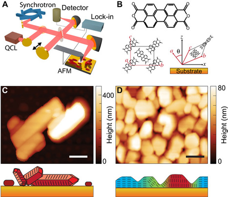
Molecular solids and polymers can form low-symmetry crystal structures that exhibit anisotropic electron and ion mobility in engineered devices or biological systems. The distribution of molecular orientation and disorder then controls the macroscopic material response, yet it is difficult to image with conventional techniques on the nanoscale. Scientists now have demonstrated a new form of optical nanocrystallography that combines scattering-type scanning near-field optical microscopy with both optical antenna and tip-selective infrared vibrational spectroscopy. From the symmetry-selective probing of molecular bond orientation with nanometer spatial resolution, they determined crystalline phases and orientation in aggregates and films of the organic electronic material perylenetetracarboxylic dianhydride. Mapping disorder within and between individual nanoscale domains, the correlative hybrid imaging of nanoscale heterogeneity provides insight into defect formation and propagation during growth in functional molecular solids.
Tokyo Medical and Dental University (TMDU) researchers developed a technique for attaching two distinct layers of cells on top of each other on an amnion-based scaffold. When osteoblasts and mesenchymal stem cells were used to form the layers, the cell constructs more effectively promoted bone regeneration after implantation onto skull defects in mice, compared with their single-cell-layer equivalents. This new approach has a range of potential applications for tissue engineering in the field of regenerative medicine.
Via Carlos Garcia Pando
|
|
Rescooped by
Dr. Stefan Gruenwald
from Vincill-Science
October 14, 2016 7:07 PM
|
Fifteen years ago, scientists celebrated the first draft of the sequenced human genome. At the time, they predicted that humans had between 25,000 and 40,000 genes that code for proteins. That estimate has continued to fall. Humans actually seem to have as few as 19,000 such genes1— a mere 1–2% of the genome. The key to our complexity lies in how these genes are regulated by the remaining 99% of our DNA, known as the genome's 'dark matter'. From efforts such as the massive Encyclopedia of DNA Elements (ENCODE) project2, launched in 2003 by the US National Human Genome Research Institute, it's clear that copious regulatory elements are at play, tuning gene expression in ways that scientists are only starting to unravel. By uncovering regulatory instructions in the genome beyond protein-coding genes, scientists are hoping to yield new ways to understand and treat disease. “It's not overstating to say that ENCODE is as significant for our understanding of the human genome as the original DNA sequencing of the human genome,” says cell biologist Bing Ren of the University of California, San Diego, Institute for Genomic Medicine in La Jolla, who is a member of the ENCODE team. The human genome is not packed with 'junk' as previously thought, but with regulatory regions that modulate gene activity. Ren is also part of a subsequent consortium called the Roadmap Epigenomics Project 3. These two initiatives — both funded by the US National Institutes of Health (NIH) — aim to map and predict the existence of elements in the genome, including in the vast stretches of non-coding portions, that drive when and where genes are expressed. Scientists have generated a list of such elements by using biochemical assays to probe DNA sequences, RNA transcripts, regulatory proteins bound to DNA and RNA and epigenetic signatures — the chemical tags on DNA and the proteins packaging it — that also affect gene expression. So far, the data suggest that there are hundreds of thousands of functional regions in the human genome whose task is to control gene expression: it turns out that much more space in the human genome is devoted to regulating genes than to the genes themselves. Scientists are now trying to validate each predicted element experimentally to ascertain its function — a mammoth task, but one for which they now have a powerful new tool. Since the gene-editing technique CRISPR–Cas9 entered the scientific arena, the speed at which researchers can test functional elements in the non-coding regions has ramped up. But it is still a daunting endeavour: more than 3 million regulatory DNA regions, thought to contain some 15 million binding sites for regulatory proteins called transcription factors, control gene expression in the human cell types studied thus far. About 150,000 may be active in any given cell type. These could be crucial to understanding disease, because most single-nucleotide changes associated with common diseases fall in regions outside protein-coding genes, and they often overlap with DNA sites highlighted by ENCODE as having regulatory function. Certain regulatory elements that normally drive gene expression are thought to underpin the mechanism of cancer, for example. Disrupting a gene's regulatory elements, the data suggest, could thus have as drastic an impact on cell function as disrupting the gene itself. Using CRISPR–Cas9, scientists now have an opportunity to test that premise by introducing targeted mutations into non-coding sequences and observing the consequences.
Via Gerd Moe-Behrens, Eric Vincill
Harnessing solid-state quantum bits, or qubits, is a key step toward the mass production of electronic devices based on quantum information science and technology. However, realizing a robust qubit with a long lifetime is challenging, particularly in semiconductors comprising multiple types of atoms.
The close collaboration between experiments in Prof. David Awschalom's group and theory and simulations in Prof. Giulia Galli's group, both in the Institute for Molecular Engineering, has enabled a crucial step toward solid-state qubits in industrially important semiconductors. In a paper, published Sept. 29 in Nature Communications, the two groups showed that electron qubits bound to atom-like defects in a commercial silicon carbide wafer can exhibit the longest electronic coherence times ever measured in a natural crystal.
"Quantum coherence underlies all quantum information technologies, such as quantum communication and quantum sensing. However, the coherence time in materials is eventually limited by the magnetic noise produced by the fluctuating nuclear spins in a crystal," said Hosung Seo, an IME postdoctoral researcher and the paper's lead author.
Defects in silicon carbide have recently attracted attention as potential candidates for solid-state qubits. Due to its extensive use in the optoelectronics and power electronics industries, silicon carbide also has a strong potential for mass production.
However, spin qubits in silicon carbide have been expected to have inherently short coherence times because of the high concentration of magnetic nuclei in the crystals. Counterintuitively, the electron coherence time in silicon carbide reaches 1.3 milliseconds—the longest time measured in a naturally isotopic crystal.
Via Mariaschnee
|
|
Scooped by
Dr. Stefan Gruenwald
October 14, 2016 5:27 PM
|
Drop-seq is a method to analyze mRNA expression in thousands of individual cells by encapsulating cells in tiny droplets for parallel analysis. Droplets—nanoliter-scale aqueous compartments formed by precisely combining aqueous and oil flows in a microfluidic device (Thorsen et al., 2001 and Umbanhowar et al., 2000)—have been used as tiny reaction chambers for PCR (Hindson et al., 2011 and Vogelstein and Kinzler, 1999) and reverse transcription (Beer et al., 2008). Scientists now used these droplets to compartmentalize cells into nanoliter-sized reaction chambers for analysis of all of their RNAs. A basic challenge of using droplets for transcriptomics is to retain a molecular memory of the identity of the cell from which each mRNA transcript was isolated. To accomplish this, the scientists developed a molecular barcoding strategy to remember the cell-of-origin of each mRNA. They critically evaluated Drop-seq, then used it to profile cell states along the cell cycle. When applied to a complex neural tissue, mouse retina, from 44,808 cell profiles they identified 39 distinct populations, each corresponding to one or a group of closely related cell types. These results demonstrate how large-scale single-cell analysis can help deepen our understanding of the biology of complex tissues and cell populations.
|
|
Scooped by
Dr. Stefan Gruenwald
October 14, 2016 5:17 PM
|
Less than a micrometer thin, bendable and giving all the colors that a regular LED display does, it still needs ten times less energy than a Kindle tablet. Researchers at Chalmers University of Technology have developed the basis for a new electronic "paper." Their results were recently published in the journal Advanced Materials. When Chalmers researcher Andreas Dahlin and his PhD student Kunli Xiong were working on placing conductive polymers on nanostructures, they discovered that the combination would be perfectly suited to creating electronic displays as thin as paper. A year later the results were ready for publication. A material that is less than a micrometer thin, flexible and giving all the colors that a standard LED display does. "The 'paper' is similar to the Kindle tablet," says Andreas Dahlin. "It isn't lit up like a standard display, but rather reflects the external light which illuminates it. Therefore it works very well where there is bright light, such as out in the sun, in contrast to standard LED displays that work best in darkness. At the same time it needs only a tenth of the energy that a Kindle tablet uses, which itself uses much less energy than a tablet LED display." It all depends on the polymers' ability to control how light is absorbed and reflected. The polymers that cover the whole surface lead the electric signals throughout the full display and create images in high resolution. The material is not yet ready for application, but the basis is there. The team has tested and built a few pixels. These use the same red, green and blue (RGB) colors that together can create all the colors in standard LED displays. The results so far have been positive, what remains now is to build pixels that cover an area as large as a display. "We are working at a fundamental level but even so, the step to manufacturing a product out of it shouldn't be too far away. What we need now are engineers." One obstacle today is that there is gold and silver in the display, which makes the manufacturing expensive. "The gold surface is 20 nanometers thick so there is not that much gold in it," says Andreas Dahlin. "But at present there is a lot of gold wasted in manufacturing it. Either we reduce the waste or we find another way to decrease the manufacturing cost."
|
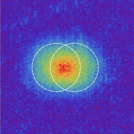
The resolution of an optical system (like a telescope or a camera) is limited by the so-called Rayleigh criterion. An international team, led by Complutense University of Madrid, has broken this limit, showing that it is not a fundamental curse. This opens the door to considerable improvement in resolution and could force the revision of Optics textbooks. This research is the culmination of a thrilling race between four groups of scientists around the world. An ideal optical system would resolve a point perfectly as a point. However, due to the wave nature of light, diffraction occurs, caused by the limiting edges of the system's apertures. The result is that the image of a point is a blur. This limits the resolution of any imaging system, including microscopes, telescopes, and cameras. The quantitative formulation of this phenomenon is the time-honored Rayleigh's limit. Rayleigh's curse limits the minimum distance that can be distinguished with visible light: on the order of 0.1 micrometer (a bacterium, for example, has a size of 2 micrometers), "which is a great limitation to our ability to see finer details", says Luis Sánchez Soto, Professor at the Faculty of Physics at Complutense University of Madrid (UCM). In cooperation with scientists from Palacký University in Olomouc (Czech Republic), the physicist has managed to break this limit, reaching resolutions up to 17 times lower than those purported by Lord Rayleigh. "Textbook Optics should be reconsidered and Rayleigh's limit shall be placed in a broader context", says Sánchez Soto, who is also a researcher at the Max-Planck Institute for the Science of Light in Erlangen (Germany). The research, published in Optica, is the culmination of a thrilling race between four teams of scientists around the world. Everyone wanted to prove the violation of this limit, but the group led by the Spanish was the first to achieve it.
Via Mariaschnee
|
|
Scooped by
Dr. Stefan Gruenwald
October 17, 2016 5:36 PM
|
A new sequencing technology, combined with a new computer algorithm that can yield detailed information about complex genomes of various organisms, has been used to produce a high-quality draft genome sequence of cabernet sauvignon, the world’s most popular red wine grape variety, reports a UC Davis genomics expert. Success of the new genome assembly, which allows researchers to assemble large segments of an organism’s DNA, also was demonstrated on the common research plant Arabidopsis thaliana and the coral mushroom (Clavicorona pyxidata). The findings are reported today, Oct. 17, in the journal Nature Methods. The three-pronged, proof-of-concept study used an open-source genome assembly process called FALCON-unzip, developed by Pacific Biosciences of Menlo Park, California. The study was led by Chen-Shan Chin, the firm’s leading bioinformatician. Lead researcher on the cabernet sauvignon sequencing effort was Dario Cantu, a plant geneticist specializing in plant and microbial genomics in the UC Davis Department of Viticulture and Enology. “For grapevine genomics, this new technology solves a problem that has limited the development of genomic resources for wine grape varieties,” Cantu said. “It’s like finally being able to uncork a wine bottle that we have wanted to drink for a long time. “The new process provides rapid access to genetic information that cabernet sauvignon has inherited from both its parents, enabling us to identify genetic markers to use in breeding new vines with improved traits,” he said. The first genome sequence for the common grapevine, Vitis vinifera, was completed in 2007. Because it was based on a grapevine variety that was generated to simplify the genome assembly procedure, rather than a cultivated variety, that sequence lacks many of the genomic details that economically important wine grape varieties possess, Cantu said. He noted that the new sequencing technology will enable his research group to conduct comparative studies between cabernet sauvignon and other historically and economically important wine grape varieties. “This will help us understand what makes cabernet sauvignon cabernet sauvignon,” he said.
|
|
Scooped by
Dr. Stefan Gruenwald
October 17, 2016 5:27 PM
|
A blue light shines through a transparent, implantable medical sensor onto a brain. The invention may help neural researchers better view brain activity. In an open-access paper published Thursday (Oct. 13, 2016) in the journal Nature Protocols, University of Wisconsin–Madison engineers have published details of how to fabricate and use neural microelectrocorticography (μECoG) arrays made with transparent graphene in applications in electrophysiology, fluorescent microscopy, optical coherence tomography, and optogenetics. Graphene is one of the most promising candidates for transparent neural electrodes, because the material has a UV to IR transparency of more than 90%, in addition to its high electrical and thermal conductivity, flexibility, and biocompatibility, the researchers note in the paper. That allows for simultaneous high-resolution imaging and optogenetic control. The procedures in the paper are for a graphene μECoG electrode array implanted on the surface of the cerebral cortex and can be completed within 3–4 weeks by an experienced graduate student, according to the researchers. But this protocol “may be amenable to fabrication and testing of a multitude of other electrode arrays used in biological research, such as penetrating neural electrode arrays to study deep brain, nerve cuffs that are used to interface with the peripheral nervous system (PNS), or devices that interface with the muscular system,” according to the paper. The researchers first announced the breakthrough in the open-access journal Nature Communications in 2014, asKurzweilAI reported. Now, the UW–Madison researchers are looking at ways to improve and build upon the technology. They also are seeking to expand its applications from neuroscience into areas such as research of stroke, epilepsy, Parkinson’s disease, cardiac conditions, and many others. And they hope other researchers do the same.
Of course, machine translation is still far from perfect. Despite its advances, GNMT can still mistranslate, particularly when it encounters proper names or rare words, which prompt the system to, again, translate individual words instead of looking at them within the context of the whole. Clearly, there is still a gap between human and machine translations, but with GNMT, it is getting smaller. By virtue of how many words, phrases, and grammar rules there are, you can only imagine how difficult it is for a person to deliver a translation from one language to another that accurately conveys the thought and nuance behind even the simplest sentence, let alone how difficult it would be for a machine. This, however, was a challenge that Google wanted to tackle head on, and today, the results of all those years spent working on a machine learning translation system were finally unveiled. Languages are naturally phrase-based, so letting machines learn as many of those phrases and the subtleties behind the language as possible so that they could be applied to the translation was essential. Getting machines to do all that requires a lot of data, and adding complex language rules into the mix requires neural networks. While Google may not be the only company that has been looking into this method for more precise and accurate translations, they managed to be the first to get it done. The Google Neural Machine Translation system (GNMT) uses state-of-the-art training techniques that significantly improve machine translation quality. It relies on long short term memory recurrent neural networks (LSTM-RNNs) and neural networks that have been trained by graphics processing units (GPUs), and tensor processing units (TPUs) to crunch data more efficiently. It all adds up to a new system that can lower translation errors by more than 55 to 85 percent.
Via Nik Peachey
|
|
Scooped by
Dr. Stefan Gruenwald
October 14, 2016 7:39 PM
|
Physicists at The University of Texas at Arlington have shown that using microwaves to activate photosensitive nanoparticles produces tissue-heating effects that ultimately lead to cell death within solid tumors. "Our new method using microwaves can propagate through all types of tissues and target deeply situated tumors," said Wei Chen, UTA professor of physics and lead author of the study published this month in he Journal of Biomedical Nanotechnology titled "A new modality of cancer treatment-nanoparticle mediated microwave induced photodynamic therapy." Photodynamic therapy kills cancer cells when a nanoparticle introduced into tumor tissue generates toxic singlet oxygen after being exposed to light. Singlet oxygen is a highly reactive type of oxygen that irreversibly damages cell mitochondria and eventually causes cell death. "Up to now, photodynamic therapy, which depends on visible, ultraviolet or near infrared light, was considered effective for skin cancers or cancers close to the skin surface," Chen said. "Our new concept combining microwaves with photodynamic therapy opens up new avenues for targeting deeper tumors and has already proven effective in rapidly and safely reducing tumor size." In prior studies, the researchers had identified a new type of nanoparticle, copper-cysteamine or Cu-Cy, that could be activated by X-rays to produce singlet oxygen and slow the growth of tumors. X-ray radiation, however, poses significant risks to patients and can harm healthy tissue. In this new lab study, the team demonstrated that the nanoparticle Cu-Cy also can be activated by microwaves, which can be targeted directly at the tumor itself without harming surrounding tissue. "Our new microwave-induced photodynamic therapy offers numerous advantages, the most significant of which is increased safety," Chen said. "Our nanoparticle Cu-Cy also demonstrates very low toxicity, is easy to make and inexpensive, and also emits intense luminescence, which means it can also be used as an imaging agent." The researchers demonstrated that both in vitro and in vivo studies on an osteosarcoma cell line showed significant cell destruction using copper cysteamine nanoparticles under microwave activation. The heating effects and the release of copper ions from copper cysteamine upon activation was the main mechanism for the generation of the reactive oxygen needed for cancer cell destruction.
|
|
Rescooped by
Dr. Stefan Gruenwald
from DNA and RNA research
October 14, 2016 7:35 PM
|
From small craft breweries to mass bottle fillers like Anheuser-Busch InBev, few would argue that the market for the most widely consumed alcoholic beverage in the world is—well, overflowing with growth potential. Research and Markets projects the global beer market to be valued at $318 billion by 2020, though similar projections fluctuate based on the emphasis given to increasingly popular microbreweries opening shop and fine-tuning their craft. Whatever data you use, it’s safe to say there’s been significant movement in this hoppy corner of the food and beverage space—and its taking biotechnology along for the ride. All of the main ingredients in beer help contribute to its flavor in some way: grains form the malt that determines how dark and heavily bodied a beer is, water can impact the minerality and even indicate the region in which the beer was produced and hops produce pounced bitter or floral undertones. But it’s the final step, in which yeast spurs fermentation, that is perhaps the most important and impactful—for both the beer consumer and scientists. Two research teams from White Labs (California) and a Belgian genetics laboratory have collaborated to map out yeasts’ genealogy, creating the first genetic family tree for brewing yeasts. The research team has already sequenced the DNA of more than 240 strains of brewing yeast from around the world, enabling them to tell how closely related two yeasts are. For example, is Brewery A’s yeast closer to the yeast used in Brewery B or Brewery C? Do Brewery A and Brewery C use yeasts derived from the same strain? Many breweries today simply reuse the same strain of yeast or rotate between two or three, but few experiment further with this aspect of brewing. Kevin Verstrepen, leader of the Belgian lab, wants to change that. “People come to the lab and they think ‘you’re working on beer,’ but it’s a product like any other. We try to make it better and more efficient, and yeast is really the key for us,” Verstrepen told Laboratory Equipment. “Yes, we do this work for flavor, but also to improve fermentation so brewers can make beers faster, or so a smaller brewer can make more beer.”
Via Integrated DNA Technologies
|
|
Scooped by
Dr. Stefan Gruenwald
October 14, 2016 7:27 PM
|
Clinicians use the Watson computer to mine cancer patients' records and provide treatment advice in Thailand, India, and soon, China. Watson for Oncology analyzes patient medical records, along with millions of pages of articles and textbooks, to make treatment recommendations. In 2011, a supercomputer won $1 million on Jeopardy! In 2016, that same supercomputer is tackling a challenge quantified not in millions of dollars but in millions of cancer patients. The goal is to use Watson’s natural language processing to mine the medical literature and a patient’s records to provide treatment advice. And this month the Watson computer system is drastically expanding its reach — from one hospital in Thailand to six in India and a planned 21 more in China. This instantiation of Watson, dubbed Watson for Oncology, is an artificial intelligence system that has access to millions of pages of medical textbooks and journal articles. Oncologists at Memorial Sloan Kettering Cancer Center trained the system to provide appropriate treatment recommendations by giving it descriptions of patients and then telling Watson the correct treatment.
|
|
Scooped by
Dr. Stefan Gruenwald
October 14, 2016 7:17 PM
|
Biomedical engineers from Harvard and MIT have managed to grow three-dimensional brain tissue in a lab using a simple and inexpensive technique. The research team borrowed processes from the semiconductor industry to create layered stacks of the tissue. The development will allow fellow scientists to study things like neuron development, as well as model the effects of drug treatments on individual patients. "We think that by bringing this kind of control and manipulation into neurobiology, we can investigate many different directions," said Utkan Demirci, part of the Harvard-MIT Division of Health Sciences and Technology (HST). Brain tissue's intricate and diverse structure meant the team needed to find a way to replicate its architectural complexity. Cells taken from the primary cortex of rats were embedded in hydrogel sheets alongside structural support components. These layers were then stacked and sealed into the required 3D shapes using light. The result is a cube of tissue containing a "diverse repertoire of brain cells" which occur in the same ratios as in natural brain tissue. The researchers eventually hope to build a cubic millimeter of tissue which would comprise of around 100,000 cells and 900,000 million connections.
|
|
Scooped by
Dr. Stefan Gruenwald
October 14, 2016 7:12 PM
|
Researchers at The University of Texas at Dallas are sweating the small stuff in their efforts to develop a wearable device that can monitor an individual's glucose level via perspiration on the skin. But for diabetics and those at risk for diabetes, self-monitoring of blood glucose, or blood sugar, is an important part of managing their conditions. "Fitness trackers that monitor heart rate and step count are very popular, but wearable, non-invasive biosensors would be extremely beneficial for managing diseases," said Prasad, the Cecil H. and Ida Green Professor in Systems Biology Science. Typical home-use blood glucose monitors require a user to obtain a small blood sample, usually through the prick of a finger and often several times a day. However, the UT Dallas textile-based sensor detects glucose in the small amount of ambient sweat on a person's skin. "In our sensor mechanism, we use the same chemistry and enzymatic reaction that are incorporated into blood glucose testing strips," Prasad said. "But in our design, we had to account for the low volume of ambient sweat that would be present in areas such as under a watch or wrist device, or under a patch that lies next to the skin." Prasad said that researchers who work with sweat often use a process called iontophoresis, which sends an electric current through the skin to generate enough perspiration for sensing experiments. However, because this method can lead to rashes and burns on the skin, the team sought an alternative that would work with small amounts of sweat. Their design works with volumes of sweat less than a microliter, which is the approximate amount of liquid that would fit in a cube the size of a salt crystal. The technology also provides a real-time response in the form of a digital readout.
|
|
Scooped by
Dr. Stefan Gruenwald
October 14, 2016 7:04 PM
|
We might think our technological innovations are driving us towards a cyborg future, but is it the brain doing all the work? Imagine a condition that leaves you fully conscious, but unable to move or communicate, as some victims of severe strokes or other neurological damage experience. This is locked-in syndrome, when the outward connections from the brain to the rest of the world are severed. Technology is beginning to promise ways of remaking these connections, but is it our ingenuity or the brain’s that is making it happen? Ever since an 18th-century biologist called Luigi Galvani made a dead frog twitch we have known that there is a connection between electricity and the operation of the nervous system. We now know that the signals in neurons in the brain are propagated as pulses of electrical potential, whose effects can be detected by electrodes in close proximity. So in principle, we should be able to build an outward neural interface system – that is to say, a device that turns thought into action. In fact, we already have the first outward neural interface system to be tested in humans. It is called BrainGate and consists of an array of micro-electrodes, implanted into the part of the brain concerned with controlling arm movements. Signals from the micro-electrodes are decoded and used to control the movement of a cursor on a screen, or the motion of a robotic arm. A crucial feature of these systems is the need for some kind of feedback. A patient must be able to see the effect of their willed patterns of thought on the movement of the cursor. What’s remarkable is the ability of the brain to adapt to these artificial systems, learning to control them better.
"Physicists have implemented the first experimental demonstration of everlasting quantum coherence—the phenomenon that occurs when a quantum system exists in a superposition of two or more states at once. Typically, quantum coherence lasts for only a fraction of a second before decoherence destroys the effect due to interactions between the quantum system and its surrounding environment.
The collaboration of physicists, led by Gerardo Adesso at The University of Nottingham and with members from the UK, Brazil, Italy, and Germany, have published a paper on the demonstration of the extreme resilience of quantum coherence in a recent issue of Physical Review Letters.
"Quantum properties can be exploited for disruptive technologies but are typically very fragile," Adesso told Phys.org. "Here we report an experiment which shows for the first time that quantum coherence in a large ensemble of nuclear spins can be naturally preserved ('frozen') under exposure to strong dephasing noise at room temperature, without external control, and for timescales as long as a second and beyond."
Quantum coherence is an inherently quantum property that arises due to the wave-like nature of matter. Most importantly for potential applications, quantum coherence allows a quantum system to occupy a superposition of states. This trait leads to quantum parallelism, which is the key ingredient that allows some quantum devices to outperform classical ones in a wide range of applications. For instance, many research groups are currently working on harnessing quantum coherence to develop quantum algorithms, quantum cryptography, quantum metrology, and other quantum technologies."
Via Mariaschnee
|
|
Scooped by
Dr. Stefan Gruenwald
October 14, 2016 5:20 PM
|
Injuries to the spinal cord can cause paralysis and other permanent disabilities because severed nerve fibers do not regrow. Now, scientists of the German Center for Neurodegenerative Diseases (DZNE) have succeeded in releasing a molecular brake that prevents the regeneration of nerve connections. Treatment of mice with "Pregabalin," a drug that acts upon the growth inhibiting mechanism, caused damaged nerve connections to regenerate. Researchers led by neurobiologist Frank Bradke report on these findings in the journal Neuron. Human nerve cells are interconnected in a network that extends to all parts of the body. In this way control signals are transmitted from head to toe, while sensory inputs flow in the opposite direction. For this to happen, impulses are passed from neuron to neuron, not unlike a relay race. Damages to this wiring system can have drastic consequences -- particularly if they affect the brain or the spinal cord. This is because the cells of the central nervous system are connected by long projections. When severed, these projections, which are called "axons," are unable to regrow. Neural pathways that have been injured can only regenerate if new connections arise between the affected cells. In a sense, the neurons have to stretch out their arms, i.e. the axons have to grow. In fact, this happens in the early stages of embryonic development. However, this ability disappears in the adult. Can it be reactivated? This was the question Professor Bradke and co-workers asked themselves. "We started from the hypothesis that neurons actively down-regulate their growth program once they have reached other cells, so that they don't overshoot the mark. This means, there should be a braking mechanism that is triggered as soon as a neuron connects to others," says Dr. Andrea Tedeschi, a member of the Bradke Lab and first author of the current publication. In mice and cell cultures, the scientists started an extensive search for genes that regulate the growth of neurons. "That was like looking for the proverbial needle in the haystack. There are hundreds of active genes in every nerve cell, depending on its stage of development. To analyze the large data set we heavily relied on bioinformatics. To this end, we cooperated closely with colleagues at the University of Bonn," says Bradke. "Ultimately, we were able to identify a promising candidate. This gene, known as Cacna2d2, plays an important role in synapse formation and function, in other words in bridging the final gap between nerve cells." During further experiments, the researchers modified the gene's activity, e.g. by deactivating it. In this way, they were able to prove that Cacna2d2 does actually influence axonal growth and the regeneration of nerve fibers. Cacna2d2 encodes the blueprint of a protein that is part of a larger molecular complex. The protein anchors ion channels in the cell membrane that regulate the flow of calcium particles into the cell. Calcium levels affect cellular processes such as the release of neurotransmitters. These ion channels are therefore essential for the communication between neurons.
|