Your new post is loading...

|
Scooped by
Dr. Stefan Gruenwald
June 27, 2017 4:32 PM
|
A hologram with a single particle of light! Until quite recently, creating a hologram of a single photon was believed to be impossible due to fundamental laws of physics. However, scientists at the Faculty of Physics, University of Warsaw, have successfully applied concepts of classical holography to the world of quantum phenomena. A new measurement technique has enabled them to register the first ever hologram of a single light particle, thereby shedding new light on the foundations of quantum mechanics. Scientists at the Faculty of Physics, University of Warsaw, have created the first ever hologram of a single light particle. The spectacular experiment, reported in the prestigious journal Nature Photonics, was conducted by Dr. Radoslaw Chrapkiewicz and Michal Jachura under the supervision of Dr. Wojciech Wasilewski and Prof. Konrad Banaszek. Their successful registering of the hologram of a single photon heralds a new era in holography: quantum holography, which promises to offer a whole new perspective on quantum phenomena. "We performed a relatively simple experiment to measure and view something incredibly difficult to observe: the shape of wavefronts of a single photon," says Dr. Chrapkiewicz. In standard photography, individual points of an image register light intensity only. In classical holography, the interference phenomenon also registers the phase of the light waves (it is the phase which carries information about the depth of the image). When a hologram is created, a well-described, undisturbed light wave (reference wave) is superimposed with another wave of the same wavelength but reflected from a three-dimensional object (the peaks and troughs of the two waves are shifted to varying degrees at different points of the image). This results in interference and the phase differences between the two waves create a complex pattern of lines. Such a hologram is then illuminated with a beam of reference light to recreate the spatial structure of wavefronts of the light reflected from the object, and as such its 3D shape. One might think that a similar mechanism would be observed when the number of photons creating the two waves were reduced to a minimum, that is to a single reference photon and a single photon reflected by the object. And yet you'd be wrong! The phase of individual photons continues to fluctuate, which makes classical interference with other photons impossible. Since the Warsaw physicists were facing a seemingly impossible task, they attempted to tackle the issue differently: rather than using classical interference of electromagnetic waves, they tried to register quantum interference in which the wave functions of photons interact. Wave function is a fundamental concept in quantum mechanics and the core of its most important equation: the Schrödinger equation. In the hands of a skilled physicist, the function could be compared to putty in the hands of a sculptor: when expertly shaped, it can be used to 'mould' a model of a quantum particle system. Physicists are always trying to learn about the wave function of a particle in a given system, since the square of its modulus represents the distribution of the probability of finding the particle in a particular state, which is highly useful.
|
|
Scooped by
Dr. Stefan Gruenwald
June 27, 2017 4:25 PM
|
Biomedical researchers are understanding the functions of molecules within the body’s cells in ever greater detail by increasing the resolution of their microscopes. However, what’s lagging behind is their ability to simultaneously visualize the many different molecules that mediate complex molecular processes in a single snap-shot. Now, a team from Harvard’s Wyss Institute for Biologically Inspired Engineering, the LMU Munich, and the Max Planck Institute of Biochemistry in Germany, has engineered highly versatile metafluorophores by integrating commonly used small fluorescent probes into self-folding DNA structures where their colors and brightness can be digitally programmed. This nanotechnological approach offers a palette of 124 virtual colors for microscopic imaging or other analytical methods that can be adapted in the future to visualize multiple molecular players at the same time with ultra-high definition. The method is reported in Science Advances. With their new method, the researchers address the problem that thus far only a limited number of molecular species can be visualized simultaneously with fluorescence microscopy in a biological or clinical sample. By introducing fluorescent DNA nanostructures called metafluorophores — versatile fluorescent dyes whose colors are determined by how their individual components are arranged in 3-dimensional structures — they overcome this bottleneck. These fluorescence images show a matrix representing 124 distinct metafluorophores, that are generated by combining three fluorescent dyes with varying intensity levels. In the future, the metafluorophore’s unique and identifiable color patterns can be used to analyze the molecular components of complex samples. “We use DNA nanostructures as molecular pegboards: by functionalizing specific component strands at defined positions of the DNA nanostructure with one of three different fluorescent dyes, we achieve a broad spectrum of up to 124 fluorescent signals with unique color compositions and intensities,” said Peng Yin, who is a Core Faculty member at the Wyss Institute and Professor of Systems Biology at Harvard Medical School. “Our study provides a framework that allows researchers to construct a large collection of metafluorophores with digitally programmable optical properties that they can use to visualize multiple targets in the samples they are interested in.” The DNA nanostructure-based approach can be used like a barcoding system to visually profile the presence of many specific DNA or RNA sequences in samples in what is called multiplexing. To enable the visualization of multiple molecular structures in tissue samples whose thickness can limit the movement of larger DNA nanostructures and make it difficult for them to find their targets, and to reduce the possibility that they attach themselves to non-specific targets producing false fluorescence signals, the team took additional engineering steps. “We developed a triggered version of our metafluorophore that dynamically self-assembles from small component strands that take on their prescribed shape only when they bind their target,” said Ralf Jungmann, Ph.D., who is faculty at the LMU Munich and the Max Planck Institute of Biochemistry and co-conducted the study together with Yin. “These in-situ assembled metafluorophores can not only be introduced into complex samples with similar combinatorial possibilities as the prefabricated ones to visualize DNA, but they could also be leveraged to label antibodies as widely used detection reagents for proteins and other biomolecules.” “This new type of programmable, microscopy-enhancing DNA nanotechnology reveals how work in the Wyss Institute’s Molecular Robotics Initiative can invent new ways to solve long-standing problems in biology and medicine. These metafluorophores that can be programmed to self-assemble when they bind their target, and that have defined fluorescent barcode readouts, represent a new form of nanoscale devices that could help to reveal complex, multi-component, biological interactions that we know exist but have no way of studying today,” said Wyss Founding Director Donald Ingber, M.D., Ph.D., who is also the Judah Folkman Professor of Vascular Biology at Harvard Medical School and the Vascular Biology Program at Boston Children’s Hospital, and Professor of Bioengineering at the Harvard John A. Paulson School of Engineering and Applied Sciences.
|
|
Scooped by
Dr. Stefan Gruenwald
June 27, 2017 1:36 PM
|
So it’s a good moment to note how good we have it here on Earth. There are longer days in our solar system, but none are quite so pleasant. If “day” refers to the time it takes for a planet to rotate exactly once on its axis (a sidereal day), then the Venusian day is the longest, lasting two hundred and forty-three Earth days. That’s even longer, by nineteen Earth days, than a Venusian year, which is the time it takes the planet to orbit the sun. If, instead, “day” refers to the period between sunrise and sunset (a solar day), Neptune’s is the longest: the gas giant orbits the sun on its side, such that one pole or the other receives daylight for forty-two years non-stop. Farther out in the universe, the days are longer still. Since 1995, some thirty-five hundred extrasolar planets have been discovered, but scientists only gained the ability to measure their spin rates in 2014. A great many of the known ones, though, orbit very close to their host stars and are probably tidally locked, with one side of the planet perpetually facing the star, just as our moon always presents the same face to Earth. “This leads to an infinitely long day, since if you are on the night side, you will never see the sun,” Konstantin Batygin, an astrophysicist at Caltech, explains. Last January, Batygin and the astronomer Mike Brown, also at Caltech, announced the possible existence of a ninth planet in the solar system, a relictual ice giant so distant that it orbits our sun once every twelve thousand to twenty thousand years. Last August, scientists discovered Proxima b, an exoplanet just 4.3 light-years away, which is about as close to us as any extrasolar planet will ever come. It, too, is probably tidally locked, its day eternal. But, even being so near, Proxima b would take us eighty thousand years (some thirty million days) to reach—a very long day’s journey into day. Summer is a separate matter. A planet’s seasons are shaped by two factors: the eccentricity of its orbit—whether it’s closer to the sun at some times of the year than at others—and the tilt of its axis. Earth’s orbit is essentially circular, so the effect on our climate is negligible. But the planet itself leans twenty-three degrees to the side; as we orbit, there comes a day when the North Pole is maximally tilted toward the sun and the Northern Hemisphere sees more daylight than it will all year. That’s today, the summer solstice. (Below the equator, it’s the winter solstice, of course, and in six months our situations will reverse.) If we weren’t off-kilter, we’d have no summer nor any seasons at all. Every day would be as long as every other, and changes in the weather would be driven more by the local geography—latitude, elevation, that mountain range to the west that keeps the rain from falling—than by shifts in the jet stream, or the massive blooms of Pacific plankton in the winter that fuel El Niño, or the decline in sunlight that triggers autumn leaves to change color. Mercury, Venus, and Jupiter, standing all but upright, are seasonless. Sad. Perhaps the weirdest summer of all unfolds on HD 131399Ab, an extrasolar gas giant that was discovered last July by Daniel Apai, an astronomer at the University of Arizona, and his colleagues. The planet belongs to a system with three stars but orbits only one of them, the biggest, which is eighty per cent larger than our sun. The other two stars orbit each other and, together, like a spinning dumbbell, orbit the big one. The view from HD 131399Ab would be spectacular if not for the ferocious winds, the lack of solid ground, and a steady rain of liquid iron. For much of the year, which lasts five hundred and fifty Earth years, the three stars appear close together in the sky, giving the planet “a familiar night side and day side, with a unique triple sunset and sunrise each day,” Kevin Wagner, one of the discoverers, remarked at the time. But as HD 131399Ab progresses in its orbit and the stars drift apart, a day arrives when the setting of one coincides with the rising of the other, and a period of near-constant daylight begins—a solstice of sorts, the start of a summer that will last about a hundred and forty Earth years.
|
|
Scooped by
Dr. Stefan Gruenwald
June 27, 2017 1:17 PM
|
Backed by the digital fortunes of Silicon Valley, biotech companies are brazenly setting out to “cure” aging. Can this really be done? Immortality, it turns out, is not an easy sell: Most people don’t like the idea of living forever. In legends of old as well as in recent popular culture, eluding death typically comes at a terrible cost; like zombies or vampires, immortal beings must feast on the living. Besides, a large percentage of today’s population also subscribes to religious beliefs in which the afterlife is something to be welcomed. When the Pew Research Center asked Americans in 2013 whether they would use technologies that allowed them to live to 120 or beyond, 56 percent said no. Two-thirds of respondents believed that radically longer life spans would strain natural resources, and that these treatments would only ever be available to the wealthy. How would the world change—socioeconomically especially—if no one ever died? Would people still have children? If they did, how long would the planet be able to sustain billions of immortals? Wouldn’t every norm predicated on our inevitable deaths break down, including all the world’s religions? What would replace them? At what point might you decide that, actually, this is enough life? After decades? Centuries? And once you made that decision, how would you make your exit?
|
|
Scooped by
Dr. Stefan Gruenwald
June 27, 2017 1:07 PM
|
For the first time ever, astronomers at The University of New Mexico say they've been able to observe and measure the orbital motion between two supermassive black holes hundreds of millions of light years from Earth - a discovery more than a decade in the making. UNM Department of Physics & Astronomy graduate student Karishma Bansal is the first-author on the paper, 'Constraining the Orbit of the Supermassive Black Hole Binary 0402+379', recently published in The Astrophysical Journal. She, along with UNM Professor Greg Taylor and colleagues at Stanford, the U.S. Naval Observatory and the Gemini Observatory, have been studying the interaction between these black holes for 12 years. "For a long time, we've been looking into space to try and find a pair of these supermassive black holes orbiting as a result of two galaxies merging," said Taylor. "Even though we've theorized that this should be happening, nobody had ever seen it until now." In early 2016, an international team of researchers, including a UNM alumnus, working on the LIGO project detected the existence of gravitational waves, confirming Albert Einstein's 100-year-old prediction and astonishing the scientific community. These gravitational waves were the result two stellar mass black holes (~30 solar mass) colliding in space within the Hubble time. Now, thanks to this latest research, scientists will be able to start to understand what leads up to the merger of supermassive black holes that creates ripples in the fabric of space-time and begin to learn more about the evolution of galaxies and the role these black holes play in it. Using the Very Long Baseline Array (VLBA), a system made up of 10 radio telescopes across the U.S. and operated in Socorro, N.M., researchers have been able to observe several frequencies of radio signals emitted by these supermassive black holes (SMBH). Over time, astronomers have essentially been able to plot their trajectory and confirm them as a visual binary system. In other words, they've observed these black holes in orbit with one another.
|
|
Scooped by
Dr. Stefan Gruenwald
June 27, 2017 1:00 PM
|
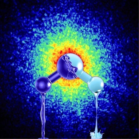
We normally consider liquid water as disordered with the molecules rearranging on a short time scale around some average structure. Now, however, scientists at Stockholm University have discovered two phases of the liquid with large differences in structure and density. The results are based on experimental studies using X-rays, which are now published in Proceedings of the National Academy of Science (US). Most of us know that water is essential for our existence on planet Earth. It is less well-known that water has many strange or anomalous properties and behaves very differently from all other liquids. Some examples are the melting point, the density, the heat capacity, and all-in-all there are more than 70 properties of water that differ from most liquids. These anomalous properties of water are a prerequisite for life as we know it. "The new remarkable property is that we find that water can exist as two different liquids at low temperatures where ice crystallization is slow", says Anders Nilsson, professor in Chemical Physics at Stockholm University. The breakthrough in the understanding of water has been possible through a combination of studies using X-rays at Argonne National Laboratory near Chicago, where the two different structures were evidenced and at the large X-ray laboratory DESY in Hamburg where the dynamics could be investigated and demonstrated that the two phases indeed both were liquid phases. Water can thus exist as two different liquids. "It is very exciting to be able to use X-rays to determine the relative positions between the molecules at different times", says Fivos Perakis, postdoc at Stockholm University with a background in ultrafast optical spectroscopy. "We have in particular been able to follow the transformation of the sample at low temperatures between the two phases and demonstrated that there is diffusion as is typical for liquids". When we think of ice it is most often as an ordered, crystalline phase that you get out of the ice box, but the most common form of ice in our planetary system is amorphous, that is disordered, and there are two forms of amorphous ice with low and high density. The two forms can interconvert and there have been speculations that they can be related to low- and high-density forms of liquid water. To experimentally investigate this hypothesis has been a great challenge that the Stockholm group has now overcome. "I have studied amorphous ices for a long time with the goal to determine whether they can be considered a glassy state representing a frozen liquid", says Katrin Amann-Winkel, researcher in Chemical Physics at Stockholm University. "It is a dream come true to follow in such detail how a glassy state of water transforms into a viscous liquid which almost immediately transforms to a different, even more viscous, liquid of much lower density".
While the definitive causes remain unclear, several genetic and environmental factors increase the likelihood of autism spectrum disorder, or ASD, a group of conditions covering a “spectrum” of symptoms, skills and levels of disability. Taking advantage of advances in genetic technologies, researchers led by Alex Nord, assistant professor of neurobiology, physiology and behavior with the Center for Neuroscience at the University of California, Davis, are gaining a better understanding of the role played by a specific gene involved in autism. The collaborative work appears June 26 in the journalNature Neuroscience. “For years, the targets of drug discovery and treatment have been based on an unknown black box of what’s happening in the brain,” said Nord. “Now, using genetic approaches to study the impact of specific mutations found in cases, we’re trying to build a cohesive model that links genetic control of brain development with behavior and brain function.” The Nord laboratory studies how the genome encodes brain development and function, with a particular interest in understanding the genetic basis of neurological disorders. There is no known specific genetic cause for most cases of autism, but many different genes have been linked to the disorder. In rare, specific cases of people with ASD, one copy of a gene called CHD8 is mutated and loses function. The CHD8 gene encodes a protein responsible for packaging DNA in cells throughout the body. Packaging of DNA controls how genes are turned on and off in cells during development. Because mice and humans share on average 85 percent of similarly coded genes, mice can be used as a model to study how genetic mutations impact brain development. Changes in mouse DNA mimic changes in human DNA and vice-versa. In addition, mice exhibit behaviors that can be used as models for exploring human behavior.
Via Mariaschnee
Some doctors dream of diagnosing diseases—or at least predicting disease risk—with a simple DNA scan. But others have said the practice, which could soon be the foundation of preventative medicine, isn’t worth the economic or emotional cost. Now, a new pair of studies puts numbers to the debate, and one is the first ever randomized clinical trial evaluating whole genome sequencing in healthy people. Together, they suggest that sequencing the genomes of otherwise healthy adults can for about one in five people turn up risk markers for rare diseases or genetic mutations associated with cancers. What that means for those people and any health care system considering genome screening remains uncertain, but some watching for these studies welcomed the results nonetheless. “It's terrific that we are studying implementation of this new technology rather than ringing our hands and fretting about it without evidence,” says Barbara Biesecker, a social and behavioral researcher at the National Human Genome Research Institute in Bethesda, Maryland. The first genome screening study looked at 100 healthy adults who initially reported their family history to their own primary care physician. Then half were randomly assigned to undergo an additional full genomic workup, which cost about $5000 each and examined some 5 million subtle DNA sequence changes, known as single-nucleotide variants, across 4600 genes—such genome screening goes far beyond that currently recommended by the American College of Medical Genetics and Genomics (ACMG), which suggests informing people of results for just 59 genes known or strongly expected to cause disease.
Via Integrated DNA Technologies
Most rod-shaped bacteria divide by splitting into two around the middle after their DNA has replicated safely and segregated to opposite ends of the cell. This seemingly simple process actually demands tight and precise coordination, which is achieved through two biological systems: nucleoid occlusion, which protects the cell's genetic material from dividing until it replicates and segregates, and the "minicell" system, which localizes the site of division around the middle of the cell, where a dividing wall will form to split it in two. But some pathogenic bacteria, e.g. Mycobacterium tuberculosis, don't use these mechanisms. EPFL scientists have now combined optical and atomic force microscopy to track division in such bacteria for the first time and have discovered that they use instead an undulating "wave-pattern" along their length to mark future sites of division. The findings are published in Nature Microbiology. The work was carried out jointly by the labs of John McKinney and Georg Fantner at EPFL. The scientists wanted to understand how bacteria that do not have the genes for nucleoid occlusion and the minicell system "decide" where and when to divide. This is important, as many pathogenic bacteria fall into this category, and knowing how they divide can open up new ways to fight them. The researchers focused on Mycobacterium smegmatis, a non-pathogenic relative of M. tuberculosis. Neither of these bacteria uses the two "conventional" biological systems for coordinating division, meaning that a non-conventional approach was needed for studying them. The researchers combined two types of microscopy to track the life cycle of the bacteria. The first technique was optical microscopy, which uses fluorescent labels for "seeing" various biological structures and biomolecules. The second technique was atomic force microscopy, which provides extremely high-resolution images of structures on the cell surface by "feeling" the surface with a tiny mechanical probe, much like a blind person can form a three-dimensional mental image of an object by passing their hands over its surface. "This experiment constitutes the longest continuous atomic force microscopy experiment ever performed on growing cells," says Georg Fantner, while John McKinney adds: "It illustrates the power of new technologies not only to analyze the things we already knew about with greater resolution, but also to discover new things that we hadn't anticipated."
Via Mariaschnee
|
|
Scooped by
Dr. Stefan Gruenwald
June 27, 2017 12:48 PM
|
The immune system is naturally equipped to protect us against cancer. Cytotoxic T lymphocytes—otherwise known as killer T cells—are especially effective at targeting tumors. However, cancers sometimes figure out how to outsmart the immune system and protect themselves. Immunotherapy aims to reverse that situation. This review is highlighting a wide scope of immune-based approaches that are already improving outcomes for patients. Many of these treatments work by either directly or indirectly enhancing the activity of T cells. Much of what we know about the immune system and its relationship to cancer was discovered by scientists affiliated with the Cancer Research Institute (CRI), which since 1953 has served as the world’s leading (and for several decades only) nonprofit organization dedicated exclusively to transforming cancer patient care by advancing immunotherapy and the science behind it. It’s clear now that immunotherapy can provide long-term benefits to sizable subsets of patients with diverse types of cancer, and new clinical breakthroughs are happening all the time. Thus far in 2017 there have been eight immunotherapy approvals, with two immunotherapies (durvalumab and avelumab) gaining approval for the first time. These clinical breakthroughs were made possible in part by decades of discoveries made by Cancer Research Institute (CRI) scientists. One of the most important figures who helped advance immunotherapy (and the science that supports it) to this point was Dr. Lloyd J. Old, who actually worked with Dr. William B. Coley’s daughter, Helen, at CRI. Now known as the “Father of Modern Tumor Immunology,” Dr. Old directed CRI’s scientific and medical efforts for 40 years (1971-2011), during which time he made major discoveries about the immune system and cancer, and helped establish the scientific foundation upon which today’s immunotherapies were developed.
|
|
Scooped by
Dr. Stefan Gruenwald
June 26, 2017 11:21 AM
|
Climate scientists are agreed that global warming will have significant long-term impacts on plankton, the creatures that underpin the health and productivity of global marine ecosystems and which play a critical role in the planetary carbon cycle, though they are less sure what exactly those impacts will be. But more immediate effects from human reliance on fossil fuels are now clearer, thanks to research that shows acoustic survey techniques used to explore the seafloor for oil and gas deposits is associated with the widespread death of plankton. The study by marine scientists from Curtin University, in Western Australia, and the University of Tasmania has beenpublished in Nature Ecology & Evolution. It concludes that “potential large-scale modification of plankton community structure and abundance due to seismic survey operations has enormous ramifications for larval recruitment processes, all higher order predators and ocean health in general” and flags the “urgent need to prioritise development and testing of alternative surveying techniques”.
|
|
Scooped by
Dr. Stefan Gruenwald
June 26, 2017 11:14 AM
|
EPFL researchers have found a way around what was considered a fundamental limitation of physics for over 100 years. They were able to conceive resonant systems that can store electromagnetic waves over a long period of time while maintaining a broad bandwidth. Their study, which has just been published in Science, opens up a number of doors, particularly in telecommunications. At EPFL, researchers challenge a fundamental law and discover that more electromagnetic energy can be stored in wave-guiding systems than previously thought. The discovery has implications in telecommunications. Working around the fundamental law, they conceived resonant and wave-guiding systems capable of storing energy over a prolonged period while keeping a broad bandwidth. Their trick was to create asymmetric resonant or wave-guiding systems using magnetic fields. The study, which has just been published in Science, was led by Kosmas Tsakmakidis, first at the University of Ottawa and then at EPFL’s Bionanophotonic Systems Laboratory run by Hatice Altug, where the researcher is now doing post-doctoral research. This breakthrough could have a major impact on many fields in engineering and physics. The number of potential applications is close to infinite, with telecommunications, optical detection systems and broadband energy harvesting representing just a few examples. Resonant and wave-guiding systems are present in the vast majority of optical and electronic systems. Their role is to temporarily store energy in the form of electromagnetic waves and then release them. For more than 100 hundred years, these systems were held back by a limitation that was considered to be fundamental: the length of time a wave could be stored was inversely proportional to its bandwidth. This relationship was interpreted to mean that it was impossible to store large amounts of data in resonant or wave-guiding systems over a long period of time because increasing the bandwidth meant decreasing the storage time and quality of storage.
|
|
Scooped by
Dr. Stefan Gruenwald
June 26, 2017 11:08 AM
|
Research from the Stowers Institute provides evidence suggesting that cancer cells might streamline their genomes in order to proliferate more easily. The study, conducted in both human and mouse cells, shows that cancer genomes lose copies of repetitive sequences known as ribosomal DNA. While downsizing might enable these cells to replicate faster, it also seems to render them less able to withstand DNA damage. The findings, published in PLoS Genetics, suggest that ribosomal DNA copy number could be used to predict which cancers will be sensitive to DNA-damaging chemotherapeutics. “Drugs that damage DNA are often used to treat cancer, but it’s not clear why they would selectively kill cancer cells,” says Jennifer L. Gerton, Ph.D., an investigator at the Stowers Institute who led the study. “Our results suggest that off-loading copies of ribosomal DNA could create instability in the genome that makes cells particularly susceptible to chemotherapy with DNA-damaging drugs.”
|
|
|
Scooped by
Dr. Stefan Gruenwald
June 27, 2017 4:30 PM
|
It is hard to believe: A tiny spiral - a micro robot - catches a single sperm, moves it directly to an egg cell and delivers it right there. The company Nanoscribe is producing the most accurate 3D-printers in the world to print structures 250 times finer than a human hair. Researchers use them to print tiny robots, which may one day move inside the body. So far, this spermbot only functions in a petri dish and with bovine sperm. But maybe one day, it could help women who wish to get pregnant, Oliver Schmidt, Professor at the Leibniz Institute for Solid State and Materials Research (IFW) Dresden told DW. "With some men, the sperm are not moving, but still healthy. We would like to propel them artificially to be able to reach their final destination," he said. But the physicist admitted there is still a long journey ahead before the technique becomes a medical application. Right now, the main challenge for using such micro robots inside a human body is imaging: "In a petri dish we can do all our experiments with high resolution microscopy," Schmidt said. "But when we operate deeply inside the tissue, the resolution fades."
Even the most modern computer tomographs, which are used to display a cross-section of a human body, are not strong enough to help guide the micro robot to its target. One would need real time imaging to observe the robot, he added. The researchers from Dresden control their spiral with a magnetic field that rotates outside around the experiment. "It can not just be a permanent magnetic field. But on the other hand, the field does not need to be very strong." Certainly, for the human body it would not cause any harm, Schmidt stresses.
|
|
Scooped by
Dr. Stefan Gruenwald
June 27, 2017 3:47 PM
|
The Koch snowflake is a remarkable tile which only tiles the plane if tiles of two (or more) different sizes are used. It is constructed by a recursive process. Begin with an equilateral triangle with side 1 and to each side attach in the middle an equilateral triangle with side 1/3. Each side of the new tile has side 1/3 also. Attach to the middle of each side an equilateral triangle of side 1/9 and so on. Alternatively, start with a regular hexagon of side 1 and cut equilateral triangles of side 1/3 from the middle of each side, and continue by cutting triangles of side 1/9 from the middle of each side, and so on. These constructions converge and for the area between two constructions after each iterations is reduced by a factor 4/9 each time. Congruent copies of the Koch snowflake do not tile the plane.
|
|
Scooped by
Dr. Stefan Gruenwald
June 27, 2017 1:20 PM
|
The People’s Bank of China has developed a digital currency that’s designed to scale to the number of transactions made every day across the country. Speeches and research papers from officials at the People's Bank of China show that the bank’s strategy is to introduce the digital currency alongside China’s renminbi. But there is currently no timetable for this, and the bank seems to be proceeding cautiously. Nonetheless the test is a significant step. It shows that China is seriously exploring the technical, logistical, and economic challenges involved in deploying digital money, something that could ultimately have broad implications for its economy and for the global financial system. A digital fiat currency—one backed by the central bank and with the same legal status as a banknote—would lower the cost of financial transactions, thereby helping to make financial services more widely available. This could be especially significant in China, where millions of people still lack access to conventional banks. A digital currency should also be cheaper to operate, and ought to reduce fraud and counterfeiting. Even more significantly, a digital currency would give the Chinese government greater oversight of digital transactions, which are already booming. And by making transactions more traceable, this could also help reduce corruption, which is a key government priority. Such a currency could even offer real-time economic insights, which would be enormously valuable to policymakers. And finally, it might facilitate cross-border transactions, as well as the use of the renminbi outside of China because the currency would be so easy to obtain. Private digital currencies, also known as cryptocurrencies, have shot to prominence in recent years following a wave of excitement, investment, and speculation focused on Bitcoin, a distributed, cryptographically secured form of money invented by an anonymous individual or group in 2008 (see “What Bitcoin Is, and Why It Matters”). Bitcoin’s distributed ledger of transactions, known as a blockchain, makes it possible for it to operate without any central authority.
|
|
Scooped by
Dr. Stefan Gruenwald
June 27, 2017 1:12 PM
|
Injecting plant-like creatures like cyanobacteria into a rat's heart can jumpstart the recovery process, study finds. Coronary artery disease is one of the most common causes of death and disability, afflicting more than 15 million Americans. Although pharmacological advances and revascularization techniques have decreased mortality, many survivors will eventually succumb to heart failure secondary to the residual microvascular perfusion deficit that remains after revascularization. A group of scientists now present a novel system that rescues the myocardium from acute ischemia, using photosynthesis through intramyocardial delivery of the cyanobacterium Synechococcus elongatus. By using light rather than blood flow as a source of energy, photosynthetic therapy increases tissue oxygenation, maintains myocardial metabolism, and yields durable improvements in cardiac function during and after induction of ischemia. By circumventing blood flow entirely to provide tissue with oxygen and nutrients, this system has the potential to create a paradigm shift in the way ischemic heart disease is treated.
|
|
Scooped by
Dr. Stefan Gruenwald
June 27, 2017 1:02 PM
|
Physicists from the University of Nebraska-Lincoln are seeing an everyday phenomenon in a new light. By focusing laser light to a brightness one billion times greater than the surface of the sun - the brightest light ever produced on Earth - the physicists have observed changes in a vision-enabling interaction between light and matter. Those changes yielded unique X-ray pulses with the potential to generate extremely high-resolution imagery useful for medical, engineering, scientific and security purposes. The team's findings, detailed June 26 in the journal Nature Photonics, should also help inform future experiments involving high-intensity lasers. Donald Umstadter and colleagues at the university's Extreme Light Laboratory fired their Diocles Laser at helium-suspended electrons to measure how the laser's photons - considered both particles and waves of light - scattered from a single electron after striking it. Under typical conditions, as when light from a bulb or the sun strikes a surface, that scattering phenomenon makes vision possible. But an electron - the negatively charged particle present in matter-forming atoms - normally scatters just one photon of light at a time. And the average electron rarely enjoys even that privilege, Umstadter said, getting struck only once every four months or so. Though previous laser-based experiments had scattered a few photons from the same electron, Umstadter's team managed to scatter nearly 1,000 photons at a time. At the ultra-high intensities produced by the laser, both the photons and electron behaved much differently than usual. "When we have this unimaginably bright light, it turns out that the scattering - this fundamental thing that makes everything visible - fundamentally changes in nature," said Umstadter, the Leland and Dorothy Olson Professor of physics and astronomy. A photon from standard light will typically scatter at the same angle and energy it featured before striking the electron, regardless of how bright its light might be. Yet Umstadter's team found that, above a certain threshold, the laser's brightness altered the angle, shape and wavelength of that scattered light. "So it's as if things appear differently as you turn up the brightness of the light, which is not something you normally would experience," Umstadter said. "(An object) normally becomes brighter, but otherwise, it looks just like it did with a lower light level. But here, the light is changing (the object's) appearance. The light's coming off at different angles, with different colors, depending on how bright it is." That phenomenon stemmed partly from a change in the electron, which abandoned its usual up-and-down motion in favor of a figure-8 flight pattern. As it would under normal conditions, the electron also ejected its own photon, which was jarred loose by the energy of the incoming photons. But the researchers found that the ejected photon absorbed the collective energy of all the scattered photons, granting it the energy and wavelength of an X-ray.
|
|
Scooped by
Dr. Stefan Gruenwald
June 27, 2017 12:58 PM
|
How fast does evolution occur? In certain bacteria, it can occur almost instantaneously, a University at Buffalo molecular biologist has discovered. Mark R. O'Brian, PhD, chair and professor of the Department of Biochemistry in the Jacobs School of Medicine and Biomedical Sciences at UB, made the surprising discovery when studying how bacteria finds and draws iron into itself. The National Institutes of Health has awarded him a $1.28 million, four-year grant to delve into the mechanisms of bacteria mutating to accept iron, and how the organism expels excess iron. The discovery was made almost by accident, O'Brian said. The bacteria Bradyrhizobium japonicum was placed in a medium along with a synthetic compound to extract all the iron. O'Brian expected the bacteria to lie dormant having been deprived of the iron needed to multiply. But to his surprise, the bacteria started multiplying. "We had the DNA of the bacteria sequenced on campus, and we discovered they had mutated and were using the new compound to take iron in to grow," he said. "It suggests that a single mutation can do that. So we tried it again with a natural iron-binding compound, and it did it again." The speed of the genetic mutations—17 days—was astounding. "We usually think of evolution taking place over a long period of time, but we're seeing evolution—at least as the ability to use an iron source that it couldn't before—occurring as a single mutation in the cell that we never would have predicted," he said. "The machinery to take up iron is pretty complicated, so we would have thought many mutations would have been required for it to be taken up," he said. The evolution of the bacteria does not mean it is developing into some other type of creature. Evolution can also change existing species "to allow them to survive," O'Brian said.
While the pupil may be the opening in the eye that lets light through to the retina, the iris is the tissue that opens and closes to determine the size of the pupil. Although mechanical irises are already a standard feature in cameras, scientists from Finland and Poland have recently created an autonomous artificial iris that’s much more similar to those found in the eye – it may even eventually be able to replace damaged or defective ones.The contact lens-like device was created by researchers from Finland’s Tampere University of Technology, along with Poland’s University of Warsaw and Wrocław Medical University. It’s made from a polymer (a liquid crystal elastomer) that expands when exposed to light, then shrinks back when the light is lessened. This causes an opening in the middle to get smaller or larger, depending on the light levels – in this way, it works very much like a natural iris. Unlike automatic irises in cameras, it requires no power source or external light detection system. With an “eye” towards one being able to use it as an optical implant, the scientists are now adapting it to work in an aqueous environment. They’re also working at increasing its sensitivity, so that its opening and closing are triggered by smaller changes in the amount of incoming light. The research is being led by Tampere’s Prof. Arri Priimägi, and was recently described in a paper published in the journal Advanced Materials.
Via Mariaschnee
|
|
Rescooped by
Dr. Stefan Gruenwald
from DNA and RNA research
June 27, 2017 12:53 PM
|
Thousands of mouse genes (around 15% of the mouse genome) are now available from the IMPC - an important milestone for genotype-to-phenotype research. Researchers at the European Bioinformatics Institute (EMBL-EBI) and their collaborators in the International Mouse Phenotyping Consortium (IMPC) have fully characterised thousands of mouse genes for the first time. Published in Nature Genetics, the results offer hundreds of new disease models and reveal previously unknown gene functions. The 3328 genes described in this publication by the IMPC represent approximately 15% of the mouse genome.
Via Integrated DNA Technologies
|
|
Rescooped by
Dr. Stefan Gruenwald
from DNA and RNA research
June 27, 2017 12:50 PM
|
In the fight against the viruses that invade everyday life, seeing and understanding the battleground is essential. Scientists at the Morgridge Institute for Research have, for the first time, imaged molecular structures vital to how a major class of viruses replicates within infected cells. “The challenge is a bit like being a car mechanic and not being able to see the engine or how it’s put together in detail,” says Paul Ahlquist, director of virology at the Morgridge Institute and professor of oncology and molecular virology at the University of Wisconsin–Madison. “This work is our first look at the engine.” The research, published June 27 in the journal eLife, uses pioneering cryo-electron tomography to reveal the complex viral replication process in vivid detail, opening up new avenues to potentially disrupt, dismantle or redirect viral machinery. One of several goals in the Ahlquist Lab is to understand genome replication for positive strand RNA viruses, the largest genetic class of viruses that includes many human pathogens such as the Zika, Dengue, SARS, and Chikungunya viruses. The group studies processes in a strategic way, focusing not on the fine details of a single important virus, but on large principles that apply to the whole class.
Via Integrated DNA Technologies
|
|
Scooped by
Dr. Stefan Gruenwald
June 27, 2017 12:41 PM
|
The medical community gradually acknowledges the importance of digital health, but they don’t yet embrace it enough or cannot get behind it with such a speed as it would require. For doing so, the first step is always getting to know what’s coming. So, here are the trends changing the pharmaceutical industry in the near future.
|
|
Scooped by
Dr. Stefan Gruenwald
June 26, 2017 11:16 AM
|
Toxins produced by three different species of fungus growing indoors on wallpaper may become aerosolized, and easily inhaled. The findings, which likely have implications for “sick building syndrome,” were published in Applied and Environmental Microbiology, a journal of the American Society for Microbiology.
“We demonstrated that mycotoxins could be transferred from a moldy material to air, under conditions that may be encountered in buildings,” said corresponding author Jean-Denis Bailly, DVM, PhD, Professor of Food Hygiene, National Veterinary School of Toulouse, France. “Thus, mycotoxins can be inhaled and should be investigated as parameters of indoor air quality, especially in homes with visible fungal contamination.”
The impetus for the study was the dearth of data on the health risk from mycotoxins produced by fungi growing indoors. (image: microscopic view of a sporulating Aspergillus, showing numerous light spores that can be easily aerosolized and inhaled together with mycotoxins. credit: Sylviane Bailly.)
In the study, the investigators built an experimental bench that can simulate an airflow over a piece of contaminated wall paper, controlling speed and direction of the air. Then they analyzed the resulting bioaerosol.
“Most of the airborne toxins are likely to be located on fungal spores, but we also demonstrated that part of the toxic load was found on very small particles—dust or tiny fragments of wallpaper, that could be easily inhaled,” said Bailly..
|
|
Scooped by
Dr. Stefan Gruenwald
June 26, 2017 11:11 AM
|
As humans put ever more heat-trapping gases into the atmosphere, the Earth heats up. These are the basics of global warming. But where does the heat go? How much extra heat is there? And how accurate are our measurements? These are questions that climate scientists ask. If we can answer these questions, it will better help us prepare for a future with a very different climate. It will also better help us predict what that future climate will be. The most important measurement of global warming is in the oceans. In fact, “global warming” is really “ocean warming.” If you are going to measure the changing climate of the oceans, you need to have many sensors spread out across the globe that take measurements from the ocean surface to the very depths of the waters. Importantly, you need to have measurements that span decades so a long-term trend can be established. These difficulties are tackled by oceanographers, and a significant advancement was presented in a paper just published in the journal Climate Dynamics. That paper, which I was fortunate to be involved with, looked at three different ocean temperature measurements made by three different groups. We found that regardless of whose data was used or where the data was gathered, the oceans are warming.
|