Your new post is loading...

|
Scooped by
Dr. Stefan Gruenwald
August 30, 2018 8:19 PM
|
The National Science Foundation (NSF) announced today that it has awarded $60 million to the Texas Advanced Computing Center (TACC) at The University of Texas at Austin for the acquisition and deployment of a new supercomputer that will be the fastest at any U.S. university and among the most powerful in the world. The new system, known as Frontera (Spanish for "frontier"), will begin operations in 2019. It will allow the nation's academic researchers to make important discoveries in all fields of science, from astrophysics to zoology, and further establishes The University of Texas at Austin's leadership in advanced computing. "Supercomputers — like telescopes for astronomy or particle accelerators for physics — are essential research instruments that are needed to answer questions that can't be explored in the lab or in the field," said Dan Stanzione, TACC executive director. "Our previous systems have enabled major discoveries, from the confirmation of gravitational wave detections by the Laser Interferometer Gravitational-wave Observatory to the development of artificial-intelligence-enabled tumor detection systems. Frontera will help science and engineering advance even further." "For over three decades, NSF has been a leader in providing the computing resources our nation's researchers need to accelerate innovation," said NSF Director France Córdova. "Keeping the U.S. at the forefront of advanced computing capabilities and providing researchers across the country access to those resources are key elements in maintaining our status as a global leader in research and education. This award is an investment in the entire U.S. research ecosystem that will enable leap-ahead discoveries." Frontera is the latest in a string of successful awards and deployments by TACC with support from NSF. Since 2006, TACC has built and operated three supercomputers that debuted in the Top 10 most powerful systems in the world: Ranger (2008), Stampede1 (2012) and Stampede2 (2017). Three other systems debuted in the Top 25. If completed today, Frontera would be the fifth most powerful system in the world, the third fastest in the U.S. and the largest at any university. For comparison, Frontera will be about twice as powerful as Stampede2 (currently the fastest university supercomputer) and 70 times as fast as Ranger, which operated until 2013. To match what Frontera will compute in just one second, a person would have to perform one calculation every second for about a billion years. Anticipated early projects on Frontera include analyses of particle collisions from the Large Hadron Collider, global climate modeling, improved hurricane forecasting and multi-messenger astronomy. The primary computing system will be provided by Dell EMC and powered by Intel processors. Data Direct Networks will contribute the primary storage system, and Mellanox will provide the high-performance interconnect for the machine. NVIDIA, GRC (Green Revolution Cooling), CoolIT Systems, and the cloud providers Amazon, Google, and Microsoft will also have roles in the project. "The new Frontera systems represents the next phase in the long-term relationship between TACC and Dell EMC, focused on applying the latest technical innovation to truly enable human potential," said Thierry Pellegrino, vice president of Dell EMC High Performance Computing. "The substantial power and scale of this new system will help researchers from Austin and across the U.S. harness the power of technology to spawn new discoveries and advancements in science and technology for years to come." "Accelerating scientific discovery lies at the foundation of the TACC's mission, and enabling technologies to advance these discoveries and innovations is a key focus for Intel," said Patricia Damkroger, Vice President in Intel's Data Center Group and General Manager, Extreme Computing Group. "We are proud that the close partnership we have built with TACC will continue with TACC's selection of next-generation Intel Xeon Scalable processors as the compute engine for their flagship Frontera system." Faculty at the Institute for Computational Engineering and Sciences (ICES) at UT Austin will lead the world-class science applications and technology team, with partners from the California Institute of Technology, Cornell University, Princeton University, Stanford University, the University of Chicago, the University of Utah and the University of California, Davis. Experienced technologists and operations partners from the sites above as well as The Ohio State University, the Georgia Institute of Technology and Texas A&M University will ensure the system runs effectively in all areas, including security, user engagement and workforce development.
|
|
Scooped by
Dr. Stefan Gruenwald
August 30, 2018 10:09 AM
|
Chemicals found in 3.4 billion-year-old rocks have confirmed them as contenders for the title of oldest evidence of life on Earth. Discovered in Western Australia back in 2013, the Strelley Pool “microfossils” are thought to be all that remains of ancient bacteria. But the passage of time takes its toll on all fossils, especially microscopic ones. This means any remains that are discovered are the subject of enormous debate in the scientific community. In their latest study, a team of scientists used the chemical traces left on the miniscule structures found at Strelley and compared them with the chemical signatures of modern-day bacteria. They also used more recent microfossil evidence from 1.9 billion year-old Canadian rock formations as an additional comparison, and found that the characteristics of each were roughly the same. “We demonstrate that the elemental and molecular characteristics of these 3.4 billion year-old microfossils are consistent with biological remains, slightly degraded by fossilisation processes,” said Dr Julien Alleon, a Massachusetts Institute of Technology scientist who led the study. “This effectively supports the biological origin of the Strelley Pool microfossils.”
|
|
Scooped by
Dr. Stefan Gruenwald
August 29, 2018 10:07 PM
|
Researchers are hopeful that, for the deadly brain cancer glioblastoma, immunotherapy might succeed where other therapies have not. As this Cancer Currents post reports, different immunotherapy approaches are being tested in clinical trials. Cancer treatments that work by engaging the immune systemto attack tumors have proven to be effective against a growing number of cancers. One notable exception thus far, however, has been brain cancer, including glioblastoma, the most common type of brain cancer in adults. Despite continued efforts over several decades to develop new therapies for glioblastoma, none has appreciably improved how long patients live. Most people with this type of brain cancer, in fact, survive for less than 2 years. Researchers who study glioblastoma have been hopeful that immunotherapy might be able to succeed where other therapies have not. And in laboratory studies and human clinical trials, they are leaving no immunotherapy stone unturned. Unfortunately, several immune-based treatments that looked highly promising in early-phase studies of patients with glioblastoma have not panned out in larger, phase 3 clinical trials. But even those failures have a silver lining, said Michael Lim, M.D., who directs the Brain Tumor Immunotherapy program at the Johns Hopkins University Sidney Kimmel Comprehensive Cancer Center. “Just the fact that we have had some phase 3 [immunotherapy] trials in glioblastoma, where for years we had a hard time getting past phase 2 trials, is an encouraging sign,” Dr. Lim said. There is undoubtedly optimism among researchers about the prospects for immunotherapy as an effective treatment for glioblastoma. At the same time, through their work in the lab and in the clinic, researchers are exploring a critical question: how to overcome the unique and daunting challenges presented by the brain and of glioblastoma itself. “We’re talking about a disease that has been one of the hardest to treat in the history of oncology,” said Mark Gilbert, M.D., director of the Neuro-Oncology Branch in NCI’s Center for Cancer Research. And trying to develop and test immune-based treatments for glioblastoma, Dr. Gilbert continued, “is a whole new realm.”
|
|
Scooped by
Dr. Stefan Gruenwald
August 29, 2018 1:08 PM
|
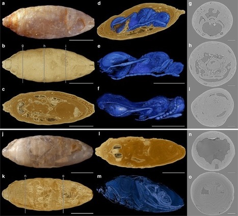
About 50% of all animal species are considered parasites. The linkage of species diversity to a parasitic lifestyle is especially evident in the insect order Hymenoptera. However, fossil evidence for host–parasitoid interactions is extremely rare, rendering hypotheses on the evolution of parasitism assumptive. Here, using high-throughput synchrotron X-ray microtomography, scientists examine 1510 phosphatized fly pupae from the Paleogene of France and identify 55 parasitation events by four wasp species, providing morphological and ecological data. All species developed as solitary endoparasitoids inside their hosts and exhibit different morphological adaptations for exploiting the same hosts in one habitat. Our results allow systematic and ecological placement of four distinct endoparasitoids in the Paleogene and highlight the need to investigate ecological data preserved in the fossil record.
|
|
Scooped by
Dr. Stefan Gruenwald
August 29, 2018 1:01 PM
|
Body weight appears to be under control of higher-level brain systems involved in cognition, decision-making, and motivation. Clinicians should consider how the way we think can make us vulnerable to obesity, and how obesity is genetically intertwined with brain structure and mental performance, according to researchers at the Montreal Neurological Institute and Hospital (The Neuro). Their study (“Neurobehavioral correlates of obesity are largely heritable"), published in the Proceedings of the National Academy of Sciences, was an examination of magnetic resonance imaging (MRI) and cognitive test data from 1,200 individuals, supplied as part of the Human Connectome Project. Researchers found that people with higher a body mass index (BMI) showed reduced cognitive flexibility, ability to delay gratification, visuospatial ability, and verbal memory. They also found that people with increased BMI tended to have a thicker left prefrontal cortex and a thinner right prefrontal cortex. Previous studies have shown damage to the right prefrontal cortex can lead to increased eating. Subjects with higher BMI also had increased volume in the left amygdala, which is believed to play a role in response to food cues. They also had decreased volume in the entorhinal-parahippocampal structures, which are associated with episodic memory and context mediation. This suggests a model where people prone to obesity are more sensitive to visual food cues, and less able to resist them by considering the negative context of eating, like weight gain. Many of the subjects were siblings, including fraternal and identical twins. This allowed researchers to determine the heritability of the traits as well as obesity, measured by BMI. Using statistical methods, researchers found that many of the cognitive and neurological traits have genetic links with obesity. This suggests the role genetics play in obesity is manifested at least partially through brain anatomy and cognitive functions. "This research will be useful in developing interventions to help people with obesity," says the study's lead author, Uku Vainik, a researcher at The Neuro and the Institute of Psychology at University of Tartu, Estonia. "Modifying neurobehavioral factors with cognitive training, to improve people's ability to resist food, for example, could hold promise. Interventions shouldn't just focus on diet, but also acknowledge the neurobehavioural profile that obesity is genetically intertwined with. Such interventions might help people to stay lean despite their genetic signature." "This work adds support to the theory that body weight in humans is partly under control of higher-level brain systems involved in cognition, decision-making, and motivation," says Alain Dagher, M.D., associate professor, McGill University and the paper's senior author. "Furthermore, individual differences in these brain systems that regulate food intake appear to be moderately heritable."
|
|
Scooped by
Dr. Stefan Gruenwald
August 28, 2018 10:06 PM
|
Since the birth of modern astronomy, scientists have sought to determine the full extent of the Milky Way galaxy and learn more about its structure, formation and evolution. According to current theories, it is widely believed that the Milky Way formed shortly after the Big Bang (roughly 13.51 billion years ago). This was the result of the first stars and star clusters coming together, as well as the accretion of gas directly from the Galactic halo. Since then, multiple galaxies are thought to have merged with the Milky Way, which triggered the formation of new stars. But according to a new study by a team of Japanese researchers, our galaxy has had a more turbulent history than previously thought. According to their findings, the Milky Way experienced a dormant era between two periods of star formation that lasted for billions of years, effectively dying before coming back to life again. Their study, titled “The formation of solar-neighbourhood stars in two generations separated by 5 billion years“, recently appeared in the scientific journal Nature. The study was conducted by Masafumi Noguchi, an astronomer from the Astronomical Institute at Tohoku University, Japan. Using a new idea known as “cold flow accretion”, Noguchi calculated the evolution of the Milky Way over a 10 billion year period.
|
|
Scooped by
Dr. Stefan Gruenwald
August 28, 2018 7:27 PM
|
New research has narrowed down the date of the most severe mass extinction known to have occurred on Earth. Additionally, the study has determined the rate at which extinctions occurred. It’s well known that Earth’s most severe mass extinction occurred about 250 million years ago. What’s not well known is the specific time when the extinctions occurred. A team of researchers from North America and China have published data which explicitly provides the date and rate of extinction. "This is the first paper to provide rates of such massive extinction," says Dr. Charles Henderson, professor in the Department of Geoscience at the University of Calgary and co-author of the paper: Calibrating the end-Permian mass extinction. "Our information narrows down the possibilities of what triggered the massive extinction and any potential kill mechanism must coincide with this time." About 95 percent of marine life and 70 percent of terrestrial life became extinct during what is known as the end-Permian, a time when continents were all one land mass called Pangea. The environment ranged from desert to lush forest. Four-limbed vertebrates were becoming diverse and among them were primitive amphibians, reptiles and a group that would, one day, include mammals. Through the analysis of various types of dating techniques on well-preserved sedimentary sections from South China to Tibet, researchers determined that the mass extinction peaked about 252.28 million years ago and lasted less than 200,000 years, with most of the extinction lasting about 20,000 years. "These dates are important as it will allow us to understand the physical and biological changes that took place," says Henderson. "We do not discuss modern climate change, but obviously global warming is a biodiversity concern today. The geologic record tells us that ‘change’ happens all the time, and from this great extinction life did recover." There is ongoing debate over whether the death of both marine and terrestrial life coincided, as well as over kill mechanisms, which may include rapid global warming, hypercapnia (a condition where there is too much CO2 in the blood stream), continental aridity and massive wildfires. The conclusion of this study says extinctions of most marine and terrestrial life took place at the same time. And the trigger, as suggested by these researchers and others, was the massive release of CO2 from volcanic flows known as the Siberian traps, now found in northern Russia.
|
|
Scooped by
Dr. Stefan Gruenwald
August 28, 2018 6:33 PM
|
How Einstein’s equivalence principle extends to the quantum world has been puzzling physicists for decades, but a team including a University of Queensland researcher has found the key to this question. UQ physicist, Dr Magdalena Zych from the ARC Centre of Excellence for Engineered Quantum Systems, and the University of Vienna’s Professor Caslav Brukner have been working to discover if quantum objects interact with gravity only through curved space-time. “Einstein’s equivalence principle contends that the total inertial and gravitational mass of any objects are equivalent, meaning all bodies fall in the same way when subject to gravity,” Dr Zych said. “Physicists have been debating whether the principle applies to quantum particles, so to translate it to the quantum world we needed to find out how quantum particles interact with gravity. “We realized that to do this we had to look at the mass.” Mass is dynamic quantity and can have different values, and in quantum physics, mass of a particle can be in a quantum ‘superposition’ of two different values. According to the famous equation E=MC2, the mass of any object is held together by energy. In a state unique to quantum physics, energy and mass can exist in a 'quantum superposition' – as if they consisted of two different values 'at the same time'. “We realized that we had to look how particles in such quantum states of the mass behave in order to understand how a quantum particle sees gravity in general,” she said. “Our research found that for quantum particles in quantum superpositions of different masses, the principle implies additional restrictions that are not present for classical particles — this hadn’t been discovered before. It means that previous studies that attempted to translate the principle to quantum physics were incomplete because they focused on trajectories of the particles but neglected the mass.” The study opens a door for new experiments that are necessary to test if quantum particles obey the additional restrictions that have been found. The study has been published in Nature Physics (DOI: 10.1038/s41567-018-0197-6)
|
|
Scooped by
Dr. Stefan Gruenwald
August 28, 2018 6:25 PM
|
Researchers at the University of Birmingham have developed a contact lens that may help people with color blindness simply by using a low cost dye, according to research published today (26 April 2018) in the journal Advanced Healthcare Materials. Color blindness -- or color vision deficiency (CVD) -- is an inherited genetic ocular disorder where some people have difficulty distinguishing certain colors (5% of males and 0.8% of females). While no cure for this disorder exists, several methods have been used to increase the color perception of those affected. However, current products on the market such as color filtering glasses are expensive, bulky and incompatible with other vision corrective glasses. Normal color vision is trichromatic -- this means any color can be created by combining the colors blue, red and green, which are perceived by a cluster of cones at the back of the eye. These cones are divided into three groups, responsible for short wavelengths -- blue -- medium wavelengths -- green -- and long wavelengths -- red. In normal vision all three are present. When any of these cones are missing, the brain receives incorrect information leading to limited ability to identify certain colors in some people. Several companies are already selling glasses and custom made lenses for color blindness correction which can be expensive for many users, however, in this research an inexpensive soft commercial contact lens was dyed with a non-toxic rhodamine derivative dye. This particular derivative of rhodamine was chosen as it is known for its ability to absorb certain wavelengths of light in the optical spectrum. Researchers found that the dye blocked the band that lies between the red and green wavelengths, which is perceived by two sets of corresponding optical cones simultaneously. The removal of this band through the dyed lens inhibited the simultaneous triggering of the cones designated for green and red wavelength bands, enabling better differentiation between red and green colors. The dyed lens was tested on people with red-green color vision deficiency (the most common form of CVD). The dyed contact lens was applied to a glass slide. The participants were asked to look at several numbers through the dyed lens, and to note whether there were any improvements to the colors or the clarity of the number. They were also asked to observe their surroundings and note whether they saw any improvements in their color perception. The results verified that dye tinted lenses can be used to enhance the color perception of people affected by color vision deficiency. Further patient studies are now underway. Dr Haider Butt, lead researcher from the University of Birmingham's Department of Mechanical Engineering and the Institute of Healthcare Technologies., said: "Contact lenses are of interest for color blindness correction because it is easier to correct the entire field of view. The dye processing we carried out does not need any complex preparation, it is not toxic to the human eye, and our method could be easily used in both glasses and contact lenses at low cost." He continued: "We are now looking into using a similar process to correct purple-blue color blindness, and also to bring together a number of dyes to make lenses perform for both red-green and purple-blue color blindness simultaneously. We are about to commence human clinical trials shortly."
|
|
Scooped by
Dr. Stefan Gruenwald
August 28, 2018 6:03 PM
|
Regions of several dozen to several hundred base pairs of extreme conservation have been found in non-coding regions in all metazoan genomes. The distribution of these elements within and across genomes has suggested that many have roles as transcriptional regulatory elements in multi-cellular organization, differentiation and development. Currently, there is no known mechanism or function that would account for this level of conservation at the observed evolutionary distances. Previous studies have found that, while these regions are under strong purifying selection, and not mutational coldspots, deletion of entire regions in mice does not necessarily lead to identifiable changes in phenotype during development. These opposing findings lead to several questions regarding their functional importance and why they are under strong selection in the first place. In this perspective, the authors discuss the methods and techniques used in identifying and dissecting these regions, their observed patterns of conservation, and review the current hypotheses on their functional significance.
|
|
Scooped by
Dr. Stefan Gruenwald
August 28, 2018 3:00 PM
|
America’s gun addiction is bad. But to understand just how bad it is, you’ve got to see the numbers.
|
|
Scooped by
Dr. Stefan Gruenwald
August 27, 2018 10:35 PM
|
While hundreds of circular RNAs (circRNAs) are abundant in mammalian brains, one big question has remained unanswered: What are they actually good for? Published in Science, Nikolaus Rajewsky and his team at the Berlin Institute of Medical Systems Biology (BIMSB) of the Max Delbrück Center for Molecular Medicine in the Helmholtz Association (MDC), as well as other collaborators within the MDC and Charité, present data that -- for the first time -- link a circular RNA to brain function. RNA is much more than the mundane messenger between DNA and the protein it encodes. Indeed, there are several different kinds of non-coding RNA molecules. They can be long non-coding RNAs (lncRNAs) or short regulatory RNAs (miRs); they can interfere with protein production (siRNAs) or help make it possible (tRNAs). In the past 20 years, scientists have discovered some two dozen RNA varieties that form intricate networks within the molecular microcosm. The most enigmatic among them are circRNAs, an unusual class of RNAs whose heads are connected to their tails to form a covalently closed ring. These structures had for decades been dismissed as a rare, exotic RNA species. In fact, the opposite is true. Current RNA-sequencing analyses have revealed that they are a large class of RNA, which is highly expressed in brain tissues. Thousands of circular RNAs exist in nematode worms, mice and humans In 2013, two pioneering studies that characterized circular RNAs appeared in the journal Nature, one of them by Nikolaus Rajewsky and his team. Intriguingly, most circular RNAs are unusually stable, floating in the cytoplasm for hours and even days on end. The systems biologists proposed that -- at least sometimes -- circRNAs serve gene regulation. Cdr1as, a large single-stranded RNA loop that is 1,500 nucleotides around, might act as a "sponge" for microRNAs. For example, it offers more than 70 binding sites for a microRNA called miR-7. MicroRNAs are short RNA molecules that typically bind to complementary sequences in messenger RNAs, thereby controlling the amounts of specific proteins produced by cells. Additionally, Rajewsky and his collaborators mined databases and discovered thousands of different circRNAs in nematode worms, mice and humans. Most of them were highly conserved throughout evolution. "We had found a parallel universe of unexplored RNAs," says Rajewsky. "Since publication the field has exploded; hundreds of new studies have been carried out." Understanding a circle that is mostly present in excitatory neurons For the Science paper, the systems biologists teamed up with Carmen Birchmeier's lab at the MDC to reconsider Cdr1as. "This particular circle can be found in excitatory neurons but not in glial cells," says Monika Piwecka, one of the first authors of the paper and coordinator of most of the experiments. "In brain tissues of mice and humans, there are two microRNAs called miR-7 and miR-671 that bind to it." In a next step, Rajewsky and his collaborators selectively deleted the circRNA Cdr1as in mice using the genome editing technology CRISPR/Cas9. In these animals, the expression of most microRNAs in four studied brain regions remained unperturbed. However, miR-7 was downregulated and miR-671 upregulated. These changes were post-transcriptional, consistent with the idea that Cdr1as usually interacts with these microRNAs in the cytoplasm. "This indicates that Cdr1as usually stabilizes or transports miR-7 in neurons by sponging them up, while miR-167 might serve to regulate levels of this particular circular RNA," says Rajewsky. If microRNA floated in the cytoplasm without binding anywhere, it would get broken down as waste. The circle would prevent that and also carry it to new places like the synapses. He adds: "Maybe we should think about Cdr1as not as a 'sponge' but as a 'boat.' It prevents its passengers from drowning and also moves on to new ports." The changes in microRNA concentration had dramatic effects on the mRNA and proteins produced by nerve cells, especially for a group called "immediate early genes." They are part of the first wave of responses when stimuli are presented to neurons. Also affected were messenger RNAs that encode proteins involved in the maintenance of the animals' sleep-wake cycles.
|
|
Rescooped by
Dr. Stefan Gruenwald
from Päivät
August 27, 2018 3:57 PM
|
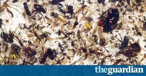
The abundance of flying insects has plunged by three-quarters over the past 25 years, according to a new study that has shocked scientists. Insects are an integral part of life on Earth as both pollinators and prey for other wildlife and it was known that some species such as butterflies were declining. But the newly revealed scale of the losses to all insects has prompted warnings that the world is “on course for ecological Armageddon”, with profound impacts on human society. The new data was gathered in nature reserves across Germany but has implications for all landscapes dominated by agriculture, the researchers said. The cause of the huge decline is as yet unclear, although the destruction of wild areas and widespread use of pesticides are the most likely factors and climate change may play a role. The scientists were able to rule out weather and changes to landscape in the reserves as causes, but data on pesticide levels has not been collected. “The fact that the number of flying insects is decreasing at such a high rate in such a large area is an alarming discovery,” said Hans de Kroon, at Radboud University in the Netherlands and who led the new research. “Insects make up about two-thirds of all life on Earth [but] there has been some kind of horrific decline,” said Prof Dave Goulson of Sussex University, UK, and part of the team behind the new study. “We appear to be making vast tracts of land inhospitable to most forms of life, and are currently on course for ecological Armageddon. If we lose the insects then everything is going to collapse.” The research, published in the journal Plos One, is based on the work of dozens of amateur entomologists across Germany who began using strictly standardised ways of collecting insects in 1989. Special tents called malaise traps were used to capture more than 1,500 samples of all flying insects at 63 different nature reserves.
Via mrhill, Skuuppilehdet
|
|
|
Scooped by
Dr. Stefan Gruenwald
August 30, 2018 7:08 PM
|
Scientists are fighting dengue fever with the help of Wolbachia, a common bacterium that stops the virus from replicating inside the mosquitoes that transmit the disease. Without it, we have few weapons against dengue. Although the bacterium is common among insects, it does not infect Aedes aegypti, a species of mosquito that is a major carrier of dengue. Instead researchers infect the mosquito with Wolbachia in the laboratory and then release A. aegypti into the wild. The goal is to reduce infections in humans by getting Wolbachia-infected mosquitoes to mate and pass the bacterium to future generations. If the method works, vast numbers of wild mosquitoes will eventually carry Wolbachia and thus be unable to transmit dengue.
|
|
Scooped by
Dr. Stefan Gruenwald
August 30, 2018 12:46 AM
|
A Rice University computer scientist and his colleagues have proposed a scalable algorithm for quantum state tomography to significantly accelerate the imposing task of validating the accuracy of quantum computers. Anastasios Kyrillidis, an assistant professor of computer science who joined Rice this year, led the development of a nonconventional method as a diagnostic tool for powerful, next-generation computers that depend on the spooky actions of quantum bits -- aka qubits -- which are switches that operate under rules that differ from the 1s and 0s in classical computers. Quantum computers exploit the principles of quantum mechanics to quickly solve tough problems that would take far longer on conventional supercomputers. They promise future breakthroughs in drug design, advanced materials, cryptography and artificial intelligence. An open-access paper by Kyrillidis and his team appears in the Nature journal Quantum Information. Like any new hardware, Kyrillidis said, quantum computer systems are prone to bugs that need to be squashed. That takes continuous testing to validate their capabilities. The sheer complexity of quantum computers that do exponentially more with every bit requires an immense amount of validation, he said. Kyrillidis' method focuses on quantum state tomography, a process inspired by medical tomography in which images of a body are captured in slices that are later reassembled into a three-dimensional map. Quantum state tomography differs, he said, as it takes "images" of the state of a quantum computer's qubits. "When a quantum computer executes an algorithm, it starts at a specific state; think of it as the input to the algorithm," Kyrillidis said. "As the computer progresses through steps of the algorithm, it's going through many states. The state at the very end is the answer to your algorithm's question." By reassembling the full state from these measurements, Kyrillidis said one can later pinpoint hardware or software errors that may have caused the computer to deliver unexpected results. That takes a lot of measurements, and the computational cost of reconstruction can be high, even for classical computers, he said. Tomography-based analysis of quantum computers with even as few as five or six qubits would be prohibitive without somehow simplifying the task -- and state-of-the-art machines have 50 qubits or more. Qubits are the basic units of information in a quantum computer. Like a bit in a classical computer, each qubit can represent either 1 or 0. Unlike a bit, a qubit can also represent 1 and 0 simultaneously, a state called superposition that exponentially raises the number of calculations an array of qubits can perform at once. To make it more interesting, the state of the qubit as determined by magnetic polarization or electron spin only exists when it's measured. Kyrillidis said even a modest increase in the number of qubits in a computer dramatically increases its power. "In a system with five qubits, the state can be represented by a 2-to-the-5 times 2-to-the-5 matrix, so it's a 32-by-32 matrix," he said. "That's not big. But in a 20-qubit system like the one at IBM, the state can be characterized by a million-by-million matrix. If we were taking full measurements with regular tomography techniques, we would need to poll the system roughly a million-squared times in order to get enough information to recover its state." Kyrillidis and his team solved the validation problem with an algorithm they call Projected Factored Gradient Decent (ProjFGD). It takes advantage of compressed sensing, a method that minimizes the amount of incoming data while still ensuring accurate results. He said the method would cut the number of measurements for a 20-qubit system to a mere million or so. "That's still a big number, but much smaller than a million squared," he said. Kyrillidis noted that IBM, where he spent a year as a research scientist before coming to Rice, has put a quantum computer in the cloud where anyone can access it and run programs. He said the company reasons that the more people learn about programming for quantum computers now, the more mature their skills will be when the platform comes of age. But there's a side benefit for him, as it gives him a ready platform to test ProjFGD. "The quantum state tomography tool is generic, and has more to do with the nature of the qubit rather than the specific architecture," Kyrillidis said. "As quantum computers get more powerful, it can definitely be scaled up to certify systems."
|
|
Scooped by
Dr. Stefan Gruenwald
August 29, 2018 1:09 PM
|
We now have the most precise estimates for the strength of gravity yet. Two experiments measuring the tiny gravitational attraction between objects in a lab have measured Newton’s gravitational constant, or Big G, with an uncertainty of only about 0.00116 percent. Until now, the smallest margin of uncertainty for any G measurement has been 0.00137 percent. The new set of G values, reported in the Aug. 30 Nature, is not the final word on G. The two values disagree slightly, and they don’t explain why previous G-measuring experiments have produced such a wide spread of estimates (SN Online: 4/30/15). Still, researchers may be able to use the new values, along with other estimates of G, to discover why measurements for this key fundamental constant are so finicky — and perhaps pin down the strength of gravity once and for all. The exact value of G, which relates mass and distance to the force of gravity in Newton’s law of universal gravitation, has eluded scientists for centuries. That’s because the gravitational attraction between a pair of objects in a lab experiment is extremely small and susceptible to the gravitational influence of other nearby objects, often leaving researchers with high uncertainty about their measurements.
|
|
Scooped by
Dr. Stefan Gruenwald
August 29, 2018 1:05 PM
|
For decades, researchers have bickered about claims to have found the oldest life on Earth – but a new analysis seems to have solved the problem, and with a surprise result. Arguments often revolve around whether tiny squiggles, blobs and tubes found in ancient rocks are fossilised lifeforms or just weird minerals. Now, a study published in the journal Nature Ecology and Evolution leapfrogs these squabbles. Rather than arguing about forms, a team from the University of Bristol in the UK, led by phylogeneticist Davide Pisane, has taken a big data approach. Using 29 DNA codes that are common to species across the tree of life, they pinpoint the emergence of the last universal common ancestor (LUCA) at an extraordinarily early 4.5 billion years ago – just an eye-blink after our planet formed. Did life come from space and did the first cells land here on Earth just after our planet cooled?
|
|
Scooped by
Dr. Stefan Gruenwald
August 28, 2018 10:16 PM
|
World’s largest laser explores high-pressure conditions at the core of giant planets. Sientists have bombarded hydrogen with 168 powerful laser beams – part of the world’s largest laser, the National Ignition Facility at Lawrence Livermore National Laboratory – to reproduce what happens to the gas under the intense pressures at the cores of giant planets. What they saw confirms predictions that if you keep hydrogen cool enough while pounding on it with six million times atmospheric pressure – twice the pressure at the center of the Earth – the hydrogen turns into a shiny metal. “It’s like squeezing air and turning it into a shiny, light version of liquid mercury. Although exotic at Earth’s surface, metallic hydrogen is the main material inside most giant planets and stars,” said Raymond Jeanloz, a UC Berkeley professor of astronomy and of earth and planetary science and a co-author of the research. The team’s optical measurements of the so-called insulator-to-metal transition in fluid hydrogen resolved discrepancies in previous experiments and established new benchmarks for calculations used to construct planetary models. Livermore Lab physicist Peter Celliers was the lead author of the paper, which was published this week in the journal Science. “These results are a true experimental tour de force and are particularly important because they provide a very stringent test on the different varieties of numerical simulations that one can use to predict the properties of planetary constituents at high pressure — necessary to model the internal structure and evolutionary processes of Jupiter and Saturn,” said Marius Millot, a physicist at LLNL and co-author of the paper.
|
|
Scooped by
Dr. Stefan Gruenwald
August 28, 2018 7:58 PM
|
Researchers from UCLA and Washington University in St. Louis have discovered the previously unknown mechanism of how proteins from Plasmodium parasites — which cause malaria — are exported into human red blood cells, a process that is vital for parasites to survive in humans. The finding could pave the way for new treatments for malaria. In a recent study, published in Nature, researchers reveal the atomic structure of the Plasmodium translocon of exported proteins, or PTEX, the protein complex that transports these malaria proteins into the red blood cell. The research also provides the first direct evidence of the proteins moving through PTEX. “For many years, scientists hypothesized that PTEX was responsible for transporting these malaria proteins,” said Z. Hong Zhou, a professor of microbiology, immunology and molecular genetics and director of the Electron Imaging Center for Nanomachines at the California NanoSystems Institute at UCLA, who led the study. “We just never knew how it worked, until now.” Malaria is an infectious disease transmitted to people through the bites of infected mosquitos. It affects more than 200 million people annually, causing nearly a half-million deaths each year. When a human is bitten, Plasmodium parasites invade human red blood cells and take a part of the red blood cell membrane to build a protective compartment, or vacuole, around itself. “Normal red blood cells are too simple to provide enough nutrients to support the actively growing parasites,” said Chi-Min Ho, a UCLA graduate student and first author of the study. “Each parasite is essentially living in an empty warehouse and has to produce hundreds of malaria ‘effector’ proteins to remodel the red blood cell into a home.” PTEX acts as a gatekeeper; without it, effector proteins become trapped in the vacuole. Ho and her colleagues wanted to learn exactly how PTEX is involved in transporting the proteins out into the red blood cell.
|
|
Scooped by
Dr. Stefan Gruenwald
August 28, 2018 7:20 PM
|
Circular RNAs (circRNAs) play critical physiologic functions, but it is not known whether human DNA viruses express circRNAs. Scientists now surveyed Epstein−Barr virus (EBV) and Kaposi’s sarcoma herpesvirus (KSHV) tumors and cell lines, and found specific circRNAs expressed from both viruses. EBV circular BamHI A rightward transcripts (circBARTs) were expressed in all EBV tumor latency forms, including all EBV-infected post-transplant lymphoproliferative disease tumors tested, whereas EBV circBHLF1 and circLMP2 were more variably expressed. KSHV expressed circvIRF4 constitutively in primary effusion lymphoma cell lines, while the polyadenylated nuclear locus promiscuously generated variable, inducible, and bidirectional circRNAs. Tumor virus circRNAs can be long-lived, unique tumor biomarkers that may also open new research opportunities into understanding how these viruses cause cancer.
Epstein−Barr virus (EBV) and Kaposi’s sarcoma herpesvirus (KSHV) cause ∼2% of all human cancers. RNase R-resistant RNA sequencing revealed that both gammaherpesviruses encode multiple, uniquely stable, circular RNAs (circRNA). EBV abundantly expressed both exon-only and exon−intron circRNAs from the BamHI A rightward transcript (BART) locus (circBARTs) formed from a spliced BART transcript and excluding the EBV miRNA region. The circBARTs were expressed in all verified EBV latency types, including EBV-positive posttransplant lymphoproliferative disease, Burkitt lymphoma, nasopharyngeal carcinoma, and AIDS-associated lymphoma tissues and cell lines. Only cells infected with the B95-8 EBV strain, with a 12-kb BART locus deletion, were negative for EBV circBARTs. Less abundant levels of EBV circRNAs originating from LMP2- and BHLF1-encoding genes were also identified. The circRNA sequencing of KSHV-infected primary effusion lymphoma cells revealed a KSHV-encoded circRNA from the vIRF4 locus (circvIRF4) that was constitutively expressed. In addition, KSHV polyadenylated nuclear (PAN) RNA locus generated a swarm (>100) of multiply backspliced, low-abundance RNase R-resistant circRNAs originating in both sense and antisense directions consistent with a novel hyperbacksplicing mechanism. In EBV and KSHV coinfected cells, exon-only EBV circBARTs were located more in the cytoplasm, whereas the intron-retaining circBARTs were found in the nuclear fraction. KSHV circvIRF4 and circPANs were detected in both nuclear and cytoplasmic fractions. Among viral circRNAs tested, none were found in polysome fractions from KSHV−EBV coinfected BC1 cells, although low-abundance protein translation from viral circRNAs could not be excluded. The circRNAs are a new class of viral transcripts expressed in gammaherpesvirus-related tumors that might contribute to viral oncogenesis.
|
|
Scooped by
Dr. Stefan Gruenwald
August 28, 2018 6:30 PM
|
The human brain, with its 100 billion neurons that control every thought, word, and action, is the most complex and delicate organ in the body. Because it needs extra protection from toxins and other harmful substances, the blood vessels that supply the brain with oxygen and nutrients are highly selective about which molecules can cross from the blood into the brain and vice versa. These blood vessels and their unique network of supporting pericyte and astrocyte cells comprise the blood-brain barrier (BBB). When the BBB is disrupted, as happens with exposure to methamphetamine ("meth") and other drugs, the brain's sensitive neurons become susceptible to harmful damage. Beyond forming a physical barrier, the BBB is thought to directly interact with the brain and help regulate its function, but figuring out exactly how the cells of the BBB and the brain influence each other has been a challenge, as in vitro models (i.e., cells in a dish) are too simplified and in vivo models (i.e., natural human brain tissue) too complex. Now, researchers at the Wyss Institute for Biologically Inspired Engineering have created a "just right" model of the BBB-brain interface using microfluidically linked Organ Chips that reacts to meth like the human brain and allows an unprecedented look into how the brain's vasculature influences and regulates its metabolic function. The research is reported in Nature Biotechnology. "Most of today's research on Organ Chips is focused on trying to increase the complexity of cell types on each chip, but we realized that the brain is already so complex that we couldn't analyze it on one chip, so we did the opposite and divided one organ onto multiple chips," said first author Ben Maoz, Ph.D., a former Technology Development Fellow at the Wyss Institute who is currently an Assistant Professor at Tel Aviv University, Israel. "The beauty of this work is that Organ Chips were able to open up another dimension for neurological research that no other method could; decoupling a very dense organ to unveil new interactions between the different structures within the brain." The BBB-Brain Chip system consists of three chips: one "influx" BBB Chip, a Brain Chip, and a second "efflux" BBB Chip, physically distinct from each other but all connected with microfluidic channels to allow the exchange of chemicals and other substances between them, much like how the supplying blood vessels, neuronal compartment, and draining blood vessels are linked in the brain. The BBB Chip has one channel lined with endothelial cells through which flows culture medium that mimics blood, separated by a porous membrane from a parallel channel containing pericytes and astrocytes that is perfused with artificial cerebrospinal fluid (aCSF). The Brain Chip has a similar aCSF flow channel that is separated by another semipermeable membrane from a compartment containing human brain neurons and their supporting astrocytes to mimic brain tissue. The three chips' aCSF channels are connected together in series, creating a fully linked system in which substances can diffuse from the vascular channel across the first BBB into the aCSF, enter the brain neuronal cell compartment, flow back into the aCSF, and ultimately diffuse out across the second BBB into another vascular channel, as happens in vivo. The team cultured human cells in the linked BBB-Brain Chips and exposed them to meth, which is known to disrupt the junctions between the cells of the BBB in vivo and cause it to "leak." When meth was flowed through the blood vessel channel of the BBB Chip, it compromised the junctions of the BBB's vascular endothelial cells and allowed the passage of molecules that normally wouldn't be able to cross the BBB into the Brain Chip. This experiment confirmed that the model worked, and established that it could be used in research to better understand drugs' effects on the human brain and develop treatments.
|
|
Scooped by
Dr. Stefan Gruenwald
August 28, 2018 6:05 PM
|
Of all the adjectives you could use to describe a crocodile’s face, “sensitive” might not be an obvious one. But their huge jaws, pointed teeth and armored scales belie a surprising secret. Their faces, and possibly their entire bodies, are covered with tiny bumps that are far more sensitive than our own fingertips. The bumps are obvious if you look carefully. Each one is a small dome, barely a millimeter wide, surrounded by a groove. There are around 4,000 of them on an alligator’s jaws and inside its mouth. Crocodiles and gharials also have the bumps on virtually every scale of their bodies, giving a total of around 9,000. All of these animals are called crocodilians. They were first described in 1895, and scientists have used them to identify different farmed species of crocodilians from endangered poached ones. But no one knew what the bumps did. Perhaps they secrete waterproofing oils? Maybe they help the croc to detect changes in saltiness, electricity, magnetic fields, or water pressure? There were plenty of hypotheses, but few actual studies. In 2002, Daphne Soares from the University of Maryland showed that American alligators could orient themselves to the ripples created by a single drop of water, even in complete darkness. The bumps on their faces seems to be involved. It was a brief study, but it intrigued Duncan Leitch from Vanderbilt University, who wanted to know what the bump were doing. Now, with Ken Catania, he has shown that they’re touch sensors, which make a crocodilian’s face exquisitely sensitive despite its hard unyielding armor.
|
|
Scooped by
Dr. Stefan Gruenwald
August 28, 2018 5:22 PM
|
The healthy function, or disease-fueling dysfunction, of any given organ stems from the behavior, or misbehavior, of the individual cells that make up that organ. Recent technological advances have enabled scientists to analyze the actions of cells, one at a time, but these technologies can yield only static snapshots of cellular activity. Capturing an individual cell’s behavior as a process that foretells its future rather than an occurrence frozen in time has thus far eluded scientists. Now, for the first time, researchers at Harvard Medical School, working with colleagues at the Karolinska Institutet in Sweden, have succeeded in going beyond such cellular freeze framing and managed to capture a cell’s decision making as a dynamic process during which a cell determines what to do and where it’s headed. The approach, described Aug. 8 in Nature, is a mathematical model that estimates RNA velocity—the rate of RNA change over time—as a predictor of cell fate on the timescale of hours. The ability to capture cellular intent can help scientists better analyze cell function and dysfunction in complex tissues and organs—those composed of various cell types. In addition, it could help monitor how organs develop and how they respond to a given drug or therapy at the cellular level—an insight that can help gauge the efficacy of a treatment. For their predictive model, scientists captured changes in mRNA, a messenger molecule that interprets the genetic code embedded in DNA and translates these instructions for cells, in essence giving them marching orders on how much of which protein to make. “Estimating RNA velocity—or the rate of RNA change over time—is akin to observing the cooks in a restaurant kitchen as they line up the ingredients to figure out what dishes they’ll be serving up next,” said Peter Kharchenko, co-senior author of the study and assistant professor of biomedical informatics at Harvard Medical School. The scientists measured trace molecules—markers already being captured during standard single-cell analyses—but often dismissed as “noise.” These molecular markers, the scientists say, provide clues into both a cell’s intent and the footprints of its past. The markers emerge at various parts of the RNA lifecycle—a process during which a cell turns genetic instructions into functioning proteins. The process occurs in five discrete steps: - Transcription, during which a cell reads parts of its DNA to produce precursor messenger RNA, or pre-mRNA, in a nascent form.
- Splicing and other processing, during which the nascent pre-mRNA is edited into a mature mRNA molecule—the written marching orders that guide the making of functional proteins.
- Nuclear export, during which the genetic marching orders, written in the mature mRNA, get exported from the cell’s nucleus—the vault where genetic code is kept—and into the cell cytoplasm for processing.
- Translation, during which the mature mRNA releases the written orders and turns them into functional proteins.
- Degradation, during which mRNA, having done its job, is chopped up and marked for destruction by recycling.
During this cycle, cells contain a mixture of mRNA in various stages—newly made, nascent pre-mRNA, mature mRNA and fragmented, chopped-up mRNA on its way to being recycled. Each stage bears a different molecular trace that can provide clues about a cell’s future direction and ultimate destiny, the team hypothesized. “We reasoned that by distinguishing mRNA molecules in different stages of the life cycle, we would capture past, present and future states of individual cells,” Kharchenko said.
|
|
Scooped by
Dr. Stefan Gruenwald
August 27, 2018 10:43 PM
|
Pioneer 10 and 11 each carry a 6 x 9-inch (15 x 23 centimeters), gold-anodized aluminum plaque. The plaque is affixed to support struts close to the spacecraft’s bus (main body). Carl Sagan and Frank Drake played key roles in designing the plaque and Linda Salzman Sagan, Sagan’s wife at the time, was the artist who actually drew the images engraved on the plaque. The most striking feature of the plaque are the figures of the man and the woman overlying the silhouette of the Pioneer spacecraft itself. While this does clearly convey our physical size and shape, as well as that sexual dimorphism is present in humans, the facial features of the couple have little detail and the sort of sensory organs are being depicted (the couple’s ears are barely shown at all) might be unclear. The man and woman both have their mouths closed, and viewers might not understand that these are even mouths at all. Given how the image is drawn, an observer could also be forgiven for not understanding that both the male and female have hair on their heads. The couple both have a bland expression (which may have been an attempt to avoid anything that could be interpreted as hostile) and the man is seen raising his right hand with the palm facing the viewer. While this gesture clearly conveys a greeting when viewed by another human, an extraterrestrial may have no way of interpreting this gesture. Could you interpret a gesture made by an antelope … or a praying mantis? It does show, however, that humans have opposable thumbs, as well as the general range of motion of the upper limbs. With regards to the scientific data presented, the top left of the plaque shows the hyperfine transition of neutral hydrogen as a means of conveying to the reader baseline units of time (0.7 nanoseconds, the frequency of the transition) and distance (21 cm, the wavelength of the light released by the transition). If one is able to deduce that the image is that of hydrogen, the time and distance should be understandable. The plaque also contains a map of our sun relative to 14 pulsars as well as the center of our galaxy, conveying both the distances to the pulsars and their frequency in binary notation. As this image conveys copious objective data, a spacefaring species might well be able to easily interpret it. Finally, the plaque contains a map of the solar system. The solar system map is likely among the more easily interpreted parts of the plaque, with Pioneer shown to have originated from the third planet. The plaque was created at a time when Pluto was still considered the ninth planet (before the discovery of other trans-Neptunian dwarf planets such as Eris and Sedna, among others), but it would still direct the reader’s attention to Earth if they were able to figure out that our solar system was the one depicted. The Voyager golden record (left) is a 12-inch gold-plated copper disc. Its cover aluminum electroplated with an ultra-pure sample of uranium-238. (Credit: NASA) Voyager’s Golden Record The Voyager record asks more of whoever finds it but gives more information in return. These phonographs, attached to the spacecraft bus, feature a cover illustration and over 90 minutes of audio on the reverse side. The cover illustration features the same image of hydrogen and the same pulsar map as found on the Pioneer plaque. Of critical importance, the Voyager records convey instructions on how to play them, such as how to affix the attached stylus, at what rate of rotation the record must be spun, and the proper waveform of signals generated by the record. It also explicitly tells the reader how to know if they are viewing the images properly via an engraving of what the first image (a circle) should look like. While this may seem very daunting, the challenge is primarily technical and might well be easily overcome by an advanced spacefaring species. An alien species might well find more difficulty in interpreting the audio samples, music, and images contained on the record. There are over 50 greeting messages in different languages. While the specifics of the messages are likely to be uninterpretable, they would at least convey to the listener the diversity of the creatures who created the Voyagers. Similarly, the musical selections chosen demonstrate a wide range of human musical styles (ranging from works of Beethoven and Stravinsky to those of Chuck Berry, among others). While the lyrics of “Johnny B. Goode” are probably gibberish to an extraterrestrial, the beat and rhythm of the song would convey a tremendous amount to an alien listener. Of perhaps the greatest importance are the 115 images encoded on the record. The first six images, if decoded properly, provide immense technical data for the reader regarding mathematical definitions, scales and sizes, as well as additional information regarding our location and how to find us. Images of the sun and its spectrum, as well as some of the planets in our solar system, could help the discoverer of the Voyagers to find us should they decide to pay the Earth a visit. There are also approximately 20 medical and scientific diagrams including the structure of DNA and detailed images of human anatomy. These images could likely be interpreted correctly given their concrete nature. The Voyager record also contains a plethora of images of humans engaged in a variety of activities (including eating, looking through a microscope, and even going on a spacewalk). While many of these images would be hard to interpret (e.g., a picture of a woman licking an ice cream cone or a photo of a string quartet) the images would at least convey that humans have created a complex civilization with some degree of advanced technology.
|
|
Scooped by
Dr. Stefan Gruenwald
August 27, 2018 4:01 PM
|
The Sun serves as the primary energy source for Earth’s climate. Some of the incoming sunlight is reflected directly back into space, especially by bright surfaces such as ice and clouds, and the rest is absorbed by the surface and the atmosphere. Much of this absorbed solar energy is re-emitted as heat (longwave or infrared radiation). The atmosphere in turn absorbs and re-radiates heat, some of which escapes to space. Any disturbance to this balance of incoming and outgoing energy will affect the climate. For example, small changes in the output of energy from the Sun will affect this balance directly.
If all heat energy emitted from the surface passed through the atmosphere directly into space, Earth’s average surface temperature would be tens of degrees colder than today. Greenhouse gases in the atmosphere, including water vapour, carbon dioxide, methane, and nitrous oxide, act to make the surface much warmer than this, because they absorb and emit heat energy in all directions (including downwards), keeping Earth’s surface and lower atmosphere warm (see Figure b1). Without this greenhouse effect, life as we know it could not have evolved on our planet. Adding more greenhouse gases to the atmosphere makes it even more effective at preventing heat from escaping into space. When the energy leaving is less than the energy entering, Earth warms until a new balance is established. The atmospheric concentrations of carbon dioxide, methane, and nitrous oxide have increased significantly since the Industrial Revolution began. In the case of carbon dioxide, the average concentration measured at the Mauna Loa Observatory in Hawaii has risen from 316 parts per million (ppm) [that is, for every million molecules in the air, 316 of them were CO2] in 1959 (the first full year of data available) to 396 ppm in 2013 (see Figure b2). The same rates of increase have since been recorded at numerous other stations worldwide. Since pre-industrial times, the atmospheric concentration of CO2 has increased by 40%, methane has increased by about 150%, and nitrous oxide has increased by roughly 20%. More than half of the increase in CO2 has occurred since 1970. Increases in all three gases contribute to warming of Earth, with the increase in CO2 playing the largest role. Learn about the sources of human emitted greenhouse gases.
|