 Your new post is loading...
Sciatica is generally described as a set of symptoms, primarily characterized by pain and discomfort, along with tingling sensations and numbness. Athletes frequently report experiencing symptoms of sciatica, however, there are many factors as well as a variety of injuries and conditions which can manifest these well-known symptoms. Piriformis syndrome is a disorder that is frequently confused with symptoms of sciatica. The piriformis muscle is commonly known among athletes and healthcare professionals as a significant muscle in the posterior hip. This muscle functions to control hip joint rotation and abduction and it is also a distinguishable muscle due to its inversion of action in rotation. The piriformis muscle also raises awareness as the various causes of piriformis syndrome, a condition suspected to be a potential source of pain and dysfunction, not only in athletes, but in the general population as well.
Athletes who participate in overhead sports and physical activities, including baseball pitchers, tennis, swimming, water polo and throwing sport athletes, exert tremendous amounts of stress on their shoulders and its surrounding structures when they participate in their specific athletics. For instance, an elite baseball pitcher’s arm has been recorded at over 7000 degrees/second, which arguably makes it the fastest human body movement from any sport. Shoulder pain is a common symptom among the overhead athlete where throwing athletes will generally complain of dead arm, defined as a condition which restricts them from throwing at pre-injury speeds or control. SLAP, or superior labrum anterior-posterior, lesions are common causes of this type of dysfunction. A SLAP tear occurs on the glenoid labrum from the anterior to posterior angle of the long head of the biceps tendon. The glenoid labrum is a wedge-shaped fibrous tissue structure that is attached to the edge of the glenoid, functioning to deepen the glenoid cavity to improve stability as well as implement muscular control and proprioception. The anatomy of the proximal long head bicep tendon may actually vary but, in a majority of cases, it originates from the posterior superior labrum and it is broader and innervated more sensory fibres than the distal tendon.
Approximately 80 percent of the population has or will experienced acute or chronic symptoms of back pain at least once throughout their lifetimes, according to several research studies. While a great majority of these issues are only temporary and they do resolve on their own, the injuries and/or conditions behind the individual's back pain can accumulate tremendous financial burdens from the medical system over time, amounting to a great increase in medical expenses, including lost employee hours and a loss of productivity from the workforce if the affected individual has to take time off to recover from their specific type of back pain. Among the many types of complications, most described as extension related low back pain, some of the most common include: spinal disc herniation and bulges; degeneration of the spine; annular tears; ligament sprains; muscle strains, particularly in the quadratus lumborum; osteoarthritis; rheumatoid and ankylosing spondylitis; facet joint sprains; and stress fractures, pars defects and spondylolisthesis. However, bone injuries causing extension related low back pain can be a series of progressive disorders or pathologies along the lumbar spine, caused by the excessive amount of uncontrolled lumbar spine extensions among many athletes. These could basically occur due to a postural, gradual onset of repetitive trauma, most frequently associated with sports, for instance, gymnastics. Two specific demographic groups experience the most extension related low back pain among the general population: the first group includes individuals who stand for prolonged periods of time, such as retailers, military, security guards, etc. Standing for extended periods of time normally forces the pelvis to begin shifting into an anterior tilt angle, placing compressive forces against the facet joints of the lumbar spine as these will also begin to shift towards a position of extension following the pelvic tilt; and the second group includes athletes who participate in extension sports, such as gymnastics, tennis, swimming, diving, football, volleyball, basketball, track and field, and cricket fast bowlers, and experience sports injuries. This may be more distinct in sports which include extension/rotation.
Super Bowl 50 will showcase the leading players in the National Football League, with Joshua Kollmann, DC and Brad Wiest, DC – team chiropractors for the Carolina Panthers – and Shawn Caldwell, DC, team chiropractor for the Denver Broncos, helping players achieve optimal performance.
The Foundation for Chiropractic Progress (F4CP), a not-for-profit organization dedicated to raising awareness about the value of chiropractic care, points out that all 32 NFL teams include the professional services of a doctor of chiropractic (DC) as part of their integrated health care team approach.
Marking their fifth year with the Carolina Panthers, Drs. Kollmann (pic. left) and Wiest (pic. right) highlight the integrative role of chiropractic care: “We are in the stadium training room twice a week – and more often during play-offs – addressing specific sports injuries or providing preventive, maintenance care that the athletes want in order to achieve peak performance. Every player is individually assessed and the treatment plan is communicated and discussed with the training staff. Since the physical nature of the sport really impacts body structure, many players look forward to their pre- and post-game spinal and extremity adjustments as well as other advanced approaches.”
Shawn Caldwell, DC, who has served the Denver Broncos since 2004 and is now preparing for his second Super Bowl, says, “I work hand-in-hand with the athletic trainers and focus on performing chiropractic spine and extremity adjustments that restore joint function. The goal is to enable players to perform optimally or heal from injuries. I am at the facility two-three times weekly or more if necessary. Some players get an adjustment every time I am in the training room, while others when they are symptomatic so they can return to the playing field.”
According to Kyle Prusso, DC, team chiropractor for the Oakland Raiders since 2005 and president of Pro Football Chiropractic Society, an organization of chiropractors who provide the highest quality chiropractic health care to the elite athletes of professional Football: “It’s great to see chiropractic care integrated in all facets of health care, especially in professional and amateur sports. One of the reasons is that athletes are asking for us, with increased player requests driving utilization of chiropractic across all sports. Athletes are very in tune with their bodies and recognize that chiropractic care can boost optimal performance levels.”
All doctors are passionate about their roles as team chiropractors to an NFL Super Bowl contender, as Dr. Kollmann says, “I am humbled by this position and want the world to know that this is a great time to be a chiropractor. It means the world to me to ignite and advance our profession – especially for those who are pursuing a chiropractic education as well as veteran professionals. We are igniting the profession and honor those DCs who pioneered the opportunities that have helped us to become a part of the NFL teams.”
Dr. Caldwell, who is also the chiropractor for the Colorado Rockies Major League Baseball team, sums it up, “Chiropractors are playing an important role in the health care and performance of professional athletes. This is a fantastic experience for us and for the players.”
Doctors of chiropractic receive a minimum of seven years of higher level education, and are qualified to diagnose, treat and manage a broad spectrum of health conditions. They are the primary care professionals for spinal health and well-being. For athletes, chiropractic care helps to reduce the risk of injuries, and improve health and performance through enhancements in range of motion, flexibility, balance, muscle strength and other key factors.
If you are interested in learning more about how to be an NFL Chiropractor you can connect with the Professional Football Chiropractic Association on their website or on their Facebook page.
No matter what the outcome of this weekends game may be, rest assured that both teams will have been well adjusted and their nervous systems tuned on and ready to perform on the biggest stage of all.
Super Bowl LI kicks-off on February 5, 2017, with team doctors of chiropractic (DC) helping professional athletes to prevent, manage and care for injuries, as well as achieve peak performance. The Foundation for Chiropractic Progress® (F4CP), the leading voice of the chiropractic profession, points to the role of DCs with all four play-off teams,and cites data from the Professional Football Chiropractic Society (PFCS) showing on average, a professional football DC performs between 30 to 50 weekly treatments during the regular season – not including training camp or the playoffs.
F4CP® celebrates Super Bowl LI with NFL doctors of chiropractic
New England Patriots Team Chiropractor of 35+ years and proud to attend his ninth Super Bowl, Dr. Michael Miller, states, “During the NFL season, I regularly visit the stadium on my afternoon off from my office, as well as on game days both at home and away. Chiropractic care is emphasized by the head coach, trainers and medical staff as a proactive regimen to prevent injuries, with most of the players receiving adjustments roughly 1-2 times per week.”
He adds that chiropractic care has proven itself over the years in the sports injury arena to be well-accepted as the choice of champions and an integral part of any injury prevention program. “We’ve also earned the respect of other team physicians as a necessary protocol, and are all committed to one goal: keeping the athletes performing at their maximum potential and preventing and managing injuries as they occur.”
He says that chiropractic care provides the athletes with the confidence they need to play without the distraction of pain. “The players are educated about the principles of chiropractic and that it is designed to eliminate the cause of their problem rather than just masking their symptoms.”
Joseph Krzemien, DC, team chiropractor, Atlanta Falcons, says that there is growing evidence and a correlation between football players who receive consistent chiropractic care and a decline in injuries during practice or play:
“During the six seasons I’ve spent with the Atlanta Falcons, my goal has always been to prepare each player’s body to better resist trauma and to speed its natural recovery time,” he says. “This season, the Falcons have been successful for a lot of reasons, but I firmly believe that regular chiropractic care has played an important role in helping us stay healthy and get to Super Bowl LI – I am proud of the role I’ve played in their success.”
For Michael Zoelle, DC, team chiropractor, Green Bay Packers, the trauma experienced by the athletes’ bodies during a game is very similar to that of a car accident.
“Ensuring that the joints are functioning properly is critical in the healing process, as well as for injury prevention and optimal performance,” says Dr. Zoelle. “The players recognize that chiropractic care helps them to perform better and heal faster, ultimately leading to better team success.”
DCs receive a minimum of seven years of higher level education, and are qualified to diagnose, treat and manage a broad spectrum of health conditions. They are the primary care professionals for spinal health and well-being. For athletes, chiropractic care helps to reduce the risk of injuries and improve health and performance through enhancements in range of motion, flexibility, balance, muscle strength and other key factors.
It's not uncommon for athletes to suffer damage or injury as a result of an accident while on the field, occasionally developing or aggravating a previous condition in the process, which can affect their performance. Sports or physical activities requiring rigorous training and performance during competitive have a higher risk of injury among athletes. Knee injuries are some of the most common complications affecting numerous athletes throughout their career, most frequently hyperextension knee injuries caused during contact sports. Hyperextension knee injuries can vary from acute to chronic and these are generally painful. The infrapatellar fat pad, abbreviated as IPFP, is one of the most frequently affected structures due to hyperextension knee injuries. In the presence of an acute knee hyperextension injury, for instance, when an athlete is tackled in rugby, the posterior cruciate ligament, or PCL and/or the posterior lateral corner, or PLC, of the knee may suffer injury.
Athletes are subjected to experience a variety of injuries or conditions due to their exposure of rigorous activities. While they may be highly trained to warm up and exercise accordingly before engaging in their specific physical activity or sport, an accident during practice or training can often be unpredictable and inevitable. From landing incorrectly after a jump to simple degeneration, athletes who frequently utilize their feet in numerous exerting ways are more prone to suffer foot injuries than the general population. Foot injuries occur frequently among athletes and they manifest in various forms. From stress fractures of the metatarsals and tarsal bones to chronic soft tissue injuries, such as plantar fasciitis and midfoot sprains to the joints between the tarsals and metatarsals. Although generally considered to occur infrequently, injuries to the midfoot, specifically the Lisfranc joint or the tarsometatarsal joint during Lisfranc injuries, these afflictions require special attention as they can be considerably devastating to most athletes. Lisfranc injuries are high speed injuries which can result in serious deformity of the midfoot joints, commonly due to dislocations and/or fractures. Automobile or motorcycle accidents, violent falls onto the foot or a severe, crushing injury to the foot, are several of the most common circumstances which can lead to Lisfranc injuries. In sport settings, this form of foot injury is less severe, often resulting from a crushing and/or twisting means to the planted weight-bearing foot. But, when it does occur, Lisfranc injuries can cause overwhelming consequences for the athlete. Approximately 16 percent of all sports injuries involve the foot. Although foot complications in athletes are reasonably common, Lisfranc injuries are quite rare. Researchers found that Lisfranc injuries account for up to 4 percent of all college level football injuries. Severe forms of the complication can unfortunately not only be season-ending but career-ending as well for the athlete.
Transverse process fractures are an uncommon type of sport injury reported in athletes. It commonly develops after direct contact injuries and/or due to a strong forceful contraction of the quadratus lumborum muscle when the trunk is in a position of side flexion. The transverse processes can become fractured through direct trauma or injury from contact sports or due to direct impact from falls off heights or from motorcycles. Automobile accidents can also cause injury to these important structures. In addition, because damage to the transverse processes usually involve high velocity forces, surrounding organs may also be affected. Also, depending on the extent of trauma, multiple transverse processes can be fractured at the same time. Furthermore, a muscle avulsion where it pulls off the transverse process can occur during a muscle contraction while in a forward bend, rotating posture or with strong lateral flexion movements, for instance, during rugby, MMA and judo. Finally, repetitive microtrauma as a result of constant and repetitive motions in certain sports and physical activities may also cause fractures, for example, as with cricket fast bowlers. The three most common mechanisms of injury recorded with professional elite level rugby athletes included: direct impact from a knee, which causes fractures to the L2 and L3 transverse processes found in the lumbar spine, without causing other forms of injury; a strong blow to the upper chest/arm area from the side, which generally causes a player to contract their muscles, including the quadratus lumborum, resulting in an avulsion to the L3 transverse processes of the lumbar spine; and last, an individual falling heavily onto their side and landing on another player, was described to cause fractures to the L2 transverse processes of the lumbar spine as well as the 11th and 12th ribs on the same side.
Within the cervical spine, the transverse processes function as points of attachment for a variety of muscles surrounding the region, such as the suboccipital muscles, the scalenes and the levator scapulae as well as several ligaments which connect to these to help support the spine. The transverse processes located within the cervical spine are unique from the others in which they contain small holes to provide the passing of the vertebral arteries that extend from the subclavian artery to the brain, granting protection to this fundamental artery. Along the thoracic spine, the unique function of the transverse process is to provide a point of attachment for the ribs through the structure known as the costotransverse joint. These articulations are essential towards the function of the ribs during breathing and movement. In the lumbar spine, the transverse processes function as a base for the attachment of many of the trunk muscles which surround it, such as the quadratus lumborum, the middle layer of the thoracolumbar fascia, which is an indirect attachment point for the internal oblique and the transverse abdominus, the psoas major and the longissimus thoracis. Because many muscles insert onto these specific transverse process, the length and shape of these is much bigger than those found along the cervical and thoracic spine. Small ligaments and proprioceptive muscles known as the intertransversi and the rotatores can be found between each transverse process, functioning as minor position sensors which allow the muscles to feel sensation during the movements of the spine. As a matter of fact, alterations which only occur during puberty may create abnormalities associated with the size and shape of these and fractures along these may occur in teenagers and young adults.
Lisfranc injuries are high speed injuries which can result in serious deformity of the midfoot joints, commonly due to dislocations and/or fractures. Automobile or motorcycle accidents, violent falls onto the foot or a severe, crushing injury to the foot, are several of the most common circumstances which can lead to Lisfranc injuries. In sport settings, this form of foot injury is less severe, often resulting from a crushing and/or twisting means to the planted weight-bearing foot. But, when it does occur, Lisfranc injuries can cause overwhelming consequences for the athlete. Approximately 16 percent of all sports injuries involve the foot. Although foot complications in athletes are reasonably common, Lisfranc injuries are quite rare. Researchers found that Lisfranc injuries account for up to 4 percent of all college level football injuries. Severe forms of the complication can unfortunately not only be season-ending but career-ending as well for the athlete. Per researchers, this type of foot injury is more frequent in sports such as football, with an increased percentage of Lisfranc injuries occurring in offensive linemen, followed by rugby, as these types of sports usually involve high levels of contact through the foot. The injury has also been reported to occur in baseball, gymnastics, horse riding, windsurfing, rodeo riding and skydiving. The skeletal structure of the foot is intricately supported by ligaments as well as the intrinsic and extrinsic muscles which extend through the plantar arch and dorsum of the foot. The Lisfranc complex is a term utilized to describe the articulation between the midfoot and forefoot. It includes the joints surrounding the proximal row of cuneiforms and cuboid along with the distal row of metatarsal heads. Injury to this area of articulations can develop in various ways.
Up to 2 million Americans describe symptoms of heel pain every year, accounting for an estimate of up to $400 million in medical bills. Regardless, not much is known about the pathophysiology and etiology of plantar heel pain. By emphasizing on the causes of plantar fasciitis as well as discussing other common mechanical issues behind heel pain, including plantar fascia tears/rupture, heel pain of neural origin, calcaneal stress fractures and atrophy of the heel pad, individuals can learn to understand the diagnostic criteria and possible treatment options of plantar heel pain. The plantar fascia is a fibrous aponeurosis which extends from the calcaneal tuberosity in the heel, to the proximal phalanges in the toe. Plantar fasciitis most commonly occurs as a result of mechanical overload due to either bio-mechanical faults, obesity and work habits. The plantar fascia functions to support the medial longitudinal arch, acting as a shock absorber, through both passive tensioning of the plantar fascia, known as the Windlass mechanism, and through the active tension of the plantar intrinsic foot muscles, including the flexor hallicus brevis, the adductor hallicus and the plantar interossei, as well as the tibialis posterior. It has been suggested that intrinsic foot muscle weakness can lead to increased loads on the plantar fascia. Because testing the strength of these muscles can be very difficult, researchers have utilized volume estimates of the intrinsic foot muscles and the tibialis posterior muscles to identify whether there’s a distinction in volume between these muscles in people with plantar heel pain. There was no particular difference in the volume of the tibialis posterior and there was only a 5 percent variation in the forefoot volume of the intrinsic foot muscles in comparison with the asymptomatic foot.
Among the many types of complications which cause extension related low back pain, some of the most common include: spinal disc herniation and bulges; degeneration of the spine; annular tears; ligament sprains; muscle strains, particularly in the quadratus lumborum; osteoarthritis; rheumatoid and ankylosing spondylitis; facet joint sprains; and stress fractures, pars defects and spondylolisthesis. However, bone injuries causing extension related low back pain can be a series of progressive disorders or pathologies along the lumbar spine, caused by the excessive amount of uncontrolled lumbar spine extensions among many athletes. These could basically occur due to a postural, gradual onset of repetitive trauma, most frequently associated with sports, for instance, gymnastics. Two specific demographic groups experience the most extension related low back pain among the general population: the first group includes individuals who stand for prolonged periods of time, such as retailers, military, security guards, etc. Standing for extended periods of time normally forces the pelvis to begin shifting into an anterior tilt angle, placing compressive forces against the facet joints of the lumbar spine as these will also begin to shift towards a position of extension following the pelvic tilt; and the second group includes athletes who participate in extension sports, such as gymnastics, tennis, swimming, diving, football, volleyball, basketball, track and field, and cricket fast bowlers. This may be more distinct in sports which include extension/rotation.
An uncommon cause of concussion involves an indirect blow where the force of the impact is spread up to the head from another area of the body, for instance, when a stationary rugby player is tackled from behind where the head is suddenly flicked back, some of the force of the tackle may pass through the brain, causing the player to suffer a concussion without receiving a direct blow to the head. In a majority of cases, although cuts and bruises may be present on the affected individual’s head and/or face from the blow, many people whom experienced a concussion never lose consciousness. Because of this, coaches and sports physicians without the proper experience may not immediately suspect the presence of a concussion or they often assume these are not a cause for concern. Although the severity can vary, there is no such thing as a minor concussion. In fact, while a single concussion shouldn’t cause permanent damage, others could lead to permanent impairment or worse complications. Prior studies support the concept best known as post-concussive vulnerability, which demonstrates how another blow to the head where the brain has already recovered from previous injury can cause worsening metabolic alterations within the cells. This indicates the importance of properly identifying a concussion as soon as possible to remove an injured athlete from the field of play and ensure another concussion doesn’t occur.
|
Athletes regularly participate in rigorous training and competition. While they routinely stretch and exercise accordingly to prevent experiencing injuries while performing their specific sport of physical activity, they constant and repetitive movements of the body can often cause damage or injury, even developing an aggravating condition regardless of the process they follow to avoid harm. Hamstring injuries are recognized as frequent injuries among athletes, particularly due to the use of the legs in a majority of sports or physical activities. Hamstring injuries are significantly common in athletes and the risk of re-injury is reasonably frequent. Researchers found that in elite-level Australian football, hamstring injuries were the most prevalent type of sports injury which required time away from competition. Researchers also determined that low-grade muscle strains occur most frequently, followed by more significant myotendinous junction tears. Fortunately, these have shown a positive response to conservative rehabilitation. Hamstring avulsions are considerably rare, same as complete ruptures originating at the hamstring. Such type of sports injuries can be debilitating. Muscle ruptures in the form of hamstring avulsions have been reported more frequently in the younger population due to an immature epiphyseal growth plate found on the ischial tuberosity in older children and adolescents. Hamstring avulsions in adults with fully fused ischial tuberosities are contributed to be ruptures of the proximal hamstring tendon or complete avulsion fractures of the ischial tuberosity. An immediate diagnosis following proper treatment methods for ischial tuberosity avulsions or tendon ruptures is essential at this point because several individuals whom were treated non-operatively for hamstring ruptures experienced residual loss of power. Further complications for hamstring avulsions include pain, weakness, cramping during locomotion and pain while sitting. As with the majority of tendon avulsions, treating the injury as soon as possible can present better outcomes than delaying treatment. According to research, receiving treatment within four weeks of injury resulted in better recovery outcomes as compared to those which received treatment after four weeks of injury.
Visit the Mercola Video Library
By Dr. Mercola
Dr. Eric Goodman is the creator of Foundation Training, a highly effective protocol. Foundation Training focuses on body weight exercises that integrate as many muscles as possible to strengthen and elongate your core and posterior chain — which includes all the muscles that connect to your pelvis, whether above or below it — thereby alleviating many chronic pain issues.
The protocol has evolved over the years, and I’ve interviewed Goodman twice before, in 2013 and the most recent two years ago in 2014, covering various updates.
In this interview, he delves into some of the details covered in his latest book, “True to Form: How to Use Foundation Training for Sustained Pain Relief and Everyday Fitness.”
Goodman, who is trained as a chiropractor, is a pioneer in the world of structural biomechanics. His program teaches you to optimize your posture, thereby decreasing bodily pain and your risk of exercise injury.
“The idea is really simple. Our body is made to help itself. As long as we can get the muscles to align it properly, our breathing patterns to align properly, our pelvic muscles to be more stabilized, our posture will involuntarily become stronger,” he explains.
“My education is in chiropractic. I’m licensed in Colorado and California, but I only really see patients if they need an adjustment for some reason that they can’t do the poses.”
Why Foundation Training?
While in chiropractic school, Goodman developed severe low back pain. His doctors suggested surgery, which he wisely rejected. Instead, his own pain set him on the path of discovering a long-term solution, which ultimately resulted in Foundation Training.
“My passive care was good. I was getting chiropractic care. I was being stretched. I was being massaged and worked on. But I wasn’t strengthening my spine myself. That’s the difference that I made,” he explains.
“I don’t think that I will ever negate chiropractic, because I love chiropractic. I love the ability and capacity to align the body, align the nervous system and create a very good environment for different process to occur.
[But] if you’re going to get your neck adjusted, I want your neck to stay long and strong afterwards, because that’s what’s going to stop you from having that same adjustment again a week later.”
While obsessively studying anatomy, alignment and exercise in an effort to resolve his back pain, Goodman began to notice that he, and many other people who were in pain, could not move the way the body was designed to move, and this was causing a degenerative effect — and those who were moving properly were able to regenerate and increase strength, while reducing injury and pain.
“I was in chiropractic school. I really understood the body well. I decided that this is going to become an obsession. I’m going to figure this out. I can’t become a doctor, have patients come to me that are asking for my advice on an injury that I have that I can’t fix. It’s not OK.
So, over the course of about four years, I did that. I became very obsessed. I used my anatomy knowledge. I used my understanding of exercise.
I was a personal trainer actually long before a chiropractor. Foundation Training is what I came up with. It’s what I do for myself every single day, and it’s what I’ve been extraordinarily fortunate to teach to thousands of people at this point.”
The secret to Foundation Training lies in its simplicity: no gyms, no specialized equipment and no complicated stretches. By incorporating a series of powerful movements into your daily routine, you can move better, breathe better and get back to using your body the way nature intended.
Addressing Back Pain
Low back pain is a very common problem, and the most common reason why people seek out Foundation Training. In the video below, Goodman demonstrates a back extension exercise that is particularly helpful for back pain relief.
The premise is simple. By strengthening the muscles in your back, they will keep your spine properly braced through all the movements you do as you go about your day-to-day life.
Overall, about 7 out of 10 people who learn Foundation Training do so to address back pain, 2 out of 10 seek to improve their sports performance and the remaining 10 percent typically seek to address knee pain, neck pain, jaw pain, plantar fasciitis, carpal tunnel and other chronic pain.
How to Get Started With Foundation Training
In addition to their latest book, “True to Form,” Foundation Training offers a variety of ways to get the benefits of their system, including their free videos on their website at www.FoundationTraining.com.
Foundation Training also offers certification for clinicians, practitioners, trainers and instructors who are looking to share this groundbreaking and highly effective protocol with their clients and patients.
“There are probably more people using our free videos to get well than using our DVDs and books,” he says. “That’s awesome. That’s why we have free videos.
Our latest book, ‘True to Form,’ is our illustrative process of bringing Foundation Training into your everyday activities — brushing your teeth, waking up from bed, reaching into the refrigerator, whatever, how to apply very simple movement patterns that make you stronger while you do that.
Then if you want to really get into it, we have DVDs and we have a new streaming website. If you’re really into it … come to our workshop or certification. If you come to our workshop and you decide you want to go through a certification, we take the amount of money you paid on the workshop, 100 percent of it, off the cost of the certification.”
While millions of people have gotten started through the free Foundation Training videos, the optimal way to get the most out of Foundation Training is the comprehensive step-by-step program offered on their website or a certified Foundation Training instructor.
The Importance of Posture for Ease of Movement and Health
As noted by Goodman, “Posture is a beautiful thing, except when it’s messed up.” Indeed, while I see many people walking the beach during my daily walks, poor posture is the norm, with hunching being one of the most prevalent posture problems. Indeed, “standing tall” is so basic yet most have forgotten how to do it. When you stand tall, your muscles actually work more effectively.
“We naturally adapt towards our chosen method of absorbing gravity … Our muscles absorb force … Whether they’re contracting or expanding, they are absorbing or providing force.
If you take the opportunity of gravity — this ever-present weight of your own body — and simply provide 5 percent more energy in absorbing it, 5 percent more conscious effort throughout the day, it’s going to change your life forever. It’s going to have people reacting to you differently. It’s going to have you reacting to things like anxiety differently. You’re going to feel the difference of a closed airway and an open airway.
You’re going to feel the tightness of your jaw from the head traveling to forward, contracting the back of your neck when you’re in a bad posture … You start interacting with people like a confident human being interacts.”
Patience and Practice
Most people have poor posture these days. Besides chronic peering down on your smartphone or tablet and excessive sitting, which encourages poor posture unless you’re paying careful and active attention to your body at all times, we’ve also been taught certain posture strategies that actually worsen posture rather than improve it.
Pulling your shoulders back to straighten your posture is one example. As noted by Goodman, this is a “compartmentalized correction” at a place that’s not actually causing the problem. More than anything else, the places that cause the problems in posture are the upper and lower ends of your spine.
A very basic explanation is that your upper and lower spine should be pulling away from each other, not compressing or falling toward each other. So the back and top of your head needs to be pulling away from your tailbone, straight up and neither forward nor backward.
As for how long it might take to correct your posture, it clearly depends on your individual and specific circumstances: how long you’ve had poor posture, how much time you dedicate to corrective exercises and the extent of your postural problems.
“It may be a matter of weeks to months. I believe for some people it may be a year [to complete] the whole process. [But] they’re going to be feeling better along the way. They’re going to notice incremental changes. They’re going to see that their body is supporting itself better steadily. They’re thinking about it less. They’re feeling less pain. They’re feeling better posture. That’s when the real changes start to happen. But there’s no set timeline.”
Walking 101
Walking is foundational for optimal health, and walking with good posture can really maximize your benefits. Your choice of footwear can have a significant influence here. When walking in sneakers or heels, your heel is raised higher than your toes. This in turn makes your arch tighter, which completely alters your range of motion. So, to start, walk barefoot or find a pair of zero-drop shoes.
The rationale behind walking barefoot has to do with plantar flexion, the shortening of the muscles on the back of your ankle and the lengthening of the muscles at the front of your ankle and shin. When walking barefoot, you alter the way the back of your body absorbs force for the better.
Dorsiflexing is a tremendously important piece of walking. This is where you’re lifting the top of your foot toward your shin. When you’re walking with plantar flexion (due to wearing heels), you rarely dorsiflex, thereby contracting and shortening the muscles of your shin.
“That’s Step 1, making sure that there is good dorsiflexion in every step,” Goodman says. “Literally lift the top toes away from the ground. Lift the balls of the feet away from the ground and try to spread your feet a little bit. That will allow your hips to follow a much more natural range of motion. As your feet dorsiflex, it triggers these muscles of the inside and outside of your legs.
Those muscles have internal rotation capacity and external rotation capacity. Both of which are very important for walking … The ability of the adductors, the medial hamstring, the muscles of the inner part of the thigh, the inner upper part of the thigh, to contract during a walk, to spin that hip a little bit more neutral towards straight ahead …
Long story short, range of motion of the hips is so significant to walking. That range of motion is limited most frequently in people at the feet with plantar flexion, excessive plantar flexion or excessive external rotation.”
Head Posture While Walking
The other piece of the puzzle is the location of your head. Most people walk as if they’re sniffing their way forward, head leading the rest of the body. A more natural posture is to lift your chest upward, which allows your shoulders to lift and your chin to retract more or less automatically. So think: “Chest up, chin back.” Initially, this may feel and even look a bit awkward, but you’ll notice it’s a significantly more powerful way to walk, allowing you to engage more muscles.
“If you simply focus on chin back, chest up, shoulders big, broad, not backing down but out — if you focus on that — you’re going to be walking very different. You’re going to experience this really rhythmic counterbalance in your walk. When the right foot comes forward, the shoulder comes forward on the opposite side.
It’s just these nice little muscular glutes, hamstrings, calves, adductors across the body to the opposite shoulder and pec … So many muscles involved. It’s the SCMs that are very important as well to keep that chin back, chest up function happen.”
Preventing iPhone Neck and Dowager’s Hump
The chest up, chin back posture will not only make you look better, but proper posture may also help prevent osteoporosis and significantly reduce your risk of developing a dowager’s hump, which tends to be quite common among the elderly, and women in particular.
This forward slumped posture tends to be related to chronic improper posturing that worsens over time, eventually leading to the development of rigid intractable calcifications. The beginning of the progression of the dowager’s hump is typically the loss of thoracic extension.
“I think the lack of movement and stagnation in bone leads to calcification and typical degenerative changes. Degenerative changes along with spine make it less mobile … It supports it because the muscles aren’t, the discs aren’t. It puts very rigid support structures in place.
Now, can you imagine 20, 50 years from now, when it’s 60 or 70 years after cellphones and iPads came around, the dowager’s humps, we can start calling them the iPad hump … The younger you are, the more capacity you have to be plastic, to engage your body’s natural tendency to respond to stimulus in such a fashion that will get better and better at doing the thing you’re asking it to do.
If you’re often asking it to look down at your phone, please often ask it to lift your chest up, to pull your chin back and to just stand very firm on the ground. Just look at your phone while keeping your chin back and chest up.”
According to Goodman, even if you’ve already started to develop a slight hump, chances are you’ll be able to significantly improve your alignment provided your spine has not yet calcified. Exceptions might be if you have ankylosing spondylitis (AS) or diffuse idiopathic skeletal hyperostosis (DISH). Naturally, the younger you are, the more room for improvement there is.
Decompression Breathing
One important aspect of Foundation Training is what Goodman refers to as compression breathing. With his breathing protocol, you literally re-educate the muscles surrounding your axial skeleton, the spine of your rib cage, teaching them to be in a state of expansion rather than contraction. You’ll find a demonstration of this technique in the video above, followed by another founder exercise.
“[This breathing technique] is something that’s always going to set our work aside from everything else. Not better. Not worse. Different. It’s an accessory,” Goodman says.
“Decompression breathing, which can be taken and applied to any movement, any exercise, any activity … will do more for your spine, more for your chest and neck, more for your dowager’s hump … than anything else I can possibly teach you. The unique thing about it is our specific protocol of learning how to engage the diaphragm more appropriately by drastically strengthening the muscles that surround the rib cage.”
Here’s a quick summary of the compression breathing exercise demonstrated above:
Position your feet so that the OUTSIDE of your feet are parallel. This will make it appear as though you’re standing slightly pigeon-toed
Pull your chin back and lift your chest
Place your thumbs at the bottom of your rib cage, and your pinkies on your pelvic bone
With each breath, your aim is to increase the distance between your thumb and pinky fingers, as well as increase the width of your upper back. This occurs as you elongate the back of your rib cage. Each inhalation expands your rib cage, and each exhalation will keep the abdomen extended and tight. So each in-breath fills up your rib cage, and each out-breath maintains the height and width of your rib cage
Repeat five to 10 rounds with three to four breaths per round
More Information
You have many options when it comes to learning Foundation Training. You can start by reading through or listening to my previous interviews with Goodman, “How Foundation Training Can Help You Maximize Strength and Freedom of Movement” and “New and Revised Foundation Training Exercises Add Even Greater Health Benefits.”
For free video demonstrations and tutorials, be sure to check out FoundationTraining.com and the free videos they have available at their site. For even more in-depth information, pick up Goodman’s latest book, “True to Form: How to Use Foundation Training for Sustained Pain Relief and Everyday Fitness.” Join Foundation Training Connect or check out their free resources at FoundationTraining.com/free-resources.
Just remember, the key is to actually DO the exercises. Just reading about them or watching a video will do you no good. The good news is, even if you’re wheel-chair bound, you can perform the compression breathing exercises, which will, at bare minimum, help you breathe better.
“I have a friend that is [in a wheelchair] and we do some of those workouts. He’s a very good guy from Oklahoma City, but had a very tragic accident. I really hope I get to spend some more time with that guy and see what we can do just based on breathing,” Goodman says.
Source:
Mercola.com
Athletes depend on their excellent, strength, flexibility and mobility to be able to perform effectively in their specific sport or physical activity. They undergo rigorous training in order to prepare for competitive seasons. While many athletes understand and use preventive measures to avoid experiencing an injury or developing a condition, the constant utilization of the different structures of the body can gradually cause the degeneration of the tissues, eventually resulting in an unexpected injury. Many athletes commonly experience sports injuries, however, one type of injury can cause tremendous hardships for the affected athlete: a torn meniscus. Within an incidence ratio of 60 to 70 for each 100,000 visits, meniscal tears of the knee are a common diagnosis among many sports injury scenarios. Meniscal tears have accumulated great amounts of lost time from physical activity and even employment, which is why understanding the risk factors for meniscal tears is essential, allowing for a quicker and more accurate diagnosis of this type of sports injury. Lesions to the meniscus frequently occur in sport settings where tremendous dynamic demands are placed against the knee during specific movements. Moreover, meniscal tears may also occur during workplace environments where repetitive, constant motions can develop complications. Basic activities, such as standing, lifting, squatting, kneeling and sitting, are believed to increase the chances of damage to the meniscus. The time between ACL, or anterior cruciate ligament, ruptures and surgical reconstruction procedures have also been acknowledged as a risk factor for meniscal lesions. Numerous elements contributing to this type of injury have been recognized, however, there’s limited research to support them.
Glioblastoma is a particularly aggressive form of brain cancer that can be difficult to treat. New research, however, may have found a drug that can inhibit the protein driving its growth.
Researchers may have found a way to inhibit the growth of brain tumors, here shown in an MRI scan.
Glioblastomas are tumors that form out of the “sticky,” supportive tissue of the brain and spinal cord.
Most of the time, glioblastomas are aggressive malignant; they are made of many different types of cells that reproduce very quickly and receive a significant blood supply. The 5-year survival rate is estimated to be less than 10 percent.
Glioblastoma can be difficult to treat because of the heterogenous nature of its cells. Some of the cells may respond to therapy while others may not.
Typically, treatment includes a combination of surgery, radiation, and chemotherapy. So far, this has resulted in a median survival of about 2 to 3 years for patients receiving standard treatment.
Patients with more severe forms of glioblastoma, who receive a combination of drugs and radiation therapy, usually survive for an average of 14.6 months, and the 2-year survival rate is approximately 30 percent. Little can be done to treat recurrent glioblastoma.
In this context, more and more researchers are exploring genetic options for treatment. Recent studies have pointed to mutations in the receptor tyrosine kinase (RTK) genes as the key driver of glioblastoma, but clinical trials aimed specifically at neutralizing these driver mutations were not successful in treating this form of cancer.
However, researchers from the Peter O’Donnell Jr. Brain Institute and Harold C. Simmons Comprehensive Cancer Center may have found a way to inhibit glioblastoma cells.
Their findings were published in the journal Cell Reports.
The team – co-led by Dr. Robert Bachoo, of the Annette G. Strauss Center for Neuro-Oncology at University of Texas Southwestern Medical Center, and Dr. Ralf Kittler, an assistant professor of pharmacology in the Eugene McDermott Center for Human Growth and Development – successfully used a drug to target different proteins that drive the growth of glioblastoma tumors.
Mithramycin inhibits glioblastoma-driving transcription factors
The new study suggests that so far, researchers have mistakenly focused on RTK gene mutations, which are only responsible for starting tumor growth, not for the continued growth of glioblastoma.
“Our work shows that the gene mutations which the pharmaceutical industry and clinicians have been focusing on are essential only for starting tumor growth. Once the tumor has advanced to the stage where patients seek treatment, these mutations are no longer required for continued tumor growth; they are in effect redundant,” explains co-senior author Dr. Bachoo.
Instead of the RTK genes, the new study found three transcription factors to be responsible for glioblastoma: Sox2, Olig2, and Zeb1.
As co-senior author Dr. Kittler explains, the study shows that these “neurodevelopmental transcription factors (master proteins that regulate the activity of hundreds of genes during normal brain development) are reactivated to drive the growth of glioblastoma.”
From runners to many other types of athletes, experiencing an accident that results in damage or injury, or developing a previous condition is not uncommon among athletes. Subsequently, a majority of athletes practice preventive measures or they stretch and exercise accordingly before performing in their specific sport or physical activity to avoid any injuries which could remove them from training or competition. Heel pain is among one of the most frequent symptoms athletes report from their sessions, especially since the feet are practically essential in most sports and physical activities. Plantar heel pain is frequently diagnosed by healthcare professionals, including chiropractors and sports clinicians, often recorded due to mechanical, neurologic, traumatic or other systemic conditions. Without a doubt, plantar fasciitis is the most common pathology among sporting environments. However, other causes of heel pain may be previously considered when evaluating an athlete with heel pain. Up to 2 million Americans describe symptoms of heel pain every year, accounting for an estimate of up to $400 million in medical bills. Regardless, not much is known about the pathophysiology and etiology of plantar heel pain. By emphasizing on the causes of plantar fasciitis as well as discussing other common mechanical issues behind heel pain, including plantar fascia tears/rupture, heel pain of neural origin, calcaneal stress fractures and atrophy of the heel pad, individuals can learn to understand the diagnostic criteria and possible treatment options of plantar heel pain.
There’s a disease that’s often mistaken for another illness or disorder at first since it can cause flu-like symptoms, fatigue, loss of appetite and other problems associated with different health concerns. But it’s much more serious than the flu. In fact, it’s a progressive, debilitating disease that affects about one in every 4,000 people. I’m talking about mitochondrial disease.
Mitochondrial disease is a disorder that’s caused by failure of the mitochondria, which results from DNA mutations that affect how someone’s genes are expressed. What do mitochondria do, and how does their failure impact someone’s health? Mitochondria are specialized “compartments” found within almost every single cell of the human body (all except red blood cells). They’re often nicknamed “the powerhouse” of cells because they help with the process of creating usable energy (ATP) within cells, but mitochondria also have numerous other roles too.
According to the United Mitochondrial Disease Foundation, mitochondria are responsible for creating more than 90 percent of the energy needed to sustain the human body (plus the bodies of most other animals too), but what might surprise you is that about 75 percent of their job is dedicated to other important cellular processes besides energy production. (1, 2) Without proper mitochondrial functioning, we wouldn’t be able to grow and development from the time of infancy or have enough energy to carry out bodily functions as adults like digestion, cognitive processes and maintaining cardiovascular/heartbeat rhythms.
There’s still a lot to learn about how mitochondrial disease develops, what risk factors might make people susceptible, how it should be properly diagnosed and what the best treatment options are. Researchers believe that the aging process itself is at least partially caused by deteriorating mitochondrial functions, and today we know of many different disorders that are tied to abnormal mitochondria processes (cancer, some forms of heart disease and Alzheimer’s, for example).
That being said, because there isn’t a cure for mitochondrial disease at this time, the goal is to help control symptoms and stop progression as much as possible through a healthy lifestyle and in some cases medications.
Natural Treatment for Mitochondrial Disease
1. See a Doctor for Early Treatment and Management
Early diagnoses and treatment of mitochondrial disease might be able to help stop cellular damage from worsening and causing permanent disabilities. Early interventions for young children can also help improve functions like talking, walking, eating and socializing.
It helps many patients manage their symptoms when they become educated on mitochondrial diseases and know what to expect. Mitochondrial disease is unpredictable and can change shape day to day, so the more a patient understands his or her own disease, the better that person can prepare for symptoms. Symptoms can worsen and progress if they’re ignored so ongoing support and early recognition are key.
2. Get Plenty of Rest
People with mitochondrial disease often experience chronic fatigue, which makes it hard to go about life normally. Things like digestion, bathing, walking and working can be hard to keep up with, so getting plenty of sleep and not overexerting yourself is important.
Many people aren’t be able to exercise, at least not vigorously, due to trouble breathing and low energy, and require more sleep than a healthy person would to manage symptoms and stay healthy. It’s also helpful to prevent fatigue by eating regularly and avoiding fasting, plus trying to stick to a normal sleep/wake cycle as much as possible.
3. Eat an Anti-Inflammatory Diet
Digesting the foods we eat is one of the hardest processes the body goes through, using a high percentage of our daily energy to metabolize nutrients, send them to our cells and discard of waste afterward. A lot of people with mitochondrial disease experience gut trouble, problems with appetite and eating regularly, and uncomfortable symptoms caused during digestion of foods, which is why a nutrient-dense diet that’s low-processed is most beneficial.
The more processed someone’s diet is (high in things like sugar, artificial ingredients, refined carbohydrates and hydrogenated fats), the harder the organs have to work to extract nutrients and get rid of toxic waste that’s left over. It’s also important to consume plenty of nutrients to help prevent even more fatigue from developing, such as B vitamins, iron, electrolytes and trace minerals.
For some people with mild forms of mitochondrial disease, getting enough rest and eating a healing diet filled with anti-inflammatory foods is enough to help manage their symptoms and improve quality of life. Some helpful tips for improving mitochondrial disease symptoms with a healthy diet include:
Avoid fasting/going too long without eating, and avoid trying to lose too much weight (both can worsen fatigue). Eat small, frequent meals to help with digestion.
Have a healthy snack before bedtime (especially one with a form of complex carbohydrates) and upon waking up.
Healthy fats seem to be helpful for some people with mitochondrial diseases, so in some cases extra fat can be taken in the form of MCT oil. (3) Each person should test his or her reaction to fats since some do better with a lower fat diet, while others must be careful about low-fat diet risks. Some people need to reduce almost all fats and consume more carbohydrates to avoid excess free fatty acids and low-energy ADP production.
Iron-rich foods should be limited and levels monitored since iron can be harmful if it’s overaccumulated. Avoid taking supplements with iron unless you’re being monitored by a doctor, and try to limit vitamin C foods around a meal rich in iron, since this boosts iron absorption even more. (4)
4. Avoid High Amounts of Stress
Stress worsens inflammation and fatigue while also hindering immune function. Stressful situations should be avoided, and many patients find they feel better when purposefully reduce stress by incorporating stress relievers like meditation, journaling, relaxing outdoors, etc. Thermal regulation is also critical for people with mitochondrial disease, which means avoiding stressing situations like very cold or very hot temperatures.
5. Build Immunity to Prevent Infections
People with mitochondrial disease are more susceptible to infections and other illnesses, so it’s crucial to keep immunity up with a healthy lifestyle. Many different natural antiviral herbs might be able to help prevent frequent infections. Tips for helping to improve immunity include:
conserving energy and pacing out activities to avoid fatigue
getting outdoors and maintaining a comfortable environment/temperature as much as possible
Avoiding exposure to lots of germs, bacteria and viruses that trigger an illness (such as in childcare settings, schools or certain work environments)
staying hydrated and eating a nutrient-dense diet
taking high-quality supplements, including: omega-3 fatty acids, a multivitamin/B vitamin complex, and antioxidants like vitamin C or vitamin E. There’s also evidence that CoQ10, a fat-soluble antioxidant used for energy production, can be helpful and is safe for most people with mitochondrial dysfunction. (5)
Facts About Mitochondrial Disease
Mitochondria disease is actually a term used to group together hundreds of different disorders that all stem from dysfunctions of mitochondria, each one with its own exact cause and symptoms.
It’s estimated that about one in 4,000 people has a type of mitochondrial disease, which is considered progressive in nature and currently without a cure. (6)
When mitochondria stop working properly, the result is that less energy in the form of ATP is generated within cells, and therefore the whole body usually suffers. Cells can become damaged or die all together, sometimes leading to a complete failure of different organs and entire bodily systems.
Damaged mitochondria can affect how the brain, heart, liver, bones, muscles, lungs, kidneys and endocrine systems (hormones) work. (7)
Children are more likely to have mitochondrial disease than adults are, although more cases of adult-onset mitochondrial disease are now being diagnosed. Infants and children might show signs of slow or abnormal development, trouble speaking or hearing, fatigue, and lack of coordination at a young age.
Mitochondrial disease can develop at any age (although it shows up in children most often) and is often mistaken for another illness or disorder at first since it can cause flu-like symptoms, fatigue, loss of appetite and other problems associated with different health concerns.
Some people experience debilitating symptoms from mitochondrial disease, like not being able to talk or walk normally, but others live a mostly normal life as long as they take care of themselves carefully.
Most patients’ symptoms fluctuate over the course of their disease, from severe to being barely noticeable. However, some people develop mitochondrial disease at a young age that causes disabilities that last their whole lifetimes. Older people can develop diseases related to mitochondrial dysfunction, including dementia and Alzheimer’s disease. (8)
Mitochondrial disease runs in families to some extent, but it’s also caused by other factors. Family members with the same disorder can experience vastly different symptoms even if they have the same genetic mutations.
How Mitochondria Work
It takes about 3,000 genes to make one mitochondria, and only about 3 percent of those genes (100 of the 3,000) are allocated for making ATP (energy) within cells. The remaining 95 percent of genes found within mitochondria are tied to cell formation and differentiation, functions of the metabolism, and various other specialized roles.
Mitochondria are needed to:
build, break down and recycle the molecular “building blocks” of cells
make new RNA/DNA within cells (from purines and pyrimidines)
produce enzymes required to make hemoglobin
help cleanse the liver and detoxify the body by boosting removal of substances like ammonia
for cholesterol metabolism
creating and balancing hormones (including estrogen and testosterone)
carrying out various neurotransmitter functions
protection against oxidative damage/free radical production
breaking down fats, proteins and carbs from our diets to be turned into ATP (energy)
As you can see, mitochondria are extremely important for development and overall health, since they help us grow from an embryo to an adult and form new tissues throughout our lives. All of the roles mitochondria have help slow down the effects of aging and defend us from disease development.
Symptoms of Mitochondrial Disease
Symptoms of mitochondrial disease can manifest in many different ways and vary in terms of intensity depending on the specific person and which organs are affected. When a large enough number of cells in one organ are damaged, symptoms become noticeable. Some common mitochondrial disease symptoms and signs include: (9)
fatigue
loss of motor control, balance and coordination
trouble walking or talking
muscle aches, weakness and pains
digestive problems and gastrointestinal disorders
trouble eating and swallowing
stalled growth and development
cardiovascular problems and heart disease
liver disease or dysfunction
diabetes and other hormonal disorders
respiratory issues like trouble breathing normally
higher risk for strokes and seizures
vision loss and other visual problems
trouble hearing
hormonal disorders including a lack of testosterone or estrogen
higher susceptibility to infections
It’s possible for mitochondrial disease to affect only one organ or group of tissues in some people, or to affect entire systems in others. Many people with a mutation of mtDNA display a cluster of symptoms that are then classified as a specific syndrome. Examples of these types of mitochondrial diseases include: (10)
Kearns-Sayre syndrome
chronic progressive external ophthalmoplegia
mitochondrial encephalomyopathy with lactic acidosis and stroke-like episodes
myoclonic epilepsy with ragged-red fibers
neurogenic weakness with ataxia and retinitis pigmentosa
many people also experience symptoms that cannot be easily classified, so they don’t fit into one particular category
Whether they’re grouped together under a specific condition/syndrome or not, research suggests that people with mitochondrial dysfunctions experience higher rates of these symptoms and illnesses than people with mitochondrial diseases:
drooping of the eyelids (ptosis)
autoimmune disorders like Hashimoto’s disease and fluctuating encephalopathy
disorders that affect the eyes, including external ophthalmoplegia, optic atrophy, pigmentary retinopathy and diabetes mellitus
exercise intolerance
irregular heartbeat rhythms and functions (cardiomyopathy)
seizures
dementia
migraines
stroke-like episodes
autism — a child with autism may or may not have a mitochondrial disease (11)
mid- and late-pregnancy loss (miscarriages)
The Causes of Mitochondrial Disease
Mitochondrial disease is the result of spontaneous mutations in mtDNA or nDNA. This leads to altered functions of either proteins or RNA molecules that live within mitochondria compartments of cells. In some cases, mitochondrial disease only affects certain tissues during the time of development and growth, which are referred to as “tissue-specific isoforms” of mitochondrial dysfunction. Researchers don’t fully understand yet why people are affected so differently by mitochondrial problems and what leads to experiencing symptoms within various organs/systems.
Because mitochondria perform hundreds of different functions in different tissues throughout the entire body, mitochondrial diseases produce a wide spectrum of problems, making proper diagnoses and treatment hard for doctors and patients. (12)
Even when researchers are able to identify that an identical mtDNA mutation occurred in two different people using genetic testing, both people still might not have identical symptoms (the term for diseases like this that are caused by the same mutation but cause different symptoms is “genocopy” diseases). Mutations in different mtDNA and nDNA can also cause the same symptoms (known as “phenocopy” diseases).
Risk Factors for Mitochondrial Diseases
The exact causes of mitochondrial disease aren’t entirely known at this time. Risk factors for mitochondrial disease and related illnesses, however, include: (13)
Having nuclear gene defects that are inherited in an autosomal recessive or autosomal dominant manner (they’re transmitted by maternal inheritance more often but can be passed on from a parent). (14) Mitochondrial disease has an approximate recurrence risk of one in 24 within the same family. Parents can be genetic carriers of a mitochondrial disease and not show symptoms of their own but still pass the defective gene onto their children.
High levels of inflammation. Inflammation has been linked to multiple degenerative diseases as well as the aging process itself, and mitochondrial alterations play a central role in these processes. (15)
Other compounding medical conditions. For example, in adults many “diseases of aging” have been found to have defects of mitochondrial function, including type 2 diabetes, Parkinson’s disease, atherosclerotic heart disease, stroke, Alzheimer’s disease and cancer.
In some cases, patients receiving immunizations show abnormal mitochondrial symptoms for the first time, or symptoms become worse. But it still isn’t totally clear if the immunizations can be blamed and how they’re involved. Some evidence suggests children should not receive vaccinations if they have underlying mitochondrial disorders that make them exponentially more vulnerable to vaccine damage. (16, 17)
Some evidence shows that inflammation and “medical stress” — caused by an unhealthy lifestyle or conditions like fevers, infections, dehydration, electrolyte imbalances and other illnesses — can activate the immune system, which worsens metabolic disorders and mitochondrial functions.
Mitochondrial Disease Takeaways
Mitochondria disease is actually a term used to group together hundreds of different disorders that all stem from dysfunctions of mitochondria, each one with its own exact cause and symptoms.
Mitochondrial disease is often mistaken for another illness or disorder at first since it can cause flu-like symptoms, fatigue, loss of appetite and other problems associated with different health concerns. It’s a progressive, debilitating disease that affects about one in every 4,000 people.
Some people experience debilitating symptoms from mitochondrial disease, like not being able to talk or walk normally, but others live a mostly normal life as long as they take care of themselves carefully.
To treat mitochondrial disease, see a doctor for early treatment and management, get plenty of rest, eat an anti-inflammatory diet, avoid high amounts of stress, and build immunity to prevent infections.
Symptoms include fatigue; loss of motor control, balance and coordination; trouble walking or talking; muscle aches, weakness and pains; digestive problems and gastrointestinal disorders; trouble eating and swallowing; stalled growth and development; cardiovascular problems and heart disease; liver disease or dysfunction; diabetes and other hormonal disorders; respiratory issues like trouble breathing normally; higher risk for strokes and seizures; vision loss and other visual problems; trouble hearing; hormonal disorders including a lack of testosterone or estrogen; and higher susceptibility to infections.
Risk factors include nuclear gene defects that are inherited in an autosomal recessive or autosomal dominant manner, high levels of inflammation, and other compounding medical conditions. Some evidence shows that inflammation and “medical stress” — caused by an unhealthy lifestyle or conditions like fevers, infections, dehydration, electrolyte imbalances and other illnesses — can activate the immune system, which worsens metabolic disorders and mitochondrial functions.
Engaging in everyday training and physical activities on a regular basis is fundamental for athletes as doing so helps increase their performance potential when participating in their specific sport or exercise. However, with constant and repetitive movements, the human body can eventually experience degenerative changes which can result in damage or injury to the structures of the body as well as develop or aggravate a previously existing condition. In addition, an accident during the practice of the athlete's specific sport or physical activity can result in various types of sports injuries. While some forms of sports injuries are more commonly diagnosed in athletes, such as ankle sprains or strains, occasionally, individuals may be exposed to a unique circumstance where a rare type of injury can occur. Hip injuries are considered infrequent sports injuries, primarily because the hip is so well supported by its surrounding structures and tissues. When they do occur though, an athletes performance can be greatly affected. Hip injuries are often uncommon types of injuries among athletes, as these don’t generally occur immediately, rather, the accumulated hours of training may progressively cause a series of worsening symptoms. Approximately 3.3 percent to 11.5 percent of long distance runners suffer sports injuries as a result of overtraining, where hip complications are believed to contribute for up to 14 percent of all athletic issues. In fact, hip injuries make up nearly a sixth of all injuries sustained by athletes. Moreover, because of the complexity of the hip and its surrounding structures, about 30 percent of hip injuries are undiagnosed. Without correcting the initial problem, recurrence or ongoing impairment may often follow.
A new, unique test has been designed where it has the ability to search for more than 100 markers which could indicate the presence of a concussion, according to the authors of the research. In previous years, researchers looked for a single marker in the blood to indicate whether an individual had suffered a concussion or not. "We were pleasantly surprised when we looked at the pattern of metabolites (or markers) and we could identify people who were injured with no other information at a greater than 90 percent certainty," stated lead researcher Dr. Douglas Fraser, a consultant in pediatric critical care medicine at the Children's Health Research Institute in London, Ontario. During the research study, Dr. Fraser and his colleagues examined 29 teen hockey players for markers of concussion. Of these individuals, some had experienced head injuries while others had not. Regardless, everyone involved in the study was convinced of the test's abilities. "It might have potential for diagnosis of concussion but these are preliminary results with only 29 patients," stated Dr. John Kuluz, director of traumatic brain injury and neuro-rehabilitation at Nicklaus Children's Hospital in Miami. According to Dr. Kuluz, the test must first be validated in a lot more patients before its effectiveness can be determined.
The hip can be described as a ball and socket joint, the ball constitutes from the head of the femur and the socket from the acetabulum of the pelvis. The depth of the socket is increased due to a specific type of tissue best known at the fibrocartilage lining of the labrum, which is almost identical to the cartilage found in the knee. The extra added depth to the acetabulum adheres the ball within the socket to allow the necessary stability to support the hip joint as well as its surrounding muscles and ligaments. The labrum is made up of multiple nerve endings which assist with the perception of pain and the awareness and balance of the joint within the body, referred to as proprioception. The structure provides forward, backward, and side to side movement to the hip, also allowing it to rotate inwards and outwards. This intricate mobility of the hip, together with the speed and power of running, is the main cause behind the different forms of hip injuries among athletes. To understand the mechanics of running and the process of impact which transfers through the body, the cycle of running can be explained into two phases. The first phase is called the stance phase, where the foot lands on the ground, and the second phase is called the swing phase, were the foot moves through the air. The stance phase initiates when the heel is in contact with the ground. Referred to as the mid-stance, this middle phase occurs when the rest of the foot follows, also referred to as the absorption phase. At this point, the knee and ankle are fully flexed in order to be able to absorb the impact against the ground, functioning as a brake to control the landing. The leg then saves this elastic energy within the muscles. The hip, knee and ankle subsequently extend using the recoil from the muscles to complete the toe-off phase and propel the body forward and upward.
The infrapatellar fat pad is identified as an extrasynovial structure which is located on the anterior of the knee, away from the area of the patella. It’s characterized as a mobile formation and its shape, volume and pressure is altered with the movement of the knee. The infrapatellar fat pad attaches anteriorly to the immediate patellar tendon and inferior pole of the patella, posteriorly attaching to the intercondylar notch of the femur and in some individuals, the ACL. It is a heavily vascularized structure, also innervated by branches of the obturator, saphenous and the well-known peroneal nerve. The fibres which denote pain from the stimulation of the nerve cells are most dense in the central and lateral sections of the infrapatellar fat pad. Injuries or conditions affecting the infrapatellar fat pad may commonly result from a direct blow or as a result of chronic irritation due to hyperextension knee injuries. Both conditions present a series of painful symptoms which can be debilitating. Individuals or athletes with these types of complications experience knees that hyperextend and they may walk with poor quad control and knee hyperextension. The IPFP, or infrapatellar fat pad, can also become injured as a result of direct trauma to the knee, either through a blunt force or through shear injury along with a patellar dislocation or ACL rupture. Individuals with hyperextension knee injuries originating from infrapatellar fat pad issues often describe a sharp, burning and/or aching deep pain on either side of the patellar tendon. Certain sports or physical activities, including maximal knee extensions or basic activities which require active knee extension, such as going upstairs or prolonged knee flexion, may aggravate the symptoms of hyperextension knee injuries from the IPFP.
Within an incidence ratio of 60 to 70 for each 100,000 visits, meniscal tears of the knee are a common diagnosis among many sports injury scenarios. Meniscal tears have accumulated great amounts of lost time from physical activity and even employment, which is why understanding the risk factors for meniscal tears is essential, allowing for a quicker and more accurate diagnosis of injury. Lesions to the meniscus frequently occur in sport settings where tremendous dynamic demands are placed against the knee during specific movements. Moreover, meniscal tears may also occur during workplace environments where repetitive, constant motions can develop complications. Basic activities, such as standing, lifting, squatting, kneeling and sitting, are believed to increase the chances of damage to the meniscus. The time between ACL, or anterior cruciate ligament, ruptures and surgical reconstruction procedures have also been acknowledged as a risk factor for meniscal lesions. Numerous elements contributing to this type of injury have been recognized, however, there’s limited research to support them. Scientists at the University of Amsterdam, published a systematic review along with a meta-analysis, which investigated several key risk factors for meniscal tears. The menisci are two semilunar ridge-like structures, which are positioned on the medial and lateral surface of the tibial plateau. The menisci function by allowing the convex surface of the femur to articulate on the concave surface of the tibia. Additionally, the menisci enable the conversion of weight as well as the absorption of shock during dynamic movements, acting to protect the articular cartilage. Meniscal lesions can be characterized as acute or degenerative, manifesting in different forms, such as a bucket handle tear, a horizontal tear, or a degenerative tear.
Shoulder pain is a common symptom among the overhead athlete where throwing athletes will generally complain of dead arm, defined as a condition which restricts them from throwing at pre-injury speeds or control. SLAP, or superior labrum anterior-posterior, lesions are common causes of this type of dysfunction. A SLAP tear occurs on the glenoid labrum from the anterior to posterior angle of the long head of the biceps tendon. The glenoid labrum is a wedge-shaped fibrous tissue structure that is attached to the edge of the glenoid, functioning to deepen the glenoid cavity to improve stability as well as implement muscular control and proprioception. The anatomy of the proximal long head bicep tendon may actually vary but, in a majority of cases, it originates from the posterior superior labrum and it is broader and innervated more sensory fibres than the distal tendon. There are four main subgroups of SLAP lesions: type 1, where the connection between the labrum and the glenoid remains intact while degeneration and some shredding has occurred. It’s believed this may not be a cause of many symptoms; type 2, the most common type of SLAP lesion which causes a majority of the symptoms and may require surgery to heal, involves the detachment of the superior labrum and the long head of the biceps tendon from the glenoid rim; type 3, where the meniscoid superior labrum tears and is removed from the joint but the connection between the tendon and the labral rim remains intact; and type 4, where the tear of the superior labrum extends into the tendon, partially removed from the joint along the superior labrum.
|


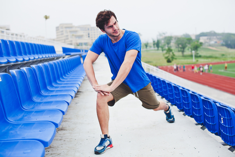


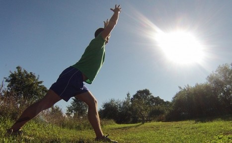


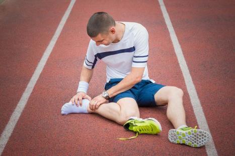


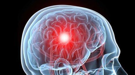


In athletes, piriformis syndrome is a common disorder identified by the irritation and inflammation of the piriformis muscle which can generally result in the compression of the sciatic nerve. This impingement of the nerves and its surrounding tissues can cause the symptoms of sciatica to manifest, characterized by pain and discomfort, along with tingling sensations and numbness, affecting an athlete's performance. For more information, please feel free to ask Dr. Jimenez or contact us at (95) 850-0900.