 Your new post is loading...
The TFL, or tensor fascia latae, is a complex muscle which is intricately arrangement anatomically with the ITB, or iliotibial band, and it performs various essential functions, such as allowing hip mobility as well as transmitting fascial tension through the fascia latae located in the thigh and the iliotibial band. The TFL also provides postural support during one-legged stance and limits the tensile stress on the femur caused by the combination of bodyweight, ground reaction force and how these create individual bending forces against the femur. When one discusses the anatomy of the TFL, the anatomy of the ITB should also be discussed as these serve a conjoined role in order to function. A study conducted to compare the TFL and ITB in humans to other primates and mammals determined that human beings are the only mammals to have a defined ITB. The study also further regarded the anatomy and function of both the tensor fascia latae and the iliotibial band. Additional studies via cadaveric and biomechanical modelling research added a substantial amount of knowledge about this often misunderstood muscle, the TFL, and its relationship to the ITB. The general agreement is that the tensor fascia latae begins on the iliac crest which starts just lateral to the origin of the sartorious, or ASIS, and extends posteriorly along the iliac crest to combine several types of tissue into the iliac crest and onto the gluteal fascia. It’s been highlighted that the muscle provides multiple functions and contains anatomically distinct heads: the anteromedial, or AM, and the posterolateral, or PM, head.
The predominance of neck injuries in sports is believed to be rising, mostly due to the increased improvements in injury recording and observation. However, the growth of physical, extreme sports has led to higher risks of injury among unprepared athletes. For instance, athletes who participate in sports such as skeleton, where individuals sprint on ice and hurtle head first down an icy, often bumpy track at elevated speeds, must learn to understand the importance of properly training their neck to avoid complications to its surrounding structures. Neck injuries are common in skeleton but these can be prevented. Neck training doesn’t simply involve avoiding the risk of suffering a neck complication, in competitive sports, such as skeleton, strengthening the neck can ultimately improve an athlete’s overall physical performance, helping them achieve their goals of triumph. In order to decrease the chance of injury, the neck needs to be strategically and individually prepared to ensure it has a greater tolerance to the increased loads it’s exposed to. But, before an athlete begins implementing this program, it’s essential for them to receive an accurate evaluation of their cervical spine in a comprehensive assessment and screening process. Neck injuries occur most frequently in motorsports and high impact, collision sports like rugby. It’s been previously described that acute force exposure through compression and distraction, axial loading and/or direct blows along with sudden acceleration and deceleration of the structures of the body, are the most common reasons for injury in these types of sports.
Many variations of SLAP lesions can primarily be treated conservatively with methods to initially improve the patho-mechanical factors that affect SLAP lesions, such as glenohumeral internal rotation deficit (GHIRD) and scapular dyskinesis. With a majority of type 1 lesions, this can be significantly effective in eliminating the symptoms of the lesion without the need for surgery. However, once an athlete has surgically treated their SLAP lesions, they can follow a similar rehabilitation process to achieve overall wellness. Minor type-1 SLAP lesions may only require a simple debridement without disrupting the biceps anchor, whereas type-2 SLAP lesions are the most commonly seen type by many healthcare providers, involving a detachment of the biceps anchor from the labrum. Type-2 lesions can be treated with arthroscopic fixation of the superior labrum to develop biceps anchor stability. Type 3 SLAP lesions are identified by bucket-handle tears along the superior labrum with intact biceps anchor. This bucket handle fragment can easily be debrided by an arthroscopic shaver, and further treatment may often not be needed. The other types of SLAP lesions are not as common as the type 2 lesions, although if they do occur, these will almost certainly require surgical intervention. A large database study found that the highest incidence of repair is among the 20-29 years and 40-49 years of age groups. This is believed to occur due to the younger population’s higher participation in sports activities. In the 40-49-year-old group however, the high incidence most likely occurs due to the degeneration of the labrum, which may primarily develop around this age. Also men have a three times higher incidence of repair. This is believed to occur due to how males are more likely to participate in a sport which may cause SLAP lesions.
The labrum increases the surface area of the acetabulum by 22 percent while it increases the volume by 33 percent, functioning accordingly to fasten the head of the femur while allowing it to rotate. From a cross-sectional view, the labrum is triangular in shape with an extra articular area of thick connective tissue which contains a rich supply of blood while the intra articular area majorly has no blood supply. When the hip’s normal range of mobility extends beyond its limit, the labrum is stressed by a strong, compressive force and a tear at his point can ultimately affect the stability of the joint and distribution of weight or load. Furthermore, the labrum is considered a pain generating structure as numerous pain receptors are located in its superior and anterior regions. It is at the anterior surface where an ALT is most vulnerable to pressure along the end point of hip flexion. On another note, abnormal alterations within the structure, such as retroverted acetabulum and coxa valga, have been simultaneously recorded in up to 87 percent of individuals with labral tears.
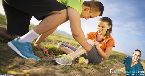
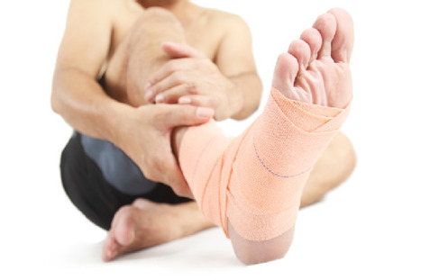
Due to the nature of lateral ankle sprains, once an individual has experienced an injury, there’s a high probability that another injury can occur. This is because damage or injury to the ligaments and joint capsules within the ankle can impair the structures ability to function appropriately. Along with any deficits in proprioception, this can often result in a high re-injury rate. Indeed, research has demonstrated that 73 percent of individuals who sprain their ankles are most likely to experience recurrent ankle injuries in the future. The most frequent factor behind a majority of ankle sprains is when a single extremity lands on an uneven surface. During this case, a quick and joined ankle plantar flexion and inversion motion can often result in an extreme lateral movement which can lead to ankle sprains. Another factor that can influence the chance of lateral ankle sprains is proprioception, or an individual’s own neuromuscular control. With the necessary proprioception, an athlete may be able to accurately determine the speed and force of a disturbance of motion in order to be able to subsequently react with an appropriate joint and muscle motion to avoid injury. However, because there’s always a chance the athlete may be surrounded by uneven terrain, even an individual with excellent proprioceptive skills can suffer harm. Moreover, the evidence for the role of proprioceptive factors in preventing ankle sprains is still unclear.
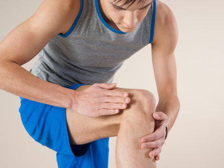
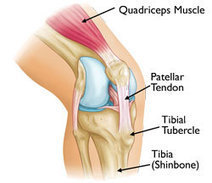
During jumping sports where great amounts of stress are suddenly placed on the lower extremities of the body, patellar tendinopathy, best known as jumper’s knee, can be a frequently reported type of injury. Patellar tendinopathy, or PT, ultimately alters an athlete’s overall performance, affecting their capacity to jump, land, run and change direction. While their decline in performance can lead to decreased training tolerance, the symptoms associated with the condition also often results in missed training days and competition for a majority of athletes. For those individuals diagnosed with PT, managing the condition can be very challenging, especially for athletes during the competition phase of a season. Because increased amounts of force are constantly being placed against the tendon, patellar tendinopathy may commonly require many treatment sessions before it can truly begin to heal. It’s essential to be patient during the rehabilitation process to achieve a full recovery. Taking this into consideration, several guidelines may be followed in order to properly formulate a rehabilitation program to restore the original state of the individual’s patella tendon and help athletes return to their regular training routines as soon as possible.
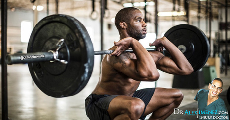
Weightlifting is a common type of sport or physical activity many individuals prefer for strengthening their upper body muscles but weightlifting also commonly results in injury. By following a few safety tips, you can prevent hurting yourself while weightlifting to continue training your body effectively. The most popular weight lifting routines today mainly focus on completing a workout within a limited amount of time. Using a fixed group of exercises, the point of this type of workout is to complete the routines as quickly as each person is individually able to. Focusing on speed during an exercise or physical activity can be a major factor for potential injury. Training is about working out accordingly at the most appropriate tempo for each weight and lift. Slowing down the tempo while weight lifting will help increase the amount of energy an individual has to finish a lift. Additionally, taking a sufficient amount of time for rest in between sets can help prepare an individual both mentally and physically for the next exercise. Then, when considering safety measures for injury prevention, an individual should note that their exercise routines for weight lifting should include lifts that strengthen and both the front and back sides of the body. Balancing exercises for the entire body will help strengthen the various muscles of the body as well as avoid overusing and underusing other muscle groups and risk an injury. Most importantly, in order to avoid injury, an individual should never sacrifice form while lifting. Performing each lift with correct form is the best way to avoid an injury. Lifting weights that are heavier than an individual’s normal lifting weight, lifting the weights rapidly, or simply being unfamiliar with the proper way to lift, are all ways that compromise form and could cause an injury.
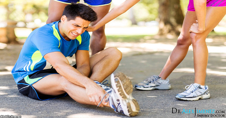
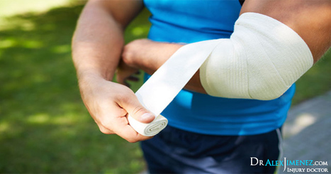
A sprain is medically defined as a stretch or tear of the ligaments, the strong cords of fibrous tissue which connect two bones together at the joints. Sprains most commonly occur on areas of the body which can be injured during a fall or sudden twisting motion, such as the ankle. According to the U.S. National Library of Medicine, approximately 2 million ankle sprains alone occur each year. An incorrect fall or abrupt twist usually causes a sprain because the unusual movement can force a joint into an abnormal position that may wind up stretching or tearing the ligament. Ankles, wrists, knees, and fingers are all frequently sprained areas of the body. A strain is medically defined as a stretch or tear of the muscle or tendon. A tendon is a fibrous band of tissue that connects the muscles to bones. Strains most commonly occur on the lower back and on the hamstring muscle located on the posterior side of the thigh, most commonly as a result of overexertion, trauma, or repetitive movements. Strains most frequently occur on the back, hamstring, and even the shoulder, because these areas are greatly mobile and highly used during strenuous physical activity, leading to a stretch or tear of a single, or multiple, muscle and tendon due to overuse. Although sprains and strains significantly differ from each other, these do share several similarities, which is the main reason individuals generally confuse the two conditions. Both sprains and strains include symptoms of pain, swelling, and limited mobility around the region of the injury. The symptoms can range from moderate to intense, according to the injury’s level of severity. Individual’s who’ve suffered an injury and are experiencing these symptoms can temporarily relieve their pain and discomfort using ice therapy to reduce the inflammation around the affected area as well as getting plenty of rest and elevation.
Many athletes, specifically running athletes, frequently seek chiropractic treatment for iliotibial band syndrome. These individuals often describe feeling radiating pain that begins on the outside of their knee that extends throughout their thighs, hips, and even their lower back. The iliotibial band, or the IT band, is a cord of connective tissue that starts at the side ridge of the pelvis, or the ilium, and travels down the side of the leg, attaching into the weight bearing bone of the lower leg, or the tibia, located near the knee. IT band syndrome is a very common condition occurring in runners and cyclists. This condition frequently results from any activity that causes the leg to turn inward repeatedly. Wearing improper or worn-out shoes, running downhill and constantly in the same direction, or simply from running too many miles, iliotibial band syndrome is an injury caused by overuse where constant rubbing between the band and the bone irritates the tissue and causes inflammation. Through spinal adjustments and manual manipulation, the chiropractor can restore the natural alignment of the spine, gradually improving the individual’s IT band syndrome and its symptoms as well as the runner’s gait and running posture to prevent further complications.
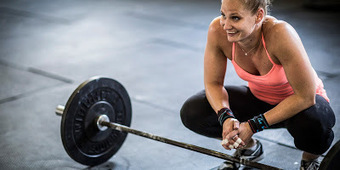
Back injuries and its associated symptoms of pain occur frequently in athletes who perform weight lifting. There is a higher risk of injury when using heavy weights during exercise, especially with dead lifts and squats, but cutting the amount of weight individuals use in their work-out routines can help ultimately avoid injury. The first step to prevent back injury and pain is to begin cutting the weight an individual is normally capable of doing by 3. For example, if you’re capable of doing squats in a Smith cage with 100lbs, you can cut the weight to 30lbs. When it comes to dead lifts, the same system can be used. An individual who normally dead lifts using 60lbs can cut the weight to 15lbs and only use one-legged dead lifts. Balance while performing these can be a challenge but after the body begins to accommodate to the altered exercise, the muscles can benefit greatly. Decreasing or not using weight or decreasing the amount of repetitions a person uses during their set can help avoid injury.
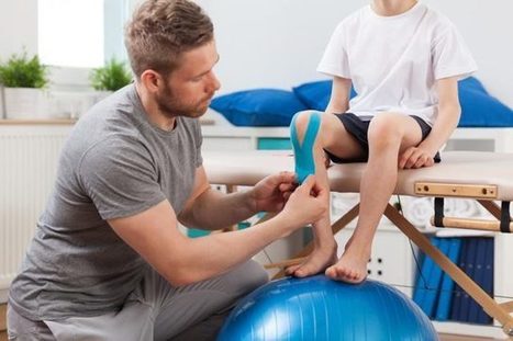
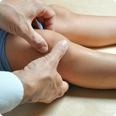
Children between the ages of 9 and 15 are most commonly diagnosed with Osgood-Schlatter disease. Osgood-Schlatter disease is a condition which occurs when the patellar tendon, the tendon over the kneecap just below the knee and on the tibia or shin-bone, pulls on the growth plate of the bone. As a result, the region becomes inflamed, swollen, tender, and painful while participating in physical activities. But, as with many other conditions, stretches, exercises, and other measures can be used to treat Osgood-Schlatter disease and alleviate the symptoms associated with the condition.
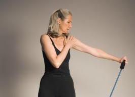
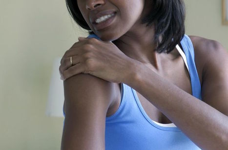
The humerus, or arm bone, extends from the shoulder to the elbow joint, helping position the hand during daily activities such as eating. A break to this lengthy bone in the upper arm requires medical attention. Treating a fractured humerus most commonly involves immobilization through the use of a collar and cuff or a cast for about six to eight weeks, depending on the severity of the fracture. Following recovery from a broken shoulder involves physical therapy exercises to help regain the shoulder joints natural range of motion, strength, and normal functioning in the arm and shoulder.
The majority of treatments for a broken shoulder include more passive types of remedies, such as alleviating the symptoms rather than resorting to surgical repair. Because most clavicle or collarbone fractures involve clean breaks, often times these type of injuries can mend on their own with the proper care and patience. According to the Cleveland Clinic, less than 20 percent of shoulder fractures will require manipulation or surgery to set the bones in the proper position for healing. Thus, for the remaining percentage of broken shoulder injuries, getting plenty of rest, using ice therapy, immobilizing the shoulder and in some cases mild use of pain relief medication can help heal a broken shoulder.
|
During single extremity weight bearing exercises, such as stance phase of walking or running, lunging and landing from a jump, amongst others, the lower extremity joints are designed to naturally absorb the impact of gravity being placed against the body. When the force of gravity acts upon the body, the joints move into distinct directions and the muscles need to properly function as to counteract these forces. Generally, these muscles function isometrically and/or eccentrically. For instance, with the absorption movements of a pelvic lateral tilt, the hip abductors work to stabilize the movement. With an anterior pelvic tilt absorption movement, the pelvic posterior tilters such as the gluteals and hamstrings work to stabilize mobility. With hip joint flexion, adduction and internal rotation, the muscles are controlled by the gluteus medius and other hip joint external rotators, such as the gemellus muscles, quadrutus femoris, obturator muscles and the piriformis. And finally, the quadriceps controls the absorption movements of a knee joint flexion, the soleus of an ankle dorsiflexion and the tibialis posterior, FHL and FDL, stabilizes midfoot pronation. The gluteus medius is a proximal hip muscle which purpose is to control proximal pelvic/hip joint motion that in turn controls lower limb kinetics around the knee and ankle. The gluteus medius attaches to the iliac crest and inserts onto the greater trochanter, functioning as a hip abductor, hip external rotator and stabilizer of the pelvis on the femur during stance phase of gait. It’s most significant role, however, is to compress the femoral head into the acetabulum during the stance phase of gait. The muscle is divided into three equal parts: anterior, middle and posterior. The fibres which make up the posterior section of the muscle travel parallel with the neck of the femur while the middle and anterior sections travel vertically from the iliac crest to the anterosuperior feature of the greater trochanter. It’s been suggested, that each individual part of the muscle functions independently from each other as each of the three portions contain their own supply of nerves which run through the superior gluteal nerve.
Muscular imbalances around the complex structures of the shoulder can develop abnormal activation patterns and inherent myofascial restrictions, both which can cause a significant decrease in the athlete’s scapular control and dyskinesis, leading to glenohumeral joint injuries resulting from instability and impingement. The serratus anterior, or SA, is one of the muscles of the scapula that functions by providing a connection between the shoulder girdle and the trunk, however, it’s often believed to be a dysfunctional muscle among shoulder pathologies. The serratus anterior is a primarily offers movement to the scapula, contributing to the maintenance of normal scapulo-humeral rhythm and motion. Due to its insertion on the inferior and medial border of the scapula, it can produce upward rotation and posterior tilting. Poor activation of the serratus anterior muscle may result in limited scapular rotation and protraction, causing a relative anterior-superior translation of the humeral head in relation to its glenoid articulation, leading to sub-acromial impingement and rotator cuff tears. The serratus anterior is characterized as a flat sheet of muscle beginning from the lateral surface of the first nine ribs. Then, it passes behind and around the thoracic wall before inserting into the anterior surface of the medial border of the scapula. The most important function of the serratus anterior, or SA, is to protract and rotate the scapula, helping to maintain it close yet away from the thoracic wall, allowing for the proper positioning of the glenoid fossa to increase the efficiency of upper extremity motion to its maximum state. The anatomy of the SA can be broken down into three anatomical components as follows: Read more.
The semitendinosus, or ST, the semimembranosus, or SM, and the biceps femoris long and short heads (BFLH and BFSH) are part of the hamstring muscle group. They primarily function with the extension of the hip and flexion of the knee as well as providing multi-directional stability of the tibia and pelvis. These three muscles which make up the hamstring muscle group, cross the posterior aspect of both the hip and the knee joints, making them bi-articular. As a result, they are consistently responding to large mechanical forces created by upper limb, trunk and lower limb locomotion as a means of concentric and eccentric mobilization. During sporting activities, these forces will tend to increase, augmenting the frequency of injury. In a study conducted at the University of Melbourne, biomechanical analysts measured the musculotendinous strain, velocity, force, power, work and other biomechanical loads experienced by the hamstrings throughout the course of over-ground sprinting and compared the biomechanical load across each individual hamstring muscle. Basically, the hamstrings are subjected to a stretch-shortening cycle when sprinting, with the lengthening phase occurring during the terminal swing and the shortening phase commencing just before each foot strike, continuing throughout the stance. Then, the biomechanical load on the bi-articular hamstring muscles were determined to be stronger during the terminal swing. BFLH had the greatest musculotendinous strain, ST displayed considerable musculotendinous lengthening velocity, and SM produced the highest musculotendinous force and both absorbed and generated the most musculotendinous power. Similar research also distinguished peak musculotendinous strain as a large contributor to eccentric muscle damage or injury, most commonly acute hamstring injuries, instead of peak muscle strength. This is why eccentric strengthening is often a rehabilitation recommendation for acute hamstring injuries.
The subscapularis begins on the anterior scapular, or subscapular fossa, and introduces onto the minor tuberosity of the humerus. It is the largest of the rotator cuff muscles and its cross-sectional area is larger than the other three rotator cuff muscles combined; the infraspinatus, the teres minor and the supraspinatus. The most essential functions of the glenohumeral joint are: depressor of the humeral head; anterior stabilizer of the humeral head, which means it glides the humeral head posteriorly relative to the glenoid fossa; and internal rotator of the shoulder together with the powerful pectoralis major and latissimus dorsi. The tendon fibres combine with the anterior capsule of the shoulder, which serve to reinforce the anterior shoulder capsule. The muscle is considered to be less substantial as a shoulder internal rotator, as the pectoralis major and latissimus dorsi are powerful internal rotators, and it’s therefore more essential as a dynamic anterior stabilizer of the glenohumeral joint due to its action in preventing an anterior shear of the humeral head. The subscapularis has a deep connection with the long head of the biceps. This is known as a capsuloligamentous complex that functions to stabilize the long head of the biceps tendon in the bicipital groove. The pulley complex is made up of the superior glenohumeral ligament, the coracohumeral ligament, and the distal attachment of the subscapularis tendon, where it is located within the rotator interval between the anterior edge of the supraspinatus tendon and the superior edge of the subscapularis tendon. Subscapularis tendon injuries may weaken the stability of the bicep. In order to maintain the biceps tendon stabilized and in place, support of the most superior insertion point of the subscapularis from behind the ligament and tension in the superior glenohumeral ligament is needed. Issues with a biceps sling is a common cause and effect of disease in many athletes, requiring the constant and effective rotation of the shoulder, just like the cocking position during baseball pitching.
Tendons are sturdy bands of connective tissue that function by connecting muscles to bones. These channel the force created by each individual muscle to move the bone. Because of this, they must be sufficiently powerful to endure the force which is conducted through them yet sufficiently flexible to act as pulleys around bony prominences. Athletes who frequently overuse their muscles or as a result of direct trauma can develop a tendon injury. Of the 32 million musculoskeletal injuries documented in the United States annually, 45% of them are injuries to tendons, ligaments, or joint capsules. The most commonly injured tendons include the tendons of the rotator cuff of the shoulder, the Achilles tendon, patellar tendon, and the elbow extensor tendon. Several factors can place additional strain on the tendon and contribute to injuries caused by overuse, including: abnormal direction of pull due to skeletal misalignment; differences in limb lengths; muscle weakness or imbalances; hypermobile joints;
inflexible muscles;
training errors; and faulty or improperly fitted equipment and/or footwear. Tendon injuries are believed to be difficult to treat as they were once historically thought of as an inflammatory condition, referred before to as tendonitis. Its treatment was therefore focused on reducing the inflammation through traditional anti-inflammatory medications and modalities and was extensively unsuccessful.
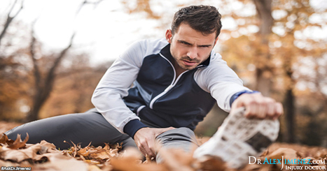
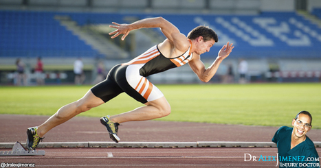
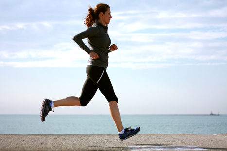
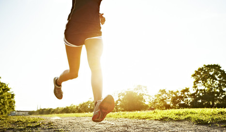
Running injuries can occur unexpectedly over a variety of reasons and situations. Once you’ve suffered an injury as a result of running, time and patience will be a key element towards recovery and it’s important to give your body the proper time to heal. Using over-the-counter medication for the symptoms of pain and cold therapy to reduce the inflammation around the injury, are all temporary ways to relieve an individual’s injury or condition. But, ultimately, chiropractic care can be a great treatment option for runners. Chiropractic treatment focuses on musculoskeletal injuries, or soft tissue injuries, which can help heal many types of injuries, symptoms and conditions as well. Runners can greatly benefit from chiropractic care.
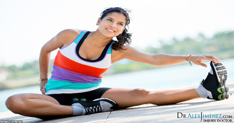
As individual’s increase their cardiovascular activity, sprains and muscle strains on the surrounding tissues of the hamstrings, quadriceps, and calf muscles become several of the most common type of injuries. Back injuries also become more frequent during periods of higher activity. More severe types of injuries can often lead to muscle, tendon, or ligament tears as well as bone fractures. A successful exercise program provides a complete training routine which equally conditions different muscle groups while reducing the chance of injury. In addition, seeking chiropractic care and physical therapy to help during injury prevention can further maximize an individual’s performance. Exercise related injuries mainly occur due to improper biomechanics. The improper movements of an individual’s structure during physical activities or sports are often the root cause of spinal misalignments and muscle imbalances. If the body is not moving or functioning the way it was designed to, it can place unnecessary pressure and stress on a single side of the body or an incorrect area of the body. An injury and its associated symptoms, can tremendously reduce an individual’s ability to perform to their fullest capacity, ultimately altering an individual’s lifestyle. Regular chiropractic care and massage therapy can help diagnose and determine the source of your injury as well as help treat the injury and its symptoms. Combining chiropractic care along with physical exercise this summer can ultimately improve your fitness, health and wellbeing.
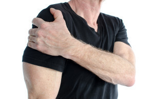
A frozen shoulder can cause pain and discomfort, greatly restricting the range of motion of the shoulder and ultimately altering an individual's lifestyle. After treating the shoulder injury with ice to reduce the inflammation, seeking medical treatment from a chiropractor or physical therapist and following through with the recommended treatment options can help prevent shoulder stiffness and a frozen shoulder.
The iliotibial band, or IT band, is a ligament that begins at the pelvis, along the iliac crest, and travels down through the outside of the thigh from the hip to the shin. Most frequently caused by overuse from running sports and activities, knots can develop along the IT band and on the lower side of the leg, causing the ligament to become tight and inflamed, a condition known as iliotibial band syndrome. Once the IT band contracts and shortens, the affected individual can experience symptoms of hip pain, lower back pain, and most commonly, knee pain. A chiropractor can both diagnose and help treat many spinal injuries and conditions, such as a possible spinal misalignment. In the majority of cases, if the spine and pelvis aren’t aligned or these aren't moving properly, the altered gait can cause muscle imbalances and may increase the chances of developing iliotibial band syndrome. Having a strong core can also be essential for runners in order to avoid developing injuries. Exercises such as planks, side planks, and crunches should be a part of a runner’s daily exercise routine to strengthen the core. The use of foam rollers can also help relieve symptoms of pain. In order to alleviate IT band syndrome, an individual has to loosen up the knots along the iliotibial band. And last, improper footwear may also cause running complications. There are a variety of shoes specifically designed to make running easier and much more comfortable. By following the above recommendations, runners can alleviate their iliotibial band syndrome pain and symptoms and return to their regular running activities.
Apophyses are growth plates made up of cartilage that can be found throughout a child's body, functioning as attachment sites between muscles and tendons. When a young athlete participates in physical activities or sports where a specific set of muscles is constantly overused, such as the thigh muscles while running or jumping, the apophyses can weaken. The repetitive stretching of the apophysis can cause microscopic cracks in the cartilage growth plate, resulting in pain and inflammation. Sinding Larsen Johansson syndrome (SLJ) is a condition that occurs at the bottom of the kneecap due to the additional stress of the apophysis being repetitively stretched during physical activities or sports. This condition can also occur from a direct, blow, fall, or one sudden jump.
Osgood-Schlatter disease is a common cause of knee pain in growing adolescents. It is characterized as an inflammation of the patellar tendon, the area just below the knee where the tendon from the kneecap attaches to the shinbone or tibia. Osgood-Schlatter disease most frequently occurs during growth spurts since the bones, muscles, tendons, and other structures of the body are changing quickly. Also, participating in physical activity can add stress on the bones and muscles and therefore, children who participate in physical activity, especially running or jumping sports, are usually at a higher risk of developing this condition. In the majority of Osgood-Schlatter disease cases though, simple measures such as plenty of rest, over-the-counter medication, and stretches and exercise can help alleviate the symptoms of the condition and restore the affected individual's regular lifestyle.
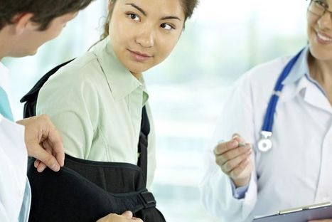
A fracture of your arm bone near the shoulder may require physical therapy to help improve normal arm function. A proximal humeral fracture is a type of injury that results near the shoulder joint. "Proximal" reffers to being close to the body and "humeral" refers to the arm bone known as the humerus. A fracture to the proximal humerus is typically causes due to significant trauma from an injury to the arm or shoulder. Falling on to the outstretched hand, a forceful pull to the arm or shoulder, and falling on to the side or shoulder are several situations that could cause fracture to the shoulder joint. These are the most common causes for bone injury but are not limited to these.
|


The tensor fascia latae, or TFL, is a well-known hip muscle among healthcare professionals and rehabilitation specialists. Because of its essential function, this muscle may be responsible for pain and dysfunction in the lower extremities, pelvis and spine. Research studies conclude that this muscle is greatly misunderstood, but with further examination, injury can be prevented. For more information, please feel free to ask Dr. Jimenez or contact us at (915) 850-0900.