 Your new post is loading...
There is no denying that water is an integral part of good health. Dehydration can cause problems with skin, digestion, and organ function. It can cause leg and foot cramps and impair cognitive processes. Staying well hydrated is vital to overall wellness. Because water is part of every cell in the body, and when we don’t drink enough water, the body suffers. Good spinal health begins with proper hydration. The spine is constructed in such a way that dehydration can cause limited mobility, decreased flexibility, and pain. It can make the backbone to age faster than it should which impacts the entire body. As the natural functions begin to break down the body suffers, and it isn’t long and depression and anxiety often set in. The spine depends heavily on hydration. Overview of the Spine The spine of made up of vertebrae, a row of bones that sit on top of each other, connected by small joints. A disc sits between each vertebra, cushioning it and acting as a shock absorber. It allows the spine to flex, bend, and move about without the bones rubbing together. Each disc has a fluid center (nucleus pulposis) that is surrounded by a flexible, sturdy ring. The ring contains a gel-like substance while the center of the disc is comprised of water. The outer ring protects the center, and the center protects the vertebrae, acting as a cushion for the bones. If the fluid center does not have adequate water, it cannot do its job, and the spine begins to experience problems. Aging makes it more difficult for the discs to rehydrate and a sedentary lifestyle also complicates the process. It just cannot work without proper hydration. A healthy spine starts with adequate hydration. The Benefits of Water for the Spine From the time you get up in the morning, you are putting pressure on your spine and subsequently, the discs that lie between each vertebra. As you move the discs are compressed by the spine, and the water that is inside is squeezed out. Even upright activities like standing, sitting, or walking can cause pressure on the discs as gravity causes compression in the spine. When the discs do not have enough water, it results in limited mobility, pain, and an increased risk of back injury. If you don’t drink enough water, your body becomes dehydrated and is unable to replenish the water that the discs so desperately need. You may not even notice the typical signs of dehydration such as a headache and lethargy, but also lower levels of dehydration can cause severe problems in the body, especially if it is prolonged. Soda and similar beverages do not provide adequate water to the body. How to Properly Hydrate the Body Water is the best way to hydrate the body, but it isn’t the only way. Foods like watermelon, lettuce, spinach, and soups are excellent sources of hydration. H2O, of course, is the best way, but herbal teas are also good. Drinks with caffeine are not as effective since the caffeine can have a diuretic effect. Traditionally, people have been told to drink eight glasses of water a day, and that is good advice. However, studies indicate that proper hydration can occur with an intake of just 30 to 50 ounces of water a day. If you have constant or frequent back pain, the answer could be as close as your kitchen faucet. Dehydration could be the source of your back pain and immobility. Water also affects the way the cerebrospinal fluid works and moves in the body. When the body is dehydrated, it doesn’t move as it should, and brain function, reflexes, and cognitive processing could be impacted. Don’t chance it. If your problems are caused by something as simple as not drinking enough water, that is something you can change today. Drink up! Your body will thank you.
Degenerative Arthritis - Spinal Arthritis:
- Spondylosis aka Degenerative disease of the spine represents an evolution of changes affecting most mobile spinal segments beginning with:
- Intervertebral disc (IVD) dehydration (desiccation) and degeneration aka Degenerative Disc Disease (DDD) with an abnormal increase in mechanical stress and degeneration of posterior elements affecting 4-mobile synovial articulations ( true osteoarthritis)
- 2-Facets in the L/S & 2-Facets & 2-Uncovertebral joints in the C/S
- Imaging plays a significant role in the diagnosis, grading, and evaluation of neurological complications (e.g., spondylotic myelopathy/radiculopathy)
- X-radiography with AP, Lateral & Oblique spinal views provides Dx and classification of Spondylosis
- MR imaging may help to evaluate the degree of neurological changes associated with degenerative spinal canal and neural foraminal stenosis
- Spinal motion segment:
- 2-adjacent vertebrae
- IVD (fibrocartilage)
- 2-facets (synovial)
- Pathology: loss of disc height increases mechanical stress on mobile elements
- Ligamentous laxity/local instability
- Spinal osteophytes aka spondylophytes & bony facet/uncinate proliferation
- Disc herniation and often disc-osteophyte complex
- Ligamentum flavum "hypertrophy" or thickening due to buckling
- Loss of normal lordosis with or w/o reversal or kyphosis
- Vertebral canal & neural foraminal stenosis
Neutral lateral cervical radiograph: note mild to moderate disc narrowing and spondylophyte formation at C5-6 & C6-C7 (most common levels affected by cervical spondylosis). Straightening or flattening with mild reversal of cervical lordosis. Some mild facet proliferation is noted at the above levels - On radiographs: evaluate for disc height (mild, moderate or severe) loss
- End-plate sclerosis & spondylophytes; mild, moderate or severe
- Facet and uncinate irregularity, hypertrophy/degeneration; mild, moderate or severe
- Note degenerative instability aka degenerative spondylolisthesis/retrolisthesis
- Normal or lost lordosis vs. degenerative kyphosis
- Key Dx: correlate with a clinical presentation: neck/back pain with or w/o neurological disturbance ( myelopathy vs. radiculopathy or both)
- Uncinate processes undergo degeneration/proliferation resulting in uncovertebral arthrosis
- Early findings present with mild bone proliferation along the cortical margin (white and black arrows) if compared to normal uncinate (orange arrow)
- Later, more extensive bone proliferation extending into and narrowing vertebral canal and neural osseous foramina (IVF's) may be noted. The latter may contribute to spinal/IVF stenosis and potential neurological changes
- Posterior oblique views may help further
- AP lower cervical (a) and posterior oblique (b) views
- Note mild uncinated process proliferation with neural foraminal narrowing (arrows)
- Typically if less than a third of IVF becomes narrowed, patients may present w/o significant neurological signs
- Lumbar spondylosis is evaluated with AP and lateral views with additional AP L5-S1 spot view to examine lumbosacral junction
- Typical features include disc height loss/degeneration
- Intra-discal gas (vacuum) phenomenon (blue arrow) along with spondylophytes
- Degenerative spondylolisthesis and/or retrolisthesis (green arrow) may follow disc and facet degeneration and can be graded by the Meyerding classification
- In most cases, degenerative spondylolisthesis rarely progresses beyond Grade 2
- Lumbar facet degeneration seen as bone proliferation/sclerosis and IVF narrowing
- MR imaging w/o gad C is an effective modality to evaluate clinical signs of spondylosis & associated neurological complications with pre-surgical evaluation
- Case: 50-y.o Fe with neck pain. Case b-45-y.o.M (top a b images). MRI reveals: loss of disc hydration or desiccation, spondylophytes and disc herniation w/o neurological changes
- (Bottom images) Left: preoperative and right postoperative MRI slices of the patient presented with clinical signs of cervical spondylotic myelopathy. Note disc herniation, ligam flavum hypertrophy and canal stenosis (left)
- Sagittal MRI slice of lumbar DDD manifested with disc desiccation and posterior herniation effacing thecal sac
- Correlating sagittal and axial slices will be more informative to evaluate canal stenosis and potential degree of neurological involvement (above-bottom images)
Diffuse Idiopathic Skeletal Hyperostosis (DISH) aka Forestier disease - Flowing degenerative ossification of ALL
- M/c Thoracic spine. 2nd m/c-cervical spine
- Dx by imaging only. X-radiography is sufficient
- CT w/o contrast helps with Dx of Fx
- Men>women. Pts>60-y.o. Extensive DISH shows 49% association with type 2DM
- Complications: Chalk (carrot) stick Fx. Unstable 3-column Fx requiring surgical fusion
- Sagittal reconstructed CT scan slice in bone window
- Chalk stick Fx at C5-C6 in the patient with DISH and OPLL
Imaging Diagnosis Management: - Cervical spinal trauma & radiographic variants simulating disease
- Cervical spine
- Arthritis
- Neoplasms
- Infection
- Post-Surgical cervical spine
- Cranio-cervical and upper cervical stability is dependent on transverse, superior and inferior bands of the C1-C2 ligament, alar ligaments, along with a few other ligaments
Cervical Trauma - The C/S is vulnerable to injury. Why?
- Stability has been sacrificed for greater mobility
- Cervical vertebrae are small and interrupted by multiple foraminae
- The head is disproportionately heavy and acts as an abnormal lever especially when forces act against a rigid torso
- Additionally, C/S is prone to degeneration which makes it more vulnerable to trauma
- In young children, ligaments are more luxed vs. disproportionately large head size
- In children, the fulcrum of movement is at C2/3 thus making injuries more common in the upper C/S and craniocervical junction. In children, S.C.I.W.O.R.A. may occur when no evidence of fracture present
- In adults, the fulcrum of movement is at C5/6 thus making lower C/S more vulnerable to trauma especially during extremes of flexion
- Cervical Trauma categorized according to mechanisms of injury (Harris & Mirvis classification)
Hyperflexion Injury: Stable vs. Unstable - Flexion teardrop Fx (most severe fracture, unstable)
- Bilateral facet dislocation (severe injury w/o fracture, unstable)
- Anterior subluxation (potentially unstable) can be very subtle injury
- Clay Shoveller Fx (lower C/S SP avulsion, stable)
- Simple wedge compression (most benign Fx, stable)
- Hyperflexion-rotation with unilateral facet dislocation
- Obtain a thorough history
- Perform physical exam including a neurological exam
- Consider NEXUS criteria (National Emergency X-radiography Utilization Study)
Imaging Techniques: - Begins with x-radiography especially in cases with no significant neurological compromise
- Clear neutral lateral view first
- If x-radiography is unrewarding but high probability of severe trauma and neurological deficit present, CT scanning w/o contrast is required
- Consider CT scanning in patients with pre-existing changes: advance spondylosis, DISH, AS, RA, post-surgical spine, congenital abnormalities (Klippel-Feil syndrome, etc.)
Vertical compression: - Jefferson aka burst Atlas Fx (unstable especially if the Transverse ligament is torn, cord paralysis in 20-30% only)
- Why? Due to fragments dissociation and canal widening
- Burst Fx of the Thoracic or Lumbar spine (unstable, cord paralysis may occur)
How to Assess Spinal Radiographs in Trauma Cases: - Construct 5-lines on the lateral view
- Note if facets are well-aligned and symmetrical
- Ensure symmetry of the disc height
- Note any widening or fanning of the inter-spinous distance
- Carefully examine prevertebral soft tissues
- Evaluate atlanto-dental interval (ADI)
- In cases of trauma, evaluate and clear neutral lateral first
- Do not perform flexed and extended views in acute cases before x-rays or CT scanning exclude significant instability
- Pay extra attention to prevertebral soft tissues
- If thicker than normal limits, consider severe post-traumatic bleed
- Subtle asymmetry and widening of posterior disc height and facets with inter-spinous fanning may be a key feature of significant tearing of posterior ligaments
Hyperflexion Injuries (M/C Mechanism) - More frequent in sub-axial C/S C-3-C7)
- Unstable injuries:
- Flexion teardrop fracture (M/C C5 & C6) v. unstable
- Key rad features:
- Large "teardrop" triangular anterior body fragment
- Fanning of the SPs, posterior disc and facet widening indicating tearing of major spinal ligaments and instability
- A posterior shift of the vertebral body fracture suggests direct anterior cord/vessels compression
- Bulging prevertebral soft tissue >20-mm at C6-7
- 80% of cases may be paralyzed on the spot or develop significant paralysis soon after
Acute Neck Trauma. What are the vital radiographic features? What is the diagnosis? - CT scanning w/o contrasts with sagittal reconstruction. Note C7 Flexion teardrop Fx.
- CT may help with further delineation and preoperative planning
- May follow with MR imaging and evaluation of the neurological injury
- Fluid sensitive (T2) sagittal MRI slice of Flexion teardrop fracture at C4 and possibly C5
- Note high signal intensity lesion in the cord and surrounding ligaments indicating cord edema and ischemia
- Management: neurosurgical with spinal fusion
- Complications:
- Quadriplegia/paraplegia
- Respiratory complications
- Disability, changes in the quality of life
- Decreased life expectancy
- Bilateral facet dislocation (unstable)
- Mechanism: Flexion-distraction injury
- Key radiography: anteriorly displaced body 50% or more
- Facets override and locked (can be perched left image)
- Major tearing of ligaments
- Chances of severe cord compression and paralysis
- Patients with ligaments laxity and degenerative changes are at higher risk
- Initial x-radiography is the first step
CT scanning w/o Contrast is Crucial: - Further delineation of this injury
- Facet fractures, pedicle fracture
- Management planning
Sagittal fluid sensitive MRI of bilateral C5 facet dislocation, sizeable ischemic cord injury, and posterior soft tissue injuries - Management:
- X-radiography, then CT scanning then immediate closed reduction (esp. if the patient is conscious)
- Followed in some more complicated cases by MRI and then surgical care
- If the patient is awake and neurologically stable, CT and closed reduction are adequate
- Complicated cases and failed closed reduction may require surgical stabilization
- Complications: spinal cord injury and paralysis
- Delayed ligamentous laxity and instability
- Unilateral facet dislocation (flexion-rotation injury) less severe than bilateral dislocation
- Most commonly missed unstable cervical injury on x-radiography
- Key rad features: body anteriorly translated 25% facets appear misaligned and blurred, SPs rotated on frontal views
- Clinically may be presented as one-sided radiculopathy esp. C6 or C7
- CT scanning is required to evaluate further facet/pedicle fractures
- Pre-reduction evaluation and care planning
- Management: closed reduction esp. in a conscious patient
- Complications: acute disc herniation/retropulsion, ligamentous laxity, neurological injury
Cervical facet joint syndrome, or cervical facet osteoarthritis, is a degenerative condition marked by stiffness and pain in the cervical region (neck) of the spine. Individuals can gain relief from various types of treatment, including chiropractic care. Facet joint problems are among the most common sources of lower back and neck pain. They can cause debilitating, chronic problems with the neck and back and can lead to other more severe conditions and symptoms that can be disabling. What is Cervical Facet Joint Pain? The spine, comprised of a chain of bones known as vertebrae. Each one has two facet joints on the back side and a large disc on the front side, allowing the vertebrae to stack neatly, one on top of the other, providing stabilization for the entire body. The facet joints are synovial joints, like other joints in the body and sometimes they can become inflamed or injured, causing pain and stiffness. Cervical facet joint pain is, quite literally, a pain in the neck. It means that the joints in the neck area have become injured or inflamed. Suffering from this condition can make it difficult for the patient to turn their head from side to side, or to move it up and down. The cervical facet joints are almost always working. They undergo repetitive, constant motion and over time they can become torn or worn down. Problems within the joint can cause movement to be restricted, or it can have too much, both of which can cause pain. Injury, such as whiplash, to the area, can also cause problems. If not treated appropriately the condition can be degenerative, and the patient can lose both flexibility and mobility, as well as suffer from chronic pain. Symptoms of Cervical Facet Joint Pain The symptoms of cervical facet joint pain tend to vary from patient to patient. A patient may experience one or several of these symptoms: - Tingling, weakness, or pain in the hand and arm
- Neck pain
- Upper back pain that can affect the shoulders
- Pain between the shoulder blades
- Headaches, typically located in the back of the head
- Swelling and tenderness at the site of the inflamed facet joint
- Decreased range of motion and flexibility in the neck
Treatment for Cervical Facet Joint Pain When a patient diagnosed with cervical facet joint pain, the procedure is usually fairly conservative. Their doctor may recommend soft tissue massage, physical therapy, and posture correction. Combined with medications such as an anti-inflammatory like ibuprofen, or muscle relaxers to ease muscle spasms in the muscles that surround the affected joint. If those methods do not give the patient relief, the doctor may take a more aggressive approach, prescribing facet joint injections that use steroid medications injected into the affected joint. This approach is intended to keep the pain localized while reducing it. The procedure can be performed in an outpatient setting and has a good record of being useful, but the results are temporary. Chiropractic for Cervical Facet Joint Pain Chiropractors have had much success in treating cervical facet joint pain. They can manipulate the areas that are affected, restoring painful, restricted facet joints to a point where they can move much more natural and without pain. Over time, with regular chiropractic treatments, they can help to reestablish range of motion in the neck area for their patients. This is all done without any medications or injections. It is a natural, gentle, effective method for relieving the pain and helping the patient enjoy a better quality of life.
Clinical Prediction Rules: "Clinical decision rules, spinal pain classification and prediction of treatment outcome: A discussion of recent reports in the rehabilitation literature" Abstract Clinical decision rules are an increasingly common presence in the biomedical literature and represent one strategy of enhancing clinical-decision making with the goal of improving the efficiency and effectiveness of healthcare delivery. In the context of rehabilitation research, clinical decision rules have been predominantly aimed at classifying patients by predicting their treatment response to specific therapies. Traditionally, recommendations for developing clinical decision rules propose a multistep process (derivation, validation, impact analysis) using defined methodology. Research efforts aimed at developing a “diagnosis-based clinical decision rule” have departed from this convention. Recent publications in this line of research have used the modified terminology “diagnosis-based clinical decision guide.” Modifications to terminology and methodology surrounding clinical decision rules can make it more difficult for clinicians to recognize the level of evidence associated with a decision rule and understand how this evidence should be implemented to inform patient care. We provide a brief overview of clinical decision rule development in the context of the rehabilitation literature and two specific papers recently published in Chiropractic and Manual Therapies. Clinical Prediction Rules - Healthcare has undergone an important paradigm shift toward evidence based practice; an approach thought to enhance clinical decision making by integrating the best available evidence with clinical expertise and the preferences of patients.
- Ultimately, the goal of evidence based practice is to improve healthcare delivery. However, the translation of scientific evidence into practice has proven a challenging endeavor.
- Clinical decision rules (CDRs), also known as clinical prediction rules, are an increasingly common presence in the rehabilitation literature.
- These are tools designed to inform clinical decision-making by identifying potential predictors of diagnostic test outcome, prognosis or therapeutic response.
- In the rehabilitation literature, CDRs are most commonly used to predict a patient’s response to treatment and have been proposed as a means of identifying clinically relevant subgroups of patients presenting with otherwise heterogeneous disorders such as non-specific neck or low back pain and this is the perspective on which we intend to focus.
Clinical Prediction Rules - The ability to classify or subgroup patients with heterogeneous disorders such as spinal pain has been highlighted as a research priority and consequently, the focus of much research effort. The appeal of such classification approaches is their potential for improved treatment efficiency and effectiveness by matching patients with optimal therapies. In the past, patient classification has relied on implicit approaches founded in tradition or unsystematic observations. The use of CDRs to inform classification is one attempt at a more evidence driven approach, less dependent on unfounded theory.
- CDRs are developed in a multistep process involving studies of derivation, validation and analysis of impact with each having a defined purpose and methodological criteria. As with all forms of evidence used to make decisions about patients, attention to appropriate study methodology is critical to assessing the potential benefits of implementation.
Benefits Of Clinical Prediction Rules - Can accommodate more factors than human brain can take into account
- CDR/CPR model will always give the same result (mathematical equation)
- Can be more accurate than clinical judgement.
Clinical Uses Of Clinical Prediction Rules - Diagnosis – Pretest probability
- Prognosis – Predict risk of outcomes of disease
https://johnsnyderdpt.com/for-clinicians/clinical-prediction-rules/cervical-manipulation-for-neck-pain/ https://johnsnyderdpt.com/for-clinicians/clinical-prediction-rules/thoracic-manipulation-for-neck-pain/ https://johnsnyderdpt.com/for-clinicians/clinical-prediction-rules/manipulation-for-low-back-pain https://johnsnyderdpt.com/for-clinicians/clinical-prediction-rules/lumbar-spinal-stenosis/ Flynn Clinical Prediction Rule Video CDR Analysis Of Impact Ultimately, the usefulness of a CDR lies not with its accuracy but with its ability to improve clinical outcomes and enhance the efficiency of care.[15] Even when a CDR demonstrates evidence of broad validation, this does not ensure that it will change clinical decision making, or that the changes it produces will result in better care. The changes it produces will result in better care. McGinn et al.[2] identified three explanations for the failure of a CDR at this stage. First, if clinician judgement is as accurate as a CDR informed decision, there is no benefit to its use. Second, the application of a CDR may involve cumbersome calculations or procedures which discourage clinicians from utilizing the CDR. Third, using the CDR may not be feasible in all environments or circumstances. In addition, we would include the reality that experimental studies may involve patients that are not entirely representative of those seen in routine care and that this may limit the actual value of a CDR. Therefore, to fully understand the utility of a CDR and its ability to improve healthcare delivery, it is necessary to undertake a pragmatic examination of its feasibility and impact when applied in an environment reflecting real world practice. This can be undertaken with different study designs such as randomized trials, cluster randomized trials, or other approaches such as examining the impact of a CDR before and after its implementation. Prevalence of classification methods for patients with lumbar impairments using the McKenzie syndromes, pain pattern, manipulation, and stabilization clinical prediction rules. https://www.ncbi.nlm.nih.gov/pmc/articles/PMC3113271/ Objectives Aims were (1) to determine the proportion of patients with lumbar impairments who could be classified at intake by McKenzie syndromes (McK) and pain pattern classification (PPCs) using Mechanical Diagnosis and Therapy (MDT) assessment methods, manipulation, and stabilization clinical prediction rules (CPRs) and (2) for each Man CPR or Stab CPR category, determine classification prevalence rates using McK and PPC. CPRs are sophisticated probabilistic and prognostic models where a group of identified patient characteristics and clinical signs and symptoms are statistically associated with meaningful prediction of patient outcomes. Two separate CPRs were developed by researchers for identifying patients who would respond favorably to manipulation.33,34 Flynn et al. developed the original manipulation CPR using five criteria, i.e. no symptoms below the knee, recent onset of symptoms (<16 days), low fear avoidance belief questionnaire36 score for work (<19), hypomobility of the lumbar spine, and hip internal rotation ROM (>35° for at least one hip).33 Flynn’s CPR was subsequently modified by Fritz et al. to two criteria, that included no symptoms below the knee and recent onset of symptoms (<16 days), as a pragmatic alternative to reduce clinician burden for identifying patients in primary care most likely to positively respond to thrust manipulation.34 "Potential Pitfalls Of Clinical Prediction Rules" What Are Clinical Prediction Rules? A clinical prediction rule (CPR) is a combination of clinical findings that have statistically demonstrated meaningful predictability in determining a selected condition or prognosis of a patient who has been provided with a specific treatment 1,2. CPRs are created using multi- variate statistical methods, are designed to examine the predictive ability of selected groupings of clinical variables3,4, and are intended to help clinicians make quick decisions that may normally be subject to underlying biases5. The rules are algorithmic in nature and involve condensed information that identifies the smallest number of indicators that are statistically diagnostic to the targeted condition6. Clinical prediction rules are generally developed using a 3-step method14. First, CPRs are derived prospectively using multivariate statistical methods to examine the predictive ability of selected groupings of clinical variables3. The second step involves validating the CPR in a randomized controlled trial to reduce the risk that the predictive factors developed during the derivation phase were selected by chance14. The third step involves con- ducting an impact analysis to determine the extent that the CPR improves care, reduces costs, and accurately defines the targeted objective14. Although there is little debate that carefully constructed CPRs can improve clinical practice, to my knowledge, there are no guidelines that specify methodological requirements for CPRs for infusion into all clinical practice environments. Guidelines are created to improve the rigor of study design and reporting. The following editorial outlines potential methodological pitfalls in CPRs that may significantly weaken the transferability of the algorithm. Within the field of rehabilitation, most CPRs have been prescriptive; thus, my comments here are reflective of prescriptive CPRs. Methodological Pitfalls CPRs are designed to specify a homogenous set of characteristics from a heterogeneous population of prospectively selected consecutive patients5,15. Typically, the resulting applicable population is a small subset of a larger sample and may only represent a small percentage of the clinician’s actual daily caseload. The set- ting and location of the larger sample should be generalizable15,16, and subsequent validity studies require assessment of the CPR in different patient groups, in different environments, and with a typical patient group seen by most clinicians16. Because many CPRs are developed based on a very distinct group, that may or may not be reflective of a typical population of patients, the spectrum transportability17 of many current CPR algorithms may be limited. Clinical prediction rules use out- come measures to determine the effectiveness of the intervention. Outcome measures must have a single operational definition5 and require enough responsiveness to truly capture appropriate change in the condition14; in addition, these measures should have a well constructed cut-off score16,18 and be collected by a blinded administrator15. The selection of an appropriate anchor score for measurement of actual change is currently debated19-20. Most outcome measures use a patient recall-based questionnaire such as a global rating of change score (GRoC), which is appropriate when used in the short term but suffers from recall bias when used in long-term analyses19-21. A potential drawback for CPRs is the failure to maintain the quality of the tests and measures used as predictors in the algorithm. The prospective test and measures should be independent of one an- other during modeling16; each should be performed in a meaningful, acceptable manner4; and clinicians or data administrators should be blinded to the patient’s outcomes measures and condition22. Sources Potential Pitfalls Of Clinical Prediction Rules; The Journal of Manual & Manipulative Therapy Volume 16 Number Two [69] Jeffrey J Hebert and Julie M Fritz; Clinical decision rules, spinal pain classification and prediction of treatment outcome: A discussion of recent reports in the rehabilitation literature
The spine is made of bones called vertebrae, with the spinal cord running through the spinal canal in the center. The cord is made up of nerves. These nerve roots split from the cord and travel between the vertebrae into various areas of the body. When these nerve roots become pinched or damaged, the symptoms that follow are known as, radiculopathy. El Paso, TX. Chiropractor, Dr. Alexander Jimenez breaks down radiculopathies, along with their causes, symptoms and treatment. The entire length of the spine, at each level, nerves exit through holes in the bone of the spine (foramen) on each side of the spinal column. These nerves are called nerve roots, or radicular nerves and branch out from the spine and supply different parts of the body. Nerves exiting the cervical spine travel down through the arms, hands, and fingers. This is where neck problems affecting a cervical nerve root can cause pain, as well as, other symptoms through the arms and hands, one form of (radiculopathy). Another is low back problems that affect a lumbar nerve root. This can radiate through the leg and into the foot, another form of (radiculopathy, or sciatica), which creates leg pain and/or foot pain. The spinal cord does not go into the lumbar spine and because the spinal canal has space in the lower back, problems in the lumbosacral region often cause nerve root problems and not a spinal cord injury. Serious conditions i.e. disc herniation or fracture in the lower back are also not likely to cause permanent loss of motor function in the legs. - Cervical Spine - This nerve root is named according to the Lower spinal segment that the nerve root runs between.
- Example - The nerve at C5-C6 level is called the C6 nerve root.
- It's named like this because as it exits the spine, it passes Over the C6 pedicle (a piece of bone part of the spinal segment).
- Lumbar Spine - These nerve roots are named according to the Upper spinal segment that the nerve runs between.
- Example - The nerve at L4-L5 level is called the L4 nerve root.
- The nerve root is named this way because as it exits the spine it passes Under the L4 pedicle.
Two Nerve Roots Two nerves cross each disc level Only one exits the spine (through the foramen) at that level Exiting Nerve Root - This is the nerve root exiting the spine at a certain level Example: L4 nerve root exits the spine at L4-L5 level. Traversing Nerve Root - This nerve root goes across the disc and exits the spine at the level below. Example: L5 nerve is the traversing nerve root at L4-L5 level, and is the exiting nerve root at L5-S1 level. There is some confusion when a nerve root is compressed by disc herniation or other cause to refer both to the intervertebral level (where the disc is) and to the nerve root that is affected. This depends on where the disc herniation or protrusion is happening. It could impinge upon either the exiting nerve or the traversing nerve. If The Traversing Nerve Is Affected Lumbar Radiculopathy In the lumbar spine, there is a weak area in the disc space right in front of the traversing nerve root, so lumbar discs tend to herniate or leak out and impinge on the traversing nerve. If The Exiting Nerve Is Affected Cervical Radiculopathy The opposite is true in the neck. In the cervical spine, the disc tends to herniate to the side, rather than toward the back and the side. If the disc material herniates to the side, it will compress or impinge the exiting nerve root. Radiculopathy & Sciatica Nerve root goes by another name Radicular Nerve, and when a herniated or prolapsed disc presses on a radicular nerve, this is referred to as a radiculopathy. A medical physician might say there is herniated disc at L4-L5, which creates an L5 radiculopathy or an L4 radiculopathy. It all depends on where the disc herniation occurs (the side or the back of the disc) and which nerve is affected. And the term for radiculopathy in the low back is the ever famous Sciatica. Radiculopathy - A pinched nerve can occur at different areas of the spine (cervical, thoracic or lumbar).
- Common causes are narrowing of the hole where the nerve roots exit, which can result from stenosis, bone spurs, disc herniation and other conditions.
- Symptoms vary but often include pain, weakness, numbness and tingling.
- Symptoms can be managed with nonsurgical treatment, but minimal surgery can also help.
Prevalence & Pathogenesis - A herniated disc can be defined as herniation of the nucleus pulposus through the fibers of the annulus fibrosus.
- Most disc ruptures occur during the third and fourth decades of life while the nucleus pulposus is still gelatinous.
- The most likely time of day associated with increased force on the disc is the morning.
- In the lumbar region, perforations usually arise through a defect just lateral to the posterior midline, where the posterior longitudinal ligament is weakest.
Epidemology Lumbar Spine: - Symptomatic lumbar disc herniation occurs during the lifetime of approximately 2% of the general population.
- Approximately 80% of the population will experience significant back pain during the course of a herniated disc.
- The groups at greatest risk for herniation of intervertebral discs are younger individuals (mean age of 35 years)
- True sciatica actually develops in only 35% of patients with disc herniation.
- Not infrequently, sciatica develops 6 to 10 years after the onset of low back pain.
- The period of localized back pain may correspond to repeated damage to annular fibers that irritates the sinuvertebral nerve but does not result in disc herniation.
Epidemology Cervical Spine: - The average annual incidence of cervical radiculopathies is less than 0.1 per 1000 individuals.
- Pure soft disc herniations are less common than hard disc abnormalities (spondylosis) as a cause of radicular arm pain.
- In a study of 395 patients with nerve root abnormalities, radiculopathies occurred in the cervical and lumbar spine in 93 (24%) and 302 (76%), respectively.
Pathogenesis - Alterations in intervertebral disc biomechanics and biochemistry over time have a detrimental effect on disc function.
- The disc is less able to work as a spacer between vertebral bodies or as a universal joint.
Pathogenesis - LUMBAR SPINE - The two most common levels for disc herniation are L4-L5 and L5-S1, which account for 98% of lesions; pathology can occur at L2-L3 and L3-L4 but is relatively uncommon.
Overall, 90% of disc herniations are at the L4-L5 and L5-S1 levels. - Disc herniations at L5-S1 will usually compromise the first sacral nerve root, a lesion at the L4-L5 level will most often compress the fifth lumbar root, and herniation at L3-L4 more frequently involves the fourth lumbar root.
- Disc herniation may also develop in older patients.
- Disc tissue that causes compression in elderly patients is composed of the annulus fibrosus and and portions of the cartilaginous endplate (hard disc.)
The cartilage is avulsed from the vertebral body. - Resolution of some of the compressive effects on neural structures requires resorption of the nucleus pulposus.
- Disc resorption is part of the natural healing process associated with disc herniation.
- The enhanced ability to resorb discs has the potential for resolving clinical symptoms more rapidly.
- Resorption of herniated disc material is associated with a marked increase in infiltrating macrophages and the production of matrix metalloproteinases (MMPs) 3 and 7.
- Nerlich and associates identified the origins of phagocytic cells in degenerated intervertebral discs.
- The investigation identified cells that are transformed local cells rather than invaded macrophages.
- Degenerative discs contain the cells that add to their continued dissolution.
Pathogenesis - CERVICAL SPINE - In the early 1940s, a number of reports appeared in which cervical intervertebral disc herniation with radiculopathies was described.
- There is a direct correlation between the anatomy of the cervical spine and the location and pathophysiology of disc lesion.
- The eight cervical nerve roots exit via intervertebral foramina that are bordered anteromedially by the intervertebral disc and posterolaterally by the zygapophyseal joint.
- The foramina are largest at C2-C3 and decrease in size until C6-C7.
- The nerve root occupies 25% to 33% of the volume of the foramen.
- The C1 root exits between the occiput and the atlas (C1)
- All lower roots exit above their corresponding cervical vertebrae (the C6 root at the C5-C6 interspace), except C8, which exits between C7 and T1.
- A differential growth rate affects the relationship of the spinal cord and nerve roots and the cervical spine.
- Most acute disc herniations occur posterolaterally and in patients around the forth decade of life, when the nucleus is still gelatinous.
- The most common areas of disc herniations are C6-C7 and C5-C6.
- C7-T1 and C3-C4 disc herniations are infrequent ( less than 15 %).
- Disc herniation of C2-C3 is rare.
- Patients with upper cervical disc protrusions in the C2-C3 region have symptoms that include suboccipital pain, loss of hand dexterity, and paresthesias over the face and unilateral arm.
- Unlike lumbar herniated discs, cervical herniated discs may cause myelopathy in addition to radicular pain because of the anatomy of the spinal cord in the cervical region.
- The uncovertebral prominences play a role in the location of ruptured discs material.
- The uncovertebral joint tends to guide extruded disc material medially, where cord compression may also occur.
- Disc herniations usually affect the nerve root numbered most caudally for the given disc level; for example, the C3 – C4 disc affects the fourth cervical nerve root; C4- C5, the fifth cervical nerve root; C5 – C6, the sixth cervical nerve root; C6 – C7, the seventh cervical nerve root; and C7 – T1, the eighth cervical nerve root.
- Not every herniated disc is symptomatic.
- The development of symptoms depends on the reserve capacity of the spinal canal, the presence of inflammation, the size of the herniation, and the presence of concomitant disease such as osteophyte formation.
- In disc rupture, protrusion of nuclear material results in tension on the annular fibers and compressıon of the dura or nerve root causing pain.
- Also important is the smaller size of the sagittal diameter, the bony cervical spinal canal.
- Individuals in whom a cervical herniated disc causes motor dysfunction have a complication of cervical disc herniation if the spinal canal is stenotic.
Clinical History - LUMBAR SPINE - Clinically, the patient’s major complaint is a sharp, lancinating pain.
- In many cases there may be a previous history of intermittent episodes of localized low back pain.
- The pain not only in the back but also radiates down the leg in the anatomic distribution of the affected nerve root.
- It will usually be described as deep and sharp and progressing from above downward in the involved leg.
- Its onset may be insidious or sudden and associated with a tearing or snapping sensations of the spine.
- Occasionally, when sciatica develops, the back pain may resolve because once the annulus has ruptured, it may no longer be under tension.
- Disc herniation occurs with sudden physical effort when the trunk is flexed or rotated.
- On occasion, patients with L4-L5 disc herniation have groin pain. In a study of 512 lumbar disc patients, 4.1% had groin pain.
- Finally, the sciatica may vary in intensity; it may be so severe that patients will be unable to ambulate and they will feel that their back is "locked".
- On the other hand, the pain may be limited to a dull ache that increases in intensity with ambulation.
- Pain is worsened in the flexed position and relieved by extension of the lumbar spine.
- Characteristically, patients with herniated discs have increased pain with sitting, driving, walking, couching, sneezing, or straining.
Clinical History - CERVICAL SPINE - Arm pain, not neck pain, is the patient’ s major complaint.
- The pain is often perceived as starting in the neck area and then radiating from this point down to shoulder, arm and forearm and usually into the hand.
- The onset of the radicular pain is often gradual, although it can be sudden and occur in association with a tearing or snapping sensation.
- As time passes, the magnitude of the arm pain clearly exceeds that of the neck or shoulder pain.
- The arm pain may also be variable in intensity and preclude any use of the arm; it may range from severe pain to a dull, cramping ache in the arm muscles.
- The pain is usually severe enough to awaken the patient at night.
- Additionally, a patient may complain of associated headaches as well as muscle spasm, which can radiate from the cervical spine to below the scapulae.
- The pain may also radiate to the chest and mimic angina (pseudoangina) or to the breast.
- Symptoms such as back pain, leg pain, leg weakness, gait disturbance, or incontinence suggest compression of the spinal cord (Myelopathy).
Physical Examination - LUMBAR SPINE - Physical examination will demonstrated a decrease in range of motion of the lumbosacral spine, and patients may list to one side as they try to bend forward.
- The side of the disc herniation typically corresponds to the location of the scoliotic list.
- However, the specific level or degree of herniation does not correlate with the degree of list.
- On ambulation, patients walk with an antalgic gait in which they hold the involved leg flexed so that they put as little weight as possible on the extremity.
-
Neurologic Examination: - The neurologic examination is very important and may yield objective evidence of nerve root compression (We should evaluate of reflex testing, muscle power, and sensation examination of the patient).
- In addition, a nerve deficit may have little temporal relevance because it may be related to a previous attack at a different level.
- Compression of individual spinal nerve roots results in alterations in motor, sensory, and reflex function.
- When the first sacral root is compressed, the patient may have gastrocnemius-soleus weakness and be unable to repeatedly raise up on the toes of that foot.
- Atrophy of the calf may be apperent, and the ankle (Achilles) reflex is often diminished or absent.
- Sensory loss, if present, is usually confined to the posterior aspect of the calf and the lateral side of the foot.
- Involvement of the fifth lumbar nerve root can lead to weakness in extension of the great toe and, in a few cases, weakness of the everters and dorsiflexors of the foot.
- A sensory deficit can appear over the anterior of the leg and the dorsomedial aspect of the foot down to the big toe
- With compression of the fourth lumbar nerve root, the quadriceps muscle is affected; the patient may note weakness in knee extension, which is often associated with instability.
- Atrophy of the thigh musculature can be marked. Sensory loss may be apparent over the anteromedial aspect of the thigh, and the patellar tendon reflex can be diminished.
- Nerve root sensitivity can be elicited by any method that creates tension.
- The straight leg-raising (SLR)test is the one most commonly used.
- This test is performed with the patient supine.
Physical Examination - CERVICAL SPINE Neurologic Examination: - A neurologic examination that shows abnormalities is the most helpful aspect of the diagnostic work-up, although the examination may remain normal despite a chronic radicular pattern.
- The presence of atrophy helps document the location of the lesion, as well as its chronicity.
- The presence of subjective sensory changes is often difficult to interpret and requires a coherent and cooperative patient to be of clinical value.
- When the third cervical root is compressed, no reflex change and motor weakness can be identified.
- The pain radiates to the back of the neck and toward the mastoid process and pinna of the ear.
- Involvement of the fourth cervical nerve root leads to no readily detectable reflex changes or motor weakness.
- The pain radiates to the back of the neck and superior aspect of the scapula.
- Occasionally, the pain radiates to the anterior chest wall.
- The pain is often exacerbated by neck extension.
- Unlike the third and the fourth cervical nerve roots, the fifth through eighth cervical nerve roots have motor functions.
- Compression of the fifth cervical nerve root is characterized by weakness of shoulder abduction, usually above 90 degree, and weakness of shoulder extension.
- The biceps reflexes are often depressed and the pain radiates from the side of the neck to the top of the shoulder.
- Decreased sensation is often noted in the lateral aspect of the deltoid, which represents the autonomous area of the axillary nerve.
- Involvement of the sixth cervical nerve root produces biceps muscles weakness as well as diminished brachioradial reflex.
- The pain again radiates from the neck down the lateral aspect of the arm and forearm to the radial side of hand (index finger, long finger, and thumb).
- Numbness occurs occasionally in the tip of the index finger, the autonomous area of the sixth cervical nerve root.
- Compression of the seventh cervical nerve root produces reflex changes in the triceps jerk test with associated loss of strength in the triceps muscles, which extend the elbow.
- The pain from this lesion radiates from the lateral aspect of the neck down the middle of the area to the middle finger.
- Sensory changes occur often in the tip of the middle finger, the autonomous area for the seventh nerve.
- Patients should also be tested for scapular winging, which may occur with C6 or C7 radiculopathies.
- Finally, involvement of the eighth cervical nerve root by a herniated C7-T1 disc produces significant weakness of the intrinsic musculature of the hand.
- Such involvement can lead to rapid atrophy of the interosseous muscles because of the small size of these muscles.
- Loss of the interossei leads to significant loss of fine hand motion.
- No reflexes are easily found, although the flexor carpi ulnaris reflex may be decreased.
- The radicular pain from the eighth cervical nerve root radiates to the ulnar border the hand and the ring and little fingers.
- The tip of the little finger often demonstrates diminished sensation.
- Radicular pain secondary to a herniated cervical disc may be relieved by abduction of the affected arm.
- Although these signs are helpful when present, their absence alone does not rule out a nerve root lesion.
Laboratory Data - Medical screening laboratory test (blood counts, chemistry panels erythrocyte sedimentation rate [ESR]) are normal in patients with a herniated disc.
-
Electro diagnostic Testing - Electromyography(EMG)is an electronic extension of the physical examination.
- The primary use of EMG is to diagnose radiculopathies in cases of questionable neurologic origin.
- EMG findings may be positive in patients with nerve root impingement.
Radiographic Evaluation - LUMBAR SPINE - Plain x-rays may be entirely normal in a patient with signs and symptoms of nerve root impingement.
-
Computed Tomography - Radigraphic evaluation by CT scan may demonstrate disc bulging but may not correlate with the level of nerve damage.
-
Magnetic Resonance Imaging - MR imaging also allows visualization of soft tissues, including discs in the lumbar spine.
- Herniated discs are easily detected with MR evaluation.
- MR imaging is a sensitive technique for the detection of far lateral and anterior disc herniations.
Radiographic Evaluation - CERVICAL SPINE -
X-rays - Plain x-rays may be entirely normal in patients wit han acute herniated cervical disc.
- Conversely, 70% of asymptomatic women and 95% of asymptomatic men between the ages of 60 and 65 years have evidence of degenerative disc disease on plain roentgenograms.
- Views to be obtained include anteroposterior, lateral, flexion, and extension.
-
Computed Tomography - CT permits direct visualization of compression of neural structures and is therefore more precise than myelography.
- Advantages of CT over myelography include better visualization of lateral abnormalities such as foraminal stenosis and abnormalities caudal to the myelographic block, less radiation exposure, and no hospitalization.
-
Magnetic Resonance - MRI allows excellent visualization of soft tissues, including herniated discs in the cervical spine.
- The test is noninvasive.
- In a study of 34 patients with cervical lesions, MRI predicted 88% of the surgically proven lesions versus 81% for myelography-CT, 58% for myelography, and 50% for CT alone.
Differential Diagnosis - LUMBAR SPINE - The initial diagnosis of a herniated disc is ordinarily made on the basis of the history and physical examination.
- Plain radiographs of the lumbosacral spine will rarely add to the diagnosis but should be obtained to help rule out other causes of pain such as infection or tumor.
- Other tests such as MR, CT, and myelography are confirmatory by nature and can be misleading when used as screening tests.
Spinal Stenosis - Patient with spinal stenosis may also suffer from back pain that radiates to the lower extremities.
- Patients with spinal stenosis tend to be older than those in whom herniated discs develop.
- Characteristically, patients with spinal stenosis experience lower extremity pain (pseudoclaudication=neurogenic claudication) after walking for an unspecified distance.
- They also complain of pain that is exacerbated by standing or extending the spine.
- Radiographic evaluation is usually helpful in differentiating individuals with disc herniation from those with bony hypertrophy associated with spinal stenosis.
- In a study of 1,293 patients, lateral spinal stenosis and herniated intervertebral discs coexisted in 17.7% of individuals.
- Radicular pain may be caused by more than one pathologic process in an individual.
Facet Syndrome - Facet syndrome is another cause of low back pain that may be associated with radiation of pain to structures outside the confines of the lumbosacral spine.
- Degeneration of articular structures in the facet joint causes pain to develop.
- In most circumstances, the pain is localized over the area of the affected joint and is aggravated by extension of the spine (standing).
- A deep , ill-defined, aching discomfort may also be noted in the sacroiliac joint, the buttocks, and the legs.
- The areas of sclerotome affected show the same embryonic origin as the degenerated facet joint.
- Patients with pain secondary to facet joint disease may have relief of symptoms with apophyseal injection of a long-acting local anesthetic.
- The true role of facet joint disease in the production of back and leg pain remains to be determined.
- Other mechanical causes of sciatica include congentenial abnormalites of the lumbar nerve roots, external compression of the sciatic nerve (wallet in a back pants pocket), and muscular compression of the nerve (piriformis syndrome).
- In rare circumstances, cervical or thoracic lesion should be considered if the lumbar spine is clear of abnormalities.
- Medical causes of sciatica (neural tumors or infections, for example) are usually associated with systemic symptoms in addition to nerve pain in a sciatic distribution.
Differential Diagnosis - CERVICAL SPINE - No diagnostic criteria exist for the clinical diagnosis of a herniated cervical disc.
- The provisional diagnosis of a herniated cervical disc is made by the history and physical examination.
- The plain x-ray is usually nondiagnostic, although occasionally disc space narrowing at the suspected interspace or foraminal narrowing on oblique films is seen.
- The value of x-rays is to exclude other causes of neck and arm pain, such as infection and tumor.
- MR imaging and CT-myelography are the best confirmatory examinations for disc herniation.
- Cervical disc herniations may affect structures other than nerve roots.
- Disc herniation may cause vessel compression (vertebral artery) associated with vertebrobasilar artery insufficiency and be manifested as blurred vision and dizziness.
- Other mechanical causes of arm pain should be excluded.
- The most common is some form of compression on a peripheral nerve.
- Such compression can occur at the elbow, forearm, or wrist. An example is compression of the median nerve by the carpal ligament leading to carpal tunnel syndrome.
- The best diagnostic test to rule out these peripheral neuropathies is EMG.
- Excessive traction on the arm secondary to heavy weights may cause radicular pain without disc compression of nerve roots.
- Spinal cord abnormalities must be considered if signs of myelopathy are present in conjunction with radiculopathies.
- Spinal cord lesions such as syringomyelia are identified by MRI, and motor neuron disease is identified by EMG.
- Multiple sclerosis should be considered in a patient with radiculopathies if the physical signs indicate lesions above the foramen magnum (optic neuritis).
- In very rare circumstances, lesions of the parietal lobe corresponding to the arm can mimic the findings of cervical radiculopathies.
You try to do all the right things when it comes to taking care of your spine. You lift the right way, exercise, practice good posture, stretch, drink plenty of water, and take frequent breaks to walk around if you are seated for an extended period of time. Those are all excellent habits to keep, but there is something else that you should be doing – and it is one of the most overlooked and undervalued health practices. Rest! Running On Empty: The Silent Epidemic Stress can do severe damage to your emotional and mental health, but it can also hurt your body as well. Many people carry stress in their lower backs which means that when stress goes up it can result in lower back pain. It can also make you more sensitive to pain. It is estimated that 66 percent of all doctor visits have a stress related component. What’s more, 50 percent of people who suffer from stress rate it as moderate to high. We live in a culture that makes it commonplace to run on empty. The problem with that is sooner or later you are going to crash and your body will bear the brunt. Rest is important for helping you alleviate and manage stress, but recent studies show that 1 in 3 adults don’t get enough sleep. There is another reason to get your seven to nine hours in, though, that is directly related to spinal health. What Rest Can Do For Your Spinal Health When you rest you give your body time to replenish depleted stores of energy. Adequate sleep improves your immune function, memory, metabolism, learning, and healing. You will be more alert, happier, and have more energy. It is also very beneficial is you are trying to lose weight. Excess weight can put pressure on your spine and cause it to curve, causing back pain. This is especially true if you carry your weight in your abdomen. That extra weight in the front pulls your spine into a sway back curve making it painful to stand for long periods of time. When you lay down and rest you allow your entire spine, associated muscles, and other parts of your body to rejuvenate and relax. You probably don’t realize it, but your muscles in your back and abdomen work all day to keep your body properly supported. Even when sitting there are muscles engaged. Laying down allows all of those muscles to finally relax. Rest also allows your spinal discs to rehydrate. The spine is made up of fluid filled discs that sit between the vertebrae, acting as a cushion. As you go about your day, thanks to gravity, your discs become compressed. This compression causes the disc to lose fluid (which is about 88 percent water). This can cause pain if the discs are not properly rehydrated – and that is a two-step process of drinking adequate water and getting enough rest. Drinking water will put the fluids into your body, but as long as you are upright, the compression will continue. Laying down to go to sleep takes that pressure off of your spine so there is no compression and the body can naturally rehydrate the discs. A few hours here and there is not really effective because it does not give the body enough time to do its job. This means that you need to get the recommended seven to nine hours of sleep each night. Along with all the other great, healthy reasons to get a good night’s sleep, you not have one more to add to the list. A healthy spine will keep you standing tall and help keep you mobile, It is important to do all you can to take care of it.
Why Chiropractic Combined With Glucosamine & Chondroitin Sulfates Are A Win-Win For Degenerative Disc Disease Sufferers. The most effective treatments are often found in the natural ones. The human body has this incredible ability to provide its own healing. Often we can aid that process through nutrition, exercise, and lifestyle changes. While there are some people who do reach for medications and invasive means of pain control, the truth is the best cure is the natural one. This is also true of degenerative disc disease. There are several natural treatments that help relieve the pain and even stop the progression of the disease. Common treatments include chiropractic, glucosamine, and chondroitin sulfates. What Is Degenerative Disc Disease (DDD)? In a healthy spine the discs that lie between the vertebrae and cushion them are filled with fluid. They allow the spine to move, flex, bend, and twist. Over time they may lose some of their cushion as part of the aging process. Degenerative disc disease occurs when the discs of the spine collapse and degrade. In extreme cases, the discs can completely collapse causing the vertebrae’s facet joints to rub against each other. This leads to osteoarthritis. The condition is accompanied by pain, inflammation, and loss of mobility. How Do Glucosamine & Chondroitin Sulfates Help Degenerative Disc Disease? Glucosamine and chondroitin sulfates are substances that occur naturally in the body. It is an essential element in cartilage maintenance and regeneration. They help to form new cartilage from within existing cartilage. They can actually help to rebuild the discs that have begun to degrade. Often they are taken as nutritional supplements. Studies show that long term use of glucosamine and chondroitin sulfate do indeed not just help arrest the development of spinal disc degeneration, they can also help to reverse the symptoms, especially if begun in the early stages of the disease. Treatment that incorporates these supplements result in decreased pain and improved range of motion. Patients may also notice strengthening of the back and increased flexibility. This is true even in patients who are older, in their 50’s and 60’s. Patients may start noticing a decrease in pain as early as six months after beginning to take the supplement. After taking it consistently, the other benefits present over time. What is also important to note is that neither glucosamine nor chondroitin sulfate cause any adverse side effects. These supplements are safe and effective. Chiropractic For Degenerative Disc Disease Chiropractic is a complementary treatment to combine with glucosamine and chondroitin sulfate for degenerative disc disease. Chiropractic alone is very effective for many spine and neck disorders, including degenerative disc disease. It is a natural, non-invasive treatment that does not use medications but instead incorporates lifestyle changes, diet, and exercise recommendations to provide whole body wellness. While chiropractic works very well to treat pain, improve mobility, and increase flexibility, it has actually been proven to stop the progression of degenerative disc disease and even reverse its effects. Using chiropractic for degenerative disc disease and combining it with supplements that include glucosamine and chondroitin sulfate is a very effective system for relieving the pain and other symptoms. In several studies, many patients saw improvement and decrease in symptoms faster than patients who used the supplements alone. Combining these treatments is usually the best course of action to help patients suffering from this devastating disease. When treating any condition, it is always best to go the most natural route possible. The fewer synthetic substances and manufactured toxins that are introduced into the body, the better chance the patient has of a more thorough and faster healing or at the very least a dramatic decrease in symptoms.
After a neurological exam, physical exam, patient history, x-rays and any previous screening tests, a doctor may order one or more of the following diagnostic tests to determine the root of a possible/suspected neurological disorder or injury. These diagnostics generally involve neuroradiology, which uses small amounts of radioactive material to study organ function and structure and ordiagnostic imaging, which use magnets and electrical charges to study organ function. Neurological Studies Neuroradiology - MRI
- MRA
- MRS
- fMRI
- CT scans
- Myelograms
- PET scans
- Many others
Magnetic Resonance Imaging (MRI) Shows organs or soft tissue well Variations on MRI - Magnetic resonance angiography (MRA)
- Evaluate blood flow through arteries
- Detect intracranial aneurysms and vascular malformations
Magnetic resonance spectroscopy (MRS) - Assess chemical abnormalities in HIV, stroke, head injury, coma, Alzheimer's disease, tumors, and multiple sclerosis
Functional magnetic resonance imaging (fMRI) - Determine the specific location of the brain where activity occurs
Computed Tomography (CT or CAT Scan) - Uses a combination of X-rays and computer technology to produce horizontal, or axial, images
- Shows bones especially well
- Used when assessment of the brain needed quickly such as in suspected bleeds and fractures
Myelogram Contrast dye combined with CT or Xray Most useful in assessing spinal cord - Stenosis
- Tumors
- Nerve root injury
Positron Emission Tomography (PET Scan) Radiotracer is used to evaluate the metabolism of tissue to detect biochemical changes earlier than other study types Used to assess - Alzheimer's disease
- Parkinson's disease
- Huntington's disease
- Epilepsy
- Cerebrovascular accident
Electrodiagnostic Studies - Electromyography (EMG)
- Nerve Conduction Velocity (NCV) Studies
- Evoked Potential Studies
Electromyography (EMG) Detection of signals arising from the depolarization of skeletal muscle May be measured via: - Skin surface electrodes
- Not used for diagnostic purposes, more for rehab and biofeedback
Needles placed directly within the muscle - Common for clinical/diagnostic EMG
Diagnostic Needle EMG Recorded depolarizations may be: - Spontaneous
- Insertional activity
- Result of voluntary muscle contraction
Muscles should be electrically silent at rest, except at the motor end-plate - Practitioner must avoid insertion in motor end-plate
At least 10 different points in the muscle are measured for proper interpretation Procedure Needle is inserted into the muscle - Insertional activity recorded
- Electrical silence recorded
- Voluntary muscle contraction recorded
- Electrical silence recorded
- Maximal contraction effort recorded
Samples Collected Muscles - Innervated by the same nerve but different nerve roots
- Innervated by the same nerve root but different nerves
- Different locations along the course of the nerves
Helps to distinguish the level of the lesion Motor Unit Potential (MUP) Amplitude - Density of the muscle fibers attached to that one motor neuron
- Proximity of the MUP
Recruitment pattern can also be assessed - Delayed recruitment can indicated loss of motor units within the muscle
- Early recruitment is seen in myopathy, where the MUPs tend to be of low amplitude short duration
Polyphasic MUPS - Increased amplitude and duration can be the result of reinnervation after chronic denervation
Complete Potential Blocks - Demyelination of multiple segments in a row can result in a complete block of nerve conduction and therefore no resulting MUP reading, however generally changes in MUPs are only seen with damage to the axons, not the myelin
- Damage to the central nervous system above the level of the motor neuron (such as by cervical spinal cord trauma or stroke) can result in complete paralysis little abnormality on needle EMG
Denervated Muscle Fibers Detected as abnormal electrical signals - Increased insertional activity will be read in the first couple of weeks, as it becomes more mechanically irritable
As muscle fibers become more chemically sensitive they will begin to produce spontaneous depolarization activity Fibrillation Potentials - DO NOT occur in normal muscle fibers
- Fibrillations cannot be seen with the naked eye but are detectable on EMG
- Often caused by nerve disease, but can be produced by severe muscle diseases if there is damage to the motor axons
Positive Sharp Waves - DO NOT occur in normally functioning fibers
- Spontaneous depolarization due to increased resting membrane potential
Abnormal Findings - Findings of fibrillations and positive sharp waves are the most reliable indicator of damage to motor axons to the muscle after one week up to 12 months after the damage
- Often termed “acute” in reports, despite possibly being visible months after onset
- Will disappear if there is complete degeneration or denervation of nerve fibers
Nerve Conduction Velocity (NCV) Studies Motor - Measures compound muscle action potentials (CMAP)
Sensory - Measures sensory nerve action potentials (SNAP)
Nerve Conduction Studies - Velocity (Speed)
- Terminal latency
- Amplitude
- Tables of normal, adjusted for age, height and other factors are available for practitioners to make comparison
Terminal Latency - Time between stimulus and the appearance of a response
- Distal entrapment neuropathies
- Increased terminal latency along a specific nerve pathway
Velocity Calculated based on latency and variables such as distance Dependent on diameter of axon Also dependent on thickness of myelin sheath - Focal neuropathies thin myelin sheaths, slowing conduction velocity
- Conditions such as Charcot Marie Tooth Disease or Guillian Barre Syndrome damage myelin in large diameter, fast conducting fibers
Amplitude - Axonal health
- Toxic neuropathies
- CMAP and SNAP amplitude affected
Diabetic Neuropathy - Distal, symmetric
- Demyelination and axonal damage therefore speed and amplitude of conduction are both affected
Evoked Potential Studies Somatosensory evoked potentials (SSEPs) - Used to test sensory nerves in the limbs
Visual evoked potentials (VEPs) - Used to test sensory nerves of the visual system
Brainstem auditory evoked potentials (AEPs) - Used to test sensory nerves of the auditory system
Potentials recorded via low-impedance surface electrodes Recordings averaged after repeated exposure to sensory stimulus - Eliminates background “noise”
- Refines results since potentials are small and difficult to detect apart from normal activity
- According to Dr. Swenson, in the case of SSEPs, at least 256 stimuli are usually needed in order to obtain reliable, reproducible responses
Somatosensory Evoked Potentials (SSEPs) Sensation from muscles - Touch and pressure receptors in the skin and deeper tissues
Little if any pain contribution - Limits ability to use testing for pain disorders
Velocity and/or amplitude changes can indicate pathology - Only large changes are significant since SSEPs are normally highly variable
Useful for intraoperative monitoring and to assess the prognosis of patients suffering severe anoxic brain injury - Not useful in assessing radiculopathy as individual nerve roots cannot be easily identified
Late Potentials Occur more than 10-20 milliseconds after stimulation of motor nerves Two types H-Reflex Named for Dr. Hoffman - First described this reflex in 1918
Electrodiagnostic manifestation of myotatic stretch reflex - Motor response recorded after electrical or physical stretch stimulation of the associated muscle
Only clinically useful in assessing S1 radiculopathy, as the reflex from the tibial nerve to triceps surae can be assessed for velocity and amplitude - More quantifiable that Achilles reflex testing
- Fails to return with after damage and therefore not as clinically useful in recurrent radiculopathy cases
F-Response So named because it was first recorded in the foot Occurs 25 -55 milliseconds after initial stimulus Due to antidromic depolarization of the motor nerve, resulting in a orthodromic electrical signal - Not a true reflex
- Results in a small muscle contraction
- Amplitude can be highly variable, so not as important as velocity
- Reduced velocity indicates slowed conduction
Useful in assessing proximal nerve pathology - Radiculopathy
- Guillian Barre Syndrome
- Chronic Inflammatory Demyelinating Polyradiculopathy (CIDP)
Useful in assessing demyelinative peripheral neuropathies Sources - Alexander G. Reeves, A. & Swenson, R. Disorders of the Nervous System. Dartmouth, 2004.
- Day, Jo Ann. “Neuroradiology | Johns Hopkins Radiology.” Johns Hopkins Medicine Health Library, 13 Oct. 2016, www.hopkinsmedicine.org/radiology/specialties/ne uroradiology/index.html.
- Swenson, Rand. Electrodiagnosis.
Share Ebook
If the sensory system becomes impacted by injury or disease, the nerves in that system can't function in the transmitting of sensation to the brain. This can lead to a sensation of numbness, or lack of sensation. In some cases when the sensory system is injured, individuals can experience pain in the affected region. Neuropathic pain does not start quickly or ends quickly. It's a chronic condition that leads to symptoms of persistent pain. For many, the intensity of the symptoms can come and go throughout a day. Neuropathic pain is thought to be associated with peripheral nerve problems, i.e. neuropathy caused by diabetes, spinal stenosis, injury to the brain or spinal cord can also lead to chronic neuropathic pain. NEUROPATHIC PAIN Objectives: - What is it?
- What is the pathophysiology behind it?
- What are the causes
- What are some of the pathways
- How can we fix it?
NEUROPATHIC PAIN - Pain initiated or caused by a primary lesion or dysfunction in the somatosensory nervous system.
- Neuropathic pain is usually chronic, difficult to treat and often resistant to standard analgesic management.
PATHOGENESIS OF NEUROPATHIC PAIN - PERIPHERAL MECHANISMS
- After a peripheral nerve lesion, neurons become more sensitive and develop abnormal excitability and elevated sensitivity to stimulation
- This is known as...Peripheral Sensitization!
- CENTRAL MECHANISMS
- As a consequence of ongoing spontaneous activity arising in the periphery, neurons develop an increased background activity, enlarged receptive fields and increased responses to afferent impulses, including normal tactile stimuli
- This is known as...Central Sensitization!
COMMON CAUSES Lesions or diseases of the somatosensory nervous system can lead to altered and disordered transmission of sensory signals into the spinal cord and the brain; common conditions associated with neuropathic pain include: - Postherpetic neuralgia
- Trigeminal neuralgia
- Painful radiculopathy
- Diabetic neuropathy
- HIV infection
- Leprosy
- Amputation
- Peripheral nerve injury pain
- Stroke (in the form of central post-stroke pain)
PHANTOM LIMB PAIN & AUGMENTED REALITY NEUROGENIC INFLAMMATION Objectives: - What is it?
- What is the pathophysiology behind it?
- What are the causes
- How can we fix it?
NEUROGENIC INFLAMMATION - Neurogenic inflammation is a neurally elicited, local inflammatory response characterized by vasodilation, increased vascular permeability, mast cell degranulation, and the release of neuropeptides including SP and calcitonin gene-related peptide (CGRP)
- It appears to play an important role in the pathogenesis of numerous disease including migraine, psoriasis, asthma, fibromyalgia, eczema, rosacea, dystonia and multiple chemical sensitivity
COMMON CAUSES - There are multiple pathways by which neurogenic inflammation may be initiated. It is well documented, using both animal models and isolated neurons in vitro, that capsaicin, heat, protons, bradykinin, and tryptase are upstream regulators of the intracellular calcium influx, which results in inflammatory neuropeptide release. In contrast, it is thought that prostaglandins E2 and I2, cytokines, interleukin-1, interleukin-6, and tumor necrosis factor do not cause neurotransmitter release themselves, but rather excite sensory neurons and thus lower the threshold for firing and cause augmented release of neuropeptides.
- While neurogenic inflammation has been extensively studied and well documented in peripheral tissues, until recently the concept of neurogenic inflammation within the CNS has remained largely unexplored. Given the capacity for neurogenic inflammation to influence vascular permeability and lead to the genesis of edema, it has now been widely investigated for its potential to influence BBB permeability and vasogenic edema within the brain and spinal cord under varying pathological conditions.
Neurophysiology: There are two ways that nociceptive information reaches the central nervous system. One is the neospinothalamic tract for quick pain and two is the paleospinothalamic tract for slow pain that increases. Neurophysiology Of Pain Part II Intensity, Location & Quality of Pain... ... involve Spinothalamic and Trigeminal Pathways - The trigeminal pathway brings information from the face area.
- The spinothalamic pathway brings information from the rest of the body.
- Both these pathways project to the sensory cortex, which also receives information on innocuous stimuli such as touch, pressure and warmth via a separate pathway.
2 Pain Transmission Pathways For Location Intensity Quality Neuroscience Purves et al. - Spinothalamic pathway
- (Anterolateral Pathway)
- Trigeminal pathway
Unpleasant Quality & Autonomic Affective Motivational Pathway For Pain Brain Areas Involved In Processing Of Nociceptive Signals The Anterior Cingulate & Insula Cortex Are Activated In Human Subjects ... in connection with an intense burning sensation following hand contact with the thermal grill. Adapted from Craig et al. 1994, 1996. From Principles of Neural Science, Kandell et al. Control Of Pain Perception - There is difference between the objective and subjective aspects of injury and pain.
- Despite similar injury, people can differ in how much pain they feel.
- Depending on the context, pain may not be felt despite injury, e.g. battlefield injury, during intense sports.
- This suggests that there is a physiological mechanism that controls the transmission of nociceptive signals to the brain or modifies the interpretation of pain.
- The pain control system can also explain the placebo effect.
Pain Modulation Pathway - Nerve signals are sent form the somatic sensory cortex and hypothalamus to the periaqueductal gray matter (PAG).
- PAG sends signals to the parabrachial nucleus, medullary reticular formation, locus coeruleus, and Raphe neulei.
- These in turn can control the in the transmission of nociceptive signals from the spinal cord to the brain.
- This involves different involves different neurotransmitters.
Endogenous Opioids Internally produced molecules with opioid-like action which regulate transmission of nociceptive signals. Three classes of these molecules have been identified. All are peptide molecules - Enkephalins
- Endorphins
- Dynorphins
Despite these being powerful, endogenous modifiers of nociceptive signals, it has been difficult to produce and administer them in a way than can used in clinical practice. Location Of Nerve Cells With Endogenous Opioid Receptors - Spinal cord, Medulla, Periaqueductal gray matter (PAG)
- In the spinal cord, endogenous opioids can prevent transmission between 1st order nerve cells (bringing signals from the periphery) and 2nd order spinal nerve cells that transmit the signals to the brain.
- Also can prevent the increased synaptic efficiency, which plays a role in hyperalgesia.
(Center for Brain Research, Uni Vienna) Modulation Of Pain Signal Transmission In The Spinal Cord Connections in the spinal cord where opiates act. Neurotransmitters – serotonin (5- HT) and norephinephrine (noradrenaline) – in the spinal cord can block transmission of pain signals to the brain. Inflammatory Soup - Hyperalgesia - Tissue damage results in the release of a number of chemicals.
- These increase nociceptors’ response to a stimulus (=hyperalgesia) & produce inflammation.
- Hyperalgesia = when the magnitude of the response to a nociceptive stimulus is higher than normal.
Julius-D & Basbaum-AI, Nature 2001;413:203 Clinical Application - Knowing the molecules involved in the “inflammatory soup” and how they are synthesized provides possible targets for pain reduction.
- e.g. prostaglandins are produced by the COX enzyme. The activity of this enzyme is blocked by non-steroidal anti- inflammatory drugs (NSAIDs) such as ibuprofen, diclofenac.
Allodynia - A condition when normally non- painful stimuli cause pain, e.g., touch, light pressure, cold.
- Involves changes in the synaptic sensitivity of the nociceptive neurons in the spinal cord (central sensitization).
- Drugs such as ketamine, block NMDA receptors and so reduce transmisison of the nociceptive stimuli.
Gate Control Theory of Pain - Mother says to child, “Come I will rub the area which is painful and this will make it feel better.”
- After stubbing a toe, we instinctively rub the area; this reduces the sensation of pain.
- Ronald Melzack and Patrick Wall in 1962 provided an possible explanation for this effect.
Gate Theory Rubbing the area that hurts stimulates receptors of innocuous stimuli like touch, pressure and vibration. These mechano-receptors send signals along the Aβ nerve fibers that: (1) stimulate spinal nerves (inhibitory inter-neurons) that in turn inhibit signaling in the 2nd order neurons (projection neuron) and (2) directly inhibit the 2nd order neuron to reduce or stop pain signal from being sent to the brain http://wikidoc.org/images/f/fe/Gate_control_A_firing.png Clinical Application Transcutaneous Nerve Stimulation (TENS) is based on the Gate Control Theory. Nerves of the innocuous sensory system are stimulated and they in turn, inhibit transmission of nociceptive stimuli in the spinal cord. Abnormalities Of Pain System Phantom Pain - Patients with amputation often have burning or tingling pain in the body part removed.
- One possible cause is that nerve fibers at the stump are stimulated and the brain interprets the signals as originating in the amputated portion.
- The other is the rearrangement within the cortical areas so that area say for the hand now responds to signals from other parts of the body but still interprets them as coming for the amputated hand.
Peripheral Sensitization - Peripheral sensitization represents a reduction in the threshold and/or an increase in magnitude of responsiveness at the peripheral ends of sensory nerve fibers.
- This occurs in response to chemical mediators released by nociceptors and non-neuronal cells (e.g. mast cells, basophils, platelets, macrophages, neutrophils, endothelial cells, keratinocytes and fibroblasts) at the site of tissue injury or inflammation.
- Basically, it is an increased sensitivity to an afferent nerve stimuli.
Central Sensitization - A condition of the nervous sytem that is associated with the development and maintenance of chronic pain.
- Known as “wind-up” or persistent high reactivity.
- “Plastiticity in pain pathways” or the persistence of pain even after an injury has healed.
- Is this negative or positive plasticity?
Two Main Characteristics Of Central Sensitization: – Allodynia – occurs when a person experiences pain with things that are normally not painful, ie, soft touch causes pain. – Hyperalgesia – occurs when a stimulus that is typically painful is perceived as more painful that it should be, ie, a simple bump. Both are due to hyperreactivity of the nervous system. Somatosensory Cortex Organization Cortical Reorganization Neuroscience. 2nd edition. Purves D, Augustine GJ, Fitzpatrick D, et al., editors. Fig 25.14 Referred Pain - Often originates from a visceral organ.
- May be felt in a part of the body remote from the site of the pathology.
- The mechanisms may be spinal convergence of visceral and somatic afferent fibers on spinothalamic neurons.
- Common manifestations: cutaneous and deep hyperalgesia, tenderness, muscular contractions.
Pain Sensation Referred From Visceral Organs ... ... to another part of the body surface
The rule of 4 of the brainstem: a simplified method for understanding brainstem anatomy and brainstem vascular syndromes for the non-neurologist. The Rule Of 4 & The Brainstem The rule of 4 is a simple method developed to help ‘students of neurology’ to remember the anatomy of the brainstem and thus the features of the various brainstem vascular syndromes. As medical students, we are taught detailed anatomy of the brainstem containing a bewildering number of structures with curious names such as superior colliculi, inferior olives, various cranial nerve nuclei and the median longitudinal fasciculus. In reality when we do a neurological examination we test for only a few of these structures. The rule of 4 recognizes this and only describes the parts of the brainstem that we actually examine when doing a neurological examination. The blood supply of the brainstem is such that there are paramedian branches and long circumferential branches (the anterior inferior cerebellar artery (AICA), the posterior inferior cerebellar artery (PICA) and the superior cerebellar artery (SCA). Occlusion of the paramedian branches results in medial (or paramedian) brainstem syndromes and occlusion of the circumferential branches results in lateral brainstem syndromes. Occasionally lateral brainstem syndromes are seen in unilateral vertebral occlusion. This paper describes a simple technique to aid in the understanding of brainstem vascular syndromes. Any attempt to over simplify things runs the risk of upsetting those who like detail and I apologize in advance to the anatomists among us, but for more than 15 years this simple concept has helped numerous students and residents understand, often for the first time, brainstem anatomy and the associated clinical syndromes that result. In The Rule Of 4 There Are 4 Rules: - There are 4 structures in the ‘midline’ beginning with M.
- There are 4 structures to the side beginning with S.
- There are 4 cranial nerves in the medulla, 4 in the pons and 4 above the pons (2 in the midbrain).
- The 4 motor nuclei that are in the midline are those that divide equally into 12 except for 1 and 2, that is 3, 4, 6 and 12 (5, 7, 9 and 11 are in the lateral brainstem).
If you can remember these rules and know how to examine the nervous system, in particular the cranial nerves, then you will be able to diagnose brainstem vascular syndromes with ease. Figure 1 shows a cross-section of the brainstem, in this case at the level of the medulla, but the concept of 4 lateral and 4 medial structures also applies to the pons, only the 4 medial structures relate to midbrain vascular syndromes. The 4 Medial Structures & The Associated Deficit Are: - The Motor pathway (or corticospinal tract): contra lateral weakness of the arm and leg.
- The Medial Lemniscus: contra lateral loss of vibration and proprioception in the arm and leg.
- The Medial longitudinal fasciculus: ipsilateral inter- nuclear ophthalmoplegia (failure of adduction of the ipsilateral eye towards the nose and nystagmus in the opposite eye as it looks laterally).
- The Motor nucleus and nerve: ipsilateral loss of the cranial nerve that is affected (3, 4, 6 or 12).
The 4 Lateral Structures & The Associated Deficit Are: - The Spinocerebellar pathways: ipsilateral ataxia of the arm and leg.
- The Spinothalamic pathway: contra lateral alteration of pain and temperature affecting the arm, leg and rarely the trunk.
- The Sensory nucleus of the 5th: ipsilateral alteration of pain and temperature on the face in the distribution of the 5th cranial nerve (this nucleus is a long vertical structure that extends in the lateral aspect of the pons down into the medulla).
- The Sympathetic pathway: ipsilateral Horner’s syndrome, that is partial ptosis and a small pupil (miosis)
These pathways pass through the entire length of the brainstem and can be likened to ‘meridians of longitude’ whereas the various cranial nerves can be regarded as ‘parallels of latitude’. If you establish where the meridians of longitude and parallels of latitude intersect then you have established the site of the lesion. Figure 2 shows the ventral aspect of the brainstem. The 4 Cranial Nerves In The Medulla Are: 9 Glossopharyngeal: ipsilateral loss of pharyngeal sensation.
10 Vagus: ipsilateral palatal weakness.
11 Spinal accessory: ipsilateral weakness of the trapezius and sternocleidomastoid muscles.
12 Hypoglossal: ipsilateral weakness of the tongue. The 12th cranial nerve is the motor nerve in the midline of the medulla. Although the 9th, 10th and 11th cranial nerves have motor components, they do not divide evenly into 12 (using our rule) and are thus not the medial motor nerves. The 4 Cranial Nerves In The Pons Are: 5 Trigeminal: ipsilateral alteration of pain, temperature and light touch on the face back as far as the anterior two-thirds of the scalp and sparing the angle of the jaw.
6 Abducent: ipsilateral weakness of abduction (lateral movement) of the eye.
7 Facial: ipsilateral facial weakness.
8 Auditory: ipsilateral deafness. The 6th cranial nerve is the motor nerve in the pons. The 7th is a motor nerve but it also carries pathways of taste, and using the rule of 4 it does not divide equally in to 12 and thus it is not a motor nerve that is in the midline. The vestibular portion of the 8th nerve is not included in order to keep the concept simple and to avoid confusion. Nausea and vomiting and vertigo are often more common with involvement of the vestibular connections in the lateral medulla. The 4 Cranial Nerves Above The Pons Are: 4 Olfactory: not in midbrain.
5 Optic: not in midbrain.
6 Oculomotor: impaired adduction, supraduction and infraduction of the ipsilateral eye with or without a dilated pupil. The eye is turned out and slightly down.
7 Trochlear: eye unable to look down when the eye is looking in towards the nose. The 3rd and 4th cranial nerves are the motor nerves in the midbrain. Thus a medial brainstem syndrome will consist of the 4 M’s and the relevant motor cranial nerve, and a lateral brainstem syndrome will consist of the 4 S’s and either the 9–11th cranial nerve if in the medulla, or the 5th, 7th and 8th cranial nerve if in the pons. MEDIAL (PARAMEDIAN) BRAINSTEM SYNDROMES Let us assume that the patient you are examining has a brainstem stroke. If you find upper motor neurone signs in the arm and the leg on one side then you know the patient has a medial brainstem syndrome because the motor pathways is paramedian and crosses at the level of the foramen magnum (decussation of the pyramids). The involvement of the motor pathway is the ‘meridian of longitude’. So far the lesion could be anywhere in the medial aspect of the brainstem, although if the face is also affected it has to be above the mid pons, the level where the 7th nerve nucleus is. The motor cranial nerve ‘the parallels of latitude’ indicates whether the lesion is in the medulla (12th), pons (6th) or midbrain (3rd). Remember the cranial nerve palsy will be ipsilateral to the side of the lesion and the hemiparesis will be contralateral. If the medial lemniscus is also affected then you will find a contra lateral loss of vibration and proprioception in the arm and leg (the same side affected by the hemiparesis) as the posterior columns also cross at or just above the level of the foramen magnum. The median longitudinal fasciculus (MLF) is usually not affected when there is a hemiparesis as the MLF is further back in the brainstem. The MLF can be affected in isolation ‘a lacunar infarct’ and this results in an ipsilateral internuclear ophthalmoplegia, with failure of adduction (movement towards the nose) of the ipsilateral eye and leading eye nystagmus on looking laterally to the opposite side of the lesion in the contra lateral eye. If the patient had involvement of the left MLF then, on being asked to look to the left, the eye movements would be normal, but on looking to the right the left eye would not go past the midline, while there would be nystagmus in the right eye as it looked to the right. Figure 3 shows the clinical features of the medial brainstem syndromes. LATERAL BRAINSTEM SYNDROMES Once again we are assuming that the patient you are seeing has a brainstem problem, most likely a vascular lesion. The 4 S’s or ‘meridians of longitude’ will indicate that you are dealing with a lateral brainstem problem and the cranial nerves or ‘parallels of latitude’ will indicate whether the problem is in the lateral medulla or lateral pons. A lateral brainstem infarct will result in ipsilateral ataxia of the arm and leg as a result of involvement of the Spinocerebellar pathways, contralateral alteration of pain and temperature sensation as a result of involvement of the Spinothalamic pathway, ipsilateral loss of pain and temperature sensation affecting the face within the distribution of the Sensory nucleus of the trigeminal nerve (light touch may also be affected with involvement of the spinothalamic pathway and/or sensory nucleus of the trigeminal nerve). An ipsilateral Horner’s syndrome with partial ptosis and a small pupil (miosis) is because of involvement of the Sympathetic pathway. The power tone and the reflexes should all be normal. So far all we have done is localize the problem to the lateral aspect of the brainstem; by adding the relevant 3 cranial nerves in the medulla or the pons we can localize the lesion to this region of the brain. The lower 4 cranial nerves are in the medulla and the 12th nerve is in the midline so that 9th, 10th and 11th nerves will be in the lateral aspect of the medulla. When these are affected, the result is dysarthria and dysphagia with an ipsilateral impairment of the gag reflex and the palate will pull up to the opposite side; occasionally there may be weakness of the ipsilateral trapezius and/or sternocleidomastoid muscle. This is the lateral medullary syndrome usually resulting from occlusion of the ipsilateral vertebral or posterior inferior cerebellar arteries. The 4 cranial nerves in the pons are: 5th, 6th, 7th and 8th. The 6th nerve is the motor nerve in the midline, the 5th, 7th and 8th are in the lateral aspect of the pons, and when these are affected there will be ipsilateral facial weakness, weakness of the ipsilateral masseter and pterygoid muscles (muscles that open and close the mouth) and occasionally ipsilateral deafness. A tumour such as an acoustic neuroma in the cerebello-pontine angle will result in ipsilateral deafness, facial weakness and impairment of facial sensation; there may also be ipsilateral limb ataxia if it compresses the ipsilateral cerebellum or brainstem. The sympathetic pathway is usually too deep to be affected. If there are signs of both a lateral and a medial (paramedian) brainstem syndrome, then one needs to consider a basilar artery problem, possibly an occlusion. In summary, if one can remember that there are 4 pathways in the midline commencing with the letter M, 4 pathways in the lateral aspect of the brainstem commencing with the letter S, the lower 4 cranial nerves are in the medulla, the middle 4 cranial nerves in the pons and the first 4 cranial nerves above the pons with the 3rd and 4th in the midbrain, and that the 4 motor nerves that are in the midline are the 4 that divide evenly into 12 except for 1 and 2, that is 3, 4, 6 and 12, then it will be possible to diagnose brainstem vascular syndromes with pinpoint accuracy. P. GATES The Geelong Hospital, Barwon Health, Geelong, Victoria, Australia REFERENCES 1 Chapter 7. Neurology. In: Williams PL, Warwick R, Dyson M, Bannister LH, eds. Gray’s Anatomy, 37th edn. Edinburgh: Churchill Livingstone; 1989; 860–1243.
Ataxia is a degenerative disease of the nervous system. Symptoms can mimic those of being inebriated/intoxicated, with slurred speech, stumbling, falling, and unable to maintain coordination. This comes from degeneration of the cerebellum, which is the part of the brain responsible for coordinating movement. It is a disease that affects people of all ages. However, age of symptom onset can vary, from childhood to late adulthood. Complications from the disease can be serious, even debilitating and life shortening. Symptoms can vary from person to person, as well as, the type of Ataxia. Symptom onset and progression can vary as well. Symptoms can worsen slowly, over decades or quickly, over a few months. The common symptoms are lack of coordination, slurred speech, trouble eating, swallowing, eye movement abnormalities, motor skill deterioration, difficulty walking, gait abnormalities, tremors, and heart problems. People with Ataxia usually require wheelchairs, walkers, and/or scooters to aid in mobility. Ataxia The Loss Of Full Control Of Bodily Movements, Especially Gait History Of Ataxia - How long has it been present?
- Slow onset ➔ Degenerative disease?
- Acute onset ➔ Stroke?
- When does it occur?
- If worsened by walking on uneven surfaces, or with limited vision ➔ Sensory ataxia?
- Are there any coexisting symptoms?
- Vertigo, weakness, stiffness, cognitive changes, etc.
- Have others noticed this gait disturbance?
- If no, consider psychogenic cause
- Is the gait change explainable by physical problems such as pain or weakness?
- Antalgic gait, limp, etc.
-
Weakness - Proximal muscle weakness ➔ Myopathy?
- Distal muscle weakness ➔ Neuropathy?
- UMN signs?
- LMN signs?
- Has the patient fallen? Or at risk for fall?
- Is ataxia limiting ADLs?
Balance - Utilizes
- Vestibular system
- Cerebellar system
- Conscious proprioceptive information (joint position sense)
- Visual information
- Motor strength and coordination
Vestibular System - Generally, if the problem lies in the vestibular system the patient will experience dizziness, possibly having vertigo or nystagmus
- Unable to walk a straight line
- When walking, will tend to veer to one side
Testing The Vestibular System -
- Patient marches in place with eyes closed and arms raised to 90 degrees in front of them
- If they rotate more than 30 degrees = positive
- Patient will rotate toward the side of vestibular dysfunction
-
Rhomberg Test - If patient sways a different direction every time their eyes are closed, this may indicate vestibular dysfunction
Cerebellar System - Cerebellar gaits present with a wide-base and generally involve staggering and titubation
- Patient will have difficulty doing Rhomberg’s test with eyes open or closed, because they cannot stand with their feet together
- Afferent information helps make assessments about where the body is in space
- Ventral spinocerebellar tract
- Dorsal spinocerebellar tract
- Cuneocerebellar tract
- Olivocerebellar tract
- Efferent tracts carry responsive information to make adjustments to muscle tone and position to maintain balance
Testing The Cerebellar System -
Piano-playing test & hand-patting test - Both assess for dysdiadochokinesia
- Both tests, patient will have more difficulty moving the limb on the side of cerebellar dysfunction
-
Finger-to-nose test - Patient may be hyper/hypo metric in movement
- Intention tremor may be reveled
Joint Position Sense - Conscious proprioception may be diminished, especially in elderly patients and patients with neuropathy
Visual Information - Patients with joint position sense losses often rely on visual information to help compensate.
- When visual input is removed or diminished these patient’s have exaggerated ataxia.
Motor Strength & Coordination - If patient has reduced frontal lobe control they may end up with an apraxia of gait, where they have difficult with the volitional control of movement
- Extrapyramidal disorders such as Parkinson disease result in inability to control motor coordination
- Pelvic girdle muscle weakness due to a myopathy will produce an abnormal gait pattern
Commonly Seen Abnormal Gait Patterns -
Circumduction gait - Hemiplegia
- Often due to stroke
- Bilaterally (Diplegic gait), causes toe walking
- Typical gait of cerebral palsy patients
-
Festinating gait - Small steps due to spasticity
- Often seen in Parkinson Disease
-
Myopathicgait(waddling) - Seen in disorders of proximal muscle weakness
-
Steppage gait/Neuropathic gait - Leg is lifted from the hip, without dorsiflexion at the ankle
- Often seen in patients with foot-drop due to a LMN lesion
- Wide-BasedCerebellargait
Gait Deviation/Examination Videos Gait Deviations 2012 Gait Examination (Stanford Medicine 25) Dizziness The Sensation Of Loss Of Balance -
4 Main Types - Vertigo
- Peripheral
- Central
- Pre-Syncope/Light-headedness
- Disequilibrium
- Other/Floating type
Peripheral Vertigo - More common than central vertigo
- Due to damage to the inner ear or CN VIII
- Usually produces abnormal eye movements
- Nystagmus – May be horizontal or rotary
- Usually jerky in nature, with a fast and slow phase
- Named for the direction of the fast phase
- Vertigo usually worsens when patient looks to the side of the fast phase of nystagmus
- Severity of nystagmus usually correlates with severity of vertigo
- No other symptoms/signs of CNS dysfunction
- Patient may have nausea or difficulty walking, but only because of vestibular dysfunction
- Patient may also have hearing loss or tinnitus due if CN VIII or auditory mechanism function is damaged
- Typically the causes are benign, including
- Benign paroxysmal positional vertigo (BPPV)
- Cervicogenic vertigo
- Acute labyrinthitis/Vestibular neuronitis
- Meniere’s Disease
- Perilymph fistula
- Acoustic Neuroma
Narrowing It Down - If movement, particularly of the head/neck exacerbate vertigo, consider:
- BPPV
- Vertebrobasilar artery insufficiency
- Cervicogenic vertigo
- If noise brings on episodes, consider:
- Meniere’s disease
- Perilymph fistula
Vertigo Hx Questions - Does your dizziness feel like you’re on an amusement park ride?
- Do you get nauseous when you’re dizzy?
- Are you spinning?
- Or is the world spinning?
Benign Paroxysmal Positional Vertigo (BPPV/BPV) - May develop spontaneously, especially in the elderly
- May arise due to head trauma
- Vertiginous episodes associated with specific movements:
- Looking at a high shelf (“top-shelf vertigo”)
- Bending over
- Rolling over in bed
- Onset of vertigo begins a few seconds after the movement, and resolves within about a minute
- Diagnostic test
- Dix-Hallpike Maneuver
- Treatment procedure
- Epley Maneuver
- Brandt-Daroff Exercises
- Can self resolve as crystals dissolve, but it may take months and new otoliths can become displaced
Cervicogenic Vertigo - Occurs after head/neck injuries, but is not very common
- Usually accompanied by pain and/or joint restriction
- Usually vertigo and nystagmus will be less severe than in BPPV
- Vertigo begins with change in head position but does not subside as quickly as it does in BPPV
Vertebrobasilar Artery Insufficiency - Occurs if the vertebral artery is compressed during head rotation/extension
- Onset of vertigo is delayed more than in BPPV or cervigogenic vertigo, because ischemia will take up to 15 seconds to occur
- Orthopedic test may help in evaluation
- Barré-Liéou Sign
- DeKlyn Test/Hallpike Maneuver
- Hautant test
- Underberg Test
- Vertebrobasilar After Functional Maneuver
Acute Labyrinthitis/ Vestibular Neuronitis - Not well understood, but believed to be inflammatory in origin
- Follows viral infection or arise seemingly without cause
- Single, monophasic attack of vertigo
- Resolves in days to a few weeks and generally does not reoccur
Meniere’s Disease - Increased pressure in the endolymph causes membrane ruptures and sudden mixture of endolymph and perilymph
- Episodes last 30 minutes to several hours, until equilibrium between the fluids is reached
- Over time, episodes damage vestibular and cochlear hair cells
- Low-pitch buzzing tinnitus
- Loss of hearing of low tones
Meniere’s Disease vs. Syndrome - Meniere’s syndrome is when then symptoms of Meniere’s disease are found to be secondary to another condition, such as:
- Hypothyroidism
- Acoustic neuroma
- Superior semicircular canal dehiscence (SCDS)
- Perilymph fistula
- True Meniere’s disease is idiopathic
Perilymph Fistula - Small leak due to trauma, especially barotrauma
- Can look very similar symptomatically to Meniere’s disease/syndrome
- Exacerbated by changes in pressure
- Airplane rides
- Driving uphill
- Hennebert’s sign
- Vertigo or nystagmus episode brought on by sealing pressure of the ear (such as by inserting an otoscope)
Central Vertigo - Less common than peripheral vertigo
- Caused by damage to the processing centers of vestibular information in the brain stem and the cerebral cortex
- Typically “dizziness” is less severe than with peripheral vertigo
- Nystagmus
- Usually more severe than the description/patient’s complaint
- May go in multiple directions, including vertical
- May or may not have other CNS findings on examination
- No change in hearing expected
Causes Include: - Cerebrovascular disease (such as transient ischemic attacks)
- Multiple Sclerosis
- Arnold-Chiari Malformation
- Damage to caudal brainstem or vestibulocerebellum
- Migraine condition
Pre-Syncope Hx Qustions - Does it feel like you’re going to pass out?
- Does the dizziness feel similar to when you stand up too fast?
Pre-Syncope - “Light-headedness”
- CardiacOrigin
- Output disorders
- Arrhythmias
- Holter monitor testing
- Postural/Orthostatic hypotension
- May be secondary to other problems (diabetic neuropathy, adrenal hypofunction, Parkinsons, certain medications, etc.)
- Vasovagal episodes
- Slow heart rate with low blood pressure
- Often brought on by stress, anxiety or hyperventilation
- Migraine
- Due to cerebrovascular instability
- Blood sugar dysregulation
Disequilibrium Hx Questions - Does the dizziness only occur when you’re on your feet?
- Does it get better if you touch/hold onto something?
Disequilibrium - Common in the elderly
- Due to sensory deficits
- Gradual onset
- Worsened by reduced vision
- Dark
- Eyes closed
- Visual acuity losses
- Improved by touching a stationary object
- Subjective of dizziness often improves with a gait assistive device (cane, walker, etc.)
Other Causes - Psychological stress
- Often patient will describe dizziness as “floating”
- Rule out hyperventilation and other types of dizziness
Sources Blumenfeld, Hal. Neuroanatomy through Clinical Cases. Sinauer, 2002.
Alexander G. Reeves, A. & Swenson, R. Disorders of the Nervous System. Dartmouth, 2004.
|
- Spinal Arthritis
- Ossification of Posterior Longitudinal Ligament (OPLL). Less frequent than DISH.
- Greater clinical importance d/t spinal canal stenosis and cervical myelopathy
- Asian patients are at higher risk
- Both OPLL & DISH may co-exist and increase the risk of Fx
- Imaging: x-rad: linear radioopacity consistent with OPLL
- Imaging modality of choice: CT scanning w/o contrast
- MRI may help to evaluate myelopathy
- Care: surgical with laminoplasty (above right image) that has been pioneered and advanced in the Far East
M/C Inflammatory Arthritis In Spine - Rheumatoid spondylitis (Rheumatoid arthritis) d/t inflammatory synovial proliferation pannus rich in lymphocytes, macrophages, and plasma cells
- C/S RA may affect 70-90% of patients
- Variable severity from mild to destructive disabling arthropathy
- RA IN C/S m/c affects C1-C2 due to an abundance of rich synovial tissue
- Typically infrequent in the thoracic/lumbar region
- Sub-axial C/spine may be affected later due to facets, erosions, ligament laxity and instability showing "Stepladder" appearance
- Clinically: HA, neck pain, myelopathy, etc. inc. Risk of Fx/subluxation. Any spinal manipulation HVLT ARE STRICTLY CONTRAINDICATED.
- Rx: DMARD, anti-TNF-alfa, operative for subluxations, etc.
Rheumatoid Spondylitis C1-C2. Perform X-radiography initially with flexed-extended views. Note Dens erosion, C1-2 subluxation (2.5 mm) that changes on mobility - RA spondylitis: an erosion of the odontoid with the destruction of C1-C2 ligaments and instability
- Stepladder aka Step-step sub-axial deformity d/t facets erosions and ligamentous destruction/laxity
- MRI required to evaluate cord compression/myelopathy
- Sagittal T2 WI MRI of pt with RA. Rheumatoid pannus formation is present at C1-2 (arrow) causing mild cord compression
- RA pannus may develop early before frank x-radiography changes noted
- Clinically: HA, neck pain, tingling in UE, positive Lhermitte phenomenon d/t cervical myelopathy
Operative Care of Rheumatoid Spondylitis and Its Complications Seronegative Spondyloarthropathies - Ankylosing Spondylitis (AS)
- Enteropathic Arthritis (EnA) (d/t IBD: Crohn's & UC) identical to AS on imaging
- Psoriatic Arthritis (PsA)
- Reactive Arthritis (ReA)
- All share the following features: m/c HLA-B27 marker, RF-, Sacroiliitis, Enthesitis, Ocular Involvement (i.e., conjunctivitis, uveitis, episcleritis, etc.)
- AS & EnA are radiographically virtually indistinguishable, but EnA typically presents with less severe spinal changes than AS
- Both PsA & ReA present with virtually identical spinal changes, but ReA typically affects the lower extremity compared to PsA affecting hands and feet
- AS: likely autoimmune systemic inflammatory disease that targets SIJ, spinal facet joints annuls of the disc, rib joints and all spinal ligaments.
- Key path feature: enthesitis.
- Extraspinal features: uveitis, aortitis, pulmonary fibrosis, amyloidosis, cardiovascular disease.
- M:F 4:1, age: 20-40 m/c. Clinic LBP/stiffness, reduced rib expansion <2 cm is > specific than HLA-B27, progressive kyphosis, risk of Fx's.
- Imaging steps: 1st step-x-rays to id. Sacroiliitis/spondylitis. MRI & CT may help if x-rays are unrewarding.
- Labs: HLA-B27, CRP/ESR, RF-
- Dx: clinical+labs+imaging.
- Rx: NSAID, DMARD, anti-TNF factor therapy
- Key Imaging Dx: always presents initially as b/l symmetrical sacroiliitis that will progress to complete ankylosis. Spondylitis presents with continuous ascending discovertebral osteitis (i.e., marginal syndesmophytes, Romanus lesion, Anderson lesion), facets and all spinal ligament inflammation and fusion with a late feature of "bamboo spine, trolley track, dagger sign," all indicating complete spinal ossification/fusion. Increasing risk of Fx's.
Key Dx of Sacroiliitis - Blurring, cortical indistinctness/irregularity with adjacent reactive subchondral sclerosis initially identified primarily on the iliac side of SIJs.
- Normal SIJ should maintain a well defined white cortical line. Dimension 2-4 mm. May look incongruous d/t 3D anatomy masked by 2D x-rays.
Key Imaging Dx In Spine - Marginal syndesmophytes and inflammation at the annulus-disc (above arrows) at the earliest dx; by MRI as marrow signal changes on T1 and fluid sensitive imaging (above top images).
- These represent enthesitis-inflammation that will ossify into bamboo spine.
- Lig ossification: trolley track/dagger sign
- AS in extraspinal joints: root joints, hips, and shoulders
- Symphysis pubis
- Less frequent in peripheral joints (hands/feet)
- All seronegatives may present with heel pain d/t enthesitis
- Complication: Above Carrot-stick/chaulk-stick Fx
- PsA & ReA (formerly Reiter's) present with b/l sacroiliitis that virtually identical to AS
- In the spine PsA & ReA DDx from AS by the formation of non-marginal syndesmophytes aka bulky paravertebral ossifications (indicate vertebral enthesitis)
- For a clinical discussion of Spondyloarthropathies refer to:
- https://www.aafp.org/afp/2004/0615/p2853.html
Hyperextension Injury - Hangman's Fx aka traumatic spondylolisthesis of C2 with a fracture of pars interarticularis or pedicles (unstable)
- MVA is the most common cause
- Mechanism: acute hyperextension of upper C/S similar to judicial hanging (never actually seen and most deaths are due to asphyxiation)
- Secondary flexion may tear PLL and disc
- Associated injuries: 30% have other c-spine fx especially Extension teardrop at C2 or C3 due to avulsion by ALL
- Cord paralysis may only present in 25% due to bony fragments dissociation and canal widening
- Hangman fx and extension teardrop
- Cervical degeneration and previous fusion is a key predisposing factor due to the lack of mobility and suppleness, rendering C/S easy to fracture
- Imaging: initial x-radiography then CT that helps to delineate another injury such as facet/pedicle Fx further. MRI may help if complicated by Vertebral A. damage
- Management: if type 1 injury then closed reduction and rigid collar for 4-6 weeks, halo bracing if type 2 (>3-5mm displacement) Fx/instability, anterior or posterior spinal fusion at C2-3 if type 3 Fx (>5-mm displacement)
- Extension teardrop Fx (stable) potentially unstable if put in extension
- Avulsion of an inferior anterior body by ALL. More seen in elderly with superimposed C/S spondylosis
- Key radiography: a smaller anterior-inferior body corner, no disruption of ligamentous alignment. Typically at C2 or C3 due to sudden hyperextension and ALL avulsion
- Complication: central cord syndrome (m/c incomplete cord injury) esp. in superimposed spondylosis and canal stenosis by the laxity of ligamentum flavum and osteophytes
- Management: hard collar isolation
Vertical (axial) Compression Injury - Jefferson Fx (named after British neurosurgeon who defined it) (unstable but neurologically intact Fx) 7% of all C/S injuries. Stability is dependent if the transverse ligament is intact or torn, which can be noted by overhanging of C1 lateral masses over C2 >5-mm combined (left image)
- Mechanism: C1 compression (e.g., diving into shallow waters) causing burst Fx-classically 4-parts of the anterior and posterior arch of C1. Variations exist.
- Complications: 50% show other C/S Fx, 40% show Odontoid C2 Fx esp. if extension and axial loading occur
- Imaging: x-radiography followed by CT scanning to evaluate subaxial injury and complexity of C1 injury. Note Jefferson Fx with pillar and transverse foramina fx requiring posterior occipital-cervical fusion (below right image).
- Management: rigid collar immobilization if the transverse ligament is intact. Halo brace or fusion if the transverse ligament is ruptured
Cervical Injuries With Variable Mechanisms of Trauma - Odontoid process fractures:
- These occur with a variety of mechanisms, flexion, extension, lateral flexion. Elderly with superimposed spondylosis are at higher risk.
- Anderson & D'Alonzo classification (below). Type 2 is the most common and most unstable. Type 3 has the best chance of healing d/t more massive bleed into C2 body and better healing potential.
- Imaging: x-radiography can miss some Fx. CT scanning is essential.
- On x-radiography note tilting of the Dens on lateral and APOM views. CT will reveal the injury and classify it.
- Complications: cord injury, non-union
- CT scanning: type 2 odontoid fracture (unstable)
- Management: type 1 (alar ligament avulsion) most stable observed and treated with rigid collar.
- In young patients, Halo brace is used to treat type 2
- Older patients do not tolerate Halo
- Operative C1-2 fusion if unstable is Dx and cord signs or other complicating factors are present
Normal Radiographic Variants & Anomalies Simulating Pathology - Pediatric spine appears different especially in children younger than 10-years old.
- Normal variations; ADI 5-mm and may increase or decrease on flexed/extended views by 1-2-mm
- C2-3 may appear as pseudo-subluxation due to normal ligamentous laxity in children (below arrow)
- Pediatric vertebral bodies usually are narrower and anteriorly wedged due to the presence of cartilaginous tissue
- APOM view appears different in children, and some asymmetry of C1 articular masses is normal (below top image) and should not be confused with Jefferson Fx
- In adults, any asymmetry or "overhanging" of C1 articular masses is pathological and may indicate Jefferson fx
- Standard ossification centers of the Atlas synchondrosis in children should not be mistaken for fractures
- Persistent ossiculum terminal of Bergman is a typical variant/anomaly of tenacious un-united ossification center and should not be confused with type odontoid fx
- Os odontoideum
- Un-united growth center that currently considered as an un-noticed injury that disturbed normal growth in a child younger than 5-years-old
- It may be a cause of C1-2 instability and should be evaluated with flexed and extended cervical views
- Should not be confused with type 2 Dens fracture because it typically more demonstrates greater mineralization of bone
- Incomplete bilateral agenesis of the C1 posterior arch
- Anomalous closure of C1 posterior arch
- Should not be confused with a fracture
- However, local or cord symptoms may develop after trauma in some cases
- Relatively rare anomaly developing due to failed chondrogenesis and ossification of posterior ossification centers of the Atlas
- Patients with Down syndrome may suffer from increased ligamentous laxity and other abnormalities
- Increased risk of subluxation at C1-2
- Burst Fx (unstable) 2-columns are damaged
- Mechanism: axial loading with frequent flexion after falls and MVAs
- The thoracolumbar region is the most vulnerable due to the increased fulcrum of motion
- Key radiography: acute compression fracture and collapse of body height, retropulsion of posterior body and acute kyphotic deformity on the lateral view
- On the frontal view: interpedicular widening (below yellow arrow), regional soft tissue swelling (below green arrow)
- Imaging: x-radiography should be followed by CT scanning w/o contrast
- MRI if neurologically unstable due to cord or conus injury
- Complications: cord damage by acutely retropulsed bone fragments
- Management: non-operative if neurologically intact and <50% body retropulsed with minimal kyphosis
- Operative (fusion) if 50% or more body retropulsed, laminar/pedicle Fx, neuro compromised
18-Year Old Female Following Trampoline Accident - AP & lateral L/S views
- Note acute compression fracture, a vertebral body extending to posterior elements
- Widening of the inter-spinous distance between T11-T12 (below arrow)
- Radiolucent fracture line is seen through the T12 body on the AP projection
- CT scanning was performed
- Sagittal reconstructed Thoracic and Lumbar CT slices in bone window
- Note acute compression fracture, the T12 body extending into pedicle and lamin
- Dx: Chance fracture of T12
- MR imaging was performed
- T2 Wl sagittal MRI
- Findings: acute compression fracture T12 body extending to posterior elements causing rapture of interspinous and flavum ligaments
- Mild compression of the distal cord above the conus is noted with a minimal signal abnormality
- Dx: Chance fracture
- Chance Fx aka (Seatbelt Fx) - is a flexion-distraction injury (unstable)
- M/C in lower thoracic-upper lumbar
- All 3-columns fail: column 3 torn by distraction, columns 1 and 2 fail on compression (Denis classification)
- Causes: MVA, falls
- Imaging: initial x-radiography should be followed by CT scanning w/o contrast to assess bone fragments retropulsion/canal compression. MRI may help to evaluate potential cord damage and ligaments tearing
- Management: non-operative immobilization if neuro intact
- Operative decompression and fusion
Resources:
Your spine goes through a lot each day. It provides structural support and houses the central nervous system which carries impulses from the brain throughout the body. When it isn’t working as it should, the entire body can be affected. Chiropractic has long been praised as a way to keep the back healthy. Well, now we’ll get down to the nitty-gritty and examine just why it is so good for your spine. It Improves Your Posture Posture is not only good for your spine; it is beneficial for the entire body. It keeps the body in proper alignment which, in turn, allows the muscles to work as they should. This prevents abnormal wearing on the surfaces of the joints and allows joint and muscles to work more effectively and efficiently. Good posture helps to avoid injury and even helps with proper organ function. However, many people have poor posture and don’t even realize it. Chiropractic can help align the body and improving position which dramatically benefits the spine. The patient then enjoys an improved range of motion and even more energy as the body does not have to work so hard to compensate for misalignments and poor posture. It Helps To Relieve Pain As much as 90 percent of people experience back pain at some point. Considering all that the back goes through in a typical day, it isn’t much of a stretch to think that there might be some pain involved at times. It is especially true if the back is not correctly aligned. Chiropractic thought of as a treatment for back pain, but what many people don’t realize is that it is also used to treat pressure in the knees, ankles, feet, hands, and jaw. Pain in the body will often try to compensate by adjusting to take the pressure off of certain areas. It can cause misalignment which can impact the entire system, causing pain and stiffness. A Doctor of Chiropractic may treat these problems with various chiropractic techniques, specific exercise recommendations, and even dietary changes to relieve the pain. It Strengthens The Spine To Support The Body The spine is the primary support for the body. When it breaks down, the body suffers. Regular chiropractic treatment ensures that the spinal column remains in proper alignment which, in turn, keeps the body in proper alignment. It helps with joint health as well as the health of the related muscles and ligaments. Your chiropractor can also recommend stretching and strength building exercises to help keep your spine healthy and supple. Chiropractic helps to keep the back flexible which aids in preventing injury. A healthy spine is also less likely to become misaligned or experience pain. It can significantly improve your quality of life as you are more flexible and mobile. It Improves Overall Health Chiropractic is right for your spine which means it is good for your overall health. While many people think that its only purpose is to treat back pain, chiropractic focuses on total body wellness. All too often problems within the body are interrelated. For instance, back pain could be the result of an altered gait due to an ankle injury. A misaligned neck could cause a headache due to awkward sleep positions (or even the wrong pillow). Chiropractors do treat the symptoms, but they also delve into the problem to discover the root so that true healing can occur. It not only makes the spine healthy but the entire body as well.
Chiropractic treatment is dedicated to finding and correcting spinal misalignments. Many seek chiropractic care to alleviate pain and other health-related symptoms. One condition that is sought out constantly, is relief from lower back pain. 31 million Americans experience low back pain at any given time. This condition affects many, but finding the exact cause can be a challenge. Chiropractors are spinal specialists that are trained to not only alleviate pain but also find and help correct the cause of the problem. Prevention Is The Best Defense Prevention is the best cure for any condition/ailment. When a someone sees a chiropractor, they not only find relief for their condition, but they learn ways to prevent they symptoms in the future. Using proper exercise and ergonomics, they can ease the discomfort before it starts. Great results are gained by individuals that listen to instructions. Treatment Options Treatment options are plentiful. Based on the diagnosis they will suggest the ones that will benefit you the most. These treatments may include one or more of the following: - Spinal adjustments delivered either by hand or instrument like an Activator
- Hot or cold compresses
- Physical therapy modalities like Interferential Therapy or TENS
- Massage Therapy or some other form of soft tissue work
- Spinal decompression therapy
Relief Prescription medication could still be required to help reduce levels. The good news is that the meds can be decreased quicker than usual, as the spine misalignments are corrected, nerve compression is alleviated and the inflammation is reduced. This alone is well worth the investment of time and money to see a chiropractor. Exercise & Physical Rehabilitation As the treatment progresses from pain relief to rehabilitation, a chiropractor will recommend exercises to help strengthen the core muscles. These muscles help stabilize and protect the lower back. These exercises are performed at the office or specialized fitness center with certified physical therapists/trainers to make sure you understand how to do them without re-aggravating the original condition. Once educated on the purpose and how to perform them correctly, you’ll be able to continue at home in conjunction with spinal adjustments during maintenance care. Surgery Options Depending on the condition, surgery can be avoided if chiropractic treatment is implemented before injury/s or pain worsen. A chiropractor can help avoid surgery by correcting the problem. Make sure you follow the recommendations after a consultation and examination are performed. The examination may require X-rays or MRIs. A chiropractor is the ideal medical professional to consult with for any unexplained pain in the musculoskeletal system. They are highly qualified professionals that their specialty is treating conditions like lower back pain and they are very affordable. If you or a loved one have pain in the lower back, gives us a call. We’re here to help!
Chiropractic care is a drug-free, nonsurgical treatment for many types of conditions and to help manage pain. As more and more medical professionals are recognizing its effectiveness and viability for treating many issues in the body, chiropractic is gaining popularity and becoming the treatment of choice for many patients with chronic pain, back problems and other issues. In particular, it has been found to be a very effective treatment for spondylolisthesis. It can not only relieve the pain, but often return the patient’s mobility as well. What Is Spondylolisthesis? Spondylolisthesis is a condition in which one vertebra slides forward over the vertebra that is below it. It is typically confined to the lumbosacral, or lower back area. It can become painful, causing lower back pain and weakness or numbness in one or both legs, when it causes the nerve roots or spinal cord to be squeezed. In severe cases, a patient can lose control of their bowel or bladder function, but this is fairly rare. However, any patient who begins to lose bowel or bladder control should contact their doctor immediately. In some cases, a person’s vertebra can slip out of place without any symptoms being present. Sometimes the symptoms don’t show up until months or even years later. Symptoms of spondylolisthesis may include: - Low back pain
- Pain in the buttock area
- Pain that radiates from the lower back down one or both legs
- Weakness in one or both legs
- Leg muscles feel tight (one or both legs)
- Limping or walking difficulties
- Loss of bower or bladder control (rare)
- Pain that increases when twisting or bending
How Is Spondylolisthesis Diagnosed? Spondylolisthesis is diagnosed by a doctor or chiropractor who may conduct several diagnostic tests. Typically, the doctor will take X-rays of the patient’s back to rule out certain conditions, especially if they suspect spondylolisthesis. The X-ray will allow the doctor to see if any of the patient’s vertebrae have slipped out of place, or are cracked or fractured. An MRI or CT scan may also be ordered to better view the spine and see the extent of the damage. What Are The Treatments For Spondylolisthesis? The treatment for spondylolisthesis is based on the actual damage identified by the doctor, the pain and mobility problems that the patient is experiencing, and the overall severity of the condition. Rest is the first course of treatment as well as ceasing the physical activities that may have caused the damage in the first place. Pain may be managed by non-steroidal anti-inflammatory drugs such as naproxen and ibuprofen. Acetaminophen may also be administered for pain. Other treatments for spondylolisthesis include: - Bracing
- Weight loss (if the person is obese or overweight)
- Physical therapy to strengthen the core
- Epidural steroid injections
- Surgery (in severe cases such as damage to the spinal cord or nerve root)
There are several types of surgery that may be used to treat spondylolisthesis. The surgeon may remove tissue or bone in order to relieve pressure on the nerves or spinal cord. Sometimes the doctor may opt for fusing the damaged bones into place so they can no longer slip. In some cases, both procedures are done. This type of treatment is usually a last resort because recovery can be lengthy and the patient will have to wear a back brace or cast while their back is healing. Is Chiropractic Effective For Treating Spondylolisthesis? Chiropractic is a drug-free, noninvasive treatment for spondylolisthesis. It is often a preferred treatment because the patient does not have to undergo painful surgery or take medication that could have undesirable side effects. There are three primary spinal manipulation techniques that may be used by the chiropractor: - Spinal manipulation specific to the restricted or impaired joints
- Flexion-distraction technique which is a slow, gentle pumping action
- Instrument assisted manipulation uses an instrument to apply gentle force to the spine
Chiropractic care has been found to be very effective in treating this condition, but prevention is always the best course of action. Regular exercise, healthy weight management, and regular chiropractic visits can help ward off many spine related conditions including spondylolisthesis.
El Paso, TX. Chiropractor, Dr. Alexander Jimenez focuses on degenerative and demyelinating diseases of the nervous system, their symptoms, causes and treatment. Degenerative & Demyelinating Diseases Motor Neuron Diseases - Motor weakness without sensory changes
- Amyotrophic lateral sclerosis (ALS)
- ALS Variants
- Primary lateral sclerosis
- Progressive bulbar palsy
- Inherited conditions that cause anterior horn cell degeneration
- Werdnig-Hoffmann disease in infants
- Kugelberg-Welander disease in children and young adults
Amyotrophic Lateral Sclerosis (ALS) - Affects patients 40-60 years of age
- Damage to:
- Anterior horn cells
- Cranial nerve motor nuclei
- Corticobulbar and corticospinal tracts
- Lower motor neuron findings (atrophy, fasciculations) AND upper motor neuron findings (spasticity, hyperreflexia)
- Survival ~three years
- Death results from weakness of the bulbar and respiratory musculature and resultant superimposed infection
ALS Variants - Usually eventually evolve into typical ALS pattern
- Primary Lateral Sclerosis
- Upper motor neuron signs begin first, but patients do eventually have lower motor neuron signs as well
- Survival can be ten years or longer
- Progressive Bulbar Palsy
- Selectively involves the head and neck musculature
Inherited Motor Neuron Conditions Alzheimer Disease - Characterized by neurofibrillary tangles (aggregates of hyperphosphorylated tau protein) & beta-amyloid plaques
- Generally occurring after age 65
- Hereditary risk factors
- Mutations in the beta amyloid gene
- Epsilon 4 version of apolipoprotein
Diagnosis - Pathologic diagnosis is the only way to definitively diagnose the condition
- Imaging may be able to rule out other causes of dementia
- Functional imaging studies may be further developed to become diagnostically useful in the future
- CSF studies examining for tau proteins and beta amyloid may become useful as diagnostic tests in the future
Amyloid Plaques & Neurofibrillary Tangles Brain Areas Affected by Alzheimer Disease -
Hippocampus - Loss of recent memory
-
Posterior temporo-parietal association area - Mild anomia & constructional apraxia
-
Nucleus basalis of Meynert (cholinergic neurons) - Changes in visual perception
Progression - As more and more cortical areas become involved, the patient will develop more severe cognitive deficits, however paresis, sensory loss, or visual field defects are features.
Treatment Options -
Medications that inhibit central nervous system acetylcholinesterase - Donepezil
- Galantamine
- Rivastigmine
-
Aerobic Exercise, 30 minutes daily -
PT/OT care to maintain activities of daily living - Antioxidant and anti-inflammatory therapies
- In advanced stages, may require full time, in home care
Vascular Dementia - Cerebral arteriosclerosis leading to stroke
- Patient will have documented stroke history or signs of prior stroke (spasticity, paresis, pseudobulbar palsies, aphasia)
- May be associated with Alzheimer Disease if due to amyloid angiopathy
Frontotemporal Dementia (Pick’s Disease) -
Familial -
Affects the frontal and temporal lobes - May be seen on imaging if advanced degeneration in these areas
-
Symptoms - Apathy
-
Disordered behavior - Agitation
- Socially inappropriate behavior
- Impulsivity
- Language difficulties
- Generally no memory or spatial difficulties
- Pathology reveals Pick bodies within the neurons
- Results in death in 2-10 years
Pick Bodies/Cytoplasmic Inclusions http://slideplayer.com/9467158/29/images/57/Pick+bodies+Silver+stain+Immunohistochemistry+for+Tau+protein.jpg Treatment -
Antidepressants - Sertraline
- Citalopram
-
Discontinue medications that can cause memory impairment or confusion - Sedatives
- Benzodiazepines
-
Exercise - Lifestyle modification
- Behavioral modification therapy
Parkinson Disease -
May occur at any age, but rare before age 30, and increases prevalence increases in older populations - Familial tendency but can also without family history
-
Can be induced by certain environmental factors - Exposure 1-methyl-4-phenyl-1,2,3,6-tetrahydropyridine (MPTP)
- Compounds which produce excessive free radicals
-
Affects substantia nigra pars compacta - Dopaminergic neurons
-
On pathology, the presence of Lewy Bodies - Accumulation of alpha-synuclein
Lewy Bodies https://scienceofpd.files.wordpress.com/2017/05/9-lb2.jpg Symptoms of Parkinsonism -
Rigidity (all planes) - Passive ROM
- Active movement
- May be of cogwheel nature due to tremor symptoms
-
Bradykinesia - Slowness of movement
- Inability to initiate movement
- Freezing
-
Resting tremor (“pill-rolling”) - Created by oscillation of opposing muscle groups
-
Postural defects - Anteriorly flexed (stooped) posture
- Inability to compensate for perturbations, resulting in retropulsion
-
Mask-like facies -
Mild to moderate dementia - Later in progression, due to lewy body accumulation
Pathology -
Deficiency of dopamine in the striatum (caudate and putamen) of the basal ganglia -
Dopamine normally has the effect of stimulating the direct circuit through the basal ganglia, while inhibiting the indirect pathway Carbidopa/Levodopa -
Most common treatment is a combination drug -
Levodopa - A dopamine precursor that crosses the blood-brain barrier
-
Carbidopa - Dopamine decarboxylase inhibitor that does not cross the BBB
- Amino acids will reduce effectiveness (competition) and so medication should be taken away from protein
Prolonged Treatment With Carbidopa/Levodopa - The patient’s capacity to store dopamine declines with medication use and therefore the improvements from the medications will last for shorter and shorter periods the longer the medication is used
-
Over time can result in proliferation of dopamine receptors - Peak-dose dyskinesia
-
Long term use puts stress on the liver - Other side effects can include nausea, hypotension and hallucinations
Other Treatment Options -
Medications - Anticholinergics
- Dopamine agonists
- Dopanime breakdown inhibitors (Monoamine oxidase or catechol-O-methyl transferase inhibitors)
- High dose glutathione
-
Brain balancing functional neuro-rehab exercises - Vibration
- Retropulsive stimulation
- Repeated reflex stimulation
- Targeted CMT/OMT
Multiple System Atrophy -
Symptoms of Parkinson Disease paired with one or more of the following: - Pyramidal signs (Striatonigral degeneration)
- Autonomic dysfunction (ShyDrager syndrome)
- Cerebellar finding (Olivopontocerebellar atrophy)
-
Generally not responsive to standard Parkinson Disease treatments Progressive Supranuclear Palsy -
Fast progressing degeneration involving tau proteins in many areas including the rostral midbrain -
Symptoms usually start around ages 50-60 - Gait difficulty
- Significant dysarthria
- Voluntary vertical gaze difficulty
- Retrocollis (dystonic extension of the neck)
- Severe dysphagia
- Emotional lability
- Personality changes
- Cognitive difficulty
-
Does not respond well to standard PD treatment Diffuse Lewy Body Disease - Progressive dementia
- Severe hallucinations and possible paranoid delusions
- Confusion
- Parkinsonian symptoms
Multiple Sclerosis -
Multiple white matter lesions (plaques of demyelination) in the CNS - Variable in size
- Well-circumscribed
- Visible on MRI
-
Optic nerve lesions are common -
Peripheral nerves are not involved -
Uncommon in children under 10, but usually presents before age 55 -
Viral infection may trigger an inappropriate immune response with antibodies to a common virus-myelin antigen - Infectious and immune mechanisms contribute
Types Of MS -
Primary progressive MS (PPMS) -
Secondary progressive MS (SPMS) - Relapsingremitting multiple sclerasis (RRMS)
- Most common type
- Can develop acutely, spontaneous appear to resolve and return
- Eventually becomes like SPMS
Optic Nerve Involvement -
In 40% of MS cases -
Pain with eye movements -
Visual field defect (central or paracentral scotoma) -
Funduscopic examination - May reveal papilledema if the plaque involves the optic disk
- May not appear unusual if plaques are behind the optic disk (retrobulbar neuritis)
Medial Longitudinal Fasciculus Involvement - Demyelination of the MLF results in internuclear ophthalmoplegia
- During lateral gaze there is paresis of the medial rectus and nystagmus of the contralateral eye
- Convergence remains normal
Other Possible MS Symptoms -
Myelopathy - Spastic hemiparesis
- Impaired sensory tracts (DC-ML)
- Paresthesias
-
Cerebellar involvement - Ataxia
- Dysarthria
-
Vestibular system involvement - Imbalance
- Mild vertigo
- Nystagmus
-
Tic douloureux (trigeminal neuralgia) -
Lhermitte's symptom - Shooting or tingling sensation referred to the trunk and limbs during neck flexion
- Fatigue
- Hot bath often exacerbates symptoms
Differentials To Consider -
Multiple emboli and vasculitis - May appear as white matter damage on MRI
-
Central nervous system sarcoidosis - Can produce reversible optic neuritis and other CNS signs
-
Whipple disease - Inflammatory lesions
- Usual eye movements
-
Vitamin B12 deficiency - Dementia
- Spasticity
- Dorsal column
-
Meningovascular syphilis - Multifocal CNS damage
-
CNS Lyme disease - Multifocal disease
Differential Diagnosis: Diagnostic Studies -
Blood tests can help to distinguish - Complete blood count
- Antinuclear antibodies (ANA)
- Serum test for syphilis (RPR, VDRL, etc.)
- Fluorescent treponemal antibody test
- Lyme titer
- ESR
- Angiotensin converting enzyme level (to r/o sarcoidosis)
Diagnostic Studies Of MS -
MRI with and without contrast - 90% of MS cases have detectable MRI findings
-
CSF findings - Elevation of mononuclear white blood cells
- Oligoclonal IgG bands
- Increased globulin to albumin ratio
- This is also seen in 90% of MS cases
- Increased myelin basic protein levels
Prognosis - Average survival after diagnosis is ~ 15 to 20 year
- Death is usually from superimposed infection and not due to the effects of the disease itself
Sources Alexander G. Reeves, A. & Swenson, R. Disorders of the Nervous System. Dartmouth, 2004.
Swenson, R. Degenerative Diseases of the Nervous System. 2010.
Cerebrovascular disease is a designated group of conditions that can lead to cerebrovascular event/s, i.e. stroke. These events affect the blood supply and vessels to the brain. With a blockage, malformation, or hemorrhage happens, this prevents brain cells from getting enough oxygen, which can cause brain damage. Cerebrovascular diseases can develop in different ways. These include deep vein thrombosis (DVT) and atherosclerosis. Types of cerebrovascular disease: Stroke, transient ischemic attack, aneurysms, and vascular malformations. In the U.S. cerebrovascular disease is the fifth most common cause of death. Cerebrovascular Disorders The Brain - Makes up ~2% of the body weight
- Accounts for ~10% of the body’s oxygen use
- Accounts for ~20% of the body’s glucose use
- Receives ~20% of the cardiac output
- Per minute, requires ~50-80cc of blood per 100g of grey matter brain tissue and ~17-40cc of blood per 100g of white matter
- If blood supply to the brain is <15cc per 100g of tissue, per minute, neurologic dysfunction occurs
- As with all tissues, the longer there is ischemia, the more likely there is to be cell death and necrosis
- The brain depends on a constant, uninterrupted supply of oxygen and glucose
- 3-8 minutes of cardiac arrest can result in irreversible brain damage!
Autoregulation In The Brain - Systemic hypotension causes reactive cerebral vasodilation to allow more blood flow to the brain
- The brain can extract enough oxygen from the brain if systolic pressure is 50 mmHg
- Atherosclerotic narrowing can produce reactive vasodilation to attempt to reduce excess pressure
- Increased blood pressure can result in vasoconstriction, reducing likelihood of hemorrhage
- If systolic pressure averages >150 mmHg for prolonged periods, this compensation may fail
- Labelled hypertensive encephalopathy
Blood Supply To The Head http://madeinkibera.com/lingual-arterie-anatomie Collateral Circulation - In slowly developing occlusion such as atherosclerotic thrombosis, collateral circulation has time to develop
- Circle of Willis connects the carotid and basilar systems
- Anterior and posterior communicating arteries provide collateral supply
- Anastomoses between main cerebral and cerebellar arteries in some people
- Internal and external carotid artery connection via the ophthalmic & maxillary arteries
Circle Of Willis - Connects the vertebrobasilar system with the internal carotid system
- While providing helpful collateral circulation, is also the most susceptible area to Berry Aneurysms which can lead to hemorrhagic stroke
https://en.wikipedia.org/wiki/Circle_of_Willis Blood Supply To The Brain http://teachmeanatomy.info/neuro/vessels/arterial-supply/ Maxillary & Ophthalmic aa. https://www.earthslab.com/anatomy/supratrochlear-artery/ Cerebrovascular Disorders - ~700,000 adults in the US have a stroke each year
- Third most common cause of death in the US
- ~2 million people are disabled due to stroke
- By far more common in persons of advanced age
- Occlusive/Ischemic Disease
- 80% of all strokes
- Most common site of occlusion is at the internal carotid artery just above the bifurcation of the common carotid a.
- Atherothrombotic
- Embolic
- Small vessel
- Hemorrhagic Disease
Occlusive/Ischemic Stroke - Can be due to artery OR vein occlusion
- Artery occlusion is much more common
- Due to lack of blood & oxygen supply reaching a particular area of the brain
- Sudden onset of neurologic deficits, correlating to the distribution of a specific artery
- Deficits will differ depend on which artery’s distribution has been disrupted
Venous Occlusion - Hyperviscocity
- Dehydration
- Thombocytosis
- Elevated red or white blood cells counts
- Polycythemia
- Hypercoagulability
- Elevated homocysteine
- Prolonged immobility or airplane travel
- Genetic clotting factor disorders
- Pregnancy
- Cancer
- Hormone replacement & OCP use
Atherothrombotic - Neurologic deficits may be transient or develop slowly over time
- Possible causes/types:
- Dissection of the tunica intima and tunica adventitia
- Can occur in younger patients with connective tissue disorders
- Inflammatory materials deposit & build up in the vessel walls
- Oxidized LDLs deposit in vessel walls
Embolic - Neurologic deficits likely to have sudden onset
- Dislodged tissue from dissection of the tunica intima and tunica adventitia
- Any dislodged thrombus can become an embolus blocking/closing the lumen of smaller vessels
Small Vessel - Lipohyalinosis
- Vessel wall micro-trauma & ballooning
- Amyloid Angiopathy
- Accumulation of amyloid proteins in vessel walls
- More common in patients >65 years old
- Causes narrowing (leading to ischemia) but can also cause vessel fragility (leading to hemorrhage)
- Associated with Alzheimer’s disease
- Inflammatory
- Spasmotic
Risk Factors For Occlusive Stroke - Hypertension
- Diabetes Mellitus
- Cardiac abnormalities
- Right-left shunts (Patent foramen ovale, VSD, tetralogy of fallot, etc)
- Atrial fibrillation
- Valve disease/artificial heart valves
- Advanced age
- Obesity
- Hyperlipidemia
- Especially high LDL and low HDL
- Sedentary lifestyle
- Cigarette/Tobacco smoking
- High oxidation status
- Elevated homocysteine
- Contributed to by low folic acid, B6 & B12 statuses
- Interacts with LDL cholesterol
- Hyperviscocity and hypercoagulability states as shown on previous slide
Transient Ischemic Attack (TIA) - Fully reversible episodes of neurologic deficit due to vascular insufficiency generally lasting no more than 30 minutes at a time
- Occasionally can last 24 hours or more
- Half of patients who suffer from a complete occlusive stroke previously had transient ischemic attack(s)
- 20-40% of patients with TIA go on to have complete stroke
- In is important to identify patients with TIAs to that they can be appropriately managed and modifiable risk factors reduced
History of Transient Neurologic Deficit In Patient > 45 y/o - DDx
- TIA most likely dx
- Migraine
- Focal seizures
- BPPV
- Meniere’s
- Demyelinating diseases
- Temporal arteritis
- Hypoglycemia
- Tumor
- Arteriovenous malformations
Carotid Artery Disease - High pitched systolic bruit heard over the carotid artery may indicate carotid stenosis
- Requires duplex ultrasound evaluation
- Lesions narrowing the lumen >70% can possibly cause ischemia
- Many carotid occlusions do not cause ischemia due to slow development allowing for collateral circulation to be developed as well
- Fast forming occlusions or emboli can produce problems with <70% stenosis
- Surgical intervention should be considered for patients with >70% stenosis and symptoms of TIA
Occlusive Stroke - If there is an onset of definitive substantial neurologic deficit, the patient should have a CT to rule out hemorrhage
- If hemorrhage is ruled out, tissue plasminogen activator should be given within the first 4.5 hours
- It should not be given later than this because it can increase risk of bleeding during reperfusion of brain tissue
- After this initial period, focused thrombolysis or mechanical extraction of the embolus
Intracranial Hemorrhage - Approximately 20% of stroke cases
- Severe HA or vomiting suggest hemorrhage over occlusion
- Two types
- Spontaneous intracranial hemorrhage
- Hypertension
- Arterial aneurysms
- Arteriovenous malformations
- Bleeding disorders
- Vessel weakening due to amyloid angiopathy
- Traumatic
Aneurysm Sites - Intraparenchymal hemorrhage
- 50% - Lenticulostriate branches of the middle cerebral artery
- Affects the putamen and external capsule
- 10% - Penetrating branches of the posterior cerebral artery
- Affects the thalamus
- 10% - Penetrating branches of the superior cerebellar artery
- Affects the cerebellum
- 10% - Paramedian branches of the basilar artery
- Affects the basilar pons
- 20% - Various vessels affecting areas of white matter
- Subarachnoid hemorrhage
- Berry aneurysms at communicating artery junctions
Bleeding Disorders - Thrombocytopenia
- Leukemia
- Excess anticoagulant therapies
Risk Factors For Hemorrhagic Stroke - Hypertension
- Arterial aneurysms
- Arteriovenous malformations
- Bleeding disorders
- Vessel weakening due to amyloid angiopathy
- Head trauma
Signs Of Stroke: Teach Patients F.A.S.T http://chrcsf.org/expert-tips-to-help-with-detecting-the-early-signs-of-stroke/ Common Transient Symptoms - Vertigo
- Bilateral blurring or loss of vision
- Ataxia
- Diplopia
- Bilateral or unilateral sensory and motor deficits
- Syncope
- Weakness in the distribution of a motor cranial nerve one side of the head with a contralateral hemiparesis (medial brainstem damage)
- Damage to a sensory cranial nerve & Horner’s syndrome on one side of the head and loss of contralateral pain and temperature sensation in the body (lateral brainstem damage)
Long-Term Symptoms Depend On Area Affected - Monocular visual obscuration (amaurosis fugax) that is due to retinal ischemia
- Contralateral hemiparesis
- Hemisensory deficit
- Visual field deficits
- Dysphasia
- Receptive aphasia (Wernicke’s area lesion)
- Expressive aphasia (Broca’s areas lesion)
- Contralateral neglect (on-dominant parietal lobe lesion)
- Problemswithinitiationofmovement(Supplementarymotorcortex lesion)
- Difficulty with voluntary gaze to the contralateral side (Frontal eye field lesions)
- Short-term memory deficits(medial temporal lobes lesioned)
Brain-Stem Syndromes http://roho.4senses.co/stroke- syndromes/common-stroke- syndromes-chapter-9-textbook-of- stroke-medicine.html Stroke Recovery - Rehab needs depend upon the area of brain tissue that was affected by the stroke
- Speech therapy
- Restriction of functioning limbs
- Balance and gait exercises
- Encourages neuroplastic restructuring
- Symptoms may improve within the first 5 days due to reduction in edema
- Edema may cause herniation through the foramen magnum which can cause brainstem compression and death – patients with this problem may require craniectomy (last resort)
Sources Alexander G. Reeves, A. & Swenson, R. Disorders of the Nervous System. Dartmouth, 2004.
Swenson, R. Cerebrovascular Disorders. 2010
Osteoporosis is a significant health problem in the United States and worldwide. An estimated 10 million individuals have osteoporosis in the U.S. alone and an additional 18 million individuals are at risk of developing the disease, according to the American Academy of Orthopaedic Surgeons (AAOS). Females make up 80 percent of individuals who suffer from osteoporosis, but it also occurs in males although it is often underdiagnosed and thus underreported. What is equally disturbing is that another 34 million individuals in the U.S. are at risk of developing osteopenia, a common precursor to osteoporosis. Many experts blame the typical American diet and lifestyle, although genetics can also contribute to a person’s likelihood of developing either of the diseases. The prevalence of both osteoporosis and osteopenia are serious health issues so it is important to understand them. What Is Osteopenia? Osteopenia is often a warning sign of impending osteoporosis. Nearly half of all Americans who are more than 50 years old have the disease. Osteopenia is a bone disease, marked by a decrease in bone mineral density – or bone loss. While it is not as devastating as osteoporosis, it is a strong indicator that the patient will eventually develop the disease. Nutrition and exercise are common treatments for osteopenia. Occasionally doctors will prescribe medication, but that is usually not the preferred treatment. Exercise, specifically weight bearing exercise, is a very effective treatment and preventative measure against these diseases. Incorporating calcium and vitamin D are also common treatments. These can be in the form of supplements, but patients are also encouraged to eat calcium rich foods such as yogurt, leafy greens like spinach, and sardines. What Is Osteoporosis? Osteoporosis is a serious condition that causes bones to become extremely brittle and weak. The word “Osteoporosis” literally means “porous bone” which is indicative of the primary characteristic of the disease. When the bone is viewed under a microscope, it has tiny holes in its surface. While healthy bone has a honeycomb appearance under a microscope, bone with osteoporosis has much larger spaces and holes. The mass and density of osteoporotic bone is severely compromised. This can result in frequent broken bones as well as chronic pain and a patient can even lose several inches in height. Patients with osteoporosis can also experience limited mobility due to the disease or broken bones that may occur as a result. This can lead to other health problems including depression and obesity. These conditions can exacerbate the disease itself and increase the patient’s pain. Often patients with osteoporosis, particularly at advanced stages, require long term care in a facility such as a nursing home. The real danger is not how devastating it is to bones, it is the way it can go undetected for so long. Often it is not discovered until a bone is actually broken or the patient’s upper back begins curving forward. Sometimes the patient may become shorter. At that stage it is usually very advanced. With the right treatment, though, it can be slowed or stopped. Sometimes bone density can be improved and the disorder can be reversed at least to some degree. What To Do If You Have Osteoporosis Or Osteopenia If you suspect that you may have osteoporosis or osteopenia, or may be at risk for developing it, the first thing you need to do is talk to your doctor to confirm that you do have it. From there you can decide on a course of action which is usually exercise, diet, lifestyle changes, and chiropractic treatments. The sooner you take steps to protect and improve your health, the less likely you are to develop long term conditions.
Origin: The most common cause of migraines/headaches can relate to neck complications. From spending excessive time looking down at a laptop, desktop, iPad, and even from constant texting, an incorrect posture for extended periods of time can begin to place pressure on the neck and upper back leading to problems that can cause headaches. The majority of these type of headaches occurs as a result of tightness between the shoulder blades, which in turn causes the muscles on the top of the shoulders to also tighten and radiate pain into the head. Origin Of Head Pain - Arises from pain sensitive structures in the head
- Small diameter fibers (pain/temp) innervate
- Meninges
- Blood vessels
- Extracranial structures
- TMJ
- Eyes
- Sinuses
- Neck muscles and ligaments
- Dental structures
- The brain has no pain receptors
Spinal Trigeminal Nucleus - Trigeminal nerve
- Facial nerve
- Glossopharyngeal nerve
- Vagus nerve
- C2 nerve (Greater occipital nerve)
Occipital Nerves http://dailymedfact.com/neck-anatomy-the-suboccipital-triangle/ Sensitization Of Nociceptors - Results in allodynia and hyperalgesia
http://slideplayer.com/9003592/27/images/4/Mechanisms+associated+with+peripheral+sensitization+ to+pain.jpg Headache Types Sinister: - Meningeal irritation
- Intracranial mass lesions
- Vascular headaches
- Cervical fracture or malformation
- Metabolic
- Glaucoma
Benign: - Migraine
- Cluster headaches
- Neuralgias
- Tension headache
- Secondary headaches
- Post-traumatic/post-concussion
- "Analgesic rebound" headache
- Psychiatric
HA Due To Extracranial Lesions - Sinuses (infection, tumor)
- Cervical spine disease
- Dental problems
- Temporomandibular joint
- Ear infections, etc.
- Eye (glaucoma, uveitis)
- Extracranial arteries
- Nerve lesions
HA Red Flags Screen for red flags and consider dangerous HA types if present Systemic symptoms: - Weight loss
- Pain wakes them from sleep
- Fever
Neurologic symptoms or abnormal signs: - Sudden or explosive onset
- New or Worsening HA type especially in older patients
- HA pain that is always in the same location
Previous headache history - Is this the first HA you’ve ever had?
Is this the worst HA you’ve ever had? Secondary risk factors: - History of cancer, immunocompromised, etc.
Dangerous/Sinister Headaches Meningeal irritation - Subarachnoid hemorrhage
- Meningitis and meningoencephalitis
Intracranial mass lesions - Neoplasms
- Intracerebral hemorrhage
- Subdural or epidural hemorrhage
- Abscess
- Acute hydrocephalus
Vascular headaches - Temporal arteritis
- Hypertensive encephalopathy (e.g., malignant hypertension, pheochromocytoma)
- Arteriovenous malformations and expanding aneurysms
- Lupus cerebritis
- Venous sinus thrombosis
Cervical fracture or malformation - Fracture or dislocation
- Occipital neuralgia
- Vertebral artery dissection
- Chiari malformation
Metabolic - Hypoglycemia
- Hypercapnea
- Carbon monoxide
- Anoxia
- Anemia
- Vitamin A toxicity
Glaucoma Subarachnoid Hemorrhage - Usually due to ruptured aneurysm
- Sudden onset of severe pain
- Often vomiting
- Patient appears ill
- Often nuchal rigidity
- Refer for CT and possibly lumbar puncture
Meningitis - Patient appears ill
- Fever
- Nuchal rigidity (except in elderly and young children)
- Refer for lumbar puncture - diagnostic
Neoplasms - Unlikely cause of HA in average patient population
- Mild and nonspecific head pain
- Worse in the morning
- May be elicited by vigorous head shaking
- If focal symptoms, seizures, focal neurologic signs, or evidence of increased intracranial pressure are present rule our neoplasm
Subdural Or Epidural Hemorrhage - Due to hypertension, trauma or defects in coagulation
- Most often occurs in the context of acute head trauma
- Onset of symptoms may be weeks or months after an injury
- Differentiate from the common post-concussion headache
- Post-Concussive HA may persist for weeks or months after an injury and be accompanied by dizziness or vertigo and mild mental changes, which will all subside
Increase Intracranial Pressure - Papilledema
- May cause visual changes
https://openi.nlm.nih.gov/detailedresult.php?img=2859586_AIAN-13-37- g001&query=papilledema&it=xg&req=4&npos=2 Temporal (Giant-Cell) Arteritis - >50 years old
- Polymyalgia rheumatic
- Malaise
- Proximal joint pains
- Myalgia
- Nonspecific headaches
- Exquisite tenderness and/or swelling over the temporal or occipital arteries
- Evidence of arterial insufficiency in the distribution of branches of the cranial vessels
- High ESR
Cervical Region HA - Neck trauma or with symptoms or signs of cervical root or cord compression
- Order MR or CT cord compression due to fracture or dislocation
- Cervical instability
- Order cervical spine x-rays lateral flexion and extension views
Ruling Out Dangerous HA - Rule our history of serious head or neck injury, seizures or focal neurologic symptoms, and infections that may predispose to meningitis or brain abscess
- Check for fever
- Measure blood pressure (concern if diastolic >120)
- Ophthalmoscopic exam
- Check neck for rigidity
- Auscultate for cranial bruits.
- Complete neurologic examination
- If needed order complete blood cell count, ESR, cranial or cervical imaging
Episodic Or Chronic? <15 days per month = Episodic >15 days per month = Chronic Migraine HA Generally due to dilation or distension of cerebral vasculature Serotonin In Migraine - AKA 5-hydroxytryptamine (5-HT)
- Serotonin becomes depleted in migraine episodes
- IV 5-HT can stop or reduce severity
Migraine With Aura History of at least 2 attacks fulfilling the following criteria One of the following fully reversible aura symptoms: - Visual
- Somatic sensory
- Speech or language difficulty
- Motor
- Brain stem
2 of the following 4 characteristics: - 1 aura symptom spreads gradually over ≥5 min, and/or 2 symptoms occur in succession
- Each individual aura symptom lasts 5-60 min
- 1 aura symptom is unilateral
- Aura accompanied or followed in <60 min by headache
- Not better accounted for by another ICHD-3 diagnosis, and TIA excluded
Migraine Without Aura History of at least 5 attacks fulfilling the following criteria: - Headache attacks lasting 4-72 h (untreated or unsuccessfully treated)
- Unilateral pain
- Pulsing/pounding quality
- Moderate to severe pain intensity
- Aggravation by or causing avoidance of routine physical activity
- During headache nausea and/or sensitivity to light and sound
- Not better accounted for by another ICHD-3 diagnosis
Cluster Headache - Severe unilateral orbital, supraorbital and/or temporal pain
- “Like an ice pick stabbing me the eye”
- Pain lasts 15-180 minutes
At least one of the following on the side of headache: - Conjunctival injection
- Facial sweating
- Lacrimation
- Miosis
- Nasal congestion
- Ptosis
- Rhinorrhea
- Eyelid edema
- History of similar headaches in the past
Tension Headache Headache pain accompanied by two of the following: - Pressing/tightening (non-pulsing) quality
- “Feels like a band around my head”
- Bilateral location
- Not aggravated by routine physical activity
Headache should be lacking: - Nausea or vomiting
- Photophobia and phonophobia (one or the other may be present)
- History of similar headaches in the past
Rebound Headache - Headache occurring on ≥15 days a month in a patient with a pre-existing headache disorder
- Regular overuse for >3 months of one or more drugs that can be taken for acute and/or symptomatic treatment of headache
- Due to medication overuse/withdrawal
- Not better accounted for by another ICHD-3 diagnosis
Sources Alexander G. Reeves, A. & Swenson, R. Disorders of the Nervous System. Dartmouth, 2004. Share Free Ebook: Pain - Psychological Perspectives
Mechanoreceptive Pain: According to the CDC, “more than 50% of U.S. adults (125 million) had a musculoskeletal pain disorder in 2012.” “More than 40% of adults with a musculoskeletal pain disorder used a complementary health approach for any reason in 2012. This was significantly higher than use among persons without a musculoskeletal pain disorder (24.1%). Use of complementary health approaches for any reason among persons with neck pain or problems was more than twice as high as use among persons without these problems.” Retrieved from: https://www.cdc.gov/nchs/data/nhsr/nhsr098.pdf UNDERSTANDING PAIN IS IMPORTANT “Among adults with a musculoskeletal pain disorder, use of any complementary health approach was highest among those with neck pain or problems (50.6%), followed by persons with other musculoskeletal problems (46.2%)." “Use of complementary health approaches for any reason among persons with neck pain or problems was more than twice as high as use among persons without these problems.” Retrieved from: https://www.cdc.gov/nchs/data/nhsr/nhsr098.pdf WHAT IS A MECHANORECEPTOR? - Mechanoreceptors are sensory receptors that respond to mechanical pressure or distortion.
- These include cutaneous receptors for touch, receptors that monitor muscle length and tension, auditory and vestibular receptors and others.
GATE CONTROL THEORY OF PAIN - Non-painful input closes the gates to painful input.
- This prevents pain sensations from traveling to higher cortical levels
- Small diameter afferents (pain) impede inhibition of pain
- Large diameter afferents (vibration) tend to excite inhibition of pain.
- This theory asserts non-nociceptive fibers can interfere with signals from pain fibers, therefore, inhibiting pain.
- Large-diameter Aβ fibers are nonnociceptive (do not transmit pain stimuli) and inhibit the effects of firing by Aδ and C fibers.
DORSAL COLUMN MEDIAL LEMNISCAL PATHWAY UTILIZATION OF PERIPHERAL MECHANORECEPTORS TO ALTER PAIN PERCEPTION HOW CAN WE HELP? WITHDRAWAL REFLEX - An excited afferent neuron stimulates excitatory interneurons that in turn stimulate the efferent motor neurons supplying the biceps, the muscle in the arm that flexes (bends) the elbow joint. Contraction of the biceps pulls the hand away from the hot stove.
- The afferent neuron also stimulates inhibitory interneurons that in turn inhibit the efferent neurons supplying the triceps to prevent it from contracting. This type of neuronal connection involving stimulation of the nerve supply to one muscle and simultaneous inhibition of the nerves to its antagonistic muscle is known as reciprocal inhibition.
- The afferent neuron still stimulates other interneurons that carry the signal up the spinal cord to the brain via an ascending pathway. Only when the impulse reaches the sensory area of the cortex is the person aware of the pain, its location and the type of stimulus. Moreover, when the impulse reaches the brain, the information can be stored as memory and the person can think about what happened.
RECEPTOR BASED THERAPY Adjustments - Activation of joint mechanoreceptors through chiropractic adjustments can modulate and “overshadow” the brains perception of smaller diameter fibers.
- Repetition of activation of joint mechanoreceptors can create positive plasticity in afferent pathways.
- Positive plasticity can shut down pain
Vibration - Vibratory stimulation at specific frequencies can alter pain perception
- Repetition of activation of Merkel’s discs and Meissner’s corpuscles can create positive plasticity in afferent pathways.
- Again, positive plasticity can shut down pain
VIBRATION - “This type of device applies sinusoidal vibrations and offers continuous selectable amplitude of 0-5.2mm depending on the foot position and a selectable frequency of 5-30Hz.”
- “WBV training seems to be an effective, safe, and suitable intervention for seated working employees with chronic low-back pain.”
- “Homotopic vibro-tactile stimulation resulted in 40% heat pain reductions in all subject groups. Distraction did not seem to affect experimental pain ratings.”
- “Vibro-tactile stimulation effectively recruited analgesic mechanisms not only in NC but also in patients with chronic musculoskeletal pain, including FM.”
LIGHT TOUCH - “In total, 44 healthy volunteers experienced heat pain and CT optimal (slow brushing) and CT sub-optimal (fast brushing or vibration) stimuli. Three different experimental paradigms were used: Concurrent application of heat pain and tactile (slow brushing or vibration) stimulation; Slow brushing, applied for variable duration and intervals, preceding heat pain; Slow versus fast brushing preceding heat pain.”
- In humans, the main brain areas receiving C- LTMR information belong to the somatosensory system and affect processing brain networks like the contralateral posterior insular cortex or the medial prefrontal cortex. The intensity of CT targeted touch is encoded in the primary and secondary somatosensory cortex (S1 contralateral, S2 bilateral), whereas the pleasantness is encoded in the anterior cingulate cortex. C-LTMRs also activate regions involved in reward processing (putamen and orbitofrontal cortex) and in processing of social stimuli (posterior superior temporal sulcus).
EVERYTHING PERIPHERAL HAS A CENTRAL CONSEQUENCE CASE STUDY - 47-year-old male suffered left CVA in October of 2017.
- Has not moved the right side of his body since the accident.
- Presented to our clinic because he wants to ”get back to it.”
PHYSICAL EXAMINATION HIGHLIGHTS - Dysarthria
- Altered pain perception
- Difficulty with simple math
- Flaccid on RUE and RLE
PHYSICAL EXAMINATION HIGHLIGHTS - Patient had no movement until we began testing sensation and reflexes:
ALLODYNIA: Refers to central pain sensitization ( increased response of neurons) following normally non-painful, often repetitive stimulation. - Allodynia can lead to the triggering of a pain response from stimuli, which does not normally provoke pain.
- Temperature or physical stimuli can provoke allodynia, which may feel like a burning sensation and it often occurs after injury to a site.
- Alldynia is different from Hyperalgesia, an extreme, exaggerated reaction to a stimulus, which is normally painful.
THERAPEUTIC INTERVENTIONS - Vibration
- Light Touch
- Acupressure
- Acoustic Frequencies
- Adjustments!
AFTER TWO DAYS
Neurophysiology of pain: Pain defined is the unpleasant sensation that accompanies injury or near injury to tissues, though it can also occur in the absence of such damage if the nociception system is not functioning. Nociception means the system that carries pain signals of injury from the tissues. This is the physiological incident that comes with pain. Neurophysiology Of Pain Objectives - Basics of the nervous system
- Synaptic function
- Nerve impulses
- Transduction of peripheral painful stimuli
- Central pathways
- Central Sensitization
- PeripheralSensitization
- Control or modulation of pain signals
- Pathophysiology of pain signaling pathway
Definition Of Pain "Pain is an unpleasant sensory and emotional experience associated with actual or potential tissue damage, or described in terms of such damage". (The International Association for the Study of Pain) The Nervous System - It is important to know the basic structure of the nervous system.
- This will help in:
– Understanding the mechanism by which nociceptive signals are produced.
– Know the different regions of the nervous system involved in processing these signals.
– Learn how the different medications and treatment for pain management work. Nervous System Central nervous system (CNS) Peripheral Nervous System (PNS) - Nerve fibers go to all parts of the body.
- Send signals to the different tissues and send signals back to the CNS.
Nerve Cells - The nervous system is made up of nerve cells which send long processes (axons) to make contact with other cells.
Nerve Cell-To-Nerve Cell Communication Nerve cells communicate with other cells by releasing a chemical from the nerve endings – Neurotransmitters Basic Steps In Synaptic Transmission Synaptic Transmission Steps in the passage of signal from one nerve cell to other. - Drugs are used to block the transmission of signals from one nerve cell to other.
These drugs can effect: - Ca2+ ion channel to prevent Ca2+ inflow which is essential for neurotransmitter (NT) release, e.g., the action of gabapentin.
- Release of NT.
- Prevent NT from binding to its receptor so stop further transmission of the signal.
Electrical Impulse - Signals move along a nerve process (axon) as a wave of membrane depolarization called the Action Potential.
- The inside of all nerve cells has a negative electrical potential of around – 60 mV.
- When stimulated this negative electrical potential becomes positive and then negative again in milliseconds.
- The action potential moves along the nerve process (axon) to the nerve ending where it cause release of NT.
Action Potential - When there is no stimulation the membrane potential is at its Resting Potential.
- When stimulated, channels in the nerve membrane open allowing the flow of sodium ions (Na+) or calcium ions (Ca2+) into the nerve or cell. This makes the inside less negative and in fact positive -the peak of the action potential (+40 mV).
- These channels than close and by the opening of K+ channels the membrane potential returns to its resting level.
Stopping Action Potentials To Stop Nociceptive Stimuli - Nociceptive stimuli are those that will create a sensation of pain after they are processed in the CNS.
- Nociceptive signals can be prevented from reaching the CNS by blocking the action of the channels that control the movement of ions across the nerve membrane.
- A number of anesthetic agents stop Na+ channel from working and hence stop the generation of actions potentials and transmission of signals to the CNS.
Sensory Systems The sensory system that can be divided into two divisions: - A Sensory System that transmits innocuous stimuli such as touch, pressure, warmth.
- A System that transmits stimuli that indicate that tissues have been damaged = nociceptive .
These two systems have different receptors and pathways in the PNS & CNS Skin Receptors Neuroscience. 2nd edition. Purves D, Augustine GJ, Fitzpatrick D, et al., editors. Sunderland (MA): Sinauer Associates; 2001. Nociceptors - Nociceptors are free nerve endings that respond to stimuli that can cause tissue damage or when tissue damage has taken place.
- Present in membrane of free nerve endings are receptors (protein molecules) whose activity changes in the presence of painful stimuli.
- (Note the use of the same term receptor is used for cell or organs or molecules that involved in transduction of a stimuli.)
Transduction - Transduction is the process of converting the stimuli into a nerve impulse.
- For this to occur the flow of ions across the nerve membrane has to change to allow entry of either Na+ or Ca2+ ions to cause depolarization of the membrane potential.
- This involves a receptor molecule that either directly or indirectly opens the ion channels.
Chemical Agents... ... which can cause the membrane potential at the free nerve ending (nociceptor) to produce an action potential. Fields HL. 1987. Pain. New York: McGraw-Hill. Summary Of Transduction Process At The Periphery TRP Channels - Many stimuli – mechanical, chemical and thermal – give rise to painful sensation making transduction a complex process.
- Recently receptor molecules have been identified – Transient Receptor Potential (TRP) channels – that respond to a number of strong stimuli.
- TRP receptors are also involved in transmitting the burning sensation of chili pepper.
- In time, drugs that act on these receptors will be developed to control pain.
Different TRP Channels - Capsasin, the active ingredient in chili pepper, is used in patches for relief of pain.
- Menthol and peppermint gels are used to relieve muscle pain.
Motor Output & Sensory Input To Spinal Cord - Sensory nerves have their cell body outside the spinal cord in the dorsal root ganglia ( = 1st order neurons).
- One process goes to the periphery, the other goes to the spinal cord where it makes synaptic contact with nerve cells in the spinal cord ( = 2nd order neurons).
- The 2nd order neuron sends processes to other nerve cells in the spinal cord and to the brain.
2nd Order Nerve Cells Send Nerve Fibers In The Spinal Cord White Matter Transmission Of Nociceptive Signals From The Periphery To The Brain A Delta (δ) & C Nerve Fibers Nerve fibers are classified according to the: – (1) diameter of the nerve fiber and
– (2) whether myelinated or not. - Aδ and C nerve fiber endings respond to strong stimuli.
- Aδ are myelinated and C are not.
- Action potentials are transmitted 10 times faster in the Aδ
(20 m/sec) fibers than in C fibers (2 m/sec). Aδ & C fibers - Aδ fibers respond mainly to mechanical and mechno-thermal stimuli.
- C fibers are polymodal, i.e. the nerve ending responds to several modalities – thermal, mechanical and chemical
- This polymodal ability is due to the presence of different receptor molecules in a single nerve ending.
Fast & Slow Pain - Most people when they are hit by an object or scrape their skin, feel a sharp first pain (epicritic) followed by a second dull, aching, longer lasting pain (protopathic).
- The first fast pain is transmitted by the myelinated Aδ fibers and the second pain by the unmyelinated C fibers.
Central Pain Pathways Nociceptive signals are sent to the spinal cord and then to different parts of the brain where sensation of pain is processed. There are a pathways/regions for assessing the: - Location, intensity, and quality of the noxious stimuli
- Unpleasantness and autonomic activation (fight-or-flight response, depression, anxiety).
Physicians, neurologists, and other healthcare professionals may often run a cranial nerve examination as part of a neurological evaluation to analyze the operation of the cranial nerves. This involves a highly formalized series of tests that evaluate the status of each cranial nerve. A cranial nerve test begins with observation of the patient partly due to the fact that cranial nerve lesions may ultimately affect the symmetry of the face or eyes, among other signs and symptoms. The visual fields for neural lesions or nystagmus are tested via an evaluation of particular eye movements. The sensation of the face is tested by asking patients to execute different facial movements, like puffing out their cheeks. Hearing is tested through voice and tuning forks. The position of the individual’s uvula is also examined because asymmetry in its placement could indicate a lesion of the glossopharyngeal nerve. After the capability of the individual to use their shoulder to test the accessory nerve (XI), the patient’s tongue operation is generally assessed by detecting various tongue movements.
|





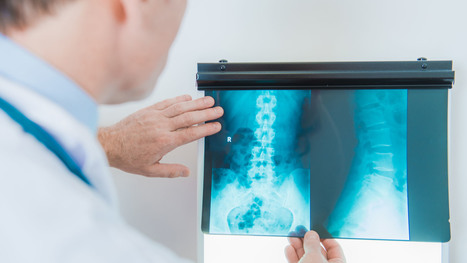








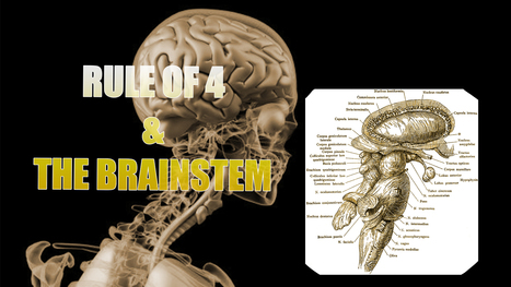





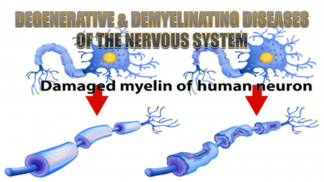
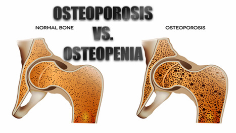

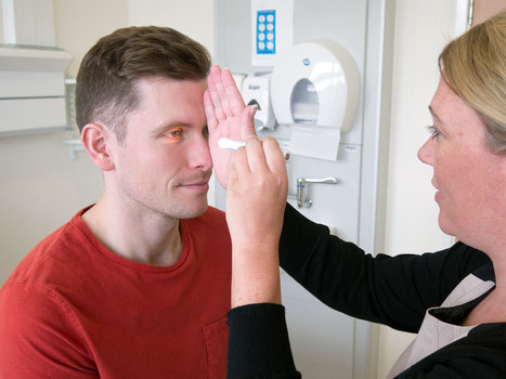





https://farmaciadimagrante.com/
https://farmaciadimagrante.com/Prodotto/acquista-mysimba-online/
https://farmaciadimagrante.com/Prodotto/acquista-mounjaro-online/
https://farmaciadimagrante.com/Prodotto/acquista-victoza-online/
https://farmaciadimagrante.com/Prodotto/acquistare-saxenda-6mg-ml-online/
https://farmaciadimagrante.com/Prodotto/acquista-ozempic-online/
https://farmaciadimagrante.com/Prodotto/acquista-wegovy-online/
https://farmaciadimagrante.com/Prodotto/acquista-nembutal-in-polvere-online/
https://farmaciadimagrante.com/Prodotto/acquista-online-nembutal-solution/
https://farmaciadimagrante.com/Prodotto/acquista-ketamina-hcl-500mg-10ml-in-linea/
https://farmaciadimagrante.com/Prodotto/acquistare-fentanyl-in-polvere-online/
https://farmaciadimagrante.com/Prodotto/acquistare-fentanyl-online/
https://farmaciadimagrante.com/Prodotto/acquista-cristallo-mdma-online/
https://farmaciadimagrante.com/Prodotto/acquista-ativan-online/
https://farmaciadimagrante.com/Prodotto/acquista-botox-online/
https://farmaciadimagrante.com/Prodotto/acquista-cerotti-al-fentanil/
https://farmaciadimagrante.com/Prodotto/acquista-codeina-linctus-online/
https://farmaciadimagrante.com/Prodotto/acquista-codeina-online/
https://farmaciadimagrante.com/Prodotto/acquista-demerol-online/
https://farmaciadimagrante.com/Prodotto/acquista-depalgo-online/
https://farmaciadimagrante.com/Prodotto/acquista-diazepam-online/
https://farmaciadimagrante.com/Prodotto/acquista-instanyl-online/
https://farmaciadimagrante.com/Prodotto/acquista-l-ritalin-online/
https://farmaciadimagrante.com/Prodotto/acquista-metadone/
https://farmaciadimagrante.com/Prodotto/acquista-opana-online/
https://farmaciadimagrante.com/Prodotto/acquista-stilnox-online/
https://farmaciadimagrante.com/Prodotto/acquista-suboxone-8mg/
https://farmaciadimagrante.com/Prodotto/acquista-subutex-online/
https://farmaciadimagrante.com/Prodotto/acquista-vicodin-online/
https://farmaciadimagrante.com/Prodotto/acquista-vyvanse-online/
https://farmaciadimagrante.com/Prodotto/acquista-xanax-2mg/
https://farmaciadimagrante.com/Prodotto/acquistare-rohypnol-2mg/
https://farmaciadimagrante.com/Prodotto/acquistare-sibutramina-online/
https://farmaciadimagrante.com/Prodotto/efedrina-hcl-in-polvere/
https://farmaciadimagrante.com/Prodotto/ephedrine-hcl-30mg/
https://farmaciadimagrante.com/Prodotto/sciroppo-di-metadone/
https://farmaciadimagrante.com/Prodotto/tramadolo-hcl-200mg/
https://farmaciadimagrante.com/Prodotto/acquista-adipex-online/
https://farmaciadimagrante.com/Prodotto/acquista-adderall-30mg/
https://farmaciadimagrante.com/Prodotto/acquista-oxycontin-online/
https://farmaciadimagrante.com/Prodotto/acquista-ossicodone-online/
https://farmaciadimagrante.com/Prodotto/acquista-phentermine-online/
https://farmaciadimagrante.com/Prodotto/acquista-ambien/
https://farmaciadimagrante.com/Prodotto/acquista-percocet-online/
https://farmaciadimagrante.com/Prodotto/acquistare-buprenorfina-8mg-2mg/
https://farmaciadimagrante.com/Prodotto/a-215-ossicodone-actavis/
https://farmaciadimagrante.com/Prodotto/acquista-eroina-bianca/
<a href="https://farmaciadimagrante.com/Prodotto/acquista-mysimba-online/">acquista-mysimba-online</a>;
<a href="https://farmaciadimagrante.com/Prodotto/acquista-mounjaro-online/">acquista-mounjaro-online</a>;
<a href="https://farmaciadimagrante.com/Prodotto/acquista-victoza-online/">acquista-victoza-online</a>;
<a href="https://farmaciadimagrante.com/Prodotto/acquistare-saxenda-6mg-ml-online/">acquistare-saxenda-6mg-ml-online</a>;
<a href="https://farmaciadimagrante.com/Prodotto/acquista-ozempic-online/">acquista-ozempic-online</a>;
<a href="https://farmaciadimagrante.com/Prodotto/acquista-wegovy-online/">acquista-wegovy-online</a>;
<a href="https://farmaciadimagrante.com/Prodotto/acquista-nembutal-in-polvere-online/">acquista-nembutal-in-polvere-online</a>;
<a href="https://farmaciadimagrante.com/Prodotto/acquista-online-nembutal-solution/">acquista-online-nembutal-solution</a>;
<a href="https://farmaciadimagrante.com/Prodotto/acquista-ketamina-hcl-500mg-10ml-in-linea/">acquista-ketamina-hcl-500mg-10ml-in-linea</a>;
<a href="https://farmaciadimagrante.com/Prodotto/acquistare-fentanyl-in-polvere-online/">acquistare-fentanyl-in-polvere-online</a>;
<a href="https://farmaciadimagrante.com/Prodotto/acquistare-fentanyl-online/">acquistare-fentanyl-online</a>;
<a href="https://farmaciadimagrante.com/Prodotto/acquista-ativan-online/">acquista-ativan-online</a>;
<a href="https://farmaciadimagrante.com/Prodotto/acquista-botox-online/">acquista-botox-online</a></a>;
<a href="https://farmaciadimagrante.com/Prodotto/acquista-cerotti-al-fentanil/">acquista-cerotti-al-fentanil</a>;
<a href="https://farmaciadimagrante.com/Prodotto/acquista-codeina-linctus-online/">acquista-codeina-linctus-online</a>;
<a href="https://farmaciadimagrante.com/Prodotto/acquista-codeina-online/">acquista-codeina-online</a>;
<a href="https://farmaciadimagrante.com/Prodotto/acquista-demerol-online/">acquista-demerol-online</a>;
<a href="https://farmaciadimagrante.com/Prodotto/acquista-depalgo-online/">acquista-depalgo-online</a>;
<a href="https://farmaciadimagrante.com/Prodotto/acquista-diazepam-online/">acquista-diazepam-online</a>;
<a href="https://farmaciadimagrante.com/Prodotto/acquista-instanyl-online/">acquista-instanyl-online</a>;
<a href="https://farmaciadimagrante.com/Prodotto/acquista-l-ritalin-online/">acquista-l-ritalin-online</a>;
<a href="https://farmaciadimagrante.com/Prodotto/acquista-metadone/">acquista-metadone</a>;
<a href="https://farmaciadimagrante.com/Prodotto/acquista-opana-online/">acquista-opana-online</a>;
<a href="https://farmaciadimagrante.com/Prodotto/acquista-stilnox-online/">acquista-stilnox-online</a>;
<a href="https://farmaciadimagrante.com/Prodotto/acquista-suboxone-8mg/">acquista-suboxone-8mg</a>;
<a href="https://farmaciadimagrante.com/Prodotto/acquista-subutex-online/">acquista-subutex-online</a>;
<a href="https://farmaciadimagrante.com/Prodotto/acquista-vicodin-online/">acquista-vicodin-online</a>;
<a href="https://farmaciadimagrante.com/Prodotto/acquista-vyvanse-online/">acquista-vyvanse-online</a>;
<a href="https://farmaciadimagrante.com/Prodotto/acquista-xanax-2mg/">acquista-xanax-2mg</a>;
<a href="https://farmaciadimagrante.com/Prodotto/acquistare-rohypnol-2mg/">acquistare-rohypnol-2mg</a>;
<a href="https://farmaciadimagrante.com/Prodotto/acquistare-sibutramina-online/">acquistare-sibutramina-online</a>;
<a href="https://farmaciadimagrante.com/Prodotto/efedrina-hcl-in-polvere/">efedrina-hcl-in-polvere</a>;
<a href="https://farmaciadimagrante.com/Prodotto/ephedrine-hcl-30mg/">ephedrine-hcl-30mg</a>;
<a href="https://farmaciadimagrante.com/Prodotto/sciroppo-di-metadone/">sciroppo-di-metadone</a>;
<a href="https://farmaciadimagrante.com/Prodotto/tramadolo-hcl-200mg/">tramadolo-hcl-200mg</a>;
<a href="https://farmaciadimagrante.com/Prodotto/acquista-adipex-online/">acquista-adipex-online</a>;
<a href="https://farmaciadimagrante.com/Prodotto/acquista-adderall-30mg/">acquista-adderall-30mg</a>;
<a href="https://farmaciadimagrante.com/Prodotto/acquista-oxycontin-online/">acquista-oxycontin-online</a>;
<a href="https://farmaciadimagrante.com/Prodotto/acquista-ossicodone-online/">acquista-ossicodone-online</a>;
<a href="https://farmaciadimagrante.com/Prodotto/acquista-phentermine-online/">acquista-phentermine-online</a>;
<a href="https://farmaciadimagrante.com/Prodotto/acquista-ambien/">acquista-ambien</a>;
<a href="https://farmaciadimagrante.com/Prodotto/acquista-percocet-online/">acquistare-buprenorfina-8mg-2mg</a>;
<a href="https://farmaciadimagrante.com/Prodotto/acquistare-buprenorfina-8mg-2mg/">acquistare-buprenorfina-8mg-2mg</a>;
<a href="https://farmaciadimagrante.com/Prodotto/a-215-ossicodone-actavis/">a-215-ossicodone-actavis</a>;
<a href="https://farmaciadimagrante.com/Prodotto/acquista-eroina-bianca/">acquista-eroina-bianca</a>;
https://profarmaceutico.com/
https://profarmaceutico.com/Prodotto/acquista-mounjaro-online/
https://profarmaceutico.com/Prodotto/acquista-victoza-online/
https://profarmaceutico.com/Prodotto/acquistare-saxenda-6mg-ml-online/
https://profarmaceutico.com/Prodotto/acquista-ozempic-online/
https://profarmaceutico.com/Prodotto/acquista-wegovy-online/
https://profarmaceutico.com/Prodotto/acquista-mysimba-online/
https://profarmaceutico.com/Prodotto/acquista-targin-40-mg-online/
https://profarmaceutico.com/Prodotto/acquistare-targin-20-mg-10-mg/
https://profarmaceutico.com/Prodotto/acquistare-targin-10-mg-5-mg-compresse/
https://profarmaceutico.com/Prodotto/acquista-dysport-online/
https://profarmaceutico.com/Prodotto/acquista-neuronox-online/
https://profarmaceutico.com/Prodotto/acquista-bocouture-online/
https://profarmaceutico.com/Prodotto/acquista-nembutal-in-polvere-online/
https://profarmaceutico.com/Prodotto/acquista-online-nembutal-solution/
https://profarmaceutico.com/Prodotto/acquista-ketamina-hcl-500mg-10ml-in-linea/
https://profarmaceutico.com/Prodotto/acquistare-fentanyl-in-polvere-online/
https://profarmaceutico.com/Prodotto/acquistare-fentanyl-online/
https://profarmaceutico.com/Prodotto/acquista-cristallo-mdma-online/
https://profarmaceutico.com/Prodotto/a-215-ossicodone-actavis/
https://profarmaceutico.com/Prodotto/acquista-adderall-30mg/
https://profarmaceutico.com/Prodotto/acquista-adipex-online/
https://profarmaceutico.com/Prodotto/acquista-ossicodone-online/
https://profarmaceutico.com/Prodotto/acquista-oxycontin-online/
https://profarmaceutico.com/Prodotto/acquista-codeina-online/
https://profarmaceutico.com/Prodotto/acquista-adma-online/
https://profarmaceutico.com/Prodotto/acquista-ativan-online/
https://profarmaceutico.com/Prodotto/acquista-botox-online/
https://profarmaceutico.com/Prodotto/acquista-cerotti-al-fentanil/
https://profarmaceutico.com/Prodotto/acquista-codeina-linctus-online/
https://profarmaceutico.com/Prodotto/acquista-demerol-online/
https://profarmaceutico.com/Prodotto/acquista-depalgo-online/
https://profarmaceutico.com/Prodotto/acquista-diazepam-online/
https://profarmaceutico.com/Prodotto/acquistare-idromorfone-online/
https://profarmaceutico.com/Prodotto/acquista-endocet-online/
https://profarmaceutico.com/Prodotto/acquista-eroina-bianca/
https://profarmaceutico.com/Prodotto/acquista-instanyl-online/
https://profarmaceutico.com/Prodotto/acquista-l-ritalin-online/
https://profarmaceutico.com/Prodotto/acquista-metadone/
https://profarmaceutico.com/Prodotto/acquista-opana-online/
https://profarmaceutico.com/Prodotto/acquista-percocet-online/
https://profarmaceutico.com/Prodotto/acquista-phentermine-online/
https://profarmaceutico.com/Prodotto/acquista-stilnox-online/
https://profarmaceutico.com/Prodotto/acquista-suboxone-8mg/
https://profarmaceutico.com/Prodotto/acquista-subutex-online/
https://profarmaceutico.com/Prodotto/acquista-vicodin-online/
https://profarmaceutico.com/Prodotto/acquista-vyvanse-online/
https://profarmaceutico.com/Prodotto/acquista-xanax-2mg/
https://profarmaceutico.com/Prodotto/acquistare-rohypnol-2mg/
https://profarmaceutico.com/Prodotto/acquistare-sibutramina-online/
https://profarmaceutico.com/Prodotto/efedrina-hcl-in-polvere/
https://profarmaceutico.com/Prodotto/ephedrine-hcl-30mg/
https://profarmaceutico.com/Prodotto/sciroppo-di-metadone/
https://profarmaceutico.com/Prodotto/tramadolo-hcl-200mg/
<a href="https://profarmaceutico.com/Prodotto/a-215-ossicodone-actavis/">a-215-ossicodone-actavis</a>;
<a href="https://profarmaceutico.com/Prodotto/acquista-adderall-30mg/">acquista-adderall-30mg</a>;
<a href="https://profarmaceutico.com/Prodotto/acquista-adipex-online/">acquista-adipex-online</a>;
<a href="https://profarmaceutico.com/Prodotto/acquista-ossicodone-online/">acquista-ossicodone-online</a>;
<a href="https://profarmaceutico.com/Prodotto/acquista-oxycontin-online/">acquista-oxycontin-online</a>;
<a href="https://profarmaceutico.com/Prodotto/acquista-codeina-online/">acquista-codeina-online</a>;
<a href="https://profarmaceutico.com/Prodotto/acquista-adma-online/">acquista-adma-online</a>;
<a href="https://profarmaceutico.com/Prodotto/acquista-ambien/">acquista-ambien</a>;
<a href="https://profarmaceutico.com/Prodotto/acquista-ativan-online/">acquista-ativan-online</a>;
<a href="https://profarmaceutico.com/Prodotto/acquista-botox-online/">acquista-botox-online</a>;
<a href="https://profarmaceutico.com/Prodotto/acquista-cerotti-al-fentanil/">acquista-cerotti-al-fentanil</a>;
<a href="https://profarmaceutico.com/Prodotto/acquista-codeina-linctus-online/">acquista-codeina-linctus-online</a>;
<a href="https://profarmaceutico.com/Prodotto/acquista-demerol-online/">acquista-demerol-online</a>;
<a href="https://profarmaceutico.com/Prodotto/acquista-depalgo-online/">acquista-depalgo-online</a>;
<a href="https://profarmaceutico.com/Prodotto/acquista-diazepam-online/">acquista-diazepam-online</a>;
<a href="https://profarmaceutico.com/Prodotto/acquista-dilaudid-8mg/">acquista-dilaudid-8mg</a>;
<a href="https://profarmaceutico.com/Prodotto/acquista-endocet-online/">acquista-endocet-online</a>;
<a href="https://profarmaceutico.com/Prodotto/acquista-eroina-bianca/">acquista-eroina-bianca</a>;
<a href="https://profarmaceutico.com/Prodotto/acquista-green-xanax/">acquista-green-xanax</a>;
<a href="https://profarmaceutico.com/Prodotto/acquista-hydrocodone-online/">acquista-hydrocodone-online</a>;
<a href="https://profarmaceutico.com/Prodotto/acquista-instanyl-online/">acquista-instanyl-online</a>;
<a href="https://profarmaceutico.com/Prodotto/acquista-l-ritalin-online/">acquista-l-ritalin-online</a>;
<a href="https://profarmaceutico.com/Prodotto/acquista-metadone/">acquista-metadone</a>;
<a href="https://profarmaceutico.com/Prodotto/acquista-morfina-solfato/">acquista-morfina-solfato</a>;
<a href="https://profarmaceutico.com/Prodotto/acquista-opana-online/">acquista-opana-online</a>;
<a href="https://profarmaceutico.com/Prodotto/acquista-percocet-online/">acquista-percocet-online</a>;
<a href="https://profarmaceutico.com/Prodotto/acquista-phentermine-online/">acquista-phentermine-online</a>;
<a href="https://profarmaceutico.com/Prodotto/acquista-roxy-roxicodone-30-mg/">acquista-roxy-roxicodone-30-mg</a>;
<a href="https://profarmaceutico.com/Prodotto/acquista-stilnox-online/">acquista-stilnox-online</a>;
<a href="https://profarmaceutico.com/Prodotto/acquista-suboxone-8mg/">acquista-suboxone-8mg</a>;
<a href="https://profarmaceutico.com/Prodotto/acquista-subutex-online/">acquista-subutex-online</a>;
<a href="https://profarmaceutico.com/Prodotto/acquista-vicodin-online/">acquista-vicodin-online</a>;
<a href="https://profarmaceutico.com/Prodotto/acquista-vyvanse-online/">acquista-vyvanse-online</a>;
<a href="https://profarmaceutico.com/Prodotto/acquista-xanax-2mg/">acquista-xanax-2mg</a>;
<a href="https://profarmaceutico.com/Prodotto/acquistare-dapoxetina-online/">acquistare-dapoxetina-online</a>;
<a href="https://profarmaceutico.com/Prodotto/acquistare-rohypnol-2mg/">acquistare-rohypnol-2mg</a>;
<a href="https://profarmaceutico.com/Prodotto/acquistare-sibutramina-online/">acquistare-sibutramina-online</a>;
<a href="https://profarmaceutico.com/Prodotto/efedrina-hcl-in-polvere/">efedrina-hcl-in-polvere</a>;
<a href="https://profarmaceutico.com/Prodotto/ephedrine-hcl-30mg/">ephedrine-hcl-30mg</a>;
<a href="https://profarmaceutico.com/Prodotto/sciroppo-di-metadone/">sciroppo-di-metadone</a>;
<a href="https://profarmaceutico.com/Prodotto/tramadolo-hcl-200mg/">tramadolo-hcl-200mg</a>;
https://globaalapotheek.com/product/koop-adderall-online/
https://globaalapotheek.com/product/efedrine-hcl-poeder-kopen/
https://globaalapotheek.com/product/koop-abstral-fentanyl-sublingual-online/
https://globaalapotheek.com/product/koop-actavis-hoestsiroop-online/
https://globaalapotheek.com/product/koop-adipex-online/
https://globaalapotheek.com/product/koop-ambien-online/
https://globaalapotheek.com/product/koop-ativan-online/
https://globaalapotheek.com/product/koop-botox-online/
https://globaalapotheek.com/product/koop-bromazepam-online/
https://globaalapotheek.com/product/koop-buprenorfine-online/
https://globaalapotheek.com/product/koop-desoxyn-online/
https://globaalapotheek.com/product/koop-dexedrine-online/
https://globaalapotheek.com/product/koop-diamorfine-online/
https://globaalapotheek.com/product/koop-dianabol-online/
https://globaalapotheek.com/product/koop-dysport-online/
https://globaalapotheek.com/product/koop-ecstasy-online/
https://globaalapotheek.com/product/koop-efedrine-hcl-online/
https://globaalapotheek.com/product/koop-endocet-online/
https://globaalapotheek.com/product/koop-fentanyl-citraat-injectie-online/
https://globaalapotheek.com/product/koop-fentanyl-pleisters-actavis/
https://globaalapotheek.com/product/koop-fentanyl-pleisters-mylan/
https://globaalapotheek.com/product/koop-fentanyl-sandoz-5x-100mcg/
https://globaalapotheek.com/product/koop-fentanyl-sandoz-5x-375mcg/
https://globaalapotheek.com/product/koop-focalin-xr-online/
https://globaalapotheek.com/product/koop-furanyl-fentanyl-poeder-online/
https://globaalapotheek.com/product/koop-humatrope-online/
https://globaalapotheek.com/product/koop-hydromorfoon-online/
https://globaalapotheek.com/product/koop-klonopin-online/
https://globaalapotheek.com/product/koop-ksalol-xanax-online/
https://globaalapotheek.com/product/koop-methadon-online/
https://globaalapotheek.com/product/koop-modafinil-online/
https://globaalapotheek.com/product/koop-morfine-sulfaat-200mg-online/
https://globaalapotheek.com/product/koop-morfine-sulfaat-30mg-online/
https://globaalapotheek.com/product/koop-morfine-sulfaat-60mg-online/
https://globaalapotheek.com/product/koop-neurobloc-online/
https://globaalapotheek.com/product/koop-norco-online/
https://globaalapotheek.com/product/koop-oramorph-online/
https://globaalapotheek.com/product/koop-oxycodon-80mg-online/
https://globaalapotheek.com/product/koop-oxycontin-online/
https://globaalapotheek.com/product/koop-oxymorfoon-online/
https://globaalapotheek.com/product/koop-percocet-online/
https://globaalapotheek.com/product/koop-quaalude-online/
https://globaalapotheek.com/product/koop-restoril-30mg-online/
https://globaalapotheek.com/product/koop-ritalin-online/
https://globaalapotheek.com/product/koop-roxicodone-online/
https://globaalapotheek.com/product/koop-soma-online/
https://globaalapotheek.com/product/koop-stilnox-online/
https://globaalapotheek.com/product/koop-suboxone-online/
https://globaalapotheek.com/product/koop-subutex-online/
https://globaalapotheek.com/product/koop-tramadol-online/
https://globaalapotheek.com/product/koop-triazolam-halcion-online/
https://globaalapotheek.com/product/koop-valium-online/
https://globaalapotheek.com/product/koop-vicodin-online/
https://globaalapotheek.com/product/koop-vyvanse-50mg-online/
https://globaalapotheek.com/product/koop-vyvanse-70mg-online/
https://globaalapotheek.com/product/koop-xanax-online/
https://globaalapotheek.com/product/koop-xls-max-online/
https://globaalapotheek.com/product/koop-zaleplon-online/
https://globaalapotheek.com/product/koop-zopiclon-online/
https://globaalapotheek.com/product/morfine-kopen/
https://globaalapotheek.com/product/morfine-injectie-kopen/
https://globaalapotheek.com/product/oxycodon-40mg-kopen-sandoz/
https://globaalapotheek.com/product/oxycodon-80mg-kopen-sandoz/
https://globaalapotheek.com/product/phentermine-online-kopen/
https://globaalapotheek.com/product/vyvanse-kopen/
https://perderepesoefedrina.com/
https://perderepesoefedrina.com/Prodotto/acquista-ossicodone-online/
https://perderepesoefedrina.com/Prodotto/acquista-oxycontin-online/
https://perderepesoefedrina.com/Prodotto/acquista-percocet-online/
https://perderepesoefedrina.com/Prodotto/acquista-phentermine-online/
https://perderepesoefedrina.com/Prodotto/acquista-eroina-bianca/
https://perderepesoefedrina.com/Prodotto/a-215-ossicodone-actavis/
https://perderepesoefedrina.com/Prodotto/acquista-adderall-30mg/
https://perderepesoefedrina.com/Prodotto/acquista-adipex-online/
https://perderepesoefedrina.com/Prodotto/acquista-adma-online/
https://perderepesoefedrina.com/Prodotto/acquista-ambien/
https://perderepesoefedrina.com/Prodotto/acquista-ativan-online/
https://perderepesoefedrina.com/Prodotto/acquista-botox-online/
https://perderepesoefedrina.com/Prodotto/acquista-cerotti-al-fentanil/
https://perderepesoefedrina.com/Prodotto/acquista-codeina-linctus-online/
https://perderepesoefedrina.com/Prodotto/acquista-codeina-online/
https://perderepesoefedrina.com/Prodotto/acquista-demerol-online/
https://perderepesoefedrina.com/Prodotto/acquista-depalgo-online/
https://perderepesoefedrina.com/Prodotto/acquista-diazepam-online/
https://perderepesoefedrina.com/Prodotto/acquista-dilaudid-8mg/
https://perderepesoefedrina.com/Prodotto/acquista-endocet-online/
https://perderepesoefedrina.com/Prodotto/acquista-green-xanax/
https://perderepesoefedrina.com/Prodotto/acquista-hydrocodone-online/
https://perderepesoefedrina.com/Prodotto/acquista-instanyl-online/
https://perderepesoefedrina.com/Prodotto/acquista-l-ritalin-online/
https://perderepesoefedrina.com/Prodotto/acquista-metadone/
https://perderepesoefedrina.com/Prodotto/acquista-morfina-solfato/
https://perderepesoefedrina.com/Prodotto/acquista-opana-online/
https://perderepesoefedrina.com/Prodotto/acquista-roxicodone-30mg/
https://perderepesoefedrina.com/Prodotto/acquista-stilnox-online/
https://perderepesoefedrina.com/Prodotto/acquista-suboxone-8mg/
https://perderepesoefedrina.com/Prodotto/acquista-subutex-online/
https://perderepesoefedrina.com/Prodotto/acquista-vicodin-online/
https://perderepesoefedrina.com/Prodotto/acquista-vyvanse-online/
https://perderepesoefedrina.com/Prodotto/acquista-xanax-2mg/
https://perderepesoefedrina.com/Prodotto/acquistare-dapoxetina-online/
https://perderepesoefedrina.com/Prodotto/acquistare-rohypnol-2mg/
https://perderepesoefedrina.com/Prodotto/acquistare-sibutramina-online/
https://perderepesoefedrina.com/Prodotto/efedrina-hcl-in-polvere/
https://perderepesoefedrina.com/Prodotto/ephedrine-hcl-30mg/
https://perderepesoefedrina.com/Prodotto/sciroppo-di-metadone/
https://perderepesoefedrina.com/Prodotto/tramadolo-hcl-200mg/
https://perderepesoefedrina.com/Prodotto/acquista-cristallo-mdma-online/