 Your new post is loading...
The cranial nerves are the nerves which come out straight from the brain, including the brainstem, in comparison to the spinal nerves, which come out from sections of the spinal cord. Of those, 10 out of 12 of these cranial nerves originate in the brainstem. Cranial nerves transfer information between the brain and parts of the human body, particularly to and from areas of the head and neck. Spinal nerves exit from the spinal cord with the spinal nerve closest to the head (C1) exiting in the space above the first cervical vertebra. The cranial nerves, however, exit from the central nervous system above this region. Each cranial nerve is paired and is present on either side of the brain. Based on the definition in humans, there are twelve, sometimes thirteen, cranial nerve pairs, which have been assigned Roman numerals I-XII for identification, sometimes including cranial nerve zero as well. The numbering of the cranial nerves is based on the order in which they emerge from the brain, or from the front to the back of the brainstem. The terminal nerves, olfactory nerves (I) and optic nerves (II) come out from the cerebrum, or forebrain, where the rest of the ten pairs of cranial nerves arise in the brainstem, which is the lower portion of the brain. The cranial nerves are considered components of the peripheral nervous system (PNS), though on a structural level, the olfactory, the optic and the trigeminal nerves are more accurately considered a portion of the central nervous system (CNS). Most commonly, humans are believed to have twelve pairs of cranial nerves (I-XII). These include: the olfactory nerve (I), the optic nerve (II), the oculomotor nerve (III), the trochlear nerve (IV), the trigeminal nerve (V), the abducens nerve (VI), the facial nerve (VII), the vestibulocochlear nerve (VIII), the glossopharyngeal nerve (IX), the vagus nerve (X), the accessory nerve (XI), and the hypoglossal nerve (XII). There may be a thirteenth cranial nerve, known as the terminal nerve, or nerve N or O, which Is quite small and may or may not be functional in humans.
Following upper limb peripheral nerve transection and surgical repair, some patients regain good sensorimotor function while others do not. Understanding peripheral and central mechanisms that contribute to recovery may facilitate the development of new therapeutic interventions. Plasticity following peripheral nerve transection has been demonstrated throughout the neuroaxis in animal models of nerve injury. However, the brain changes that occur following peripheral nerve transection and surgical repair in humans have not been examined. Furthermore, the extent to which peripheral nerve regeneration influences functional and structural brain changes has not been characterized. Therefore, we asked whether functional changes are accompanied by grey and/or white matter structural changes and whether these changes relate to sensory recovery? To address these key issues we (i) assessed peripheral nerve regeneration; (ii) measured functional magnetic resonance imaging brain activation (blood oxygen level dependent signal; BOLD) in response to a vibrotactile stimulus; (iii) examined grey and white matter structural brain plasticity; and (iv) correlated sensory recovery measures with grey matter changes in peripheral nerve transection and surgical repair patients. Compared to each patient’s healthy contralesional nerve, transected nerves have impaired nerve conduction 1.5 years after transection and repair, conducting with decreased amplitude and increased latency. Compared to healthy controls, peripheral nerve transection and surgical repair patients had altered blood oxygen level dependent signal activity in the contralesional primary and secondary somatosensory cortices, and in a set of brain areas known as the ‘task positive network’. In addition, grey matter reductions were identified in several brain areas, including the contralesional primary and secondary somatosensory cortices, in the same areas where blood oxygen level dependent signal reductions were identified. Furthermore, grey matter thinning in the post-central gyrus was negatively correlated with measures of sensory recovery (mechanical and vibration detection) demonstrating a clear link between function and structure. Finally, we identified reduced white matter fractional anisotropy in the right insula in a region that also demonstrated reduced grey matter. These results provide insight into brain plasticity and structure-function-behavioural relationships following nerve injury and have important therapeutic implications. Keywords: cortical thickness; fMRI; diffusion tensor imaging; plasticity; peripheral nerve injury
Abbreviations: BA=Brodmann area; BOLD=blood oxygen level dependent; fMRI=functional magnetic resonance imaging;
PNIr=peripheral nerve transection and surgical repair; S1=primary somatosensory cortex; S2=secondary somatosensory cortex Introduction Following upper limb peripheral nerve transection and surgical repair (PNIr), �25% of patients have not returned to work 1.5 years after surgery (Jaquet et al., 2001). In addition, �57% of patients with nerve injuries are between 16–35 years of age (McAllister et al., 1996); thus, a long life of disability and economic difficulties may accompany upper limb nerve transection. Understanding the central and peripheral ramifications of peripheral nerve injury may facilitate the development of new therapeutic strategies and intervention programs. It is not known how the brain responds to PNIr in humans. However, animal studies have established that plasticity within the somatosensory cortex begins immediately following peripheral nerve transection, and that 1 year after complete nerve transection and surgical repair, cortical maps contain patchy, non- continuous representations of the transected and adjacent nerves (Wall et al., 1986). The mechanisms that facilitate functional plasticity are thought to include the immediate unmasking of pre-existing projections from adjacent cortical and subcortical levels, and long term sprouting of axons at multiple levels of the neuroaxis, including the primary somatosensory cortex (S1) (Florence and Kaas, 1995; Hickmott and Steen, 2005). Human brain imaging studies have corroborated the findings from animal models with the identification of altered functional MRI activation maps due to spinal cord injury, amputation, toe- to-thumb transfer, and in patients with carpel tunnel syndrome (Lotze et al., 2001; Manduch et al., 2002; Jurkiewicz et al., 2006; Napadow et al., 2006). Furthermore, structural MRI studies have recently visualized grey and white matter changes following traumatic injuries and in diverse pathological conditions, including limb amputation and chronic pain (Apkarian et al., 2004; Draganski et al., 2006; Davis et al., 2008; Geha et al., 2008; May, 2008). Grey matter changes are thought to be related to changes in cell size, atrophy and/or loss of neurons or glia, whereas white matter changes are impacted by axonal degenera- tion and loss of myelin (Beaulieu, 2002; May, 2008). A powerful approach to delineate mechanisms of pathology and plasticity is to combine functional and structural grey and white matter imaging techniques. We previously reported that patients with complete upper limb PNIr retained profound somatosensory deficits that persist 41.5 years following surgery (Taylor et al., 2008a). Based on these findings, we reasoned these patients would exhibit functional and structural brain changes in key somatosensory brain areas. Therefore, in this study, we hypothesized that PNIr patients would have: (i) reduced blood oxygen level dependent (BOLD) responses to vibratory stimulation of the transected nerve territory, in the region of S1 that represents the injured upper limb and in the secondary somatosensory cortex (S2); (ii) a corresponding reduction in cortical thickness in these regions of the contralesional S1 and S2; (iii) a correlation between changes in cortical thickness and psychophysical measures of somatosensory function (vibration and touch detection thresh- olds); and (iv) reduced fractional anisotropy (a measure of white matter integrity) in white matter feeding into/out of these somatosensory cortical areas. Methods Subjects We recruited 27 patients with complete transection of the median and/or ulnar nerve followed by surgical repair from plastic surgeons affiliated with the University of Toronto Hand Program between June 2006 and May 2008. From this larger cohort, 14 pain-free patients (three females, 11 males; 34 � 10 years) with complete transection of the right median and/or ulnar nerve were included in the study [to avoid confounds related to the presence of pain and laterality patients with pain (n=6) and left sided lesions (n=7) were excluded from this analysis]. All patients underwent microsurgical nerve repair at least 1.5 years prior to study enrolment (recovery time varied from 1.5 to 8 years). In addition, we recruited 14 age- and sex-matched healthy controls (3 females, 11 males; 34 � 10 years). All subjects gave informed written consent to procedures approved by the University Health Network Research Ethics Board. All subjects were right handed (determined using the Edinburgh handedness inventory: Oldfield, 1971) and had no history of neurological injury or chronic pain (either before or after nerve transection). See Table 1 for demographic details. Study Design All subjects participated in an imaging session that included: (i) functional magnetic resonance imaging (fMRI) in response to vibrotactile stimuli applied to the right index finger (within the median nerve territory); (ii) a high-resolution anatomical scan of the whole brain, acquired for image registration and for the analysis of cortical grey matter; and (iii) two diffusion tensor imaging scans for the assessment of white matter integrity. Prior to imaging, subjects were instructed in the basic design of the experiment and reminded to remain as still as possible throughout the duration of the scan. Subjects were free to withdraw from the study at any time. In addition, a sensory and motor assessment was performed for all subjects (Taylor et al., 2008a). As touch and vibration detection thresholds were correlated with cortical thickness a description of these methods is included below (other psychophysical measures will be reported elsewhere). Vibration Threshold Vibration detection thresholds were determined using a hand held Bio-Thesiometer (Bio-Medical Instrument Company, USA). The device has a 12-mm probe that was placed on the distal phalanx of the right index finger (D2). Thresholds were determined using the method of limits: the amplitude (voltage) was gradually increased until the subject indicated that they perceived the stimulus. Vibration thresholds were acquired three times and an average value was calculated. During vibration threshold testing, subjects were instructed to close their eyes and rest the back of their hand on a supportive cushion. Mechanical Detection Threshold Mechanical detection thresholds were determined using a standardized set of von Frey filaments (OptiHair2 Marstock Nervtest, Germany) containing 12 logarithmically spaced calibrated filaments that delivered forces from 0.25–512 mN. The contact surface diameter of all 12 filaments was ~0.4mm. Trials were conducted with the subject’s eyes closed and hands resting on a soft cushion. Probes were applied in an ascending series and subjects were required to make a response every time they felt a probe touch the right D2 fingertip. This process was repeated three times. The force for the filament that was detected in at least two of three trials was reported as that subject’s mechanical detection threshold. Nerve Conduction Testing Patients participated in bilateral sensory and motor nerve conduction studies at the Toronto Western Hospital electromyography (EMG) clinic. For motor nerve conduction, the stimulating electrode was placed at the wrist and elbow (separately) and the recording electrode was placed over the abductor pollicis brevis, for median nerve assessment, or the abductor digiti minimi for ulnar nerve assessment. For sensory nerve testing the recording electrode was placed at the wrist and the stimulating electrode was placed at digits D2, D3 and D5. A senior, experienced neurologist from the Toronto Western Hospital EMG Clinic (Dr Peter Ashby) reviewed all clinical assessments to deter- mine which nerves demonstrated normal/abnormal responses. As amplitude and latency measures are known to vary substantially between subjects (due to factors such as the density of innervation, the depth of the nerve and the thickness of an individual subject’s skin) (Kimura, 2001) each patient’s untransected nerve served as their own control for comparison with values from the transected side. In those patients with detectable nerve conduction responses, paired t-tests were performed to assess the difference in latency or amplitude measures between each patient’s transected and contralesional untransected nerves. Imaging Parameters Brain imaging data were acquired using a 3T GE MRI system fitted with an eight-channel phased array head coil. Subjects were placed supine on the MRI table and each subjects’ head was padded to reduce movement. Whole-brain fMRI data was acquired using echo planar imaging (28 axial slices, field of view (FOV) = 20 x 20 cm, 64 x 64 matrix, 3.125 x 3.125 x 4mm voxels, echo time (TE) = 30 ms, repetition time (TR) = 2000 ms). The scan time was 5 min and 8 s (154 frames). During scanning, a non-painful, 12 Hz vibrotactile stimulus was applied to the distal phalanx of the right D2 using balloon diaphragms driven by compressed air (Device man- ufactured by Dr Christo Pantev; www.biomag.uni-muenster.de). Stimuli were delivered in blocks of 10s interleaved with 20s of rest, for a total of 10 blocks of stimulation and 10 blocks of rest. The first 8 s (4 TRs) of data acquired from each run were discarded to allow for fMRI signal equilibration. Subjects were instructed to keep their eyes closed throughout scanning and focus on the stimuli. A whole brain three dimensional (3D) high-resolution anatomical scan (124 sagittal slices, 24 x 24 cm FOV, 256 x 256 matrix, 1.5 x 0.94 x 0.94 mm voxels) was acquired with a T1-weighted 3D spoiled gradient echo sequence (one signal average, flip angle = 20� , TE �5 ms). In addition, two diffusion tensor imaging scans (38 axial slices, FOV 24 x 24 cm, 128 x 128 matrix, 1.875 x 1.875 x 3 mm voxels) were acquired along 23 directions with a b-value of 1000smm–2. Each run also contained two volumes with no diffusion weighting. fMRI Analysis Data were analysed using Brainvoyager QX v1.8 (Brain Innovaton, Maastricht, Netherlands). Pre-processing included: 3D motion correction, slice scan-time correction, linear trend removal, high-pass filtering (five cycles per run), and spatial smoothing with a 6mm full width at half maximum (FWHM) Gaussian kernel. fMRI data sets were interpolated to 3 x 3 x 3 mm voxels, registered to the high-resolution ana- tomical image, and normalized to standard Talairach space (Talairach and Tournoux, 1988). Voxels are reported as 1 x 1 x 1 mm. Data were analysed using the general linear model; the model was obtained by convolving the boxcar function of the time course of tactile stimulation with the standard haemodynamic response function. To identify between group differences in activation patterns a fixed effects analysis was performed with the contrasts: (i) healthy controls: stimulation 4 rest; (ii) PNIr: stimulation 4 rest; and (iii) healthy controls 4 PNIr. Activation maps were thresholded at a corrected value of P50.05 (derived from an uncorrected P50.0001 and 120mm3 contiguous voxels as previously reported: Taylor and Davis, 2009); this was also validated by running a Monte Carlo Simulation with AlphaSim application implemented in the Analysis of Functional Neuroimage (AFNI) software. This analysis included only the 11 patients that sustained transection of the right median nerve (n=9) or the right median and ulnar nerve (n = 2) (i.e. the three patients with a pure right ulnar nerve transection were not included in this analysis). Cortical Thickness Analysis Cortical thickness analysis was performed using Freesurfer (http:// surfer.nmr.mgh.harvard.edu); methods have been outlined in detail elsewhere (Dale et al., 1999; Fischl et al., 1999a, b; Fischl and Dale 2000). Briefly, high-resolution T1-weighted anatomical data sets were registered to the Talairach atlas (Talairach and Tournoux, 1988). This was followed by intensity normalization, skull stripping and separation of the hemispheres. Subsequently, the white/grey matter (called the white surface) and grey/CSF (called the pial surface) boundaries were identified and segmented. The distance between the white and pial surfaces was then calculated at every point in each hemisphere of the brain. To identify group differences between the 14 patients and 14 age/sex-matched controls, a general linear model analysis was performed at every point on the brain. As individual’s cortical topography is inherently heterogeneous, a 5mm FWHM spatial smoothing kernel was applied prior to statistical analysis. Data are displayed at a corrected P50.05 (derived from an uncorrected P50.0075 and 102 contiguous vertices); this was calculated by running a Monte Carlo simulation with AlphaSim. A vertex represents a point on a two dimensional sheet, and, in this study, the distance between two vertices is 0.80mm2. As patients exhibited significant deficits in somatosensory function within the transected nerve territory, we hypothesized that measures of somatosensory function (vibration and touch detection) would correlate with cortical thickness in the contralesional post-central gyrus (primary and secondary somatosensory cortices). Therefore, we performed correlation analyses in the patient group between: (i) cortical thickness and vibration detection threshold; and (ii) cortical thickness and touch detection thresholds. One patient did not complete psychophysical assessment; therefore, this analysis included 13 PNIr patients. In addition, to determine if there was a relationship between cortical thickness and recovery time a correlation analysis was also performed between these two measures. These correlation analyses were restricted to the contralesional post-central gyrus by including a mask (taken from Freesurfer’s built in atlas) in the general linear model. A Monte Carlo simulation was performed that was restricted to the number of vertices within contralesional post-central gyrus; images are displayed with a corrected P50.05 (derived from an uncorrected P50.0075 and 68 contiguous vertices). Diffusion Tensor Imaging Analysis Diffusion tensor image processing was performed with DTiStudio (www.MriStudio.org) and FSLv.4.0 (www.fmrib.ox.ac.uk/fsl/). Images were first realigned with the Automatic Image Registration tool implemented in DTiStudio, using the first B0 image in the first series acquired as the template. This process corrects for subject motion and eddy-current distortion. All images were then inspected visually to assess image quality and the alignment of the separate diffusion tensor imaging runs. If an artefact was detected, the slice was removed prior to calculating the average of the two separate diffusion tensor imaging runs. Individual FA maps were calculated using the DTIFIT tool implemented in FSL. Voxel-wise statistical analysis was performed to identify group differences in the mean fractional anisotropy using Tract Based Spatial Statistics; for a full description of these methods see Smith et al. (2006). Briefly, images were non-linearly registered to a target image (MNI152), the mean image was then created from all datasets and this image was subsequently thinned to represent all tracts that were common to all subjects. Each subject’s highest fractional anisotropy values were then projected onto the skeleton by searching in white matter perpendicular to each point on the white matter skeleton. A whole-brain voxel-wise statistical analysis was then performed between groups (14PNIr and 14 healthy controls) and images were whole brain corrected at P50.05. In addition, a region of interest analysis was performed in white matter tracts adjacent to the contralateral S1, thalamus and bilateral anterior and posterior insula. These regions were chosen as they have previously been implicated in aspects of somatosensation and because they correspond with regions that were identified in the fMRI and cortical thickness analysis (CTA) group analyses. Regions of interest were drawn on the white matter skeleton as follows: (i) The contralateral S1 region of interest originated medially at the junction between the white matter skeleton of the corona-radiata and the skeleton section feeding into the post- central gyrus; terminating at the end of the tract within a given slice. In the z direction the region of interest extended from z=49 to 57; white matter tracts supplying the hand region. (ii) The contralateral thalamus region of interest was restricted to white matter tracks surrounding the posterior and medial thalamic nuclei (nuclei involved in somatosensory function), extending from z = –1 to 4. (iii) Insular regions of interest were drawn bilaterally within white matter adjacent to the anterior and posterior insula based on criteria previously published by our lab (Taylor et al., 2008b). The region of interest extended from z = 2 to 8. Fractional anisotropy values were extracted from each of these regions of interest and a multivariate analysis of variance (MANOVA) was performed using the Statistical Package for the Social Sciences v13.0 (SPSS Inc, Chicago), which included fractional anisotropy values for all six regions of interest. Results Table 1 provides demographic details for study participants. All 14 patients sustained a complete transection of the right median and/or ulnar nerve followed by microsurgical repair at least 1.5 years prior to study enrolment. The time from surgery to testing ranged from 1.5 to 8 years with a mean (�SD) of 4.8 � 3 years. Patients and controls did not (34 � 10 years both groups; t = 0.04; P = 0.97). Psychophysics Vibration thresholds were calculated from all three measurements since one-way repeated measures analysis of variance (ANOVA) indicated no significant differences between the three trials [F (25, 1)=0.227, P=0.64]. Vibration and mechanical detection thresh- olds were significantly impaired in PNIr patients compared to healthy controls (vibration: t = 4.77, P50.001, Fig. 3A; mechanical: t=3.10, P=0.005, Fig. 3D). Nerve Conduction Testing Amplitude and latency measures obtained from each patient’s contralesional nerves were classified as normal by an experienced neurologist at the Toronto Western Hospital EMG Clinic. Nine of the 14 patients completed nerve conduction testing. Table 2 displays the average increase/decrease latency and amplitude data for sensory nerve conduction from the wrist to the abductor pollicis brevis (median) or abductor digiti minimi (ulnar) muscles and for sensory conduction from the wrist to D2 (median) and D5 (ulnar) compared to each patients uninjured contralesional nerve. Out of nine, seven patients had transections that included the median nerve. Of these seven, one patient had no detectable response during motor testing and another patient had no detectable response during sensory testing. In the six patients with detectable responses, motor conduction latencies were increased by 43% (t=6.2; P=0.002) and amplitudes were decreased by 38% (t=–2.6; P=0.045) when each patient’s transected nerve was compared to their non-injured side. Sensory conduction in median nerves also revealed a 26% increase in latency (t=3.9; P=0.011) and a 73% decrease in amplitude (t=–8.0; P=0.000) compared to normal contralesional nerves. In the four patients with ulnar nerve transections one patient had no detectable response during sensory nerve testing. In those patients with responses, ulnar nerve motor latencies were not significantly elevated (t = 2.8; P = 0.070); however, amplitudes were significantly decreased by 41% (t = –5.9; P = 0.010). Sensory testing of the ulnar nerve demonstrated a 27% increase in latency (t = 4.3; P = 0.049) but no significant increase in amplitude (t = �3.5; P = 0.072). Functional Plasticity In The Primary Somatosensory Cortex Functional MRI maps were calculated from the 11 PNIr patients with right median nerve transections (patients with ulnar nerve transections were excluded from this analysis) and 11 age- and sex-matched healthy controls. From Fig. 1A, it is clear that PNIr patients have significantly less activation, compared to healthy controls, in a region of S1 corresponding to Brodmann area 2 (BA2) (Talairach and Tournoux, 1988) and S2 (see Table 3 for details). The average event-related responses from these regions of interest highlight the attenuated BOLD response within the patients’ left BA2 and left S2 (Fig. 1B and C, respec- tively). Curiously, vibrotactile stimulation in the patients activated a more superior part of the post-central gyrus (probably BA1/3) (Talairach and Tournoux, 1988) (Fig. 1A and Table 3). An event- related average (Fig. 1D) demonstrates that healthy controls had minimal activation in this region. Furthermore, patients had significantly more activation in brain regions collectively known as the task positive network (asterisks in Fig. 1). See Table 3 for the full list of task positive brain areas activated. This network includes lateral prefrontal, lateral parietal, premotor and inferior temporal cortices (Table 3): brain areas that are activated during the performance of an attention demanding task and suppressed or inactive during rest or tasks that are not cognitively or attentionally challenging (Fox et al., 2005; DeLuca et al., 2006; Seminowicz and Davis 2007). Reduced Grey Matter In The Primary Somatosensory Cortex Correlates With Sensory Recovery Cortical thickness analysis in all 14 patients and 14 age/ sex-matched healthy controls revealed several loci of significant cortical thinning in the PNIr group (Fig. 2 and Table 4). Specifically, patients had a 13%–22% reduction in cortical thick- ness in the left (contralesional) S1, S2, pregenual anterior cingulate gyrus, ventrolateral prefrontal cortex and right anterior insula, anterior/posterior mid cingulate gyrus and paracentral lobule. Interestingly, the locations of grey matter thinning within the post-central gyrus coincide with the regions of reduced BOLD following vibrotactile stimulation (Table 4). Since we had prior knowledge of the patients’ sensory deficits and recovery time (i.e. time since microsurgical repair), we next asked whether the patients’ cortical thickness in the post-central gyrus correlated with their sensory mechanical and vibration detection thresholds, or with their recovery time. These analyses revealed a negative correlation between cortical thickness and vibration detection thresholds in a region encompassing BA1/2 and S2 (P50.001, r=�0.80 and �0.91, for BA1/2 and S2, respectively; Fig. 3 and Table 5). In addition, mechanical detection thresholds were also negatively correlated with cortical thickness in a slightly more superior BA2 region and the same S2 region (P50.001, r = �0.83 and �0.85, for BA2 and S2, respectively; Fig. 3 and Table 5). However, we did not identify a significant relationship between recovery time and cortical thickness. Therefore, in the post-central gyrus cortical thinning was associated with more severe sensory deficits. However, we did not identify a significant relationship between recovery time and cortical thickness. Again, there was a correspondence between the cortical thinning in areas negatively correlated with vibratory stimuli and the regions showing group fMRI and CTA abnormalities. White Matter Abnormalities Following Nerve Transection To assess white matter integrity we utilized a region of interest approach to examine white matter group differences based on a priori hypotheses. Regions of interest were restricted to white matter tracts surrounding and feeding into the contralesional S1 and thalamus. In addition, we also drew regions of interest in white matter adjacent to left and right, anterior and posterior insula. The insula was chosen as it is implicated in somatosensory processing and because we identified reduced grey matter in the right anterior insular with CTA. This region of interest approach revealed that patients had significantly reduced white matter fractional anisotropy values (MANOVA including all six regions of interest) adjacent to the right anterior [F (1, 26) = 4.39, P = 0.046; Fig. 4A] and posterior insula [F (1, 26) = 5.55, P = 0.026; Fig. 4B], but there were no group differences in the white matter adjacent to the left insula (left anterior insula: P = 0.51; left posterior insula: P=0.26), thalamus (P=0.46) or S1 (P=0.46) Discussion Here, we have demonstrated for the first time that there is functional plasticity and both grey and white matter structural abnormalities in several cortical areas following upper limb peripheral nerve transection and surgical repair. This plasticity may arise from incomplete peripheral nerve regeneration (peripheral cell death and/or incomplete re-myelination), as nerve conduction measures in these patients demonstrated severe abnormalities. In addition, our data demonstrate that decreased vibrotactile-evoked fMRI responses in the post-central gyrus correspond with grey matter thinning in the patient group. These results suggest that reduced BOLD responses may be facilitated by a reduction in cortical grey matter and/or a decrease in the afferent input to the post-central gyrus. In addition, cortical thickness within these same parts of the post-central gyrus negatively correlated with behavioral measures of somatosensory function. That is, increased somatosensory deficits were correlated with thinner cortex; both of which may be related to afferent input. Taken together, our data suggest that incomplete peripheral nerve regeneration contributes to somatosensory impairments, cortical grey matter atrophy and reduced fMRI activation (see Fig. 5 for a summary of these findings). It is well known that cortical plasticity following peripheral nerve transection and surgical repair can occur throughout the CNS in non-human primates (Kaas, 1991). This plasticity is thought to be due to the unmasking of previously silent synapses or axonal sprouting into deafferented territory (Wall et al., 1986; Florence and Kaas, 1995). In the primate model, 1 year following nerve transection and surgical repair, the denervated cortex is characterized by incomplete and disorderly representations of the regenerated and adjacent (intact) nerves. This patchy representation is attributed to incomplete peripheral regeneration resulting in a partial recovery of the denervated cortical space (Kaas, 1991). To assess the extent of peripheral regeneration in our patient population we performed sensory and motor nerve conduction studies across the transected area. Our nerve conduction results demonstrate that PNIr patients have significantly decreased amplitude and increased latency in both sensory and motor nerves compared to their own untransected side. Decreased amplitude combined with increased latency is indicative of peripheral fibre loss (i.e. cell death) and/or abnormal or incomplete re-myelination following transection (Kimura, 1984). In addition, it is well established that between 20% and 50% of dorsal root ganglion neurons die following nerve transection (Liss et al., 1996). Thus, afferent cell death and incomplete regeneration can result in decreased afferent input to the cortex, which may account for ongoing sensory deficits and decreased BOLD response in BA2 and S2. Furthermore, this diminished afferent input could also account for the cortical thinning we observed in the same regions of the cortex. Sensory deprivation has been shown to cause trans- neuronal degeneration in several regions of the CNS, including the dorsal horn following sciatic nerve section (Knyihar-Csillik et al., 1989), and may involve second- and third-order neurons (Powell and Erulkar, 1962). Transneuronal degeneration is characterized by cell shrinkage and is thought to be related to decreased, or non-existent, afferent input (Knyihar-Csillik et al., 1989). Thus, cortical grey matter loss (or atrophy) could also be directly related to decreased afferent input. We also demonstrated increased activation in the post-central gyrus in a region corresponding to BA1/3 (Talairach and Tournoux, 1988). Electrophysiological, anatomical tracing and neuroimaging studies have established that for the majority of cutaneous mechanoreceptive afferents the first cortical destina- tions are BA1 and BA3b. These cytoarchitectonic brain areas each possess a somatotopic body map with small receptive fields. In addition, these areas respond to many features of tactile information, such as texture and roughness, velocity and curvature of stimuli (Bodegard et al., 2001). fMRI studies have demon- strated that activity within the somatosensory cortex is influenced by attention such that fMRI responses to tactile stimuli in S1 are increased when subjects attend to a tactile stimulus, but are atte- nuated when subjects are distracted (Arthurs et al., 2004; Porro et al., 2004). Furthermore, our patients activated a network of brain areas known as the task positive network (DeLuca et al., 2006) more than healthy controls. These brain areas are activated during attention demanding processes (Fox et al., 2005; Seminowicz and Davis, 2007). Together, these findings imply that patients must attend to the stimulus more than controls because of their impaired sensory input. This increased attention may also account for the increased activation in BA1/3b. Of course, the increased activation in BA1/3b may also reflect plasticity that is unrelated to attentional load. BA2 and S2 both receive projections from BA1/3b and also from distinct parts of the ventroposterior thalamic complex (Pons et al., 1985; Friedman and Murray, 1986). Both of these brain areas have large, often multi-digit (BA2) or bilateral (S2) receptive fields (Pons et al., 1985; Iwamura et al., 2002). Based on ana- tomical projections and neuronal response properties, hierarchical processing of tactile information has been demonstrated from BA1/3b to BA 2 (Kaas et al., 2002). In addition, electrophysiolo- gical studies in macaques (Pons et al., 1987) and magnetoence- phalography data acquired in humans, suggest that serial processing of tactile inputs occurs from S1 to S2 in higher primates (Frot and Mauguiere 1999; Disbrow et al., 2001). Several studies have demonstrated that BA2 is preferentially activated by shape and curvature (Bodegard et al., 2001), while S2 may be involved in tactile learning (Ridley and Ettlinger 1976; Murray and Mishkin, 1984), supporting the notion that these brain areas are involved in higher-order somatosensory processing. Our psychophysical assessment demonstrated that patients were significantly impaired at the detection of simple tactile stimuli, and in the Shape Texture Identification test 1.5 years after surgery (Taylor et al., 2008a). This latter test assesses a patient’s ability to recognize character- istics of an object while actively exploring a shape or texture, requiring the integration of sensory information across regions of the body (Rosen and Lundborg, 1998). Taken together, one inter- pretation of our data is that PNIr patients attend more to the vibrotactile stimulus, leading to increased activation of the task positive network and BA1/3. However, in these patients, our data imply that higher-order processing areas, such as BA2 and S2, did not receive tactile information, which, in turn, may result in cortical thinning and reduced BOLD responses. The insula is thought to play a role in integrating multimodal information important for sensorimotor, emotional, allostatic/ homeostatic and cognitive functions (Devinsky et al., 1995; Critchley, 2004; Craig, 2008) and has been designated a limbic sensory cortex (Craig, 2008). Several studies have reported insular activation in response to tactile stimulation (Gelnar et al., 1998; Downar et al., 2002) and anatomical tracing studies in primates have demonstrated that the insula is reciprocally connected to frontal, parietal and temporal lobes (Augustine, 1996). In our patients, the right anterior insula was the only cortical area that demonstrated significant cortical thinning in conjunction with reduced fractional anisotropy values in the adjacent white matter, suggesting that the cortical thinning within this region is associated with a loss of fibres projecting to or from this structure. The right anterior insula has been implicated in interoception as it is situated to integrate homeostatic input from the body with motivational, emotional and social conditions (Craig, 2008). Furthermore, Critchley et al. (2004) reported a correlation between interoceptive abilities and the grey matter volume of the right anterior insula. Given our finding that the patients have decreased grey matter in the right anterior insula, it would be of interest to assess interoceptive capabilities following peripheral nerve injury in a future study. Taken together, we have demonstrated for the first time that functional and structural alterations are present in the human cerebral cortex 1.5 years after a complete transection of an upper limb peripheral nerve that was microsurgically repaired. In addition, nerve conduction measures indicate incomplete peripheral regeneration in these patients. Furthermore, we show that cortical thickness is related to psychophysical measures of recovery, in that thinner cortex within BA2 and S2 was associated with poorer somatosensory function. These data suggest that the reestablishment of normal functional activation maps is directly associated with the successful regeneration of peripheral afferents. Keri S. Taylor,1,2 Dimitri J. Anastakis2,3,4 and Karen D. Davis1,2,3 1 Division of Brain, Imaging and Behaviour – Systems Neuroscience, Toronto Western Research Institute, University Health Network, Toronto, Canada M5T258
2 Institute of Medical Science, University of Toronto, Canada
3 Department of Surgery, University of Toronto, Canada
4 Clinical Studies Resource Centre, Toronto Western Research Institute, University Health Network, Toronto, Canada M5T2S8 Correspondence to: Karen D. Davis, Ph.D.,
Division of Brain, Imaging and Behaviour – Systems Neuroscience, Toronto Western Research Institute,
Toronto Western Hospital,
University Health Network,
Room MP14-306, 399 Bathurst Street,
Toronto, Ontario,
Canada M5T 2S8
E-mail: kdavis@uhnres.utoronto.ca Acknowledgements The authors thank Mr. Geoff Pope, Dr. Adrian Crawley, Mr. Eugene Hlasny and Mr. Keith Ta for expert technical assistance. The authors would like to thank Dr. Peter Ashby and Mr. Freddy Paiz from the Toronto Western Hospital EMG Clinic for conduct- ing the nerve conduction tests and for providing expert assess- ment of the findings. The authors also thank Drs Dvali, Binhammer, Fialkov and Antonyshyn for collaborating with this project. Dr. Davis is a Canada Research Chair in Brain and Behaviour (CIHR MOP 53304). Funding The Physicians’ Services Incorporated and a joint seed grant from the University of Toronto Centre for the Study of Pain/ AstraZeneca. Supplementary material Supplementary material is available at Brain online. References: Apkarian AV, Sosa Y, Sonty S, et al. Chronic back pain is associated with decreased prefrontal and thalamic gray matter density. J Neurosci 2004; 24: 10410–5. Arthurs OJ, Johansen-Berg H, Matthews PM, Boniface SJ. Attention differentially modulates the coupling of fMRI BOLD and evoked potential signal amplitudes in the human somatosensory cortex 7. Exp Brain Res 2004; 157: 269–74. Augustine JR. Circuitry and functional aspects of the insular lobe in primates including humans. Brain Res Brain Res Rev 1996; 22: 229–44. Beaulieu C. The basis of anisotropic water diffusion in the nervous system - a technical review. NMR Biomed 2002; 15: 435–55. Bodegard A, Geyer S, Grefkes C, Zilles K, Roland PE. Hierarchical processing of tactile shape in the human brain. Neuron 2001; 31: 317–28. Craig AD. Interoception and Emotion: A Neuroanatomical Perspective. In: Lewis M, Haviland-Jones J, Barrett L, editors. Handbook of emotions. New York: Guildford Press; 2008. p. 272–87. Critchley HD. The human cortex responds to an interoceptive challenge. Proc Natl Acad Sci USA 2004; 101: 6333–4. Critchley HD, Wiens S, Rotshtein P, Ohman A, Dolan RJ. Neural systems supporting interoceptive awareness. Nat Neurosci 2004; 7: 189–95. Dale AM, Fischl B, Sereno MI. Cortical surface-based analysis. I. Segmentation and surface reconstruction. Neuroimage 1999; 9: 179–94. Davis KD, Pope G, Chen J, Kwan CL, Crawley AP, Diamant NE. Cortical thinning in IBS: implications for homeostatic, attention, and pain processing. Neurology 2008; 70: 153–4. DeLuca M, Beckmann CF, De SN, Matthews PM, Smith SM. fMRI resting state networks define distinct modes of long-distance interactions in the human brain. Neuroimage 2006; 29: 1359–67. Devinsky O, Morrell MJ, Vogt BA. Contributions of anterior cingulate cortex to behaviour. Brain 1995; 118 (Pt 1): 279–306. Disbrow E, Roberts T, Poeppel D, Krubitzer L. Evidence for interhemispheric processing of inputs from the hands in human S2 and PV. J Neurophysiol 2001; 85: 2236–44. Downar J, Crawley AP, Mikulis DJ, Davis KD. A cortical network sensitive to stimulus salience in a neutral behavioral context across multiple sensory modalities. J Neurophysiology 2002; 87: 615–20. Draganski B, Moser T, Lummel N, et al. Decrease of thalamic gray matter following limb amputation. Neuroimage 2006; 31: 951–7. Fischl B, Dale AM. Measuring the thickness of the human cerebral cortex from magnetic resonance images. Proc Natl Acad Sci USA 2000; 97: 11050–5. Fischl B, Sereno MI, Dale AM. Cortical surface-based analysis. II: Inflation, flattening, and a surface-based coordinate system. Neuroimage 1999a; 9: 195–207. Fischl B, Sereno MI, Tootell RB, Dale AM. High-resolution intersubject averaging and a coordinate system for the cortical surface. Hum Brain Mapp 1999b; 8: 272–84. Florence SL, Kaas JH. Large-scale reorganization at multiple levels of the somatosensory pathway follows therapeutic amputation of the hand in monkeys. J Neurosci 1995; 15: 8083–95. Fox MD, Snyder AZ, Vincent JL, Corbetta M, Van E, Raichle ME. The human brain is intrinsically organized into dynamic, anticorrelated functional networks. Proc Natl Acad Sci USA 2005; 102: 9673–9678. Friedman DP, Murray EA. Thalamic connectivity of the second somatosensory area and neighboring somatosensory fields of the lateral sulcus of the macaque. J Comp Neurol 1986; 252: 348–73. Frot M, Mauguiere F. Timing and spatial distribution of somatosensory responses recorded in the upper bank of the sylvian fissure (SII area) in humans. Cereb Cortex 1999; 9: 854–63. Geha PY, Baliki MN, Harden RN, Bauer WR, Parrish TB, Apkarian AV. The brain in chronic CRPS pain: abnormal gray-white matter interactions in emotional and autonomic regions. Neuron 2008; 60: 570–81. Gelnar PA, Krauss BR, Szeverenyi NM, Apkarian AV. Fingertip representation in the human somatosensory cortex: an fMRI study. Neuroimage 1998; 7: 261–83. Hickmott PW, Steen PA. Large-scale changes in dendritic structure during reorganization of adult somatosensory cortex. Nat Neurosci 2005; 8: 140–42. Iwamura Y, Tanaka M, Iriki A, Taoka M, Toda T. Processing of tactile and kinesthetic signals from bilateral sides of the body in the postcentral gyrus of awake monkeys. Behav Brain Res 2002; 135: 185–90. Jaquet JB, Luijsterburg AJ, Kalmijn S, Kuypers PD, Hofman A, Hovius SE. Median, ulnar, and combined median-ulnar nerve injuries: functional outcome and return to productivity. J Trauma 2001; 51: 687–92. Jurkiewicz MT, Crawley AP, Verrier MC, Fehlings MG, Mikulis DJ. Somatosensory cortical atrophy after spinal cord injury: a voxelbased morphometry study. Neurology 2006; 66: 762–4. Kaas JH. Plasticity of sensory and motor maps in adult mammals. Annu Rev Neurosci 1991; 14: 137–67. Kaas JH, Jain N, Qi HX. The organization of the somatosensory system in primates. In: Nelson RJ, editor. The somatosensory system. Washington, D.C.: CRC Press; 2002. p. 1–25. Kimura J. Electrodiagnosis in diseases of the nerve and muscle: principles and practice. Oxford: Oxford University Press; 2001. Kimura J. Principles and pitfalls of nerve conduction studies. Ann Neurol 1984; 16: 415–29. Knyihar-Csillik E, Rakic P, Csillik B. Transneuronal degeneration in the Rolando substance of the primate spinal cord evoked by axotomy-induced transganglionic degenerative atrophy of central primary sensory terminals. Cell Tissue Res 1989; 258: 515–25. Liss AG, af Ekenstam FW, Wiberg M. Loss of neurons in the dorsal root ganglia after transection of a peripheral sensory nerve. An anatomical study in monkeys. Scand J Plast Reconstr Surg Hand Surg 1996; 30: 1–6. Lotze M, Flor H, Grodd W, Larbig W, Birbaumer N. Phantom movements and pain. An fMRI study in upper limb amputees. Brain 2001; 124: 2268–77. Manduch M, Bezuhly M, Anastakis DJ, Crawley AP, Mikulis DJ. Serial fMRI of adaptive changes in primary sensorimotor cortex following thumb reconstruction. Neurology 2002; 59: 1278–81. May A. Chronic pain may change the structure of the brain. Pain 2008; 137: 7–15. McAllister RM, Gilbert SE, Calder JS, Smith PJ. The epidemiology and management of upper limb peripheral nerve injuries in modern practice. J Hand Surg (Br) 1996; 21: 4–13. Murray EA, Mishkin M. Relative contributions of SII and area5 to tactile discrimination in monkeys 2. Behav Brain Res 1984; 11: 67–83. Napadow V, Kettner N, Ryan A, Kwong KK, Audette J, Hui KK. Somatosensory cortical plasticity in carpal tunnel syndrome-a crosssectional fMRI evaluation. Neuroimage 2006; 31: 520–30. Oldfield RC. The assessment and analysis of handedness: the Edinburgh inventory. Neuropsychologia 1971; 9: 97–113. Pons TP, Garraghty PE, Cusick CG, Kaas JH. The somatotopic organization of area 2 in macaque monkeys 6. J Comp Neurol 1985; 241: 445–66. Pons TP, Garraghty PE, Friedman DP, Mishkin M. Physiological evidence for serial processing in somatosensory cortex. Science 1987; 237: 417–20. Porro CA, Lui F, Facchin P, Maieron M, Baraldi P. Percept-related activity in the human somatosensory system: functional magnetic resonance imaging studies. Magn Reson Imaging 2004; 22: 1539–48. Powell TP, Erulkar S. Transneuronal cell degeneration in the auditory relay nuclei of the cat. J Anat 1962; 96: 249–68. Ridley RM, Ettlinger G. Impaired tactile learning and retention after removals of the second somatic sensory projection cortex (SII) in the monkey. Brain Res 1976; 109: 656–60. Rosen B, Lundborg G. A new tactile gnosis instrument in sensibility testing. J Hand Ther 1998; 11: 251–7. Seminowicz DA, Davis KD. Pain enhances functional connectivity of a brain network evoked by performance of a cognitive task. J Neurophysiol 2007; 97: 3651–9. Smith SM, Jenkinson M, Johansen-Berg H, et al. Tract-based spatial statistics: voxelwise analysis of multi-subject diffusion data. Neuroimage 2006; 31: 1487–1505. Talairach J, Tournoux P. Co-planar stereotaxic atlas of the human brain. New York: Thieme Medical Publishers Inc.; 1988. Taylor KS, Anastakis DJ, Davis KD. Chronic pain following peripheral nerve injury is associated with pain catastrophizing and neuroticism. Int Ass Stud Pain 2008a; 267. Taylor KS, Davis KD. Stability of tactile- and pain-related fMRI brain activations: an examination of threshold-dependent and threshold-independent methods. Hum Brain Mapp 2009; 30: 1947–62. Taylor KS, Seminowicz DA, Davis KD. Two systems of resting state connectivity between the insula and cingulate cortex. Hum Brain Mapp 2008b;DOI:10.1002/hbm.20705. Wall JT, Kaas JH, Sur M, Nelson RJ, Felleman DJ, Merzenich MM. Functional reorganization in somatosensory cortical areas 3b and 1 of adult monkeys after median nerve repair: possible relationship to sensory recovery in humans. J Neurosci 1986; 6: 218–33.
El Paso, TX. Chiropractor, Dr. Alexander Jimenez continues with the cerebellum overview. The cerebellum is one of the most identifiable parts of the brain based on its unique shape and location. It is an extremely important part of the brain. It is responsible for being able to perform everyday voluntary tasks likes walking and writing. And it's essential for being able to keep balance and remain upright. People who have suffered from a damaged cerebellum struggle with balance and maintaining proper muscle coordination. EVERYTHING PERIPHERAL HAS A CENTRAL CONSEQUENCE! CASE STUDY Cerebellar Ataxia 54-YEAR-OLD FEMALE PRESENTED TO OUR CLINIC FOR FEELINGS OF “UNSTEADINESS” - Patient woke up one morning over one year ago with vertigo.
- Patient has difficulty with balance and walking. She sometimes resorts to using a cane. Extreme difficulty walking downstairs
- Patient has been proactive in her weight loss, however, this has served as a speed bump in her plan of getting back to health.
- She has not been able to exercise like she had in the past.
- Patient has been to several vestibular rehabilitation clinics to no avail.
PHYSICAL EXAMINATION HIGHLIGHTS - Cranial nerves I-XII WNL
- Wide-based gait
- Right cerebellar findings
- Provocative Romberg testing produced significant sway in the right posterior and left anterior canal position.
THERAPEUTIC INTERVENTIONS - Right Posterior Canal Repositioning Maneuver.
- Right Cerebellar Novel Exercises.
- Chiropractic Adjustments
AFTER 1ST DAY - Marked improvement in balance.
- Comfortable walking and standing with more narrow- based gait.
- Ability to walk down stairs without holding handrail.
CASE STUDY Meet Aaron & McKayla **Permission given to use names, images and whatever else needed to spread the word A 39-year-old retired Explosives Ordinance Disposal Technician who in 2011... And in 2015... WHAT CAN FUNCTIONAL NEUROLOGY DO FOR AARON? HOW CAN WE HELP HIS BALANCE? IF YOU DON’T USE IT.... WHAT DO YOU SEE? WHAT DOES IT MEAN? AFFERENTATION WITH METABOLIC CONSIDERATIONS A-BETA - MECHANORECEPTORS - Merkel’s disc – slow adapting to pressure and texture. Sharpest resolution for spatial patterning. “steady light pressure”
- Meissner’s Corupuscle – superfiicial motion detection. Two point discretion.
- Ruffini’s Corpuscle – located in dermis. Steady skin stretch and joint pressure.
- Pacinian Corpuscle – rapid adapter, Associated with vibration.
GOLGI TENDON ORGAN IB FIBERS - Responds to muscle tension changes.
1A IIA SOMATOSENSORY - Muscle spindle fiber is the largest fiber in the human body.
- Respond to the rate of change in muscle length, as well to change in velocity, rapidly adapting.
- This will require the most demands on metabolic capacity.
BACK TO THE CASE - In 2011, Aaron had lost both of his eyes in an IED explosion.
- Due to the blast, Aaron also lost his sense of smell and taste.
- After several months of rehab, Aaron learned how to ”be really good at being blind.”
- Although he could not see, balance was no major issue. “I was climbing mountains, running marathons, kayaking...you name it.”
- In 2015, a few months after running the Boston Marathon, Aaron was on the phone with Mckayla.
- “He said he was not feeling well and was going to go lie down. I was concerned but did not think much of it.”
- After a day and a half of waiting for his call, McKayla found out Aaron contracted meningitis and was intubated in the ICU.
-
Finding out Aaron is completely deaf after meningitis... -
The meningitis obliterated his hearing and left him completely deaf for 5 months. -
Not only that, the meningitis wreaked havoc on Aaron’s balance centers (his vestibulocerebellum) and he suffered from severe vertigo and difficulty standing and walking. After recovering from meningitis: -
“You can see how he's walking on the treadmill in the very beginning. It took so much out of him to be able to do that.” – Mckayla -
Remember “metabolic capacity?” -
Aaron was actually able to get himself back into running shape and ran one of his best times in Ohio, but not without struggle. -
“Every little change in pace and every little movement was a huge calibration for me and it took a lot out of me.” -
“I still have a lot of work to do...” CHALLENGE ACCEPTED -
Sooooo....back to the basics! -
We utilized different surfaces to challenge his balance system (foam pads, wobble boards, etc.... -
We also had him do most of his therapies barefoot to increase afferentation to the somatosensory cortex Updates from McKayla: -
“Pace is a 7:30 and he's doing 6 miles. Completed core work too.” -
Typically in the OVARD we would spin Aaron in specific directions and he would tell us which direction he was spinning in. -
At first this was very difficult and he could not perceive the movement, however it was not long until he was sensing each direction of his spin. -
We let him have a little fun in this particular video.... -
I asked Aaron and McKayla how they felt therapy was going. -
They responded “great, but we won’t really know until he goes for a run outside...” -
So we went on a seven mile run at an 8 minute pace. -
Here we are working on turns. -
Cured! -
Aaron is back home in Florida continuing his training for Boston in two weeks. -
He is continuing at-home exercises and vestibular rehab with specialists -
He and I are running a half marathon together in the not-so-distant future SOME SIMPLE CEREBELLAR THERAPIES GENERAL CEREBELLAR EXERCISES -
Spinning in desk chair will stimulate ipsilateral cerebellum -
Passive muscle stretch will stimulate ipsilateral cerebellum -
Squeezing tennis ball will stimulate ipsilateral cerebellum -
Passive or active non-linear complex movements will stimulate ipsilateral cerebellum -
Finger to nose pointing will stimulate ipsilateral cerebellum Vermal & Paravermal Exercises -
Passive and active gaze stabilization exercises with central fixation -
Wobble board/unsteady surface exercises -
Balance beam exercises and tandem walking -
Bouncing a ball against the ground or throwing it against the wall -
Core exercises such as planks, sit-ups and yoga -
Learning how to balance on a bicycle -
Supine cross crawl activity Lateral Cerebellum Exercises -
Cognitive processes -
Learning a musical instrument -
Tracing a maze -
Playing “catch” -
Tapping fingers/hand or toes/feet to the beat of a metronome -
Trying to write with eyes closed -
Strategic board games THE LANGUAGE OF THE BRAIN IS REPETITION! By RYAN CEDERMARK, RN BSN MSN DC DACNB
El Paso, TX. Chiropractor, Dr. Alexander Jimenez continues the discussion on the anatomy of nerve fibers, receptors, spinal tracts and brain pathway/s. As the spinal nerve nears the spinal cord, it splits into the dorsal and ventral roots. The dorsal root only contains the axons of sensory neurons. While the ventral roots contain only the axons of motor neurons. Some of the branches synapse with local neurons in the dorsal root ganglion, posterior (dorsal) horn, and even the anterior (ventral) horn, at the spine where they enter. Other branches travel short distances up or down the spine to interact with neurons at other levels of the spinal cord. A branch can also turn into the posterior (dorsal) column white matter to connect with the brain. Spinal nerve systems that connect to the brain are contralateral, in that the right side of the body is connected to the left side of the brain and the left side of the body is connected to the right side of the brain. Cranial nerves convey specific sense information from the head and neck directly to the brain. Whereas spinal information is contralateral, cranial nerve systems are for the most part ipsilateral, meaning that a cranial nerve on the right side of the head is connected to the right side of the brain. Some cranial nerves contain only sensory axons. Other cranial nerves have both sensory and motor axons, including the trigeminal, facial and glossopharyngeal. General senses of somatosensation for the face travel through the trigeminal system. PATHWAYSTHE POSTERIOR COLUMN– MEDIAL LEMNISCUS SYSTEM CONVEYS INFORMATION ABOUT TOUCH AND LIMB POSITIONPOSTERIOR COLUMN MEDIAL LEMNISCAL PATHWAY- The term posterior column refers to the entire contents of a posterior funiculus, exclusive of its share of the propriospinal tract. The posterior columns consist mainly of ascending collaterals of large myelinated primary afferents carrying impulses from various kinds of mechanoreceptors (although substantial numbers of second-order fibers and unmyelinated fibers are also included). This has traditionally been considered the major pathway by which information from low-threshold cutaneous, joint, and muscle receptors reaches the cerebral cortex.
DAMAGE TO THE POSTERIOR COLUMN–MEDIAL LEMNISCUS SYSTEM CAUSES IMPAIRMENT OF PROPRIOCEPTION AND DISCRIMINATIVE TACTILE FUNCTIONS“As might be expected from the types of afferents contained in the posterior columns, this pathway carries information important for the conscious appreciation of touch, pressure, and vibration and of joint position and movement. However, because input from cutaneous receptors also reaches the cortex by other routes, damage to the posterior columns causes impairment, but not abolition, of tactile perception. Complex discrimination tasks are more severely affected than is the simple detection of stimuli. Other functions, such as proprioception and kinesthesia, are classically considered to be totally lost after posterior column destruction. The result is a distinctive type of ataxia (incoordination of movement); the brain is unable to direct motor activity properly without sensory feedback about the current position of parts of the body. This ataxia is particularly pronounced when the patient’s eyes are closed, preventing visual compensation.” Given the role of the posterior column, the patient should be screened for any abnormalities regarding their sense of fine touch, vibration, barognosis, graphesthesia, stereognosis, kinaesthesia, two-point discrimination and conscious proprioception: - A common way of testing for fine touch is to ask the patient to recognize common objects placed within a cloth using their touch.
- Vibration sense can be tested using a low pitched C128 tuning fork placed along a bony prominence of the desired corresponding spinal level(s) to be tested.
- Barognosis refers to the ability to determine the approximate weight of an object.
- Graphesthesia refers to the ability to recognize writing on the skin by touch. The practitioner can draw out a letter on the patients skin as a way of testing.
- Kinaesthesia refers to ones own sense of body motion (excluding equilibrium which is controlled in part by the inner ear) and is commonly tested using the subject’s ability to detect an externally imposed passive movement, or the ability to reposition a joint to a predetermined position.
- Proprioception is often assessed using the Rombergs test. This examination is based on the notion that a person requires at least two of the three following senses to maintain balance while standing: proprioception; vestibular function and vision. A patient who has a defect within their proprioceptive mechanism can still maintain balance by using vestibular function and vision. In the Romberg test, the patient is stood up and asked to close their eyes. A loss of balance is interpreted as a positive Romberg sign.
THE SPINOTHALAMIC TRACT CONVEYS INFORMATION ABOUT PAIN AND TEMPERATUREA GOOD BRAIN CAN MODULATE PAINSPINOTHALAMIC TRACT- Pain is a complex sensation, in that a noxious stimulus leads not only to the perception of where it occurred but also to things such as a rapid increase in level of attention, emotional reactions, autonomic responses, and a greater likelihood that the event and its circumstances will be remembered. Corresponding to this complexity, multiple pathways convey nociceptive information rostrally from the spinal cord. One of them (the spinothalamic tract) is analogous to the posterior column–medial lemniscus pathway.
SPINOTHALAMIC TRACTSTwo main parts of the Spinothalamic Tract (STT)- Lateral Spinothalamic Tract
- Transmission of pain and temperature
- Anterior Spinothalamic Tract
- Transmission of crude touch and firm pressure
DAMAGE TO THE ANTEROLATERAL SYSTEM CAUSES DIMINUTION OF PAIN AND TEMPERATURE SENSATIONSExamination:Given the role of the spinothalamic tract, the patient should be screened for any abnormalities regarding their sense of touch, pain, temperature, and pressure sensation. Screening for such abnormalities is commonly done using gentle pin pricks and cotton wool, to contrast between sharp and soft, following cutaneous sensory nerve root distributions. Hot and cold discrimination can be ascertained using the cold metal arm of a tuning fork, and a warm palm or heated object. HAUSER ET AL. FIBROMYALGIA, 2015- “Pain processing and its modulation: Activation of peripheral pain receptors (also called nociceptors) by noxious stimuli generates signals that travel to the dorsal horn of the spinal cord via the dorsal root ganglion. From the dorsal horn, the signals are carried along the ascending pain pathway or the spinothalamic tract to the thalamus and the cortex. Pain can be controlled by nociception- inhibiting and nociception-facilitating neurons. Descending signals originating in the supraspinal centers can modulate activity in the dorsal horn by controlling spinal pain transmission. CNS, central nervous system.”
SPINAL INFORMATION REACHES THE CEREBELLUM BOTH DIRECTLY AND INDIRECTLYThe spinal cord is an important source of information used by the cerebellum in the coordination of movement. This information reaches the cerebellar cortex and nuclei both directly, by way of spinocerebellar tracts, and indirectly, by way of relays in brainstem nuclei. A number of spinocerebellar tracts have been described, some representing the upper extremity and others the lower extremity. Only three have been well characterized. DESCENDING PATHWAYS INFLUENCE THE ACTIVITY OF LOWER MOTOR NEURONSBy RYAN CEDERMARK, DC DACNB RN BSN MSN
Neurons are believed to establish neuronal connections through innate predetermined programs during the developmental process of the brain. It’s also believed that neurons gravitate to areas of attraction and move away from areas of repulsion in a theory known as the chemoaffinity hypothesis. The Chemoaffinity hypothesis claims that neurons first make connections with their targets based on interactions with specific molecular markers and, therefore, that the first wiring diagram of an organism is indirectly determined by its genotype. These markers are created during cellular differentiation and aid not just with synaptogenesis, but also act as guidance cues for their individual axon. The development of the mature nervous system formations demands axons to navigate to their correct targets in order to establish neuronal connections or synaptic connections. Growing axons create highly motile structures, known as growth cones, which direct the axon to its goal. They do it by responding to specific guidance molecules that either attract or repel the growth cone.
Daniel Alvarado, owner of PUSH Fitness, first met Dr. Alex Jimenez through the connection of a good friend and they became workout partners. Daniel Alvarado learned a lot regarding chiropractic alignment and physiology by training with Dr. Jimenez. After experiencing upper and mid back pain as well as shoulder pain, Daniel Alvarado began receiving regular chiropractic care with Dr. Alex Jimenez in order to restore the original alignment of his spine and improve the symptoms of his injury. Daniel Alvarado highly recommends Dr. Jimenez as the non surgical choice for sports injuries, as he discusses how Dr. Alex Jimenez's relationship with his patients can ensure a positive environment for a better, more improved treatment. Together with Dr. Alex Jimenez's chiropractic care and his own rehabilitation and sports therapy, Daniel Alvarado emphasizes the dynamic of their services. Sports injuries are injuries which occur in athletic activities or exercising. There are kids and approximately 30 million teens alone that participate in some form of sport. About 3 million sports athletes experience sports injuries annually, which causes some loss of time of participation in the sport. Prevention helps reduce sport injuries. It's crucial to set up participation in warm-ups and exercises which focus on primary muscle groups utilized in the game of interest. Also, establishing an accident prevention program as a staff, including education on rehydration, nutrition, tracking staff members "in danger", tracking behavior, skills, and techniques. Our team has takes great pride in bringing our families and injured patients only clinically proven treatments protocols. By teaching complete holistic wellness as a lifestyle, we also change not only our patients lives but their families as well. We do this so that we may reach as many El Pasoans who need us, no matter the affordability issues. Our uplifting southwest community surrounded by it limitless beauty is an amazing place to live and enjoy our families; it is therefore our mission to help each of our patients to live, to love, to matter and to thrive pain free in this wonderful special place. There is no reason we cannot help you. �� If you have enjoyed this video and/or we have helped you in any way please feel free to subscribe and share us. Thank You & God Bless. Dr. Alex Jimenez DC, C.C.S.T Facebook Clinical Page: https://www.facebook.com/dralexjimenez/ Facebook Sports Page: https://www.facebook.com/pushasrx/ Facebook Injuries Page: https://www.facebook.com/elpasochiropractor/ Facebook Neuropathy Page: https://www.facebook.com/ElPasoNeuropathyCenter/ Facebook Fitness Center Page: https://www.facebook.com/PUSHftinessathletictraining/ Yelp: El Paso Rehabilitation Center: http://goo.gl/pwY2n2 Yelp: El Paso Clinical Center: Treatment: https://goo.gl/r2QPuZ Clinical Testimonies: https://www.dralexjimenez.com/category/testimonies/ Information: LinkedIn: https://www.linkedin.com/in/dralexjimenez Clinical Site: https://www.dralexjimenez.com Injury Site: https://personalinjurydoctorgroup.com Sports Injury Site: https://chiropracticscientist.com Back Injury Site: https://www.elpasobackclinic.com Rehabilitation Center: https://www.pushasrx.com Fitness & Nutrition: http://www.push4fitness.com/team/ Pinterest: https://www.pinterest.com/dralexjimenez/ Twitter: https://twitter.com/dralexjimenez Twitter: https://twitter.com/crossfitdoctor
George Lara, now retired, found relief with Dr. Alex Jimenez, back pain specialist and chiropractor, for his degenerative disk disease following two back injuries he experienced several years ago. After using drugs/medications and experiencing constant symptoms due to his DDD, Mr. Lara describes how Dr. Jimenez's chiropractic treatment greatly helped restore his quality of life as well as promote his overall health and wellness. George Lara highly recommends Dr. Alex Jimenez as a non-surgical treatment choice for degenerative disc disease, and praises his dedication for treating his patients. Degenerative disk disease, or DDD, refers to the natural breakdown of an intervertebral disk of the spine. Despite its title, DDD isn't regarded as a disorder, nor is it degenerative. To the contrary, disk degeneration is frequently the consequence of ordinary daily stresses and minor accidents that cause spinal disks to slowly eliminate water in the anulus fibrosus, or even through the stiff outer layer of a disk. As water content decreases, they start to collapse. This could lead to pressure being placed on the nerves causing weakness and pain. While not necessarily symptomatic, DDD may lead to acute or chronic low back or neck pain in addition to nerve pain based on the positioning of the affected disk and the amount of strain it puts around the surrounding nerve roots. Please Recommend Us: If you have enjoyed this video and/or we have helped you in any way please feel free to recommend us. Thank You. Recommend: Dr. Alex Jimenez – Chiropractor Health Grades: http://www.healthgrades.com/review/3SDJ4 Facebook Clinical Page: https://www.facebook.com/dralexjimenez/reviews/ Facebook Sports Page: https://www.facebook.com/pushasrx/ Facebook Injuries Page: https://www.facebook.com/elpasochiropractor/ Facebook Neuropathy Page: https://www.facebook.com/ElPasoNeuropathyCenter/ Yelp: http://goo.gl/pwY2n2 Clinical Testimonies: https://www.dralexjimenez.com/category/testimonies/ Information: Dr. Alex Jimenez – Chiropractor Clinical Site: https://www.dralexjimenez.com Injury Site: https://personalinjurydoctorgroup.com Sports Injury Site: https://chiropracticscientist.com Back Injury Site: https://www.elpasobackclinic.com Linked In: https://www.linkedin.com/in/dralexjimenez Pinterest: https://www.pinterest.com/dralexjimenez/ Twitter: https://twitter.com/dralexjimenez Twitter: https://twitter.com/crossfitdoctor Recommend: PUSH-as-Rx ®™ Rehabilitation Center: https://www.pushasrx.com Facebook: https://www.facebook.com/PUSHftinessathletictraining/ PUSH-as-Rx: http://www.push4fitness.com/team/
Chiropractic Benefits: Curvature of the spine, even slight, can cause pain and postural problems. When the curve is more than 10 degrees, it is considered to be scoliosis. The primary symptom of scoliosis is a significant curvature of the spine and is the majority of cases the cause is not known. Even mild cases can cause pain and a decrease in mobility. In more advanced cases the effects of the condition are more pronounced. Chiropractic has been a regular course of therapy for many scoliosis patients and recent studies provide even more evidence that it is highly effective and that there are many benefits to using it as a treatment. Chiropractic BenefitsDetection Of Scoliosis In Early StagesTypically, slight curvatures in the spine are ignored in traditional medicine. Many times scoliosis is not diagnosed until the curvature presents significant distortion, pain, or indications of structural destruction. Chiropractic treatment enables early detection by identifying minor degrees of curvature or distortion. This essentially has the probability of detecting scoliosis at an early enough stage to halt progression of the condition or treat it before the symptoms negatively impact the patient’s mobility or quality of life. Relieve Pain And Mobility Caused by ScoliosisPain and mobility can be debilitating for the scoliosis patient. While there is no solid evidence at this time that supports chiropractic as a cure for significant scoliosis but it also has not been shown to worsen the curvatures either. However, adjustments of the spine through chiropractic treatment, both pain and mobility have been shone to improve. Studies are currently being conducted and some recent research suggests that chiropractic can significantly improve the pain and mobility caused by scoliosis, as well as help with other symptoms the patient may have. Improvement In Cobb AngleCobb angle is a term used to describe the degree of spinal deformities a patient experiences. It is broadly used to describe spinal damage due to injury or disease, but it is also commonly used to describe the curvature of a scoliosis patient’s spine. This measurement is used to track progression of the condition and determine what therapies or treatments are required. In a study published in September 2011, 28 patients were evaluated and monitored in two clinics in Michigan. All patients, ranging from age 18 to 54, had been diagnosed with scoliosis. The study involved exposing the subjects to regular, consistent multimodal chiropractic rehabilitation treatment over a period of time. Once their treatment cycle was complete, the patients were monitored or a period of 24 months. At the conclusion of the study, the patients reported improvement in pain and mobility. Additionally, the Cobb angle of each patient as well as the level of disability improved during the treatments and at the conclusion of the treatment cycle. What was most remarkable, though, was that in the subsequent follow ups, even at the end of the study 24 months later, the patients were still reporting these improvements. Current StudiesCharles A Lantz, D.C., Ph.D. of the Life Chiropractic College West in San Lorenzo, California, where he is the Director of Research, is currently involved in a research project studying the effectiveness of chiropractic for scoliosis in children. The subjects range from 9 years old to 15 years old and have been diagnosed with scoliosis at a mild to moderate level (curve is less than 25). Lantz embarked on this project to answer a need for more research on the topic. Currently, there are few formal research endeavors regarding scoliosis and chiropractic as an effective treatment. In 1994, Lantz published an article in the October issue of Chiropractic: The Journal of Chiropractic: Research and Clinical Investigation, Volume 9, Number 4. The article, titled Conservative Management of Scoliosis, stressed Lantz’s observation that more clinical trials are needed to be conducted for adults as well as adolescents with scoliosis to study and measure how chiropractic benefits scoliosis.
Back pain is a daily issue for millions of Americans, with a variety of medial issues being the culprit. The results of lower back pain on the economy as a whole are far reaching, from tons of lost work time to enormous medical costs. Maignes Syndrome is estimated to be the cause of a great deal of the instances of lower back pain. Never heard of it? Lucky you because those who are diagnosed with Maignes Syndrome suffer pain that sometimes lasts for weeks or even months, and can become quite severe. Discomfort is increased sometimes when the patient twists his torso, or lifts a heavy object. What Is Maignes Syndrome?Also called Thoracolumbar Junction Syndrome, Maignes Syndrome is a spinal disorder that is located in the nerves in the upper lumbar region of the back, causing pain to radiate along the nerves from the site. This spinal condition creates difficult to diagnose symptoms, since it often results in pain in a different part of the body than the actual source. It is believed this "condition exists because of the facet joint issues at the junction between the middle spine and lower spine.". The pain from Maignes Syndrome usually shows up in the hip, lower back, or groin. If you are experiencing lower back pain, you may suffer from Maignes Syndrome. Schedule a chiropractic visit as soon as possible, because a chiropractor benefits Maignes Syndrome sufferers in four important ways. Doctor Of Chiropractic Can……Help Correctly Diagnose ItUnfortunately, the nature of the pain and location of the condition frequently cause Maignes Syndrome to be misdiagnosed. Sacroiliac joint pain is sometimes the diagnosis they receive, which hinders proper treatment. For this reason, the patient needs to make certain they are working with an experienced chiropractor who understands the subtle differences of the two conditions. …Adjust The Area Where The Issue OriginatesIn order to minimize the symptoms of the condition, a chiropractor can administer adjustments on and around the area causing the issue, the thoracolumbar facet joints. Aligning this area correctly, and loosening the area that may have become tight from overcompensation, assists in relieving pain from Maignes Syndrome. …Offer At Home Exercises To Help With HealingFortunately, there are exercises that can aid Maignes Syndrome, both in loosening the tightness of the afflicted area, and building up the surrounding muscle strength so the body can compensate for the issue. A chiropractor who understands this spinal condition can walk you through a step-by-step exercise regimen of the types of exercises that will help your body adapt to and heal from Maignes Syndrome. …Promote Your Body's Ability To Heal ItselfChiropractic care is a broad-based approach to the body's inner function and balance. Experienced chiropractors understand that all parts fit together for overall health. A patient with Maignes Syndrome benefits from chiropractic care because of this. Your chiropractor will make a series of adjustments that help the nervous system work at optimum capacity, which promotes healing to the entire body. Attacking Maignes Syndrome directly at the site and through the body as a whole promotes faster healing and increased mobility. Individuals with Maignes Syndrome unfortunately face an uphill battle that begins with being correctly diagnosed. The complexity of the spinal condition is the primary reason to seek a professional chiropractor's opinion at the first sign of ongoing lower back pain. Once Maignes Syndrome is correctly pinpointed, the chiropractor will be able to design an in-house and at-home blend of treatment options to minimize your healing time and achieve a pain-free, fully functioning back.
"Mom, it hurts. It hurts!" Parents of children suffering from ear infections are all too familiar with this cry. Tugging at their ears, crying, and trouble sleeping are all signs of an ear infection in your little one. Children are more susceptible to ear infections than adults because of their smaller Eustachian tubes, and their less effective immune systems. Unfortunately, chronic ear infections can quickly become a painful, ongoing problem. Parents have a variety of options at their disposal to treat their child's ear infections. Drops, antibiotics, and ventilation tubes placed by surgical procedure are all commonly used treatments for chronic infections. Chiropractic care is another option available and is rapidly gaining acceptance and popularity for the effective treatment of ear infections. According to HealthyChild.com, ear infections are the reason for 35% of pediatrician visits, and ear pain is the number one reason a child visits a chiropractor. Children who suffer from recurring ear infections benefit from chiropractic care in several ways that are not available through other, more traditional forms of treatment. Here are a few of the benefits chiropractic care offer for ear infections are: Ear Infections: It Is Non-InvasiveUnlike the surgical procedure that is often used to combat ear infections by inserting tubes in the child's ears, chiropractic adjustments provide a non-invasive form of treatment. This treatment option works well because a little one's spine may be moved out of alignment from the birthing process, or from any number of spills he or she makes learning to walk, run, or navigate stairs. An experienced chiropractor can map out a treatment plan that includes gentle adjustments of the child's neck and spine. The goal is to decrease fluid build up within the ear canals that bring on ear infections and pain. It Is A Painless Option Of TreatmentThe last thing parents want in any medical treatment is to cause their child more pain. A negative experience at a doctor's office can create a fear that lasts for years to come. Children with ear infections have nothing to fear from visiting a chiropractor. During a chiropractic visit, the child will participate in adjustments that are pain free. The treatments will be able to reduce the pain in a short amount of time in many instances, and reduce the chance of dealing with another ear infection down the road. It Treats The Underlying CauseOther options of treatment may decrease the symptoms of an ear infection, but never get to the actual root issue. Chiropractic care strives to return the body in its entirety to its normal function. This is the main building block concept for chiropractic in general. By re-aligning the spine, the body as a whole can be healthier and more resistant to infection and disease. Chiropractic care helps adjust the child's body back to a normal state, thus providing a way for it to heal itself and fight off future ear (and other) infections better than simply using drops and antibiotics. If your child is susceptible to ear infections, there are steps you can take to minimize the chances of an occurrence in the first place. Frequent hand washing to prevent the spread of germs and avoiding cigarette smoke are two simple ways to combat ear infections. Chiropractic treatment continues to show promise in treating the underlying issues that cause a child to suffer from ear infections. If you are looking for a treatment that is pain free, not intimidating to the child, and an alternative to invasive surgery, enlisting the help of a chiropractor is a fantastic option. So if your child is experiencing chronic ear infections, give us a call. Our Doctor of Chiropractic is here to help!
Sitting: It's no big secret that exercise is good for you. Many articles and books have been written and studies completed on the subject of physical fitness. However, lately it's been discussed as to whether that five mile run or spin class is really enough. Is our work environment undoing any positive effort we put into staying healthy? The short answer is YES. Human bodies were not built to sit for long periods of time. Our twenty-first century workplace — working behind a desk, typing on the computer, endless video conferences — unfortunately stacks against us to keep us on our… well, not our feet. There are four harmful results on a person's body that stem from sitting at work. Sitting For Many Hours Each Day Takes A Toll On Our Backs.Working at a desk for many hours a day causes employees to stay in one position for an extended length of time. This puts a great deal of pressure on their backs. Over time, painful back problems can develop and, if left untreated, continue to worsen. Sitting Also Takes A Toll On Our Waistline.Obesity is at an all time high in the United States, and at least a good chunk of the reason is our sedentary lifestyle. Sitting for a long period (8 or more hours a day) decreases our metabolism, causing us to burn fewer calories. The time at our desks can end up packing on the pounds. Sitting Can Increase The Chance Of Developing A Life-Threatening Disease.Studies show individuals with sedentary jobs have more health problems than their active counterparts. Cardiovascular disease and Type 2 diabetes are both examples of health issues that arise more often in people who regularly sit down most of the day. Sitting Can Cause Premature Death.This may sound melodramatic, but it's true. As we talked about above, sitting for prolonged periods of time puts you at greater risk of diseases that may end up killing you. A study published in the Archives of Internal Medicine, "found people who sat for over 11 hours a day had a 40% greater risk of dying within three years from any cause than people who sat less than 4 hours a day." So, what should a sedentary office person do to improve their health and decrease the risk many hours of daily sitting causes? - Get on your feet! Schedule times during the day to stand up and walk around. If you can't remember to do it, add an alarm on your cell phone. Even a couple minutes on your feet every hour will help balance prolonged sitting.
- Learn to sit correctly. If you must sit, make certain your chair isn't causing more damage. Select a chair that is height and angle adjustable. The seat should support your lower body, and the back should fit the curves of your spine. Special bonus points go to chairs with lumbar support and that rock.
- Visit your chiropractor. Back problems brought on by a job behind a desk are not going to magically go away, and can get worse over time. Make a chiropractor appointment, get examined, and work to correct the issue.
- Invest in a standing desk. A growing trend is to turn a sitting job into a standing job. Desks that are taller have the ability to keep you on your feet longer, which will provide many health benefits in the long run.
Good health is one of our greatest assets, and it pays to protect it. By understanding the risks of a sedentary working environment, we can be proactive in increasing activity and promoting our individual fitness. If you or a loved one needs more insight on how chiropractic can guide you toward a healthier lifestyle, give us a call. We’re here to help!
Myth 1: You Only Have To Sit Up Straight.Back pain myths are like any other, however, your mom was not totally wrong; hunching can certainly be bad for your back. But the opposite is true as well. Strain can be also caused by sitting up for too long without a break. If you work make sure that your chair is at a height where your knees are at a 90-degree angle, your feet can rest flat on the ground, and you have back support. Make sure that you stand up, stretch, and take a walk several times each day to keep from becoming stiff or causing injury. Myth 2: You Need The Firmest Mattress Possible.Back pain sufferers can actually experience increased pain if their mattress is too firm since it puts more strain on heavy points like the hips and shoulders. On the other hand too soft a mattress could lack the support required to allow proper movement. In both instances, the person wakes up stiff and in pain. Studies show that a medium-firm mattress provides the right amount of support to help prevent injury. Myth 3: Back Pain Is Caused By Exercise.A poll by the North American Spine Society revealed this as the number one back pain myth. Sure, if you don't work out then try to win a competition, you could experience injury. You can help prevent back pain by preparing your body and workouts with proper warm-up and great stretching exercises. (Take a cue from professional athletes that factor stretching and warm ups in their everyday routine.) Strengthen your back by strengthen your core, through exercises focused on strengthening your stomach and back muscles as well as cardio. Myth 4: Back Pain Is An Unavoidable Side-Effect Of Getting Older.Getting older does not mean life has to be painful. While there are aches and pains that come with an aging body, staying physically healthy (see Myth #3) through exercises that keep our bodies strong, flexible and limber are a huge benefit. There are several exercise options to try, T’ai Chi, Pilates, yoga and treatment options which range from acupuncture to physical therapy to advanced treatment options both surgical and nonsurgical. Bottom line is you do not have to live with back pain. Treating Back Pain Myth 5: Back Pain Came Out Of Nowhere.Another back pain myth is sufferers often claim one wrong twist or simply bending over was the cause of their injury. But that was likely the result of several other factors. Overdoing a workout, using poor technique when lifting heavy objects, bad posture, and especially weight gain can all put strain on the spine and lead to "out of nowhere" spasms. As with more serious conditions such as joint and disk disorders a spine doctor is recommended to find the source of the pain. Myth 6: A Hot Bath Can Bring ReliefThere are few things as relaxing as a nice spa, but doing so after injuring your back may actually make your situation worse by increasing inflammation. Doctors recommend applying ice to the area for 20 minutes at a time during the first two or three times in order to reduce pain and inflammation. An exception, people who suffer from chronic pain can find relief taking a warm bath. Play it safe and check with your doctor for the best treatment. Myth 7: If I See A Doctor, I'll Probably Have Surgery.Most people will experience some level of back pain in their lifetime, but the overwhelming majority will find relief through modifications such as over the counter anti-inflammatory medicines, exercise, physical therapy, or even just by waiting it out. In actuality, spine surgery is recommended for a small percentage of patients and until all other treatment methods have been tried. These patients often suffer from degenerative spine or joint problems that cause pain that is chronic. Whether you understand the origin of your pain or not, a fear of surgery shouldn't prevent you from seeking medical help.
Is Your Mobility Slipping Away ?
Mobility is more than moving from one place to another. It’s freedom and independence.
Whether you have a back or other condition that impacts how you walk, or your body doesn’t move as effortlessly as it once did, decreased mobility impacts a lot of people. But, if you notice changes early on, you and your doctor may be able to work on a plan to keep you moving comfortably for longer.
To see some red flags that may signal a decline in healthy mobility. Do any sound familiar?
Short Walks Become Long Walks
How do you feel after walking a quarter of a mile? Is it painful or taxing on your body?
Unless you have a spine condition or other disease that limits your ability to walk, you should be able to walk your entire life. If it’s challenging to complete short walks, that’s a red flag your mobility is at risk.
Changes In Gait
It’s not just walking distance that foreshadows a decline in mobility—how you walk is just as revealing.
Gait is the medical term for how you walk. While gait changes are a typical part of aging, some gait characteristics aren’t normal. For example: If you walk in a shuffling, unsteady, or cautious manner, you should see your doctor. He or she will help assess your gait and hopefully find the reason for it and ways to improve your movement.
Your “Get Up & Go” Is A Bit Slow
Another way to test your mobility is by doing the “get up and go” test.
You’ll need a friend to time you during this task. First, start by sitting in a chair. When your friend says “go,” get up and walk 10 feet in a straight line, then turn around and walk back to the chair and sit down. Most people who have healthy mobility can complete the exercise in 10 seconds or less.
While speed is an important part of this exercise, think about how you feel afterward. Do you feel any pain in your back muscles or in your legs or joints? If so, you should talk to your doctor to find ways to ease these movements’ impact on your quality of life.
Golf & Chiropractic Care
Daily Tasks Aren’t So Routine
Is cooking a meal or taking a shower an ordeal when it used to be just a typical part of your day? Do you have work-arounds for tasks that were once easy?
One can overlook how a once simple part of your daily routine has slowly morphed into a challenging task. You may adjust your lifestyle to accommodate a decline in mobility (like putting a seat in the shower), but that’s a sign it’s time to see your doctor.
Your doctor can get to the bottom of your movement problems and suggest ways to help, such as using an assistive device, like a cane or walker, so you can move easier.
You Don’t Travel Far From Home
This isn’t about getting on a plane—you don’t travel to the other side of town. Avoiding going away from home, even if it’s a grocery store a few miles from your house, could be a sign your ability to move is declining.
Think about it: Running errands across town for an hour or two takes physical endurance. If you feel mentally and physically exhausted over the thought of completing a few errands, that’s a red flag your mobility health is at risk.
The Best Way To Improve Mobility
Want to keep walking well into your later years? The answer is simple: Stay active.
Physical activity is excellent for your heart, lungs, muscles, and spine, and helps preserve your independence. It’s never too late to start exercising.
Caring for your mobility is essential—not just for spine health but for your total health.
|
Human Cranial nerves are a set of 12 paired nerves that come directly from the brain. The first two (olfactory and optic) come from the cerebrum, with the remaining ten come from the brain stem. The names of the these nerves relate to what function they perform and are also numerically identified in roman numerals (I-XII). The nerves serve in functions of smell, sight, eye movement, and feeling in the face. These nerves also control balance, hearing, and swallowing. Cranial Nerves: Review - CN I – Olfactory
- CN II – Optic
- CN III – Oculomotor
- CN IV – Trochlear
- CN V – Trigeminal
- CN VI – Abducens
- CN VII – Facial
- CN VIII – Vestibulocochlear
- CN IX – Glossopharyngeal
- CN X – Vagus
- CN XI – Accessory
- CN XII - Hypoglossal
Location Of Nerves CN I – Olfactory CN I Clinically - Lesions resulting in anosmia (loss of the sense of smell) can be caused by:
- Trauma to the head, especially patient’s hitting the back of their head
- Frontal lobe masses/tumors/SOL
- Remember that loss of the sense of smell is one of the first symptoms seen in Alzheimer's and early dementia patients
Testing CN I - Have the patient close their eyes and cover one nostril at a time
- Have them breathe out through their nose, THEN place the scent under the nostril while they breathe in.
- Ask them “do you smell anything?”
- This tests if the nerve is functioning
- If they say yes, ask them to identify it
- This tests if the processing pathway (temporal lobe) is functional
Cranial Nerve II – Optic Cranial Nerve II Clinically Lesions to this nerve can be the result of: - CNS disease (such as MS)
- CNS tumors and SOL
- Most problems with the visual system arise from direct trauma, metabolic or vascular diseases
- FOV lost in the periphery can mean SOL affecting the optic chiasm such as a pituitary tumor
Testing Cranial Nerve CN II -
Can the patient see? - If patient has vision in each eye, nerve is functional
- Visual acuity testing
- Snellen chart (one eye at a time, then two eyes together)
- Distance vision
- Rosenbaum chart (one eye at a time, then two eyes together)
- Near vision
Associated Testing For Visual System - Ophthalmoscopic/Funduscopic exam
- Assessment of A/V ratio and vein/artery health
- Assessment of cup to disc ratio
- Field of vision testing
- Intraoccular pressure testing
- Iris shadow test
Cranial Nerve III – Oculomotor Cranial Nerve III Clinically - Diplopia
- Lateral strabismus (unopposed lateral rectus m.)
- Head rotation (yaw) away from the side of the lesion
- Dilated Pupil (unopposed dilator pupillae m.)
- Ptosis of the eyelid (loss of function of levator palpebrae superioris m.)
- Lesions to this nerve can be the result of:
- Inflammatory diseases
- Syphilitic and tuberculous meningitis
- Aneurysms of the posterior cerebral or superior cerebellar aa.
- SOL in the cavernous sinus or displacing the cerebral peduncle to the opposite side
Testing Cranial Nerve CN II & III - Pupillary reflex testing
- Move the light in front of the pupil from the lateral side and hold 6 seconds
- Watch for direct (ispilateral eye) and consensual (contralateral eye) pupillary constriction
Testing Cranial Nerve CN II & III Cranial Nerve IV – Trochlear Cranial Nerve IV Clinically - Patient has diplopia & difficulty in downward gaze
- Often complain of difficulty walking down stairs, tripping,falling
- Extortion of the affected eye (unopposed inferior oblique m.)
- Head tilt (roll) to the unaffected side
- Lesions to this nerve can be the result of:
- Inflammatory diseases
- Aneurysms of the posterior cerebral or superior cerebellar aa.
- SOL in the cavernous sinus or superior orbital fissure
- Surgical damage during mesencephalon procedures
Head Tilt In Superior Oblique Palsy (CN IV Failure) Cranial Nerve VI – Abducens Cranial Nerve VI Clinically - Diplopia
- Medial strabismus (unopposed medial rectus m.)
- Head rotation (yaw) toward the side of the lesion
- Lesions to this nerve can be the result of:
- Aneurysms of the posterior inferior cerebellar or basilar aa.
- SOL in the cavernous sinus or 4th ventricle (such as a cerebellar tumor)
- Fractures of the posterior cranial fossa
- Increased intracranial pressure
Testing Cranial Nerve CN III, IV & VI - H-Pattern testing
- Have the patient follow an object no larger than 2 inches
- Patient’s can have focus difficulty if the item is too large
- It’s also important not to hold the object too close to the patient.
- Convergence and accommodation
- Bring object close to the bridge of the patient’s nose and back out. Perform at least 2 times.
- Look for pupillary constriction response as well as convergence of the eyes
Cranial Nerve V – Trigeminal Cranial Nerve V Clinically - Decreased bite strength on the ipsilateral side of lesion
- Loss of sensation in V1, V2 and/or V3 distribution
- Loss of corneal reflex
- Lesions to this nerve can be the result of:
- Aneurysms or SOL affecting the pons
- Specifically tumors at the cerebellopontine angle
- Skull fractures
- Facial bones
- Damage to foramen ovale
- Tic doloureux (Trigeminal neuralgia)
- Sharp pain in V1-V3 distributions
- Tx with analgesic, anti-inflammatory, contralateral stimulation
Testing Cranial Nerve CN V - V1 – V3 pain & light touch testing
- Testing is best done toward the more medial or proximal areas of the face, where V1, V2 &V3 are better delineated
- Blink/Corneal reflex testing
- Puff of air or small tissue tap from the lateral side of the eye on the cornea, if normal, the patient blinks
- CN V provides the sensory (afferent) arc of this reflex
- Bite strength
- Have patient bite down on tongue depressor & try to remove
- Jaw jerk/Masseter Reflex
- With patient’s mouth slightly open place thumb on patient’s chin and tap your own thumb with a reflex hammer
- Strong closure of the mouth indicates UMN lesion
- CN V provides both the motor and sensory of this reflex
Cranial Nerve VII – Facial Cranial Nerve VII Clinically - As with all nerves, symptoms describe the location of the lesion
- Lesion in the lingual nerve will result in loss of taste, general sensation in tongue & salivary secretion
- Lesion proximal to the branching of the chorda tympani such as in the facial canal will result in the same symptoms without the loss of general sensation of the tongue (because V3 has not yet joined the CN VII)
- Corticobulbar innervation is asymmetric to the upper and lower parts of the Facial Motor Nucleus
- If there is an UMN lesion (lesion to the corticobulbar fibers) the patient will have paralysis of the muscles of facial expression in the contralateral lower quadrant
- If there is a LMN lesion (lesion to the facial nerve itself) the patient will have paralysis of the muscles of facial expression in the ipsilateral half of the face
- Bell’s Palsy
Testing Cranial Nerve CN VII - Ask the patient to mimic you or follow instructions to make certain facial expressions
- Be sure to assess all four quadrants of the face
- Raise eyebrows
- Puff cheeks
- Smile
- Close eyes tightly
- Check for strength of the buccinator muscle against resistance
- Ask patient to hold air in their cheeks as you press gently from the outside
- Patient should be able to hold air in against resistance
Cranial Nerve VIII - Vestibulocochlear Cranial Nerve VIII Clinically - Changes in hearing alone are most often due to
- Infections (otitis media)
- Skull fracture
- The most common lesion to this nerve is caused by an acoustic neuroma
- This affects CN VII and CNVIII (cochlear AND vestibular divisions) due to proximity in the internal auditory meatus
- Symptoms include nausea, vomiting, dizziness, hearing loss, tinnitus, and bell’s palsy etc.
Testing Cranial Nerve CN VIII - Otoscopic Exam
- Scratch Test
- Can the patient hear equally on both sides?
- Weber Test
- Tests for lateralization
- 256 Hz tuning fork placed on top of the patient’s head in the center, is it louder on one side than the other?
- Rinne Test
- Compares air conduction to bone conduction
- Normally, air conduction should last 1.5-2 as long as bone conduction
Testing Cranial Nerve CN VIII Cranial Nerve IX – Glossopharyngeal Cranial Nerve IX Clinically - This nerve is rarely damaged alone, due to it’s proximity to CN X & XI
- Look for signs of CN X & XI damage as well if CN IX involvement is suspected
Cranial Nerve X – Vagus Cranial Nerve X Clinically - Patient may have dysarthria (difficulty speaking clearly) and dysphagia (difficulty swallowing)
- May present as food/liquid coming out their nose or frequent choking or coughing
- Hyperactivity of the visceral motor component can cause hypersecretion of gastric acid leading to ulcers
- Hyper-stimulation of the general sensory component can cause coughing, fainting, vomiting and reflex visceral motor activity
- The visceral sensory component of this nerve only provides general feelings of un-wellness, but visceral pain is carried on the sympathetic nerves
Testing Cranial Nerve IX & X - Gag reflex
- CN IX provides the afferent (sensory) arc
- CN X provides the efferent (motor) arc
- ~20% of patients have a minimal or absent gag reflex
- Swallowing, gargling, etc.
- Requires CN X function
- Palatal elevation
- Requires CN X function
- Is it symmetrical?
- Palate elevates and uvula deviates contralateral to damaged side
- Auscultation of the heart
- R CN X innervates SA node (more rate regulation) and L CN X the AV node (more rhythm regulation)
Cranial Nerve XI – Accessory Cranial Nerve XI Clinically - Lesions may result from radical surgeries in the neck region, such as removal of laryngeal carcinomas
Testing Cranial Nerve XI - Strength test SCM m.
- Patient will have difficulty turning head against resistance toward the side opposite of the lesion
- Strength test trapezius m.
- Patient will have difficulty with shoulder elevation on the side of the lesion
Cranial Nerve XII – Hypoglossal Cranial Nerve XII Clinically - On tongue protrusion, the tongue deviates toward the side of the inactive genioglossus m.
- This could be contralateral to a corticobulbar (UMN) lesion OR ipsilateral to a hypoglossal n. (LMN) lesion
Testing Cranial Nerve XII - Ask patient to stick out their tongue Look for deviation as in above slide
- Have patient place tongue inside cheek and apply light resistance, one side at a time
- Patient should be able to resist moving the tongue with pressure
Sources Blumenfeld, Hal. Neuroanatomy through Clinical Cases. Sinauer, 2002.
Pauwels, Linda Wilson, et al. Cranial Nerves: Anatomy and Clinical Comments. Decker, 1988.
Epigenetic: A fine interplay exists between sensory experience and innate genetic programs leading to the sculpting of neuronal circuits during early brain development. Recent evidence suggests that the dynamic regulation of gene expression through epigenetic mechanisms is at the interface between environmental stimuli and long lasting molecular, cellular and complex behavioral phenotypes acquired during periods of developmental plasticity. Understanding these mechanisms may give insight into the formation of critical periods and provide new strategies for increasing plasticity and adaptive change in adulthood. Introduction During early development, neuronal circuits are created and connections between neurons undergo remodeling as they develop their adult functional properties in response to the surrounding environment. The adult brain loses this extraordinary plasticity. Recent findings support a key role of epigenetic factors in mediating the effects of sensory experience on site-specific gene expression, synaptic transmission, and behavioral phenotypes. Here we review recent evidence implicating multiple epigenetic mechanisms in experience-dependent changes during development and discuss their role in critical period expression in the developing and adult brain. Epigenetics: Molecular Mechanisms Of Gene Regulation The term ‘epigenetic’ refers to chromatin modifications which alter gene expression without affecting DNA sequence. The factors that contribute to the epigenetic regulation of transcriptional activity are numerous and include microRNA [1], DNA methylation [2,3] and post- translational modifications of nucleosomal histones [2,4]. DNA methylation refers to a chemical modification to DNA whereby cytosine is converted to 5-methylcytosine with the consequence of reduced accessibility of the DNA to transcription factors (Figure 1a–d). These modi- fications can be stable and heritable and provide a critical mechanism in cellular differentiation [3]. The process of methylation is dependent on the presence of methyl donors (provided by nutrients such as folic acid, meth- ionine and choline) and methyltransferases which med- iate either maintenance (i.e. DNMT1) or de novo DNA methylation (i.e. DNMT3). Transcriptional repression associated with DNA methylation is further sustained through methyl-binding proteins such as MeCP2 [5]. Epigenetic control of gene expression is also mediated through multiple post-translational modifications of histone proteins, including methylation, acetylation and ubiquination, which can alter the accessibility of DNA and the density of chromatin structure (Figure 1e,f). In particular, histone acetylation is associated with increased transcriptional activity whereas histone deacetylation is associated with transcriptional repression. The acetylation state of these nucleosomal proteins is controlled by the presence of histone acetyltransferases (HATs), histone deacetylases (HDACs), which are recruited by methyl-binding proteins, and by HDAC inhibitors, which effectively increase gene expression through shifting histones to an acetylated state [2,6]. The timing and degree of gene expression are controlled through these complex mechanisms, thus providing a link between single genotypes and multiple phenotypes. Epigenetic Factors & The Influence Of Early Life Experiences In mammalian development, the prenatal and postnatal periods are characterized by rapid changes in neuronal organization, thus providing a critical window of opportunity during which environmental experiences can lead to long-term influences on brain and behavior. There is increasing evidence for the role of epigenetic factors in mediating the relationship between these experiences and long-term outcomes. Mueller and Bale [7] have recently demonstrated decreased DNA methylation of the corticotrophin-releasing-factor (CRF) gene promotor and increased methylation of the glucocorticoid receptor (GR) exon 17 promotor region in hypothalamic tissue of adult male mice born to gestationally stressed females. These epigenetic modifications are associated with exposure to stress during the early stages of prenatal development and may involve dysregulation of placental gene expression. The nutritional environment during fetal development has likewise been demonstrated to influence growth, metabolism and brain development and there is increasing evidence that dietary levels of methyl-donors can epigenetically alter gene expression in offspring [8,9]. In rats, Lillycrop et al. [10] illustrate that GR 110 and PPARa (peroxisome proliferator-activated receptor alpha) gene promotor methylation is reduced in the hepatic tissue of offspring born to protein restricted dams whereas methylation is increased in offspring of dams whose diet is supplemented with methyl donors [10,11]. These effects may be related to DNMT1 expression, which is likewise decreased with dietary protein restriction [11]. Prenatal nutritional regulation of DNA methylation has similarly been observed in brain tissue associated with levels of DNMT1 expression [12], suggesting that in the rapid period of cell division ocurring during fetal development, the level of methyl donors can have a significant impact on transcriptional activity that is maintained into adulthood. The role of epigenetic modification in sustaining the effects of environmental experience has also been demonstrated in the context of postnatal mother–infant interactions. Individual variations in maternal care during the immediate postpartum period in rats are associated with changes in offspring hypothalamic-pituitary-adrenal (HPA) activity, neuroendocrine systems involved in reproduction and hippocampal plasticity [13]. Analyses of levels of promotor methylation within the hippocampal GR 17 and hypothalamic ERa genes in offspring of rat dams that provide high vs. low levels of maternal care indicate that high levels of care are associated with decreased promotor methylation and thus increased gene expression [14,15]. Though the route through which these epigenetic changes are mediated is not yet clear, there is evidence for increased binding of nerve growth factor-inducible protein A (NGFI-A) to the GR exon 17 promoter amongst offspring who receive high levels of care in infancy [15] and in vitro models suggest that NGFI-A up-regulation is associated with histone acetylation, DNA demethylation, and activation of the exon 17 GR promoter [16]. The relevance of these effects in humans has recently been demonstrated by Oberlander et al. [17] in the analysis of methylation status of the GR promotor at NGFI-A binding sites in cord blood mononuclear cells of infants exposed to third trimester maternal depressed or anxious mood. Maternal depression was found to be associated with increased GR 1F promotor methylation in fetal blood samples and these methylation patterns predicted HPA reactivity in infants at 3 months of age [17]. Analysis of hippocampal tissue from suicide victims with a history of childhood abuse similarly indicates lower GR expression and higher GR 1F promotor methylation associated with disruptions of the early environment and confirms the findings from rodent studies that differential NGFI-A binding is a functional consequence of these epigenetic effects [18]. However, the impact of perinatal mother– infant interactions is not limited to GR regulation as illustrated by Roth et al. [19] examining the effects of postnatal abuse on offspring brain derived neurotrophic factor (BDNF) methylation [19]. In rats, an increase in methylation of exon IV of the BDNF promotor and consequent decrease in BDNF mRNA in the prefrontal cortex was found in association with exposure to periods of abusive maternal care (dragging, rough handling, etc.). As was the case with the effects of individual differences in maternal care, these effects emerged in infancy and were sustained into adulthood. Moreover, these effects on BDNF exon IV methylation are perpetuated to the F1 generation suggesting a role for epigenetic mechanisms in transgenerational effects [20]. Development Across The Lifespan: Epigenetics & Experience Dependent Plasticity The previous section highlights the stable effects of early life experiences and how these events become encoded at a molecular level. Another approach to the study of epigenetics and development comes from studies of synaptic plasticity during the expression of long-term potentiation (LTP) and memory consolidation. High levels of maternal care and exposure to juvenile environ- mental enrichment (EE) have been demonstrated to improve capacity for learning and memory associated with LTP enhancement [21,22]. Moreover, recent evidence suggests that EE modulates NMDAr/p38/LTP signaling pathways in the hippocampus and improves contextual fear memory formation across generations such that offspring of enriched mothers likewise show enhanced LTP even when cross-fostered at birth to non- enriched mothers [23]. Environmental enrichment has been associated with increased histone acetylation in the hippocampus and improved spatial memory [24,25]. Pharmacological targeting of the epigenome has been used to demonstrate the role of histone acetylation and DNA methylation in the consolidation of long-term memory [26]. Treatment with zebularine (an inhibitor or DNA methyltransferases) has been shown to block memory formation and reduce histone acetylation following con- textual fear conditioning in adult rats [27] whereas treatment with the HDAC inhibitor sodium butyrate lead to enhanced formation of contextual fear memories [28]. The particular HDAC target of these inhibitors may be HDAC2 as recent evidence has emerged illustrating decreased synaptic plasticity and memory formation in mice over-expressing HDAC2 but not HDAC1; with the converse effect in HDAC2-deficient mice [29]. These studies illustrate a possible relationship between synaptic activity and histone acetylation/DNA methylation in mature neurons, suggesting that there is continued plasticity in these epigenetic systems beyond the prenatal and postnatal periods of development. Epigenetic Mechanism & The Regulation Of Synaptic Transmission Activity-dependent changes in gene expression within neuronal pathways during development may serve as a critical pathway linking experience of the external environment and epigenetic modifications within the cell nucleus. In a recent study, Monteggia and colleagues elegantly demonstrated that spontaneous synaptic trans- mission in hippocampal neurons is regulated by alterations in DNA methylation that occur in response to synaptic activity [30]. Treatment with a DNMT inhibitor lead to a significant decrease in frequency of miniature excitatory post-synaptic currents (mEPSCs) and rate of spontaneous synaptic vesicle fusion correlated with a decrease in BDNF promoter I methylation and increased BDNF expression. This effect was blocked with inhibition of synaptic activity and reductions in mEPSCs were prevented in the absence of MeCP2. These results strongly suggest a role for DNA methylation/MeCP2 pathways in the control of synaptic function. Activity- dependent phosphorylation of MeCP2 via Ca2+-calmodulindependent kinase II has been shown to cause MeCP2 dissociation from target genes and relieve transcriptional repression [31]. Consequently, genes such as BDNF are increased in expression leading to normal dendritic patterning and dendritic spine development [32]. These findings suggest an epigenetic mechanism through which neurons can monitor alterations in activity level and adjust neurotransmitter output via altered gene expression with consequences for network excitability and circuit refinement. Impairments in these MeCP2 pathways may lead to several neurodevelopmental abnormalities including Rett syndrome, infantile autism, mental retardation, and schizophrenia [33] and targeted deletion of MeCP2 in the amygdala has recently been demonstrated to impair learning and memory and lead to increased anxiety-like behavior in mice [34]. Epigenetic Control Of Critical Period Plasticity Though epigenetic mechanisms have certainly been implicated in mediating the high levels of plasticity in early development, it is also possible to view the decreased plasticity and sensitivity that occurs later in development from an epigenetic perspective. Neocortical circuits are extremely sensitive to manipulations of the sensory environment during restricted temporal windows of postnatal development called ‘critical periods’. For example, an imbalance in binocular vision during child- hood affects perception leading to amblyopia or ‘lazy eye’. Monocular deprivation (MD) reproduces this classical paradigm of experience-dependent plasticity [35]. The striking physiological effect of MD is a shift in visual cortical neuron response in favor of the non-deprived eye; an example of ocular dominance (OD) plasticity. The critical period during which this OD plasticity occurs is defined by the activation and subsequent inhibition of specific molecular pathways involving signaling molecules such as aCaMKII, calcineurin, PKA, ERK, and CREB [36]. Recently, Pizzorusso and colleagues identified rapid increases in ERK-dependent phosphorylation of histones associated with activation of the juvenile visual cortex and a developmental downregulation of this effect in older mice [37]. In adult mice, the reduced OD plasticity can be reinstated through treatment with the HDAC inhibitor trichostatin A (TSA). Multiple cellular mechanisms might contribute to experience-dependent plasticity expression [38]. Further work is necessary to understand if epigenetic mechanisms are generally acting in all cellular substrates or only within a specific subset. Myelin maturation has also been proposed as one of the major factors contributing to decreased neuronal plasticity. During the onset of critical period plasticity, oligodendrocytes start to express specific myelin structural proteins, including myelin basic protein (MBP), myelin-associated glycoprotein (MAG), myelin oligodendrocyte glycoprotein (OMgp) and myelin-associated oligodendrocyte basic protein (MOBP) [39]. As myelination reaches adult levels, OD plasticity is strongly reduced or absent. MAG and OMgp may contribute to critical period closure through activation of Nogo receptors. Indeed, mice lacking Nogo receptors exhibit OD plasticity even in adulthood [40]. Manipulation of epigenetic status of oligodendrocytes may also be an effective strategy for modulating plasticity. Casaccia-Bonnefil and colleagues have shown that histone modifications are involved in oligodendrocyte precursor cell (OPC) differentiation during development and in recovery from injury [41– 43]. Administration of the HDAC inhibitor valproic acid during the critical period of myelination onset was found to prevent the OPC maturation into myelinating cells. These results suggest that HDAC activity during a specific temporal window of postnatal development is required for OPC differentiation and myelination. At later developmental stages, histone deacetylation subsides and is replaced by repressive histone methylation and the establishment of a compact chromatin structure, characteristic of the differentiated oligodendrocyte phenotype [43]. Shen et al. [44] found that in response to damage of oligodendrocytes, robust remyelination occurred in juvenile but not in older animals with the new myelin synthesis preceded by down regulation of oligodendrocyte differentiation inhibitors and neural stem cell markers and the recruitment of HDACs to promoter regions. This HDAC recruitment is inefficient in older brains, allowing for the accumulation of transcriptional inhibitors and prevention of myelin gene expression. This age-depend- ent effect can be induced in young mice treated with HDAC inhibitors during the period when damage to oligodentrocytes is occurring. Thus, there are epigenetic changes that are characteristic of periods of developmental plasticity that could provide a target for therapeutic intervention in the event of CNS damage. The use of HDAC inhibitors to increase plasticity in the brain may be a promising therapeutic approach as there is conver- ging evidence from rodent models that treatment with these compounds (1) can lead to dramatic shifts in gene expression and behavior in adult offspring who have received low levels of maternal care [15] and (2) mimic the effects of EE on reversal of neurodevelopmental abnormalities [24]. Rather than producing a generalized increase in transcription, these compounds lead to acti- vation of a specific subset of genes [45–47], suggesting possible targeted intervention to reinstate plasticity in adult brain. Conclusions There is converging evidence for the role of epigenetic modifications such as histone acetylation and DNA meth- ylation in both the stability and plasticity of developing neuronal circuits. The persistent effects on gene expression that can be achieved through these mechanisms provide a biological route through which environmental experiences can become embedded, leading to long-term changes in neurobiology and behavior. Enhancing plasticity in the adult brain is an exciting prospect and there is certainly evidence emerging that suggest the possible use of epigenetic factors to induce a ‘younger’ brain. The challenge of future studies is to establish the pathways through which site-specific and gene-specific transcriptional modifications can be achieved and to better understand the route through which experiences across the lifespan induce this molecular plasticity. Michela Fagiolini 1, Catherine L Jensen 2 and Frances A Champagne 2 Current Opinion in Neurobiology 2009, 19:1–6
This review comes from a themed issue on Development
Edited by Takao Hensch and Andrea Brand
0959-4388/$ – see front matter Published by Elsevier Ltd.
DOI 10.1016/j.conb.2009.05.009 Corresponding author: Champagne, Frances A (fac2105@columbia.edu) References: 1. Mattick JS, Amaral PP, Dinger ME, Mercer TR, Mehler MF: RNA regulation of epigenetic processes. Bioessays 2009, 31:51-59. 2. Feng J, Fouse S, Fan G: Epigenetic regulation of neural gene expression and neuronal function. Pediatr Res 2007, 61:58R63R. 3. Razin A: CpG methylation, chromatin structure and gene silencing-a three-way connection. EMBO J 1998, 17:4905-4908. 4. Fukuda S, Taga T: Cell fate determination regulated by a transcriptional signal network in the developing mouse brain. Anat Sci Int 2005, 80:12-18. 5. Fan G, Hutnick L: Methyl-CpG binding proteins in the nervous system. Cell Res 2005, 15:255-261. 6. Strathdee G, Brown R: Aberrant DNA methylation in cancer: potential clinical interventions. Expert Rev Mol Med 2002, 2002:1-17. 7. Mueller BR, Bale TL: Sex-specific programming of offspring emotionality after stress early in pregnancy. J Neurosci 2008, 28:9055-9065. Rodent study illustrating alterations in DNA methylation of placental and brain tissue following exposure to gestational stress, providing a possible mechanism mediating the long-term neurobiological effects of prenatal exposure to elevated maternal HPA activity 8. Hoet JJ, Hanson MA: Intrauterine nutrition: its importance during critical periods for cardiovascular and endocrine development. J Physiol 1999, 514(Pt 3):617-627. 9. Zeisel SH: Importance of methyl donors during reproduction. Am J Clin Nutr 2009, 89:673S-677S. 10. Lillycrop KA, Phillips ES, Torrens C, Hanson MA, Jackson AA, Burdge GC: Feeding pregnant rats a protein-restricted diet persistently alters the methylation of specific cytosines in the hepatic PPAR alpha promoter of the offspring. Br J Nutr 2008, 100:278-282. 11. Lillycrop KA, Slater-Jefferies JL, Hanson MA, Godfrey KM, Jackson AA, Burdge GC: Induction of altered epigenetic regulation of the hepatic glucocorticoid receptor in the offspring of rats fed a protein-restricted diet during pregnancy suggests that reduced DNA methyltransferase-1 expression is involved in impaired DNA methylation and changes in histone modifications. Br J Nutr 2007, 97:1064-1073. Illustrates the impact of methyl donors in the maternal diet on offspring DNA methylation and histone acetylation patterns, providing an important link between nutrition and gene regulation 12. Kovacheva VP, Mellott TJ, Davison JM, Wagner N, LopezCoviella I, Schnitzler AC, Blusztajn JK: Gestational choline deficiency causes global- and Igf2 gene- DNA hypermethylation by upregulation of Dnmt1 expression. J Biol Chem 2007, 282:31777-31788. 13. Meaney MJ: Maternal care, gene expression, and the transmission of individual differences in stress reactivity across generations. Annu Rev Neurosci 2001, 24:1161-1192. 14. Champagne FA, Weaver IC, Diorio J, Dymov S, Szyf M, Meaney MJ: Maternal care associated with methylation of the estrogen receptor-alpha1b promoter and estrogen receptoralpha expression in the medial preoptic area of female offspring. Endocrinology 2006, 147:2909-2915. 15. Weaver IC, Cervoni N, Champagne FA, D’Alessio AC, Sharma S, Seckl JR, Dymov S, Szyf M, Meaney MJ: Epigenetic programming by maternal behavior. Nat Neurosci 2004, 7:847-854. 16. Weaver IC, D’Alessio AC, Brown SE, Hellstrom IC, Dymov S, Sharma S, Szyf M, Meaney MJ: The transcription factor nerve growth factor-inducible protein a mediates epigenetic programming: altering epigenetic marks by immediate-early genes. J Neurosci 2007, 27:1756-1768. 17. Oberlander TF, Weinberg J, Papsdorf M, Grunau R, Misri S, Devlin AM: Prenatal exposure to maternal depression, neonatal methylation of human glucocorticoid receptor gene (NR3C1) and infant cortisol stress responses. Epigenetics 2008, 3:97-106. Provides evidence for the relevance of epigenetic mechanisms in mediating the effects of maternal mood on infant development. Illustrates the translation of experimental approaches conducted in animal models to the study of clinically relevant issues in humans 18. McGowan PO, Sasaki A, D’Alessio AC, Dymov S, Labonte B, Szyf M, Turecki G, Meaney MJ: Epigenetic regulation of the glucocorticoid receptor in human brain associates with childhood abuse. Nat Neurosci 2009, 12:342-348. Provides compelling evidence for the presence of differential methylation of GR with consequences for gene expression in the human hippocampus as a function of early childhood abuse using brain tissue obtained from suicide victims 19. Roth TL, Lubin FD, Funk AJ, Sweatt JD: Lasting epigenetic influence of early-life adversity on the BDNF gene. Biol Psychiatry 2009, 65:760-769. Rodent study of the transgenerational impact of exposure to maternal abuse in infancy and the role of differential methylation of BDNF in the prefrontal cortex in mediating these effects 20. Champagne FA: Epigenetic mechanisms and the transgenerational effects of maternal care. Front Neuroendocrinol 2008, 29:386-397. 21. Bruel-Jungerman E, Laroche S, Rampon C: New neurons in the dentate gyrus are involved in the expression of enhanced long-term memory following environmental enrichment. Eur J Neurosci 2005, 21:513-521. 22. Champagne DL, Bagot RC, van Hasselt F, Ramakers G, Meaney MJ, de Kloet ER, Joels M, Krugers H: Maternal care and hippocampal plasticity: evidence for experience-dependent structural plasticity, altered synaptic functioning, and differential responsiveness to glucocorticoids and stress. J Neurosci 2008, 28:6037-6045. 23. Arai JA, Li S, Hartley DM, Feig LA: Transgenerational rescue of a genetic defect in long-term potentiation and memory formation by juvenile enrichment. J Neurosci 2009, 29:1496- 1502. Rodent study illustrating the transgenerational impact of environmental enrichment on LTP suggesting that genetically induced deficits can be overcome through environmental conditions experienced by a previous generation 24. Fischer A, Sananbenesi F, Wang X, Dobbin M, Tsai LH: Recovery of learning and memory is associated with chromatin remodelling. Nature 2007, 447:178-182. Rodent study showing that environmental enrichment increases histone acetylation in the hippocampus. HDAC inhibitors induce increased spatial memory in a neurodegenerative disorder mouse model 25. Williams BM, Luo Y, Ward C, Redd K, Gibson R, Kuczaj SA, McCoy JG: Environmental enrichment: effects on spatial memory and hippocampal CREB immunoreactivity. Physiol Behav 2001, 73:649-658. 26. Sweatt JD: Experience-dependent epigenetic modifications in the central nervous system. Biol Psychiatry 2009, 65:191-197. 27. Lubin FD, Roth TL, Sweatt JD: Epigenetic regulation of BDNF gene transcription in the consolidation of fear memory. J Neurosci 2008, 28:10576-10586. Recent paper from a series of investigations by the Sweatt lab illustrating the dynamic changes to DNA methylation which occur during the process of learning and the critical role of these modifications in the consolidation of memory 28. Levenson JM, Roth TL, Lubin FD, Miller CA, Huang IC, Desai P, Malone LM, Sweatt JD: Evidence that DNA (cytosine-5) methyltransferase regulates synaptic plasticity in the hippocampus. J Biol Chem 2006, 281:15763-15773. 29. Guan JS, Haggarty SJ, Giacometti E, Dannenberg JH, Joseph N, Gao J, Nieland TJ, Zhou Y, Wang X, Mazitschek R et al.: HDAC2 negatively regulates memory formation and synaptic plasticity. Nature 2009, 459:55-60. Study in mice examining the particular HDAC target through which HDAC inhibitors exert enhancements in synaptic plasticity and memory. Using targeted up- and downregulation of HDAC2 the authors illustrate the importance of levels of this enzyme in mediating cognitive enhancement 30. Nelson ED, Kavalali ET, Monteggia LM: Activity-dependent suppression of miniature neurotransmission through the regulation of DNA methylation. J Neurosci 2008, 28:395-406. This paper focuses on the regulation of DNA methylation by NMDA receptor-mediated synaptic activity within mature neurons and how epigenetic alterations affect basal synaptic function. These findings suggest a synaptic basis for neurological symptoms associated with neurodevelopmental disorders such as Rett syndrome 31. Chen WG, Chang Q, Lin Y, Meissner A, West AE, Griffith EC, Jaenisch R, Greenberg ME: Derepression of BDNF transcription involves calcium-dependent phosphorylation of MeCP2. Science 2003, 302:885-889. 32. Zhou Z, Hong EJ, Cohen S, Zhao WN, Ho HY, Schmidt L, Chen WG, Lin Y, Savner E, Griffith EC et al.: Brain-specific phosphorylation of MeCP2 regulates activity-dependent Bdnf transcription, dendritic growth, and spine maturation. Neuron 2006, 52:255-269. 33. Moretti P, Zoghbi HY: MeCP2 dysfunction in Rett syndrome and related disorders. Curr Opin Genet Dev 2006, 16:276-281. 34. Adachi M, Autry AE, Covington HE 3rd, Monteggia LM: MeCP2- mediated transcription repression in the basolateral amygdala may underlie heightened anxiety in a mouse model of Rett syndrome. J Neurosci 2009, 29:4218-4227. 35. Tropea D, Van Wart A, Sur M: Molecular mechanisms of experience-dependent plasticity in visual cortex. Philos Trans R Soc Lond B Biol Sci 2009, 364:341-355. 36. Medini P, Pizzorusso T: Visual experience and plasticity of the visual cortex: a role for epigenetic mechanisms. Front Biosci 2008, 13:3000-3007. 37. Putignano E, Lonetti G, Cancedda L, Ratto G, Costa M, Maffei L, Pizzorusso T: Developmental downregulation of histone posttranslational modifications regulates visual cortical plasticity. Neuron 2007, 53:747-759. The authors identify ERK/MAPK-dependent regulation of histone modifications as a new mechanism underlying the expression of ocular dominance plasticity 38. Hensch TK: Critical period mechanisms in developing visual cortex. Curr Top Dev Biol 2005, 69:215-237. 39. Quarles RH: Myelin sheaths: glycoproteins involved in their formation, maintenance and degeneration. Cell Mol Life Sci 2002, 59:1851-1871. 40. McGee AW, Yang Y, Fischer QS, Daw NW, Strittmatter SM: Experience-driven plasticity of visual cortex limited by myelin and Nogo receptor. Science 2005, 309:2222-2226. 41. He Y, Dupree J, Wang J, Sandoval J, Li J, Liu H, Shi Y, Nave KA, Casaccia-Bonnefil P: The transcription factor Yin Yang 1 is essential for oligodendrocyte progenitor differentiation. Neuron 2007, 55:217-230. 42. Shen S, Casaccia-Bonnefil P: Post-translational modifications of nucleosomal histones in oligodendrocyte lineage cells in development and disease. J Mol Neurosci 2008, 35:13-22. 43. Shen S, Li J, Casaccia-Bonnefil P: Histone modifications affect timing of oligodendrocyte progenitor differentiation in the developing rat brain. J Cell Biol 2005, 169: 577-589. 44. Shen S, Sandoval J, Swiss VA, Li J, Dupree J, Franklin RJ, Casaccia-Bonnefil P: Age-dependent epigenetic control of differentiation inhibitors is critical for remyelination efficiency. Nat Neurosci 2008, 11:1024-1034. This paper provides mechanistic insight into how oligodendrocytes precursors cell differentiation is epigenetically regulated during remyelination and how these mechanisms change with aging. 45. Fass DM, Butler JE, Goodman RH: Deacetylase activity is required for cAMP activation of a subset of CREB target genes. J Biol Chem 2003, 278:43014-43019. 46. Vecsey CG, Hawk JD, Lattal KM, Stein JM, Fabian SA, Attner MA, Cabrera SM, McDonough CB, Brindle PK, Abel T et al.: Histone deacetylase inhibitors enhance memory and synaptic plasticity via CREB:CBP-dependent transcriptional activation. J Neurosci 2007, 27:6128-6140. 47. Weaver IC, Meaney MJ, Szyf M: Maternal care effects on the hippocampal transcriptome and anxiety-mediated behaviors in the offspring that are reversible in adulthood. Proc Natl Acad Sci U S A 2006, 103:3480-3485.
El Paso, TX. Chiropractor Dr. Alexander Jimenez presents an introduction to the cerebellum. The brain is a complex structure that has billions of nerve cells. The basic anatomy is easily understandable. But there is one part of the brain, the cerebellum, which is involved in virtually all movement. This is the part of the brain that helps a person drive, throw a ball, or walk across the street. Problems with the cerebellum are uncommon and mostly involve movement and coordination difficulties. This article will give an overview of the anatomy, purpose, and disorders of the cerebellum, as well as, how to keep the brain healthy. FAGIOLINI ET AL. EPIGENETIC INFLUENCES ON BRAIN DEVELOPMENT AND PLASTICITY CURR OPIN NEUROBIOL, 2009 - “Enhancing plasticity in the adult brain is an exciting prospect and there is certainly evidence emerging that suggest the possible use of epigenetic factors to induce a “younger” brain.”
- “Recent findings support a key role of epigenetic factors in mediating the effects of sensory experience on site-specific gene expression, synaptic transmission, and behavioral phenotypes.”
TAYLOR ET AL. CUTTING YOUR NERVE CHANGES YOUR BRAIN BRAIN, 2009 - “Animal studies have established that plasticity within the somatosensory cortex begins immediately following peripheral nerve transection, and that 1 year after complete nerve transection and surgical repair, cortical maps contain patchy, noncontinuous representations of the transected and adjacent nerves.”
- “Here, we have demonstrated for the first time that there is functional plasticity and both grey and white matter structural abnormalities in several cortical areas following upper limb peripheral nerve transection and surgical repair.”
�THE CEREBELLUM IMPORTANT FUNCTIONAL AREAS OF THE CEREBELLUM -
Spinocerebellum -
Vestibulocerebellum -
Cerebrocerebellum SPINOCEREBELLUM -
Responsibilities: - Regulation of muscle tone for posture and locomotion
- Balance
-
Patient Complaints: - Difficulty with balance
- Difficulty walking in the dark
- Difficulty going down stairs
- Sway to one side while walking
-
Examination Findings: - Wide based gait
- Sway in Romberg’s position
http://www.neuroexam.com/neuroexam/content.php?p=37 WHAT DO YOU SEE? WHAT CAN YOU DO? -
Have the patient perform balance exercises: - Practice Romberg’s
- Practice one leg standing
- Bosu Ball exercises
- Foam Pad exerscises
- Balance Board exercises
-
Increase core stability: - Plank’s
- Yoga
-
Increase proprioception: - Adjust!
- But which side?
VESTIBULOCEREBELLUM - Responsibilities:
- Regulation of vestibular system
- Regulation of balance
- Assistance with eye movements (encoding retinal slip)
-
Patient Complaints: - Postural muscle fatigue
- Dizziness
- Disorientation
- Difficulty riding in a car
- Nausea
-
Examination Findings: - Wide based gait
Sway in Romberg’s position - Nystagmus
- Impaired VOR
- Impaired smooth pursuits
- Hypermetric Saccades
http://www.neuroexam.com/neuroexam/content.php?p=37 VOR WHAT DO YOU SEE? EYE MOVEMENT REVIEW WHAT CAN YOU DO? -
Have the patient perform gaze stability exercises: - Sit arms length away
- Fixate on dot
- Rotate head in different directions
-
Rotation exercises: - Activate VOR
- Activate side less active
-
Provide OPK stimulation: - Which side do you stimulate?
CEREBROCEREBELLUM - Responsibilities:
- Coordination of fine movements
- Coordination of speech
- Coordination of thought
-
Patient Complaints: - Clumsiness with hands
- Clumsiness with feet
- Tripping over feet
- Hand shaking with intention
-
Examination Findings: - Intention tremor
- Termination tremor
- Dysmetria
- Dysdiadochokinesia
http://www.neuroexam.com/neuroexam/content.php?p=37 WHAT CAN YOU DO? - Have the patient perform coordinated movements!
- Example: piano playing, finger taping, finger to nose, etc.
TAYLOR ET AL. CUTTING YOUR NERVE CHANGES YOUR BRAIN BRAIN, 2009 - “Animal studies have established that plasticity within the somatosensory cortex begins immediately following peripheral nerve transection, and that 1 year after complete nerve transection and surgical repair, cortical maps contain patchy, noncontinuous representations of the transected and adjacent nerves.”
- “Here, we have demonstrated for the first time that there is functional plasticity and both grey and white matter structural abnormalities in several cortical areas following upper limb peripheral nerve transection and surgical repair.”
By RYAN CEDERMARK, RN BSN MSN DC DACNB
El Paso, TX. Chiropractor, Dr. Alexander Jimenez discusses the anatomy of nerve fibers, receptors, spinal tracts and brain pathways. Regions of the Central Nervous System (CNS) coordinate various somatic processes using sensory inputs and motor outputs of peripheral nerves. Important areas of the CNS that play a role in somatic processes are separated in the spinal cord brain stem. Sensory pathways that carry peripheral sensations to the brain are referred to an ascending pathway, or tract. Various sensory modalities follow specific pathways through the CNS. Somatosensory stimuli activate receptors in the skin, muscles, tendons, and joints throughout the entire body. The somatosensory pathways are divided into two separate systems based on the location of the receptor neurons. Somatosensory stimuli from below the neck run along the sensory pathways of the spinal cord, and the somatosensory stimuli from the head and neck travel through cranial nerves. ANATOMY OF RECEPTORS, NERVE FIBERS, SPINAL CORD TRACTS AND BRAINSTEM PATHWAYSRECEPTORS AND RECEPTOR BASED THERAPYNEURONS NEED THREE THINGS TO SURVIVE!FUNCTIONAL NEUROLOGY KEY CONCEPTSThe cell needs three things to survive. - Oxygen, glucose and stimulation.
- Stimulation = Chiropractic, exercise, etc.
- Stimulation leads to neuronal growth
- Neuronal growth leads to plasticity
- Subluxations alter the frequency of firing of neurons
- Activation of one side will stimulate ipsilateral cerebellum and contralateral cortex (usually)
- Proper stimulation CAN reduce pain.
CHIROPRACTIC IS RECEPTOR-BASED THERAPYINTRODUCTION- The ongoing activity and output of the CNS are greatly influenced, and sometimes more or less determined, by incoming sensory information.
- The basis of this incoming sensory information is an array of sensory receptors, cells that detect various stimuli and produce receptor potentials in response, often with astonishing effectiveness.
- The health of the neuron, however, plays a huge role in how neurons can produce receptor potentials, the endurance of the neuron and the ability to create plasticity.
- ”Neurons that fire together, wire together.” Hebbian Theory
TYPES OF RECEPTORS- Chemoreceptors
- Smell, taste, interoceptors
- Thermoreceptors
- Temperature
- Mechanoreceptors
- Cutaneous receptors for touch, auditory, vestibular, proprioceptors
- Nociceptors
- Pain
PARTS OF RECEPTORSAlthough their morphologies vary widely, all receptors have three general parts: 1. Receptive Area
2. Area Rich In Mitochondria - Health of the neurons within the receptors will determine its response to stimulation
3. Synaptic Area To Pass Messages To The CNS RECEPTIVE FIELDS- These are particular areas in the periphery where application of an adequate stimulus causes the receptors to respond.
- Neurons in successive levels of sensory pathways (second- order neurons, thalamic and cortical neurons-also have receptive fields, although they may be considerably more elaborate than those of the receptors.
TRANSDUCTIONSensory receptors use ionotropic and metabotropic mechanisms to produce receptor potentials - Sensory receptors transduce some physical stimulus into an electrical signal – a receptor potential – that the nervous system can understand.
- Sensory receptors are similar to postsynaptic membranes as their adequate stimuli are analogous to neurotransmitters.
THE DIAMETER OF A NERVE FIBER IS CORRELATED WITH ITS FUNCTIONBIGGER = FASTERLarger fibers conduct action potentials faster than do smaller fibers. - Aα fibers are the largest and most rapidly conducting myelinated fibers.
- The slowest conducting fibers of the body are the C fibers
RECEPTORS IN MUSCLES AND JOINTS DETECT MUSCLE STATUS AND LIMB POSITIONMUSCLE SPINDLESMuscle spindles (Fig. 9-14) are long, thin stretch receptors scattered throughout virtually every striated muscle in the body. - These muscle spindles sense muscle length and proprioception (“one’s own” perception).
- They are quite simple in principle, consisting of a few small muscle fibers with a capsule surrounding the middle third of the fibers.
- These fibers are called intrafusal muscle fibers (fusus is Latin for “spindle,” so intrafusal means “inside the spindle”), incontrast to the ordinary extrafusal muscle fibers (“outside the spindle”).
- The ends of the intrafusal fibers are attached to extrafusal fibers, so whenever the muscle is stretched, the intrafusal fibers are also stretched.
- The central region of each intrafusal fiber has few myofilaments and is noncontractile, but it does have one or more sensory endings applied to it.
- When the muscle is stretched, the central part of the intrafusal fiber is stretched, mechanically sensitive channels are distorted, the resulting receptor potential spreads to a nearby trigger zone, and a train of impulses ensues at each sensory ending.
GOLGI TENDON ORGANS- Golgi tendon organs are spindle-shaped receptors found at the
- junctions between muscles and tendons. They are similar to Ruffini endings in their basic organization, consisting of interwoven collagen bundles surrounded by a thin capsule (Fig. 9-16).
- Large sensory fibers enter the capsule and branch into fine processes that are inserted among the collagen bundles. Tension on the capsule along its long axis squeezes these fine processes, and the resulting distortion stimulates them.
- If tension is generated in a tendon by making its attached muscle contract, tendon organs are found to be much more sensitive and can actually respond to the contraction of just a few muscle fibers.
- Thus Golgi tendon organs very specifically monitor the tension generated by muscle contraction and come into play whe
- n fine adjustments in muscle tension need to be made (e.g., when handling a raw egg).
- Thus the mode of action of Golgi tendon organs is quite different from that of muscle spindles (Fig. 9-17). If a muscle contracts isometrically, tension is generated across its tendons, and the tendon organs signal this; however, the muscle spindles signal nothing because muscle length has not changed (assuming that the activity of the gamma motor neurons remains unchanged).
- In contrast, a relaxed muscle can be stretched easily, and the muscle spindles fire; the tendon organs, however, experience little tension and remain silent. A muscle, by virtue of these two types of receptors, can have its length and tension monitored simultaneously.
https://www.ncbi.nlm.nih.gov/pmc/articles/PMC4668288/ https://www.ncbi.nlm.nih.gov/pubmed/23709641 By RYAN CEDERMARK, DC DACNB RN BSN MSN
Neuroembryology: As an embryo all of the cells in the body are identical. But as time passes, some cells develop into neurons and others develop into skin, hair, or muscle cells. Why do some cells become neurons? How do neurons become organized in the spinal cord and brain in order to allow us to walk, talk, see, recall life events, feel pain, keep balance, and think? The answers will help us understand how we develop from an embryo into a full-grown person and how our body and brain constantly adapts, throughout our lives to the environments changes. El Paso, TX. Chiropractor, Dr. Alexander Jimenez discusses neuroembryology, with the intent of educating patients, past and present, and the general public about the how the spine is connected to everything and where everything begins. INTRODUCTION- Focusing attention on the development of the nervous system can increase a clinician's insight into brain functionality and treatment possibilities.
- Dysfunctions that may be associated with developmental abnormalities of the brain may range from a mild reduction in cortical function to conditions such as autism and schizophrenia.
- The development of the nervous system is influenced by both endogenous and exogenous mechanisms.
- Endogenous referring to genetics, exogenous referring to the embryo’s environment.
- Development is an ongoing process....NEUROPLASTICITY
Neuroembryology: DEVELOPMENT- Development of the nervous system can first be identified at about 3 weeks after conception.
- At 3 weeks, in response to underlying chemical signaling from the mesoderm, the neural plate forms, which quickly transitions into the neural groove.
- At the beginning of the 4th week, the two folds forming the neural groove begin to fuse starting the formation of the neural tube
- Fusion proceeds cranially and caudally and the entire neural tube is closed by the end of the 4th week.
- This process is known as primary neurulation.
- As the neural tube closes, it progressively separates from the ectodermal surface and leaves behind neural crest cells.
- Neural crest cells develop into the PNS.
- The neural tube develops into virtually the entire CNS.
- A longitudinal groove forms on the lateral wall of the neural tube during the fourth week of development
- This is known as the sulcus limitans, which separates the tube into dorsal and ventral halves.
- The gray matter of the dorsal half forms an alar plate and the ventral half forms a basal plate.
- This distinction is of great functional importance because the alar plate plays a role in sensory processing and the basal plate plays a role in motor output.
- This similarity is seen in the adult spinal cord with the posterior gray matter receiving sensory input and the anterior gray matter producing motor output.
THREE PRIMARY VESICLES- Prosencephalon
- Mesencephalon
- Rhombencephalon
FIVE SECONDARY VESICLES- Telencephalon
- Diencephalon
- Mesencephalon (remains unchanged)
- Metencephalon
- Myelencephalon
ESTABLISHMENT OF NEURONAL CONNECTIONS- Neurons may have innate predetermined programs that lay out the basic patterns of connections to be formed initially in their development.
- Theoretically, neurons gravitate to areas of attraction and move away from areas of repulsion – Chemoaffinity hypothesis
- Several mechanisms allow axons to accurately find their way to their target destinations:
- Selective axonal fasciculation
- Axon substrate interactions
- Axonal tropisms
- Gradient effects
SYNAPTOGENESIS- Synaptogenesis
- The formation of synapsis between neurons.
- This is VERY important during the embryological development for good neuronal growth
- Based on the laws of plasticity, if you don’t use it, you will lose it (or never really get it).
NEUROPLASTICITY - There is both good and bad plasticity.
- Good plasticity is adaptation to positive environmental stimulation.
- Bad plasticity can be related to insult or injury to the nervous system or chronic pain pathways.
By RYAN CEDERMARK, DC DACNB RN BSN MSN
Allergy Sufferers! As winter gives way to spring, seasonal allergies can really get you down. Whether you get a few sniffles and some sneezing or you are down for the count with every terrible allergy symptom known to man, it can make spring pretty unbearable. There is no shortage of allergy medications on the market, but they come with their own issues. The majority of them cause drowsiness and other unpleasant side effects, leaving you barely able to function. Those that are made from a “non drowsy” formula sound great, but if you have certain health conditions, like high blood pressure, you are out of luck – and stuck either taking the ones that make you sleep sucking it up and dealing with your allergies sans medication. That’s no way to live. What Are Allergies?When your immune produces histamines in response to an allergen that you encounter the physiological reaction that you experience is broadly referred to as allergies or hay fever. The allergens may be simple substances that normally do not affect people, but when your body is out of balance, it can cause a variety of problems. Symptoms of allergies include: - Runny nose
- Stuffy nose
- Headache
- Sneezing
- Itchy eyes
- Coughing or scratchy throat
- Skin Rash or Hives
- Swelling
- Diarrhea
- Nausea
- Fatigue
Anaphylaxis, severe, life threatening allergies can include swelling of the airways, tongue, and throat, inability to breathe due to blocked airway, and other dangerous symptoms. The allergens can be something you come in contact with, like poison ivy, something you breathe in, like mold or dust, or it can be something you ingest, like strawberries or peanuts. Different people will have different allergies, but those who are allergic to the same things may not have the same reaction. Often a doctor or allergist will diagnose your allergies. Chiropractic Care For Allergy SufferersChiropractic treatments have been found to be very effective for relieving allergy symptoms and even stopping allergies at their source. It reduces the severity of allergy symptoms as well as the frequency of occurrence. It does not work like allergy medications which have an anti-histamine effect and only work as a short term fix for your allergy symptoms. Chiropractic treatments help your body become more balanced so that it is better equipped for combating allergies at the source. When your spine is not aligned it can impact your nervous system leading to a variety of problems – including allergies. Your immune system can be affected, causing it to malfunction. A chiropractor can help relieve the stress on your nervous system by aligning your spine. This takes the pressure off of nerves, allowing your immune system to function at a more optimal level. This makes it easier for your body to ward off infections while recognizing allergens as harmless. When your immune system encounters allergens it doesn’t overreact to them. Instead, the reaction is much more subdued, or even nonexistent. Chiropractic has also been found to help asthma patients breathe easier. Asthma symptoms are diminished. Chiropractic care is more than just spinal manipulation, though. It promotes whole body wellness. Patients are taught exercise, stress relief, and nutrition so that the entire system is treated. The whole body treatment plan for chiropractic patients will help you be allergy free in a short time. It is important to follow your chiropractic plan thoroughly and consistently. Get plenty of rest and take time to destress. The more you can relax and take care of yourself, the healthier you will be overall. Chiropractic care can help so many health conditions; it can actually make you healthier. Allergy sufferers or if you are struggling with allergies for the first time, give chiropractic care a try you just might be surprised.
Hydration: There is no denying that a healthy diet is integral to overall wellness and staying hydrated is absolutely vital. Every organ, every cell in your body contains water. In fact, when you don’t drink enough water and keep your body properly hydrated, it does not function as it should. The health of your spine and back depends heavily on whether you are properly hydrated. In fact, if you don’t drink enough water plain, pure water, it could affect your back, causing pain and limiting mobility. HydrationOverview Of Spinal ConstructionTo understand water’s role in spinal health, you first need to understand how the spine is constructed. The row of bones that make up the spine are called vertebrae. Between each vertebrae is a disc. This disc works like a shock absorber so as you bend, flex, and move about the disc provides a buffer so the bones do not rub together. Each disc is comprised of two parts. The center of the disc is the nucleus pulposis. It is made up mostly of water. The nucleus pulposis is surrounded by a tough, flexible ring that contains a gelatinous substance. It protects the inner area which is the cushion for the vertebrae. Two factors that further complicate the rehydration of the discs are aging and sedentary lifestyles. How Water Is Good For Your BackAs you go about your daily activities, each time you move, the spine compresses the disc, squeezing out the water within. Even walking or sitting upright can cause this as gravity causes the spine to compress. When the disc does not have adequate water, the result is pain and lack of mobility as well as increased risk of spinal injury. When you are not properly hydrated your body cannot replenish the water in the discs, causing them to remain compressed. Beverages like soda are not adequate for effective hydration. You need to make sure that you drink enough water every day. For years we were told that 8 eight ounce glasses of water a day was the rule for proper hydration, and that works for many people. However, an article in the Harvard Health Letter suggests that the body can be properly hydrated with 30 to 50 ounces of water a day. The article goes on to suggest that water can also be found in foods like spinach, watermelon, soups, and lettuce are also good sources for hydration. Spinal Problems Caused By DehydrationWhen the body is dehydrated the discs remain compressed instead of refilling. When that happens the overall function of the spine is compromised. Your constant back pain may actually be caused by dehydration. When the discs cannot refill they can’t do their job. This leads to immobility and a compromised range of motion. Hydration also plays an important role in how the cerebrospinal fluid moves and works. While it does require more than water to function properly, water does play an important role. When the body is dehydrated this fluid cannot move as it should which can even affect brain function. On a larger scale, when the body is dehydrated it begins to retrieve water from other parts of the body so it can reroute it to the vital organs and sustain life. The extremities are the first places it pulls water from and the spine is another. When it deprives the spine of water in order to supply the organs then you remain in a constant deficit which can cause pain and mobility problems. How The Spine RehydratesAs the body rehydrates itself, it does not assign the spine as a priority. The vital organs take precedence so the spine basically gets what is left over. If there is already a deficiency present, then there is nothing left over to rehydrate the spine. When there is adequate water in the body, normal activity and movement aids in the rehydration of the discs. The most significant rehydration occurs while you sleep though. When you lie down and rest your body is best able to initiate the process of rehydrating your spinal discs via osmosis. Chiropractic care is another way to adjust the spine and encourage the refilling of the discs. That depends, of course, on proper water intake. Be good to your spine; it’s the only one you have.
Chiropractic care for spinal disc problems is steadily increasing in popularity. As more and more research proves the effectiveness for chiropractic for these types of problems, doctors and therapists are incorporating it into treatment plans for their patients. Spinal disc problems can be debilitating, causing significant pain and severely limiting mobility. Degenerative disc disease is one of the most common spinal problems, causing back pain that, like other spinal conditions, can extend into the hip and leg or even the arm and hand. It is caused by degeneration or thinning of the spinal joints or discs which can disrupt the mechanics of the spine and contribute to bulging discs that press on spinal nerves. Traditional treatment of this condition usually consists of strong pain medication and sometimes surgery. Patients who are dealing with pain from discs that are bulging or ruptured as well as herniated, or slipped discs, do respond well to chiropractic care. Where surgery has long been a primary medical answer to pain relief for these spinal conditions, chiropractic treatments offer a nonsurgical alternative that has been shown to be very effective. Most spinal or disc problems are marked by a specific set of symptoms including neck and back pain, stiffness, arm pain, leg pain, and tenderness in the spinal muscles or the spine itself. Other more serious symptoms can present at the onset of the condition or over a period of time. These symptoms indicate a dangerous problem that could even be life threatening; they include: - Back pain accompanied by fever
- Loss of control of bladder or bowel
- Severe weakness that inhibits the ability to use arms, legs, walk, etc.
- Loss of sensation in both arms and/or both buttocks
- Inability to defecate or urinate
More serious or life threatening symptoms may require medical intervention, but most spinal conditions can be helped or even corrected with chiropractic care. Through the careful, precise manipulation of the spine, a chiropractor can make adjustments to the body that help relieve the pain and pressure. This treatment can halt the debilitating progression of certain spinal problems while relieving pain and increasing mobility. Spinal problems are usually diagnosed by a combination of complete physical examination and a thorough patient history. During the examination the patient’s musculoskeletal and nervous systems are carefully assessed. A doctor of chiropractic will examine the patient by moving the patient into different positions using their legs, arms, and back while applying pressure to the joints. Other diagnostic tools include x-rays and magnetic resonance imaging (MRI). The preferable course of treatment for spinal problems is more conservative in nature. The earlier the problem can be diagnosed and treatment can begin, the better. There are several different treatments that a doctor of chiropractic may do to control and treat the pain. Modalities such as heat and ice applied to the affected area have been proven effective in controlling injury related pain in the lower back. Movement is also crucial to healing and the sooner the patient gets back on their feet, the better. For most injuries or back problems, those problems are exacerbated when the patient stays in bed or coddles their back instead of walking and moving. It can result in long term back problems. Spinal manipulation by a doctor of chiropractic has been proven to be an effective, safe treatment for disc problems and associated pain. In some cases therapeutic exercise may be added to the treatment and this too has been shown to be very effective. In most cases, chiropractic care is sufficient and surgery of the spine or injections are unnecessary to manage the problems. In fact, these more invasive therapies often create more problems than they cure. Chiropractic care should be the first course of action for spine related pain, injury, or problems. So if you or a loved one are suffering from spinal disc problems, make sure you give us a call. We’re here to help!
The autonomic concomitants of cervical pathomechanics
(the posterior cervical sympathetic syndrome) have been
widely reported. The literature documenting the autonomic
manifestations of thoracic and lumbar articular dysfunctions
has not been as extensive. The present study
attempts to determine the incidence and nosography of
vertebrogenic autonomic dysfunction (V.A.D.) in a sample
of 250 consecutive back pain subjects. Thirty-nine per cent
of all back pain subjects exhibited probable evidence of
V.A.D. The incidence of V.A.D. was distributed as follows:
cervicogenic cephalalgia - 60%76 (i.e., disturbed
vision, dysequilibrium, gastrointestinal upset); thoracalgia
- 54% (ie., nausea, flatus); and lumbalgia - 31% (i e.,
constipation, urinary frequency, menstrual disturbances). It is a well known clinical fact that diseases of the internal
organs may produce functional changes and symptoms
or signs in the musculoskeletal system of the body. These
somatic manifestations of visceral disease are fairly commonplace
and are considered to be of importance in the
diagnosis and localization of internal disease. All clinicians
have observed the conspicuous costovertebral angle muscle
spasm resulting from acute renal disorders (1). Abdominal
wall tension and tenderness are characteristic of certain
intra-abdominal and intrapelvic diseases (2-7). Coronary
disease is frequently accompanied by painful trigger areas
in the muscles of the chest and shoulder (8-9).
The premise that disorders of the musculoskeletal
system may reflexly cause autonomic dysfunction and
symptoms attributable to visceral malfunction is common
to both the chiropractic (10) and osteopathic schools (11),
but is not widely accepted by the medical profession. Physiologic
research, based primarily on animal experimentation,
has demonstrated that somatovisceral reflex activity
is indeed a physiologic fact (12-19); however, much remains
to be done to understand the importance of these
reflexes in normal and abnormal human physiology. The
literature of the osteopathic and chiropractic disciplines
has historically contained an abundance of anecdotal and
conceptual data in support of the somatovisceral reflex
hypothesis as a mechanism for symptom production in
man. Very little controlled clinical data has been presented
to support this hypothesis. Palmer in 1895 (20) was probably one of the first to
report a simple cause and effect relationship regarding
somatovisceral symptom production in a patient. His subject
had apparently been working in a cramped position
and felt something "give in his back." He claimed that he
simultaneously became deaf. Palmer examined the
patient's spine and found a "displaced fourth dorsal vertebra"
(sympathetic vasomotor nerves to the cranium) and
corrected it with a manipulation. The patient's hearing
was restored. Palmer thus deduced that the hearing loss
had been vertebrogenic in origin. Three decades later, the allopathic observers, Barre in
1925 and Lieou in 1928, reported similar series of cases in
which disorders of the cervical spine were accompanied by
dysfunction of the organs of the head (21,22). In addition
to hearing loss, the following cervicogenic symptoms have
been observed: vertigo, dysequilibrium, tinnitus, scotomata,
decreased vision, dysphagia, dysphonia, cough, anxiety
and asthenia. (23) The Barr6-Lieou syndrome (posterior cervical sympathetic
syndrome) has been repeatedly recorded in the literature
since 1928. This syndrome represents a generally
accepted classic example of somatovisceral reflex
pathology and is to be found in much of the authoritative
orthopaedic literature of today (24). Reports of somatically-induced visceral dysfunction have
not been confined to the cervical region, nor have they
been restricted to the literature of the chiropractic and
osteopathic schools. This is noteworthy, insofar as allopathic
training does not usually emphasize (or even include)
studies of somatovisceral reflex physiology. One
would therefore expect that allopathic observers would
approach their clinical observations with an absence of
bias and a low index of suspicion for somatovisceral
pathology. Nevertheless, such disorders have been obvious
enough to be noted and reported. Table 1 summarizes allopathic
observations in this regard as reported by Wills
(25), Ussher (26), Travell (27,28,29), Jackson (24), Cooper
(30), Lewit (31), Ushio et al (32), Love (33) and Ver Brugghen
(34). The literature cited here would tend to indicate that
somatically induced visceral dysfunction and symptom
production is indeed a clinical problem. The prevalence of
the problem is not known. The investigators reviewed in
Table 1 have for the most part, not attempted to discover
the percentile incidence of autonomic symptoms in musculoskeletal
disease, nor have they made it clear whether
their data was based on consecutive groups of first-contact
patients or on treatment-resistant subjects who were referred
to their departments for special care. The purpose of this investigation was to determine the frequency of symptoms attributable to vertebrogenic autonomic dysfunction in a consecutive group of first-contact chiropractic patients. MethodA pilot study, of the prospective-descriptive format, was designed. The focus of the study was on the anamnesis, with particular attention given to the sequence of clinical events. (For example, do patients with back pain simultaneously develop autonomic symptoms? If so, how often? 52 Do the autonomic symptoms disappear upon recovery from the spinal pain?) The emphasis then, was on seeking a parallelism between the development and disappearance of back pain with the development and disappearance of visceral symptoms. Inclusion/Exclusion and Data Gathering:The following guidelines for data gathering were adhered to throughout the study: 1. Two hundred and fifty consecutive first-contact patients who presented with back pain (cervical, thoracic or lumbar) comprised the sample for analysis. 2. After a thorough case-history was obtained from each subject, careful notation was made of associated symptoms. The phrasing of this latter aspect of the interview was as follows: "Have you, since the onset of your back pain, developed any other seemingly unrelated symptoms for example, have you felt generally ill? Have you been constipated? Have you had to empty your bladder more frequently - or less frequently?" Usually, four or five examples of known vertebrogenic symptoms were mentioned. See Table 1. A systems review followed. As additional symptoms were uncovered, it was determined whether or not their onset in any way paralleled the onset of back pain. 3. The patient was not given a r6sume prior to the beginning of treatment for his back pain. No further comment was made regarding the subject's visceral symptoms, if present. No mention was made of any relationship between the spinal pain and the visceral symptoms. A chart of the autonomic nervous system was removed from the office. The patient was not told that analytical data was being gathered. The attempt here was to limit, as much as possible, the introduction of accentuated placebogenic and/or Hawthorne effects (35) into the study. 4. The subjects who were accepted as chiropractic patients were treated with spinal manipulative therapy (primarily chiropractic high velocity, controlled amplitude adjustment). In some cases, ancillary physical therapeutic methods were utilized as indicated. Patients who were referred to other health disciplines for care (i.e. surgery) remained in the study providing that follow-up data were available. (This study was not designed to test the effectiveness of manipulative therapy its sole purpose was to investigate spinovisceral symptom parallelism see illustrative case 4.) 5. The subjects were re-evaluated when they became asymptomatic with respect to their back pain or were much improved (at least 80%o by mutual patient-examiner agreement). The patients were at this time questioned regarding the status of any associated visceral symptoms that had been previously reported. The observation that recovery from visceral symptoms paralleled recovery from the spinal pain was taken to be confirmatory evidence of vertebrogenic autonomic dysfunction in that subject. Subjects were excluded from the study for the following reasons: non-acceptance as a patient because of organic pathology or other contraindications, non-compliance, self-discharge from care, unsatisfactory recovery from the spinal complaint and incomplete admission or follow-up records. When a patient was dropped from the study, the next consecutive patient history was admitted to the data pool so that the target number of 250 subjects could be met. A survey of drug usage by patients in this study revealed the following: 52% of patients did not use drugs at all; 32% has self-administered approximately 1 to 6 non-prescription analgesic tablets (i.e. aspirin, codeine) and had subsequently sought professional care; 16% were on regular therapeutic dosages of physician prescribed or self-administered pharmaceuticals either for spinal pain or for other unrelated conditions. Most of the subjects who had used pharmaceuticals remained in the study. They were included or excluded after careful analysis of the individual data (see data analysis guidelines). Vertebrogenic Data Analysis:The presence or absence of vertebrogenic autonomic dysfunction (V.A.D.) was determined from the admission and follow-up records on each subject. The likelihood of vertebrogenicity was determined on the strength of the gathered data. The information on each case was then categorized as yielding probable, possible or negative evidence of vertebrogenic autonomic dysfunction. The guidelines for categorization were as follows: 1. Probable: Patients who exhibited an unequivocal parallelism between the onset and abatement of back pain with the onset and abatement of visceral symptoms were considered to have evidence of probable vertebrogenic autonomic dysfunction. 2. Possible: If there was any doubt regarding the accuracy of the gathered data (e.g. inconsistency of the subject's responses to questions) the findings were categorized as "possible V.A.D." If any discrepancies were noted in spinovisceral symptom parallelism, the data was considered to yield evidence of possible vertebrogenic autonomic dysfunction. For example, patients who fully recovered from spinal pains, but were left with vestiges of autonomic symptoms or patients who recovered from autonomic symptoms but were left with some spinal pain, were placed in the V.A.D. possible category. 3. Negative: Subjects who developed back pain but did not experience any symptoms attributable to autonomic dysfunction were classified as "V.A.D. negative". As stated in the previous section, data from patients who had used pharmaceuticals were individually scrutinized for inclusion or exclusion, and categorization. This analytical process would best be illustrated by the following examples: Case 1: This patient developed acute lumbalgia. He self-administered four 222 tablets® (total 32 mgs. codeine) on day 1 and presented here the next morning. He reported constipation as an associated symptom. The constipation resolved on day 3 although the lumbalgia continued. He remained in the study and was classified as "negative V.A.D." Case 2: This patient developed cervicalgia and self-administered 6 aspirin over day 1. She presented on day 2 and reported dizziness, gastric upset and flatus as associated symptoms. She discontinued her aspirin, but the autonomic symptoms persisted throughout the course of her treatment. All spinal and associated symptoms abated during the 3rd week. She remained in the study and was classified as "possible V.A.D." Case 3: This patient developed a severe cervical-brachial neuralgia. Her family physician prescribed Tylenol® and diazepam. She presented on day 10. She reported anorexia, nausea, flatus, blurred vision, dizziness and mental fogging as associated symptoms. On day 24 her cervicalgia and all associated symptoms had cleared, yet she remained on the prescribed medication. She was classified as "probable V.A.D." ResultsOf the original 250 subjects who met the criteria for admission to the study, 22 were subsequently lost (20 with self-discharges and/or poor results, 2 with organic disease found on continuing examination) and replaced by next consecutive patients. Ninety-eight (39%) of all subjects exhibited "probable" and thirty-three (13%) exhibited "possible" autonomic dysfunction. Also noteworthy was the observation that four (2%) of the subjects experienced reactivation (relapse) of previously quiescent visceral disease during their back pain episodes (2 duodenal ulcers, 2 lower genitourinary infections). The levels of spinal complaint fell into the following four categories: 1. cervicalgia with cephalalgia, 2. cervicalgia (with or without upper extremity pain), 3. thoracalgia, and 4. lumbalgia (with or without lower extremity pain). The frequency of autonomic dysfunction for each category is given in Table 2. The frequency distributions of the various autonomic manifestations observed are given in Tables 3 - 6. While Tables 3 - 6 give the frequency of symptoms observed in the present study, they do not give an accurate picture of vertebrogenic autonomic dysfunction as actually seen in clinical practice. Many of the subjects had multiple autonomic symptoms and these symptom complexes are not accurately portrayed by tables. The following case studies are instructive: Case 4: Mrs. F. presented with neck pain and headaches. She had sustained a whiplash injury 2 years previously and her symptoms had cleared with chiropractic therapy. Her present symptoms had recurred 2 months prior to her consultation here. She complained of a constant, moderate to severe upper cervical ache which radiated into the occipital-frontal regions. The cephalalgia occurred daily and intensified in the mid afternoon. She reported associated dizziness and difficulty in focusing her eyes. She could not clearly focus on close objects and stated that while driving, the windshield seemed to move back and forth in relation to her eyes. She was constantly squinting in an effort to see clearly. She had been seen by an optometrist 3 weeks previously with negative findings. On examination, cervical extension, right side-bending and left rotation were painful and limited. There was palpable paravertebral muscle spasm and restriction of motion in the upper cervical articulations. Trigger points were found here which reproduced the cephalalgia. Cervicogenic autonomic dysfunction was objectivated with the following manoeuvres: 1. Triggers: Firm pressure over the right lamina of C2 precipitated an immediate bout of blurred vision and diplopia. 2. Resisted Motion: Resisted cervical extension (with the head fixed so as to exclude vestibular motion) resulted in an episode of "swooning" and vertigo. 3. Cervical Torsion: The patient's head was fixed by an assistant (so as to exclude vestibular motion) and the torso was rotated, flexed, extended, side-bent and circumducted under the immobilized cranium. These maneuvers precipitated vertigo. Cervicogenic autonomic dysfunction was further confirmed by the therapeutic test. The patient was treated with the manual adjustment of the upper cervical spine. The results were as follows: 1. The initial manipulation caused a transitory aggravation of the dizziness and the visual symptoms. 2. This was quickly relieved by manual traction. 3. The patient was fully recovered from all articular (pain) and non-articular (autonomic) symptoms by the tenth office visit. She remained asymptomatic over a 4 month follow-up period. Case 5: Mrs. J. presented with low cervical, right scapular and right mid-thoracic pain of several months duration. There was associated dizziness, staggering and blurred vision. She had been referred to an ENT specialist who was non-commital in regards to a diagnosis. On examination, a trigger point was located on the antero-lateral aspect of C6-7 which reproduced her pain. The cervical torsion test was strongly positive the patient requiring support because of the precipitated vertigo. The lower cervical segments were adjusted and all symptoms abated after 5 visits. The patient has had several relapses over the ensuing months. Dysequilibrium has been a constant concomitant of each attack. Case 6: Mr. R. Complained of a constant, dull mid-thoracic ache of 3 weeks duration. The symptoms had occurred after the patient spent several days working in the stooped forward position. The patient also complained of nausea and a feeling of a "lump" in the epigastric region. He stated that his food remained in his stomach for several hours after ingestion. He regurgitated frequently and was troubled with belching and flatus. He occasionally had crampy abdominal pains. On examination, the spinous processes and right costotransverse articulations of T4 to T7 were exquisitely tender. There was rhomboid. spasm on the right and mid-thoracic motion was restricted. A single adjustment to this level was followed by immediate pain relief together with several minutes of massive gaseous eructation. All other symptoms cleared over the next few hours and did not recur over a 3 month follow-up period. Case 7: Mr. V. presented with acute lumbo-sciatica of one week's duration. The pain had occurred after a straight-legged lift. Mr. V. reported that since the occurrence of his injury he had been troubled with alternating bouts of constipation and diarrhea, urinary frequency, nocturia, partial urinary retention, impotence and "retraction" of the testes. He exhibited sciatic kyphoscoliosis. All trunk movements were limited and painful. Straight-leg raising, foot dorsiflexion and Valsalva maneuvers were positive. Deep springing of the L4 vertebra aggravated his pain. Sphincter tone was normal. A trial of manipulation afforded only temporary (hours-days) relief. The left Achilles reflex became sluggish and the patient was referred for neurosurgery. An L4 discotomy resulted in complete recovery from all spinal and visceral symptoms. Case 8: Mrs. R. presented with an acute lumbalgia which referred to the left groin. The pain had occurred 4 days previously and since that time she had been troubled with constipation, flatus, urinary frequency and a burning dysuria. Her urinalysis was negative for abnormality. Lower lumbar and left sacroiliac dysfunctions were corrected with manipulation. All symptoms had cleared by the fourth office visit. At the time of discharge from care, Mrs. R. asked if her bowel and bladder symptoms could have anything to do with the lower back. She stated that every time she had a lower back episode she developed the same pelvic symptoms. She, had had several unremarkable bowel, gall bladder and urinary investigations in regards to these symptoms. Discussion: Vertebrogenic Autonomic DysfunctionThe exact mechanism of spinovisceral symptom production is not conclusively known. Several pathophysiologic hypotheses could be advanced to explain these clinical phenomena. It is also possible that different pathological processes are operant in different individuals. Postulated mechanisms of spinovisceral symptom production are summarized below: Pain: The autonomic concomitants of severe pain are well known. They are due to mass sympathetic stimulation and include such symptoms as agitation, hyperhidrosis, pupillary dilation and vomiting. Stress-Endocrine: Selye (36) has pointed out that many disease states are accompanied by two groups of symptoms - the symptoms produced by the stressor and another group of symptoms resulting from the body's endocrinal defense responses to the stressor. Using the eosinophil count as a stress indicator, I presented evidence which would suggest that 54% of severe lumbalgic episodes are accompanied by an alarm-endocrine reaction (37). It is possible that some of the symptoms observed in this study have an endocrine-chemical basis. Somato-Psycho-Visceral Reflexes: It is well known that certain patients look at their pains through a psychological magnifying glass and develop all manner of unrelated symptoms. Psychogenic symptom production, no doubt, introduces an artifact into this study. It is, however, very interesting to note that most of the symptoms observed here, bear a segmental relationship to the level of spinal lesion (only one patient in this series had any knowledge of the anatomy of the autonomic nervous system). These segmental spino-psycho-visceral relationships may be explained by osteopathic research. In a brilliant experiment, Korr et al have shown that when the psyche is stimulated, maximum sympathetic outflow occurs at hyperirritable cord levels which have been previously sensitized (facilitated) by proprioceptive input from preexisting spinal joint lesions (38). Somato-Visceral Reflexes: Somato-autonomic reflexes in the laboratory animal are a physiologic fact. They are observed under certain conditions in man. The postulated modus operandi in spinal lesions is as follows: the lesioned spinal joints trigger an increased afferent input (pain, proprioception) into the related cord segments. The lateral horn cells are facilitated via the internuncial neurons and impulses spill over into the sympathetic efferents causing activation (motor, vasomotor, secretory) of the target 56 viscera. Such sustained pathophysiologic activity could well be responsible for spinovisceral symptoms observed in back pain subjects. Nerve Compression: It is well known that discal lesions, vertebral exostoses and stenosis may compress nervous tissue and cause autonomic dysfunction or paresis. It is also possible that root compression may interfere with axoplasmic flow. If, for the purpose of survey, one considers paresthesia/anesthesia to be indicative of nerve-tissue compromise, then 30% of first-contact patients presenting at this office have nerve compression syndromes. This could be responsible for autonomic dysfunction in selected patients. Vascular Compromise: Cervical degenerative lesions may compromise the vertebral arteries and cause cranial symptoms. This mechanism could have been operative in aged patients; however, the slowly progressive cerebral dysfunction (39), which allegedly occurs with sustained neck torsion in this condition, was not observed in any of the subjects. Proprioceptive Cross-Talk: It is possible that unequal cervical muscle tensions might feed the central nervous system with confusing proprioceptive information so as to disturb normal righting reflexes. This mechanism could explain the high incidence of dysequilibrium in subjects with neck injuries. Other: The constipation observed in severe lumbalgic patients may be antalgic or due to lack of exercise. The spinovisceral syndromes described here are typical of the symptom complexes observed in the everyday clinical practices of all manipulative practitioners. Further studies, to more precisely explore these common clinical problems, are warranted. The present study represents a first-attempt prospective investigation and the presented statistics may be considered to be "ball-park" figures only. A much larger subject population is required. Analysis of the gathered data in a preliminary study such as this, almost invariably exposes deficiencies in the original prospective design which could be corrected to improve future investigations (40). The following recommendations should be considered: Investigator Artifact: Solo investigators who study their own patient populations may be subject to impaired objectivity. Future studies should utilize a multi-disciplinary team of investigators to design and carry out the project. Data Artifacts: All data gathering methods (verbal interviews and questionnaires) are subject to error (40,41). The interview method was used in the present study. Future studies should include both verbal interviews and written questionnaires to facilitate cross-checking of the accuracy of the gathered data. Psychologic Artifacts: Clinical studies are frequently criticized for failing to establish psychological base-lines for the studied subjects. Written questionnaires could incorporate standard psychological testing proforma. Pharmacologic Artifacts: Investigator judgement (bias) was used in categorizing data on drug users. Future studies should exclude these subjects, or at least analyze their data in a separate category. Statistical Artifacts: A much larger study population is required in order to establish accurate prevalence rates for vertebrogenic autonomic dysfunction. Formal statistical analysis is also required. Follow-Up: The foregoing study was designed to be an acute investigation of a common clinical syndrome. It asked simple, straight-forward questions regarding the sequence of events during back pain episodes. While many of the subjects were followed for months, and even years, it is debatable if long term follow-up would bring forth any additional answers to the questions asked. It is important to note, however, that historical chiropractic and osteopathic theory, in common with the psychosomatic school, hypothesizes that long standing functional disorders may eventually lead to organic pathology. Follow-up of patients with recurrent spinal lesions, over many years, might constitute one way of testing this hypothesis. Conclusions: Vertebrogenic Autonomic DysfunctionThe autonomic manifestations of back pain have been explored. This preliminary study indicates that 39%o of all back pain subjects have symptoms attributable to irritation of the- autonomic nervous system by spinal lesions. Further studies, which incorporate more rigid protocol, are required to accurately describe the nosographic features of these common clinical syndromes. References: 1. Guyton A. Textbook of medical physiology. Fifth edition. Philadelphia:
W B Saunders, 1976.
2. Brobeck JR. Physiologic basis ofLmedical practice. Tenth edition.
Baltimore: Williams and Wilkins, 1979.
3. Guyton A. Basis human physiology. Second edition. Philadelphia:
W B Saunders, 1977.
4. Beeson P, McDermott W. Textbook of medicine. Fourteenth edition.
Philadelphia: W B Saunders, 1975.
5. Cecil R, Loeb R. Textbook of medicine. Ninth edition. Philadelphia:.
W B Saunders, 1955.
6. Adams J. Outline of orthopaedics. Sixth edition. Edinburgh: Livingston,
1967.
7. Pottenger F. Symptoms of visceral disease. Seventh edition. St Louis:
CV Mosby, 1953.
8. Pinzler S, Travell J. Therapy directed at the somatic component of
cardiac pain. Am H J 1948; 35: 248-268.
9. Travell J, Pinzler S. The myofascial genesis of pain. Postgraduate
Medicine 1952; 11: 425-430.
10. Verner J. The science and logic of chiropractic. Englewood: J Verner,
1941.
11. MacDonald G, Hargrave-Wilson W. The osteopathic lesion. London:
Heinemann, 1935.
12. Sato A, Schmidt R. Somato sympathetic reflexes: afferent fibers,
central pathways, discharge characteristics. Physiological Reviews
1973; 53: 916-947.
13. Sato A, Sato Y, Shimada F, Torigata Y. Changes in vesical function
produced by cutaneous stimulation in rats. Brain Research 1975; 94:
465-474.
14. Sato A, Sato Y, Shimada F, Torigata Y. Changes in gastric motility
produced by nociceptive stimulation of the skin in rats. Brain Research
1975; 87: 151-159.
15. Sato A, Sato Y, Shimada F, Torigata Y. Varying changes in heart
rate produced by nociciptive stimulation of the skin in rats at different
temperatures. Brain Research 1976; 110: 301-311.
16. Haldeman S. Interactions between the somatic and visceral nervous
systems. JCCA 1971; 15(3): 20-25.
17. Sato A. The importance of somato-autonomic reflexes in the regulation
of visceral organ function. JCCA 1976; 20(4): 32-38.
18. Coote J. Somatic sources of afferent input as factors in aberrant
autonomic, sensory and motor function. In: Korr IM, ed. The neurobiologic
mechanisms in manipulative therapy. New York: Plenum
Press, 1978: 91-127.
19. Appenzeller 0. Somatoautonomic reflexology - normal and abnormal.
In:- Korr IM, ed. The neurobiologic mechanisms in nmanipulative
therapy. New York: Plenum Press, 1978: 179-217.
20. Palmer D. The science, art and philosophy of chiropractic. Portland:
Portland Printing House, 1910: 18.
21. Barre J. Rev Neurol 1926; 33: 1246
22. Lieou Y. Syndrome sympathique cervical posterieur et arthrite cervicale
chronique. These de Strasbourg, 1928. (Fre)
23. Gayral L, Neuwirth E. Oto-neuro-ophthalmologic manifestations of
cervical origin. NY State J Med 1954; 54: 1920-1926.
24. Jackson R. The cervical syndrome. Springfield: Charles C Thomas,
1966: l31-144.
25. Wills 1, Atsatt R. The viscerospinal syndrome: a confusing factor in
surgical diagnosis. Arch Surg 1934; 29: 661-668.
26. Ussher N. The viscerospinal syndrome - a new concept of visceromotor
and sensory changes in relation to deranged spinal structures.
Ann Int Med 1940; 13(2): 2057-2090.
27. Travell J, Bigelow N. Role of somatic trigger areas in the patterns of
hysteria. Psychsom Med 1947; 2: 353-363.
28. Travell J. Referred pain from skeletal muscle. NY State J Med; 1955
Feb: 331-340.
29. Travell J. Mechanical headache. Headache 1967 Feb: 23-29.
30. Cooper A. Trigger-point injection: its place in physical medicine.
Arch Phys Med Rehab 1961; 704-709.
31. Lewit K. Menieres disease and the cervical spine. Rev Czechoslovak
Med 1961; 7(2): 129-139.
32. Ushio N, Hinoki M, Hine S, Okada S, Ishida Y, Koike S, Shizuba S.
Studies on ataxia of lumbar origin in cases of vertigo due to whiplash
injury. Agressologie 1973; 14(D): 73-82.
33. Love J, Schorn V. Thoracic disc protrusions. JAMA 1%5;
43-62.
34. Ver Brugghen A. Massive extrusions of lumbar intervertebral discs.
Surg Gynecol Obstet 1945; 81: 269.
35. Treece E, Treece J. Elements of research in nursing. First edition.
Saint Louis: CV Mosby, 1973.
36. Selye H. The stress of life. New York: McGraw-Hill, 1956.
37. Johnston R. Vertebrogenic stress eosinopenia. JCCA 1974; 18(4):
14-20.
38. Korr l, Thomas P, Wright R. Symposium on the functional implications
of segmental faciliation. JAOA 1955; 54: 173.
39. Houle J. Assessing hemodynamics of the vertebro-basilar complex
through angiothlipsis. JCCA 1972 June: 35-36, 41.
40. Friedman G. Primer of epidemiology. First edition. New York:
McGraw-Hill, 1974.
41. Koran L. The reliability of clinical- methods, data and judgments.
New Engl J Med 1975; 293: 642-646.
Golfers, does this sound familiar? It's a warm sunny day with a bit of a breeze, you are on the back nine about to sink a putt. When you swing, your back seizes up with severe pain. The beautiful day of golf turns into painfully riding in the golf cart back to the clubhouse, and you limping painfully to your car. If you have ever strained your back during a golf game, you are not alone. It's estimated of the 30 million golfers in the United States, 80% have experienced some sort of back pain. As fun as it is, swinging at golf balls puts an individual's body in an awkward position, opening up the opportunity for injury. While some golfers suffer through the pain by popping over-the-counter medications, others back away from playing as often, or stop altogether. There's another way to combat back injuries caused by golfing, without meds. It's not a magic wand, it's chiropractic care! Golfers are increasingly finding chiropractic care to be a valuable tool to help them deal with back injuries. Here are FORE! ways chiropractors can help injured golfers get off the couch and back on the green. Golfers: Consistent Adjustments Can Avoid Injuries In The First Place.Golfing, or any activity, is more enjoyable and causes less chance of injury if an individual's body is in top condition and operating normally. Periodic spinal adjustments keep the body functioning at maximum capacity, and reduce the chances of being injured. If the neck and back are aligned correctly, awkward positioning such as a golf swing will have a less negative impact. Chiropractic Treatment Can Reduce A Golfer's Pain.Back injuries can be extremely painful, and many turn to pain medications to gain relief and comfort. By treating the origin of the pain instead of just the symptoms, a chiropractor helps their patients manage the pain through manipulations, instead of drugs. Over the course of a few treatments, pain is often drastically diminished and much more manageable. A Golf Injury Can Heal Quicker With Chiropractic Care.Injuries to the back or neck can heal faster when chiropractors treat them than on their own. An experienced chiropractor can adjust the spine, and also work on the joints and surrounding tissue that can cause pain and hinder healing. Chiropractic evaluation considers the body as a whole. By treating the entire body, it promotes quicker healing of the injury. Increased Mobility Can Be Gained With Chiropractic Visits.Golfers who play often as well as those who only play a few times a year know mobility is essential to a good golf game. Stiff joints and a weak back not only mess with the golf game, but can be the very issues that end up causing an injury. A chiropractic treatment schedule keeps the body loose and strong, and working in optimum fashion. This prevents injuries and increases the chances of playing the game of your life. Golfers need to realize the sport can cause serious injuries just like "rougher" sports like football and rugby. It's a good idea to stretch before playing, stay hydrated, and avoid overexertion. If you are a golfer, chiropractic care is a valuable tool in staying healthy. Regular adjustments and manipulations keep your body on track and performing with maximum mobility at the top of your game. If you do suffer an injury, a chiropractor can help you manage your pain and decrease the time it takes to heal. Contact us today for more information on how we can help reduce the chance of injury for golfers, and promote healing.
Although chiropractic is dedicated to finding and correcting vertebral subluxations (also known as spinal misalignments), many patients seek chiropractic care to alleviate pain and other health-related symptoms. One condition that chiropractic patients seek relief from is consistent low back pain. According to the American Chiropractic Association, 31 million Americans experience low back pain at any give time. Even though low back pain plagues many people, finding the exact cause can be a challenge. However, chiropractors are spinal specialists that are trained extremely well to not only help alleviate your pain but also find the cause of the problem. As you seek help from your local chiropractor, you’ll want to keep the following things in mind: Low Back Pain: Prevention Is KeyPrevention is often the best cure for low back pain. When a patient sees a chiropractor, they’ll not only find relief for the low back pain they’re experiencing, but they’ll also learn ways to prevent such pain in the future. By using proper exercise and ergonomic techniques, they can ease their pain before it even starts. Amazing results are easily obtained simply by patients listening to the instructions given by their chiropractic doctor. Treatment Options Are AvailableFortunately, there are many treatment options for low back pain. Based off of the diagnosis provided by your Doctor of Chiropractic, he or she will be able to suggest the ones that will benefit you the most. These treatments may include one or more of the following: - Spinal adjustments delivered either by hand or instrument like an Activator
- Hot or cold compresses
- Physical therapy modalities like Interferential Therapy or TENS
- Massage Therapy or some other form of soft tissue work
- Spinal decompression therapy
Getting Relief From Your PainIf you were prescribed pain medication by a medial doctor before seeing your chiropractor, it may still be required to help reduce your pain levels. However, the good news is that you may be able to decrease your pain medication quicker than usual as spinal misalignments are corrected, nerve compression is alleviated and inflammation is reduced. That alone is well worth the investment of time and money to see your local chiropractor. Rehab Through ExerciseAs your care progresses from pain relief to rehabilitation of the spine, your chiropractor will recommend certain exercises to help strengthen your core muscles which, in return, will help stabilize and protect your lower back. Typically, these exercises are performed at the chiropractic office to make sure you understand how to do them without re-aggravating your original complaint. Once you’ve been educated on their purpose and know how to perform them correctly without supervision, you’ll be able to continue them at home in conjunction with the spinal adjustments you receive at the office during maintenance care. Surgery May Be AvoidedDepending on your condition, you may be able to avoid surgery if you choose to see a chiropractor before your injuries or pain become worse. In some instances, a chiropractor can help you to avoid surgery entirely by helping correct the problem instead of just masking it through pain relief. The key is to make sure you follow the recommendations of your chiropractor after a thorough consultation and examination are performed. Part of the examination procedures may require X-rays or MRIs. These not only benefit the chiropractor when he or she is developing your treatment plan but will also give you the peace of mind that the problem will be found. The bottom line is that a chiropractor is the ideal professional to consult with for any unexplained pain in the musculoskeletal system. They’re not only well-qualified to treat conditions like low back pain but also achieve great results in a very affordable and effective manner. If you or a loved one are suffering from low back pain, gives us a call. We’re here to help!
Chiropractor, Dr. Alexander Jimenez brings insight to a cool diaper changing technique created by fellow chiropractor Emily Puente, that will protect your baby’s spine. According to Puente changing diapers the traditional way, involves lifting both of the baby’s legs up over his or her head—puts a lot of stress and strain on the low back. And since babies who are not crawling have not yet developed their lordotic curve in their lumbar spine, all this stress can have a negative impact as baby grows. Instead, Puente recommends rolling your baby to change diapers in order to limit the strain on the spine. Watch her demonstrate the handy technique in a video she made for the Bridge Family Chriopractic website: Diaper Changing Video “By doing this roll technique it’s going to take the stress off that low back and make it so we’re not going to have an effect on the cervical curve,” she explains. “After your baby is crawling you don’t have to be as careful about that…but up until that point we do want to be more careful.” So easy, in fact, that Puente told us she employs the method with her 20-month-old! Looks simple enough, right? What do you think--will you try it? Source: Parents
|



















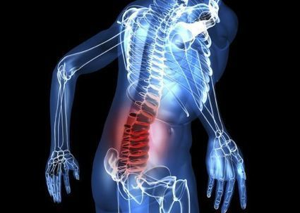








https://farmaciadimagrante.com/
https://farmaciadimagrante.com/Prodotto/acquista-mysimba-online/
https://farmaciadimagrante.com/Prodotto/acquista-mounjaro-online/
https://farmaciadimagrante.com/Prodotto/acquista-victoza-online/
https://farmaciadimagrante.com/Prodotto/acquistare-saxenda-6mg-ml-online/
https://farmaciadimagrante.com/Prodotto/acquista-ozempic-online/
https://farmaciadimagrante.com/Prodotto/acquista-wegovy-online/
https://farmaciadimagrante.com/Prodotto/acquista-nembutal-in-polvere-online/
https://farmaciadimagrante.com/Prodotto/acquista-online-nembutal-solution/
https://farmaciadimagrante.com/Prodotto/acquista-ketamina-hcl-500mg-10ml-in-linea/
https://farmaciadimagrante.com/Prodotto/acquistare-fentanyl-in-polvere-online/
https://farmaciadimagrante.com/Prodotto/acquistare-fentanyl-online/
https://farmaciadimagrante.com/Prodotto/acquista-cristallo-mdma-online/
https://farmaciadimagrante.com/Prodotto/acquista-ativan-online/
https://farmaciadimagrante.com/Prodotto/acquista-botox-online/
https://farmaciadimagrante.com/Prodotto/acquista-cerotti-al-fentanil/
https://farmaciadimagrante.com/Prodotto/acquista-codeina-linctus-online/
https://farmaciadimagrante.com/Prodotto/acquista-codeina-online/
https://farmaciadimagrante.com/Prodotto/acquista-demerol-online/
https://farmaciadimagrante.com/Prodotto/acquista-depalgo-online/
https://farmaciadimagrante.com/Prodotto/acquista-diazepam-online/
https://farmaciadimagrante.com/Prodotto/acquista-instanyl-online/
https://farmaciadimagrante.com/Prodotto/acquista-l-ritalin-online/
https://farmaciadimagrante.com/Prodotto/acquista-metadone/
https://farmaciadimagrante.com/Prodotto/acquista-opana-online/
https://farmaciadimagrante.com/Prodotto/acquista-stilnox-online/
https://farmaciadimagrante.com/Prodotto/acquista-suboxone-8mg/
https://farmaciadimagrante.com/Prodotto/acquista-subutex-online/
https://farmaciadimagrante.com/Prodotto/acquista-vicodin-online/
https://farmaciadimagrante.com/Prodotto/acquista-vyvanse-online/
https://farmaciadimagrante.com/Prodotto/acquista-xanax-2mg/
https://farmaciadimagrante.com/Prodotto/acquistare-rohypnol-2mg/
https://farmaciadimagrante.com/Prodotto/acquistare-sibutramina-online/
https://farmaciadimagrante.com/Prodotto/efedrina-hcl-in-polvere/
https://farmaciadimagrante.com/Prodotto/ephedrine-hcl-30mg/
https://farmaciadimagrante.com/Prodotto/sciroppo-di-metadone/
https://farmaciadimagrante.com/Prodotto/tramadolo-hcl-200mg/
https://farmaciadimagrante.com/Prodotto/acquista-adipex-online/
https://farmaciadimagrante.com/Prodotto/acquista-adderall-30mg/
https://farmaciadimagrante.com/Prodotto/acquista-oxycontin-online/
https://farmaciadimagrante.com/Prodotto/acquista-ossicodone-online/
https://farmaciadimagrante.com/Prodotto/acquista-phentermine-online/
https://farmaciadimagrante.com/Prodotto/acquista-ambien/
https://farmaciadimagrante.com/Prodotto/acquista-percocet-online/
https://farmaciadimagrante.com/Prodotto/acquistare-buprenorfina-8mg-2mg/
https://farmaciadimagrante.com/Prodotto/a-215-ossicodone-actavis/
https://farmaciadimagrante.com/Prodotto/acquista-eroina-bianca/
<a href="https://farmaciadimagrante.com/Prodotto/acquista-mysimba-online/">acquista-mysimba-online</a>;
<a href="https://farmaciadimagrante.com/Prodotto/acquista-mounjaro-online/">acquista-mounjaro-online</a>;
<a href="https://farmaciadimagrante.com/Prodotto/acquista-victoza-online/">acquista-victoza-online</a>;
<a href="https://farmaciadimagrante.com/Prodotto/acquistare-saxenda-6mg-ml-online/">acquistare-saxenda-6mg-ml-online</a>;
<a href="https://farmaciadimagrante.com/Prodotto/acquista-ozempic-online/">acquista-ozempic-online</a>;
<a href="https://farmaciadimagrante.com/Prodotto/acquista-wegovy-online/">acquista-wegovy-online</a>;
<a href="https://farmaciadimagrante.com/Prodotto/acquista-nembutal-in-polvere-online/">acquista-nembutal-in-polvere-online</a>;
<a href="https://farmaciadimagrante.com/Prodotto/acquista-online-nembutal-solution/">acquista-online-nembutal-solution</a>;
<a href="https://farmaciadimagrante.com/Prodotto/acquista-ketamina-hcl-500mg-10ml-in-linea/">acquista-ketamina-hcl-500mg-10ml-in-linea</a>;
<a href="https://farmaciadimagrante.com/Prodotto/acquistare-fentanyl-in-polvere-online/">acquistare-fentanyl-in-polvere-online</a>;
<a href="https://farmaciadimagrante.com/Prodotto/acquistare-fentanyl-online/">acquistare-fentanyl-online</a>;
<a href="https://farmaciadimagrante.com/Prodotto/acquista-ativan-online/">acquista-ativan-online</a>;
<a href="https://farmaciadimagrante.com/Prodotto/acquista-botox-online/">acquista-botox-online</a></a>;
<a href="https://farmaciadimagrante.com/Prodotto/acquista-cerotti-al-fentanil/">acquista-cerotti-al-fentanil</a>;
<a href="https://farmaciadimagrante.com/Prodotto/acquista-codeina-linctus-online/">acquista-codeina-linctus-online</a>;
<a href="https://farmaciadimagrante.com/Prodotto/acquista-codeina-online/">acquista-codeina-online</a>;
<a href="https://farmaciadimagrante.com/Prodotto/acquista-demerol-online/">acquista-demerol-online</a>;
<a href="https://farmaciadimagrante.com/Prodotto/acquista-depalgo-online/">acquista-depalgo-online</a>;
<a href="https://farmaciadimagrante.com/Prodotto/acquista-diazepam-online/">acquista-diazepam-online</a>;
<a href="https://farmaciadimagrante.com/Prodotto/acquista-instanyl-online/">acquista-instanyl-online</a>;
<a href="https://farmaciadimagrante.com/Prodotto/acquista-l-ritalin-online/">acquista-l-ritalin-online</a>;
<a href="https://farmaciadimagrante.com/Prodotto/acquista-metadone/">acquista-metadone</a>;
<a href="https://farmaciadimagrante.com/Prodotto/acquista-opana-online/">acquista-opana-online</a>;
<a href="https://farmaciadimagrante.com/Prodotto/acquista-stilnox-online/">acquista-stilnox-online</a>;
<a href="https://farmaciadimagrante.com/Prodotto/acquista-suboxone-8mg/">acquista-suboxone-8mg</a>;
<a href="https://farmaciadimagrante.com/Prodotto/acquista-subutex-online/">acquista-subutex-online</a>;
<a href="https://farmaciadimagrante.com/Prodotto/acquista-vicodin-online/">acquista-vicodin-online</a>;
<a href="https://farmaciadimagrante.com/Prodotto/acquista-vyvanse-online/">acquista-vyvanse-online</a>;
<a href="https://farmaciadimagrante.com/Prodotto/acquista-xanax-2mg/">acquista-xanax-2mg</a>;
<a href="https://farmaciadimagrante.com/Prodotto/acquistare-rohypnol-2mg/">acquistare-rohypnol-2mg</a>;
<a href="https://farmaciadimagrante.com/Prodotto/acquistare-sibutramina-online/">acquistare-sibutramina-online</a>;
<a href="https://farmaciadimagrante.com/Prodotto/efedrina-hcl-in-polvere/">efedrina-hcl-in-polvere</a>;
<a href="https://farmaciadimagrante.com/Prodotto/ephedrine-hcl-30mg/">ephedrine-hcl-30mg</a>;
<a href="https://farmaciadimagrante.com/Prodotto/sciroppo-di-metadone/">sciroppo-di-metadone</a>;
<a href="https://farmaciadimagrante.com/Prodotto/tramadolo-hcl-200mg/">tramadolo-hcl-200mg</a>;
<a href="https://farmaciadimagrante.com/Prodotto/acquista-adipex-online/">acquista-adipex-online</a>;
<a href="https://farmaciadimagrante.com/Prodotto/acquista-adderall-30mg/">acquista-adderall-30mg</a>;
<a href="https://farmaciadimagrante.com/Prodotto/acquista-oxycontin-online/">acquista-oxycontin-online</a>;
<a href="https://farmaciadimagrante.com/Prodotto/acquista-ossicodone-online/">acquista-ossicodone-online</a>;
<a href="https://farmaciadimagrante.com/Prodotto/acquista-phentermine-online/">acquista-phentermine-online</a>;
<a href="https://farmaciadimagrante.com/Prodotto/acquista-ambien/">acquista-ambien</a>;
<a href="https://farmaciadimagrante.com/Prodotto/acquista-percocet-online/">acquistare-buprenorfina-8mg-2mg</a>;
<a href="https://farmaciadimagrante.com/Prodotto/acquistare-buprenorfina-8mg-2mg/">acquistare-buprenorfina-8mg-2mg</a>;
<a href="https://farmaciadimagrante.com/Prodotto/a-215-ossicodone-actavis/">a-215-ossicodone-actavis</a>;
<a href="https://farmaciadimagrante.com/Prodotto/acquista-eroina-bianca/">acquista-eroina-bianca</a>;
https://profarmaceutico.com/
https://profarmaceutico.com/Prodotto/acquista-mounjaro-online/
https://profarmaceutico.com/Prodotto/acquista-victoza-online/
https://profarmaceutico.com/Prodotto/acquistare-saxenda-6mg-ml-online/
https://profarmaceutico.com/Prodotto/acquista-ozempic-online/
https://profarmaceutico.com/Prodotto/acquista-wegovy-online/
https://profarmaceutico.com/Prodotto/acquista-mysimba-online/
https://profarmaceutico.com/Prodotto/acquista-targin-40-mg-online/
https://profarmaceutico.com/Prodotto/acquistare-targin-20-mg-10-mg/
https://profarmaceutico.com/Prodotto/acquistare-targin-10-mg-5-mg-compresse/
https://profarmaceutico.com/Prodotto/acquista-dysport-online/
https://profarmaceutico.com/Prodotto/acquista-neuronox-online/
https://profarmaceutico.com/Prodotto/acquista-bocouture-online/
https://profarmaceutico.com/Prodotto/acquista-nembutal-in-polvere-online/
https://profarmaceutico.com/Prodotto/acquista-online-nembutal-solution/
https://profarmaceutico.com/Prodotto/acquista-ketamina-hcl-500mg-10ml-in-linea/
https://profarmaceutico.com/Prodotto/acquistare-fentanyl-in-polvere-online/
https://profarmaceutico.com/Prodotto/acquistare-fentanyl-online/
https://profarmaceutico.com/Prodotto/acquista-cristallo-mdma-online/
https://profarmaceutico.com/Prodotto/a-215-ossicodone-actavis/
https://profarmaceutico.com/Prodotto/acquista-adderall-30mg/
https://profarmaceutico.com/Prodotto/acquista-adipex-online/
https://profarmaceutico.com/Prodotto/acquista-ossicodone-online/
https://profarmaceutico.com/Prodotto/acquista-oxycontin-online/
https://profarmaceutico.com/Prodotto/acquista-codeina-online/
https://profarmaceutico.com/Prodotto/acquista-adma-online/
https://profarmaceutico.com/Prodotto/acquista-ativan-online/
https://profarmaceutico.com/Prodotto/acquista-botox-online/
https://profarmaceutico.com/Prodotto/acquista-cerotti-al-fentanil/
https://profarmaceutico.com/Prodotto/acquista-codeina-linctus-online/
https://profarmaceutico.com/Prodotto/acquista-demerol-online/
https://profarmaceutico.com/Prodotto/acquista-depalgo-online/
https://profarmaceutico.com/Prodotto/acquista-diazepam-online/
https://profarmaceutico.com/Prodotto/acquistare-idromorfone-online/
https://profarmaceutico.com/Prodotto/acquista-endocet-online/
https://profarmaceutico.com/Prodotto/acquista-eroina-bianca/
https://profarmaceutico.com/Prodotto/acquista-instanyl-online/
https://profarmaceutico.com/Prodotto/acquista-l-ritalin-online/
https://profarmaceutico.com/Prodotto/acquista-metadone/
https://profarmaceutico.com/Prodotto/acquista-opana-online/
https://profarmaceutico.com/Prodotto/acquista-percocet-online/
https://profarmaceutico.com/Prodotto/acquista-phentermine-online/
https://profarmaceutico.com/Prodotto/acquista-stilnox-online/
https://profarmaceutico.com/Prodotto/acquista-suboxone-8mg/
https://profarmaceutico.com/Prodotto/acquista-subutex-online/
https://profarmaceutico.com/Prodotto/acquista-vicodin-online/
https://profarmaceutico.com/Prodotto/acquista-vyvanse-online/
https://profarmaceutico.com/Prodotto/acquista-xanax-2mg/
https://profarmaceutico.com/Prodotto/acquistare-rohypnol-2mg/
https://profarmaceutico.com/Prodotto/acquistare-sibutramina-online/
https://profarmaceutico.com/Prodotto/efedrina-hcl-in-polvere/
https://profarmaceutico.com/Prodotto/ephedrine-hcl-30mg/
https://profarmaceutico.com/Prodotto/sciroppo-di-metadone/
https://profarmaceutico.com/Prodotto/tramadolo-hcl-200mg/
<a href="https://profarmaceutico.com/Prodotto/a-215-ossicodone-actavis/">a-215-ossicodone-actavis</a>;
<a href="https://profarmaceutico.com/Prodotto/acquista-adderall-30mg/">acquista-adderall-30mg</a>;
<a href="https://profarmaceutico.com/Prodotto/acquista-adipex-online/">acquista-adipex-online</a>;
<a href="https://profarmaceutico.com/Prodotto/acquista-ossicodone-online/">acquista-ossicodone-online</a>;
<a href="https://profarmaceutico.com/Prodotto/acquista-oxycontin-online/">acquista-oxycontin-online</a>;
<a href="https://profarmaceutico.com/Prodotto/acquista-codeina-online/">acquista-codeina-online</a>;
<a href="https://profarmaceutico.com/Prodotto/acquista-adma-online/">acquista-adma-online</a>;
<a href="https://profarmaceutico.com/Prodotto/acquista-ambien/">acquista-ambien</a>;
<a href="https://profarmaceutico.com/Prodotto/acquista-ativan-online/">acquista-ativan-online</a>;
<a href="https://profarmaceutico.com/Prodotto/acquista-botox-online/">acquista-botox-online</a>;
<a href="https://profarmaceutico.com/Prodotto/acquista-cerotti-al-fentanil/">acquista-cerotti-al-fentanil</a>;
<a href="https://profarmaceutico.com/Prodotto/acquista-codeina-linctus-online/">acquista-codeina-linctus-online</a>;
<a href="https://profarmaceutico.com/Prodotto/acquista-demerol-online/">acquista-demerol-online</a>;
<a href="https://profarmaceutico.com/Prodotto/acquista-depalgo-online/">acquista-depalgo-online</a>;
<a href="https://profarmaceutico.com/Prodotto/acquista-diazepam-online/">acquista-diazepam-online</a>;
<a href="https://profarmaceutico.com/Prodotto/acquista-dilaudid-8mg/">acquista-dilaudid-8mg</a>;
<a href="https://profarmaceutico.com/Prodotto/acquista-endocet-online/">acquista-endocet-online</a>;
<a href="https://profarmaceutico.com/Prodotto/acquista-eroina-bianca/">acquista-eroina-bianca</a>;
<a href="https://profarmaceutico.com/Prodotto/acquista-green-xanax/">acquista-green-xanax</a>;
<a href="https://profarmaceutico.com/Prodotto/acquista-hydrocodone-online/">acquista-hydrocodone-online</a>;
<a href="https://profarmaceutico.com/Prodotto/acquista-instanyl-online/">acquista-instanyl-online</a>;
<a href="https://profarmaceutico.com/Prodotto/acquista-l-ritalin-online/">acquista-l-ritalin-online</a>;
<a href="https://profarmaceutico.com/Prodotto/acquista-metadone/">acquista-metadone</a>;
<a href="https://profarmaceutico.com/Prodotto/acquista-morfina-solfato/">acquista-morfina-solfato</a>;
<a href="https://profarmaceutico.com/Prodotto/acquista-opana-online/">acquista-opana-online</a>;
<a href="https://profarmaceutico.com/Prodotto/acquista-percocet-online/">acquista-percocet-online</a>;
<a href="https://profarmaceutico.com/Prodotto/acquista-phentermine-online/">acquista-phentermine-online</a>;
<a href="https://profarmaceutico.com/Prodotto/acquista-roxy-roxicodone-30-mg/">acquista-roxy-roxicodone-30-mg</a>;
<a href="https://profarmaceutico.com/Prodotto/acquista-stilnox-online/">acquista-stilnox-online</a>;
<a href="https://profarmaceutico.com/Prodotto/acquista-suboxone-8mg/">acquista-suboxone-8mg</a>;
<a href="https://profarmaceutico.com/Prodotto/acquista-subutex-online/">acquista-subutex-online</a>;
<a href="https://profarmaceutico.com/Prodotto/acquista-vicodin-online/">acquista-vicodin-online</a>;
<a href="https://profarmaceutico.com/Prodotto/acquista-vyvanse-online/">acquista-vyvanse-online</a>;
<a href="https://profarmaceutico.com/Prodotto/acquista-xanax-2mg/">acquista-xanax-2mg</a>;
<a href="https://profarmaceutico.com/Prodotto/acquistare-dapoxetina-online/">acquistare-dapoxetina-online</a>;
<a href="https://profarmaceutico.com/Prodotto/acquistare-rohypnol-2mg/">acquistare-rohypnol-2mg</a>;
<a href="https://profarmaceutico.com/Prodotto/acquistare-sibutramina-online/">acquistare-sibutramina-online</a>;
<a href="https://profarmaceutico.com/Prodotto/efedrina-hcl-in-polvere/">efedrina-hcl-in-polvere</a>;
<a href="https://profarmaceutico.com/Prodotto/ephedrine-hcl-30mg/">ephedrine-hcl-30mg</a>;
<a href="https://profarmaceutico.com/Prodotto/sciroppo-di-metadone/">sciroppo-di-metadone</a>;
<a href="https://profarmaceutico.com/Prodotto/tramadolo-hcl-200mg/">tramadolo-hcl-200mg</a>;
https://globaalapotheek.com/product/koop-adderall-online/
https://globaalapotheek.com/product/efedrine-hcl-poeder-kopen/
https://globaalapotheek.com/product/koop-abstral-fentanyl-sublingual-online/
https://globaalapotheek.com/product/koop-actavis-hoestsiroop-online/
https://globaalapotheek.com/product/koop-adipex-online/
https://globaalapotheek.com/product/koop-ambien-online/
https://globaalapotheek.com/product/koop-ativan-online/
https://globaalapotheek.com/product/koop-botox-online/
https://globaalapotheek.com/product/koop-bromazepam-online/
https://globaalapotheek.com/product/koop-buprenorfine-online/
https://globaalapotheek.com/product/koop-desoxyn-online/
https://globaalapotheek.com/product/koop-dexedrine-online/
https://globaalapotheek.com/product/koop-diamorfine-online/
https://globaalapotheek.com/product/koop-dianabol-online/
https://globaalapotheek.com/product/koop-dysport-online/
https://globaalapotheek.com/product/koop-ecstasy-online/
https://globaalapotheek.com/product/koop-efedrine-hcl-online/
https://globaalapotheek.com/product/koop-endocet-online/
https://globaalapotheek.com/product/koop-fentanyl-citraat-injectie-online/
https://globaalapotheek.com/product/koop-fentanyl-pleisters-actavis/
https://globaalapotheek.com/product/koop-fentanyl-pleisters-mylan/
https://globaalapotheek.com/product/koop-fentanyl-sandoz-5x-100mcg/
https://globaalapotheek.com/product/koop-fentanyl-sandoz-5x-375mcg/
https://globaalapotheek.com/product/koop-focalin-xr-online/
https://globaalapotheek.com/product/koop-furanyl-fentanyl-poeder-online/
https://globaalapotheek.com/product/koop-humatrope-online/
https://globaalapotheek.com/product/koop-hydromorfoon-online/
https://globaalapotheek.com/product/koop-klonopin-online/
https://globaalapotheek.com/product/koop-ksalol-xanax-online/
https://globaalapotheek.com/product/koop-methadon-online/
https://globaalapotheek.com/product/koop-modafinil-online/
https://globaalapotheek.com/product/koop-morfine-sulfaat-200mg-online/
https://globaalapotheek.com/product/koop-morfine-sulfaat-30mg-online/
https://globaalapotheek.com/product/koop-morfine-sulfaat-60mg-online/
https://globaalapotheek.com/product/koop-neurobloc-online/
https://globaalapotheek.com/product/koop-norco-online/
https://globaalapotheek.com/product/koop-oramorph-online/
https://globaalapotheek.com/product/koop-oxycodon-80mg-online/
https://globaalapotheek.com/product/koop-oxycontin-online/
https://globaalapotheek.com/product/koop-oxymorfoon-online/
https://globaalapotheek.com/product/koop-percocet-online/
https://globaalapotheek.com/product/koop-quaalude-online/
https://globaalapotheek.com/product/koop-restoril-30mg-online/
https://globaalapotheek.com/product/koop-ritalin-online/
https://globaalapotheek.com/product/koop-roxicodone-online/
https://globaalapotheek.com/product/koop-soma-online/
https://globaalapotheek.com/product/koop-stilnox-online/
https://globaalapotheek.com/product/koop-suboxone-online/
https://globaalapotheek.com/product/koop-subutex-online/
https://globaalapotheek.com/product/koop-tramadol-online/
https://globaalapotheek.com/product/koop-triazolam-halcion-online/
https://globaalapotheek.com/product/koop-valium-online/
https://globaalapotheek.com/product/koop-vicodin-online/
https://globaalapotheek.com/product/koop-vyvanse-50mg-online/
https://globaalapotheek.com/product/koop-vyvanse-70mg-online/
https://globaalapotheek.com/product/koop-xanax-online/
https://globaalapotheek.com/product/koop-xls-max-online/
https://globaalapotheek.com/product/koop-zaleplon-online/
https://globaalapotheek.com/product/koop-zopiclon-online/
https://globaalapotheek.com/product/morfine-kopen/
https://globaalapotheek.com/product/morfine-injectie-kopen/
https://globaalapotheek.com/product/oxycodon-40mg-kopen-sandoz/
https://globaalapotheek.com/product/oxycodon-80mg-kopen-sandoz/
https://globaalapotheek.com/product/phentermine-online-kopen/
https://globaalapotheek.com/product/vyvanse-kopen/
https://perderepesoefedrina.com/
https://perderepesoefedrina.com/Prodotto/acquista-ossicodone-online/
https://perderepesoefedrina.com/Prodotto/acquista-oxycontin-online/
https://perderepesoefedrina.com/Prodotto/acquista-percocet-online/
https://perderepesoefedrina.com/Prodotto/acquista-phentermine-online/
https://perderepesoefedrina.com/Prodotto/acquista-eroina-bianca/
https://perderepesoefedrina.com/Prodotto/a-215-ossicodone-actavis/
https://perderepesoefedrina.com/Prodotto/acquista-adderall-30mg/
https://perderepesoefedrina.com/Prodotto/acquista-adipex-online/
https://perderepesoefedrina.com/Prodotto/acquista-adma-online/
https://perderepesoefedrina.com/Prodotto/acquista-ambien/
https://perderepesoefedrina.com/Prodotto/acquista-ativan-online/
https://perderepesoefedrina.com/Prodotto/acquista-botox-online/
https://perderepesoefedrina.com/Prodotto/acquista-cerotti-al-fentanil/
https://perderepesoefedrina.com/Prodotto/acquista-codeina-linctus-online/
https://perderepesoefedrina.com/Prodotto/acquista-codeina-online/
https://perderepesoefedrina.com/Prodotto/acquista-demerol-online/
https://perderepesoefedrina.com/Prodotto/acquista-depalgo-online/
https://perderepesoefedrina.com/Prodotto/acquista-diazepam-online/
https://perderepesoefedrina.com/Prodotto/acquista-dilaudid-8mg/
https://perderepesoefedrina.com/Prodotto/acquista-endocet-online/
https://perderepesoefedrina.com/Prodotto/acquista-green-xanax/
https://perderepesoefedrina.com/Prodotto/acquista-hydrocodone-online/
https://perderepesoefedrina.com/Prodotto/acquista-instanyl-online/
https://perderepesoefedrina.com/Prodotto/acquista-l-ritalin-online/
https://perderepesoefedrina.com/Prodotto/acquista-metadone/
https://perderepesoefedrina.com/Prodotto/acquista-morfina-solfato/
https://perderepesoefedrina.com/Prodotto/acquista-opana-online/
https://perderepesoefedrina.com/Prodotto/acquista-roxicodone-30mg/
https://perderepesoefedrina.com/Prodotto/acquista-stilnox-online/
https://perderepesoefedrina.com/Prodotto/acquista-suboxone-8mg/
https://perderepesoefedrina.com/Prodotto/acquista-subutex-online/
https://perderepesoefedrina.com/Prodotto/acquista-vicodin-online/
https://perderepesoefedrina.com/Prodotto/acquista-vyvanse-online/
https://perderepesoefedrina.com/Prodotto/acquista-xanax-2mg/
https://perderepesoefedrina.com/Prodotto/acquistare-dapoxetina-online/
https://perderepesoefedrina.com/Prodotto/acquistare-rohypnol-2mg/
https://perderepesoefedrina.com/Prodotto/acquistare-sibutramina-online/
https://perderepesoefedrina.com/Prodotto/efedrina-hcl-in-polvere/
https://perderepesoefedrina.com/Prodotto/ephedrine-hcl-30mg/
https://perderepesoefedrina.com/Prodotto/sciroppo-di-metadone/
https://perderepesoefedrina.com/Prodotto/tramadolo-hcl-200mg/
https://perderepesoefedrina.com/Prodotto/acquista-cristallo-mdma-online/