Your new post is loading...

|
Scooped by
Gilbert C FAURE
November 2, 2017 6:30 AM
|
Research, reviews, highlights, methods and more on glial biology from across the Nature Research journals.
|
|
Scooped by
Gilbert C FAURE
October 27, 2017 11:47 AM
|
|
|
Scooped by
Gilbert C FAURE
October 26, 2017 12:18 PM
|
The mechanisms leading to autoimmune and inflammatory diseases in the CNS have not been elucidated. The environmental triggers of the aberrant presence of CD4+ T cells in the CNS are not known. In this article, we report that abnormal β-catenin expression in T cells drives a fatal neuroinflammatory disease in mice that is characterized by CNS infiltration of T cells, glial activation, and progressive loss of motor function. We show that enhanced β-catenin expression in T cells leads to aberrant and Th1-biased T cell activation, enhanced expression of integrin α4β1, and infiltration of activated T cells into the spinal cord, without affecting regulatory T cell function. Importantly, expression of β-catenin in mature naive T cells was sufficient to drive integrin α4β1 expression and CNS migration, whereas pharmacologic inhibition of integrin α4β1 reduced the abnormal T cell presence in the CNS of β-catenin–expressing mice. Together, these results implicate deregulation of the Wnt/β-catenin pathway in CNS inflammation and suggest novel therapeutic strategies for neuroinflammatory disorders.
|
|
Scooped by
Gilbert C FAURE
October 16, 2017 10:16 AM
|
Of interest to the etiology of demyelinating autoimmune disease is the potential to aberrantly activate CD4+ T cells due to cross-recognition of multiple self-epitopes such as has been suggested for myelin oligodendrocyte glycoprotein epitope 35–55 (MOG35–55) and neurofilament medium protein epitope 15–35 (NFM15–35). NFM15–35 is immunogenic in C57BL/6 mice but fails to induce demyelinating disease by polyclonal T cells despite having the same TCR contact residues as MOG35–55, a known encephalitogenic Ag. Despite reported cross-reactivity with MOG-specific T cells, the polyclonal response to NFM15–35 did not expand threshold numbers of MOG38–49 tetramer–positive T cells. Furthermore, NFM lacked functional synergy with MOG to promote experimental autoimmune encephalomyelitis because NFM-deficient synonymous with knockout mice developed an identical disease course to wild-type mice after challenge with MOG35–55. Single-cell analysis of encephalitogenic T cells using the peptide:MHC monomer-based two-dimensional micropipette adhesion frequency assay confirmed that NFM was not a critical Ag driving demyelinating disease because NFM18–30–specific T cells in the CNS were predominantly reactive to MOG38–49. The absence of NFM contribution to disease allowed mapping of the amino acids required for encephalitogenicity and expansion of high-affinity, MOG-specific T cells that defined the polyclonal response. Alterations of N-terminal residues outside of the NFM15–35 core nonamer promoted expansion of high-affinity, MOG38–49 tetramer–positive T cells and promoted consistent experimental autoimmune encephalomyelitis induction, unlike mice challenged with NFM15–35. Although NFM15–35 is immunogenic and cross-reactive with MOG at the polyclonal level, it fails to expand a threshold level of encephalitogenic, high-affinity MOG-specific T cells.
|
|
Scooped by
Gilbert C FAURE
October 16, 2017 7:12 AM
|
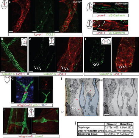
One of the characteristics of the CNS is the lack of a classical lymphatic drainage system. Although it is now accepted that the CNS undergoes constant immune surveillance that takes place within the meningeal compartment[–] , the mechanisms governin
|
|
Scooped by
Gilbert C FAURE
October 13, 2017 9:42 AM
|
J Neuroinflammation. 2016 Dec 1;13(1):301.
|
|
Scooped by
Gilbert C FAURE
October 10, 2017 8:34 AM
|
A blog on multiple sclerosis research
|
|
Scooped by
Gilbert C FAURE
October 6, 2017 3:27 AM
|
Scientists say they have found the first evidence that our brains may drain waste, a surprising finding that has eluded discovery for two centuries.
|
|
Scooped by
Gilbert C FAURE
September 26, 2017 8:12 AM
|
Parkinson’s disease is an insidious disease: by the time it manifests as the typical motor dysfunctions such as tremors or muscle rigidity, portions o
|
|
Scooped by
Gilbert C FAURE
September 22, 2017 9:47 AM
|
Multiple sclerosis can be inhibited or reversed using a novel gene therapy technique that stops the disease’s immune response in mouse models, University of Florida Health researchers have found. By combining a brain-protein gene and an existing medication, the researchers were able to prevent the...
|
|
Scooped by
Gilbert C FAURE
September 16, 2017 1:45 PM
|
Neurotherapeutics. 2017 Aug 15. doi: 10.1007/s13311-017-0565-4. [Epub ahead of print] Review
|
|
Scooped by
Gilbert C FAURE
September 16, 2017 8:05 AM
|
|
|
Scooped by
Gilbert C FAURE
September 10, 2017 4:43 AM
|
Deep sequencing of lymphocyte receptor repertoires has made it possible to comprehensively profile the clonal composition of lymphocyte populations. This opens the door for novel approaches to diagnose and prognosticate diseases with a driving immune component by identifying repertoire sequence patterns associated with clinical phenotypes. Indeed, recent studies support the feasibility of this, demonstrating an association between repertoire-level summary statistics (e.g., diversity) and patient outcomes for several diseases. In our own prior work, we have shown that six codons in VH4-containing genes in B cells from the cerebrospinal fluid of patients with relapsing remitting multiple sclerosis (RRMS) have higher replacement mutation frequencies than observed in healthy controls or patients with other neurological diseases. However, prior methods to date have been limited to focusing on repertoire-level summary statistics, ignoring the vast amounts of information in the millions of individual immune receptors comprising a repertoire. We have developed a novel method that addresses this limitation by using innovative approaches for accommodating the extraordinary sequence diversity of immune receptors and widely used machine learning approaches. We applied our method to RRMS, an autoimmune disease that is notoriously difficult to diagnose. We use the biochemical features encoded by the complementarity determining region 3 of each B cell receptor heavy chain in every patient repertoire as input to a detector function, which is fit to give the correct diagnosis for each patient using maximum likelihood optimization methods. The resulting statistical classifier assigns patients to one of two diagnosis categories, RRMS or other neurological disease, with 87% accuracy by leave-one-out cross-validation on training data (N = 23) and 72% accuracy on unused data from a separate study (N = 102). Our method is the first to apply statistical learning to immune repertoires to aid disease diagnosis, learning repertoire-level labels from the set of individual immune repertoire sequences. This method produced a repertoire-based statistical classifier for diagnosing RRMS that provides a high degree of diagnostic capability, rivaling the accuracy of diagnosis by a clinical expert. Additionally, this method points to a diagnostic biochemical motif in the antibodies of RRMS patients, which may offer insight into the disease process.
|
Intrathecal immunoglobulin G (IgG) synthesis, cerebrospinal fluid (CSF) oligoclonal IgG bands and lesional IgG deposition are seminal features of multiple sclerosis (MS) disease pathology. Both the specific targets and pathogenic effects of MS antibodies remain poorly characterized. We produced IgG1 monoclonal recombinant antibodies (rAbs) from clonally-expanded plasmablasts recovered from MS patient CSF. Among these were a subset of myelin-specific MS rAbs. We examined their immunoreactivity to mouse organotypic cerebellar slices by live binding and evaluated tissue injury in the presence and absence of human complement. Demyelination, glial and neuronal viability, and complement pathway activation were assayed by immunofluorescence microscopy and compared to the effects of an aquaporin-4 water channel (AQP4)-specific rAb derived from a neuromyelitis optica (NMO) patient. MS myelin-specific rAbs bound to discrete surface domains on oligodendrocyte processes and myelinating axons. Myelin-specific MS rAbs initiated complement-dependent cytotoxicity to oligodendrocytes and induced rapid demyelination. Demyelination was accompanied by increased microglia activation; however, the morphology and survival of astrocytes, oligodendrocyte progenitors and neurons remained unaffected. In contrast, NMO AQP4-specific rAb initiated complement-dependent astrocyte damage, followed by sequential loss of oligodendrocytes, demyelination, microglia activation and neuronal death. Myelin-specific MS antibodies cause oligodendrocyte loss and demyelination in organotypic cerebellar slices, which are distinct from AQP4-targeted pathology, and display seminal features of active MS lesions. Myelin-specific antibodies may play an active role in MS lesion formation through complement-dependent mechanisms.
Via Krishan Maggon
|
|
Scooped by
Gilbert C FAURE
October 26, 2017 12:57 PM
|
The blood–brain barrier (BBB) keeps neurotoxic plasma-derived components, cells, and pathogens out of the brain. An early BBB breakdown and/or dysfunction have been shown in Alzheimer’s disease (AD) before dementia, neurodegeneration and/or brain atrophy occur. However, the role of BBB breakdown in neurodegenerative disorders is still not fully understood. Here, we examine BBB breakdown in animal models frequently used to study the pathophysiology of AD, including transgenic mice expressing human amyloid-β precursor protein, presenilin 1, and tau mutations, and apolipoprotein E, the strongest genetic risk factor for AD. We discuss the role of BBB breakdown and dysfunction in neurodegenerative process, pitfalls in BBB measurements, and how targeting the BBB can influence the course of neurological disorder. Finally, we comment on future approaches and models to better define, at the cellular and molecular level, the underlying mechanisms between BBB breakdown and neurodegeneration as a basis for developing new therapies for BBB repair to control neurodegeneration.
|
|
Scooped by
Gilbert C FAURE
October 16, 2017 10:18 AM
|
Gut microbiota and MS in mice
|
|
Scooped by
Gilbert C FAURE
October 16, 2017 9:57 AM
|
Abstract. Movement disorders are a prominent and common feature in many autoantibody-associated neurological diseases, a group of potentially treatable conditi
|
|
Scooped by
Gilbert C FAURE
October 16, 2017 4:51 AM
|
Antibodies to different brain proteins have been recently found to be associated with an increasing number of different autoimmune diseases. They need to penetrate the blood brain barrier in order to bind antigens within the central nervous system (CNS). They can target either neuronal or non-neuronal antigen and result in damage either by themselves or in synergy with other inflammatory mediators. Antibodies can lead to acute brain pathology, which may be reversible; alternatively, they may trigger irreversible damage that persists even though the antibodies are no longer present. In this review we will describe two different autoimmune conditions and the role of their antibodies in causing brain pathology. In Systemic Lupus Erythematosus (SLE), patients can have double stranded DNA antibodies that cross react with the neuronal N-methyl-D-aspartate receptor (NMDAR) which have been recently linked to neurocognitive dysfunction. In Neuromyelitis Optica (NMO), antibodies to astrocytic aquaporin-4 (AQP4) are diagnostic of disease. There is emerging evidence that pathogenic T cells also play an important role for the disease pathogenesis in NMO since they infiltrate in the CNS. In order to enable appropriate and less invasive treatment for antibody-mediated diseases we need to understand the mechanisms of antibody mediated pathology, the acute and chronic effects of antibody exposure, if the antibodies are produced intrathecally or systemically, their target antigen and what triggers their production. Emerging data also show that in utero exposure to some brain reactive antibodies, such as those found in SLE can cause neurodevelopmental impairment since they can penetrate the embryonic blood brain barrier. If the antibody exposure occurs at a critical time of development this can result in irreversible damage of the offspring that persists throughout adulthood.
|
|
Scooped by
Gilbert C FAURE
October 13, 2017 9:42 AM
|
Delmont et al. identify neurofascin-186, the isoform of neurofascin at nodes of Ranvier, as the target of autoantibodies in a subset of patients with chronic in
|
|
Scooped by
Gilbert C FAURE
October 6, 2017 3:40 AM
|
Meningeal lymphatic vessels are present in human and nonhuman primates (common marmoset monkeys) and they can be noninvasively imaged and mapped in vivo with high-resolution, clinical MRI.
|
|
Scooped by
Gilbert C FAURE
September 29, 2017 6:40 AM
|
Microglia are tissue macrophages of the central nervous system (CNS). Their key tasks are immune surveillance as well as responding to infections or other pathological states such as neurologica
|
|
Scooped by
Gilbert C FAURE
September 23, 2017 2:15 AM
|
Light bulb moments in multiple sclerosis (MS) 1421 First known case of MS In 1421, Saint Lidwina was ice skating in Schiedam, Holland, when she suddenly became weak and fell, breaking a rib at age 15.
|
|
Scooped by
Gilbert C FAURE
September 22, 2017 9:46 AM
|
Multiple sclerosis can be inhibited or reversed using a novel gene therapy technique that stops the disease's immune response in mouse models, researchers have found.
|
|
Scooped by
Gilbert C FAURE
September 16, 2017 1:44 PM
|
Translational Psychiatry explores the more translational area between the research in neuroscience and conceptually novel treatments
|
|
Scooped by
Gilbert C FAURE
September 14, 2017 12:17 PM
|
Herpes simplex virus–1 (HSV-1) is the most common cause of sporadic viral encephalitis, which can be lethal or result in severe neurological defects even with antiviral therapy. While HSV-1 causes encephalitis in spite of HSV-1–specific humoral and cellular immunity, the mechanism by which HSV-1 evades the immune system in the central nervous system (CNS) remains unknown. Here we describe a strategy by which HSV-1 avoids immune targeting in the CNS. The HSV-1 UL13 kinase promotes evasion of HSV-1–specific CD8+ T cell accumulation in infection sites by downregulating expression of the CD8+ T cell attractant chemokine CXCL9 in the CNS of infected mice, leading to increased HSV-1 mortality due to encephalitis. Direct injection of CXCL9 into the CNS infection site enhanced HSV-1–specific CD8+ T cell accumulation, leading to marked improvements in the survival of infected mice. This previously uncharacterized strategy for HSV-1 evasion of CD8+ T cell accumulation in the CNS has important implications for understanding the pathogenesis and clinical treatment of HSV-1 encephalitis.
|


 Scoop.it!