Your new post is loading...

|
Scooped by
Gilbert C FAURE
June 5, 2018 9:26 AM
|
Introduction Previous studies have reported that impaired cytotoxic activity of natural killer (NK) cells in peripheral blood is associated with multiple sclerosis (MS) activity. Furthermore, NK cell phenotype could be associated with new lesions on MRI.
|
|
Scooped by
Gilbert C FAURE
May 30, 2018 1:55 PM
|
Alison Abbott considers a persuasive case for the link between body and mind.
|
|
Scooped by
Gilbert C FAURE
May 16, 2018 5:30 AM
|
Griveau et al. show that Olig2+ glioma cells invade by single-cell vessel co-option,
whereas Olig2− glioma cells promote angiogenesis and that anti-VEGF treatment selects
for the Olig2+/Wnt7+ phenotype. Wnt7 is necessary for vessel co-option, and Wnt inhibition
enhances the response to temozolomide treatment.
|
|
Scooped by
Gilbert C FAURE
May 15, 2018 4:52 AM
|
This review evaluates the literature for the epidemiological evidence of features of α-synucleinopathies that occur before clinical onset of disease, proposes a...
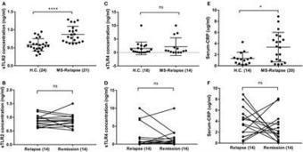
Multiple sclerosis (MS) is an immune-mediated inflammatory demyelinating disease of the central nervous system. It was previously shown that Toll-like receptor (TLR)-2 signalling plays a key role in the murine experimental autoimmune encephalomyelitis (EAE) model of MS, and that TLR2-stimulation of regulatory T cells (Tregs) promotes their conversion to T helper 17 (Th17) cells. Here, we sought potential sources of TLR2 stimulation and evidence of TLR2 activity in MS patient clinical samples. Soluble TLR2 (sTLR2) was found to be significantly elevated in sera of MS patients (n=21), in both relapse and remission, compared to healthy controls (HC) (n=24). This was not associated with the acute phase reaction (APR) as measured by serum C-reactive protein (CRP) level, which was similarly increased in MS patients compared to controls. An independent validation cohort from a different ethnic background showed a similar upward trend in mean sTLR2 values in relapsing-remitting MS (RRMS) patients, and significant differences in sTLR2 values between patients and healthy controls were preserved when the data from the two cohorts were pooled together (n=41 RRMS and 44 HC, P=0.0006). TLR2-stimulants, measured using a human embryonic kidney (HEK-293) cell transfectant reporter assay, were significantly higher in urine of MS patients than healthy controls. A screen of several common urinary tract infections (UTI)-related organisms showed strong induction of TLR2-signalling in the sam
Via Krishan Maggon
|
|
Scooped by
Gilbert C FAURE
April 26, 2018 2:41 AM
|
New horizons for multiple sclerosis therapeutics: milestones in the development of ocrelizumab Jessica Frau,* Giancarlo Coghe,* Lorena Lorefice, Giuseppe Fenu, Eleonora Cocco Multiple Sclerosis Center Binaghi Hospital, Department of Medical Sciences and Public Health, University of Cagliari,...
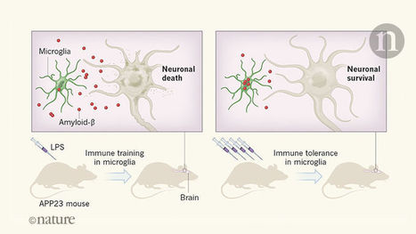
The brain’s resident immune cells retain a long-lasting memory of peripheral inflammation. This memory can influence the response to stroke and the progression of Alzheimer’s disease in mouse models.
Via Krishan Maggon
|
|
Scooped by
Gilbert C FAURE
April 20, 2018 4:30 AM
|
Objective To report the clinical characteristics of 12 patients with limbic encephalitis (LE) who were antibody‐negative after a comprehensive immunological study. Methods Review of clinical records of 163 patients with LE. Immunohistochemistry on rat brain, cultured neurons, and cell‐based assays were used to identify neuronal autoantibodies. Patients were included if 1) there was adequate clinical, CSF, and MRI information to classify the syndrome as LE, 2) MRI images were accesible for central review, and 3) serum and CSF were available and confirmed negative for neuronal antibodies. Results Twelve (7%)/163 LE patients (median age: 62 years; range: 40‐79; 9 [75%] male) without neuronal autoantibodies were identified. The most frequent initial complaints were deficits in short‐term memory leading to hospital admission in a few weeks (median time: 2 weeks; range: 0.5‐12). In four patients the short‐term memory dysfunction remained as isolated symptom during the entire course of the disease. Seizures, drowsiness, and psychiatric problems were unusual. Four patients had solid tumors (1 lung, 1 esophagus, 2 metastatic cervical adenopathies of unknown primary tumor) and 1 chronic lymphocytic leukemia. CSF showed pleocytosis in 7 (58%) with a median of 13 white blood cells /mm3 (range: 9‐25). Immunotherapy included corticosteroids, intravenous immunoglobulins, and combinations of both drugs or with rituximab. Clinical improvement occurred in 6 (54%) of 11 assessable patients. Conclusions Despite the discovery of new antibodies, 7% of LE remains seronegative. Antibody‐negative LE is more frequent in older males and usually develops with predominant or isolated short‐term memory loss. Despite the absence of antibodies, patients may have an underlying cancer and respond to immunotherapy. This article is protected by copyright. All rights reserved.
|
|
Scooped by
Gilbert C FAURE
April 17, 2018 3:06 PM
|
Multiple Sclerosis (MS) is an inflammatory disorder of the central nervous system where evidence implicates an aberrant adaptive immune response in the accrual of neurological disability. The inflammatory phase of the disease responds to immunomodulation to varying degrees of efficacy, however no therapy has been proven to arrest progression of disability. Recently, more intensive therapies, including immunoablation with autologous haematopoietic stem cell transplantation (AHSCT) have been offered as a treatment option to retard inflammatory disease, prior to patients becoming irreversibly disabled. Empirical clinical observations support the notion that the immune reconstitution that occurs following AHSCT is associated with a sustained therapeutic benefit, however neither the pathogenesis of MS nor the mechanism by which AHSCT results in a therapeutic benefit has been clearly delineated. Although the antigenic target of the aberrant immune response in MS is not defined, accumulated data suggest that immune reconstitution following AHSCT results in an immunotolerant state through deletion of pathogenic clones by a combination of direct ablation and induction of a lymphopenic state driving replicative senescence and clonal attrition. Restoration of immunoregulation is evidenced by changes in regulatory T cell populations following AHSCT and normalisation of genetic signatures of immune homeostasis. Furthermore, some evidence exists that AHSCT may induce a rebooting o
|
|
Scooped by
Gilbert C FAURE
April 16, 2018 9:16 AM
|
![Figure][1]
Peripherally derived macrophages infiltrate the brain after bone marrow transplantation and during central nervous system (CNS) inflammation. It was initially suggested that these engrafting cells were newly derived microglia and that irradiation was essential for engraftment to occur. However, it remains unclear whether brain-engrafting macrophages (beMφs) acquire a unique phenotype in the brain, whether long-term engraftment may occur without irradiation, and whether brain function is affected by the engrafted cells. In this study, we demonstrate that chronic, partial microglia depletion is sufficient for beMφs to populate the niche and that the presence of beMφs does not alter behavior. Furthermore, beMφs maintain a unique functional and transcriptional identity as compared with microglia. Overall, this study establishes beMφs as a unique CNS cell type and demonstrates that therapeutic engraftment of beMφs may be possible with irradiation-free conditioning regimens.
[1]: pending:yes
|
|
Scooped by
Gilbert C FAURE
April 14, 2018 2:07 AM
|
|
|
Scooped by
Gilbert C FAURE
April 12, 2018 5:13 AM
|
Clinical and pathogenic significance of IgG, IgA, and IgM antibodies against the NMDA receptor
From
|
|
Scooped by
Gilbert C FAURE
April 6, 2018 4:00 AM
|
|
|
|
Scooped by
Gilbert C FAURE
June 1, 2018 2:27 AM
|
Interleukin-27 (IL-27) and its subunit P28 (also known as IL-30) have been shown to inhibit autoimmunity, and have been suggested as potential immunotherapeutic for autoimmune diseases such as multiple sclerosis (MS). However, the potential of IL-27 and IL-30 as immunotherapeutic, and their mechanisms of action have not been fully understood. In this study, we evaluated the efficacy of adeno-associated viral vector (AAV)-delivered IL-27 (AAV-IL-27) and IL-30 (AAV-IL-30) in a murine model of MS. We found that one single administration of AAV-IL-27, but not AAV-IL-30 completely blocked the development of experimental autoimmune encephalomyelitis (EAE). AAV-IL-27 administration reduced the frequencies of Th17, Treg and GM-CSF-producing CD4+ T cells, and induced T cell expression of IFN-, IL-10 and PD-L1. However, experiments involving IL-10-deficient mice and PD-1 blockade revealed that AAV-IL-27-induced IL-10 and PD-L1 expression were not required for the prevention of EAE development. Surprisingly, neither AAV-IL-27 nor AAV-IL-30 treatment inhibited EAE development and Th17 responses when given at disease onset. We found that mice with established EAE had significant expansion of CD11b+Gr-1+ cells, and AAV-IL-27 treatment further expanded these cells and induced their expression of Th17-promoting cytokines such as IL-6. Adoptive transfer of AAV-IL-27-expanded CD11b+Gr-1+ cells enhanced EAE development. Thus, expansion of CD11b+Gr-1+ cells provides an explanation for th
|
|
Scooped by
Gilbert C FAURE
May 16, 2018 1:40 PM
|
Fortunately, they're hot on the heels of the mechanisms that allow them to affect brain cells.
Inflammation is a hallmark of different central nervous system (CNS) pathologies. It has been linked to neurodegenerative disorders as well as primary and metastatic brain tumors. Microglia, the brain resident immune cells are emerging as a central player in regulating key pathways in CNS...
Via Krishan Maggon
|
|
Scooped by
Gilbert C FAURE
May 10, 2018 4:13 AM
|
Autoimmune encephalitis is a group of encephalitis syndromes that cause altered mentality, memory decline, or seizures in association with the presence of serum and cerebrospinal fluid (CSF) autoantibodies (auto-Abs).
|
|
Scooped by
Gilbert C FAURE
April 26, 2018 2:42 AM
|
Read a Science commentary that explores the potential of using immunotherapy to treat Alzheimer’s disease.
|
|
Scooped by
Gilbert C FAURE
April 23, 2018 1:30 PM
|
Multiple sclerosis (MS) is the most common chronic inflammatory demyelinating disease of the CNS. Myelin-specific CD4+ Th lymphocytes are known to play a major role in both MS and its animal model experimental autoimmune encephalomyelitis (EAE). CCR7 is a critical element for immune cell trafficking and recirculation, that is, lymph node homing, under homeostatic conditions; blocking CCR7+ central memory cells from egress of lymph nodes is a therapeutic approach in MS. To define the effect of CD4+ T cell–specific constitutive deletion of CCR7 in the priming and effector phase in EAE, we used an active EAE approach in T cell reconstituted Rag1−/− mice, as well as adoptive transfer EAE, in which mice received in vitro–primed CCR7−/− or CCR7+/+ myelin Ag TCR-transgenic 2d2 Th17 cells. Two-photon laser scanning microscopy was applied in living anesthetized mice to monitor the trafficking of CCR7-deficient and wild-type CD4+ T cells in inflammatory lesions within the CNS. We demonstrate that CD4+ T cell–specific constitutive deletion of CCR7 led to impaired induction of active EAE. In adoptive transfer EAE, mice receiving in vitro–primed CCR7−/− 2d2 Th17 cells showed similar disease onset as mice adoptively transferred with CCR7+/+ 2d2 Th17 cells. Using two-photon laser scanning microscopy CCR7−/− and CCR7+/+ CD4+ T cells did not reveal differences in motility in either animal model of MS. These findings indicate a crucial role of CCR7 in neuroinflammation during the priming of autoimmune CD4+ T cells but not in the CNS.
|
|
Scooped by
Gilbert C FAURE
April 20, 2018 8:19 AM
|
Neuropsychiatric symptoms in systemic lupus erythematosus are not uncommon, yet the mechanisms underlying disease initiation and progression in the brain are incompletely understood. Although the role of T cells in other lupus target organs such as the kidney is well defined, which T cells contribute to the pathogenesis of neuropsychiatric systemic lupus erythematosus is not known. The present study was aimed at characterizing the CD4 T cell populations that are present in the choroid plexus of MRL/MpJ-faslpr mice, the primary site of brain infiltration in this classic lupus mouse model which exhibits a prominent neurobehavioral phenotype. T cells infiltrating the choroid plexus of MRL/MpJ-faslpr mice were characterized and subset identification was done by multiparameter flow cytometry. We found that the infiltrating CD4 T cells are activated and have an effector function. Importantly, CD4 T cells have a T follicular helper cell (TFH) like phenotype, as evidenced by their surface markers and signature cytokine, IL-21. In addition, CD4 TFH cells also secrete significant levels of IFN-γ and express Bcl-6, thereby conforming to a pathogenic T helper population that can drive the disease progression. Interestingly, the regulatory axis comprising CD4 T regulatory cells is diminished. These results imply that accumulation of pathogenic CD4 T follicular helper cells in the brain of MRL/MpJ-faslpr mice contributes to the neuropsychiatric manifestations of systemic lupu
|
|
Scooped by
Gilbert C FAURE
April 19, 2018 5:11 AM
|
Format: AbstractSend to Sci Rep. 2018 Feb 1;8(1):2171. doi: 10.1038/s41598-018-20622-8. Contraction of T cell richness in lung cancer brain metastases. Mansfield AS1, Ren H2, Sutor S3, Sarangi V4, Nair A4, Davila J4, Elsbernd LR3, Udell JB5, Dronca RS6, Park S7, Markovic SN6, Sun Z4, Halling KC2, Nevala WK3, Aubry MC2, Dong H3, Jen J8,9. Author information 1 Division of Medical Oncology, Mayo Clinic, Rochester, MN, USA. mansfield.aaron@mayo.edu. 2 Department of Laboratory Medicine and Pathology, Mayo Clinic, Rochester, MN, USA. 3 Department of Immunology, Mayo Clinic, Rochester, MN, USA. 4 Department of Health Sciences Research, Mayo Clinic, Rochester, MN, USA. 5 Center for International Blood and Marrow Transplant Research, Minneapolis, MN, USA. 6 Division of Medical Oncology, Mayo Clinic, Rochester, MN, USA. 7 Department of Radiation Oncology, Mayo Clinic, Rochester, MN, USA. 8 Department of Laboratory Medicine and Pathology, Mayo Clinic, Rochester, MN, USA. Jen.Jin@mayo.edu. 9 Genome Analysis Core and the Biomarker Discovery Program, Center for Individualized Medicine, Mayo Clinic, Rochester, MN, USA. Jen.Jin@mayo.edu. Abstract Very little is known about how the adaptive immune system responds to clonal evolution and tumor heterogeneity in non-small cell lung cancer. We profiled the T-cell receptor β complementarity determining region 3 in 20 patients with fully resected non-small cell lung cancer primary lesions and paired brain metastases. We characterized the richness, abundance and overlap of T cell clones between pairs, in addition to the tumor mutation burden and predicted neoantigens. We found a significant contraction in the number of unique T cell clones in brain metastases compared to paired primary cancers. The vast majority of T cell clones were specific to a single lesion, and there was minimal overlap in T cell clones between paired lesions. Despite the contraction in the number of T cell clones, brain metastases had higher non-synonymous mutation burdens than primary lesions. Our results suggest that there is greater richness of T cell clones in primary lung cancers than their paired metastases despite the higher mutation burden observed in metastatic lesions. These results may have implications for immunotherapy. PMID: 29391594 PMCID: PMC5794798 DOI: 10.1038/s41598-018-20622-8 Free PMC Article Images from this publication.See all images (3)Free text LinkOut - more resources Full Text Sources PubMed Commons home PubMed Commons 0 comments
|
|
Scooped by
Gilbert C FAURE
April 17, 2018 2:13 PM
|
|
|
Scooped by
Gilbert C FAURE
April 16, 2018 9:15 AM
|
J Neuroinflammation. 2018 Apr 14;15(1):107. doi: 10.1186/s12974-018-1148-y. Review
|
|
Scooped by
Gilbert C FAURE
April 13, 2018 8:48 AM
|
Multiple sclerosis (MS) is an autoimmune disease of the central nervous system, in which myelin and oligodendrocytes are the main target
|
|
Scooped by
Gilbert C FAURE
April 9, 2018 11:48 AM
|
![Figure][1]
Drug development for Alzheimer’s disease has endeavored to lower amyloid β (Aβ) by either blocking production or promoting clearance. The benefit of combining these approaches has been examined in mouse models and shown to improve pathological measures of disease over single treatment; however, the impact on cellular and cognitive functions affected by Aβ has not been tested. We used a controllable APP transgenic mouse model to test whether combining genetic suppression of Aβ production with passive anti-Aβ immunization improved functional outcomes over either treatment alone. Compared with behavior before treatment, arresting further Aβ production (but not passive immunization) was sufficient to stop further decline in spatial learning, working memory, and associative memory, whereas combination treatment reversed each of these impairments. Cognitive improvement coincided with resolution of neuritic dystrophy, restoration of synaptic density surrounding deposits, and reduction of hyperactive mammalian target of rapamycin signaling. Computational modeling corroborated by in vivo microdialysis pointed to the reduction of soluble/exchangeable Aβ as the primary driver of cognitive recovery.
[1]: pending:yes
|