 Your new post is loading...

|
Scooped by
Gilbert C FAURE
January 31, 2019 6:21 AM
|
médecine/sciences (M/S), revue internationale dans le domaine de la recherche biologique, médicale et en santé
|
|
Scooped by
Gilbert C FAURE
January 28, 2019 2:34 PM
|
Multiple distinct populations of potential antigen-presenting cells (APCs) are interspersed among the different anatomical components of the brain, including microglia, B lymphocytes, macrophages, and dendritic cells (DCs). Mundt et al. investigated which steady-state APC types are responsible for displaying peptide fragments of myelin proteins to pathogenic CD4+ T cells with the capacity to initiate neuroinflammatory disorders such as human multiple sclerosis. Adoptive transfer of myelin-reactive CD4+ T cells to mice with conditional deletion of MHC class II molecules in specific brain APC subsets identified conventional DCs as the essential APCs enabling initiation of T cell–mediated immunopathology. The results of this study will assist in the precision targeting of immunotherapies aimed at restraining rogue T cells responsible for human neuroinflammatory diseases.
The central nervous system (CNS) is under close surveillance by immune cells, which mediate tissue homeostasis, protection, and repair. Conversely, in neuroinflammation, dysregulated leukocyte invasion into the CNS leads to immunopathology and neurological disability. To invade the brain parenchyma, autoimmune encephalitogenic T helper (TH) cells must encounter their cognate antigens (Ags) presented via local Ag-presenting cells (APCs). The precise identity of the APC that samples, processes, and presents CNS-derived Ags to autoaggressive T cells is unknown. Here, we used a combination of high-dimensional single-cell mapping and conditional MHC class II ablation across all CNS APCs to systematically interrogate each population for its ability to reactivate encephalitogenic TH cells in vivo. We found a population of conventional dendritic cells, but not border-associated macrophages or microglia, to be essential for licensing T cells to initiate neuroinflammation.
|
|
Scooped by
Gilbert C FAURE
January 26, 2019 2:26 PM
|
Identification and sequencing of circulating tumour DNA in the cerebrospinal fluid of patients with glioma.
|
|
Scooped by
Gilbert C FAURE
January 26, 2019 1:38 PM
|
Myeloid cells, such as dendritic cells and macrophages, in the central nervous system (CNS) play critical roles in the initiation and exacerbation of multiple sclerosis (MS). Jordão et al. combined high-throughput single-cell RNA sequencing and intravital microscopy to compile a transcriptional atlas of myeloid subsets in experimental autoimmune encephalomyelitis (EAE), a mouse model of MS. Microglia and other CNS-associated macrophages expanded and transformed into various context-dependent subtypes during EAE. Furthermore, dendritic cells and monocyte-derived cells, but not resident macrophages, played a critical role by presenting antigen to pathogenic T cells. This exhaustive characterization may inform future therapeutic targeting strategies in MS.
Science , this issue p. [eaat7554][1]
### INTRODUCTION
Under homeostasis, the central nervous system (CNS) hosts microglia (MG) and CNS-associated macrophages (CAMs). During experimental autoimmune encephalomyelitis (EAE), myeloid complexity drastically increases, with dendritic cells (DCs) and monocytes seeding the CNS. However, which disease-specific populations can be found during neuroinflammation remains largely unknown.
### RATIONALE
An important step for the initiation of EAE and multiple sclerosis (MS) is the infiltration of the CNS by encephalitogenic T cells, which potentially become reactivated by encountering their self-cognate antigens presented at the brain interfaces. Myeloid cells have been shown to play a critical role in antigen presentation. Consequently, their transcriptomic profile and dynamics during neuroinflammation are crucial for understanding neuroinflammatory pathology.
### RESULTS
High-throughput single-cell sequencing (scRNA-seq) of CD45+ cells isolated from several CNS compartments (including leptomeninges, perivascular space and parenchyma, and choroid plexus) allowed us to assemble a transcriptional atlas comprising 3461 immune cells, identified as homeostatic (“h”) or disease-associated (“da”) myeloid subsets. Profiling of all CAMs unraveled a core signature that consists of Mrc1 , Pf4 , Ms4a7 , Stab1 , and Cbr2 . During disease, only Ms4a7 remained stably expressed, and a strong increase of antigen-presentation molecules (such as Cd74 ) was observed. Microglia expressed genes that included P2ry12 , Tmem119 , Sparc , and Olfml3 . Although most of the core genes were down-regulated during disease, Sparc and Olfml3 expression remained unaltered and were accompanied by an up-regulation of Ly86 . Several monocyte populations were observed during EAE, including monocyte-derived cells expressing Mertk and Mrc1 or expressing Zbtb46 and Cd209a . Although DCs were scarce in the homeostatic CNS, their density highly increased during disease, and diverse disease-associated DCs could be identified.
We next established the spatiotemporal relationship between infiltrating monocytes and resident macrophages using the Cx3cr1CreERT2 system. Local proliferation of resident macrophages occurred alongside continuous monocytic infiltration up to the peak of disease. Monocytes were transiently integrated into the CNS, and resident macrophages underwent apoptosis during the chronic phase. An evaluation of microglial expansion by using Cx3cr1CreER:R26Confetti mice revealed their clonal expansion during neuroinflammation.
We then investigated the capacity of resident and hematopoietic stem cell–derived myeloid cells for antigen presentation. Time-lapse imaging of Cx3cr1CreERT2:R26tdTomato : Cd2GFP and Ccr2RFP : Cd2GFP mice showed prolonged T cell interactions with circulating myeloid cells rather than tissue-resident macrophages during neuroinflammation. Accordingly, MOG35-55 immunization of Cx3cr1CreERT2:H2-Ab1flox mice showed no overt changes in disease development, indicating that resident macrophages are redundant for antigen presentation. By contrast, Cd11cCre:H2-Ab1flox mice were highly resistant to EAE, pointing to the potential role of DCs and monocyte-derived cells in EAE onset.
### CONCLUSION
In this study, we unraveled the complexity of the CNS myeloid landscape and the dynamics of several myeloid populations during neuroinflammation. Although CNS-resident macrophages quickly generated context-dependent subsets during disease, their role as APCs was irrelevant for the initiation of pathology. DCs and monocyte-derived cells, highly diverse during EAE, remain the major players in antigen presentation. The comprehensive characterization presented here will provide a strong basis for their future targeting.
![Figure][2]
Myeloid cell diversity during neuroinflammation.
The homeostatic CNS includes microglia and different CAMs. During disease, microglia clonally expand, and the transcriptomic profile of microglia and CAMs drastically change. Diverse DC and monocyte subsets simultaneously populate the CNS. The role of resident macrophages for antigen presentation is redundant, whereas DCs and/or monocyte-derived populations show high antigen-presentation capacity, pointing to their crucial role in experimental autoimmune encephalomyelitis.
The innate immune cell compartment is highly diverse in the healthy central nervous system (CNS), including parenchymal and non-parenchymal macrophages. However, this complexity is increased in inflammatory settings by the recruitment of circulating myeloid cells. It is unclear which disease-specific myeloid subsets exist and what their transcriptional profiles and dynamics during CNS pathology are. Combining deep single-cell transcriptome analysis, fate mapping, in vivo imaging, clonal analysis, and transgenic mouse lines, we comprehensively characterized unappreciated myeloid subsets in several CNS compartments during neuroinflammation. During inflammation, CNS macrophage subsets undergo self-renewal, and random proliferation shifts toward clonal expansion. Last, functional studies demonstrated that endogenous CNS tissue macrophages are redundant for antigen presentation. Our results highlight myeloid cell diversity and provide insights into the brain’s innate immune system.
[1]: /lookup/doi/10.1126/science.aat7554
[2]: pending:yes
|
|
Scooped by
Gilbert C FAURE
January 25, 2019 2:47 PM
|
Multiple sclerosis (MS) is the leading cause of non‐traumatic neurological disability in the United States in young adults, but current treatments are only partially effective, making it necessary to develop new, innovative therapeutic strategies. Myelin‐specific interleukin (IL)‐17‐producing T helper type 17 (Th17) cells are a major subset of CD4 T effector cells (Teff) that play a critical role in mediating the development and progression of MS and its mouse model, experimental autoimmune encephalomyelitis (EAE), while regulatory T cells (Treg) CD4 T cells are beneficial for suppressing disease. The IL‐6/signal transducer and activator of transcription 3 (STAT‐3) signaling pathway is a key regulator of Th17 and Treg cells by promoting Th17 development and suppressing Treg development. Here we show that three novel small molecule IL‐6 inhibitors, madindoline‐5 (MDL‐5), MDL‐16 and MDL‐101, significantly suppress IL‐17 production in myelin‐specific CD4 T cells in a dose‐dependent manner in vitro. MDL‐101 showed superior potency in suppressing IL‐17 production compared to MDL‐5 and MDL‐16. Treatment of myelin‐specific CD4 T cells with MDL‐101 in vitro reduced their encephalitogenic potential following their subsequent adoptive transfer. Furthermore, MDL‐101 significantly suppressed proliferation and IL‐17 production of anti‐CD3‐activated effector/memory CD45RO+CD4+ human CD4 T cells and promoted human Treg development. Together, these data demonstrate that these novel small molecule IL‐6 inhibitors have the potential to shift the Teff : Treg balance, which may provide a novel therapeutic strategy for ameliorating disease progression in MS.
|
|
Scooped by
Gilbert C FAURE
January 23, 2019 11:13 AM
|
|
|
Scooped by
Gilbert C FAURE
January 22, 2019 4:54 AM
|
Blood vessels help macrophage entry. Zhou et al. show that activated microvessels serve as critical portals for macrophage entry to boost inflammation after spinal cord injury.
Accumulation and aggregation of Beta-Amyloid (Aβ) and Alpha-Synuclein (α-Syn) are considered as central or even ca...
Via Krishan Maggon
Vascular contributions to cognitive impairment are increasingly recognized1,2,3,4,5 as shown by neuropathological6,7, neuroimaging4,8,9,10,11, and cerebrospinal fluid biomarker4,12 studies. Moreover, small vessel disease of the brain has been estimated to contribute to approximately 50% of all dementias worldwide, including those caused by Alzheimer’s disease (AD)3,4,13. Vascular changes in AD have been typically attributed to the vasoactive and/or vasculotoxic effects of amyloid-β (Aβ)3,11,14, and more recently tau15. Animal studies suggest that Aβ and tau lead to blood vessel abnormalities and blood–brain barrier (BBB) breakdown14,15,16. Although neurovascular dysfunction3,11 and BBB breakdown develop early in AD1,4,5,8,9,10,12,13, how they relate to changes in the AD classical biomarkers Aβ and tau, which also develop before dementia17, remains unknown. To address this question, we studied brain capillary damage using a novel cerebrospinal fluid biomarker of BBB-associated capillary mural cell pericyte, soluble platelet-derived growth factor receptor-β8,18, and regional BBB permeability using dynamic contrast-enhanced magnetic resonance imaging8,9,10. Our data show that individuals with early cognitive dysfunction develop brain capillary damage and BBB breakdown in the hippocampus irrespective of Alzheimer’s Aβ and/or tau biomarker changes, suggesting that BBB breakdown is an early biomarker of human cognitive dysfunction independent of Aβ and tau.
Via Krishan Maggon
|
|
Scooped by
Gilbert C FAURE
January 15, 2019 8:57 AM
|
Abstract Neuropathies associated with plasma cell dyscrasias are a major cause of morbidity for patients managed by medical oncologists. Because of similarities in clinical presentation and on nerve conduction studies, identifying the underlying disease leading to a paraproteinemic neuropathy can often be difficult. In addition, the degree of neurologic deficit does not strictly correlate with the extent of abnormalities on common clinical laboratory testing. Fortunately, with increasing understanding into the biologic mechanisms of underlying hematologic diseases, additional biomarkers have recently been developed, thus improving our diagnostic capacity. Neuropathies associated with plasma cells dyscrasias are seen with Monoclonal gammopathy of undetermined significance (MGUS) particularly IgM subtype, followed by IgG and IgA MGUS, multiple myeloma, Waldenström's macroglobulinemia, amyloid, Castleman's disease, and POEMS syndrome. The mechanisms of neuronal injury associated with plasma cell dyscrasia vary based on underlying diagnosis and include malignant infiltration, immune‐mediated antibody deposition, or local compression of nerve roots. The polyneuropathies are frequently demyelinating, although axonal and mixed neuropathies can also be seen. As demonstrated by the cases included in this review, patients frequently present with symmetric sensory disturbance, followed by progressive motor weakness. Unfortunately, because of the complexity of diagnostic testing, patients are frequently examined late, often after receiving several ineffective therapies. The aim of this case‐based review is to provide clinicians with insight on how to properly recognize these atypical neuropathies and send the appropriate diagnostic work, increasing the likelihood of accurately classify the patient's underlying hematologic disorder. 1 INTRODUCTION Approximately 10% of all idiopathic neuropathies are associated with an underlying monoclonal protein (M protein).1, 2 M proteins are immunoglobulins composed of 4 polypeptides: 2 heavy chains of the same class and subclass (IgG, IgM, IgA, IgD, or IgE) and 2 light chains (κ or λ) of the same type. By definition, M proteins are electrophoretically and immunologically homogenous and can therefore be detected via electrophoresis and immunofixation from blood or urine samples.1-3 The majority of paraproteinemic neuropathies are associated with an underlying monoclonal gammopathy of undetermined significance (MGUS), although Waldenström's macroglobulinemia (WM), light chain amyloid disease, POEMS (polyneuropathy, organomegaly, endocrinopathy, monoclonal gammopathy, skin changes) syndrome, or Castleman's disease are also possible etiologies.4, 5 Peripheral neuropathies (PN) are heterogeneous in their presentations and can range from a length‐dependent sensory neuropathy to a length‐independent process with significant motor symptoms and associated debility. The following cases are illustrative of “typical” clinical presentations of paraproteinemic neuropathies and their diagnostic work up. 2 CASE 1 A 57‐year‐old Caucasian woman presented to a neurologist with 9 months of numbness in her lower extremities and new onset ataxia. She described a progressive burning pain in her left toes that over several months progressed to involve both feet. The pain interfered with her sleep and the ataxia impaired her balance, resulting in a fall. On lower extremity examination, she had reduced sensation to temperature, light touch, and vibratory sensation bilaterally in her feet with absent ankle reflexes and diminished strength of great toe extension. Laboratory testing revealed a total protein level of 9.6 g/dL, globulin level of 5.1 g/dL, and a serum IgM level of 320 mg/dL (normal range: 50‐300 mg/dL). Serum protein electrophoresis (SPEP) detected abnormal M proteins, identified by immunofixation as IgM Kappa at a concentration of 0.3 g/dL. Urine protein electrophoresis (UPEP) with immunofixation demonstrated nonquantifiable Kappa Bence Jones proteins. Kappa and Lambda free light chain levels and their ratios were normal. A skeletal survey was negative for evidence of lytic lesions, PET‐CT found no hypermetabolic activity, and MRI total spine was negative for bony abnormalities. Nerve conduction studies (NCS) demonstrated low evoked response amplitudes, slowed conduction velocities, and prolonged distal motor latencies in both upper and lower extremities, consistent with a demyelinating sensorimotor polyneuropathy. Electromyography (EMG) was normal. IgM antibodies to sulfate‐3‐glucuronyl paragloboside (SGPG) were present at 5.07 IV (0‐0.99 IV). She was initially treated with gabapentin, oxycodone, and duloxetine but only found minimal relief. Subsequently, she was started on rituximab, a chimeric anti‐CD‐20 monoclonal antibody. After an episode of angioedema with rituximab infusion, the patient was switched to IV immunoglobulin; however, the weakness in her distal lower extremities progressed and her gait became wide based. Given her hypersensitivity reaction to rituximab, the patient was referred to a hematologist for further evaluation. A bone marrow biopsy demonstrated 5% lymphoplasmacytic B‐cells present with a mild predominance of Kappa‐positive plasma cells; the MYD88L265P mutation was also detectable. In addition, bone marrow cells were positive for clonal IgH gene rearrangement consistent with a clonal lymphoid population. Given these constellation of findings, she was examined with a PN secondary to WM with the presence of autoantibodies against glycolipid sulfoglucuronyl paragloboside (SGPG). She was treated with ofatumumab, a human anti‐CD‐20 monoclonal antibody, weekly for 6 weeks. The repeat NCS 3 months after completion of therapy demonstrated an improved demyelinating neuropathy. Total serum IgM had decreased from 320 to 168 mg/dL. After 6 months, her neurological exam was notable for measurable ankle reflexes and increased strength in her toes bilaterally. While her functional status slightly improved, her neuropathic symptoms persisted. 3 CASE 2 A 78‐year‐old man with hypertension, hyperlipidemia, and a prior ischemic stroke presented to the neurology clinic with tingling and numbness up to his knees bilaterally that had progressed to involve his left hand. His neurological exam demonstrated baseline weakness in his right upper extremity after a stroke, as well as bilateral lower extremity weakness and reduced vibration and temperature sensation up to the level of the knees in the lower extremities. Reflexes were absent below the knees and he had a wide‐based gait with a positive Romberg's sign. Labs were notable for a creatinine of 1.6 mg/dL and a hemoglobin of 12.3 g/dL. Serum protein electrophoresis with immunofixation revealed monoclonal IgM Kappa protein at a concentration of 0.9 g/dL, and UPEP detected Kappa Bence Jones proteins at an unquantifiable concentration. Serum IgM level was 1100 mg/dL (50‐300 mg/dL) and antibodies to SGPG and MAG were detected at levels of 6.93 IV (0‐0.99 IV) and 24 962 TU (0‐999 TU), respectively. Kappa free light chain level was 5.02 mg/dL (0.33‐1.94 mg/dL) and Lambda free light chain level was 2.52 mg/dL (0.57‐2.63 mg/dL) with a ratio of 1.99 (0.26‐1.65). Nerve conduction studies found markedly prolonged distal motor latencies and conduction velocities. Fibrillations were found on EMG with long duration motor unit potentials (MUPs) and reduced recruitment on maximal effort, consistent with a moderately severe polyneuropathy with demyelinating features. PET‐CT was negative for evidence of a hypermetabolic malignancy, and fat‐pad biopsy was negative for amyloid deposition. Bone marrow biopsy revealed a 10% to 20% lymphoplasmacytic marrow infiltration. Flow cytometry on marrow cells confirmed the presence of a monoclonal B‐cell lymphocytosis with a small population of Kappa light chain restricted B‐cells expressing CD5, CD19, CD20, CD25, CD79a, CD 138 and possibly CD13, and sIgM. PCR analysis revealed the presence of an MYD88L265P point mutation in the bone marrow aspirate. There was also evidence of IgH gene rearrangement; these findings were consistent with a diagnosis of WM. The patient was treated with induction rituximab therapy with subsequent improvement in his distal sensory function, although his ataxia did not improve. Given his clinical improvement, he was started on maintenance Rituximab every 2 months and reported continued improvement in his sensation bilaterally at his 8 month follow‐up visit. 3.1 Discussion Waldenstrom's macroglobulinemia is a B‐cell non‐Hodgkin's lymphoma, frequently also referred to as lymphoplasmacytic lymphoma, characterized by a monoclonal lymphoplasmacytosis within the bone marrow and/or the peripheral lymphatic tissue accompanied by an elevated serum IgM M protein.6, 7 There are no universally accepted criteria for the diagnosis of the disease. The diagnostic criteria developed from the Second International Workshop on WM does not require a minimum threshold of bone marrow plasma cell involvement or serum M protein level for diagnosis.8 Patients without evidence of organ damage, such as anemia, constitutional symptoms, hyperviscosity, lymphadenopathy, hepatosplenomegaly, or neuropathy, are considered to have asymptomatic, or smoldering disease.9 As seen in the 2 clinical vignettes above, neuropathy can be a debilitating symptom seen in WM, but the diagnosis is still frequently delayed because of the complexity of diagnostic testing. Patients presenting with distal numbness and/or pain, combined with decreased ankle reflexes, muscle atrophy, and characteristic abnormalities on NCS are most likely to be correctly examined (Table 1).10 Paraprotein Clinical symptoms Neuropathy pattern Electrodiagnostic findings Pathology MGUS (IgM) IgM‐κ Lower extremity numbness, ataxia, tremor Distal, symmetric, sensory Slow MCV, markedly prolonged DML, low TLI, conduction blocks, reduced sensory potentials Loss of myelinated fibers with evidence of remyelination; IgM antibody bound to myelin; separation of myelin lamellae MGUS (IgG or IgA) IgG‐κ or IgA‐κ Upper and lower extremity weakness and numbness Proximal and distal, symmetric, sensorimotor Slow MCV, prolonged DML Endoneurial deposits and/or possible widening of myelin lamellae MM IgG‐κ Heterogeneous: weakness and/or numbness of the hands and/or feet Heterogeneous: distal, symmetric, sensory, motor, or sensorimotor Mild slowing of MCV, mildly reduced DL, and low‐absent CMAPs and SNAPs Axonal degeneration with occasional segmental demyelination WM IgM κ Lower extremity numbness, ataxia, tremor Distal, symmetric, sensorimotor Reduced motor amplitude, increased fibrillation potentials IgM antibody bound to myelin; separation of myelin lamellae POEMS IgG‐λ Ascending weakness, tingling, and burning Distal, symmetric, motor > sensory Uniform slow CV, normal TLI, no conduction blocks, reduced motor amplitude, increased fibrillation potentials Demyelination with secondary axonal degeneration; increased thickness of the basal lamina and narrowing of endoneurial vessels; uncompacted myelin AL amyloidosis IgG‐λ or only λ Lower extremity tingling, burning, and weakness Distal, symmetric, sensory followed by motor, with autonomic failure Mild slowing of MCV, mild reduction in CMAP, mildly prolonged DL; absent SNAPs; increased fibrillation potentials Endoneurial deposition of amyloid Abbreviations: CIDP, chronic inflammatory demyelinating polyneuropathy; CMAP, compound muscle action potential; CV, motor nerve conduction velocity; DADS, distal acquired demyelinating syndrome; DL, distal latency; DML, distal motor latencies; MCV, mean corpuscular volume; MGUS, monoclonal gammopathy of undetermined significance; MM, multiple myeloma; TLI, terminal latency index; SNAP, sensory neuron action potential. The incidence of WM is 5 cases per 1 million people per year.11, 12 Peripheral neuropathies have been found in almost one quarter of patients with WM, and half of such patients are found to have myelin‐associated glycoprotein (MAG) antibodies on serologic testing.13, 14 Constitutional symptoms, pancytopenia, organomegaly, or hyperviscosity may be present at the time of diagnosis, although many patients are asymptomatic.9, 15 The neuropathy is typically a slowly progressive, symmetric, predominantly distal sensorimotor neuropathy, affecting the lower greater than upper extremities.13, 14, 16 The suspected mechanism of neuronal injury is antibody deposition within the myelin sheath, although it is unclear whether antibodies are deposited before or after the nerve damage.17 On NCS, WM patients may demonstrate features consistent with demyelination or axonal loss.18 Biopsy of the sural nerve often demonstrates demyelination with IgM deposits bound to nerve structures, IgM deposition within myelin, or no IgM present at all.13, 19, 20 A number of mutations have been identified that predict disease burden in WM. The most common mutation is a T‐>C somatic variant on chromosome 3p22.2 causing an amino acid change (L265P) in the MYD88 protein, resulting in increased NF‐κB signaling via Bruton tyrosine kinase and interleukin‐1 receptor–associated kinases (IRAK1 and IRAK4). This, in turn, leads to WM cell growth and survival (Figure 1). The second most frequent mutation is in chemokine receptor type 4 (CXCR4), a WHIM (warts, hypogammaglobulinemia, infection and myelokathexis)‐like mutation that increases cell signaling downstream.21 Mutations in ARID1A and MLL2 have also been found.22 Mutations in both MYD88 and CXCR4 are associated with increased disease activity.23 In WM, morphologic findings on bone marrow aspiration and biopsy typically include cells ranging from small lymphocytes to plasmacytoid lymphocytes, to well‐formed plasma cells. On flow cytometry, a monotypic lymphocytic component is most often detected with surface markers CD19, CD20, and immunoglobulin light chain. The lymphoid component lacks CD10, while the plasmacytic component is positive for CD138 and lacks typical B‐cell antigens. Lymphoplasmacytic cells generally express surface IgM.24 Serum IgM levels have not been shown to correlate with the degree of neuropathic symptoms or overall disease activity.25 The International Prognostic Staging System for Waldenström's Macroglobulinemia (IPSSWM) was developed to stratify WM patients into 3 risk groups based on 5‐year survival rates. The 5 features that are predictive of adverse outcomes include age greater than 65 years, hemoglobin less than 11.5 g/dL, platelet count less than 100 × 10,9 β2‐microglublin greater than 3 mg/L, and monoclonal IgM concentration greater than 7.0 g/dL.26 Similar to other “indolent” lymphomas, at present, WM is incurable. While the median survival is 5 years, approximately 10% of patients can be managed with a watch‐and‐wait strategy for over 10 years.27, 28 As no curative treatment is available, considerable attention must be directed at determining when to initiate therapy. The Mayo Clinic guidelines recommend treating WM patients with peripheral neuropathy, who are otherwise asymptomatic, with single‐agent anti‐CD20 antibody.9 While patients will often have a hematologic response, neurological recovery is either delayed or not achieved because of degeneration of affected nerves. As such, for patients with chronic longstanding peripheral neuropathy, the goal is to prevent symptomatic progression as myelin regeneration occurs slowly. A combination of dexamethasone, rituximab, and cyclophosphamide (DRC) is advised for patients with significant disease burden. A recent phase II trial of DRC found an 83% response rate, with a median progression‐free survival time of 35 months, 12 months longer than with single‐agent rituximab.29, 30 Bortezomib is best avoided in patients with paraprotein‐related neuropathy, because of neurotoxicity associated with this agent.31 Many novel agents are being studied and have shown promise in the treatment of WM, including proteasome inhibitors (carfilzomib, ixazomib, and oprozomib) and rapamycin pathway inhibitors (everolimus and perifosine). Ibrutinib, a BTK inhibitor, has also demonstrated efficacy in WM patients, including an improvement in neuropathic symptoms following treatment failure with Ritiximab.32, 33 It is recommended to base therapy decisions on both the manifestations of WM and the patient's underlying comorbidities. 4 CASE 3 A 38‐year‐old man with a past medical history of obesity and obstructive sleep apnea presented to the neurology clinic with lower back pain, numbness in his extremities, progressive leg weakness, erectile dysfunction, and leg swelling. Six months prior, he developed numbness in the toes of his right foot that soon progressed to his left foot. He then noted difficulty climbing stairs or running and suffered a mechanical fall at work. The numbness progressed to his bilateral fingertips. His neurological exam was significant for proximal and distal weakness, as well as decreased vibratory and light touch sensation in the upper and lower extremities bilaterally. Reflexes were absent diffusely and the Romberg test was positive. Nerve conduction studies with EMG revealed a decreased evoked sensory response, decreased CMAP amplitudes, slowed conduction velocities, fibrillations with positive sharp waves, and reduced recruitment, consistent with a demyelinating sensorimotor polyneuropathy. Serum IgG level was 693 mg/dL (700‐1600 mg/dL). Serum protein electrophoresis was negative for M proteins, but serum immunofixation revealed an abnormal IgG Lambda that could not be quantified. Urine protein electrophoresis detected 240 mg of protein without M proteins and serum free light chains were unremarkable. Bone marrow biopsy demonstrated adequately cellular marrow with trilineage maturing hematopoiesis, minimal lymphoplasmacytosis, and megakaryocytic atypia (16 per high power field). In addition, bone trabeculae were mildly thickened with osteoblastic rimming, suggestive of osteosclerosis. An MRI spine found an enhancing lesion in the T10 vertebral body and a hyperintense T2 and low T1 signal lesion within the midline L3 vertebral body. Computed tomography (CT) of the upper and lower extremities revealed an area of subchondral lucency within the anteroinferior right glenoid with thin sclerotic margination. The T10 lesion was biopsied and revealed a lambda‐restricted plasma cell neoplasm. Flow cytometry confirmed the aberrant lambda‐restricted plasma cell population, which was positive for CD138, CD38, CD45 (dim), and CD56 (subset), while negative for CD19, CD20, and CD27. Plasma VEGF level drawn after plasmapheresis was 575 pg/mL (9‐86 pg/mL). His testosterone level was 243.2 ng/dL (249‐836 ng/dL). Given the polyneuropathy, M protein and hypogonadism, the patient met diagnostic criteria for POEMS syndrome. Prior to receiving a diagnosis of POEMS, he was presumed to have chronic inflammatory demyelinating polyneuropathy (CIDP) and had received treatment with corticosteroids, IVIG, and plasma exchange. After the diagnosis of POEMS was confirmed, he was treated with melphalan conditioning followed by autologous stem cell transplant (ASCT). On day 30, he developed progressive weakness and a new right wrist drop. His serum VEGF level had increased to 2020 pg/mL. PET scan showed an FDG avid expansile lytic lesion in T10 vertebral body (SUV 23.8) (Figure 2). He received local radiation therapy to T10. After 2 months, his wrist drop resolved and VEGF level decreased to 148. 4.1 Discussion POEMS syndrome, or osteosclerotic myeloma, is a rare paraneoplastic syndrome caused by an underlying plasma cell dyscrasia with an estimated prevalence of 0.3 per 100 000 individuals.34 In 1980, the acronym POEMS was coined by Bardwick and colleagues based on the 5 main features of the disease, namely, polyneuropathy, organomegaly, endocrinopathy, monoclonal gammopathy and skin changes.35, 36 The predominant findings include a demyelinating neuropathy and M protein disorder (classically lambda); other possible disease features include endocrine dysfunction, papilledema, sclerotic bone lesions, and pulmonary hypertension.37, 38 On bone marrow evaluation, aspirate morphology typically reveals a monotypic plasma cell population expressing the lambda light chain, plasma cell rimming around lymphoid aggregates, and megakaryocyte hyperplasia. However, one‐third of patients do not have clonal plasma cells on iliac crest biopsy. A plasma VEGF cut‐off level of 200 pg/mL has a 95% specificity and a 68% sensitivity for diagnosis of POEMS syndrome.39 The neuropathy is characteristically ascending, peripheral, symmetric, and demyelinating on NCS.38, 40 Conduction slowing is predominant in intermediate nerve segments, and axonal loss mostly occurs in the lower extremities.40 The mechanism for neurologic injury may be due to VEGF‐induced ischemia within the endoneurium or supporting vascular supply. Other neurologic manifestations of POEMS syndrome include pain and ocular involvement, of which papilledema is the most common finding on exam.38 Serum VEGF levels have been shown to correlate with disease activity. VEGF is expressed by osteoblasts, macrophages, tumor cells, and megakaryocytes/platelets; it targets endothelial cells and triggers an increase in vascular permeability and angiogenesis. The plasma cells are lambda light chain restricted in 95% of cases with restricted immunoglobulin light chain variable gene usage.38, 41 Autologous stem cell transplant has been demonstrated to provide clinical benefit in most patients. Other options include melphalan and dexamethasone or local treatment with radiation.42 Response to systemic therapy can be delayed up to 6 months, whereas maximal response can be delayed up to 3 years posttreatment.43 As seen in this patient's case, relapse is possible following ASCT and patients may still benefit from localized radiotherapy. As such, we suggest that all patients presenting with lambda monoclonal disorders and a demyelinating neuropathy be screened for POEMS syndrome. 5 CASE 4 A 43‐year‐old man with a history of hypertension presented to the emergency department after an episode of syncope in his home. He described feeling dizzy and light‐headed for the past 5 months. He denied fevers, chest pain, or palpitations, but did note having unintentional weight loss of nearly 50 pounds, as well as diarrhea, fatigue, a tingling sensation in his feet bilaterally, and foamy urine. Physical exam was unremarkable. Laboratory studies were significant for 3+ protein on a urine dipstick with a random protein level of 291 mg/dL and a protein/creatinine ratio of 4.04. His initial electrocardiogram (EKG) was normal; however, his echocardiogram demonstrated mild left ventricular hypertrophy (LVH) with preserved ejection fraction (EF). Serum protein electrophoresis with immunofixation detected increased IgM lambda that could not be quantified because of low concentration. Twenty‐four‐hour UPEP with immunofixation was positive for 9765 mg of albumin protein and the presence of Bence Jones proteins that were too low to quantify. Kappa/Lambda free light chain ratio was in the normal range. He underwent a renal biopsy, which demonstrated small foci of mesangial and capillary wall replacement by pale amorphous material, which was positive for Congo red stain. Immunofluorescence and mass spectrometry confirmed a diagnosis of AL Amyloidosis. On bone marrow biopsy, 10% lambda‐restricted plasma cell involvement with acellular deposits and normal cytogenetics were identified. The deposits also stained positive with Congo red. Cardiac MRI was also consistent with a diagnosis of AL Amyloidosis. Upper and lower endoscopies with biopsies of the stomach, small and large bowel, were also consistent with amyloid deposition, which was found in the gastric body, duodenum, ascending and descending colon, and the rectum. Clinically, his disease course was marked by progressively worse dizziness, fatigue, and refractory orthostatic hypotension associated with nearly daily episodes of syncope. Neurologic exam was significant for decreased vibration and proprioception in the bilateral feet, with decreased reflexes throughout. Nerve conduction studies were significant for decreased motor nerve conduction velocities, reduced CMAP amplitude, prolonged F‐wave latencies, and slow sensory nerve conduction velocities with low evoked response amplitudes. EMG revealed polyphasic long‐duration MUPs and reduced recruitment on submaximal effort, consistent with a sensorineural polyneuropathy. Autonomic nervous system testing demonstrated severe parasympathetic cardiovagal and peripheral sympathetic vasoconstrictor dysfunction, consistent with severe autonomic dysfunction. He was treated with lenalidomide and dexamethasone and achieved a complete remission. Unfortunately, his autonomic neuropathic symptoms were very debilitating. He was treated with midodrine, fludrocortisone, and pyridostigmine with modest improvement in his symptoms. 5.1 Discussion AL amyloidosis is a plasma cell dyscrasia in which monoclonal plasma cells produce immunoglobulin light chain fragments that abnormally fold into a ß‐pleated sheet configuration. These misfolded proteins form amyloid fibrils and cause organ damage when they are deposited in tissues, such as the heart or kidney. Multiple genetic mutations leading to amyloid fibril formation have been identified, although AL amyloidosis is the most common form of systemic amyloidosis.44-46 Amyloid fibrils are composed of the N‐terminal portion of a monoclonal light chain, more commonly the lambda isotype and may contain the whole light chain or only the variable domain (VL).47 After amyloid has seeded an organ, growth of fibrils can be exponential.48 The genetic abnormalities in AL amyloidosis are either due to chromosomal gains (frequently in chromosomes 5, 9, and 15), rearrangements, or deletions.49 Identifying the underlying genetic abnormality may help identify patients with worse prognostic features and facilitate early aggressive therapy.50 The incidence of AL amyloid in the United States is between 5 and 13 cases per million person per year.51 The diagnosis requires histological confirmation, which can be accomplished through Congo red staining or mass spectrometry of biopsied tissue. Green birefringence under a polarized light microscope is pathognomonic for amyloid fibrils. Immunohistochemistry, proteomic analysis, and mass spectrometry are used to differentiate the type of amyloid.52, 53 Classic clinical manifestations of systemic amyloidosis include nephrotic syndrome, cardiomyopathy, PN, and hepatomegaly. Systemic symptoms, such as fatigue and weight loss are commonly described at diagnosis. Approximately 50% of patients have renal involvement and 25% have cardiac involvement during their disease course. Close to 15% of patients present with progressive numbness, tingling or burning or stabbing pain in the distal extremities, followed by weakness. Carpal tunnel syndrome and autonomic neuropathies are also common.54 It is not uncommon for patients to have neuropathic symptoms for an extended period of time prior to a diagnosis of amyloidosis; the median duration of neuropathy symptoms prior to diagnosis is 29 months.55 The PN in AL amyloidosis is typically a progressive, distal, symmetric sensory neuropathy with autonomic dysfunction. Pathology of damaged nerve fibers reveals preservation of large myelinated fibers and damage of small myelinated and unmyelinated fibers, leading to pain, decreased sensation, and autonomic disturbances. Autonomic involvement can manifest as orthostatic hypotension, gastrointestinal abnormalities, urogenital symptoms, dry mouth and dry eyes, or sweating abnormalities.56 Nerve conduction studies usually demonstrate a distal axonal degeneration neuropathy with decreased CMAP amplitude, decreased or no SNAPs, normal conduction velocity, and little evidence of demyelination. On needle EMG, fibrillations and abnormal MUPs are usually present, consistent with abnormal axonal degeneration.57 Amyloid deposition has been found in the endoneurium and epineurium, and infrequently in the perineurium of nerve fibers. Axonal degeneration is the primary finding and is commonly accompanied by moderate myelinated fiber loss. Unfortunately, amyloidosis‐associated PN responds poorly to systemic treatment of the underlying plasma cell dyscrasia.57-61 The treatment of systemic AL amyloidosis is based on treatment protocols for multiple myeloma (MM). Patients with significant systemic disease and disseminated soft tissue involvement, including cardiac, renal, GI tract, or peripheral nerve involvement, are considered for treatment. The most frequently offered treatment regimen is CyBorD regimen (cyclophosphamide, bortezomib, and dexamethasone); however, patients with neuropathies are difficult to manage with a bortezomib‐based regimen because of the incidence of treatment associated neuropathy. Treatment options for these patients include lenalidomide and dexamethasone or melphalan and dexamethasone. Patients with minimal disease can be treated with ASCT prior to any induction treatment. Posttransplant adjuvant therapy has also demonstrated favourable overall survival.62-68 Given that amyloid‐associated PN infrequently responds to systemic therapy, treatment approaches are generally aimed at improving symptoms related to autonomic dysfunction. Patient education is important, as the first approach to orthostatic hypotension is usually “increased fluid and salt intake” followed by the addition of pharmacologic therapies, including mineralocorticoid or sympathomimetic agents. Compression garments, such as stockings or abdominal binders, may be used if medical therapy does not suffice. Bowel hypomotility can be combatted with increased fiber intake, liberal fluid intake, stool softeners, and osmotic laxatives as needed, while hypermotility can be avoided with dietary modification, such as restricting gluten or lactose‐containing foods from the diet. Medications such as cholestyramine, clonidine, somatostatin analogues, pancreatic enzymes, or metronidazole may also be of help. The treatment of urinary retention includes intermittent straight catheterization or cholinergic agonists.69, 70 We recommend that patients with fair performance status and debilitating PN symptoms be treated under a clinical trial, if possible. 6 MONOCLONAL GAMMOPATHY OF UNDETERMINED SIGNIFICANCE Monoclonal gammopathy of undetermined significance is a nonmalignant B‐cell disorder and the most common plasma cell dyscrasia.71 It is characterized by an M‐spike less than or equal to 3 g/dL, with less than 10% plasma cells on bone marrow biopsy, and no evidence of multiple myeloma‐like end organ damage.1 The overall transformation rate of MGUS to multiple myeloma or myeloma‐related disorders is 1% per year, although heterogeneity exists. High‐risk features of transformation include an M protein level greater than or equal to 1.5 g/dL, an elevated free light chain ratio or a non‐IgG subclass.72, 73 The prevalence of MGUS is 5% in those greater than 70 years of age and greater than 7% in those over 85 years of age.74 IgM paraproteinemia is more frequently associated with neuropathy than IgG or IgA, with neuropathy occurring in greater than 30% of cases with an IgM clonal population. Because the prevalence of both MGUS and PN increases with age, a causative association between the 2 diseases is not clear in all cases.75-77 The neuropathy in IgM MGUS may be subclinical in up to half of patients.75 Three different patterns of PN in MGUS have been described: a distal demyelinating symmetric neuropathy, a CIDP‐like presentation and an axonal sensorimotor neuropathy.76, 78 The most typical presentation in IgM MGUS is a slowly progressive sensory neuropathy with predominantly distal numbness, ataxia, and mild to no weakness. This presentation is also frequently referred to as distal acquired demyelinating symmetric neuropathy.79, 80 IgG and IgA gammopathies have a more heterogeneous presentation and may present with sensory and/or motor dysfunction with evidence of demyelination and/or axonal loss.78, 81, 82 Neuropathy in IgM MGUS is believed to result from the reaction of M protein with MAG and, less commonly, secondary to interaction with other glycoconjugates, such as P0 or SGPG.83-85 The mechanism of nerve damage is not certain; evidence implicates the initiation of complement by M proteins, leading to demyelination of peripheral nerve fibers.84 Detectable antibodies to myelin are usually not present in IgG and IgA MGUS.71, 86 Patients with a positive MAG antibody are less likely to have a debilitating disease than patients without, but older age and positive evidence of demyelination are prognostic for worsened disability.87 On NCS, IgM MGUS associated with anti‐MAG antibodies typically demonstrates uniform, symmetric, and predominantly distal reduced conduction velocities, no conduction blocks, and prolonged distal latencies.79, 88, 89 In IgG and IgA MGUS, there are more heterogeneous abnormalities in both motor and sensory distributions, with sensory nerve action potentials and conduction blocks more frequently present than IgM‐related disease.90 Treatment regimens for MGUS‐associated PN vary by institution but have included corticosteroids, cyclophosphamide, plasma exchange, and rituximab.91, 92 There is evidence that IgM MGUS associated with anti‐MAG antibodies responds poorly to treatment when compared to idiopathic CIDP.79 IgG‐ and IgA‐associated neuropathies have demonstrated better outcomes than those with IgM MGUS, with 81% of patients with IgA or IgG MGUS and demyelinating neuropathy responding to therapy in a Cochrane analysis.92, 93 While the response in IgM anti‐MAG disease may be less robust, rituximab has been demonstrated to improve the disease course.94 A consensus statement by international experts cautioned that only patients with significant symptoms and morbidity should be treated, as the risks of treatment may outweigh the benefits. Symptomatic treatments that are frequently used with uncertain efficacy include α‐lipoic acid, acetyl‐L‐carnitine, benfotiamine, methylcobalamin, topical capsaicin, gabapentin, and pregabalin.71 7 MULTIPLE MYELOMA Multiple myeloma is a malignant neoplasm of clonal plasma cells derived from B‐cells in the bone marrow that produce an M protein. IgG is the most common M protein produced in MM, followed by IgA, isolated free light chains, and IgD.95 Multiple myeloma has an annual incidence of 4 per 100 000 people and accounts for 1% of all cancers and 10% of all hematologic malignancies in the United States.1, 96, 97 The International Myeloma Working Group has outlined the diagnostic criteria for diagnosis of 3 types of MM. Smoldering, or asymptomatic, myeloma is defined by at least 10% plasma cell involvement in bone marrow, a serum M protein concentration greater than 3 g/dL and an absence of end organ damage. Symptomatic myeloma is defined by at least 10% plasma cell involvement in bone marrow or evidence of a plasmacytoma on biopsy, plus evidence of end organ damage (hypercalcemia, renal failure, anemia, or lytic bone lesions). A recent update of the Myeloma Working Group included biomarkers of malignancy, including greater than 60% plasma cells in the marrow, a free light chain ratio of greater than 100, or more than one 5‐mm lesion seen on MRI. These biomarkers help predict the cases of smoldering myeloma with a very high rate of progression and facilitates early treatment prior to development of symptoms. Nonsecretory MM, a rare form of the disease, occurs when no M protein is found in the blood or urine.1, 98-100 The major clinical manifestations of MM include fatigue, bone pain, and increased susceptibility to infection.99 The fatigue is primarily due to anemia, which is present in close to 75% of MM cases, while bone pain is the result of plasma cell invasion. Up to two‐thirds of patients present with bone pain at diagnosis and up to 90% eventually develop bone lesions. Other common symptoms include hypercalcemia, renal failure, weight loss, and neuropathy.8, 95, 101, 102 The incidence of PN in MM is anywhere from 1% to 13%.103 In most cases, the neuropathy is a mild, slowly progressive, and predominantly distal sensorimotor neuropathy. MM related PN can be demyelinating, axonal, or both, and may involve the autonomic nervous system.104 Early onset subclinical sensory neuropathy due to decreased Meissner's corpuscle density is very common and leads a deficiency in mechanosensation, sensorimotor tasks, and thermal detection. Approximately one‐third of newly examined patients have evidence of some degree of PN. Patients with preexisting neuropathy have more severe drug‐induced neuropathic side effects from agents like thalidomide and bortezumb.105-107 The PN of MM has been referred to as “sclerosing interstitial perineuritis,” and is often compared to the neuropathy associated with carcinomas. Histopathological assessment of peripheral nerves in patients with MM have demonstrated segmental demyelination and axonal degeneration.108, 109 The mechanism of nerve damage is thought to be due to amyloid deposition or direct damage from plasma cell infiltration.110 Treatment for MM‐induced PN includes disease‐specific therapies and symptom control. The most common initial regimens used to treat MM involve a combination of an immunomodulatory agent (lenalidomide or thalidomide), proteasome inhibitor (bortezomib), and low‐dose dexamethasone.111-113 It should be noted that PN is a common side effect of both thalidomide and bortezomib, and their use may exacerbate an underlying neuropathy. Eligible patients may be considered for ASCT with melphalan conditioning, which improves overall survival and response rates.114-116 Maintenance therapy with lenalidomide or bortezomib, depending on initial treatment regimen and the risk‐stratification of the disease, has demonstrably increased progression free survival.117-120 New therapies are currently in development and many have received approval for use in relapsed or refractory MM but are beyond the scope of this review. Options for symptomatic treatment of PN include gabapentin, pregabalin, duloxetine, opioids, or oxcarbazepine.121 8 SUMMARY The development of a paraproteinemia‐related neuropathy can be a significant additional source of morbidity for patients already suffering from an underlying hematologic disorder. A high index of suspicion is needed to identify affected patients who may be candidates for therapeutic intervention. Various strategies, including many of the treatment options described above, can be used to arrest the progression of symptoms or reverse the neurologic damage inflicted by clonal plasma cells and M proteins. Therefore, further research is needed to elucidate the mechanism of nerve damage in plasma cell dyscrasias and to identify additional treatment options that can alleviate neuropathic symptoms in these patients. CONFLICT OF INTEREST The authors have no competing interest. REFERENCES Citing Literature Number of times cited: 3 , Rituximab, Reactions Weekly, 1695, 1, (320), (2018). Crossref Alessandra Tedeschi, Concetta Conticello, Rita Rizzi, Giulia Benevolo, Luca Laurenti, Maria Teresa Petrucci, Francesco Zaja and Marzia Varettoni, Diagnostic framing of IgM monoclonal gammopathy: Focus on Waldenström macroglobulinemia, Hematological Oncology, , (2018). Wiley Online Library Chiara Briani, Andrea Visentin, Marta Campagnolo, Alessandro Salvalaggio, Sergio Ferrari, Tiziana Cavallaro, Renzo Manara, Roberto Gasparotti and Francesco Piazza, Peripheral nervous system involvement in lymphomas, Journal of the Peripheral Nervous System, , (2019). Wiley Online Library
|
|
Scooped by
Gilbert C FAURE
January 15, 2019 4:18 AM
|
Individuals with schizophrenia also have higher level of antibodies against the Epstein-Barr virus, according to new research.
|
|
Scooped by
Gilbert C FAURE
January 14, 2019 2:42 AM
|
Summary The importance of the gut microbiome in the regulation of non‐infectious diseases has earned unprecedented interest from biomedical researchers. Widespread use of next‐generation sequencing techniques has prepared a foundation for further research by correlating the presence of specific bacterial species with the onset or severity of a disease state, heralding paradigm‐shifting results. This review covers the mechanisms through which a dysbiotic gut microbiota contributes to the pathological symptoms in an autoimmune neurodegenerative disorder, multiple sclerosis (MS). Although the central nervous system (CNS) is protected by the blood–brain barrier (BBB), it is unclear how gut dysbiosis can trigger potential immunological changes in the CNS in the presence of the BBB. This review focuses on the immunoregulatory functionality of microbial metabolites, which can cross the BBB and mediate their effects directly on immune cells within the CNS and/or indirectly through modulating the response of peripheral T cells to stimulate or inhibit pro‐inflammatory chemokines and cytokines, which in turn regulate the autoimmune response in the CNS. Although more research is clearly needed to directly link the changes in gut microbiome with neuroinflammation, focusing research on microbiota that produce beneficial metabolites with the ability to attenuate chronic inflammation systemically as well as in the CNS, can offer novel preventive and therapeutic modalities against a wide array of inflammatory and autoimmune diseases. Introduction Multiple sclerosis (MS) is a neurodegenerative autoimmune disorder in which the host immune system recognizes the myelin sheath surrounding axon terminals to be immunogenic, resulting in breakdown of tolerance. This triggers chronic inflammation within the central nervous system (CNS), lesion formation, demyelination of axons, along with breakdown in the blood–brain barrier (BBB), and gradual paralysis stemming from an inability to perpetuate action potentials over time. Multiple sclerosis has various forms with varying degrees of severity possessing distinct phenotypic characteristics; however, most cases share a common theme of varying symptoms at early stages followed by unrelenting neurodegeneration over time.1 The inflammation in the CNS in patients with MS is mediated primarily by the T helper type 1 (Th1) and Th17 T‐cell subsets producing pro‐inflammatory cytokines, which leads to infiltrating monocytes and macrophages in the CNS following damage to the BBB. These infiltrating mononuclear cells carry out these inadvertent effector functions resulting in neurodegeneration.2 Experimental autoimmune encephalomyelitis (EAE) is a CD4+ T‐cell‐mediated autoimmune disease induced in model organisms (primarily mouse and rat) in the research setting.3, 4 EAE is widely accepted as a reference standard disease model for in vivo laboratory research geared towards studying the physiology and potential treatments of MS. EAE is inducible in mice through administration of antigens from the myelin sheath along with an adjuvant, which leads to induction of inflammatory T helper cells that infiltrate the CNS. These cells typically include Th1 and Th17 subsets that produce cytokines and chemokines, including interleukin‐17 (IL‐17), tumour necrosis factor‐α (TNF‐α), interferon‐γ and CCL5, observed both locally in the CNS as well as systemically in circulation.2, 5-7 Along with the up‐regulation of pro‐inflammatory markers following induction of EAE, there is suppression of functionality of an anti‐inflammatory T‐cell subset known as T regulatory (Treg) cells.8 Treg cells are CD4+ cells characterized primarily by their expression of the transcription factor Foxp3 and production of the inhibitory cytokines such as IL‐10 and transforming growth factor‐β.3 The production of these suppressive cytokines inhibiting T helper cell proliferation and function is the key mechanism by which Treg cells accomplish their effector function of inducing immune tolerance.9 In addition to producing inhibitory cytokines, Treg cells accomplish their functions through cell–cell contact.9 Treg cells have garnered great interest as a potential focal point for MS treatment due to their induction of tolerance displaying protective effects against demyelination.10 The microbiota is a microbial collective composed of commensal organisms and opportunistic pathogens alike that reside along barrier sites of the host organism. The microbial diversity along these barrier sites plays a regulatory role in digestion, immune system activation, protection from development of opportunistic pathogens, and various other physiological processes.11-13 The importance of a tightly regulated microbiota on host physiology has gained a greater appreciation over recent years due largely to novel technologies giving a previously unavailable ability to culture fastidious organisms, rapidly sequence various macromolecules, and characterize microbe–host interactions with greater detail. The commensal relationship between host and microbial community possesses a highly complicated web of modulatory cross‐talk among organisms and has been recognized as an undeniable factor affecting host response in disease states.14 The count of living microbial cells, as well as genes expressed by those cells, possesses a towering presence over the host it resides within. The relative parity between these numbers is disputed due to difficulties in calculating definitive quantities in an ever‐changing microbiota; however, the ratio is believed to be at the very least a 10‐fold difference favouring the microbial flora.15 Gut–brain axis in MS and EAE Interactions between the host microbiota and the gut‐associated lymphoid tissue (GALT) help shape the immune system both locally and systemically throughout the host.16 The GALT has become a focal point in research targeting autoimmune disorders because of Treg activation by bacterial strains inducing immune tolerance within host organisms.17 The gut–brain axis is a term used to describe the interactions between host and microbiota residing within the gut and the lingering effects between these interactions that take effect within the CNS. The biodiversity present within the gastrointestinal tract has a collective metabolome very different from that of the human host that regulates the availability of nutrients for the host.18 The result of bacterial metabolism under dysbiotic conditions can either prevent nutrients from reaching the host through bacterial sequestering or provide an excess of unwanted xenobiotic metabolites with detrimental effects. The study of outward products of bacterial metabolism in the gut–brain axis is relatively new but rapidly expanding. In combination with microbial metabolism, the interactions between bacteria and the immune cells are an important aspect of the gut–brain axis and are often dependent on the products of bacterial metabolism. Interventional therapies focusing on regulation of microbial diversity as well as the metabolic traits of the microbiome have started showing progress in the treatment of MS, including the use of established non‐pathogenic probiotic regimens in the treatment of EAE.19 Although there are numerous reviews on the immune mechanisms that regulate the pathogenesis of EAE and the role of the microbiome in shaping the immune response in the gut, there is a paucity of reviews that help to understand the effects of microbiota in the gut–brain axis. It was therefore the primary objective of the current review to elucidate the complex interactions between the microbial metabolome found in the gut and how it can regulate immune response in the CNS in EAE and MS. Amino acid metabolism Cysteine metabolism Desulfovibrionaceae are strictly anaerobic bacteria that desulfinate the amino acid ‘cysteine’ for downstream purposes as a primary carbon source.20 The presence of Desulfovibrionaceae within the human gut has been shown in healthy colons; however, intestinal dysbiosis resulting from elevated levels of Desulfovibrionaceae has been shown in studies by sequencing the intestinal microbiota in both patients with MS and EAE‐induced mice.21, 22 An overabundance of Desulfovibrionaceae plays a vital role in the host metabolism due to its sequestering of the amino acid cysteine within the gastrointestinal tract. Abnormal availability of the amino acid cysteine can have widespread effects throughout the host body because it is a building block required for host production of sulphur‐containing bioactive compounds, such as glutathione. The rate‐limiting step of glutathione production, which is a tripeptide antioxidant, is the condensation reaction of cysteine with glutamate to form γ‐glutamylcysteine.23 Glutathione levels in particular are garnering focus due to their role as the primary countermeasure against reactive oxygen species (ROS) ‐related damage within the CNS. Currently, clinical studies are being performed to better understand the pathogenic role of underlying glutathione deficiencies in MS patients at different stages of the disease.24 Damage related to ROS in glutathione‐deficient patients has been shown to be reversible by the dietary supplementation of cysteine and glycine.25 Supplementation of sulphur‐containing precursor molecules was also shown to reduce oxidative stress in studies focusing on EAE‐induced mice.7 These studies suggest that the link between the bioavailability of these precursor molecules and the effector function of glutathione to eliminate elevated levels of ROS are directly related in both mice and humans. Elevated levels of reactive oxygen species within the CNS arise from residential microglia, infiltrating macrophages and general mitochondrial activity in combination with faulty countermeasures to rid the tissues of free radicals. The multifaceted role of ROS in MS pathogenesis has been gaining a greater appreciation within the research community in recent years26 due to elevated concentrations of ROS in CNS tissues playing potential roles in the initiation of lesions, recruitment of lymphocytes and the phagocytosis of myelin. High levels of superoxide in the CNS initiate lesions along the BBB due to disruption of the tight cell–cell junctions within cerebral endothelial cells as well as instigating a chemotaxis of monocytes towards the BBB.27 The phagocytosis of myelin results in the loss of action potentials being perpetuated throughout the nervous system (causing the symptoms of paralysis) and is carried out by infiltrating macrophages. This phagocytosis of myelin sheathing has been shown to be dependent on the concentration of ROS present in the surrounding microenvironment.28 In addition to an elevated ROS production, there is an increased expression of genes involved in the detoxification of ROS in an effort to alleviate self‐inflicted damage caused by inflammation targeting the CNS.29 This increased demand for antioxidant molecules locally within the CNS exposes and further exacerbates a potential susceptibility, if substantial levels of antioxidant precursor molecules are not readily available. Collectively, the above studies suggest that pathological symptoms directly caused by accumulation of ROS during an inflammatory response in the CNS can be attributed at least in part to a lack of sulphur‐containing precursor molecules related to either diet or bacterial dysbiosis. Dysbiosis arising from an overabundance of Desulfovibrionaceae within the host gut, provides a linkable mechanism of cysteine sequestering inducing a host deficiency of the sulphur‐containing bioactive antioxidant glutathione. This glutathione deficiency aids in an abundance of ROS, which degrade the BBB, aid in the recruitment of immune cells to the CNS and are required for myelin phagocytosis. Tryptophan metabolism Bacterial metabolism of tryptophan can be linked to the modulation of inflammation through the production of indole‐based compounds.30 The enzyme tryptophanase, unique to bacteria, breaks down the amino acid residues into indole, ammonia and pyruvate for further bacterial use as an energy source or building block.31 The outward indole by‐products of this reaction can come with a variety of side chains attached to the third carbon. Some of the known side‐chain functional groups include, but are not limited to, acetate, aldehyde and propionate.30 These indole‐based by‐products have the capability to bind as well as activate the aryl hydrocarbon receptor (AHR) present in host lymphocytes, similar to plant‐derived indoles.32 The AHR is a cytosolic receptor that upon activation undergoes conformational changes, binds a carrier protein to import it into the nucleus, and carries out an effector function within the nucleus as a transcription factor.33 The ligands for AHR are primarily planar hydrophobic molecules. Each of the mentioned indole by‐products containing unique side chains possesses a unique binding efficiency to the AHR ligand‐binding site. AHR ligands come from both endogenous and exogenous sources. Endogenous molecules produced by the host add a layer of complexity to the biological activity of ligands as environmental products and can either work as agonists activating the receptor, or as antagonists occupying the binding site with low efficacy to defer the basal levels of endogenous ligand activation.34 This contribution of various indole‐based compounds from the microbiome, which then compete with endogenous ligands produced by the host, results in disruption of balance between commensal organisms and host immune tissue. Modulation of AHR activity through ligand binding by environmental pollutants such as 2,3,7,8‐tetrachlorodibenzodioxin to dietary indoles has been shown primarily to lead to induction of Treg cells.35-37 However, some endogenous ligands such as 6‐formylindolo[3,2‐b]carbazole have also been shown to induce Th17 cells.32, 38, 39 The ability of AHR ligands to induce Treg cells versus Th17 cells may depend on their capacity to cause epigenetic changes including microRNA,37 and clearly more research is needed to understand the complexity of the receptor. Studies focusing on AHR activation and inhibition in EAE have resulted in polarized effects of either amelioration or perpetuation of symptoms, through induction of Treg cells and Th17 cells, respectively.40-42 It has become apparent that AHR activation via bacterial derivatives of tryptophan metabolism has a net positive effect on symptomatic outcomes of EAE. It was shown in one study that using a tryptophan‐depleted diet worsened the overall disease state of EAE in mice, whereas tryptophan supplementation resulted in an amelioration of symptoms.41 This alleviation of symptoms within the CNS was found to be mediated in part by repressing the expression of Nos2 and Ccl2 in astrocytes.41 An up‐regulation of Nos2 and Ccl2 expression within astrocytes aggravated EAE symptoms due to a localized pro‐inflammatory phenotype. Ccl2 is a cytokine involved in chemotaxis of memory T cells and monocytes to peripheral tissue sites, helping to initiate local inflammatory responses.43 Nos2, commonly known as nitric oxide synthase, produces the reactive molecule nitric oxide (NO), which contributes to vasodilatation and is used as a ROS in targeted killing.44 There does appear to be promise in the protective nature of tryptophan by‐products against autoimmune inflammation in the CNS through the gut–brain axis. However, this area of research is relatively new and will require much more focus in the coming years to pinpoint the exact roles of these dynamic processes in EAE. Carbohydrate metabolism Short‐chain fatty acid production from dietary fibres The gut microbiota also plays a key role in the production of short chain fatty acids (SCFAs) through the fermentation of carbohydrates that are indigestible to the host organism. These SCFAs direct inflammatory responses throughout the body by modulating the chromatin structure within the nuclei of lymphocytes favouring gene products that result in the proliferation of anti‐inflammatory Foxp3+ Treg cells.45, 46 SCFAs regulate T‐cell populations through the inhibition and activation of histone‐modifying enzymes that either up‐regulate (through acetylation) or down‐regulate (through deacetylation) transcription at proximal promoter regions. Treg induction by SCFA is accomplished through the inhibition of histone deacetylase activity near the Foxp3 promoter region while simultaneously promoting acetylation of histone 3 at the Foxp3 promoter region.47 The actual SCFA binding sites among CD4+ T cells for inhibition of histone deacteylase enzymes are currently disputed and could occur through either intracellular binding affecting the mTOR pathway,48 or through the cell surface G‐protein‐coupled receptors such as GPR41 and GPR43, specific for SCFAs.49 The SCFA ligand binding site on CD4+ T cells is currently debated due to GPR41 and GPR43 double knockouts having seemingly no effect on Treg cell proliferation as well as SCFAs being entirely permeable through the plasma membrane requiring no need for active transport.50 Butyrate production stems from prokaryotic fermentation of acetyl CoA precursors originating from dietary fibre catalysis. A diverse collection of species within the gastrointestinal tract have shown capabilities to ferment acetyl CoA into butyrate within the colon, however there is a primary focus on the Clostridium clusters XIVa and IV and the phylum Bacteroidetes, which both produce large quantities of butyrate.51, 52 Clinical studies focusing on sequencing the microbiome have shown a relative depletion of Bacteroidetes as well as the Clostridium clusters XIVa and IV in human samples from patients with MS when compared with healthy individuals.53 The direct effect of butyrate induction within the colon has been tested through both high‐fibre diet supplementation as well as direct oral butyrate treatments. Observed in both of these routes of butyrate supplementation was an amelioration of clinical symptoms of EAE using in vivo scoring algorithms combined with an increase in the numbers of Foxp3+ CD4+ T cells.45 Propionic acid is another SCFA produced from bacterial fermentation of indigestible fibres that could play a role in the prevention and relief of symptoms in inflammatory disorders. Propionic acid is produced by fermentation within the succinate pathway and modulates histone modifications at the Foxp3 promoter region in a manner similar to butyrate.46, 52 A large portion of the intestinal flora has been linked to the production of propionic acid, including the Gram‐negative phylum Bacteroidetes and the Gram‐positive phylum Firmicutes, which contains the genus Clostridium. Studies have shown that faecal transplants high in propionic acid in combination with propionic‐acid‐producing bacterial species have been beneficial in ameliorating the clinical symptoms of EAE in mice.54 Short‐chain fatty acids are found within the body in staggering concentrations for biologically active compounds, relatively ~150 mm when pooled as a collective within the large intestine of healthy individuals, making changes in compositional and collective abundance more than sufficient to have changes in the phenotypic properties of lymphocytes within the GALT.52 Lipid metabolism Polyunsaturated fatty acids Another mechanism modulating the inflammatory response throughout the host is perpetuated by the metabolism of polyunsaturated fatty acids, namely omega‐6 and omega‐3 fatty acids, from host cells as well as bacterial cells within the gut. These long hydrocarbon chains are directly involved in the regulation of metabolic endotoxaemia, a disorder in which an overabundance of lipopolysaccharide (LPS) ‐producing gut bacteria leads to activation of Toll‐like receptor 4 (TLR4), producing widespread chronic low‐grade inflammation.55 Omega‐3 fatty acids display an anti‐inflammatory phenotype when ingested in higher proportions than omega‐6 fatty acids due to the activation of intestinal alkaline phosphatase, an antimicrobial peptide that targets Gram‐negative organisms while simultaneously promoting the growth of Gram‐positive organisms.56 In addition to reducing Gram‐negative abundance, intestinal alkaline phosphatase exhibits anti‐toxic effects by hydrolysing phosphate groups coupled to Lipid‐A present on degraded LPS, reducing its binding efficiency to the TLR4 100‐fold.56 TLR4 activation leads to cryopyrin inflammasome activation releasing subsequent amounts of pro‐inflammatory cytokines, TNF‐α, IL‐1 and IL‐6 circulating in host organisms.57 This chronic low‐grade systemic inflammation connects the effects of microbial dysbiosis instigated by host diet to perpetual worsening of autoimmune disorders. This potential explanation coincides with the geographic distribution of diets high or low in omega‐3 fatty acids. Regions of the world with diets rich in omega‐3 fatty acids tend to have fewer instances of MS in comparison with those of the world that have a lower ratio of omega‐3 fatty acids to omega‐6 fatty acids,58 adding a potential geographical proof to polyunsaturated fatty acids as a contributing factor. With focus on polyunsaturated fatty acids as a non‐invasive intervention in the treatment of MS, clinical trials have tested the effects of supplementation of unsaturated fatty acids and found that there was a positive trend in the attenuation of symptoms, although it did not produce statistically significant results.59 Saturated long‐chain fatty acids Saturated long‐chain fatty acids, such as lauric acid, have been shown to have antimicrobial properties in high concentrations.60 These lipid molecules have a tendency to primarily target Gram‐positive microbes. They have even been implicated recently as a potential topical therapy for inflammatory skin conditions caused by Gram‐positive organisms such as Propionibacterium acnes.60 Because saturated fatty acids form an increasingly substantial portion of energy‐supplying molecules in westernized diets, there has been growing interest in the role of lauric acid in EAE due to shaping the microbiota with antimicrobial properties as well as shaping the GALT with TLR4 agonist properties similar to that of LPS.61 In vitro experiments supplementing media with saturated long‐chain fatty acids, namely lauric acid, showed an up‐regulation of Th17 cells and Th1 cells alongside a down‐regulation of Treg cells.62 In depth analysis of the transcriptome from these experiments showed an increase in pro‐inflammatory markers (TNF‐α, interferon‐γ and Csf2) as well as a decrease in the anti‐inflammatory marker Foxp3. Interestingly, these differential cytokine levels coincided with an up‐regulation of the AHR, which shows a potential increased susceptibility to both endogenous and microbiota‐produced compounds to compete as antagonists/agonists to drive further T‐cell proliferation towards either an inflammatory or anti‐inflammatory phenotype. To focus on the effects of microbial by‐products, researchers included faecal filtrates from saturated long‐chain fatty acid‐fed mice to cell cultures in Th17 cell polarizing conditions, which resulted in higher proportions of activated Th17 cells than that of control groups when compared with media that were supplemented with the faecal filtrates of mice fed a normal diet.62 Sequencing analysis from these experiments showed decreased abundance of Bacteroidetes families as well as of Prevotellaceae. Both of these bacterial populations have previously been implicated in having protective roles against the progression of EAE symptoms through production of the SCFA butyrate,51, 52 which in turn up‐regulates Treg cell proliferation. Passage through the BBB Passage of compounds from the peripheral blood supply into the CNS through the vascular endothelium composing the BBB is dependent on characteristics such as lipid solubility, tertiary structure, concentration, molecular weight and charge of the compound.63 Generally compounds that can freely pass through the endothelium are low‐molecular‐weight lipid‐soluble molecules with little to no charge. Majority of the bioactive compounds previously covered in this review are either theorized to be BBB permeable due to having all of the key characteristics of BBB‐positive compounds, or have been proven to be BBB permeable through previous experimentation. Cytokines, generally hydrophilic in nature, present in the peripheral blood can modulate immunological functions within the CNS due to transporter‐protein‐mediated passage through the BBB and deposition into the CNS.64-66 The passage of cytokines produced by peripheral lymphatic tissue through the BBB endothelium presents a bridge for BBB‐negative compounds to have lasting effects on the physiology of the CNS through the modulation of peripheral lymphocytes. Intravenously injected indole compounds similar in structure to those produced by bacterial tryptophan metabolism have been shown to be able to freely pass through the BBB when present in the peripheral blood in high enough concentrations.67 While structurally similar to many BBB‐positive compounds, the indole‐based microbial by‐products of tryptophan metabolism have not been characterized as BBB‐negative or ‐positive in vivo. The antioxidant glutathione is known to be BBB‐positive due to well‐defined transport proteins along the BBB that permit the deposition of glutathione in the non‐oxidized state into the CNS.68 LPS has been shown to be minimally invasive through the BBB endothelium of mice in vivo.69 The neuroinflammatory effects of LPS can be accomplished by TLR activation in peripheral tissues inducing secondary effects within the CNS through BBB‐positive mediators such as pro‐inflammatory cytokines. SCFAs are BBB‐positive, used by the CNS, and are integral in the development of proper permeability within the BBB.70, 71 It is currently unknown how much of the protective effects displayed by SCFAs in EAE are the result of direct interaction with the CNS versus interactions in the periphery. Summary and conclusion Multiple sclerosis is a debilitating autoimmune disease and, similar to 80 other autoimmune disorders, has no cure and limited treatment options aimed at reducing the clinical symptoms. Although there is increasing evidence suggesting that gut microbiota may play a critical role in the development or progression of MS, the precise mechanisms through which the gut microbiome can influence immune functions in the CNS despite the presence of BBB is unclear. This review systematically analysed the role of microbial metabolites and how they may regulate the immune functions of the host both within and outside the CNS, thereby providing evidence to support the hypothesis on the gut–brain axis (Fig 1). So far, research on the role of microbiota in MS has been fruitful in developing a better understanding of the pathogenesis of MS, but much more research is clearly needed in this field. Specifically, whether MS results from alterations in the microbiota caused by environmental factors and whether the microbiota influence the severity of the disease through regulation of immunological tolerance, needs additional research. Focusing research on probiotic organisms that produce beneficial metabolites or introducing changes in diet or other environmental factors to induce dysbiosis that is associated with induction of Treg cells or other immunosuppressive cytokines to attenuate chronic inflammation, can offer novel preventive and therapeutic modalities against a wide array of inflammatory and autoimmune diseases. Additionally, identification of specific deleterious microbiota triggering or promoting MS, and approaches to replace them with those that dampen chronic inflammation, can yield microbially based therapies that can help patients with minimally invasive procedures as well as cutting down on the costs of expensive long‐term medications and their associated toxicity. Disclosures The authors declare that there are no conflicts of interest. Funding NIH grants R01ES019313, R01MH094755, R01AI123947, R01AI129788, P01AT003961, P20GM103641, and R01AT006888. References Citing Literature Number of times cited: 3 Daniel M. Altmann, Neuroimmunology and neuroinflammation in autoimmune, neurodegenerative and psychiatric disease, Immunology, 154, 2, (167-168), (2018). Wiley Online Library Daniel M. Altmann, Establishing the new playbook for interactions among microbiota, bacterial metabolites, adaptive immunity, autoimmune disease and metabolic syndrome, Immunology, 154, 4, (533-534), (2018). Wiley Online Library Antonio Inserra, Claudio Alberto Mastronardi, Geraint Rogers, Julio Licinio and Ma-Li Wong, Neuroimmunomodulation in Major Depressive Disorder: Focus on Caspase 1, Inducible Nitric Oxide Synthase, and Interferon-Gamma, Molecular Neurobiology, 10.1007/s12035-018-1359-3, (2018). Crossref
|
|
Scooped by
Gilbert C FAURE
January 10, 2019 4:27 AM
|
Increased mRNA expression of IL-23 in the peripheral blood of patients with multiple sclerosis, Journal of Immunological Sciences...
|
|
|
Scooped by
Gilbert C FAURE
January 29, 2019 1:14 PM
|
Liu et al. employ a genetic knockin reporter system in mice to track and reciprocally
delete and/or express IL-1 receptor 1 (IL-1R1) in specific CNS cell types. They define cell-type-specific roles for IL-1 signaling, including an essential role for endothelial IL-1R1 in mediating sickness...
|
|
Scooped by
Gilbert C FAURE
January 28, 2019 1:37 PM
|
Abstract Myelin oligodendrocyte glycoprotein (MOG) is localized at the outermost layer of the myelin sheath and is accessible for autoantibodies. Although experimental autoimmune encephalitis induced with MOG immunization has been studied for 30 years, the results of previous reports on MOG immunoglobulin G (IgG) detection with enzyme‐linked immunosorbent assay and Western blot are confusing. However, after the development of human MOG‐transfected cell‐based assay to detect conformational‐sensitive MOG‐IgG, unique groups of patients have been found seropositive, and MOG‐IgG‐associated disease has become a hot topic in clinical neuroimmunology. Currently, the clinical spectrum of MOG‐IgG‐associated disease includes idiopathic optic neuritis, acute disseminated encephalomyelitis, encephalitides (brainstem and cerebral cortical), idiopathic myelitis, atypical multiple sclerosis, aquaporin‐4 IgG‐negative neuromyelitis optica spectrum disorders and others. MOG‐IgG‐associated disease occurs in both children and adults, and the female:male ratio is almost 1:1. Pleocytosis in the cerebrospinal fluids during acute exacerbation is often seen, but oligoclonal IgG bands are usually negative. T helper cell 17, B cells and neutrophil‐related cytokines appear to be upregulated intrathecally. The pathology of acute lesions is characterized by active inflammation demyelination with deposition of immunoglobulins and complements. Just like aquaporin 4 IgG‐positive neuromyelitis optica spectrum disorders, some disease‐modifying drugs for multiple sclerosis seem to be inefficacious in MOG‐IgG‐associated disease, and chronic immunosuppression is required to prevent relapse, especially in patients persistently positive for MOG‐IgG. Our understanding of this newly recognized disease remains insufficient, and the progress of research is much expected. Introduction Inflammatory diseases of the central nervous system in humans include multiple sclerosis (MS), acute disseminated encephalomyelitis (ADEM), idiopathic optic neuritis and myelitis, Balo's concentric sclerosis and others.1-4 Neuromyelitis optica spectrum disorders (NMOSD) had long been considered a member of this clinical entity, but after the discovery of aquaporin‐4 (AQP4) immunoglobulin G (IgG), an NMOSD‐specific autoantibody, the pathological data on NMOSD has accumulated, and AQP4‐IgG‐seropositive NMOSD is now recognized as an astrocytopathic disease.5, 6 Myelin oligodendrocyte glycoprotein (MOG) is a myelin protein,7, 8 and MOG peptides have been used as an immunogen to induce experimental autoimmune encephalomyelitis (EAE).9 However, the results of previous studies on human disease for the presence of MOG‐IgG detected by enzyme‐linked immunosorbent assay and Western blot have been confusing. With cell‐based assay (CBA) to detect conformation‐sensitive MOG‐IgG, a unique group of patients have been found seropositive for the myelin autoantibody.10-16 In this article, we present a brief overview of the biology of MOG, MOG‐IgG assay, the clinical, neuroimaging and laboratory findings, pathology and pathomechanisms, and therapy of MOG‐IgG‐associated diseases. Biology of MOG MOG is a type I integral membrane protein, and is localized in the extracellular surface of oligodendrocytes and the outermost lamellae of myelin sheath, suggesting that MOG might be accessible for autoantibodies.7, 8, 17 The extracellular portion has an immunoglobulin‐like domain. MOG has monomeric and dimeric species. The primary transcript of the MOG gene has 15 561 nucleotides, and human MOG has 139 amino acids. Human MOG cDNA is homologous to rats, mice, and bovine, and the amino acid sequences are highly conserved among animal species (>90%). The function of MOG is not fully understood, but MOG seems to serve as a homophilic adhesion receptor, and contribute to compaction and maintenance of myelin. MOG‐EAE resembles NMO MOG is highly immunogenic, and can stimulate both T‐ and B‐cell responses. Such MOG peptides as MOG1‐22, MOG35‐55 and MOG92‐106 are encephalitogenic in EAE, and MOG35‐55 can induce various clinical phenotypes of EAE (relapsing–remitting and progressive). MOG‐EAE has been studied for 30 years, and is known to mimic human NMO to some extent.9 Of particular note is a report on spontaneous bilateral optic neuritis and lumbar myelitis closely resembling Devic's disease in a double‐transgenic mouse model of autoimmune T‐cell/B‐cell cooperation (recombination‐competent immune cells expressing a T‐cell receptor‐alpha/beta specific for MOG35‐55 peptide in the context of I‐Ab along with an immunoglobulin J region replaced by the recombined heavy chain of a monoclonal antibody binding to a conformational epitope on MOG).18 However, in those days, AQP4‐IgG attracted a great deal of attention as a newly‐discovered, NMO‐specific biomarker, and the MOG story was not so conspicuous in the research of the human disease. Disappointment at MOG‐IgG seropositivity A number of studies on MOG‐IgG with enzyme‐linked immunosorbent assay, Western blot or immunohistochemistry had been published, but they were not able to reveal a unique group of patients.10, 16 Later, MOG‐IgG took center stage in MS research when the patients with clinically isolated syndrome who were seropositive for both MOG‐IgG and myelin basic protein (MBP) IgG in the blood were reported to be highly likely to convert to clinically definite MS.19, 20 Unfortunately, subsequent studies did not confirm the predictive value of the myelin antibodies. CBA to detect MOG‐IgG The MOG protein does not retain the native three‐dimensional structure in enzyme‐linked immunosorbent assay or Western blot, and there are some amino acid sequence changes between humans and rodents. Recently, human MOG‐transfected CBA was developed to detect conformation‐sensitive MOG‐IgG, and the CBA was able to identify unique patient groups.10-16 Although MOG‐IgG seropositivity is greatly influenced by which transfectants and secondary antibodies are used, recent studies have shown that a combination of full‐length human MOG‐transfected cells and secondary antibody specific to Fc or IgG1 is relatively good to detect unique patient groups. However, false positive and false negative results can occur, and further studies will be required to establish a highly reliable standard assay for MOG‐IgG. As for human MOG‐IgG reactivity: (i) the CC′‐loop of MOG is most commonly recognized; (ii) no epitope spreading occurs over time, and (iii) human MOG‐IgG does not recognize rodent MOG, which is important in developing animal models of MOG‐IgG‐associated disease.21 As some patients with NMO are MOG‐IgG‐positive, it is interesting to know if there are cases doubly positive for MOG‐IgG and AQP4‐IgG. In most recent studies with MOG‐IgG‐CBA, no patients possessed both MOG‐IgG and AQP4‐IgG, but some doubly positive cases have been reported.22, 23 Clinical, magnetic resonance imaging and laboratory features The average age of onset of MOG‐IgG‐associated disease is approximately 30 years, but it ranges widely from <5 years to >70 years, and the ratio of women to men is almost 1:1. This disease seems to be distributed nationwide and worldwide, but large‐scale epidemiological surveys have not been carried out.24-29 The clinical spectrum of MOG‐IgG‐associated disease is shown in Fig. 1. MOG‐IgG assayed by CBA is detectable in some patients with idiopathic optic neuritis, idiopathic myelitis, ADEM, AQP4‐IgG‐seronegative NMOSD, atypical MS, and brainstem and cerebral cortical encephalitides. At least half of the patients have a relapsing disease (~40 relapses). Generally speaking, the frequency of MOG‐IgG‐positive cases in the clinical phenotypes appears to be higher in children than in adults. Coexisting other autoimmune disorders are less common in MOG‐IgG‐positive disease than in AQP4‐IgG‐positive NMOSD. Nevertheless, such cases are occasionally seen (we experienced a patient positive for both NMDA receptor antibody and MOG‐IgG who had recurrent central nervous system demyelinating episodes).30 In cerebrospinal fluid (CSF) examination during acute exacerbation, pleocytosis (mononuclear cells and polymorphonuclear cells) is often seen, but oligoclonal IgG bands are mostly negative and the IgG index is not high. Clinical phenotypes of MOG‐IgG‐associated disease Case examples of magnetic resonance imaging (MRI) abnormalities are shown in Fig. 2. Optic neuritis Optic neuritis is probably the most common clinical phenotype of MOG‐IgG‐associated disease. It can be unilateral or simultaneous bilateral, but in contrast to AQP4‐IgG‐positive optic neuritis, optic chiasmal involvement is rare.24, 31 In the acute phase, severe visual impairment occurs, but functional recovery is relatively good, and unlike AQP4‐IgG‐positive optic neuritis, blindness is uncommon in MOG‐IgG‐positive optic neuritis. Orbital MRI might show hyperintense lesions along the optic nerve with gadolinium enhancement, and the optic nerve is often tortuous as a result of severe swelling. The perineural tissue is often gadolinium enhanced as well. In the chronic phase, the retinal nerve fiber layer thickness measured in optical coherence tomography is thicker in MOG‐IgG‐associated disease than in AQP4‐IgG‐seropositive NMOSD, suggesting milder neuronal damage in MOG‐IgG‐positive optic neuritis.32 Myelitis MOG‐IgG‐positive myelitis can be transverse, and approximately half of them are longitudinally extensive (longer than three vertebral segments) on MRI.24, 33 The cervical and thoracic cord are commonly involved, but as compared with AQP4‐IgG‐positive myelitis, the lumbosacral region is relatively commonly involved. Sensory disturbance and dysuria are common, and some patients might not have leg weakness. ADEM Most patients with ADEM are children, and generally speaking, MOG‐IgG‐positive ADEM is characterized by large, widespread brain MRI lesions, longitudinally extensive transverse myelitis and favorable outcome.34 AQP4‐IgG‐seronegative NMOSD In adults, patients with definite NMO (optic neuritis + longitudinally extensive transverse myelitis) are mostly AQP4‐IgG‐positive (~80%), and MOG‐IgG‐positive definite NMO is a minor population (~3%). Meanwhile, in pediatric patients with definite NMO, MOG‐IgG might be positive in approximately 40% of the total cases. MOG‐IgG‐positive NMO is generally milder than AQP4‐IgG‐positive NMO.24 Interestingly, among the 17 cases compiled by Gault and Devic in Gault's thesis in 1894, the average onset age was 31 years (range 12~52 years), there were more men than women, optic neuritis was bilateral in 15 patients and the course was monophasic in 14 patients. They are features of MOG‐IgG‐positive cases rather than AQP4‐IgG‐positive NMO. Some of the case series might have been MOG‐IgG‐positive. Atypical MS Typical MS cases are largely MOG‐IgG‐negative. In a study of 50 Japanese cases with AQP4‐IgG‐negative opticospinal MS, just two were MOG‐IgG‐positive, but they had some features atypical for MS, such as bilateral optic neuritis, longitudinally extensive transverse myelitis or moderate pleocytosis.35 In a German study, 5% of MS with severe involvement of the spinal cord, optic nerve or brainstem were MOG‐IgG‐positive and showed poor responses to multiple disease‐modifying drug therapies, although they were oligoclonal band‐positive and had typical brain MRI lesions.36 Approximately 10% of pediatric MS cases have been reported to develop longitudinally extensive transverse myelitis, and AQP4‐IgG‐seropositivity in those cases is low relative to adult patients with such long spinal cord lesions. At least a certain percentage of pediatric MS patients might be MOG‐IgG‐positive. The brain lesions of relapsing–remitting (RR) MS fulfill the MRI criteria of: (i) at least one lesion adjacent to the body of the lateral ventricle and in the inferior temporal lobe; (ii) the presence of a subcortical U‐fiber lesion; or (iii) a Dawson's finger‐type lesion, with high sensitivity and specificity, and the criteria is useful to distinguish relapsing–remitting MS from MOG‐IgG‐associated disease, and AQP4‐IgG‐positive NMOSD with brain lesions.37 MOG‐IgG‐associated disease frequently develops fluffy brainstem lesions, often located in the pons and/or adjacent to the fourth ventricle.38 Brainstem and cerebral cortical encephalitides Various brain stem lesions can develop, but area‐postrema syndrome manifesting intractable hiccups, nausea and vomiting is less common in MOG‐IgG‐positive disease than in AQP4‐IgG‐positive NMOSD. Recently, we reported MOG‐IgG‐positive unilateral cerebral cortical encephalitis with epilepsy in adults that is distinct from other known autoimmune encephalitis.39 The cortical lesions were best shown by fluid‐attenuated inversion recovery images. Some of the patients developed optic neuritis as well. Epileptic seizures in those patients were often resistant to anti‐epilepsy drug therapy alone, but responded swiftly to high‐dose intravenous methylprednisolone. The fluid‐attenuated inversion recovery‐hyperintense cortical lesions disappeared after several months. We also reported a MOG‐IgG‐positive patient who initially presented with paraparesis as a result of bilateral medial frontal cortex encephalitis.40 Pathology and pathomechanisms A small number of brain‐biopsied cases with MOG‐IgG‐positive tumefactive brain lesions have been reported, and the pathological findings are characterized by so‐called MS type II pathology (active inflammatory demyelination with deposition of immunoglobulins and complements).41, 42 Our studies on cell damage markers in the CSF during acute exacerbations showed that regardless of the clinical phenotypes, the CSF MBP level was high, whereas the CSF‐glial fibrillary acidic protein level was not elevated at all, clearly showing that MOG‐IgG‐positive disease is an inflammatory demyelinating disease, even if the clinical phenotype is NMO.43, 44 Data on immunological abnormalities in MOG‐IgG‐positive disease are insufficient, but T helper cell 17, B cells and neutrophil‐related cytokines seem to be upregulated in the CSF during the acute phase of the disease.45 Although pathogenicity of MOG‐IgG has not been clarified, there have been some reports supporting it. An Australian group showed that MOG‐IgG affected oligodendrocyte cytoskeleton.46 MOG‐IgG derived from an NMO patient caused reversible lesions in the mouse brain (independent of complement).47 More recently, a study from Denmark showed that MOG‐IgG and complement was sufficient to induce central nervous system demyelination, and was dependent on type I interferon signaling.48 In fact, the major IgG subclass of MOG‐IgG is IgG1, and thus MOG‐IgG can efficiently activate complements. Treatment Therapy for MOG‐IgG‐positive disease has not been established and existing evidence is very limited. However, in the acute phase, as with MS and NMOSD, the first‐line therapy is intravenous methylprednisolone and most patients respond well, but if refractory to intravenous methylprednisolone, plasma exchange should be started as a rescue therapy.14, 25, 49 Immunosuppression (corticosteroid, mycophenolate mofetil, methotrexate etc.) is the mainstay for treatment to prevent relapse.25, 46 In common with AQP4‐IgG‐positive NMOSD, disease‐modifying drugs for MS, such as like interferon‐beta and fingolimod, might not be efficacious in MOG‐IgG‐positive patients.50, 51 The persistence of MOG‐IgG might be crucial for relapse of MOG‐IgG‐associated disease, and useful in considering relapse prevention. Meanwhile, MOG‐IgG is only transiently positive in some patients, and the disease in those patients tends to be monophasic. Curiously, a fraction of patients persistently positive for MOG‐IgG might also be relapse‐free without chronic immunosuppression for over a long period of time. Long‐term follow‐up data should be accumulated to address the issue. Conflict of interest Professor Kazuo Fujihara serves on scientific advisory boards for Bayer Schering Pharma, Biogen Idec, Mitsubishi Tanabe Pharma Corporation, Novartis Pharma, Chugai Pharmaceutical, Ono Pharmaceutical, Nihon Pharmaceutical, Merck Serono, Alexion Pharmaceuticals, Medimmune and Medical Review; has received funding for travel and speaker honoraria from Bayer Schering Pharma, Biogen Idec, Eisai, Mitsubishi Tanabe Pharma Corporation, Novartis Pharma, Astellas Pharma, Takeda Pharmaceutical Company Limited, Asahi Kasei Medical, Daiichi Sankyo and Nihon Pharmaceutical; serves as an editorial board member for Clinical and Experimental Neuroimmunology (2009 to present), and is an advisory board member of Sri Lanka Journal of Neurology; has received research support from Bayer Schering Pharma, Biogen Idec Japan, Asahi Kasei Medical, The Chemo‐Sero‐Therapeutic Research Institute, Teva Pharmaceutical, Mitsubishi Tanabe Pharma, Teijin Pharma, Chugai Pharmaceutical, Ono Pharmaceutical, Nihon Pharmaceutical and Genzyme Japan; is funded by Grants‐in‐Aid for Scientific Research from the Ministry of Education, Culture, Sports, Science and Technology of Japan (#22229008, 2010‐2015; #26293205, 2014–2016), and by Grants‐in‐Aid for Scientific Research from the Ministry of Health, Welfare and Labor of Japan (2010 to present). Dr Douglas Kazutoshi Sato has received a scholarship from the Ministry of Education, Culture, Sports, Science and Technology (MEXT) of Japan; Grants‐in‐Aid for Scientific Research from the Japan Society for the Promotion of Science (KAKENHI 15K19472); research support from CAPES/Brasil (CSF‐PAJT—88887.091277/2014‐00); and speaker honoraria from Novartis. Dr Ichiro Nakashima has received funding for travel and received speaker honoraria from Tanabe Mitsubishi Pharma Corporation; has received research funding from LSI Medience Corporation; and Grants‐in‐Aid for Scientific Research from the Ministry of Education, Culture, Sports, Science and Technology of Japan. Dr Yoshiki Takai has received research support from Grants‐in‐Aid for Scientific Research from the Ministry of Education, Culture, Sports, Science and Technology of Japan. Dr Shuhei Nishiyama has received research support from Grants‐in‐Aid for Scientific Research from the Ministry of Education, Culture, Sports, Science and Technology of Japan. Dr Hiroshi Kuroda has received research support from Grants‐in‐Aid for Scientific Research from the Ministry of Education, Culture, Sports, Science and Technology of Japan. Professor Masashi Aoki has received research support from Grants‐in‐Aid for Scientific Research from the Ministry of Education, Culture, Sports, Science and Technology, and the Ministry of Health, Labor and Welfare of Japan. Dr Tatsuro Misu has received speaker honoraria from Bayer Schering Pharma, Biogen Idec Japan, Mitsubishi Tanabe Pharma Corporation, Asahi Kasei Medical and Astellas Pharma, and has received research support from Bayer Schering Pharma, Biogen Idec Japan, Asahi Kasei Kuraray Medical, The Chemo‐Sero‐Therapeutic Research Institute, Teva Pharmaceutical, Mitsubishi Tanabe Pharma Corporation and Teijin Pharma; and Grants‐in‐Aid for Scientific Research from the Ministry of Education, Culture, Sports, Science and Technology, and the Ministry of Health, Labor and Welfare of Japan. The other authors report no conflict of interest. References
|
|
Scooped by
Gilbert C FAURE
January 26, 2019 1:58 PM
|
Treatment of multiple sclerosis
|
|
Scooped by
Gilbert C FAURE
January 25, 2019 2:47 PM
|
The prognosis of glioblastoma has changed little over the past two decades, with only minor improvements in length of overall survival through the addition of temozolomide (temodal) to standard of care and the recommended use of alternating electric field ...
There are no new oligodendrocytes in potentially remyelinated multiple sclerosis shadow plaques, although oligodendrocyte generation is increased in the normal appearing white matter of patients with aggressive disease, informing the development of new therapies.
Via Krishan Maggon
|
|
Scooped by
Gilbert C FAURE
January 22, 2019 8:48 AM
|
Rituximab is a widely used B-cell-depleting monoclonal antibody. It is unlicensed for use in neurological disorders and there are no treatment guidelines. However, as a rapidly acting, targeted therapy with growing evidence of efficacy and tolerability in several neuroinflammatory disorders, it is...
|
|
Scooped by
Gilbert C FAURE
January 22, 2019 4:51 AM
|
An estimated 20% of all patients with cancer will develop brain metastases, with the majority of brain metastases occurring in those with lung and breast cancer and melanoma. This Primer describes the mechanisms by which cancer cells migrate to the brain and the key advances necessary to improve the prognosis for patients with brain metastases.
|
|
Scooped by
Gilbert C FAURE
January 21, 2019 8:46 AM
|
Mult Scler. 2019 Jan 18:1352458518819098. doi: 10.1177/1352458518819098.[Epub ahead of print]...
|
|
Scooped by
Gilbert C FAURE
January 15, 2019 2:26 PM
|
Trial suggests patients’ own stem cells could help in the relapsing form of the disease
|
|
Scooped by
Gilbert C FAURE
January 15, 2019 7:49 AM
|
Treatment of multiple sclerosis
|
|
Scooped by
Gilbert C FAURE
January 14, 2019 9:05 AM
|
Ocrelizumab, a humanized monoclonal anti-CD20 antibody, has shown pronounced effects in reduction of disease activity in multiple sclerosis (MS) patients and has recently been approved for the treatment of patients with relapsing MS (RMS) and primary progressive MS (PPMS).
|
|
Scooped by
Gilbert C FAURE
January 14, 2019 2:42 AM
|
Parkinson’s Disease is a peer-reviewed, Open Access journal that publishes original research articles, review articles, and clinical studies related to the epidemiology, etiology, pathogenesis, genetics, cellular, molecular and neurophysiology, as well as the diagnosis and treatment of Parkinson’s...
|


 Scoop.it!


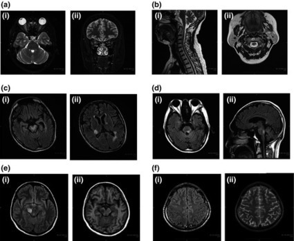











sujet abordé sur https://www.scoop.it/t/neuroimmunology?q=lymphatics