Your new post is loading...

|
Scooped by
Gilbert C FAURE
May 3, 2019 2:33 PM
|
Curr Neuropharmacol. 2019;17(2):142-150. doi: 10.2174/1570159X15666171109161140.Review...
|
|
Scooped by
Gilbert C FAURE
April 29, 2019 9:35 AM
|
Objective: We present the diagnostic and clinical course of the first multicenter case series of pediatric patients with autoimmune encephalitis associated with...
|
|
Scooped by
Gilbert C FAURE
April 26, 2019 2:51 PM
|
Multiple sclerosis (MS) is an autoimmune disease of the central nervous system (CNS). Microglia are the resident innate immune cells in the CNS; they play an important role in the processes of demyelination and remyelination in MS.
|
|
Scooped by
Gilbert C FAURE
April 23, 2019 5:09 AM
|
A research team at Massachusetts Eye and Ear has shown that microglia, the primary immune cells of the central nervous system—including the retina of the eye—serve as "gatekeepers," or biosensors and facilitators, of ...
|
|
Scooped by
Gilbert C FAURE
April 18, 2019 8:36 AM
|
Using animal models and clinical samples, the authors report that glioblastoma metabolites activate the transcription factor aryl hydrocarbon receptor in tumor-associated macrophages to modulate their function and T cell immunity, promoting tumor growth.
|
|
Scooped by
Gilbert C FAURE
April 7, 2019 2:25 PM
|
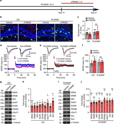
05 Apr 2019 As if the biological functions of membrane-bound TREM2 weren’t complicated enough, researchers also have to contend with the actions of its freewheeling counterpart, soluble TREM2. In the March 25 Nature Communications, researchers led by Guojun Bu at the Mayo Clinic in Jacksonville, Florida, and Xiao-Fen Chen of Xiamen University in China reported that injecting soluble TREM2 (sTREM2) into the brains of AD model mice activated microglia and rallied them to encircle and engulf plaques. The soluble fragment, which is shed from the microglial membrane, also corrected synaptic and memory deficits in the animals, the researchers claim. They have yet to nail down the enigmatic receptor for sTREM2, but zeroed in on microglia as the cells that express it. Upping sTREM2 boosted plaque clearance in 5xFAD mice. The fragment recruited microglia to plaques, and enhanced their phagocytosis. It restored synaptic function and rescued memory loss. “Studies like this are crucial to understand the functions of both full-length receptor and its cleaved ectodomain, and are extremely instrumental in aiding design strategies to therapeutically target TREM2,” commented Gernot Kleinberger of ISAR Bioscience in Planegg, Germany. Since the discovery of TREM2 as a major AD risk factor (see Research Timeline, 2012), enormous strides have been made in unraveling its complex function in the brain. In a nutshell, stimulation of the receptor by ligands including phospholipids, Aβ, and ApoE, summon microglia to clean up debris, including plaques or degenerating cells (Feb 2015 news; Oct 2015 news). In some animal models of amyloidosis and tauopathy, signaling through Trem2 can switch from beneficial to detrimental as disease progresses (Jul 2018 news). Making matters more complex, ADAM proteases cleave the receptor, shedding the extracellular sTREM2 domain into the interstitial environs. This soluble bit has been spotted mingling with neurons around Aβ plaques in mice (Jan 2018 news). In people, sTREM2 spikes in the cerebrospinal fluid in the early stages of AD (Jan 2016 news). In 2017, Bu and colleagues reported that sTREM2 bolstered microglial survival and upped pro-inflammatory cytokines (Feb 2017 news). sTREM2 Lessens Plaques. Plaque load dropped by a third in the hemisphere of hippocampus injected with sTREM2 (right). [Courtesy of Zhong et al., Nature Communications, 2019.] In their new paper, the researchers extended their investigation of sTREM2 to the 5xFAD model. First authors Li Zhong and Ying Xu, both at Xiamen University, and colleagues injected a recombinant form of sTREM2 into the right hippocampus of plaque-ridden, 7-month-old mice. They injected saline into the left hippocampus. A week later, they found that the plaque burden in the right hippocampus was a third lower than in the left. The sTREM2 injection led to the greatest reduction in large plaques, which dropped by more than half, but also reduced the total area of dystrophic neurites. Attack the Plaque. More microglia (red) swarmed Aβ plaques (green) in mice injected with sTREM2 (bottom panels). [Courtesy of Zhong et al., 2019] How did sTREM2 vanquish plaques? As suspected, the receptor riled up microglia. The glial cells proliferated in the hippocampus in the days following sTREM2 injection, and pumped out pro-inflammatory cytokines, including IL-1β and TNF. The microglia also surrounded plaques and expressed the phagocytic marker CD68. In a dish, primary microglia migrated toward sTREM2, and treatment with sTREM2 triggered microglia to gobble up fluorescently labeled Aβ42 via phagocytosis. Overall, the data suggested that sTREM2 both attracted microglia and stoked their appetite for Aβ. Curiously, the effects of sTREM2 appeared to go beyond plaque clean-up. In hippocampal slice cultures from 5xFAD mice, it rescued deficits in long-term potentiation of Schaffer collaterals, a major hippocampal circuit. Soluble TREM2 also boosted expression of synaptic receptors in both the pre- and postsynapses. Notably, when the researchers treated 5xFAD mice with PLX3397, a CSF1R receptor inhibitor that depletes the brain of microglia, sTREM2 treatment no longer boosted plaque clearance, microglial activation, or synaptic plasticity. This suggested that the beneficial effects of sTREM2—even on synaptic signaling—are mediated through microglia. If it boosts plasticity, would sTREM2 rescue memory problems? To find out, the researchers expressed it long-term. They injected neonatal wild-type and 5xFAD mice with an adeno-associated virus carrying an sTREM2 construct, then put their spatial memory to the test in the Morris water maze six months later. Among mice expressing a control virus, wild-type mice learned the location of a submerged platform faster than did 5xFAD mice, but sTREM2 overexpression rescued this deficit in the 5xFAD animals. The AAV-TREM2 dramatically reduced plaque load, recruited microglia to plaques, and boosted synaptic function. Bu proposed that in a physiological setting, sTREM2 amplifies and maintains the protective responses instigated by full-length TREM2. By the early symptomatic stage of AD, Aβ plaques are rampant, tau pathology has spread into the cortex, and neurons are degenerating. These injury signals might exacerbate the shedding of sTREM2 from microglia surrounding plaques and damaged cells, Bu said. In addition to binding many of the same ligands its full-length counterpart latches onto, sTREM2 likely binds an unknown receptor on microglia as well, recruiting more of the cells and potentiating their activation, Bu added. He hypothesized that the subsequent drop in sTREM2 in the CSF reflects the die-off of microglia. “I find it very interesting that sTREM2 can promote functions that we thought are associated with membrane bound full-length TREM2,” commented Christian Haass of the German Center for Neurodegenerative Diseases in Munich. “This indeed makes therapeutic modulation of TREM2 even more tricky.” Haass added that identification of the sTREM2 receptor could shed more light on the complicated functions of microglia. Kleinberger added that it would have been interesting to test whether mutant forms of sTREM2, such as the AD-linked R47H variant, would generate a similar loss-of-function phenotype as described for the full-length mutant receptor. The full-length receptor has so far been associated with beneficial functions such as microglial phagocytosis (Kleinberger et al., 2014). However, one study indicated that too much sTREM2 could be harmful: People harboring the H157Y mutation, which enhances sTREM shedding, had an increased risk of AD (Jiang et al. 2016).—Jessica Shugart REFERENCES News Citations TREM2 Buoys Microglial Disaster Relief Efforts in AD and Stroke 28 Feb 2015 Alzheimer’s Risk Genes Interact in Immune Cells 2 Oct 2015 TREM2: Diehard Microglial Supporter, Consequences Be DAMed 13 Jul 2018 New Mouse Models Reveal Unexpected Property of TREM2 18 Jan 2018 TREM2 Goes Up in Spinal Fluid in Early Alzheimer’s 27 Jan 2016 Does Soluble TREM2 Rile Up Microglia? 24 Feb 2017 Research Models Citations 5xFAD (B6SJL) Paper Citations Kleinberger G, Yamanishi Y, Suárez-Calvet M, Czirr E, Lohmann E, Cuyvers E, Struyfs H, Pettkus N, Wenninger-Weinzierl A, Mazaheri F, Tahirovic S, Lleó A, Alcolea D, Fortea J, Willem M, Lammich S, Molinuevo JL, Sánchez-Valle R, Antonell A, Ramirez A, Heneka MT, Sleegers K, van der Zee J, Martin JJ, Engelborghs S, Demirtas-Tatlidede A, Zetterberg H, Van Broeckhoven C, Gurvit H, Wyss-Coray T, Hardy J, Colonna M, Haass C. TREM2 mutations implicated in neurodegeneration impair cell surface transport and phagocytosis. Sci Transl Med. 2014 Jul 2;6(243):243ra86. PubMed. Jiang T, Tan L, Chen Q, Tan MS, Zhou JS, Zhu XC, Lu H, Wang HF, Zhang YD, Yu JT. A rare coding variant in TREM2 increases risk for Alzheimer's disease in Han Chinese. Neurobiol Aging. 2016 Jun;42:217.e1-3. Epub 2016 Mar 3 PubMed. Other Citations Research Timeline, 2012 FURTHER READING News Unbiased Screen Fingers TREM2 Ligands That Promote Aβ Uptake 22 Jul 2016
|
|
Scooped by
Gilbert C FAURE
April 1, 2019 1:36 PM
|
Microglia play a pivotal role in the coordination of brain development and have emerged as a critical determinant in the progression of neurodegenerative diseases; however, the role of microglia in the onset and progression of neurodevelopmental disorders is less clear. Here we show that conditional deletion of αVβ8 from the central nervous system ( Itgb8ΔCNS mice) blocks microglia in their normal stepwise development from immature precursors to mature microglia. These “dysmature” microglia appear to result from reduced TGFβ signaling during a critical perinatal window, are distinct from microglia with induced reduction in TGFβ signaling during adulthood, and directly cause a unique neurodevelopmental syndrome characterized by oligodendrocyte maturational arrest, interneuron loss, and spastic neuromotor dysfunction. Consistent with this, early (but not late) microglia depletion completely reverses this phenotype. Together, these data identify novel roles for αVβ8 and TGFβ signaling in coordinating microgliogenesis with brain development and implicate abnormally programmed microglia or their products in human neurodevelopmental disorders that share this neuropathology.
|
|
Scooped by
Gilbert C FAURE
March 30, 2019 8:17 AM
|
A new salvo in the debate over whether humans still make new brain cells as we get older
|
|
Scooped by
Gilbert C FAURE
March 19, 2019 5:21 AM
|
This Review considers the link between pain and the immune system. Nociceptors are directly activated by immune mediators and microbial products and, in turn, release neuropeptides that shape immune responses.
|
|
Scooped by
Gilbert C FAURE
March 19, 2019 5:10 AM
|
Meningitis, a group of serious diseases which infect the brain's lining, leaves its mark and can affect the body's ability to fight such infections in the future. According to a new study published in Nature Immunology, infections ...
|
|
Scooped by
Gilbert C FAURE
March 16, 2019 2:07 AM
|
Conventional therapies for Parkinson disease (PD) include dopamine replacement and therapies targeting symptomatic relief, but these approaches fail to modify the underlying disease. This Review explores the novel PD treatment strategies currently being investigated, including pharmaceuticals, cell therapies, immunotherapies, gene therapies and new technologies.
|
|
Scooped by
Gilbert C FAURE
March 12, 2019 4:08 PM
|
Microglia play a pivotal role in the coordination of brain development and have emerged as a critical determinant in the progression of neurodegenerative diseases; however, the role of microglia in the onset and progression of neurodevelopmental disorders is less clear. Here we show that conditional deletion of αVβ8 from the central nervous system ( Itgb8ΔCNS mice) blocks microglia in their normal stepwise development from immature precursors to mature microglia. These “dysmature” microglia appear to result from reduced TGFβ signaling during a critical perinatal window, are distinct from microglia with induced reduction in TGFβ signaling during adulthood, and directly cause a unique neurodevelopmental syndrome characterized by oligodendrocyte maturational arrest, interneuron loss, and spastic neuromotor dysfunction. Consistent with this, early (but not late) microglia depletion completely reverses this phenotype. Together, these data identify novel roles for αVβ8 and TGFβ signaling in coordinating microgliogenesis with brain development and implicate abnormally programmed microglia or their products in human neurodevelopmental disorders that share this neuropathology.
|
|
Scooped by
Gilbert C FAURE
March 10, 2019 2:41 PM
|
The aim of this study was to test whether autoantibodies against neurologic surface Ags are found in nonneurologic autoimmune diseases, indicating a broader loss of tolerance. Patient and matched healthy donor (HD) sera were derived from four large cohorts: 1) rheumatoid arthritis (RA) ( n = 194,...
|
|
|
Scooped by
Gilbert C FAURE
April 30, 2019 2:22 PM
|
Bacterial ventriculitis is a common complication in children with temporary external ventricular drains (EVD) and the diagnosis is challenging. The present study compared the diagnostic accuracy of novel cerebrospinal fluid (CSF) marker - CD64 expression on neutrophils measured as neutrophil CD64...
|
|
Scooped by
Gilbert C FAURE
April 29, 2019 4:12 AM
|
Curr Opin Neurol. 2018 Oct;31(5):635-639. doi: 10.1097/WCO.0000000000000599. Research Support, Non-U.S. Gov't; Review
|
|
Scooped by
Gilbert C FAURE
April 23, 2019 8:01 AM
|
21 Apr 2019 Microglia are known as the brain’s sometimes-overzealous clean-up crew, but what happens to people if these immune cells are missing from the get-go? Two studies published March 29 in the American Journal of Human Genetics describe nine people who carry mutations in both copies of the CSF-1R gene, a receptor that is vital for development and maintenance of microglia. In the most severe cases, key brain structures were not there, such as the corpus callosum that connects the two hemispheres. Bones were overly dense and malformed. While it is uncertain what the findings portend about the role of microglia in late-onset neurodegenerative disease, they make clear that the cells are indispensable components of brain development and function. Researchers describe nine cases of bi-allelic CSF-1R mutations. Severe cases had no microglia and profound abnormalities in brain and bones. Symptom onset varied from prenatal to early adulthood. Microglial progenitors derive from the embryonic yolk sac and migrate into the budding brain early in development. There, their renewal and expansion are driven by colony stimulating factor 1. Some animal studies suggest that the cells are pivotal in supporting myelination of the developing brain, assembling brain circuits, and pruning synapses (Squarzoni et al., 2014; Wlodarczyk et al., 2017; Hammond et al., 2018). People who carry one nonfunctional copy of CSF-1R develop a condition known as adult-onset leukoencephalopathy with axonal spheroids and pigmented glia (ALSP). This neurodegenerative disease degrades white matter and leads to dementia and motor impairment (Rademakers et al., 2011). In some cases, the mutated copy of CSF-1R may exert a dominant-negative effect—corralling the healthy copy and stifling signaling by more than half (Pridans et al., 2013). What would happen in people who had two mutant copies of CSF-1R? Researchers led by Tjakko van Ham of Erasmus University in the Netherlands and James Bennett of the University of Washington in Seattle described two such cases, an infant and a 24-year-old man. The baby came into Bennett’s care when he was born at Seattle Children’s Hospital. His parents had traveled from Alaska to have their child there after abnormalities were detected in utero. He was born with multiple congenital brain abnormalities, including complete absence of a corpus callosum, enlarged ventricles, and a cerebellar defect called a Dandy-Walker malformation, in which parts of the cerebellum fail to develop. He had low blood calcium and dense bones, a condition the researchers diagnosed as osteopetrosis. During his short life, he had trouble breathing, was unable to eat properly, and had epilepsy. He died from a bacterial infection at 10 months of age. Upon autopsy, the researchers found no microglia in the brain, although they did spot some cells expressing Iba1, a microglial marker that can also be expressed on circulating myeloid cells, and CD68, which marks phagocytes. Genetic testing revealed that the infant carried two copies of CSF-1R with a point mutation that disrupted splicing, leading to a protein product with a nonfunctional kinase domain. The parents, who were cousins, each carried a single copy of the variant. Neither had yet reported symptoms of ALSP, but were only 40 years old at last contact. Missing Microglia. Immunocytochemistry revealed Iba1+ microglia throughout the brain of a control, with ramified processes (G and H). An infant with homozygous mutations in CSF-1R only had scant Iba1+ cells near blood vessels (I), which were rounded (J and K). [Courtesy of Oosterhof et al., American Journal of Human Genetics, 2019.] The 24-year-old patient is still alive. At age 12, he started having seizures and regressed developmentally. Now he is unable to walk, talk, or feed himself. An MRI revealed abnormalities similar to the infant’s, including a partial loss of the corpus callosum, enlarged ventricles, atrophy of the cerebellar vermis, and white-matter loss. However, he had neither hypocalcemia nor dense bones. Two of his siblings had died at ages 4 and 21 with similar symptoms. He carries a homozygous mutation that was also predicted to affect function of the kinase domain of CSF-1R. The researchers speculated that the brain abnormalities in both cases were caused by a lack of microglia, and that the skeletal problems in the infant were likely the result of a loss of osteoclasts. These tissue-resident macrophages break down extra bone tissue, and also depend on CSF-1R signaling. The Dutch researchers had previously generated CSF-1R-deficient zebrafish, and reported that the minnows had no microglia, dense bones, and died prematurely (Oosterhof et al., 2018). A proteomic analysis found the fish lacked Cux1a, a homolog of human CUX1. This neuronal transcription factor is required for the projection of axons that make up the corpus callosum. The researchers went back to the postmortem samples from their first case, and found about half as many CUX1+ neurons as in an age-matched control. They proposed that microglia likely play an instrumental role in the formation of the corpus callosum by supporting CUX1+ neurons. The second paper, by researchers led by Shiro Ikegawa at the RIKEN Center for Integrative Medical Sciences in Tokyo, described seven people with bi-allelic CSF-1R mutations among three families. The most severe case was a 5-year-old Brazilian boy, who had similar brain deformities as those found by Bennett and colleagues. He had severe bone abnormalities, which the researchers diagnosed as dysosteosclerosis, a condition distinct from osteopetrosis, Ikegawa emphasized. He noted that the diagnosis of osteopetrosis in the infant in Bennett’s paper was likely incorrect. The other cases—one from a Japanese family and five from an extended family of Chaldeans, who are Iraqi Christians, in the U.S.—had varying degrees of brain abnormalities, dysosteosclerosis, and symptom onsets and severities. While many had seizures, intellectual disabilities, and motor problems, such as rigidity or trouble walking, one completed college before troubles with coordination and speech started to emerge. The carriers in the Brazilian and Japanese families were compound heterozygotes, meaning that they had a different mutation in each copy of the CSF-1R, while the Chaldean carriers were homozygous. The researchers reported that the mutations nixed kinase activity of the receptor by varying degrees. Authors of both studies proposed that the severity of disease across cases correlated with the extent to which CSF-1R function was lost. They placed the disease phenotypes along the same spectrum as those in heterozygous carriers with ALSP. Though people with ALSP have a later onset and do not have skeletal problems, their disease profoundly affects white matter, including the corpus callosum, van Ham pointed out. He thinks that heterozygous carriers of CSF-1R mutations may start life with enough microglia for the brain to develop, but cell numbers gradually wane over time as CSF-1R levels are too low to support their renewal. At that point, scarcity of trophic factors made by microglia, such as insulin-like growth factor-1, may lead white-matter structures to crumble, he added. In homozygous carriers, these structures may never develop in the first place, leading to more severe developmental abnormalities, he said. Florent Ginhoux of the Agency for Science, Technology, and Research in Singapore commented that these cases provide invaluable insight into human biology. He, too, noted that the wide spectrum of clinical and pathological manifestations caused by the mutations likely reflect their severity. He said work is needed to quantify the loss of CSF-1R function caused by each mutation, and understand how that correlates with the severity of the disease. Kim Green of the University of California, Irvine, had a different take on the data. He agrees that the extent of the abnormalities, particularly in the infant with homozygous CSF-1R mutations, illustrates that microglia play a critical role in brain development. However, he thinks people with residual CSF-1R function, such as heterozygous mutation carriers, or homozygous carriers with less severe mutations, have a fundamentally different disorder caused by malfunction of mutated microglia. He noted that mice expressing a single copy of CSF-1R have more microglia than wild-type mice, a curious finding he is investigating (Chitu et al., 2015). He suspects that people with residual CSF-1R likely have a disorder caused by the actions of those dysregulated cells, rather than their loss. Van Ham believes the zebrafish model, in which loss of CSF-1R correlates with loss of microglia, may more accurately reflect the human situation. Van Ham added that people with ALSP share several of the same pathological characteristics, such as axonal spheroids, as observed in the infant with homozygous mutation. They also have fewer microglia in some regions of the brain. What can these cases tell scientists about the role of microglia in late-onset neurodegenerative disease? To van Ham, the findings highlight a pivotal function for microglia in supporting axonal growth and maintaining proper wiring in the brain. He speculated that in AD, microglia could become chronically distracted by plaques, leading to a loss of physiological functions, and perhaps the erosion of white-matter tracts the cells usually support. On the flip side, Green found that using a CSF-1R inhibitor to deplete microglia from adult mice did not cause cognitive problems, and even assuaged neuroinflammation and synaptic damage in mouse models of amyloidosis (Apr 2014 news; Spangenberg et al., 2016). Researchers have also reported that nixing the cells with this inhibitor prevented Aβ plaque formation in 5xFAD transgenic mice, while another study reported that genetic ablation of the cells in adult APP/PS1 and APP23 mice appeared to have no effect on plaque development (Mar 2018 news; Oct 2009 news). However, van Ham said it is important to consider all physiological functions of microglia before toying with reducing their numbers.—Jessica Shugart REFERENCES News Citations Mutations in Microglia Protein Cause Rare Dementia 6 Jan 2012 Microglial Magic: Drug Wipes Them Out, New Set Appears 18 Apr 2014 Wiping Out Microglia Prevents Neuritic Plaques 8 Mar 2018 Research Models Citations 5xFAD (B6SJL) APPswe/PSEN1dE9 Paper Citations Squarzoni P, Oller G, Hoeffel G, Pont-Lezica L, Rostaing P, Low D, Bessis A, Ginhoux F, Garel S. Microglia modulate wiring of the embryonic forebrain. Cell Rep. 2014 Sep 11;8(5):1271-9. Epub 2014 Aug 21 PubMed. Hammond TR, Robinton D, Stevens B. Microglia and the Brain: Complementary Partners in Development and Disease. Annu Rev Cell Dev Biol. 2018 Oct 6;34:523-544. Epub 2018 Aug 8 PubMed. Pridans C, Sauter KA, Baer K, Kissel H, Hume DA. CSF1R mutations in hereditary diffuse leukoencephalopathy with spheroids are loss of function. Sci Rep. 2013 Oct 22;3:3013. PubMed. Oosterhof N, Kuil LE, van der Linde HC, Burm SM, Berdowski W, van Ijcken WF, van Swieten JC, Hol EM, Verheijen MH, van Ham TJ. Colony-Stimulating Factor 1 Receptor (CSF1R) Regulates Microglia Density and Distribution, but Not Microglia Differentiation In Vivo. Cell Rep. 2018 Jul 31;24(5):1203-1217.e6. PubMed. Chitu V, Gokhan S, Gulinello M, Branch CA, Patil M, Basu R, Stoddart C, Mehler MF, Stanley ER. Phenotypic characterization of a Csf1r haploinsufficient mouse model of adult-onset leukodystrophy with axonal spheroids and pigmented glia (ALSP). Neurobiol Dis. 2015 Feb;74:219-28. Epub 2014 Dec 9 PubMed. Spangenberg EE, Lee RJ, Najafi AR, Rice RA, Elmore MR, Blurton-Jones M, West BL, Green KN. Eliminating microglia in Alzheimer's mice prevents neuronal loss without modulating amyloid-β pathology. Brain. 2016 Apr;139(Pt 4):1265-81. Epub 2016 Feb 26 PubMed. Grathwohl SA, Kälin RE, Bolmont T, Prokop S, Winkelmann G, Kaeser SA, Odenthal J, Radde R, Eldh T, Gandy S, Aguzzi A, Staufenbiel M, Mathews PM, Wolburg H, Heppner FL, Jucker M. Formation and maintenance of Alzheimer's disease beta-amyloid plaques in the absence of microglia. Nat Neurosci. 2009 Nov;12(11):1361-3. PubMed. Other Citations APP23 External Citations Wlodarczyk et al., 2017 FURTHER READING Papers Guo L, Elcioglu NH, Karalar OK, Topkar MO, Wang Z, Sakamoto Y, Matsumoto N, Miyake N, Nishimura G, Ikegawa S. Dysosteosclerosis is also caused by TNFRSF11A mutation. J Hum Genet. 2018 Jun;63(6):769-774. Epub 2018 Mar 22 PubMed. News Long Live the Microglia! Studies Trace Their Lifespans in Mice and Humans 28 Aug 2017 Nature Versus Nurture: What Gives Microglia Their Identity? 19 Feb 2015
|
|
Scooped by
Gilbert C FAURE
April 18, 2019 8:37 AM
|
The aim of this study was to test whether autoantibodies against neurologic surface Ags are found in nonneurologic autoimmune diseases, indicating a broader loss of tolerance. Patient and matched healthy donor (HD) sera were derived from four large cohorts: 1) rheumatoid arthritis (RA) ( n = 194,...
|
|
Scooped by
Gilbert C FAURE
April 18, 2019 2:59 AM
|
Sickness behavior and cognitive dysfunction occur frequently during RNA virus infection
by unknown mechanisms. Prinz and colleagues show that virus-induced sickness behavior is induced by interferon receptor chain 1 (IFNAR1) engagement on brain endothelial and epithelial cells that in turn...
|
|
Scooped by
Gilbert C FAURE
April 4, 2019 9:03 AM
|
Brain. 2019 Apr 1. pii: awz054. doi: 10.1093/brain/awz054.[Epub ahead of print]...
|
|
Scooped by
Gilbert C FAURE
March 31, 2019 7:12 AM
|
Multiple sclerosis (MS) is an inflammatory and neurodegenerative autoimmune disease of the central nervous system (CNS). Disease-modifying therapies (DMT) targeting inflammation have been shown to reduce disease activity in patients with relapsing–remitting MS (RRMS).
|
|
Scooped by
Gilbert C FAURE
March 28, 2019 3:48 PM
|
TREM2 is a genetic risk factor for Alzheimer’s disease, and soluble TREM2 (sTREM2) in the CSF correlates with AD progression. Here the authors study the role of sTREM2 in a mouse model of Alzheimer’s disease, and find it reduces amyloid accumulation and increases the numbers of plaque-associated...
|
|
Scooped by
Gilbert C FAURE
March 19, 2019 5:21 AM
|
|
|
Scooped by
Gilbert C FAURE
March 16, 2019 7:43 AM
|
Abstract Epilepsy is a serious neurological condition exhibiting complex pathology and deserving of more serious attention. More than 30% of people with epilepsy are not responsive to more than 20 anti‐epileptic drugs currently available, reflecting an unmet clinical need for novel therapeutic strategies. Not much is known about the pathogenesis of epilepsy, but evidence indicates that neuroinflammation might contribute to the onset and progression of epilepsy following acquired brain insults. However, the molecular mechanisms underlying these pathophysiological processes are yet to be fully understood. The emerging research suggests that high‐mobility group box protein 1 (HMGB1), a DNA‐binding protein that is both actively secreted by inflammatory cells and released by necrotic cells, might contribute to the pathogenesis of epilepsy. HMGB1 as an initiator and amplifier of neuroinflammation, and its activation is implicated in the propagation of seizures in animal models. The current review will highlight the potential role of HMGB1 in the pathogenesis of epilepsy, and implications of HMGB1‐targeted therapies against epilepsy. HMGB1 in this context is an emerging concept deserving further exploration. Increased understanding of HMGB1 in seizures and epilepsy will pave the way in designing novel and innovative therapeutic strategies that could modify the disease course or prevent its development. Abbreviations used Abbreviations used AEDs anti‐epileptic drugs BBB blood‐brain‐barrier CA cornu ammonis CNS central nervous system CXCR4 C‐X‐C chemokine receptor type 4 DAMPs damage associated molecular patterns DRE drug resistant epilepsy EAE experimental autoimmune encephalomyelitis FCD focal cortical dysplasia FS febrile seizure HMGB1 high mobility group box 1 IHC immunohistochemical staining JAK‐STAT Janus kinases‐signal transducer and activator of transcription proteins KO knock out LPS‐RS lipopolysaccharide from rhodobacter sphaeroides mAb monoclonal antibodies MAPK mitogen‐activated protein kinase MES maximal electro shock NF‐κB nuclear Factor kappa‐light‐chain‐enhancer of activated B cells NMDAR N‐methyl‐d‐aspartate receptor PG prostaglandin RAGE receptor for advanced glycation end products SE status epilepticus SRS spontaneous recurrent seizures TBI traumatic brain injury TIM‐3 T‐cell immunoglobulin and mucin domain‐3 TLE temporal lobe epilepsy TLR‐4 toll‐like receptor 4 TNF tumor necrosis factor TREM‐1 triggering receptor expressed on myeloid cells‐1 Epilepsy is a serious neurological disorder characterized by the recurrent, periodic, and unpredictable occurrence of epileptic seizures which might be due to imbalance in excitatory and inhibitory pathways within the central nervous system (CNS) (Ana et al. 2019). Epilepsy has emerged as a serious health concern affecting 50–70 million people globally accounting 0.75% of global health burden (Trinka et al. 2018). Frequent and serious epileptic seizures are thought to contribute to further brain injury and persistent neurobehavioral and neuropsychiatric disorders, with significant consequences for patients, their families, and society (Yang et al. 2017). But much remains to be elucidated about the precise mechanisms of epileptogenesis – however, a plethora of findings over the past decade have highlighted the crucial pathophysiological role of brain inflammation in epilepsy (Vezzani et al. 2013; Webster et al. 2017; Paudel et al. 2018b). The currently available anti‐epileptic drugs (AEDs), which are not disease‐modifying AEDs but rather anti‐convulsants or anti‐seizure drugs, provide only symptomatic relief. Further, the mainstream AEDs exert anti‐epileptic effects largely by inhibiting voltage‐gated Na+, K+, T‐type Ca2+ channels (Simonato 2018), or by enhancing γ‐aminobutyric acid signaling, and/or by inhibiting glutamate transmission to dampen the neuronal hyperexcitability (Mertens et al. 2018). But these AEDs are not the ultimate solution, with more than 30% of patients developing drug‐resistant epilepsy (Chen et al. 2018), and increasing reports of adverse effects such as cognitive impairments, fatigue, and behavioral dysfunction in a significant proportion of patients treated with AEDs chronically (Sarkis et al. 2018). Despite recent advances in research, the etiology and the mechanisms of epileptogenesis still remain elusive, reinforcing the need for a deeper understanding of novel molecular targets. The ultimate goal of treatment is to prevent seizures with no side effects. Therefore, there is a pressing need for exploring novel treatment alternatives to minimize seizure burden as well as to prevent the related comorbidities. Currently, huge efforts have been made to understand the pathogenesis of epileptogenesis, and the pathways associated with epileptogenesis which exhibits great potentials in treating epilepsy. Considerable efforts ongoing in this regard include EPITARGET (Targets and Biomarkers for Anti‐epileptogenesis) (EPITARGET 2018) and EpiBioS4Rx (Epilepsy Bioinformatics Study for Anti‐Epileptogenic Therapy) (Vespa et al. 2018). EPITARGET is focusing mainly on identifying novel biomarkers and exploring the involvement of multiple mechanisms that might contribute to the process of epileptogenesis (EPITARGET 2018). Whereas EpiBioS4Rx mainly focused on anti‐epileptogenesis in post‐traumatic epilepsy following traumatic brain injury (TBI) (EpiBioS4Rx 2018). These both are large‐scale consortium of researchers collecting information from patient populations, utilizing rigid and reliable standardized animal models, and cutting‐edge analytical methods to analyze data from both animal and human for trans‐species comparisons, which will help in the development of anti‐epileptogenic therapies (EpiBioS4Rx 2018; EPITARGET 2018). One of the emerging potential targets is high mobility group box 1 (HMGB1), and current understanding about the role of HMGB1 in epilepsy is rapidly growing. Moreover, the concept of understanding HMGB1 as a biomarker of epileptogenesis is also growing and has been well reported (Walker et al. 2016; Ravizza et al. 2017; Paudel et al. 2018a), which will aid in assessing disease progression and early prediction of disease onset. HMGB1 protein is known to contribute to epileptogenesis via a toll‐like receptor (TLR) 4‐dependent pathway to trigger tissue damage and inflammatory responses (Kleen and Holmes 2010). HMGB1 protein is a key initiator released by dying cells in the brain, and binds to TLR4 receptors and are abnormally released through neurons and astrocytes during an epileptic seizures (Maroso et al. 2010). The HMGB1‐TLR4 regulatory axis is involved in the generation and exacerbation of epileptic seizures, and developing any innovative therapeutics which acts via inhibiting this signaling axis might represent a novel anti‐epileptic treatment strategy. HMGB1 has emerged as a novel target against epileptogenesis, but much remained to be explored before translating it to patients (Fu et al. 2017). Herein, we highlight and update on key concepts ranging from HMGB1 biology, its role in epileptogenesis, available HMGB1 inhibitors and experimental evidence of HMGB1 as an anti‐epileptic therapy. Based on available literature, it is postulated that HMGB1 is an emerging target against epilepsy and that pharmacological modulation of HMGB1 will provide disease‐modifying effects. Insights into HMGB1 biology HMGB1 is an intriguing protein with complex biochemistry. It is a known danger signal or damage associated molecular pattern (Hei et al. 2018), and was first reported in 1973 as a non‐histone chromosomal protein (Ranzato et al. 2010). Intracellular and extracellular HMGB1 protein has demonstrated several crucial roles in inflammation, including being implicated in sterile inflammation, immunity and neurodegenerative conditions, making it an attractive, and emerging target for therapeutic intervention (Andersson and Tracey 2011). Out of four members (HMGB1, HMGB2, HMGB3, and HMGB4) of the HMGB group of proteins (Štros 2010), HMGB1 is the only one that has been extensively studied with respect to epilepsy. The precise understanding of basic HMGB1 chemistry is crucial to understanding its function, as it is composed of different boxes with specific binding sites resulting in different activities. Structural analysis of HMGB1 revealed two DNA binding domains (box A and box B) and a negatively charged C‐terminal (Ulloa and Messmer 2006; Yang et al. 2018). Box A contains the antagonistic site of box B, and demonstrates anti‐inflammatory properties in both in vivo and in vitro models of inflammation (Girard 2007). In addition, box A alone serves as a competitive antagonist for HMGB1 and inhibits HMGB1 activity (Shen and Li, 2015). Box B is the functional domain which is recognized by TLR4, whereas both box B and box A can bind to DNA and play a role in folding and distorting the double‐strand DNA (Dumitriu et al. 2005). The specific amino acid domains of HMGB1 such as 89–108 and 150–183 are responsible for binding to the TLR4 and receptor for advanced glycation end products (RAGE) respectively (Huttunen et al., 2002). Moreover, the N‐terminus contains heparin‐binding motifs, whereas the box B exhibits pro‐inflammatory activity to the protein and is involved in binding to RAGE (Huttunen and Rauvala 2004) (Fig. 1). HMGB1 post‐translational modification and receptor system HMGB1 is reported to exist in three isoforms: fully reduced HMGB1, disulfide HMGB1, and sulfonyl HMGB1 (Antoine et al. 2014; Aucott et al. 2018a) (Table 1). Under normal conditions, HMGB1 is mainly located in the nucleus as a non‐acetylated and thiol form whereas after tissue injury, non‐acetylated thiol‐HMGB1 is released from dead and dying cells, and can be further converted to disulfide‐HMGB1 (Venereau et al. 2016). In addition, after cell activation or injury, translocation of HMGB1 from the nucleus to the cytoplasm may occur, followed by inflammasome activation and pyroptosis (Xu et al. 2014). Functional activity of HMGB1 is mainly determined by redox modification of 3 key cysteine residues, C23, C45, and C106. The disulfide and reduced isoforms of HMGB1 have mutually exclusive functions. S.N. Name Schematic overview Description References 1. Fully reduced (Fr) HMGB1 HMGB1 with no posttranslational modifications Thiol group is present on each of the cysteine residues Possess cell migration‐inducing activity Lacks cytokine‐inducing activity No HMGB1‐TLR4 mediated cytokine production Antoine et al. (2014), Andersson et al. (2018b) and Frigerio (2017) 2. Disulfide (Ds) HMGB1 Contain disulphide bridge between C23 and C45 and a reduced C106 residues Is a TLR4 ligand Lacks cell migration‐induced activity Possess cytokine‐inducing activity Aucott et al. (2018a) and Frigerio (2017) 3. Sulfonyl (Ox) HMGB1 All the cysteine residues are terminally oxidized Lacks both cell migration‐induced activity and cytokine‐inducing activity No HMGB1‐TLR4 mediated cytokine production Aucott et al. (2018b) and Frigerio (2017) HMGB1, high mobility group box 1; TLR4, Toll‐like receptor. The fully reduced isoform of HMGB1 resides within the cell, and upon release, acts as a chemoattractant via complexation with CXCL12, binding exclusively through C‐X‐C chemokine receptor type 4 (Schiraldi et al. 2012). Based on its redox state, HMGB1 can stimulate cells through a range of receptors, including TLR, acting alone or in combination with other immune stimulants (Harris et al. 2012). Moreover, the reduced form of HMGB1 coordinates tissue regeneration whereas disulfide‐HMGB1 triggers and sustains inflammation via the TLR4/myeloid differentiation factor‐2 and RAGE receptors, but it is not involved in tissue regeneration (Tirone et al. 2018). HMGB1 also activates RAGE but the binding potency of each isoform for RAGE is yet to be known (Walker et al. 2017). A diverse range of extracellular binding partners of HMGB1 has been reported and this includes RAGE, TLR9, TLR4, TLR2, integrin, synuclein filaments, proteoglycans, T‐cell immunoglobulin and mucin domain, triggering receptor expressed on myeloid cells‐1, cluster of differentiation 24 (CD24), C‐X‐C chemokine receptor type 4, and N‐methyl‐d‐aspartate (NMDA) receptor (NMDAR) (Kang et al. 2014). However, RAGE and TLR4 are the two important binding partners that have been implicated in HMGB1 signaling as well as being studied extensively in epileptic conditions (Maroso et al. 2010, 2011). HMGB1 is predominantly fully reduced within the cell but can also be oxidized via reactive oxygen species upon translocation to the cytoplasm or post‐extracellular release. Release pattern of HMGB1 from cells has been hypothesized via two distinct mechanisms: either from cells undergoing necrosis but not apoptosis, or secreted actively from cells after inflammatory cytokine stimulation (Tian et al. 2007). Upon its nuclear to cytoplasmatic translocation during seizures, following tissue injury, HMGB1 is extracellularly released and acts as a pro‐inflammatory cytokine (Iori et al. 2013). The hyperacetylated form of HMGB1 is the one that regulates transcription of several pro‐inflammatory cytokines, including IL‐1β, via binding with TLR2, TLR4 as well as RAGE (Bianchi and Manfredi 2009; Maroso et al. 2011). Binding of HMGB1 with RAGE and TLR4 elicits different responses. HMGB1/RAGE signaling regulates cell growth, proliferation, and migration via activation of mitogen‐activated protein kinase and nuclear factor kappa‐light‐chain‐enhancer of activated B cells (NF‐κB) pathways, whereas HMGB1/TLRs signaling mainly modulates inflammatory responses (Ding et al. 2017). RAGE is a pro‐inflammatory receptor whereas TLR4 is the known HMGB1 receptor for cytokine production (Andersson et al. 2018a). HMGB1 demonstrates its biological activity by activating TLR4 receptor, but evidences which support the concept of a critical role of HMGB1 induced TLR4 activation in disease pathogenesis is lacking or not substantial (Branco‐Madeira and Lambrecht 2010). Mechanistic insights of HMGB1 in epileptogenesis: Insights from animal models Inflammation‐induced via several brain‐disrupting events such as trauma, stroke, and infection are associated with epileptic seizures (Pitkänen and Sutula 2002). HMGB1 is one of the most influential pro‐inflammatory cytokines and activates inflammatory pathways by stimulating two principal receptors: RAGE and TLR4 (Weber et al. 2015), and activation of these receptors have been implicated in epileptogenesis as depicted in Fig. 2. This process appears to be mediated by RAGE and TLR4, with evidence in particular of activation of HMGB1/TLR4 axis in experimental and human epilepsy and ictogenesis (Maroso et al. 2010). In addition, increased levels of pro‐inflammatory cytokines [IL‐1β, tumor necrosis factor (TNF‐α), HMGB1, S100β] and downstream inflammatory mediators (prostaglandins and the complement system) have been documented in epileptogenic tissues obtained from patients with different epilepsy etiologies (Aronica and Crino 2011; Vezzani and Friedman 2011). Moreover, the pro‐inflammatory effect of HMGB1 is dependent on the activation of NF‐κB or other pathways that promote chemotaxis and the production of cytokines (Li et al. 2016). NF‐κB regulates immune and inflammatory responses and is also one of the crucial downstream transduction molecules in the TLR4 and RAGE signaling axis (Xie et al. 2013). IL‐1 is a family of pro‐inflammatory cytokines and mediates innate immune response and IL‐1β from the family is the one extensively investigated against epilepsy (Webster et al. 2017). The impact of IL‐1R/TLR signaling in epileptogenesis, characterized by neuronal hyperexcitability which might be provoked by IL‐1β and HMGB1 provides insight into how IL‐1R/TLR4 signaling might contribute in seizure generation after induction of inflammatory cascades (Iori et al. 2017). Activation of IL‐1R1/TLR4 axis in receptor‐expressing neurons promotes excitotoxicity and seizures through enhancing Ca2+ influx via NMDA receptors (Iori et al. 2013; Balosso et al. 2014) as well as might be a potential target for disease‐modifying effects. Local CNS injury, or even a peripheral inflammatory challenge such as an infection, leads to the activation of microglia, astrocytes, and neurons in the brain. These cells then release pro‐inflammatory cytokines such as IL‐1β and HMGB1, that further initiate a sequence of inflammatory events in the target cells (neurons and glia) through activation of IL‐1R1 and TLR4 (Vezzani et al. 2011). Transcriptional activation of inflammatory genes in glia might play a role in the perpetuation of brain inflammation which in turn results in the generation of individual seizures via lowering the threshold of neuronal excitability. This seizure exacerbation further activates inflammatory cascades via establishing feedback of events that contribute to the development of epilepsy (Maroso et al. 2011). A role for HMGB1 in mediating the activation of glial cells in epilepsy has also been unraveled, by mechanisms including the TLR4/NF‐κB signaling axis. In a coriaria lactone (CL)‐induced epilepsy model, CL was found to increase the levels of HMGB1, TLR4, RAGE, NF‐κB p65, and inducible nitric oxide synthases in human microglia (HM) cells. Immunohistochemical staining suggested the nuclear to cytoplasmic translocation of HMGB1 in neurons and glial cells and reported its release into the extracellular space. HMGB1 contributes in epileptogenesis mainly through microglial activation, by the TLR4‐NF‐κB signaling axis activation (Shi et al. 2018). In a Picrotoxin/low Mg2+ entorhinal cortex (EC) slice model, IL‐1β and HMGB1 favor ictal‐like discharge production whereas in focal seizure model or 4‐aminopyridine (4‐AP) model it lowered ictal‐like discharge threshold. In the 4‐AP model, Ca2+ imaging experiments demonstrated that NMDA pulse applied to IL‐1β and HMGB1‐treated slices evoked a higher activation of neurons and astrocytes as compared to saline‐treated slices. IL‐1β or HMGB1 might decrease ictal frequency by elevating the sensitivity of neurons to NMDA thus causing a greater recruitment of neurons into the initial episode of NMDA receptor‐mediated excitation in a local circuit. These results signify that IL‐1β and HMGB1 can greatly exaggerate the generation of epileptiform activities (Chiavegato et al. 2014). Maroso and colleagues were the pioneers to shed light on the contribution of HMGB1 in epileptogenesis. The study elaborates the nature of HMGB1/TLR4 signaling, its intracellular signaling, and the contribution of HMGB1/TLR4 axis in kainic acid (KA) and bicuculline models of epileptogenesis. The elevated level of TLR4 and HMGB1 expression was observed in neurons, astrocytes, and microglia in the rat hippocampus as well as in human hippocampus from intractable temporal lobe epilepsy (TLE) patients (Maroso et al. 2010). Taken together, these findings suggest that TLR4 and, at a more downstream level, NMDA receptors, contribute to the pro‐convulsant action of HMGB1 and suggest the plausible role of HMGB1‐TLR4 signaling in the development and perpetuation of seizures. However, the precise mechanism behind the epileptogenesis remained enigmatic. Of note, mice lacking TLR4 or RAGE were less prone to developing epilepsy which suggests that HMGB1 is implicated in epileptogenesis as well as ictogenesis in a TLR4‐dependent way. Moreover, an antagonist of HMGB1 and TLR4 retard seizure precipitation and decrease recurrence of acute and chronic seizure (Maroso et al. 2010) implicating HMGB1‐TLR4 axis as an emerging target against epileptic seizures. HMGB1 has gained increased attention after these findings, and numerous investigations focusing on the involvement of HMGB1 in epileptogenesis has been emerging in recent days. A recent study using intrahippocampal KA to induce a model of mesial TLE reported that the role of RAGE in seizures appears to be less prominent than that of TLR4. This statement was made on the basis of the significant decrease in KA seizures in RAGE knock out (KO) mice, whereas no delay in seizure onset was detected in TLR4 KO mice (Iori et al. 2013). Further, the proictogenic effect of exogenously‐applied HMGB1 was reduced to a greater extent in RAGE KO compared to TLR4 KO mice. One mechanism by which HMGB1 and other inflammatory mediators may exert pro‐seizure effects may be via blood‐brain‐barrier (BBB) disruption. The BBB is important in establishing and maintaining the microenvironment of the CNS that permits proper neuronal function (Liebner et al. 2018). Several vasoactive or inflammatory compounds, which includes bradykinin, complement 3a, adenosine triphosphate (ATP), histamine and serotonin from mast cells, interleukins (IL), arachidonic acid and its metabolites, interferon alpha and beta, prostaglandins, and tumor necrosis factor (TNF), have all been reported to alter BBB permeability (Oby and Janigro 2006). The major players in the synthesis of these inflammatory compounds are brain‐resident cells, namely activated microglia, astrocytes, and neurons (Devinsky et al. 2013). Invading peripherally‐derived leukocytes are also posited to play a key role in epileptogenesis, especially when the BBB permeability is disrupted (Fabene et al. 2008). Pro‐inflammatory mediators can induce and sustain BBB breakdown by modulating the endothelial tight junctions and the basal membrane as well as they might contribute in seizure activity by modulating excitability and seizure threshold in epileptic conditions (Abbott et al. 2006; Devinsky et al. 2013). It is not surprising that BBB permeability exhibits an important role in the pathogenesis of epilepsy (Oby and Janigro 2006). BBB disruption during epilepsy might allow HMGB1 and other inflammatory cytokines to intrude into the brain and aggravate a seizure. Disruption in BBB architecture can contribute to the development of epilepsy after status epilepticus (SE) through a direct mechanism (neuronal depolarization via the influx of potassium) or through a sequelae of events aggravated by leakage of serum proteins leading to activation of glial cells, impairment in potassium buffering, inflammation, and synaptogenesis (Gorter et al. 2015). After pro‐convulsant (pilocarpine) administration in mice, a release of HMGB1 into the CNS and peripheral bloodstream was found to contribute to the induction of BBB disruption and activation of inflammatory mediators leading to epilepsy (Fu et al. 2017). In spite of growing advances in our understanding of HMGB1, not much is known about its precise mechanism in the development of epileptogenesis. But there is a growing notion that HMGB1 might play role in the development of epileptogenesis, mainly disruption of BBB and induction of inflammatory processes. In an experimental model of pilocarpine‐induced SE, there was a significant increment in the leakage of Evans Blue in the thalamus and hypothalamus regions indicating BBB disruption. Administration of recombinant human HMGB1 promoted Evans Blue leakage further in a dose‐dependent fashion, which suggests that HMGB1 promotes the BBB breakdown in an epileptic state (Fu et al. 2017). The putative role of HMGB1 isoforms in epilepsy is increasingly being established and has been well reviewed earlier (Ravizza et al. 2017). Recent evidence, however, also suggests an association between oxidative stress and neuroinflammation in the generation of the disulfide HMGB1 isoform. Precisely, oxidative stress during epileptogenesis is highly associated with de novo brain and blood generation of disulfide HMGB1 (Pauletti et al. 2017). In addition, the combination of anti‐oxidant drugs (N‐acetyl‐cysteine and sulforaphane) when administered to epileptic rats reduced the onset of spontaneous seizures, inhibited disease progression and delayed seizure frequency, exerts neuroprotective effects and ameliorated cognitive impairments (Pauletti et al. 2017). These results implicate that the combinational drug inhibited the generation of disulfide HMGB1, via the reduction of oxidative stress, thus suggesting a potential novel therapeutic approach. Moreover, others have reported that the acetylated, disulfide form of HMGB1 is responsible for the detrimental inflammatory effects of epilepsy in animal models of epilepsy (Walker et al. 2016). HMGB1 in its oxidized (disulfide) form, most likely determined by the redox state of the extracellular milieu, potentiates NMDA‐mediated calcium (Ca2+) influx in pyramidal neuron cell bodies, by activation of neuronal TLR4 colocalized with NMDARs. HMGB1‐TLR4 axis triggers neutral sphingomyelinase and Src kinases activities, and the phosphorylation of the NMDAR subunit 2B (Balosso et al. 2008). This rapid onset of post‐translational pathway underlies HMGB1‐NMDAR interactions, leading to intracellular Ca2+ increase and contributing to seizures and cell loss (Balosso et al. 2014). Necrotic cell death leads to the passive release of nonacetylated HMGB1. Disulfide HMGB1 binds and signals via TLR4 and induces pro‐inflammatory and neuromodulatory effects through activation of NF‐κβ (Yang et al. 2012). A plausible role of HMGB1 in epilepsy of different etiologies is now advancing. Mesial TLE is known to be the common refractory focal epilepsy syndrome which usually emerges during childhood. Some of the key pathological characteristics including neuronal loss, axonal sprouting, and synaptic reorganization of hippocampal structures, which are also the pathological basis of refractory epilepsy (Toller et al. 2015). Activation of HMGB1/TLR4 signaling has been implicated in the pathogenesis of mesial TLE though the precise mechanism still remains elusive. In a pilocarpine‐induced animal model of mesial TLE, quantitative real time‐polymerase chain reaction (qRT‐PCR) and western blot findings demonstrated a significant elevation of HMGB1 and TLR4 gene expression in the hippocampal tissues as compared to the control group. Moreover, enzyme‐linked immunosorbent assay (ELISA) from neuron culture supernatants found that exogenously‐applied HMGB1 increased IL‐1β and TNF‐α levels in hippocampal neurons (Yang et al. 2017). Focal cortical dysplasia (FCD) is one of the major causes of refractory epilepsy and is the most common histopathological type in children with lesional epilepsy (Blümcke et al. 2009). Western blot analysis revealed that HMGB1 protein expression in the cytoplasm and TLR4 protein expression in FCD lesion tissue from FCD type II patients was higher than that in peri‐FCD tissue. MyD88 protein expression levels normalized to TLR4 in FCD lesions were higher than those in peri‐FCD tissue. Moreover, the expression ratio of K63 polyubiquitin chain to TRAF6 was increased in FCD lesion tissue, compared to that in peri‐FCD tissue. These findings suggested that that the HMGB1‐TLR4 pathway was up‐regulated in the neurons and astrocytes inside FCD type II lesions, and the up‐regulation of HMGB1‐TLR4 led to an increase in the release of downstream pro‐inflammatory cytokines (Zhang et al. 2018). Febrile seizure (FS) is the most common form of childhood seizure occurring in 2–5% of children younger than 6 years (Kwon et al. 2018). Though HMGB1 has not been directly implicated in the pathogenesis of FS, elevated levels of serum HMGB1 along with cytokines (IL‐1β, IL‐6, IL‐10, interferon‐γ, and TNF‐α) have been reported in patients (Choi et al. 2011). These findings implicate that HMGB1 and the cytokine network might also contribute to the generation of FS in children. Taken together, these findings support the observations made initially in experiment models, that HMGB1 and its receptors (RAGE and TLR4) levels are increased in epileptic tissue and may play a role in the development and perpetuation of epileptic seizures. HMGB1 dynamics during epilepsy Understanding the dynamics of HMGB1 translocation and activity is an important factor that will be helpful in assessing the disease pathology. Arrays of studies have repeatedly reported the release of HMGB1 from nuclei in a range of neurological disorders including epilepsy (Fu et al. 2017; Zhao et al. 2017; Angelopoulou et al. 2018; Sun et al. 2018). The nuclear to cytosol translocation of HMGB1 and its release from neural cells is associated with post‐translational modifications and is implicated in the epileptic seizure generation (Fig. 3). Current understanding of the dynamics of HMGB1 in epilepsy remains limited. Evidence of HMGB1 translocation after different pro‐convulsant insults (KA, pentylenetetrazol, and Pilocarpine) are summarized in (Table 2) and elaborated in the text below. This translocated HMGB1 participates in the series of events leading to seizure generation with the interaction with RAGE (Zurolo et al. 2011) or TLR4 (Maroso et al. 2010). S.N. Model Observation Detection method References 1 Pilocarpine‐induced SE HMGB1 translocation and release occurred in the CA1 and cerebral cortex regions ↓ HMGB1 levels in the brain and ↑ HMGB1 level in plasma in mice Immunofluorescence staining, Western blot and ELISA Fu et al. (2017 2 PTZ‐induced seizure HMGB1 was activated, translocated and released from nuclei in epileptic group HMGB1 was localized in the nuclei of neurons and astrocytes in control group Immunohistochemical Staining Zhao et al. (2017) 3 KA‐induced epilepsy HMGB1 was substantially increased in the pyramidal neurons in epileptic group HMGB1 immunoreactivity was detected mostly in the nuclei of the pyramidal neurons in control group Immunohistochemical Staining Chen et al. (2015) 4 KA‐induced epilepsy Expression level of HMGB1 in epileptic group was lower than the control group (p < 0.05) at 24 h, and became higher than the control group at 72 h (p < 0.05) which implicates a nucleus‐to‐cytoplasm translocation of HMGB1 Sombati's cell model (for in vitro study) and Immunohistochemistry Huang et al. (2015) 5 KA‐induced epilepsy HMGB1 was induced in CA1 and CA3 hippocampal regions and peaked at 3 h and at 6 days post‐KA administration Immunoblot analysis Luo et al. (2014) 6 eFSE HMGB1 was localized to the nuclei of cells in control CA1 neurons HMGB1 appeared in the cytoplasm after 1and 3 h of eFSE, suggesting translocation of HMGB1 protein Immunohistochemistry Patterson et al. (2015) 7 KA‐induced epilepsy HMGB1 is present in the nuclei of pyramidal neurons, granule cells and dentate gyrus in control hippocampus Between 1 and 3 h of seizure onset progressive increase in nuclear and perinuclear HMGB1 staining observed in astrocytes Immunohistochemical Staining Maroso et al. (2010a) ↓, decreased; ↑, increased; SE, status epilepticus; PTZ, pentylenetetrazol; KA, kainic acid; HMGB1, high mobility group box protein 1; ELISA, enzyme linked immunosorbent assay; eFSE, experimental febrile status epilepticus; CA, cornu ammonis. The concept of HMGB1 post‐translational modifications has been advanced in the context of epilepsy‐related hyperexcitability. Immunoprecipitation (IP) of neurons exposed to KA excitotoxicity revealed that HMGB1 was acetylated, phosphorylated and ubiquitinated, implicating that post‐translational modifications after KA administration caused HMGB1 to dissociate from chromatin DNA and promoted its release into the extracellular setting. Moreover, HMGB1 modulates the expression level of glutamate metabolism‐associated enzymes in epilepsy‐related hyperexcitability, postulating that HMGB1 signaling contributes in the generation of epileptic seizures (Kaneko et al. 2017). A similar line of evidence was reported which documented the expression level of HMGB1 in epileptic and control group at an interval of 24 and 72 h. In a KA‐induced epilepsy model, the HMGB1 expression level in the epileptic group was lower as compared to control group (p < 0.05) at 24 h and increased compared to the control group at 72 h (p < 0.05). As well as in vivo analysis discovered a nucleus‐to‐cytoplasm translocation suggesting that HMGB1 is widely distributed in the cytoplasm in the epileptic group while HMGB1 is confined to the nucleus in control group (Huang et al. 2015). Similarly, in an experimental febrile SE (eFSE) model, HMGB1 was localized inside the nuclei of cells in cornu ammonis (CA1) neurons of controls. After 1 and 3 h of eFSE, HMGB1‐IR appeared in the cytoplasm, suggesting the translocation of HMGB1. Moreover, cytoplasmic HMGB1 was absent 24 h after eFSE and no differences in hippocampal HMGB1 mRNA levels were reported at any duration. Finally, mRNA expression of TLR4 was elevated significantly at 24 h after eFSE, and normalized to baseline levels 4 days after the eFSE. This finding implicates a transient translocation of hippocampal HMGB1 from the nucleus to the cytoplasm but not in microglia or astrocytes (Patterson et al. 2015). Immunoblot analysis suggest that HMGB1 was induced in the CA1 and CA3 hippocampi region in KA‐induced model, and it peaked twice at 3 h and 6 days after‐KA administration. The significant amount of HMGB1 was accumulated in serum at 12 h post‐KA which might be due to HMGB1 release due to KA‐induced neuronal death (Luo et al. 2014). Immunofluorescence staining study reveals that pilocarpine administration caused HMGB1 translocation from the nuclei, resulting in the low HMGB1 fluorescence intensity in the nuclei and an increased number of released HMGB1 particles. These findings support the notion that there is HMGB1 translocation and release in the CA1 and cerebral cortex area during an acute epileptic state (Fu et al. 2017). In a KA‐induced C57BL/6 mice, expression level and distribution of HMGB1 after KA induction suggest that HMGB1 immunoreactivity was mostly located inside the nuclei of the pyramidal neurons in the normal group. Moreover, in the stratum radiatum (SR), stratum lacunosum, and stratum moleculare, decentralized glial cells with nuclear staining and neurons with both nuclear and cytoplasmic staining were visualized. On the other side, cytoplasmic staining of HMGB1 was significantly elevated increased in the pyramidal neurons in an epileptic group. In addition to the glial cells with nuclear staining, glial cells with nuclear and cytoplasmic staining in SR, stratum lacunosum, and stratum moleculare of CA3 regions were reported. These findings corroborate to the nuclear to cytoplasmic translocation of HMGB1 in neurons and glial cells and postulated its release into the extracellular setting (Chen et al. 2015). Precise understanding on the dynamics of HMGB1 release and translocation after pro‐convulsant insult will strengthen the knowledge in evaluating the therapeutic efficacy of HMGB1 inhibiting agent which probably inhibits the translocation of HMGB1 from nuclei into the neighbouring areas. HMGB1‐targeting therapeutic agents in epilepsy: An Update Several strategies for the inhibition of HMGB1 have been explored. They range from HMGB1 antagonists capable of interacting with RAGE, small‐molecule inhibitors of HMGB1, anti‐HMGB1 antibodies (Ab), peptide and protein inhibitors of HMGB1, and oligonucleotide‐based inhibitors of HMGB1 (Musumeci et al. 2014). Therapeutic strategies that target HMGB1 with specific Ab or antagonists have a potential for minimizing epileptic seizures characterized by excessive HMGB1 release. Nevertheless, the appreciable outcome has been achieved from experimental studies targeting extracellular HMGB1 only by anti‐HMGB1 monoclonal antibodies (mAb) and HMGB1 inhibitors against epilepsy (Table 3). In addition, biological agents including anti‐HMGB1 Ab, the recombinant A box peptide antagonist and glycyrrhizin (GL), a naturally‐occurring HMGB1 antagonist derived from licorice root, are worthy of being explored against epilepsy as they can inhibit HMGB1 released by both activated macrophages and necrotic cells (Girard 2007). S.N. Interventions Model Treatment schedule Observations References 1 Anti‐HMGB1 mAb Pilocarpine‐induced SE Single bolus immediately after pilocarpine injection Prevented the BBB permeability ↓ HMGB1 translocation Inhibited the expression of inflammation‐related factors Protected against neural cell apoptosis Delay in latency and frequency of stage 5 seizure Fu et al. (2017) 2 Anti‐HMGB1 mAb PTZ, KA and MES‐induced seizure Single bolus 3 h before PTZ 3 h post‐SE for 7 days (KA) 1–48 h after bolus injection (MES) Anti‐HMGB1 mAb attenuates MES and PTZ‐induced acute seizures as well as the translocation of HMGB1 Anti‐HMGB1 alleviates KA induced chronic epilepsy Anti‐HMGB1 mAb prevents epileptogenesis following SE Zhao et al. (2017) 3 Box A (co‐administered with IL‐1Ra) Electrical SE in rats Single bolus 1 h post‐SE onset, then daily injections for 7 days ↓ in progression of SRS frequency and ↓ number SRS Walker et al. (2017) 4 Box A KA and Bicucullineinduced seizure Single bolus 15 min prior to KA or Bicuculline administration ↓ in seizure frequency and total duration; delay in seizure onset Maroso et al. (2010a) 5 N‐acethyl‐cysteine and Sulforaphane. Electrical SE in rats Bolus injections 1–6 h post‐SE onset then and then daily injections for 7–14 days Prevented disulfide HMGB1 generation in brain and blood which is responsible for seizure generation ↓ in epilepsy onset, SRS and ↓ in SRS progression; Amelioration of cognitive deficits and exhibited neuroprotection Pauletti et al. (2017) 6 LPS‐RS KA and Bicuculline induced seizure Single bolus 15 min prior to KA or Bicuculline administration ↓ seizure number, duration as well as delay in seizure onset Maroso et al. (2010a) 7 Glycyrrhizin (GL) Pilocarpine induced SE GL (50 mg/kg, i.p.) 30 min prior to pilocarpine administration GL suppresses oxidative stress after SE GL attenuates expression of inflammatory markers after SE No anti‐epileptogenic effects noted González‐Reyes et al. (2016) 8 GL KA induced seizure 300 μL of GL 30 min prior KA injection GL suppressed HMGB1 inductions in hippocampus GL exhibited anti‐inflammatory and anti‐excitotoxic functions Luo et al. (2014) 9 GL KA induced seizure GL (10 mg/kg, i.p.) 30 min prior KA administration GL inhibited KA‐induced astrocyte and microglia activations in CA1 and CA3 hippocampal region GL suppressed KA‐induced proinflammatory marker production GL demonstrated anti‐excitotoxic effects in primary cortical cultures Luo et al. (2013) 10 Celecoxib (CCX) KA induced seizure CCX (20 mg/kg) post‐KA CCX showed immunomodulatory effects on HMGB1‐TLR4 pathway as evidenced by ↓ expression of HMGB1, TLR4 and COX‐2 in cortex and hippocampus CCX ↓ frequency and severity of KA‐induced seizure Morales‐Sosa et al. (2018) ↓, decreased; SE, status epilepticus; SRS, spontaneous recurrent seizure; PTZ, pentylenetetrazol; KA, kainic acid; MES, maximal electroshock; HMGB1, high mobility group box protein 1; mAb, monoclonal antibody; TLR4, toll‐like receptor 4; GL, glycyrrhizin; BBB, blood‐brain‐barrier; Box A (competitive inhibitor of endogeneous HMGB1); LPS‐RS, TLR4 antagonist (Lipopolysaccharide from Rhodobacter sphaeroides); IL‐1Ra, interleukin‐receptor antagonist; CA, cornu ammonis; CCX, celecoxib; COX‐2, cyclooxygenase‐2. Anti‐HMGB1 mAb Anti‐HMGB1 mAb has emerged as a novel approach against several HMGB1 mediated pathologies (Liu et al. 2007; Okuma et al. 2012; Sasaki et al. 2016). To date, anti‐HMGB1 Abs have been used mainly to confirm the contribution of HMGB1 in several pathological conditions, and the efficacy of its inhibition (Nishibori 2018). The therapeutical potential of anti‐HMGB1 Abs has already been demonstrated in a range of HMGB1‐mediated pathologies including experimental arthritis (Schierbeck et al. 2011), brain infarction (Liu et al. 2007), brain edema (Nosaka et al. 2018), and BBB permeability (Zhang et al. 2011). Nevertheless, very few studies to date have evaluated the potential role of anti‐HMGB1 mAb against different models of epilepsy and reported noteworthy outcomes. In a pilocarpine‐induced seizure model, intravenous (i.v.) administration of anti‐HMGB1 mAb exerts protective effects on neuronal apoptosis, in association with blockage of HMGB1 release, preventing the BBB disruption and inhibition of inflammation induced by pilocarpine (Fu et al. 2017). The reported anti‐epileptic effect of anti‐HMGB1 mAb might be due to the inhibition of BBB rupture, inflammatory responses, and neuronal cell death. Moreover, anti‐HMGB1 mAb treatment was sufficient to suppress acute SE‐induced translocation of HMGB1 into the peripheral setting (Fu et al. 2017). The overall result suggests that anti‐HMGB1 mAb therapy might provide a novel strategy for preventing epileptogenesis. The similar line of evidence was reported in another study incorporating both acute and chronic seizure models. Anti‐HMGB1 mAb treatment attenuates maximal electroshock and pentylenetetrazol induced acute seizures, as well as inhibits the translocation and/or release of HMGB1 in astrocytes and neurons, as evidenced by demonstration of HMGB1 confined to cell nuclei (Zhao et al. 2017). But anti‐HMGB1 mAb did not demonstrate any anti‐seizure effect on TLR4 KO mice, which supports the notion that the HMGB1‐TLR4 regulatory axis contributes to ictogenesis (Zhao et al. 2017). The anti‐seizure effect of anti‐HMGB1 mAb in the KA‐induced seizure model and in the brain slices obtained from surgical resection of clinically drug‐resistant human patients suggest that anti‐HMGB1 mAb exhibits significant clinical therapeutic value for patients with medically refractory TLE (Zhao et al. 2017). A limited number of studies have also demonstrated the neuroprotective role of anti‐HMGB1 Ab in neuronal damage and inflammation after SE. In a KA‐induced model of SE, anti‐HMGB1 Ab demonstrated dose‐dependently inhibition of the mRNA expression of IL‐1β and TNF‐a, microglial activation, and neuronal damage implicating its neuroprotective potential. But this study did not report the anti‐epileptic activity of anti‐HMGB1 Ab (Li et al. 2013). The therapeutic action of anti‐HMGB1 mAb against epilepsy has been reported to initiate at 1 h and lasted until 24 h indicating that the mAb has a potential for clinical epilepsy treatment. Moreover, the anti‐epileptic potential of anti‐HMGB1 mAb on the human epileptic brain slices has not to be washed out implicating that anti‐HMGB1 mAb binds solidly with HMGB1 solidly and has promising potentially long‐term anti‐epileptic activity (Zhao et al. 2017). In regards to the safety profile of anti‐HMGB1 mAb, it only targets the activated, translocated HMGB1, and even in a high dose (25 mg/kg in mice) did not show any impairment in basic physical functions, body growth rate and thermoregulation (Zhao et al. 2017). However, the Ab‐based strategy to target HMGB1 is limited by issues such as the probability of conformational switches in the tertiary structure of the Ab‐recognized region (Musumeci et al. 2014). In spite of that, anti‐HMGB1 mAb demonstrated enough potential and specificity for therapy and has good drug‐ability with wider therapeutic window, exhibiting enormous justification for the further development of this anti‐HMGB1 mAb for the clinical management of epilepsy. Thus the development of human mAbs against HMGB1 and evaluation of their efficacy against several experimental epilepsy models should be the objectives of the future research. HMGB1 inhibitors A widely studied small‐molecule inhibitor of HMGB1 is GL, investigated in a large number of HMGB1‐mediated conditions, and reported to inhibit extracellular HMGB1 cytokine activity (Musumeci et al. 2014). GL (20β‐carboxyl‐11‐oxo‐30‐norolean‐12‐en‐3β‐yl‐2‐O‐β‐d‐glucopyranuronosyl‐β‐d‐glucopyranosiduronic acid) is the main bioactive component of Glycyrrhiza radix. GL is reported as a natural anti‐inflammatory compound compared to an anti‐HMGB1 mAb (Sun et al. 2018). Earlier studies have documented that GK has the potential to bind directly to both HMG boxes in HMGB1, thereby demonstrating its neuroprotective effects in a wide variety of neurological diseases (Gong et al. 2014). GL has exhibited protection against TBI via inhibition of HMGB1‐RAGE interaction (Okuma et al. 2014), experimental autoimmune encephalomyelitis via inhibiting HMGB1 expression and neuronal HMGB1 release (Sun et al. 2018), and focal cerebral ischemia (Gong et al. 2014). Intracerebroventricular (i.c.v.) injection of KA‐induced seizure in mice reported translocation of HMGB1 from the nuclei of neurons and astrocytes by 3 h post‐KA injection. However, GL lowered the expression of HMGB1 in the hippocampus, and also decreased its release into serum after KA seizures (Luo et al. 2014). In a similar experiment, others have also reported that GL administration attenuates the KA‐induced neuronal death in the CA1 and CA3 hippocampus region, which corresponds with decreases in the severity and duration of KA‐induced seizures (Luo et al. 2013). In addition, GL protects rat hippocampus and olfactory bulb from oxidative and inflammatory damage induced by pilocarpine via inhibiting IL‐1β and TNF‐α in an experimental model of SE, although no anti‐epileptic properties were reported in this study (González‐Reyes et al. 2016). Recently, GL attenuated neuronal damage and altered disease progression post‐SE by inhibiting HMGB1 activity and its translocation, and protected BBB integrity in an experimental SE model induced by lithium‐pilocarpine (Li et al. 2018). Surprisingly, the current state of knowledge on the anti‐epileptic potential of GL via targeting HMGB1 in any seizure model is still lacking. Being a pharmacological inhibitor of HMGB1, GL has repeatedly demonstrated the suppression of HMGB1 expression in ranges of neurological disorders (Chen et al. 2017; Ieong et al. 2018). In this regard, GL deserves to be explored further in several acute and chronic seizure models. Miscellaneous therapies targeting HMGB1 HMGB1 has been used as a target of interest in few studies, where the molecules have demonstrated anti‐epileptic effects via inhibition of HMGB1. Box A (a competitive inhibitor of endogenous HMGB1), Ifenprodil (NR2B antagonist) and the TLR4 antagonist, lipopolysaccharide from Rhodobacter sphaeroides (LPS‐RS), demonstrated their seizure retarding potential in a range of seizure models (Maroso et al. 2010). In spite of reported beneficial activities of box A in multiple experimental models, its mechanism of action has not been yet fully understood. Since box A is a fragment of HMGB1, the common understanding is that either it binds to HMGB1 receptors without activating them, or activating them very weakly (Venereau et al. 2016). Similarly, LPS‐RS when administered 15 min before KA and Bicuculine in acute symptomatic seizures and spontaneous recurrent seizures (SRS) in mice models, delays seizure onset, seizure number, and total duration and delay SRS number and total duration respectively. Ifenprodil blocks HMGB1 and IL‐1β and reduce the SRS number and duration in a model of SRS (Maroso et al. 2010). These results support the notion that HMGB1‐TLR4 contributes to the seizure generation and severity. Therefore, the HMGB1 and TLR4 antagonist can effectively block seizure generation and seizure severity in a range of experimental models. P‐Glycoprotein (P‐gp), known as an efflux transporter protein encoded by multidrug resistance 1, is a component of the BBB and reportedly highly associated with inflammation in the epileptic brain. HMGB1 is implicated in the upregulated expression of P‐gp in the epileptic brain. In the KA‐induced seizure model, HMGB1 up‐regulated the expression level of P‐gp, TLR4, and RAGE in bEnd.3 cells as detected by western blotting. Similarly, pre‐treatment with Box A, LPS‐RS, FPS‐ZM1 (RAGE antagonist) and SN50 (NF‐κB inhibitor) attenuated HMGB1‐induced up‐regulation of P‐gp in bEnd. Three cells indicating that TLR4, RAGE, and NF‐κB play important roles in HMGB1‐induced overexpression of P‐gp (Chen et al. 2015). This findings pave the way for future elucidation of the novel mechanisms underlying the overexpression of P‐gp induced by seizure activities, and implicate that pharmacological intervention targeting HMGB1, TLR4, RAGE, and NF‐κB could be effective against drug resistant epilepsy. Micro RNA (MiRs) have also emerged as key players that regulates seizure‐induced neuronal death and the innate immune response in the modulation of astrocyte‐mediated inflammation (Liu et al. 2013). In an experimental model of autoimmune encephalomyelitis (AE)‐related epilepsy, MiRs‐129‐5p prevents the development of AE‐related epilepsy via targeting HMGB1 expression and inhibiting TLR4/NF‐kB signaling axis. Inhibition of HMGB1 by MiRs‐129‐5p has been suggested from luciferase reporter gene assay (Liu et al. 2017). These results pave the way in utilizing single‐stranded RNAs that target HMGB1 which can provide a promising contribution in regards to the future treatment of epilepsy and related diseases. Future perspective of HMGB1 The significance of targeting HMGB1 has been reported in several diseases, but considerable efforts are still required before it can be precisely targeted for any novel therapeutic strategies with clinical applicability (Venereau et al. 2016). Pharmacological modulation of the HMGB1‐TLR/RAGE signaling axis with receptor antagonists or inactivating Ab might also represent a potentially novel anti‐epileptic strategy. Developing a novel therapeutic approach for targeting HMGB1 requires a deeper understanding about its signaling pathways and the precise mechanisms involved in HMGB1 mediated disease pathology. Further research should be dedicated to creating more robust assays to evaluate functional bioactivity of HMGB1 antagonists (Andersson et al. 2018a). Moreover, a range of HMGB1 inhibitors such as ethyl pyruvate and salicylic acid should be evaluated for their therapeutic efficacy against epilepsy. In addition, future research should be in line with developing more powerful drugs that inhibit HMGB1 release, directly bind with HMGB1 and that impede the binding of HMGB1 to its receptors. Moreover, future HMGB1 antagonist should be designed in a way that it would selectively block to harmful disulfide isoform of HMGB1 overcoming the limitation of currently available HMGB1 antagonist which binds to all the three isoforms of HMGB1 (Andersson et al. 2018a). In addition it is equally important to investigate either one of the HMGB1 isoform dominates or regulates the activity of other isoforms (Gaskell et al. 2018) which is essential to elucidate the importance of HMGB1 in epileptogenesis and to design HMGB1 targeted therapies that specifically focus each HMGB1 isoform. It is worth noting that in spite of the encouraging results demonstrated by HMGB1 inhibiting therapies against epilepsy in experimental studies, no appreciable clinical outcome has yet been reported. In order to translate the usability of HMGB1 as a target in a clinical setting, the development of a human monoclonal antibody (mAb) against HMGB1 is essential and further assessment of its efficacy not only against epilepsy, but also against epilepsy‐induced cognitive dysfunction, is an unmet clinical need. Anti‐HMGB1 mAb has demonstrated encouraging results and should be studied extensively as they have a disease modifying, long‐term anti‐epileptogenic effect, as evident by a reduced seizure frequency and improved cognitive functions in experimental studies (Zhao et al. 2017). However, developing novel therapeutic strategies that could regulate intracellular HMGB1 dynamics must still await a precise understanding of intracellular HMGB1 functions. Recent progress in understanding the fundamental biology of HMGB1 under physiological and pathological conditions will eventually provide headway towards developing anti‐epileptic strategies targeting extracellular HMGB1. On a pressing note, future investigations should focus to establish HMGB1 as a target for epilepsy and related comorbid conditions that share a bidirectional relationship with it; namely cognitive decline, depression, anxiety, autism, and schizophrenia (Paudel et al. 2018b). Among this array of comorbid conditions, cognitive dysfunction stands out as being highly associated with persons with epilepsy (PWE). Moreover, HMGB1 has been implicated in memory impairment by mediating the effects of RAGE and TLR4 (Mazarati et al. 2011). An earlier study alarmingly revealed that one half of PWE exhibit cognitive dysfunctions (Jensen 2011) in one or more domains, including learning, memory, attention, and executive functioning, though memory impairment is the most common (Allendorfer and Arida 2018). Currently, there is no anti‐seizure drugs available with dual effects that can retard seizure frequency as well as improve associated cognitive dysfunction. Moreover, current AEDs are also reported to produce cognitive decline, in addition to its main role of minimizing seizures (Lagae 2006; Cavanna et al. 2010). This represents an immediate need to ameliorate the cognitive deficit experienced by PWE. Notably, there is a knowledge gap in terms of understanding the relationship between epilepsy and cognitive deficits, and that targeting the inflammatory response to prevent epileptogenesis (e.g. via HMGB1 inhibition) could add benefit of attenuating cognitive impairment. Surprisingly, in spite of its demonstrated role in epilepsy and cognitive dysfunctions, no study has reported the plausible role of HMGB1 in epilepsy induced cognitive decline. Hence, an immediate task would be to investigate the plausible role of the HMGB1 protein in epilepsy induced cognitive decline, which would further pave the way toward designing therapy that would improve the quality of life of PWE by ameliorating cognitive decline. Its worth noting that, recently anti‐HMGB1 mAb prevented cognitive dysfunction induced by TBI implicating the role of HMGB1 in TBI induced cognitive decline (Okuma et al. 2018). In this regard, HMGB1 might be a suitable candidate to be modeled as a therapy that can retard the epileptic seizure, as well as ameliorate cognitive decline. Challenges of HMGB1 as a therapy The challenges not only lie in understanding the release pattern and downstream consequences of HMGB1 activity, but also extend toward the targeting of specific HMGB1 isoforms. To date, the role of HMGB1 redox forms in the context of epileptogenesis remains mostly unexplored, opening promising new avenues of investigation in this field. The design and development of isoform‐specific HMGB1 inhibitors might exert pharmacological control of inflammation‐related neurological conditions like epilepsy (Venereau et al. 2016) though it has not been yet developed. Blocking disulfide HMGB1‐activated cell signaling might mediate significant effects in several epilepsy models. As all‐thiol isoform of HMGB1 demonstrated tissue protective role during inflammation, injury, and regeneration, it is challenging as well as desirable to design specific HMGB1 antagonists that do not simultaneously interfere with healing and the resolution of inflammation (Andersson et al. 2018a). Moreover, the chemical makeup of HMGB1 itself seems to be a challenge in developing HMGB1‐based therapy as different isoforms behave differently (Agalave et al. 2015), as well as different boxes of HMGB1 protein itself possess different activity (Fig. 1 and Table 1). Different facets of HMGB1 such as its metabolism, stability, and biological activity of breakdown products generated from extracellular proteolytic cleavage are still intriguing. Though promising, HMGB1 as a therapy might be hindered due to several limiting factors such as safety issues, complexity of HMGB1 biology as well as its ability to interact with multiple signaling systems (Harris et al. 2012). Moreover, the current findings regarding the expression level of HMGB1 in epileptic subjects is contradictory deserving further extensive investigations. A number of studies have reported an elevated expression level of HMGB1 in pilocarpine (Yang et al. 2017) and KA‐induced seizure models in immature experimental animals (Morales‐Sosa et al. 2018) as well as in clinical studies (Walker et al. 2014). On the contrary, decreased brain HMGB1 levels and increased plasma HMGB1 levels have also been reported in pilocarpine‐treated mice (Fu et al. 2017) which lends credence to the idea that HMGB1 translocate from cell nuclei to the surrounding areas, including the bloodstream, after pilocarpine administration (Fu et al. 2017). Moreover, in a pilocarpine model of epilepsy, serum levels, but not brain levels of HMGB1 was elevated (Walker 2015). The discrepancies in studies on the levels of HMGB1 after pro‐convulsant administration in different type of seizure models, leaves open the intriguing possibility that the nature of HMGB1 might be dependent on the seizure model, or severity, or brain region, and this warrants further investigation. Moreover, different notions of HMGB1 have also emerged regarding the role of HMGB1 in disease pathogenesis, but not purely on ongoing seizures (Walker et al. 2017). Though ranges of clinical and experimental findings reported HMGB1 is implicated in the pathogenesis if seizure disorders (Maroso et al. 2010) but either up‐regulation occurred as a consequence of brain insult, seizures, epileptogenesis or the chronic epileptic state still remained poorly understood. It is possible that there are other, non‐seizure related factors that are involved in the expression of HMGB1 during epilepsy (Walker 2015). This reflects the need to understand the relationship between seizure frequency and HMGB1 expression using different seizure model as well as an extensive investigation relating HMGB1 with epilepsy. Summary and Conclusion Current enthusiasm in exploring HMGB1 as an emerging target against epilepsy is increasing. The study of HMGB1 is a growing field with complex biology that deserves more attention. Progress in understanding the precise mechanisms underlying the pathogenic role of HMGB1 in epilepsy is instrumental for developing a treatment strategy with potential long‐term disease‐modifying effects by targeting HMGB1. Considering the role of HMGB1 in the pathogenesis of epilepsy, the identification of HMGB1 inhibitors and assessing their anti‐epileptic effects could have significant experimental and clinical interest. The current review connects the dots between HMGB1 biology, its plausible contribution in the pathogenesis of epilepsy and available HMGB1 blocking therapies. Herein, we have explored several insights into the contribution of HMGB1 in epilepsy and provided a perspective on the future development of novel treatments alternative targeting this extracellular protein, such as a specific antagonist against disulfide HMGB1. The concepts discussed herein provide compelling evidence that it is crucial to have a better understanding of the molecular and cellular changes underlying epilepsy and to identify the most promising HMGB1 based
|
|
Scooped by
Gilbert C FAURE
March 13, 2019 3:46 PM
|
Abstract Introduction N‐methyl‐D‐aspartate receptor (NMDAR) antibody encephalitis is mediated by immunoglobulin G (IgG) autoantibodies directed against the NR1 subunit of the NMDAR. Around 20% of patients have an underlying ovarian teratoma, and the condition responds to early immunotherapies and ovarian teratoma removal. However, despite clear therapeutic relevance, mechanisms of NR1‐IgG production and the contribution of germinal center B cells to NR1‐IgG levels are unknown. Methods Clinical data and longitudinal paired serum NR1‐reactive IgM and IgG levels from 10 patients with NMDAR‐antibody encephalitis were determined. Peripheral blood mononuclear cells from these 10 patients, and two available ovarian teratomas, were stimulated with combinations of immune factors and tested for secretion of total IgG and NR1‐specific antibodies. Results In addition to disease‐defining NR1‐IgG, serum NR1‐IgM was found in 6 of 10 patients. NR1‐IgM levels were typically highest around disease onset and detected for several months into the disease course. Moreover, circulating patient B cells were differentiated into CD19+CD27++CD38++ antibody‐secreting cells in vitro and, from 90% of patients, secreted NR1‐IgM and NR1‐IgG. Secreted levels of NR1‐IgG correlated with serum NR1‐IgG (p < 0.0001), and this was observed across the varying disease durations, suggestive of an ongoing process. Furthermore, ovarian teratoma tissue contained infiltrating lymphocytes which produced NR1‐IgG in culture. Interpretation Serum NR1‐IgM and NR1‐IgG, alongside the consistent production of NR1‐IgG from circulating B cells and from ovarian teratomas suggest that ongoing germinal center reactions may account for the peripheral cell populations which secrete NR1‐IgG. Cells participating in germinal center reactions might be a therapeutic target for the treatment of NMDAR‐antibody encephalitis. Ann Neurol 2018;83:553–561 Immunoglobulin G (IgG) autoantibodies to the NR1 subunit of the N‐methyl‐D‐aspartate receptor (NMDAR) cause a severe and diffuse encephalitis characterized by psychosis, amnesia, and a complex movement disorder.1, 2 The autoantibodies are typically present in serum and cerebrospinal fluid (CSF) at disease onset and persist for several years despite various immunotherapies. Around 20% of patients have an associated ovarian teratoma which expresses the NR1 subunit of the NMDAR,3, 4 making this a likely site of primary immunization. This finding, together with the higher concentrations of NR1‐IgG in serum than CSF,2, 5 suggest that the autoantibodies are initially generated in the periphery. Indeed, especially given the recent description of NR1‐specific B cells and antibody‐secreting cells (ASCs) in the CSF, the consistent observation of NR1‐IgG intrathecal synthesis is most likely secondary to the cells, and some autoantibodies, crossing the blood–brain barrier after the primary systemic immunization.1, 2, 6, 7 Overall, around 50% of patients with NMDAR‐antibody encephalitis fail to respond to first‐line therapies with corticosteroids, intravenous immunoglobulin (IVIG), and/or plasma exchange. Of these refractory cases, 50% respond to second‐line therapies including anti‐CD20 monoclonal antibodies (eg, rituximab) or cyclophosphamide.8 Anti‐CD20 antibodies cause B‐cell depletion, and cyclophosphamide inhibits lymphocyte proliferation. Yet, given that ASCs express little CD20 and few proliferate, it remains unclear how these drugs function to ameliorate features of NMDAR‐antibody encephalitis. Moreover, recent small series of patients with NMDAR‐antibody encephalitis describe some success with the proteasome inhibitor, bortezomib, a plasma cell–directed therapy.9 Despite increasingly widespread clinical use of these varied empirical interventions, no study has examined the immunological basis of NMDAR‐antibody production. Traditional concepts in immunology outline two largely competing mechanisms of medium‐ to long‐term antibody production.10, 11 The first hypothesizes that, throughout the disease, ongoing germinal center reactions generate NR1‐specific B cells. These reactions cyclically differentiate B cells into ASCs, which initially secrete NR1‐specific IgM and subsequently IgG. Thereafter, several of these B cells and ASCs spillover into the blood, and some may reach the central nervous system (CNS). The alternative model predicts that a single temporally remote germinal center immunization may generate a pool of NR1‐specific B cells, and NR1‐specific IgM generation is limited to this initial immunization. Thereafter, some of the B cells differentiate into long‐lived plasma cells (LLPCs), some of which retain CD19, and survive to secrete NR1‐directed IgG in niches such as bone marrow, and perhaps the CNS, often for many years. In this second model, there is no further de novo generation of antigen‐specific LLPCs or precursor B cells, and this model may explain lifelong immunity to several infectious agents.12 Dissection of these dichotomous pathways of antibody secretion has potential clinical relevance. The former hypothesis predicts that attenuation of the germinal center response should reduce antibody levels and hence disease progression and relapses, whereas the second hypothesis suggests that CD19‐directed drugs or bortezomib, which delete some ASCs including many LLPCs, should be effective therapies for NMDAR‐antibody encephalitis. To clarify whether germinal center responses represented an important mechanism for NR1‐antibody generation, we examined relevant serological and cellular parameters from peripheral blood in patients with NMDAR‐antibody encephalitis. Patients and Methods A prospectively assessed consecutive cohort of 10 patients were recruited with known CSF NR1‐IgG antibodies who met diagnostic guidelines for definite NMDAR‐antibody encephalitis.13 From these 10 patients, clinical details, including treatments (Table and Supplementary Table 1), peripheral blood mononuclear cells (PBMCs; sampled at 1–112 months after symptom onset [median, 16; mean, 25]), and longitudinal sera, were obtained. At time of PBMC sampling, no patient had received rituximab; 1 had received cyclophosphamide. Written informed consent was obtained (ethical approval: REC16/YH/0013 and 16/SC/0224). Available longitudinal serum samples were assessed for NR1‐specific IgG and IgM antibodies by live HEK293T‐cell‐based assays and diluted to end point, as described previously.2 Anti‐human IgM Fc µ‐specific (A21216; Invitrogen, Carlsbad, CA) and IgG Fc γ‐specific (709‐585‐098; Jackson Labs, Bar Harbor, ME) Alexa Fluor 594–conjugated secondary antibodies were used for live cell‐based assays, and protein G sepharose beads (17‐0618‐01; GE, Little Chalfont, UK) and anti‐IgM‐conjugated agarose beads (19935; Sigma‐Aldrich, St. Louis, MO) were used to confirm subclass specificities. IgM and IgG binding to live hippocampal neurons, cultured as described previously,14 was also assessed. A commercial antibody directed against the extracellular domain of the NR1 subunit was used for colocalization experiments (#AGC‐001; Alomone, Jerusalem, Israel). Control sera (n = 116) were obtained from patients with aquaporin‐4 antibodies (n = 25), neuropsychiatric lupus (n = 24), myelin‐oligodendrocyte glycoprotein antibodies (n = 25), multiple sclerosis (n = 17), and from 25 healthy controls. Ficoll density‐isolated patient (n = 10) and healthy control (n = 10) PBMCs (2 × 10⁵) were cultured in 96‐well plates. Each well contained 150 µl of RPMI (5% fetal calf serum plus penicillin‐streptomycin) with select combinations of cytokines, including interleukin (IL)‐2 (50ng/ml; PeproTech, Rocky Hill, NJ), IL‐1β (1ng/ml; PeproTech), IL‐21 (50ng/ml; PeproTech), IL‐6 (10ng/ml; R&D Systems, Minneapolis, MN), tumor necrosis factor α (TNFα; 1ng/ml; Peprotech), B‐cell activating factor (BAFF; 200ng/ml; R&D Systems), and other immune factors, including CD40‐ligand (in soluble form, 50 ng/ml; R&D Systems; and membrane‐bound form, CD40‐ligand‐expressing 3T3 cells after irradiation at 70 Gy) and R848 (a toll‐like receptor agonist; 2.5 μg/ml; Enzo Life Sciences, Farmingdale, NY). After 7 days in culture, cells were labelled at 4 °C with antibodies for flow cytometry. Antibodies were directed against CD3 (clone UCHT1, Pacific Blue; BioLegend, San Diego, CA), CD14 (clone HCD14, Pacific Blue; BioLegend), CD19 (clone SJ25C1, ApC‐Cy7; BD Biosciences, San Jose, CA), CD27 (clone O323; BV605, BioLegend), CD20 (clone 2H7, FITC; BD Biosciences), IgD (clone IA6‐2, PE‐CF594; BD Biosciences), IgM (clone G20‐127, APC; BD Biosciences), IgG (clone G18‐145, Alexa Fluor 700; BD Biosciences), CD38 (clone HB7, PE‐Cy7; BD Biosciences), and CD138 (clone B‐B4, PE; Miltenyi Biotec, Somerville, MA). A BD LSRII flow cytometer and FlowJo (v10.1r5; FlowJo, LLC, Ashland, OR) software was used for analysis. In parallel experiments, after 14 days in culture, nonsaturating quantities of 5‐10 μl of cell culture supernatants were tested for total IgG levels (IgG ELISA set; Bethyl Laboratories, Montgomery, TX). Another 50 μl of culture supernatant was tested by live cell‐based assay and, if positive, end‐point diluted to quantify NR1‐IgG and IgM. From 2 patients, ovarian teratoma explants or intratumoral lymphocytes were cultured under select conditions described above for PBMC experiments. In addition, 5 µm of formaldehyde‐fixed teratoma sections underwent automated staining using various commercial antibodies (CD3, Leica LN10; CD20, Dako [Carpinteria, CA] L26; CD138, Leica Microsystems [Wetzlar, Germany] MI15; CD38, Abcam [Cambridge, MA] ab140799; CD27, Abcam ab70103; and NR1, Thermo Fisher Scientific [Waltham, MA] 1H13L3) and developed with 3,3'‐diaminobenzidine, as per the manufacturer's instructions (Leica Biosystems). Statistical Analysis GraphPad Prism (version 7; GraphPad Software Inc., La Jolla, CA) was used for statistical analyses and data presentation. Results All 10 patients (mean age, 29 years; range, 16–71; all female) had serum NR1‐IgG antibodies. In addition, NR1‐specific IgM antibodies were detected in the first available serum samples from 6 of 10 (60%) patients (Fig 1A; Table). NR1‐IgG and NR1‐IgM specificities were confirmed by consistent colocalization with surface NR1‐EGFP (enhanced green fluorescent protein) expression (Fig 1A) and with an anti‐NR1 commercial antibody (Fig 1B). Furthermore, in both live NR1‐transfected cells and in live hippocampal neuronal cultures, complete depletion of either IgG (with protein G) or IgM (with anti‐IgM‐coated beads) abrogated NR1‐IgG or NR1‐IgM binding, respectively (Fig 1C,D). In addition, none of the NR1‐reactive sera bound to leucine‐rich glioma inactivated‐1 or aquaporin‐4‐transfected HEK cells (data not shown), and NR1‐IgM were present in only 1 of 116 healthy and disease control sera (Fig 1A; p < 0.0001, Fisher's exact t test, 1 patient with aquaporin‐4 antibodies). Longitudinally, in 4 patients, serum NR1‐IgM was detected at low levels (1:20–1:100 end‐point dilutions), in 2 patients they closely mirrored NR1‐IgG levels (up to 1:640 end‐point dilutions) and in 4 patients NR1‐IgM were absent throughout the disease course (representative examples in Fig 1E–G). Overall, when present, the NR1‐IgM levels were highest around disease onset and were detected at 50% of their peak levels after approximately 6 months (Fig 1H). This duration is far longer than the 5‐day half‐life of IgM,15 and strongly suggested ongoing production rather than persistence of NR1‐IgM. This active production implicated antigen‐specific germinal center responses continued for several months into the course of NMDAR‐antibody encephalitis. Next, to understand whether the germinal centers were also actively generating NR1‐specific B cells, circulating B cells were differentiated into ASCs to examine NR1‐antibody secretion. From unfractionated PBMC cultures, in vitro generation of human ASCs was especially efficient under conditions that included CD40‐ligand, IL‐2, and toll‐like receptor stimulation (R848), consistent with T‐cell help and previous reports (Fig 2A).10 These conditions, and less so those with IL‐21, generated a substantial population of CD19+ CD27++CD38++ ASCs at day 7 in vitro (Fig 2A), and 25% of the CD19+CD27++CD38++ cells (range, 15–38) expressed CD138, a known ASC‐subset marker. The peak CD19+CD27++CD38++ population correlated well with the total IgG produced in vitro (Fig 2A,B; r2 = 0.34; p < 0.0001). Indeed, by day 14, total IgM and IgG secretion had plateaued and, intriguingly, NR1‐IgM and ‐IgG became detectable in some supernatants (Fig 2A). Overall, as shown in Figure 3, exposure of the 10 patient PBMCs to eight varying experimental conditions revealed NR1‐IgG in one or more culture supernatants from 9 of 10 patients (90%). The NR1‐IgG were unlikely to have arisen from pre‐existing circulating ASCs derived ex vivo, given that they were not generated under conditions known to maintain ASCs in culture (IL‐6 ± BAFF).16 By contrast, under conditions known to proliferate circulating B cells (including IL‐2 and R848),10 NR1‐IgG was consistently detected in culture supernatants both with and without CD40‐ligand (Fig 3). NR1‐IgM was also observed in several wells, typically at lower levels. Therefore, both NR1‐specific IgG and IgM could be generated from peripheral B‐cell cultures, and neither were detected in culture supernatants from 10 age‐ and sex‐matched healthy controls. Because NR1‐IgG has proven pathological relevance in NMDAR‐antibody encephalitis,2, 8, 13 next we explored factors related to its production. The total amount of NR1‐IgG generated in vitro for an individual patient correlated with their serum NR1‐IgG levels (r2 = 0.88; p < 0.0001; Supplementary Fig 1), and not with parameters including total IgG production in vitro (r2 = 0.38; p = 0.08), time since disease onset (r2 < 0.01; p = 0.83), time since immunotherapy initiation (r2 = 0.10; p = 0.40), or types of immunotherapy (data not shown). Indeed, this NR1‐IgG production was observed at a variety of times from disease onset (see Table). Finally, the capacity for NR1‐antibody generation from two ovarian teratomas was studied (Fig 4). The teratomas contained germinal center–like structures with numerous B cells (CD20), clusters of T cells (CD3), and scattered plasma cells (CD138; Fig 4A). The T cells and B cells both expressed CD27; CD38 was prominent in the B cell regions, and the NR1 subunit was densely expressed throughout. More detailed B cell analysis from the aspirate of a cystic teratoma confirmed presence of IgM, IgG, and IgD expressing CD19+ cells, in addition to less frequent CD27++CD38++ ASCs (Fig 4B–D). In culture, these aspirated cells and the teratoma explants both produced IgG (1–3 µg/ml) and IgM (1–4 µg/ml) and, more importantly, NR1‐IgG (Fig 4E,F). Overall, NR1‐IgG was produced both under conditions which favored maintenance of resident ASCs (IL‐6) and B‐cell proliferation (R848, soluble CD40‐ligand, and IL‐2; Fig 4F). These observations, together with the higher NR1‐IgG levels observed in tumor aspirates than patient serum (Fig 4F), confirm intratumoral synthesis of NR1‐IgG, likely from a combination of resident B cells and ASCs. Discussion These experiments show that, for several years through the course of NMDAR‐antibody encephalitis, NR1‐specific IgM can be detected in patient sera, together with NR1‐specific B cells both in the circulation and in the teratoma tissue. Furthermore, under the experimental conditions presented, the circulating B cells can produce NR1‐IgG at levels proportional to serum NR1‐IgG. These collective findings suggest that persistent germinal center activity may be responsible for the ongoing production of NR1‐IgG and argue against a dominant contribution from LLPCs to circulating NR1‐IgG.9, 17, 18 Based on these data, future immunotherapies might aim to interfere with the active germinal center responses in NMDAR‐antibody encephalitis to terminate ongoing antibody production. This study has several other important clinical implications. First, the in vivo differentiation of these NR1‐specific B cells into short‐lived ASCs may mediate the observed correlation between the in vitro NR1‐IgG generation and serum NR1‐IgG levels. If so, rituximab might be partially effective in reducing NR1‐specific B cells.8 However, the efficacy of rituximab may be limited by the known relative insensitivity of lymph node–resident B cells, as observed in 1 postmortem case of NMDAR‐antibody encephalitis.18, 19 Alternatively, it remains possible that the bone marrow–niched LLPCs produce NR1‐IgG at levels proportional to the circulating B cell‐derived ASCs, and this possibility can only robustly be addressed with lymphocyte cultures directly from bone marrow samples. However, two pools of preformed active antigen‐specific ASCs—one in bone marrow and another in circulation—would be inconsistent with traditional models of antibody secretion,11 and would not account for the duration of NR1‐IgM production which we observed. To date, studies that found NR1‐IgM have not related their presence to ongoing de novo production from recent germinal center reactions.20, 21 However, in our study, NR1‐IgM rates were surprisingly high in NMDAR‐antibody encephalitis patients and low in disease controls.22 The higher detection rate in the target population may be explained by use of live cell‐based assays, which are known to enhance sensitivity of NR1‐IgG detection.23 Furthermore, perhaps exclusion of intracellular epitope binding explains the low rate observed in disease controls. Also, few PBMCs were sampled in the acute phase, and none preceding immunotherapy: these aspects require further study in larger cohorts. Nevertheless, this human cell culture model appears translatable to ongoing similar studies in patients with neuromyelitis optica24 and faciobrachial dystonic seizures25 (Makuch, Waters and Irani, unpublished), suggesting the presence of circulating antigen‐specific B cells in patients with many autoantibody‐mediated conditions. Second, it is of potential etiological interest that NR1‐specific B cells were efficiently expanded in vitro by toll‐like receptor agonism because this is an established property of herpes simplex virus (HSV),26 and HSV is both a known trigger of classical NMDAR‐antibody encephalitis,27, 28 and of NR1‐IgG and NR1‐IgM generation in patients without typical clinical features of NMDAR‐antibody encephalitis.21, 29 However, this hypothesis would necessitate preformed NR1‐reactive cells. Also, toll like receptor activation may expand T cells within PBMC cultures. Alternatively, given that HSV often causes de novo generation of NMDAR‐antibodies, it may expose CNS antigens, including the NMDAR, which then preferentially initiate an immune reaction in the draining cervical lymph nodes.30 As yet unknown immunogens may also operate through this mechanism in NMDAR‐antibody encephalitis. Third, together with ongoing NR1‐IgM production, the presence of circulating B cells with the capacity to produce NR1‐antibodies may suggest that lymph nodes are actively producing NR1‐reactive B cells. This effect may be through antigen‐dependent B cell generation or through polyclonal bystander activation of preformed memory B cells. By analogy, the ovarian teratomas express the NR1 autoantigen and neurons with abnormal morphology, which may break immune tolerance.3, 4 In addition, we and others have observed intra‐teratoma lymphocyte‐rich structures with dense T and B cell infiltrations,31 and our data show that these tumor‐resident B cells also have the capacity to generate NR1‐specific antibodies. Therefore, the ovarian teratoma contains foci highly reminiscent of germinal center structures, and these collective observations provide a scientific basis to explain why early teratoma removal improves patient outcomes.1, 2, 8 However, NR1‐specific cells may exit this putative site of immunization and seed other lymphoid organs, explaining ongoing antibody production after teratoma excision. Because both lymph nodes and teratomas contain germinal center–like structures, it is intriguing that somatic hypermutation was limited in many NR1‐specific B cells isolated from CSF.6 Given that NR1‐reactive IgM and IgA are frequently detected in many disease populations,20 and CD40‐ligand had a limited influence on NR1‐IgG production in our study, it may be that naïve autoreactive B cells with few immunoglobulin mutations are sufficient for pathogenic NR1‐IgG production. Nevertheless, it should be noted that some patients had undetectable NR1‐IgM levels, and teratomas are observed in the minority of patients, and hence it remains possible that germinal center reactions may not be active in all patients. Finally, the presence of active germinal centers in the periphery, together with the higher absolute levels of NR1‐IgG in serum than CSF and the disease association with ovarian teratomas, all strongly support the initial peripheral generation of NR1 antibodies.2, 5 How the peripherally generated NR1‐specific cells cross the blood–brain barrier and are retained to produce relative intrathecal synthesis of NR1‐IgG, upon normalization for total IgG levels, is a separate question, and it may be that their activation in the periphery is sufficient to induce ligands which facilitate transmigration into the CNS. In summary, this study demonstrates: NR1‐IgG and ‐IgM production from circulating PBMC cultures, teratoma‐based germinal center–like structures which can generate NR1‐IgG, and the detection of serum NR1‐IgM for long durations after disease onset. Taken together, these data suggest that germinal centers have the capacity to generate NMDAR‐antibodies. Therapeutic interventions in humans with NMDAR‐antibody encephalitis are required to further examine this hypothesis. Acknowledgment S.R.I. is supported by the Wellcome Trust (104079/Z/14/Z), BMA Research Grants – 2013 Vera Down grant and 2017 Margaret‐Temple grant, Epilepsy Research UK (P1201), and by the Fulbright UK‐US commission (MS‐Research Society Award). P.W. is supported by EuroImmun AG. The UK NMO commissioning group supports P.W. A.A.D. is the recipient of a Wellcome Trust clinical research training fellowship no. 205126/Z/16/Z. JV is supported by the Association of British Neurologists and Guarantors of Brain. JT was supported by the Neurological Foundation of New Zealand, VJ Chapman Research Fellowship Application. We are grateful to Professor Nick Willcox for very helpful advice and to the flow cytometry suites in the Weatherall Institute of Molecular Medicine and Biomedical Research Centre, John Radcliffe Hospital. NHS Blood & Transplant have provided material in support of the research: The views expressed in this publication are those of the author(s) and not necessarily those of NHS Blood & Transplant. We acknowledge the contribution to this study made by the Oxford Centre for Histopathology Research and the Oxford Radcliffe Biobank, which are supported by the NIHR Oxford Biomedical Research Centre. In particular, we are grateful to Tasneem Hassan‐Ali. The work is supported by the National Institute for Health Research (NIHR) Oxford Biomedical Research Centre (BRC). The views expressed are those of the authors and not necessarily those of the NHS, the NIHR, or the Department of Health. Author Contributions S.R.I., M.M., R.W., and A.A.D. contributed to study concept and design, data acquisition and analysis, and drafting the manuscript and figures. J.V., A.K.K., J.T., A.B., D.F., B.L., M.I.L., and P.W. contributed to data acquisition and analysis. Potential Conflicts of Interest S.R.I. and P.W. are co‐applicants and receive royalties on patent application WO/2010/046716 entitled “Neurological Autoimmune Disorders.” The patent has been licensed to Euroimmun AG for the development of assays for LGI1 and other VGKC‐complex antibodies. Patient No. Age, y Serum NR1‐IgG End‐Point Dilution Serum NR1‐IgM End‐Point Dilution Duration Since Disease Onset Treatments Duration of Immunotherapy 1 56 960 0 1,237 Pred, MMF 1,192 2 16 640 0 81 IVMP 57 3 22 320 320 59 IVIG 17 4 25 160 160 3,377 Nil NA 5 17 120 160 348 Pred 273 6 19 120 20 295 Pred 245 7 26 100 40 797 Pred 736 8 22 100 0 635 Pred 148 9 26 80 0a 40 Pred, IVIG 18 10 71 80 0 767 IVMP, Pred 645 The clinical features of 10 female patients whose peripheral blood mononuclear cells (PBMC) were utilized in experiments. Treatments, durations, and NR1‐IgG and NR1‐IgM end‐point dilutions were evaluated at time of sampling. a Patient 9 had NR1‐IgM detected from their first serum sample (1:20 end‐point dilution). All durations in days. Ig = immunoglobulin; IVIG = intravenous immunoglobulin; IVMP = intravenous methylprednisolone; MMF = mycophenolate mofetil; mRS = modified Rankin Score; Pred = oral prednisolone. Supporting Information References Citing Literature Number of times cited according to CrossRef: 9 William J. Scotton, Abid Karim and Saiju Jacob, Glutamate Receptor Antibodies in Autoimmune Central Nervous System Disease: Basic Mechanisms, Clinical Features, and Antibody Detection, Glutamate Receptors, 10.1007/978-1-4939-9077-1_15, (225-255), (2019). Crossref Carsten Finke, A transdiagnostic pattern of psychiatric symptoms in autoimmune encephalitis, The Lancet Psychiatry, 10.1016/S2215-0366(19)30038-0, (2019). Crossref Aude Chefdeville, Isabelle Treilleux, Marie-Eve Mayeur, Coline Couillault, Géraldine Picard, Chloé Bost, Karima Mokhtari, Alexandre Vasiljevic, David Meyronet, Véronique Rogemond, Dimitri Psimaras, Bertrand Dubois, Jérôme Honnorat and Virginie Desestret, Immunopathological characterization of ovarian teratomas associated with anti-N-methyl-D-aspartate receptor encephalitis, Acta Neuropathologica Communications, 10.1186/s40478-019-0693-7, 7, 1, (2019). Crossref Valentina Damato, Bettina Balint, Anne‐Kathrin Kienzler and Sarosh R. Irani, The clinical features, underlying immunology, and treatment of autoantibody‐mediated movement disorders, Movement Disorders, 33, 9, (1376-1389), (2018). Wiley Online Library Yoonhyuk Jang, Kyung Ah Woo, Soon‐Tae Lee, Sung‐Hye Park, Kon Chu and Sang Kun Lee, Cerebral autoinflammatory disease treated with anakinra, Annals of Clinical and Translational Neurology, 5, 11, (1428-1433), (2018). Wiley Online Library Chaosheng Zeng, Lin Chen, Bocan Chen, Yi Cai, Pengxiang Li, Limin Yan and Dehua Zeng, Th17 cells were recruited and accumulated in the cerebrospinal fluid and correlated with the poor prognosis of anti-NMDAR encephalitis, Acta Biochimica et Biophysica Sinica, 10.1093/abbs/gmy137, 50, 12, (1266-1273), (2018). Crossref Sudarshini Ramanathan and Sarosh R. Irani, Autoantibody-Mediated Forms of Encephalitis, Reference Module in Neuroscience and Biobehavioral Psychology, 10.1016/B978-0-12-809324-5.23545-1, (2018). Crossref Sophie Binks, James Varley, Wanseon Lee, Mateusz Makuch, Katherine Elliott, Jeffrey M Gelfand, Saiju Jacob, M Isabel Leite, Paul Maddison, Mian Chen, Michael D Geschwind, Eleanor Grant, Arjune Sen, Patrick Waters, Mark McCormack, Gianpiero L Cavalleri, Martin Barnardo, Julian C Knight and Sarosh R Irani, Distinct HLA associations of LGI1 and CASPR2-antibody diseases, Brain, 10.1093/brain/awy109, (2018). Crossref Adam E. Handel, Sarosh R. Irani and Georg A. Holländer, The role of thymic tolerance in CNS autoimmune disease, Nature Reviews Neurology, 10.1038/s41582-018-0095-7, (2018). Crossref
|
|
Scooped by
Gilbert C FAURE
March 12, 2019 5:52 AM
|
Summary Neuroimmunology as a separate discipline has its roots in the fields of neurology, neuroscience and immunology. Early studies of the brain by Golgi and Cajal, the detailed clinical and neuropathology studies of Charcot and Thompson’s seminal paper on graft acceptance in the central nervous system, kindled a now rapidly expanding research area, with the aim of understanding pathological mechanisms of inflammatory components of neurological disorders. While neuroimmunologists originally focused on classical neuroinflammatory disorders, such as multiple sclerosis and infections, there is strong evidence to suggest that the immune response contributes to genetic white matter disorders, epilepsy, neurodegenerative diseases, neuropsychiatric disorders, peripheral nervous system and neuro‐oncological conditions, as well as ageing. Technological advances have greatly aided our knowledge of how the immune system influences the nervous system during development and ageing, and how such responses contribute to disease as well as regeneration and repair. Here, we highlight historical aspects and milestones in the field of neuroimmunology and discuss the paradigm shifts that have helped provide novel insights into disease mechanisms. We propose future perspectives including molecular biological studies and experimental models that may have the potential to push many areas of neuroimmunology. Such an understanding of neuroimmunology will open up new avenues for therapeutic approaches to manipulate neuroinflammation. Introduction Neuroimmunology encompasses fundamental and applied biology, immunology, chemistry, neurology, pathology, psychiatry and virology of the central nervous system (CNS). Scientists in the field study the interactions of the immune and nervous system during development, homeostasis and response to injuries with the major aim of developing approaches to treat or prevent neuroimmunological diseases. The immune system has been generally regarded as autonomous and the brain protected by the blood–brain barrier, (BBB) and in the words of Rudyard Kipling (Barrack‐room ballads, 1892), ‘never the twain shall meet’. In the past decades these dogmas have been strongly challenged and dispelled with the wealth of evidence showing that not only does the nervous system receive messages from the immune system, but that signals from the brain regulate immune functions that subsequently control inflammation in other tissues 1. Communication between the immune system and the CNS is exemplified by the finding that many molecules associated with the immune system are widely expressed and functional in the nervous system and vice versa. Cross‐talk between microglia and neurones is known to be essential for maintaining homeostasis, yet such cross‐talk also occurs between oligodendrocytes and microglia 2. Disturbance in this communication due to peripheral infections in mice are known to trigger microglia activation and augment neurodegeneration 3. Similarly, recent experimental studies show that maternal infections lead to long‐term changes in microglia and abnormal brain development in the offspring 4, 5. Despite this evidence, it is surprising that the term ‘neuroimmunology’ was only first used on PubMed in 1982, coinciding with the first Neuroimmunology Congress in Stresa, Italy (Fig. 1) and following the launch of the Journal of Neuroimmunology in 1981. Although neuroimmunology research has focused on multiple sclerosis (MS; using the search term ‘neuroimmunology’, 43% of papers on PubMed in 2018 were on MS), immune responses are also observed in Guillain–Barré syndrome (GBS), white matter diseases, psychiatric disorders, infections, trauma and neurodegenerative diseases traditionally considered to be ‘cell autonomous’ (Table 1). Disease Clinical characteristics Immune involvement Ref ADEM Lethargy, visual problems, paralysis associated with viral infection or vaccination Demyelination, inflammation, axonal loss, hypertrophic astrocytes, activated microglia 6 ALS motor neurone disease Fatal motor neurone disease affecting the motor neurones leading to weakness of voluntary muscles Systemic immune activation, microglia activation and hypertrophic astrocytes. Complement deposition 7-9 AD Progressive cognitive decline. Amyloid plaques, synaptic loss and neurofibrillary tangles. Anti‐inflammatory drugs associated with reduced risk Microglia, astrocytes, complement and cytokines in plaques. Aβ binds and activates microglia. Aβ reactive T cells in blood, immunoglobulin in CSF 10, 11 Autoimmune encephalitis Psychiatric symptoms may predominate Autoantibodies directed against neuronal surface proteins including adhesion molecules, ion channels and receptors used as biomarkers of disease 12, 13 CFS Chronic dysfunction including fatigue, headaches and cognitive impairment PET imaging shows microglia activation. Immune dysregulation in cytokine profiles and T and B cells, immunoglobulin and natural killer cell cytotoxicity 14 CNS vasculitis Fatigue, impaired cognition, speech problems, seizures, paralysis Inflammation of blood vessels in the CNS 15 Depression Anxiety, cognitive impairment, panic attacks. Changes in serotonergic or glutamatergic transmission Increased T cells and cytokines. Injection of inflammatory mediators, e.g. interleukin‐2 and interferon gamma induce symptoms of depression 16, 17 Epilepsy Seizures associated with cognitive and psychological sequelae Innate and adaptive immune responses. Antibodies deposits on BBB. Anti‐inflammatory agents control forms of epilepsy 18, 19 GBS Acute paralytic neuropathy. High cerebrospinal fluid protein levels Disease seen following Zika virus infection Pathogenic antibodies to gangliosides arise due to molecular mimicry in Campylobacter jejuni lipo‐oligosaccharide infection 20, 21 HD and other polyQ diseases Mutant huntingtin protein (or other polyQ) aggregates. Neostriatal atrophy and neuronal loss in putamen and caudate nucleus Microglia express mutant huntingtin (and other polyQ) protein are dysfunctional. Expression of complement components in associated with severe atrophy 22 Infections Encephalitis, encephalomyelitis, meningitis, polyradiculitis or polyneuritis Immune responses to infectious agent Some viruses induce immunosuppression (e.g. HIV, EBV, Herpes simplex virus) 23 Leucodystrophies e.g. X‐ALD: progressive cognitive and motor function impairment and eventually total disability. Accumulated levels of very long chain fatty acids (VLCFA) X‐ALD: severe lymphocytic response. VLCFA impair monocytes. Activated microglia and astrocytes become dystrophic 24, 25 MS Relapsing remitting or progressive neurological dysfunction. Oligoclonal cerebrospinal fluid bands Demyelination and axonal loss in CNS associated with innate and adaptive immune cell activation 26 MG and other channel‐opathies Clinical features depend on antibody e.g. synaptic dysfunction, neuronal excitability due to inhibition of ion channel function Antibody‐mediated disorders of the neuromuscular junction, e.g. antibodies to AChR in MG 27, 28 Neuromyelitis optica (Devic’s disease) Inflammatory disorder affecting optic nerves and spinal cord Presence of antibodies to aquaporin 4 in 80% cases damage astrocytes 29 Paraneoplastic disorders Immune mediated disorders triggered by tumour expressing neuronal antigens. Clinical manifestations depend on target of antibody Disease associated with antibody deposits on neuromuscular junction, Purkinje cell or peripheral nerves. T cells and immunoglobulin in cerebrospinal fluid 30 Parkinson’s disease Progressive movement disorder associated with loss of dopaminergic neurones Microglia and astrocyte activation associated with neuronal loss. IL‐1b gene polymorphisms associated with early onset. CD4+ and CD8 T cells in animal models 31 SLE, PSS, diabetes, gluten ataxia SLE: cognitive decline, depression, seizures, chorea. PSS: optic neuritis, vasculitis, results neurological syndrome. Gluten ataxia: cerebellar ataxia and atrophy SLE: vasculitis, autoantibodies, immune complexes 30 PSS: inflammation mimicking MS. Gluten ataxia: loss of Purkinje cells associated with immune activation Stroke Blockage of blood vessel or haemorrhage deprives CNS of oxygen resulting in various levels of unconsciousness Systemic and local inflammation triggered to clear debris 32 Traumatic spinal injury Contusions and bruising due to fracture or dislocation leading to paralysis, or degrees of dysfunction below level of injury Injury triggers inflammation that may contribute to secondary tissue damage 33 Neuroinfections Virus Clinical characteristics Neuroimmune involvement Ref HIV dementia Cognitive changes HIV‐infected monocytes and T cells produce chemokines and cytokines 34 Arbovirus Depends on infection Virus infects neurones, local immune response, microglia and macrophages present viral antigens to T cells. Antibodies may control spread 35, 36 TBE, e.g. Zika Depends on infection, e.g. Zika virus: microcephaly, GBS and CNS disorders Role of myeloid cells in facilitating viral spread and pathology 37 Rabies Encephalitis Immune responses crucial to clear neurotrophic virus 38 HSV Fever can induce anti‐NMDAR encephalitis Innate and adaptive immune responses control infection. Virus evades CD8+ T cells. TLR‐3 polymorphisms associated with susceptibility 39 EBV Febrile illness, meningeal signs, epileptic insults, depression polyradiculomyelitis, cognitive disorders, encephalitis EBV‐related lymphomas in CNS. Increased mononuclear leucocytes. Evidence that EBV infection is linked to MS and CFS 40, 41 SSPE Fatal complication of measles infection. Latency period of 4–10 years leading to coma Immaturity of immune response leads to widespread infection 42 CFS = chronic fatigue syndrome; HSV = herpes simplex virus; NMDAR = N‐methyl‐D‐aspartate receptor; PSS = primary Sjögren’s syndrome; SSPE = subacute sclerosing panencephalitis; TBE = tick‐borne encephalitis virus; AChR = acetylcholine receptor; AD = Alzheimer’s disease; ADEM = acute demyelinating encephalomyelitis virus; ALS = amyotrophic lateral sclerosis; CNS = central nervous system; CSF = cerebrospinal fluid; EBV = Epstein–Barr virus; GBS = Guillain–Barré syndrome; HD = Huntington’s disease; MS = multiple sclerosis; MG = myasthenia gravis; SLE = systemic lupus erythematosus; TLR = Toll‐like receptor. One of the greatest misconceptions that impeded progress in neuroimmunology was the idea that the blood–brain barrier (BBB) and the perceived immunological privilege of the brain prevent cross‐talk between the CNS and immune systems. This long‐standing dogma has been challenged by recent studies and the discovery of glymphatics and meningeal lymphatic vessels 43. Although this paradigm shift is a recent advancement in thinking of nervous‐immune system cross‐talk, such changes in the field, beginning over 150 years earlier, have been generally linked to technological advances, some of which have yielded Nobel Prizes in neuroimmunology (Table 2), including the development of mutant and transgenic mice to examine disease mechanisms, stem cell technologies and the novel CRISPR/cas9 system, that allows gene editing enabling personalized treatments. Year Recipient Topic Influence on neuroimmunology field 1901 Emile A. Behring Serum therapy Opened a new road in medical science for treating diseases 1906 Camillo Golgi and Santiago Ramón y Cajal Structure of the nervous system Impregnation method allowed microscopy of neuroglia 1908 Ilya I. Metchnikoff and Paul Ehrlich Recognition of work on immunity. Metchnikoff discovered types and functions of phagocytes. Ehrlich identified types of blood leucocytes Formulating the concept of antibody: antigens complexes Antibodies are the foundation for immunohistochemistry and for some therapies 1919 Jules Bordet Discoveries relating to immunity Interaction of antibodies and complement. Of diagnostic importance and understanding mechanisms of cell death 1927 Julius Wager‐Jauregg Therapeutic value of malaria inoculation in the treatment of dementia paralytica The link between infection, inflammation and neurological diseases 1945 Alexander Fleming, Ernst B. Chain and Howard W. Florey Discovery of penicillin and treatment for various infectious diseases Key approach to managing bacterial infections including central nervous system (CNS) diseases, e.g. brain abscesses 1951 Max Theiler Yellow fever and how to combat it Controlling arboviruses using live attenuated viruses. Paved the way for controlling neurotrophic viruses 1953 Watson and Crick Structure of DNA Understanding genetic disorders and potential of gene therapy 1954 John F. Enders, Thomas H. Weller and Frederick C. Robbins Ability of poliomyelitis viruses to grow in cultures of various types of tissue In‐vitro testing of vaccines, neutralizing antibodies, typing infectious agents and cytopathic effects 1960 Frank Macfarlane Burnet and Peter B. Medawar Acquired immunological tolerance Self/non‐self‐discrimination led to approaches to induce tolerance to self‐antigens in neuroinflammatory diseases 1972 Gerald M. Edelman and Rodney R. Porter Discoveries concerning the chemical structure of antibodies Role of antibodies in disease, use in technologies, e.g. vaccine development, enzyme‐linked immunosorbent assay 1976 Baruch S. Blumberg and D. Carleton Gajdusek New mechanisms for the origin and dissemination of infectious diseases Idea of persistent infections and slow viruses (spongiform encephalopathies) 1980 Baruj Benacerraf, Jean Dausset and George D. Snell Genetically determined structures on the cell surface regulating immunological reactions Relevance of major histocompatibility complex (MHC) to developing neuroinflammatory disorders, e.g. DR2 in multiple sclerosis 1984 Niels K. Jerne, Georges J.F. Köhler and César Milstein Specificity in development and control of the immune system. Principle for production of monoclonal antibodies Development of monoclonal antibody (mAb) for therapies in neuroinflammatory diseases. mAb for characterizing immune molecules and role in diseases using immunohistochemistry 1987 Susumu Tonegawa Genetic principle for generation of antibody diversity Autoantibodies to peripheral nervous system (PNS) and CNS surface proteins, e.g. ion channels, receptors, myelin, axons 1996 Peter C. Doherty and Rolf M. Zinkernagel specificity of the cell mediated immune defence MHC class I and II restricted immune response applicable to infections and autoimmunity 1997 Stanley B. Prusiner Prions: a new biological principle of infection Modes of action may be applicable to neurodegenerative diseases 2002 Sydney Brenner, H. Robert Horvitz and John E. Sulston Genetic regulation of organ development and programmed cell death Cell death mechanism key to regulating neuronal development, neurodegeneration and control of immune responses 2003 Paul C. Lauterbur and Sir Peter Mansfield Magnetic resonance imaging Imaging neuroinflammatory diseases and response to therapy 2006 Andrew Z. Fire and Craig C. Mello RNA interference: gene silencing by double‐stranded RNA Therapeutic approaches targeting aberrant gene associated with neurological disorders 2007 Mario R. Capecchi, Martin J. Evans and Oliver Smithies Principles for introducing gene modifications in mice using embryonic stem cells The approach allows the study specific gene function and to create animal models for, e.g. neuroinflammatory diseases 2011 Bruce A. Beutler, Jules A. Hoffmann and Ralph M. Steinman Discoveries concerning activation of innate immunity (B.A.B., J.A.H.). Role of dendritic cells in adaptive immunity (R.M.S.) How innate and adaptive immune responses are activated are key to understanding and manipulation of immune responses to control diseases 2012 John B. Gurdon and Shinya Yamanaka Mature cells can be reprogrammed to become pluripotent Stem cells will facilitate regeneration within the nervous system to replace damaged cells and tissues Here, we review the developments in neuroimmunology since its roots in the first descriptions of immunological processes and neurological diseases, as well as the development of technologies and clinical trials for such diseases. Important events are given in major timelines or eras, along with the Nobel Prizes considered relevant by their impact on the field of neuroimmunology. The review also includes a perspective on the future of neuroimmunology that should herald prospective approaches to understanding these diseases, and we address several outstanding questions in the field. The long‐term goal of this rapidly developing field of neuroimmunology is to further the understanding of how immune responses shape the nervous system during development and ageing, how such responses lead to neurological diseases, and ultimately to develop new pharmacological treatments. These aspects are thus the major topics of the International Society of Neuroimmunology meetings (ISNI) (Fig. 1) and the educational topics of the global schools in neuroimmunology. Historical beginnings The first descriptions of many neuroinflammatory disorders come from personal notes, early authors and diarists. The earliest report purported to be MS was in an Icelandic woman (in approximately 1200) and Saint Lidwina of Schiedam (1380–1433), while the detailed personal diaries of Sir Augustus d’Esté, born in 1794 (grandson of King George III of England) and the British writer W. N. P. Barbellion (1889–1919) reveal their daily struggle with symptoms of MS 44, 45. Examples of early reports of other neuroinflammatory diseases include Sir Thomas Willis, credited with the first description of myasthenia gravis (MG) in 1672 46 (Fig. 2), as well as in early medical documents and diaries descriptions of encephalitis. Neuroinflammatory disorders were also documented in (albeit) fictional characters in novels such as those by Charles Dickens 47, 48. Early detailed descriptions of many neurological diseases expanded in the early 1800s (Fig. 2), due in part to Jean‐Martin Charcot (1825–1893), who systematically identified many neurological diseases including Charcot–Marie–Tooth, MS, Parkinson’s disease (PD; only later in 1872 was Parkinson credited for his earlier description, Fig. 2) and amyotrophic lateral sclerosis (ALS), by linking the clinical disease in patients with detailed studies of the anatomy and microscopy of diseased tissues 49. The link between neurology and immunology gained momentum with the refinement of the microscope and development of staining techniques to allow detailed studies of tissue. For example, the identification of different types of glial cells in the CNS and peripheral nervous system (PNS) was aided by the use of chemicals to enhance the microscopic visibility of nerve cells 50, 51, approaches for which Camillo Golgi and Santiago Ramon y Cajal received the Nobel Prize for Medicine in 1906 (Table 2). It was also with these new staining techniques that Alois Alzheimer identified the pathology underlying dementia that later became known as Alzheimer’s disease (AD) (1906) 52, and allowed Dawson to perform detailed microscopic examinations of MS (1916) 53 showing inflammation around blood vessels in CNS lesions. Purkinje is credited for the first descriptions of neurones in 1837 54, and only later did Golgi describe glial cells (1871), although Virchow had introduced the name ‘neuroglia’ and created the concept that nerve cells are held together by ‘glia’ (meaning glue) in 1856 55. Alongside the descriptions of neurological disease, various aspects of immunology were also investigated (Fig. 2). Metchnikoff revealed the rudimentary immune cells in freshwater starfish (1880) 56, and used the term ‘phagocytosis’, which became the basis of his research for which he was awarded the Nobel Prize in 1908 with Paul Ehrlich for discovery of blood leucocytes (Table 2). Later, Rio‐Hortega showed that cells in the brain (microglia) were able to phagocytose (1919) 57. In the same year, Jules Bordet was awarded the Nobel Prize for identifying factors (antibodies) in blood arising after vaccination 58, although it was not until 70 years ago that B cells were found to be important producers of antibodies in 1948 59. Immunology at the time was focused on the vaccine development for infectious diseases after the published work on the first vaccine for smallpox by British physician Edward Jenner in 1796 60. More relevant for the neuroimmunological field was the discovery of the vaccine for the neurotrophic rabies virus by Louis Pasteur (1885) 61 and the vaccine for polio by Jonas Edward Salk (1953) 62. Importantly, Pasteur used dried virus‐infected rabbit spinal cord for immunization which occasionally induced a post‐vaccine encephalomyelitis in humans. That the disease did not reflect rabies indicated that brain components in the vaccine were antigenic. In the 1940s adjuvants were developed to potentiate vaccines, and several vaccines as well as infections have been linked to neuroinflammatory diseases such as, for example, e.g. MS and acute disseminated encephalomyelitis (ADEM) (Table 1). The serendipitous finding of post‐rabies vaccination encephalitis was later exploited for immunization strategies to deliberately induce experimental autoimmune diseases (Fig. 2). Of relevance to the immune privilege nature of the CNS, in 1890 Gilman Thomson showed that brain cells can be transplanted without being rejected, many years before Sir Frank Macfarlane Burnet and Peter B. Medawar’s seminal studies, for which they received the Nobel prize in 1960 (Table 2). 1960–1980 Further to the identification and description of diseases, this era prompted the development of precise criteria for diagnosis of neuroinflammatory diseases, as well as examining the pathological mechanisms underlying disease and testing therapeutic approaches (Fig. 3). Technically, the development of computed tomography scans, positron emission spectroscopy (PET) and magnetic resonance imaging (MRI) allowed the first images of living brain, revolutionizing the diagnosis of neuroinflammatory diseases and allowing non‐invasive monitoring of disease progression as well as response to therapy. There was a surge in discoveries related to antibodies after the antibody structure was discovered (1959) 63. In this era associations were made linking antibodies to diseases such as MG and other neuroinflammatory diseases 64. For some diseases the target of the antibodies were identified 65, and the impact of pathogenic antibodies shown in vitro 66. A key development in the immunology field was the generation of monoclonal antibodies (mAb) 67. Not only were mAb key to the development of assays such as enzyme‐linked immunosorbent assay and other techniques key to linking immune cells to neurological diseases 68, this advancement also allowed development of specific therapeutic approaches in which mAb were designed to block or deplete specific cells of the immune system. The involvement of immune responses in neurological diseases prompted new approaches to treat disease and development of animal models of human diseases. While adjuvants developed in the 1940s were essential for inducing clinical disease in the case of experimental autoimmune encephalitis (EAE) 69 and experimental autoimmune neuritis (EAN), injection of antibodies to acetylcholine receptor (AchR) and from patients with myasthenia gravis (MG) induced experimental disease in rabbits. The therapy used for antibody‐mediated diseases included plasma exchange 70, while broad immunosuppressive approaches, e.g. adrenocorticotrophic hormone, were implemented for MS [Food and Drug Administration (FDA)‐approved in 1978]. Study of the immune system differentiated between cellular and humoral immunity and recognized T and B cell interactions, as well as the discovery of the first interleukins. Key to further developments in immune‐mediated diseases was Zinkernagel and Doherty’s finding (1974) that elimination of virus‐infected cells killer T cells required not only to recognize the virus but also the major histocompatibility complex (MHC) molecule of the host 71. Around this time the realization grew that cells later named as dendritic cells, due to their morphology, were intricately linked with adaptive immune responses, a notion that would later earn Steinman the Nobel Prize 72. Studies in this era supporting Cajal’s idea, that glia assist neurones, were aided by the development of the electron microscope and electrophysiological studies, although how this impacted on neuroinflammatory disease was as yet unknown. 1981–2000 This era saw major steps in putting neuroimmunology on the map as a new field with the launch of the Journal of Neuroimmunology by Cedric Raine and colleagues (1981), the first PubMed term of neuroimmunology (1981), the initiation of Neuroimmunology Congresses in Stresa, Italy (1982), the foundation of the ISNI (1987) and the launch of the Journal of Clinical and Experimental Neuroimmunology in 1988. If the previous era was dedicated to the role of antibodies in disease for which Tonegawa received the Nobel Prize in 1987 (Table 2) 73, this era was that of T cells in neuroimmunology and the recognition of the importance of innate immunity (Fig. 3). Following Doherty and Zinkernagel’s discovery in 1974, for which they were awarded the Nobel Prize in 1996, major steps were made in identifying the T cell receptor (1983–1987) 74, 75 (Table 2), classification of T cells (1986) 76, the role of MHC peptide complex in triggering T cell responses (1991) 77 and how T cells are regulated (1995) 78 or modified using altered peptide ligands (1998) 79. Models also made use of the emerging field of transgenic mice designed to express human proteins such as human leucocyte antigens (HLA), T cells expressing specific T cell receptors (TCRs), markers such as green fluorescent protein (GFP) to allow tracking of cells or generated to lack specific molecules (knock‐out or deficient mice). Many of the studies examining the pathogenic role of T cells focused on the EAE model of MS (1981–1984) 80-82 although inflammation was also reported in depression (1983) 83 and neurodegenerative diseases, e.g. AD, which up to that point had been widely assumed to be due to neuronal degeneration. While many studies focused on immune‐mediated damage, studies also revealed the importance of the immune response in shaping neuronal development. For example, while microglia were reported to be crucial for synaptic pruning, new studies from the Shatz laboratory revealed that neuronal expression of MHC class I was key to long‐term structural and synaptic modifications 84. The focus on pathogenic T cells in EAE models of MS increased and experiments using antibodies to block TCRs were performed 85, 86. Further studies highlighted the importance of other myelin antigens as targets for the demyelinating response and induction of chronic relapsing clinical disease to model the disease course in MS more clearly 87. Although T cells were at the forefront of many studies, therapeutic approaches targeting pathogenic antibodies such as trials using intravenous immunoglobulin (IVIg) in GBS, or use of therapeutic mAB to block adhesion molecules on immune cells, revealed the importance of cell trafficking across the BBB 88. Although such approaches were effective in animal models, blocking immune cell entry in the CNS in humans had serious side effects. Other strategies focused on repairing damage in the nervous systems were examined. These strategies included transplanting oligodendrocyte progenitor cells for remyelination 89 and stem cells that, although originally designed to replace damaged cells, they were later recognized to be neuroprotective via the release of growth factors and immune modulatory molecules (i.e. therapeutic plasticity) 90. This era saw the emergence of the human immunodeficiency virus (HIV), the isolation of HTLV‐1‐like retrovirus from tropical spastic paraparesis cases, the link between Campylobacter jejuni infection and GBS and the Nobel Prize to Prusiner for his studies on prions as new infectious particles promoting neurological disease (Table 2). These findings clearly highlighted the role of infectious agents in triggering neuroinflammatory disorders, although it was unclear how the different infections triggered disease. One innovative concept at the time was proposed by Janeway (1989) 91, suggesting that microbes act via receptors on innate immune cells. Only later was this concept validated by the discovery of Toll‐like receptors (TLR) and other innate receptors, as well as dendritic cells (Nobel Prize: Beutler, Hoffman, Steinman 2011). Further revelations were made in 1994, when Matzinger proposed the ‘danger model’ (1994) to include the concept that changes in the host’s tissues due to ‘dangerous’ situations, i.e. trauma or disease, could also activate innate immunity 92. Another technological leap during this era was the use of genetic engineering that enabled the generation of mice expressing antigen‐specific TCR, such as against the myelin basic protein, and humanized mice expressing certain HLA haplotypes in an attempt to understand how human genes contributed to neuroinflammmatory diseases. 2001–2018 Accumulating evidence during the last two decades shows that immune senescence is associated with late‐onset neurodegenerative diseases such as AD, PD, spinal cerebellar ataxia, ALS and Huntington’s disease, thus broadening the range of diseases falling within the neuroimmunology field (Table 1). Further evidence that the immune response is also key to neuronal development was highlighted by the finding that the complement component C1q is expressed by synapses of postnatal but not adult neurones 93 (Fig. 4). Studies in this era have also expanded ideas of how microbes, such as the newly emerging Zika virus, the re‐emergence of Ebola and the gut microbiome, influence susceptibility to neuroinflammatory disease. In line with this, clinical trials have highlighted the need to develop more specific approaches in neuroimmune diseases other than broad immunosuppression or blocking cells from entering into the CNS, in order to avoid the emergence of opportunistic infections. Thus, specific approaches such as cell depletion therapies (e.g. of B cells in MS), tolerance‐inducing strategies and the use of stem cells have been a major focus in MS, while gene therapy approaches have been initiated in an attempt to correct genetic mutations in ALS 94 (Fig. 4). Probing neuroinflammatory diseases has been aided with improved higher‐resolution MRI, single photon emission computed tomography and PET ligands 95, 96, and optical coherence tomography to visualize the progression of disease in patients and for some modes the contribution of inflammation. Similarly, in‐vivo optical imaging, for example of GFP‐labelled T cells, glia or transplanted human induced pluripotent stem cells (iPSC), in experimental models has greatly influenced our knowledge of the cross‐talk between the immune and nervous systems 97. Although mainly limited to in‐vitro and animal studies, genetic modification has proved to be an indispensable tool to study gene function in normal development and disease and has yielded several Nobel Prizes in this area (2006, Fire and Mello; 2007, Capecchi, Evans, Smithies). Breakthroughs in this era include the generation of human iPSCs for which Gurdon and Yamanaka received the Nobel Prize in 2012; gene‐targeting approaches and genome‐editing tools, the most effective for interrogation of neuroimmune disease being the CRISPR/Cas9 system (derived from clustered regularly interspaced short palindromic repeats) originating from early discoveries in bacteria 98. While yet to prove applicable to human disorders, such gene editing has allowed genetic manipulation of iPSC from humans, ALS models and elimination of viral infections by targeting viral genomes. Future perspectives While current therapies aim to modulate neuroinflammation arising during the disease, future approaches should aim at disease prevention. For some diseases, the aetiological agents are known, and thus vaccination strategies are key for disease prevention. In other cases, the specific genes or environmental agents triggering disease require clarification. Prophylactic approaches for genetic disorders could exploit genetic modification during development, while cell therapy strategies may aid regeneration of the damaged nervous system. Exploitation of infections agents may also be beneficial, as demonstrated by the recent clinical trial using a non‐pathogenic poliovirus for treating glioblastomas 99. For disease prevention, rapid and specific diagnosis as well as adequate ways to monitor the disease course and response to therapy are crucial. Thus, advancements in biomarker research will be key to faster diagnosis and more efficient monitoring in clinical trials, speeding up drug development and reducing costs. Biomarkers of neuroimmunological diseases may include markers of BBB disruption, demyelination, oxidative stress and excitotoxicity, axonal/neuronal damage, gliosis, remyelination and repair, but should also focus on markers of altered immune function such as cytokines, chemokines, antibodies, adhesion molecules, antigen presentation and changes in cellular subpopulations 100. Ideally, detection and collection of new biomarkers will be minimally invasive, specific for the disease and reflect response to therapy Additionally, well‐characterized tissue biobanks will be crucial to these advancements in biomarkers. Another important aspect of future neuroimmunology research and developments will be in disease modelling. The highly effective CRISPR/cas9 system will allow precision engineering of the genome, and has the potential to speed up the generation of transgenic animal models, generating single‐gene mutations in adult animals. Model systems making use of iPSCs from patients will also allow better translation of fundamental research data to the clinic. Further advances in CRISPR/cas9 or similar systems to increase transgene efficiency or to regulate gene expression using inducible expression systems will allow genes to be regulated once gene editing is completed. Such approaches will herald better treatments in the form of personalized medicine, gene editing (taking into account the ethical issues) and improved clinical trial design. The increase in data generated by next‐generation sequencing is expected to aid identification of genetic variants in neuroimmunological diseases. Such data are already contributing to designing algorithms, development of pharmacogenomics and personalized medicine. These approaches will be fundamental in reducing risks in drug development by avoiding adverse drug reactions, and minimizing cost by limiting drug administration solely to those patients who will benefit 101. While drug discovery is increasingly costly and prolonged, artificial intelligence (AI) may be key to reversing this trend. AI will use previously collected data and molecular dynamic predictions to reduce the number of compounds to be screened, repurpose compounds, predict interactions between compounds and their target and refine clinical trial populations 102. Advancements in targeted drug delivery will also reduce side‐effect profiles of compounds and aid in those compounds that will readily cross the BBB 103. Both Big Pharma and academia have the potential to increase drug discovery efficiency by embracing AI, pharmacogenomics, personalized medicine and targeted drug delivery to provide future treatments of neuroimmunological diseases. Conclusions The field of neuroimmunology has evolved from early studies recognizing that immune responses are present in the CNS and PNS during disease, to sophisticated approaches for manipulation of the immune system. The list of neuroimmune diseases has expanded from the prototypical cases of MS, GBS and MG to incorporate diseases considered to be purely neurological such as AD, PD, ALS as well as behavioural and mood disorders. Neuroimmunology has evolved to encompassed less disease‐orientated fields by addressing how the immune system impacts upon the developing nervous systems during pregnancy, how neural stem cells play an immune regulatory role, the contribution of immune‐senescence to ageing, how microbiota influence the immune system, and how this impacts upon development and susceptibility to neurological diseases. Understanding the delicate balance between the beneficial and pathological effects of the immune system with neuronal development and diseases has already allowed the development of rational approaches for treating neuroimmune disorders. Further advances are expected to address the following points. How pathogenic (auto)antibodies arise and how they contribute to immune‐mediated neurological disorders While the source of pathogenic antibodies in paraneoplastic neurological syndrome (PNS) are well described, a significant number of neurological diseases in which pathogenic antibodies directed to neuronal structures are not related to cancer. Uncovering how these antibodies arise, how they enter the nervous systems and approaches to inhibit antibody formation will be key to developing effective therapeutic approaches. The role of memory B cells in autoimmune diseases For several autoimmune disorders, e.g. MS, rheumatoid arthritis and Graves’ disease, among others, an association has been made between Epstein–Barr virus (EBV) and development of disease. The recent awareness that effective therapies target memory B cells makes the hypothesis that EBV triggers autoreactive B cells and/or antibodies is very compelling. Exactly how EBV triggers autoimmune neurological diseases will be an important step in understanding neuroimmunological diseases such as MS. Inflammaging and neurological diseases The term ‘inflammaging’ has been used to describe the chronic, low‐grade inflammation associated with ageing. Senescence in the immune and nervous systems covers a multitude of factors, including lowered response to vaccination, decline in effective autophagy and increased susceptibility to cancer and autoimmune diseases. Why such changes occur will be aided by studying healthy aged cohorts of different backgrounds and races and highlight how environmental factors such as diet, gut microbiota or genes and lifestyle contribute to the immune imbalance associated with ‘inflammaging’. A key question will thus be: ‘Can we manipulate the immune response to combat the effects of ageing?’. Neuroimmunology of pregnancy and development Maternal stress or infections during pregnancy have been linked to impaired cognitive development and psychiatric disorders in the offspring. The recent emergence of Zika virus has underscored not only how the brain may be shaped by infections during development, but that such infections may predispose to autoimmune diseases later in life. A future challenge will thus be to understand how maternal immune factors, including immune cells and cytokines, influence brain development in utero and modulate the beneficial factors to enhance brain development to prevent and limit the detrimental effects of the immune system that may contribute to behavioural and mood disorders. Human stem cell technology and personalized medicine The advances in reprogramming somatic cells into iPSCs has allowed the culture of patient‐specific stem cells, e.g. neuronal stem cells (NSC), to study the disease specific pathways. This technology will allow the development of human in‐vitro models to study disease and patient‐specific pathways. More importantly, these models should also allow approaches to modulate disease‐specific factors aiding personalized medicine. For some neuroimmunological diseases the use of NSC has already proved effective in experimental settings to not only repair the nervous system but examine an unexpected trait by which NSC modulate immune responses. While in its infancy, gene‐editing approaches are expected to develop to the point that genetic neurological diseases may be treatable and modulate the immune and nervous systems to combat neuroimmunological disease, and in the meantime allow standardization of iPSC cells. Acknowledgements This review was written in celebration of the 60 years’ anniversary of the foundation of the British Society of Immunology, and following the 13th ISNI meeting in 2018 in Brisbane. We thank our colleagues in the neuroimmunology field who have contributed to this review in terms of discussions and their invaluable insight into neuroimmune diseases. Specifically, we thank Dr Hans van Noort and Dr Gareth Pryce for their input with critical feedback on the paper. To the neuroimmunologists whom we have not mentioned due to lack of space, we warmly encourage them and readers to make suggestions of omissions. Disclosures None. References
|




 Scoop.it!