 Your new post is loading...

|
Scooped by
Gilbert C FAURE
July 30, 4:30 AM
|
Sexual transmission is the main route of human immunodeficiency virus 1 (HIV-1) infection, and novel interventions are needed to prevent this crucial first step.Mucosal dendritic cells play a key role by capturing HIV-1 via attachment receptors, leading to dendritic cell infection and subsequent ...
|
|
Scooped by
Gilbert C FAURE
November 15, 2022 4:56 AM
|
The SARS-CoV-2 pandemic provides a natural opportunity for the collision of coronavirus disease-2019 (COVID-19) with chronic infections, which place numerous individuals at high risk of severe COVID-19.
|
|
Scooped by
Gilbert C FAURE
March 2, 2022 4:37 AM
|
This study identifies a trio of broadly HIV-1 neutralizing IgA and IgG antibody lineages in a HIV-1 viremic controller that all target a unique viral site of vu
|
|
Scooped by
Gilbert C FAURE
October 29, 2020 2:51 PM
|
UCL Discovery is UCL's open access repository, showcasing and providing access to UCL research outputs from all UCL disciplines.
|
|
Scooped by
Gilbert C FAURE
October 8, 2020 4:09 AM
|
KEY POINTS Rectal balloon collections minimize blood contamination of mucosal Ig. Rectal balloon collections require ring lubrication to reduce blood contamination. Rectal balloon collection is quick and well tolerated in humans. Abstract Measurements of IgG and IgA in human rectal secretions are used to evaluate the Abs elicited by HIV vaccines or the bioaccumulation following immunoprophylaxis at the sites of HIV exposure. To improve sampling methods and tolerability of the procedure, we optimized a balloon device (OriCol) for rectal microbiome sampling requiring 10 second inflation and compared this method to a 5 minute collection using sponges. Lubrication of the device did not interfere with IgG, IgA, or hemoglobin ELISA. Lubricated OriCols inflated to 30 cc minimized hemoglobin contamination (<4.68 ng/ml) compared with collections with two sponge types (Weck-Cel: 267.2 ng/ml, p < 0.0001; and Merocel: 59.38 ng/ml, p = 0.003). Median human serum albumin for OriCols was 14.9 μg/ml, whereas Merocels and Weck-Cels were 28.57 μg/ml (p = 0.0005) and 106.2 μg/ml (p = 0.0002), respectively. Consistent with reduced systemic contamination, the median IgG measured in OriCol-collected rectal secretions (986 ng) was lower than secretions from sponges (Weck-Cel: 8588 ng, p < 0.0001; Merocel: 2509 ng, p = 0.0389). The median IgA yield of samples using the OriCol method (75,253 ng) was comparable to that using Merocel (71,672 ng; p = 0.6942) but significantly higher than Weck-Cel sponges (16,173 ng, p = 0.0336). Median recovery volumes for OriCols were 800 μl, whereas Merocels and Weck-Cels were 615 μl (p = 0.0010) and 655 μl (p = 0.0113), respectively. The balloon device was acceptable among 23 participants, as 85.1% experiencing their first collection ranked it as “seven: acceptable – a lot” or “six: acceptable – somewhat” in a seven-point Likert scale. Therefore, lubricated OriCols inflated to 30 cc allowed for a rapid, well-tolerated, blood-free collection of human rectal secretions. Footnotes This work was supported by the National Institutes of Health/National Institute of Allergy and Infectious Diseases (NIAID) under award numbers UM1AI068618 (to M.J.M.), UM1AI069481 (to M.J.M.), U19AI128914 (to M.J.M.), and P30AI027757. The phase 1 study was also supported by NIAID Grants UM1AI068614 and UM1AI068635. The content is solely the responsibility of the authors and does not necessarily represent the official views of the National Institutes of Health. Abbreviations used in this article: AFAB assigned female at birth AMAB assigned male at birth Hb hemoglobin HSA human serum albumin IQR interquartile range PI1 protease inhibitors type I RAI receptive anal intercourse. Received March 24, 2020. Accepted August 18, 2020. Copyright © 2020 by The American Association of Immunologists, Inc.
|
|
Scooped by
Gilbert C FAURE
September 2, 2020 9:28 AM
|
Oral and genital mucosal epithelia are multistratified epithelial barriers with well-developed tight and adherens junctions. These barriers serve as the first line of defense against many pathogens, including human immunodeficiency virus (HIV).
|
|
Scooped by
Gilbert C FAURE
August 2, 2020 5:27 AM
|
The recent announcement that a replication defective adenovirus-type 5 Gag-Pol-Nef HIV-1 vaccine developed by Merck failed in the STEP human Phase IIb efficacy trial to either prevent HIV-1 infection or to suppress viral load in subjects who subsequently ...
|
|
Scooped by
Gilbert C FAURE
May 29, 2020 9:08 AM
|
As we continue to struggle to contain the pandemic of our age, scientists are exploring vaccines that do not prevent initial infection but may prevent or limit viral replication and delay disease p...
|
|
Scooped by
Gilbert C FAURE
March 29, 2020 4:35 AM
|
PubMed comprises more than 30 million citations for biomedical literature from MEDLINE, life science journals, and online books. Citations may include links to full-text content from PubMed Central and publisher web sites.
|
|
Scooped by
Gilbert C FAURE
October 20, 2019 4:34 AM
|
We compared outer and inner foreskin tissue from adolescent males undergoing medical male circumcision to better understand signals that increase HIV target cell availability in the foreskin. We measured chemokine gene expression and the impact of sexually transmitted infections (STIs) on the...
Abstract:
As our understanding of mucosal immunity increases, it is becoming clear that the host response to HIV-1 is more complex and nuanced than originally believed. The mucosal landscape is populated with a variety of specialized cell types whose functions include combating infectious agents while preserving commensal microbiota, maintaining barrier integrity, and ensuring immune homeostasis. Advances in multiparameter flow cytometry, gene expression analysis and bioinformatics have allowed more detailed characterization of these cell types and their roles in host defense than was previously possible. This review provides an overview of existing literature on immunity to HIV-1 and SIVmac in mucosal tissues of the female reproductive tract and the gastrointestinal tract, focusing on major effector cell populations and briefly summarizing new information on tissue-resident memory T cells, Treg, Th17, Th22 and innate lymphocytes (ILC), subsets that have been studied primarily in the gastrointestinal mucosa.
Keywords: HIV-1, SIV, mucosa, gut, T-cell, adaptive, innate.
Via Krishan Maggon

|
Suggested by
Société Francaise d'Immunologie
May 24, 2019 3:49 AM
|
Abstract The high mortality rate of cholangiocarcinoma (CCA) is due, in part, to the lack of non‐invasive approaches able to accurately detect this silent tumour at early stages, when therapeutic options can be potentially curative or may at least increase the overall survival of patients. The fact that the majority of CCA tumours are not linked to any known aetiological factor highly compromises the monitoring of patients at risk for tumour development and also their early diagnosis. Combination of clinical/biochemical features, imaging techniques and analysis of non‐specific tumour biomarkers in serum are commonly used to help in the diagnosis of CCA, but tumour biopsy is usually required to confirm the diagnosis. Moreover, no prognostic biomarkers are currently used in the clinical setting, deserving more innovative research, and international validation and consensus. Important efforts have been made in the last few years to identify accurate non‐invasive biomarkers, by using innovative techniques and high‐throughput omics technologies. This review summarizes and discusses the advances in the investigation of novel diagnostic and prognostic biomarkers in CCA and envisions the future directions in this field of research. Abbreviations Abbreviations AUC area under the ROC curve CA19‐9 carbohydrate antigen 19‐9 CCA cholangiocarcinoma CEA carcinoembryonic antigen cfDNA cell‐free DNA cfRNA cell‐free RNA CTCs circulating tumour cells dCCA distal cholangiocarcinoma eCCA extrahepatic cholangiocarcinoma EVs extracellular vesicles HCC hepatocellular carcinoma iCCA intrahepatic cholangiocarcinoma IL‐6 interleukin 6 lncRNA long non‐coding RNAs miR microRNA pCCA perihilar cholangiocarcinoma PSC primary sclerosing cholangitis Key Points Currently available diagnostic biomarkers for CCA are inaccurate. Potential new biomarkers in liquid biopsies may include nucleic acids, proteins and metabolites, extracellular vesicles and circulating tumour cells. New omics technologies facilitate the profiling and analysis of disease‐specific signatures and are powerful sources of biomarkers. Novel promising biomarkers for the diagnosis of CCA need to be validated in large international cohorts of patients, including appropriate control groups of individuals/patients. 1 INTRODUCTION Cholangiocarcinoma (CCA) comprises a highly aggressive and heterogeneous group of biliary malignancies that can originate at any site of the biliary tree, and account for ~15% of all primary liver cancers.1 Its incidence is increasing worldwide and currently represents ~2% of all cancer‐related deaths per year. According to the anatomical localization, CCAs are classified into intrahepatic (iCCA), perihilar (pCCA) and distal (dCCA).1 The aetiology of the majority (~80%) of CCAs is unknown, but there are several risk factors that may predispose to its development, including age, obesity, diabetes, inflammatory liver diseases (primary sclerosing cholangitis [PSC], hepatolithiasis, cirrhosis), infectious agents (Opisthorchis viverrini, Clonorchis sinensis, hepatitis B [HBV], hepatitis C [HCV], human immunodeficiency virus [HIV]), drugs/toxins (alcohol, smoking, thorotrast, nitrosamines, asbestos, oral contraceptive pills, etc), and congenital disorders (choledochal cysts, Caroli disease, congenital hepatic fibrosis).1 The diagnosis of CCA is usually made throug a combination of clinical, biochemical, radiological and histological information. Different imaging techniques may be used for the diagnosis of each CCA subtype: ultrasound (US), computed tomography (CT), magnetic resonance imaging/magnetic resonance cholangiopancreatography (MRI/MRCP) and positron emission tomography (PET) for iCCA, MRCP for pCCA and dCCA, percutaneous transhepatic cholangiography for pCCA, and endoscopic retrograde cholangiopancreatography or endoscopic ultrasound for dCCA.2 Histological analysis is mandatory to confirm the diagnosis and can provide valuable information for the clinical management of patients,3 but it is not always recommended due to the location of the tumours and the risk of peritoneal seeding. Moreover, the serum levels of non‐specific tumour biomarkers, such as carbohydrate antigen 19‐9 (CA19‐9), are currently measured to help in the diagnosis of CCA, but these are unreliable due to their low sensitivity and specificity, particularly in early stages of the disease.4 As such, most patients with CCA are diagnosed late, when the disease is in an advanced stage, and when therapeutic options are reduced, resulting in dismal prognosis. However, in the small proportion of patients in which tumours are detected in early stages (~35%), surgical resection of the tumour or liver transplantation (in cirrhotic livers) can be potentially curative or, at least, significantly increase the overall survival of patients.1 Therefore, there is an urgent need to identify accurate non‐invasive biomarkers for the diagnosis of these tumours, and thus increase the number of potential resectable cases. In recent years, new innovative studies have been conducted in the quest for accurate biomarkers for the early diagnosis of CCA and also to predict prognosis, risk of relapse after surgery, and select the best therapeutic regimen(s) for patients. These strategies involve “omics” approaches in blood, bile, urine, extracellular vesicles (EVs) and tissues, and have resulted in promising candidates that can change the current paradigm. 2 Non‐INVASIVE BIOMARKERS 2.1 Circulating nucleic acids Circulating nucleic acids can be found in most biofluids and comprise fragments of genomic DNA (cell‐free DNA [cfDNA]) and RNA (cfRNA; typically microRNAs [miRs], but also long non‐coding RNAs [lncRNAs]). Whether actively exported or originated from dying cells, circulating nucleic acids embody potential diagnostic and/or prognostic tools for human disease, including CCA.4-7 2.1.1 Cell‐free DNA cfDNA was first shown to reflect changes in cancer aggressiveness and tumour size in the late 70’s,8 highlighting its potential as a diagnostic and/or prognostic biomarker. The ability to screen for mutations in cfDNA is particularly appealing when compared to the primary tumour, as it more accurately reflects the overall mutational pattern of these heterogeneous tumours. This concept was recently validated in CCA; plasma samples from CCA patients with known tumour genomic background were screened for 31 oncogenic mutations in KRAS, NRAS, BRAF and PIK3CA genes by multiplex digital PCR.9 For each patient, the exact mutations in the tumour were also found in the plasma. Of note, the cfDNA from patients with CCA tumours, but wild type for the 31 studied mutations, was used as negative control and strongly supported the data. These results suggest that the use of cfDNA screening in patients with CCA may be helpful in order to determine the mutational characteristics of the primary tumours and to guide potential mutation‐based therapeutic interventions. In this regard, an integrative genomic characterization of cfDNA, primary tumours, and metastases of iCCA patients with FGFR2 mutations and with acquired resistance to BGJ398, a pan‐FGFR inhibitor, revealed de novo point mutations in the FGFR2 kinase domain that were detected in cfDNA.10 Despite the small sample size, given that FGFR2 mutations are found exclusively in iCCA and account for ~10% of the diagnosed cases,11 screening for these alterations in cfDNA may represent an important approach to guide clinical decisions. 2.1.2 Cell‐free non‐coding RNA Throughout the last decade, cfRNAs, particularly miRs, have been envisioned as promising disease biomarkers due to their abundance and stability in biofluids. In comparison to other putative biomarkers, such as proteins or metabolites, miRs are less resistant to degradation and/or modification and can be easily detected and amplified. In the context of CCA, few studies have investigated the circulating miR profiles in patients (Figure 1), but two meta‐analyses have suggested their overall potential diagnostic value.12, 13 Data analysis on both meta‐analyses pinpointed miRs as promising tools for CCA diagnosis, with pooled sensitivities of 0.83 and 0.76, and specificities of 0.79 and 0.91 respectively. In both studies, pooled area under the ROC curve (AUC) was ~0.9. Further, bile was the biological fluid showcasing the highest diagnostic efficiency (AUC of 0.95), followed by serum (0.913), tissue (0.846) and urine (0.745).12 Circulating miR‐21 is one of the most well‐characterized miRs in terms of its potential as a biomarker for CCA. Increased serum and plasma levels of miR‐21 were already shown to allow for the differential diagnosis between patients with iCCA and healthy controls,5 with an AUC of 0.91 in serum and 0.94 in plasma.14 More recently, serum miR‐21 levels were also found to positively correlate with tumour stage (TNM criteria) and poor survival. Indeed, miR‐21 serum levels decreased after tumour resection, highlighting the value of circulating miR‐21, not only as a diagnostic tool, but also as a putative prognostic biomarker.15 However, the levels of this miR, considered an onco‐miR,16 are also found elevated in serum of patients with hepatocellular carcinoma (HCC)17-20 and other cancers,21-24 probably limiting its specificity to discriminate between tumours, particularly primary liver tumours. Serum miR‐26a levels are also increased in patients with CCA and, similarly to miR‐21, they positively correlate with clinical stage, metastasis, tumour differentiation status and poor survival. In terms of its diagnostic value, serum miR‐26a yielded an AUC value of 0.90 (sensitivity: 84.8%; specificity: 81.8%) in distinguishing CCA from healthy controls.5 Decreased serum levels of miR‐106a in CCA patients appear to also act as a predictor of poor prognosis, while signalling a higher likelihood of lymph node metastasis.25 miR‐150 represents another potential CCA diagnostic biomarker, although apparently contradictory results have been published. A microarray study using plasma from patients with CCA reported reduced miR‐150 levels in CCA compared with healthy individuals and patients with PSC.26 In contrast, a different study highlighted miR‐150 as being upregulated in iCCA compared with controls without cancer (AUC: 0.764; sensitivity: 80.6%; specificity: 58.1%).5 Of note, the above study also showed that the combination of miR‐150 with CA19‐9 improved the diagnostic accuracy of both biomarkers. Indeed, it is now apparent that merging different miRs into a panel of biomarkers can offer greater sensitivity and specificity. For instance, an miR profile of eight plasma miRs (483‐5p, 505‐3p, 874, 885‐5p, 320b, 92b‐3p, 1275, 1307‐3p) was shown to associate with iCCA, regardless of the degree of tumour differentiation.27 The levels of miR‐192 were also shown to be increased in serum from patients with O viverrini‐related CCA compared with healthy controls (AUC: 0.803; sensitivity: 74%; specificity: 71%), positively correlating with lymph node metastasis and poor survival.28 Of note, in this study, the authors also reported increased serum levels of both miR‐21 and miR‐150, further supporting their potential diagnostic value, while miR‐26a was found downregulated, conflicting with previous data and thus rising some questions regarding its diagnostic accuracy for all forms of the disease. Since PSC is a well‐established risk factor for CCA development, some studies have evaluated and compared the serum miR profile between patients with isolated PSC and those with PSC‐derived CCA. For instance, increased serum levels of miR‐222 (AUC: 0.71) and miR‐483‐5p (AUC: 0.70) were found in CCA patients compared with PSC patients. The combination of both miRs increased the AUC to 0.77, suggesting that they could help in the monitoring of PSC patients for early CCA detection.29 In another study, a panel composed of five serum miRs (26a, 30b, 122, 126 and 1281) was markedly different in patients with CCA compared with PSC patients,30 further underlining their potential diagnostic value. Nevertheless, it is worth mentioning that these studies did not specifically include CCA patients with a PSC background, which would be of great importance in order to find accurate diagnostic biomarkers for PSC patients who might be at risk for CCA development. Cholangiocytes have a pivotal role in bile formation, regulation and transport. In turn, circulating bile miRs may also embody a key diagnostic and/or prognostic value in CCA. miRs have already been profiled in bile samples from patients with CCA through high‐throughput PCR miRNA microarray, with miR‐9 being highlighted as a potential diagnostic biomarker (AUC: 0.98; sensitivity: 88.9%; specificity: 100%).31 Furthermore, the levels of miR‐150‐5p were found lower in bile from patients with CCA compared with healthy individuals.26 On the other hand, the levels of miR‐412, −640, −1537 and −3189 were found increased in bile from patients with PSC‐derived CCA compared to those with isolated PSC, allowing the differential diagnosis of these two diseases with an AUC value of ~0.8. Of note, combination of miR‐1537 with CA19‐9 provided a higher diagnostic value when compared solely with CA19‐9.30 Some studies have also explored the potential of urinary miRs as biomarkers for CCA (Figure 1). For instance, urinary levels of miR‐192 and miR‐21 have been found markedly increased in patients with O viverrini‐related CCA compared with healthy individuals, and the combination of these two miRs increased its diagnostic value, when comparing with each one used alone (AUC: 0.85; sensitivity: 81.8%; specificity: 71.4%).32 Overall, since most data on circulating nucleic acids as diagnostic and prognostic biomarkers in CCA have resulted from proof‐of‐concept studies, larger and international evaluations are eagerly awaited to validate their potential clinical value. 2.2 Cytokines/proteins CA19‐9 and carcinoembryonic antigen (CEA) are the most widely clinically used biomarkers to help in the diagnosis and/or monitoring of CCA, but there are large differences in sensitivity and specificity among the different published studies,33-35 limiting their diagnostic and prognostic value. For CA19‐9, the most recent data resulting from a large meta‐analysis described a pooled sensitivity and specificity of 72% and 84%, respectively, regarding the distinction between CCA and healthy controls or patients with benign biliary disease.36 Similarly, the diagnostic sensitivity and specificity of CEA range from 42% to 85% and 70% to 89%37-39 respectively. Increased serum levels of CEA and CA19‐9 have been proposed as an indicator of reduced overall survival in resectable or inoperable CCAs.39 However, while an important number of studies describe CEA and CA19‐9 as independent prognostic markers,39-41 prognostic cut‐off values vary significantly between reports and large meta‐analyses are still lacking. Nonetheless, when elevated in CCA, CA19‐9 was further suggested as biomarker to monitor response to chemotherapy and predict outcome in CCA patients.42 Other promising circulating diagnostic and prognostic biomarkers (Table 1) include cytokeratin‐19 fragment (CYFRA 21‐1), matrix metalloproteinase‐7 (MMP‐7) and osteopontin. CYFRA 21‐1 is elevated in patients with iCCA compared to patients with benign biliary diseases (sensitivity: 75.6%; specificity: 96.2%), presenting superior diagnostic values than CA19‐9 and CEA.43 In addition, serum levels of CYFRA 21‐1 correlate with disease stage, and represent an independent predictor of impaired relapse‐free and overall survival.43, 44 MMP‐7 serum levels are also elevated in patients with CCA compared to patients with benign biliary (sensitivity: 75%; specificity: 78%),45, 46 but its prognostic relevance is still unclear. Circulating osteopontin, a secreted glycol phosphoprotein, is also elevated in CCA patients compared to healthy controls or patients with PSC. It is important to note that high pre‐ and post‐operative osteopontin levels were associated with reduced overall survival after tumour resection.47 Protein/Cytokine Source Levels Comparison SEN (%) SPE (%) AUC Reference MMP7 Serum Up CCA (n = 44) vs benign biliary tract disease (n = 36) 75 78 0.730 46 Osteopontin Serum Up CCA (n = 80) vs healthy controls (n = 42) 88 100 0.964 47 IL‐6 Serum Up CCA (n = 26) vs healthy controls (n = 23) 73 92 0.875 49 S100A6 Serum Up CCA (n = 29) vs healthy controls (n = 22) 86 91 0.909 50 DKK1 Serum Up iCCA (n = 37) vs healthy controls (n = 50) 76 100 0.872 51 SSP411 Serum Up CCA (n = 35) vs “cholangitis (n = 13) and healthy controls (n = 23)” 90 83 0.913 53 SEN, sensitivity; SPE, specificity. Circulating cytokines have also been proposed as diagnostic and prognostic biomarkers in CCA patients. The pro‐inflammatory cytokine interleukin 6 (IL‐6), secreted by CCA cells48 is found elevated in serum of patients with CCA compared to healthy individuals (sensitivity: 73%; specificity: 92%), and was further proposed as marker for therapy monitoring.49 Other potential biomarker candidates reported to diagnose CCA are S100A6,50 DKK1,51 KL‐6‐Mucin52 and SSP411.53 However, larger studies are warranted to investigate/confirm their diagnostic and/or prognostic relevance in CCA. 2.3 Metabolites Metabolomics or metabolic profiling, defined as the analysis of low molecular weight metabolites (<1500 Da) in biological samples, is a promising approach for the identification of potential biomarkers useful in the diagnosis and prognosis of different diseases, including different types of cancers, like CCA.4 Due to the large number of molecules present in biological specimens, powerful bioinformatic tools for data mining and visualization are used to present the results in a comprehensive way (Figure 2). Cancer cells present profound alterations in their metabolism,54 which represent an opportunity for diagnosis and monitoring. The analysis of the metabolome in body fluids (blood/serum/plasma, bile or urine) is emerging as a new diagnostic strategy in cancer, since changes in metabolites may reflect, at least partly, what is happening in tumour cells. However, the identification of specific metabolites is a challenging goal due to the presence of many confounding factors, including age, gender, diet, underlying liver diseases, concomitant diseases, drugs and others. To date, only a reduced number of studies have investigated the usefulness of metabolites in body fluids in the diagnosis of CCA. In bile, the analysis of bile acid concentration and composition in patients with biliary tract cancer (iCCA, pCCA or extrahepatic CCA [eCCA]), biliary tract stones and healthy controls showed a reduction in the proportion of secondary bile acids in patients with CCA compared with those with biliary tract stones and healthy individuals.55 This finding was associated with an alteration of bile acid transport that could explain the exposure of the bile duct epithelium to cocarcinogenic bile acids.56 Another study analysed bile compounds in patients with CCA, PSC and benign biliary diseases and showed that changes in phosphatidylcholines, bile acids and lipids were able to discriminate CCA from other conditions (sensitivity: 88.9%; specificity: 78.1%).57 The analysis of metabolites in bile of patients with CCA, HCC, non‐malignant liver diseases and healthy individuals58 showed a decrease in glycine‐ and taurine‐conjugated bile acids, phospholipids and cholesterol in patients with CCA compared to control groups and, to a certain extent, also to HCC patients. In contrast, another study that analysed the metabolites in bile of patients with inoperable pCCA or dCCA and non‐malignant biliary diseases without cholestasis, including PSC, found increased levels of glycine‐conjugated bile acids and phosphatidylcholines in patients with CCA, and constructed models that were able to discriminate CCA patients from those with non‐malignant biliary diseases (sensitivity: 80%; specificity: 95%).59 From all these studies, bile acid species60 are positioned as some of the metabolites with potential as biomarkers. However, future studies should confirm in larger and international cohorts of patients the usefulness of the determination of bile acids and phospholipids in bile for the diagnosis of biliary tumours. Other studies have demonstrated that the analysis of metabolites in serum could help in the diagnosis of CCA. Serum analysis in two independent Chinese cohorts of patients with CCA identified several metabolites useful in the early diagnosis of this tumour, as well as to distinguish iCCA from eCCA. In particular, an increase in serum 21‐deoxycortisol and bilirubin levels and a decrease of lysophosphatidylcholines LPC(14:0) and LPC(15:0) levels were found in patients with CCA compared with healthy individuals.61 Moreover, the combination of the four candidate biomarkers was useful for distinguishing CCA from healthy controls with high accuracy (99%). A recent study analysing the metabolomics profile in biopsy‐proven patients with iCCA, HCC, PSC and healthy individuals has demonstrated that specific changes in serum concentrations of certain metabolites can help in the early and differential diagnosis of these diseases. Several metabolites presented higher diagnostic values for iCCA vs the other groups under study, with superior AUC than that found for CA19‐9.62 An algorithm combining six metabolites; three sphingomyelins (SMs), two phosphatidylcholines (PCs) and one ceramide (Cer) – SM(42:3), SM(43:2), PC(O‐16:0/20:3), PC(O‐18:0/18:2), SM(d18:2/16:0) and Cer(d18:1/16:0) – accurately differentiated iCCA from HCC (AUC: 0.9; sensitivity: 80%; specificity: 90%). Another algorithm that combined PC(34:3) and histidine accurately differentiated PSC from iCCA (AUC: 0.990; sensitivity: 100%; specificity: 70%). These interesting data, however, should be confirmed in patients with CCA arising from PSC. Remarkably, all of these results were successfully validated in another independent cohort of patients. An integrated analysis of the transcriptome and metabolome in surgically resected tumour tissue of patients with iCCA and HCC showed specific profiles of genes and metabolites that could be useful in the diagnosis of iCCA.63 In addition, the integration of genomics, transcriptomics and metabolomics in tumour tissue has also been proposed for the stratification of molecular subtypes of iCCA and HCC with similar prognosis.64 2.4 Extracellular vesicles In terms of minimally invasive biomarkers, EVs became of particular interest during the last few years. EVs can be found in all body fluids including blood,65 saliva,66 urine67 and bile.68 Commonly, EVs include two main subclasses that can be differentiated according to their size and biogenesis.69 According to the generally accepted nomenclature, larger EVs (also called microvesicles [MVs]) roughly range from 100 to 1000 nm in size and directly bud from the plasma membrane of their parental cell, whereas small EVs (also called exosomes) are considerably smaller (below 100 nm) and originate from accumulated intraluminal vesicles within the endomembranous system, forming so‐called multivesicular bodies (MVBs). The fusion of the MVBs with the plasma membrane results in the release of exosomes into the extracellular space.70, 71 EVs contain a variety of biomolecules including lipids, nucleic acids and proteins/antigens, and act as physiological mediators of cell communication.72 Furthermore, EVs amount and content reflect the pathobiological state of the cells they originate from.69 EVs have been shown to support the generation of tumour stroma during CCA development by inducing the differentiation of mesenchymal stem cells to fibroblasts,73 thus preparing their own tumour niche. Proteomic profiling of EVs derived from human serum has revealed promising candidate proteins (FCN2, ITIH4, FIBG) for the differential diagnosis of early‐stage CCA and PSC patients (AUC: 0.96; sensitivity: 0.88; specificity: 0.88).74 Importantly, some of the identified serum EV protein biomarkers allowed the accurate and early diagnosis of CCA (AMPN, VNN1 and PIGR; AUC: 0.88, 0.88 and 0.84 respectively) and HCC (LG3BP, PIGR and A2MG; AUC: 0.90, 0.84 and 0.80 respectively) compared to healthy individuals, as well as the differential diagnosis of iCCA and HCC (FIBG, A1AG1, VTDB; AUC: 0.89, 0.85 and 0.82 respectively), which currently comprises a major challenge nowadays. Of note, the combinations of some of these biomarkers increased their diagnostic accuracy. Moreover, the analysis of the surface antigen composition of serum EVs allowed to diagnose CCA from healthy individuals and other cancer entities with up to 90% sensitivity, but was unable to differentiate between CCA and HCC.75 Additionally, a correlation between CCA/HCC tumour burden and EV levels specific for those cancer entities was observed, highlighting the prognostic value of EVs, especially in terms of early detection of small tumours. The concentration of EVs per se in bile and serum was found increased in patients with CCA and pancreatic carcinoma, discriminating malignant from non‐malignant pancreatobiliary diseases with 100% sensitivity in bile and 47% in serum.76 Another study used a combined approach comprising EV isolation followed by microRNA content profiling. In particular, a miRNA panel (miRs 191, 486‐3p, 1274b, 16 and 484) showed good diagnostic values for CCA diagnosis compared to non‐malignant biliary diseases (sensitivity: 67%; specificity: 96%).77 An overview of the conducted studies summarizing their diagnostic capability for EV‐based CCA diagnosis can be found in Table 2. Biomarker Source Method Controls SEN SPE AUC Reference FCN2, ITIH4, FIBG Serum EVs Proteomics PSC 92‐100 81 0.88‐0.96 74 EpCAM+ ASGPR1+ CD133+ Serum EVs FACS Healthy 90 50 0.82 75 Total amount Serum EVs NTA Non‐malignant bile duct stenoses 47 80 0.81 76 Total amount Bile EVs NTA Non‐malignant bile duct stenoses 100 100 0.10 77 miR‐191 miR‐486‐3p miR‐1274b miR‐16 miR‐484 Serum EVs miR arrays Non‐malignant bile duct stenoses 67 96 — 78 AUC, area under (ROC) curve; EV, extracellular vesicles; FACS, fluorescence‐activated cell sorting; miR, microRNA; NTA, nanoparticle tracking analysis; SEN, sensitivity; SPE, specificity; PSC, primary sclerosing cholangitis. 2.5 Circulating tumour cells Due to the difficulty in obtaining a histological or cytological diagnosis in biliary‐pancreatic cancers, circulating tumour cells (CTCs) are of great interest. CTCs are released by primary tumours into the bloodstream at a concentration of about 106 CTCs/g of tumour/day78 and, although at very low concentrations, may be a route for metastasis of some solid neoplasms. Few studies have evaluated the presence of CTCs as a diagnostic tool for CCA, although there are emerging data in other cancers such as glioblastoma multiforme,79 hepatocellular carcinoma,80 and pancreatic,81 breast,82 and colorectal83 cancers. A number of technologies have been developed to isolate and identify CTCs from peripheral blood, including enzyme‐linked immunosorbent spot (ELISPOT) assay, real time PCR, flow cytometry, and immunocytochemistry, and automated or semi‐automated systems (CellSearch, CellSpotter, or iChip). To date, the only tool approved by the US Food and Drug Administration for the detection of CTCs is the CellSearch System, a semi‐automated platform for the preparation and subsequent capture of CTCs using epithelial cell‐specific EpCAM antibodies, prior to labelling with immunofluorescent markers. To further strengthen the discriminating capability of this tool, cells are also sorted for their positivity to DAPI, cytokeratins 8/18 (markers of hepatocytes), and 19 (marker of cholangiocytes in the liver) and their negativity for CD45 (marker of leukocytes).84 This system has the disadvantage that, apparently, few CCA tumours (10%‐20%) have significant elevation of EpCAM expression.85 Using this system, the prevalence of CTCs in blood from large cohorts of patients with metastatic carcinomas was 36% (≥2 CTCs per 7.5 mL of blood) compared to 0.3% in healthy and non‐malignant disease subjects.86 A different technology, based on a microfluidic platform capable of separating CTCs from peripheral whole blood samples using EpCAM‐coated microposts to differentiate between epithelial cells and blood leukocytes (the ‘CTC‐chip’), was developed and the efficiency of CTC capture determined in clinical specimens was 65%‐71% and the sensitivity and specificity were close to 100% in the wide variety of solid‐organ cancers tested.87, 88 A further refinement of this technology is the CTC‐iChip, which has been used to sort very rare CTCs from patients suffering from prostate cancer.89 To date, the number of studies suggesting that circulating CTCs are associated with poor prognosis in patients with advanced CCA are scarce.84, 90, 91 However, it remains unclear whether they have any diagnostic role in CCA. The first pioneering study on biliary‐pancreatic cancers study was conducted in patients using a reverse transcriptase‐PCR approach to detect CEA in blood samples; this study outlined a correlation between increased CEA expression and hematogenous dissemination and worst prognosis.92 In addition, it was also reported, using the CellSearch System, that only 25% of patients with biliary tract cancer had elevated CTCs (>2 per 7.5 mL of blood),84 suggesting the possibility of using CTCs as non‐invasive biomarkers to predict the outcome of patients.93 Of note, the presence of CTCs has been shown to correlate with higher tumour extent and lower overall survival in patients with CCA.90 Moreover, an elevation of CTCs count over baseline, together with other circulating markers, predicted a worse overall and disease‐free survival in patients with CCA.94 From a biological point of view, it was hypothesized that CTCs in the bloodstream could be putatively sustained by the interaction of the tumour cells with immune cells and CD105+ CD14+ myeloid fibroblasts, and be responsible for the metastatic spread of CCA.81 CTCs have also been isolated from portal venous blood in patients with hepatobiliary‐pancreatic malignancies.95 Although mainly focusing on patients with pancreatic ductal adenocarcinoma, portal vein derived CTCs were also examined in a small subgroup of CCA patients. Blood samples were obtained by direct intraoperative venipuncture during pancreaticoduodenectomy. CTCs were isolated by fluorescence‐activated cell sorting (CD44+, CD147+, EpCAM+, CD45−) and characterized for mRNA expression and acetylated chromatin encoding K‐RAS exon 12 mutation (K‐RAS mut). K‐RAS mut mRNA was detected at low levels and with high variability in CCA patients. The authors hypothesize that K‐RAS mut gene expression may be a useful indicator for aggressive adenocarcinoma CTCs. These cells might retain malignant potential in portal venous blood even after successful tumour resection with high risk for disease recurrence and metastatic progression.95 In line with this, another study was conducted to examine KRAS mutation‐positive CTCs in the portal venous blood of patients with hepatobiliary‐pancreatic malignancies, including a small subgroup of patients with dCCA.81 The CTCs isolated from the portal circulation were shown to be highly proliferative and resistant to apoptosis. CTCs recruited multiple immune cell types, including myeloid fibroblasts suggesting that CTC survival inside the portal venous circulation is supported by their interactions with immune cells within multi‐cell type clusters that could represent a source of local recurrence and metastatic progression. 3 BIOMARKERS IN TUMOUR TISSUE Biomarkers in tumour tissue may be of particular value for resected CCAs, as they could predict prognosis (ie, overall survival and tumour recurrence) (Table 3) and response to potential adjuvant therapies. In this regard, specific genomic and transcriptomic signatures have already been identified. High genomic heterogeneity was reported in CCA, with the most prevalent alterations related to DNA repair (TP53),96-100 growth pathways (KRAS, BRAF, SMAD4, FGFR2, PTPN3),96, 99-105 chromatin remodelling (KMT2C, ARID1A, PBRM1 and BAP1)97, 98 and developmental pathways that significantly impact the cancer growth, such as Notch and Wnt signalling pathways (NOTCH1, NICD, WNT7B and WNT10A).106, 107 Noteworthy, FGFR2 gene fusions, usually found in 5.5% to 13.6% patients with iCCA, deserve special attention since they are pharmacologically targetable and are specifically found in iCCA tumours, while being absent in any other liver malignancy, harbouring also a diagnostic value.11, 100, 101, 103, 109 IDH1 and IDH2 gene mutations are also frequently found in non‐infectious CCA, mainly in iCCA, accounting for 4.9% to 36% of the cases.97, 98, 100, 102, 110, 111 These alterations were also described in CCA cells and correlated with their methylation status.115 In a high‐throughput screening of several cancer cell lines, including 17 biliary tract cancer cells, IDH‐mutant iCCA cells exhibited good response to dasatinib, a multi‐tyrosine kinase inhibitor, which was also shown to increase apoptosis and tumour regression in IDH‐mutant xenografts.116 Consequently, a clinical trial is now being conducted to evaluate the therapeutic efficacy of dasatinib in IDH‐mutant advanced iCCA (NTC02428855). Other clinical trials are also evaluating the clinical efficacy of IDH inhibitors for CCA (NCT02989857; NCT02381886). Gene Description Expression (high/low) Method Overall survival Recurrence‐free survival Reference KRAS Kirsten rat sarcoma viral oncogene homolog High TES/ WES Decreased Decreased 117 TP53 Tumour protein 53 Low TES/ WES Decreased Decreased PROM 1 Prominin‐1/CD133 High IHC Decreased — 134 CTGF Connective tissue growth factor High IHC Increased — 135 VIM Vimentin High IHC Decreased — 136 DKK1 Dickkopf WNT signalling pathway inhibitor 1 High IHC/ PCR Decreased — 51 SOX 2 SRY‐box 2 High IHC Decreased — 137 SOX17 SRY‐box 17 Low PCR Decreased — 138 MUC1 Mucin 1, cell surface associated High PCR Decreased — 139 PTEN Phosphatase and tensin homologue Low ISH Decreased — 5 PTPN14 Protein tyrosine phosphatase, non‐receptor type 14 Low ISH Decreased — 5 lnc RNA AFAP1‐AS1 AFAP1 antisense RNA 1 High PCR Decreased — 140 lnc RNA PANDAR Promoter Of CDKN1A antisense DNA damage activated RNA High PCR Decreased — 141 CEACAM 6 Carcinoembryonic antigen‐related cell adhesion molecule 6 High PCR/ IHC — Decreased 142 CD151 Cluster of differentiation 151 High PCR/ IHC/ WB Decreased Decreased 143 C‐met MET proto‐oncogene, receptor tyrosine kinase Low PCR/ IHC Increased Increased 143 BECN1 Beclin 1 High PCR Increased Increased 144, 145 STAT3 Signal transducer and activator of transcription 3 High PCR/ IHC Decreased Decreased 146 CAPN4/CAPNS1 Calpain small subunit 1 High PCR/ IHC/ WB Decreased Decreased 147 SOX9 SRY‐box 9 High IHC Decreased — 148 CDH1 E‐cadherin Low IHC Decreased — 136, 149 FASCIN/FSCN1 Fascin actin‐bundling protein 1 High IHC Decreased — 136 S100A4 S100 calcium‐binding protein A4 High IHC Decreased — 150 EGFR Epidermal growth factor receptor High IHC Decreased — 151 VEGF Vascular endothelial growth factor High IHC Decreased — 152 MUC4 Mucin 4, cell surface associated High IHC Decreased — 153 MUC16/CEA 125 Mucin 16, cell surface associated High IHC Decreased — 154 CD44 Cluster of differentiation 44 High IHC Decreased — 155 FBXW7 F‐box and WD repeat domain containing 7 Low IHC Decreased Decreased 156 CDKN1B/p27 Cyclin‐dependent kinase inhibitor 1B Low IHC Decreased Decreased 157 CCND1 Cyclin D1 High IHC Decreased Decreased 158 HDGF Heparin‐binding growth factor High IHC Decreased — 152 KRT103 Keratin103 Low IHC Increased — 159 HDAC1 Histone deacetylase 1 High IHC Decreased Decreased 160 NOTCH4 Notch4 High IHC Decreased — 161 PTP4A3/PRL3 Protein tyrosine phosphatase type IVA, member 3 High IHC Decreased — 162 AKT1 AKT serine/threonine kinase 1 High IHC Increased — 163, 164 MTOR Mechanistic target of rapamycin kinase High IHC Increased — 163, 164 SMAD7 SMAD family member 7 High IHC Decreased Decreased 165 FOXC2 Forkhead box C2 High IHC Decreased Decreased 166 SKP2 S‐phase kinase‐associated protein 2 High IHC Decreased — 167 CTL4 Cytotoxic T‐lymphocyte antigen 4 High mRNA microarray — Decreased 120 IL‐33 Interleukin 33 High PCR/ ISH — Increased 121 MIR21 MicroRNA 21 High ISH/ PCR Decreased Decreased 5, 126 IHC, immunohistochemistry; ISH, in situ hybridization; PCR, polymerase chain reaction; TES, targeted exome sequencing; WB, western blot; WES, whole‐exome sequencing. Particular aetiological and/or risk factors for CCA may indeed select specific mutations that allow cancer development, progression and evolution.98, 102, 104, 106, 108 In this regard, mutations in TP53 seem to be highly frequent (58%) in CCA arising from patients with HBV.117 Therefore, the mutational genomic analysis and gene expression profiles of CCA may allow a precise stratification of patients, paving the path for personalized therapy.101 Mutations in KRAS (12%‐16%) and TP53 (13%‐20%) have been associated with worse prognosis, that is, lower overall survival and higher tumour recurrence than mutations in IDH1/2 or undetermined, in two large and independent cohorts of patients with iCCA undergoing tumour resection.103, 117 The analysis of the transcriptome of iCCA tumours revealed two distinct types of iCCA: the “inflammation type”, which is mainly characterized by the increased expression of inflammatory‐related genes and the “proliferation type”, which shows the worst outcome and is characterized by the activation of oncogenes.104 Further, a panel of 36 biomarkers that corelated with poor survival was identified in an mRNA microarray from patients with surgically resected iCCA.118 Moreover, mutations on KRAS/BRAF genes were directly linked to patients’ poor prognosis, along with an increased expression of human epidermal growth factor receptor 2 (HER2), which was completely absent in CCA with good prognosis. Furthermore, 73 studies based on immunohistochemical analysis of 4126 CCA patients were combined in a meta‐analysis that allowed the identification of 77 prognostic biomarkers in CCA patients that underwent surgical resection.119 The results from this meta‐analysis indicated that fascin, EGFR, mucin 1 (MUC1), MUC4 and p27 are independently associated with overall survival in resected CCA patients. Furthermore, the analysis of 53 patients with biliary tract cancer who underwent tumour resection revealed 39 transcriptomic prognostic biomarkers, all of them related with T‐cell activation and immune response. For instance, the expression levels of cytotoxic T‐lymphocyte antigen 4 (CTL4) and forkhead box P3 (FOXP3) correlated with recurrence‐free survival, suggesting an enrichment in T regulatory cells in the tumour microenvironment.120 Moreover, high IL‐33 tissue expression correlated with favourable prognosis in patients with iCCA or pCCA,121 but data on circulating IL‐33 levels were lacking. In parallel, individual case reports have further described Granulocyte colony‐stimulating factor (G‐CSF)‐expressing CCAs and suggested monitoring circulating G‐CSF to diagnose disease relapse after radical tumour resection.122, 123 Differential miR expression profiles were also identified in CCA tumour tissue compared to non‐tumour liver tissue. Several miRs were already shown to be deregulated in CCA6, 7 and interestingly, the oncomiR miR‐21 has arisen as a promising biomarker in tumour tissue. Remarkably, in two different studies, miR‐21 was found to be overexpressed in CCA tumour tissue, regardless of its aetiology, and provided a 95% sensitivity and 100% specificity in the differential identification of CCA and normal bile duct specimens or non‐tumour liver tissue respectively.124, 125 The expression of miR‐21 in tumour tissue positively correlated with the clinical stage at diagnosis and with the tumour differentiation status and, more importantly, increased levels of miR‐21 in iCCA were directly associated with poor overall and progression‐free survival.5, 126 High miR‐21 expression levels were also evident in CCA cell lines compared with non‐malignant cholangiocytes,127 while experimental inhibition of miR‐21 was shown to dampen CCA growth in vivo.5, 128 It is worth mentioning that miR‐21 expression is also increased in tumour tissue from patients with HCC, compared to healthy individuals.129, 130 Specific tissue miRNA expression profiles may also be used for diagnostic purposes since distinct miR signatures were associated with different subtypes and histological grade of O viverrini‐induced iCCA.132 Furthermore, a panel of seven miRs was found differentially expressed in tumour tissue of patients with CCA and pancreatic adenocarcinoma, further proposing that different miR expression tissue profiles may also allow the differential diagnosis of tumours with similar clinical presentations.133 4 SUMMARY AND FUTURE PERSPECTIVES The early and accurate non‐invasive diagnosis of CCA remains a major challenge. This is particularly important in order to increase the number of patients eligible for surgical tumour resection or liver transplantation, which are the only potential curative options nowadays. For this purpose, it is of pivotal importance to develop novel diagnostic strategies as well as to determine the unknown aetiologies of the majority of CCAs, which is key for monitoring patients at risk and early diagnose tumour development. Several novel approaches have been recently investigated in the search of non‐invasive biomarkers for CCA, including CTCs, EVs, miRNAs and metabolites, and multiple potential biomarkers have been described. However, the most promising biomarkers (single or clusters) need to be internationally validated in large biopsy‐proven cohorts of patients, with appropriate control groups. Moreover, future studies should investigate the accuracy of potential candidate biomarkers for all types of CCA, or for specific subgroups associated with known risk factors. In this regard, the European Network for the Study of Cholangiocarcinoma (ENS‐CCA: www.enscca.org / www.cholangiocarcinoma.eu), a pan‐European and multidisciplinary collaborative group, represents an ideal platform for these types of validation studies, for the generation of consensus statements, and for accelerating the translation of biomarkers into the clinics. ACKNOWLEDGEMENT The authors of this review article are members of the European Network for the Study of Cholangiocarcinoma and participate in the European Cholangiocarcinoma Network Action granted by the European Commission (Horizon 2020; CA18122). CONFLICT OF INTEREST The authors declare no competing interests. REFERENCES
|
|
Scooped by
Gilbert C FAURE
May 11, 2019 10:40 AM
|
Investigators tested a laboratory-made version of a naturally occurring protein (recombinant fragment of human Surfactant Protein D or rfhSP-D) on bioengineered vaginal tissues, immune cells and microbes to determine if the drug candidate could help prevent HIV transmission safely.
|
|
|
Scooped by
Gilbert C FAURE
October 12, 2023 11:46 AM
|
HIV-1 infection causes severe alterations of gut mucosa, microbiota and immune system, which can be curbed by early antiretroviral therapy. Here, we investigate how treatment timing affects intestinal memory B-cell and plasmablast repertoires of HIV-1-infected humans. We show that only class-switched memory B cells markedly differ between subjects treated during the acute and chronic phases of infection. Intestinal memory B-cell monoclonal antibodies show more prevalent polyreactive and commensal bacteria-reactive clones in late- compared to early-treated individuals. Mirroring this, serum IgA polyreactivity and commensal-reactivity are strongly increased in late-treated individuals and correlate with intestinal permeability and systemic inflammatory markers. Polyreactive blood IgA memory B cells, many of which egressed from the gut, are also substantially enriched in late-treated individuals. Our data establish gut and systemic B-cell polyreactivity to commensal bacteria as hallmarks of chronic HIV-1 infection and suggest that initiating treatment early may limit intestinal B-cell abnormalities compromising HIV-1 humoral response. HIV-1 infection is known to impact the gut mucosa, effecting the microbiota and immune system, but early antiretroviral therapy is linked to partial reversal of this phenomena. Here the authors explore the impact of early commencement of antiretroviral therapy and show this can limit the abnormal responses of intestinal B cells associated with HIV-1 infection.
|
|
Scooped by
Gilbert C FAURE
August 18, 2022 5:11 AM
|
Dysbiosis of vaginal microbiota is associated with increased HIV-1 acquisition, but the underlying cellular mechanisms remain unclear. Vaginal Langerhans cells (LCs) protect against mucosal HIV-1 infection via autophagy-mediated degradation of HIV-1.
|
|
Scooped by
Gilbert C FAURE
December 9, 2020 9:09 AM
|
Citation: Langel SN, Otero CE, Martinez DR, Permar SR (2020) Maternal gatekeepers: How maternal antibody Fc characteristics influence passive transfer and infant protection. PLoS Pathog 16(3): e1008303. https://doi.org/10.1371/journal.ppat.1008303 Editor: Matthew J. Evans, Mount Sinai School of Medicine, UNITED STATES Published: March 26, 2020 Copyright: © 2020 Langel et al. This is an open access article distributed under the terms of the Creative Commons Attribution License, which permits unrestricted use, distribution, and reproduction in any medium, provided the original author and source are credited. Funding: SNL is supported by an NIH National Institute of Allergy and Infectious Diseases (NIAID: https://www.niaid.nih.gov) Ruth L. Kirschstein National Research Service Award T32 AI007392. CEO is supported by an NIH NIAID Ruth L. Kirschstein National Research Service Award T32 CA009111. DRM is supported by an NIH NIAID Ruth L. Kirschstein National Research Service Award T32 AI007151 and a Burroughs Wellcome Postdoctoral Enrichment Award (www.bwfund.org). SRP is supported by NIH NIAID R01 AI122909, P01 AI129859, and P01 AI117915. The funders had no role in study design, data collection, analysis, decision to publish, or preparation of the manuscript. The content is solely the view of the authors and does not necessarily represent the official views of the National Institutes of Health. Competing interests: I have read the journal's policy and have the following conflicts: SRP serves as a consultant for Pfizer, Sanofi, Moderna, and Merck vaccines and has a sponsored program on preclinical cytomegalovirus vaccine development with Merck and Moderna. All other authors declare no competing interests. Introduction Maternal antibodies (MatAbs) passively transferred across the placenta and into breast milk are critical for protection against infectious disease and immune development during the first year of life [1]. Passive transfer in the placenta and mammary gland (MG) is dependent on MatAbs binding to crystallizable fragment (Fc) receptors (FcRs) on polarized epithelial cells. For example, immunoglobulin G (IgG) transfers through the placenta by Fc domain binding to the Fc receptor neonatal (FcRn) on syncytiotrophoblasts [2], providing the fetus with a systemic source of protective IgG antibodies [3]. Additionally, maternal dimeric immunoglobulin A (dIgA) antibodies transfer into breast milk by binding to the polymeric immunoglobulin receptor (pIgR) on MG epithelial cells through the antibody joining chain (J-chain) [4] and provide immune protection in the gut while shaping microbiota colonization [4,5]. Yet MatAbs can interfere with the neonatal immune response, particularly after vaccination [6]. This Pearl explores the role of monomeric IgG, the only antibody isotype to cross the placenta, and polymeric IgA, the major antibody species in breast milk, and their Fc domain characteristics on passive transfer to and functional activity in the newborn. The IgG Fc domain and its effector functions in the context of MatAb passive transfer Antibodies contain 2 domains that exert a wide range of effector functions. The antigen-binding fragment (Fab) domain binds foreign antigens and drives antibody diversity [7], whereas the Fc is responsible for initiating innate immune cell activation and passive antibody transfer [8]. The classical FcRn-driven IgG transport mechanism is responsible for shuttling IgG within acidified endosomes across the syncytiotrophoblast cell barrier from maternal to fetal circulation (Fig 1A) [2]. Once in the neonate, the IgG Fc domain can engage classical type I Fc gamma (Fcγ) receptors (activating [FcγRI, FcγRIIa, FcγRIIc, FcγRIIIa, FcγRIIIb]; inhibitory [FcγRIIb]) or complement to mediate nonneutralizing functions like antibody-dependent cell-mediated cytotoxicity (ADCC) and antibody-dependent cellular phagocytosis (ADCP), or complement-dependent cytotoxicity (CDC), respectively (Fig 1A) [9]. Nonclassical type II FcRs are C-type lectin receptors, including CD209 (DC-SIGN) and CD23, which bind IgG to facilitate immune complex formation [9]. Considering each family of Fc receptors initiates distinct effector functions, the diversity of the Fc domain allows tailoring of nonneutralizing Fc-mediated activity to protect against viruses like HIV, influenza, and cytomegalovirus [10–12]. Alternatively, pathogens such as dengue virus utilize complement and FcR pathways for antibody-dependent enhancement of disease [13]. Download: PPT PowerPoint slide PNG larger image TIFF original image Fig 1. Maternal antibody passive transfer and functional activity in the neonate. (A) IgG passive transfer in the placenta influences FcγR-mediated cell cytotoxicity, phagocytosis, and complement activation in the developing fetus/newborn. (B) IgA passive transfer in the mammary gland results in FcαR- and IgA-mediated cell activation and microbiota regulation, respectively. Fab, antigen-binding fragment; Fc, crystallizable fragment; FcαR, Fc alpha receptor; FcRn, Fc receptor neonatal; FcγR, Fc gamma receptor; IgA, immunoglobulin A; IgG, immunoglobulin G; J-chain, joining chain; pIgR, polymeric immunoglobulin receptor. https://doi.org/10.1371/journal.ppat.1008303.g001 The IgG Fc domain mediates considerable heterogeneity of its effector functions depending on the subclass and glycan profile. For example, each IgG subclass (IgG1-4) has one N-glycosylation site in each CH2 domain, an important binding site for FcγRs (Fig 2). Interestingly, there are up to 36 possible antibody glycan profiles that could theoretically be present on each CH2 domain. This allows for combinatorial diversity of the Fc region with 144 different potential functional states for the 4 IgG subclasses [14]. This is relevant in the context of maternal–fetal immunity, as FcRn has different binding affinities to each IgG subclass, which may reflect their placental transfer efficiency [15]. Additionally, recent data suggest that Fc glycan profiles create antibody transfer hierarchies in the placenta of both healthy and HIV-infected pregnant women. For example, in healthy pregnant women, there is a shift toward IgG galactosylated antibodies, which have higher FcRn-binding affinity, are more efficiently transferred across the placenta, and enhance natural killer (NK) cell degranulation and chemokine secretion [16]. Additionally, binding of tetanus toxoid–specific IgG to placental FcγRIIa H131, FcγRIIa R131, and FcγRIIIa F158 (but not canonical FcRn) was positively associated with placental IgG transfer efficiency in HIV-infected women, suggesting that noncanonical placental FcRs may also play a role in IgG placental transfer [17,18]. Fc-mediated differential selection of IgG antibodies in the placenta is likely an adaptive evolutionary mechanism to passively transfer the most effective antibodies to the infant, which can be altered by disease status. Download: PPT PowerPoint slide PNG larger image TIFF original image Fig 2. Schematic representation of IgA and IgG glycosylation. N-linked glycosylation is depicted as yellow circles, whereas O-linked glycosylation is depicted as green stars. IgA, immunoglobulin A; IgG, immunoglobulin G; sIgA2, secretory IgA. https://doi.org/10.1371/journal.ppat.1008303.g002 Do IgA Fc region characteristics influence IgA passive transfer or effector function in breast milk? IgA antibodies bind their own unique Fc receptors that facilitate epithelial cell transcytosis and innate immune cell activation. dIgA antibodies are composed of 2 monomers, linked by a 15-kDa J-chain. Transport of dIgA into breast milk is dependent on C-terminal binding of the J-chain to a portion of pIgR, known as the secretory component, on the basolateral surface of MG epithelial cells [19]. Without the J-chain, IgA antibodies are secreted as monomers and are not actively transported across the mucosal epithelium [20]. After transport of the J-chain/pIgR complex to the apical portion of the cell, pIgR is cleaved, releasing secretory IgA (sIgA) into breast milk and other mucosal fluids (Fig 1B) [21]. IgA also binds to Fc alpha receptor (FcαR) [22] on the surface of myeloid cells. Monomeric serum IgA induces inhibitory signals, whereas IgA immune complexes have increased avidity to and cross-link FcαRI, resulting in proinflammatory responses [23]. The dominant immunoglobulin class in breast milk sIgA has decreased affinity for FcαRI likely due to steric hindrance from the attached secretory component [24]. Although the opsonic activity of sIgA is poor compared with monomeric and dIgA [25], sIgA can initiate macrophage phagocytosis and neutrophil respiratory burst [26,27]. Further defining the anti- and proinflammatory effects of IgA subclass–FcR interactions would allow fine tuning of breast milk immunity and may represent an attractive therapeutic strategy. Considering breast milk sIgA protects from pathogenic insult and facilitates maturation of the microbiota in early life [28], understanding breast milk antibody Fc–mediated effector functions is integral to neonatal intestinal health (Fig 1B). This is further supported by the fact that bacteria have evolved mechanisms to block IgA and FcαR interactions [29]. Additionally, it is likely that the more complex and extensive glycosylation pattern of IgA antibodies (Fig 2) impacts effector function in milk. Indeed, mucosal secretions, including breast milk, contain mostly IgA2 [30], which has 2 [IgA2m(1)] or 3 [IgA2m(2)] additional conserved N-glycans compared with IgA1 [31], which dominates in serum [32]. Recent evidence demonstrates that IgA glycan–bacteria interactions regulate gut microbiota composition and metabolism as well as retrograde transport of sIgA immune complexes back to the lamina propria independent of antibody–epitope interactions [33–35]. Additionally, the sialic acid in IgA antibody’s C-terminal tail competes with receptor binding to some viruses, providing an innate line of defense against infection [36]. This is relevant to breast milk IgA passive transfer, considering that the leading causes of severe pediatric gastroenteritis worldwide (rotavirus and norovirus) both utilize sialic acid receptors for intestinal infection [37,38]. Studies are needed to define the mechanisms of Fc-mediated IgA effector functions in breast milk, including their interactions with the developing infant microbiome and protection against intestinal viral infections. MatAb interference is influenced by MatAb Fc domain–receptor interactions Despite the well-documented benefits of MatAbs on early life immunity [3], a mounting body of evidence indicates that MatAbs can inhibit immune responses to certain infant vaccinations [6,39]. A recent meta-analysis demonstrated that MatAbs acquired transplacentally inhibited antibody responses to priming vaccinations and these effects were not overcome by administration of a booster dose [40]. This highlights the “window of susceptibility” that exists for infants when MatAbs are not high enough for seroprotection yet still interfere with infant vaccine responses. Multiple mechanisms have been proposed to describe both Fab- and Fc-mediated MatAb interference. These include live virus vaccine neutralization, inhibition of B-cell responses by epitope masking [39], and IgG Fc binding to FcγRIIB [41]. Kim and colleagues demonstrated that B-cell responses to a live attenuated measles vaccine were inhibited by passively transferred measles-specific IgG antibodies in a FcγRIIB-dependent manner, suggesting that IgG Fc region characteristics contribute to suppression of the immune response [41]. Additionally, removing the glycans from IgG2b abolished its immunosuppressive activity both in vitro and in vivo [42,43]. Considering that the mechanisms of MatAb interference likely differ depending on vaccine type (live attenuated, inactivated, subunit), route of delivery (oral, subcutaneous [SQ], intramuscular [IM]), and adjuvant formulation, defining glycan-dependent passive transfer of maternal IgG antibody subclasses in the placenta is crucial for developing effective maternal immunization strategies. Although less studied, IgA antibodies in breast milk may also contribute to interference of immune responses to oral infant vaccines such as rotavirus and poliovirus [44,45]. Indeed, the 2 oral rotavirus vaccines Rotarix and Rotateq demonstrate lower efficacy and immunogenicity in infants from some low- and middle-income countries (LMICs) [46,47] where women tend to have higher titers of antirotavirus IgA antibodies and neutralizing activity in milk [48,49]. The high rotavirus neutralizing activity in breast milk of women from LMICs may partially explain the decrease in rotavirus vaccine efficacy; however, IgA MatAb interference is not well defined [49]. Although infant CD4+ T-cell responses are mostly unaffected by MatAb interference [6,39], recent work demonstrated that MatAbs dampen mucosal T-cell responses against commensal bacteria [50] and limit the expansion of T follicular helper (TFH) cells in the germinal center (GC) [39]. The premature decline in GC TFH cells resulted in the reduction or prevention of plasma cell and memory B-cell generation in a MatAb- and antigen dose–dependent manner [39]. Interestingly, at low or intermediate titers, MatAbs did not prevent the induction of memory B cells, suggesting a gradient effect of MatAbs on infant immune responses [39]. Defining the functional consequences of MatAb gradients will be essential for infant vaccine design and immunization timing. For example, and in addition to previously discussed mechanisms, there is evidence that preexisting antibodies can promote higher affinity antibody responses due to competitive binding in the GC [51] and increased uptake and antigen presentation through immune complexes (ICs) [52] in a Fc glycosylation–dependent manner [53,54]. However, more research is needed to determine the effects of these mechanisms in the setting of passively transferred MatAbs and infant GC responses. Harnessing MatAb Fc region characteristics and receptor interactions to fine-tune maternal immunizations that maximize infant protection Passive transfer of MatAbs is central to pathogen protection and immune system development in early life. However, MatAb-mediated interference dampens antibody responses to vaccinations, leaving children more susceptible to infections while increasing transmission rates to unvaccinated cohorts. Recent work demonstrated that maternal IgG antibodies are differentially transferred across the placenta in an Fc glycan–dependent manner [16,17]. Considering vaccination strategies could direct antigen-specific antibody glycosylation [55], defining MatAb glycan profiles represents an adaptable and powerful mechanism to fine-tune maternal immunizations that maximize infant protection while limiting MatAb interference. Future studies are needed to determine (1) how maternal vaccination and their distinct adjuvant mixtures alter IgG Fc domain glycosylation and whether certain glycan profiles are associated with IgG Fc-mediated MatAb interference and (2) whether or not the IgA Fc domain regulates passive transfer in the MG or effector function in breast milk. Defining the molecular mechanisms of Fc-mediated functional activity at the maternal–fetal/neonatal interface is critical for developing next-generation maternal vaccines and antibody-based therapeutics to improve the health of the mother–neonatal dyad. References 1. Hurley WL, Theil PK. Perspectives on immunoglobulins in colostrum and milk. Nutrients. 2011 Apr;3(4): 442–474. pmid:22254105; PubMed Central PMCID: PMC3257684. View Article PubMed/NCBI Google Scholar 2. Simister NE, Story CM, Chen HL, Hunt JS. An IgG-transporting Fc receptor expressed in the syncytiotrophoblast of human placenta. Eur J Immunol. 1996 July;26(7): 1527–1531. pmid:8766556 View Article PubMed/NCBI Google Scholar 3. Fouda GG, Martinez DR, Swamy GK, Permar SR. The Impact of IgG transplacental transfer on early life immunity. Immunohorizons. 2018 Jan 1;2(1): 14–25. pmid:29457151 View Article PubMed/NCBI Google Scholar 4. Rogier EW, Frantz AL, Bruno MEC, Wedlund L, Cohen DA, Stromberg AJ, et al. Secretory antibodies in breast milk promote long-term intestinal homeostasis by regulating the gut microbiota and host gene expression. Proc Natl Acad Sci U S A. 2014 Feb 25;111(8):3074–3079. pmid:24569806 View Article PubMed/NCBI Google Scholar 5. Sadeharju K, Knip M, Virtanen SM, Savilahti E, Tauriainen S, Koskela P, et al. Maternal antibodies in breast milk protect the child from enterovirus infections. Pediatrics. 2007 May;119: 941–946. pmid:17473095. View Article PubMed/NCBI Google Scholar 6. Niewiesk S. Maternal antibodies: clinical significance, mechanism of interference with immune responses, and possible vaccination strategies. Front Immunol. 2014 Sep 16;5: 446. pmid:25278941; PubMed Central PMCID: PMC4165321. View Article PubMed/NCBI Google Scholar 7. Li Z, Woo CJ, Iglesias-Ussel MD, Ronai D, Scharff MD. The generation of antibody diversity through somatic hypermutation and class switch recombination. Genes Dev. 2014 Jan 1;18(1): 1–11. pmid:14724175. View Article PubMed/NCBI Google Scholar 8. Lu LL, Suscovich TJ, Fortune SM, Alter G. Beyond binding: antibody effector functions in infectious diseases. Nat Rev Immunol. 2018 Jan;18(1):46–61. pmid:29063907 View Article PubMed/NCBI Google Scholar 9. Pincetic A, Bournazos S, DiLillo DJ, Maamary J, Wang TT, Dahan R, et al. Type I and type II Fc receptors regulate innate and adaptive immunity. Nat Immunol. 2014 Aug;15(8):707–16. pmid:25045879 View Article PubMed/NCBI Google Scholar 10. Horwitz JA, Bar-On Y, Lu CL, Fera D, Lockhart AAK, Lorenzi JCC, et al. Non-neutralizing Antibodies Alter the Course of HIV-1 Infection In Vivo. Cell. 2017 Aug 10;170(4):637–648.e10. pmid:28757252 View Article PubMed/NCBI Google Scholar 11. Henry Dunand CJ, Leon PE, Huang M, Choi A, Chromikova V, Ho IY, et al. Both Neutralizing and Non-Neutralizing Human H7N9 Influenza Vaccine-Induced Monoclonal Antibodies Confer Protection. Cell Host Microbe. 2016 Jun 8;19(6):800–813. pmid:27281570 View Article PubMed/NCBI Google Scholar 12. Nelson CS, Huffman T, Jenks JA, Cisneros de la Rosa E, Xie G, Vandergrift N, et al. HCMV glycoprotein B subunit vaccine efficacy mediated by nonneutralizing antibody effector functions. Proc Natl Acad Sci U S A. 2018 Jun 12;115(24):6267–6272. pmid:29712861 View Article PubMed/NCBI Google Scholar 13. Katzelnick LC, Gresh L, Halloran ME, Mercado JC, Kuan G, Gordon A, et al. Antibody-dependent enhancement of severe dengue disease in humans. 2017 Nov 17;358(6365):929–932. pmid:29097492 View Article PubMed/NCBI Google Scholar 14. Jennewein MF, Alter G. The Immunoregulatory Roles of Antibody Glycosylation. Trends Immunol. 2017 May;38(5):358–372. pmid:28385520. View Article PubMed/NCBI Google Scholar 15. Palmeira P, Quinello C, Silveira-Lessa AL, Zago CA, Carneiro-Sampaio M (2012) IgG placental transfer in healthy and pathological pregnancies. Clin Dev Immunol. 2012;2012: 985646. pmid:22235228; PubMed Central PMCID: PMC3251916. View Article PubMed/NCBI Google Scholar 16. Jennewein MF, Goldfarb I, Dolatshahi S, Cosgrove C, Noelette FJ, Krykbaeva M, et al. Fc Glycan-Mediated Regulation of Placental Antibody Transfer. Cell. 2019 Jun 27;178(1):202–215.e14. pmid:31204102; PubMed Central PMCID: PMC6741440. View Article PubMed/NCBI Google Scholar 17. Martinez DR, Fong Y, Li SH, Yang F, Jennewein MF, Weiner JA, et al. Fc Characteristics Mediate Selective Placental Transfer of IgG in HIV-Infected Women. Cell. 2019 Jun 27;178(1):190–201.e11. pmid:31204101; PubMed Central PMCID: PMC6727200. View Article PubMed/NCBI Google Scholar 18. Martinez DR, Fouda GG, Peng X, Ackerman ME, Permar SR. Noncanonical placental Fc receptors: What is their role in modulating transplacental transfer of maternal IgG? PLoS Pathog. 2018 Aug 30;14(8):e1007161. pmid:30161231; PubMed Central PMCID: PMC6117057. View Article PubMed/NCBI Google Scholar 19. Johansen FE, Kaetzel CS. Regulation of the polymeric immunoglobulin receptor and IgA transport: new advances in environmental factors that stimulate pIgR expression and its role in mucosal immunity. Mucosal Immunol. 2011 Nov;4(6):598–602. pmid:21956244; PubMed Central PMCID: PMC3196803. View Article PubMed/NCBI Google Scholar 20. Johansen FE, Braathen R, Brandtzaeg P. Role of J chain in secretory immunoglobulin formation. Scand J Immunol. 2000 Sep;52(3): 240–248. pmid:10972899 View Article PubMed/NCBI Google Scholar 21. Johansen F-E, Braathen R, Brandtzaeg P. The J Chain Is Essential for Polymeric Ig Receptor-Mediated Epithelial Transport of IgA. J Immunol. 2001 Nov 1;167(9):5185–5192. pmid:11673531. View Article PubMed/NCBI Google Scholar 22. Aleyd E, Heineke MH, van Egmond M. The era of the immunoglobulin A Fc receptor FcalphaRI; its function and potential as target in disease. Immunol Rev. 2015 Nov;268(1):123–138. pmid:26497517. View Article PubMed/NCBI Google Scholar 23. Hansen IS, Baeten DLP, den Dunnen J. The inflammatory function of human IgA. Cell Mol Life Sci. 2019 Mar;76(6):1041–1055. pmid:30498997; PubMed Central PMCID: PMC6513800 View Article PubMed/NCBI Google Scholar 24. Bonner A, Furtado PB, Almogren A, Kerr MA, Perkins SJ (2008) Implications of the near-planar solution structure of human myeloma dimeric IgA1 for mucosal immunity and IgA nephropathy. J Immunol. 2008 Jan 15;180(2):1008–1018. pmid:18178841. View Article PubMed/NCBI Google Scholar 25. Bakema JE, van Egmond M. Immunoglobulin A: A next generation of therapeutic antibodies? mAbs. 2011 Jul 1;3(4): 352–361. pmid:21691145 View Article PubMed/NCBI Google Scholar 26. Stewart WW, Kerr MA. The specificity of the human neutrophil IgA receptor (Fc alpha R) determined by measurement of chemiluminescence induced by serum or secretory IgA1 or IgA2. Immunology. 1990 Nov;71(3): 328–334. pmid:2269470; PubMed Central PMCID: PMC1384427. View Article PubMed/NCBI Google Scholar 27. Gorter A, Hiemstra PS, Leijh PC, van der Sluys ME, van den Barselaar MT, van Es LA, et al. IgA- and secretory IgA-opsonized S. aureus induce a respiratory burst and phagocytosis by polymorphonuclear leucocytes. Immunology. 1987 Jul; 61(3): 303–309. pmid:3610212; PubMed Central PMCID: PMC1453393. View Article PubMed/NCBI Google Scholar 28. Fadlallah J, El Kafsi H, Sterlin D, Juste C, Parizot C, Dorham, K, et al. Microbial ecology perturbation in human IgA deficiency. Sci Transl Med. 2018 May 2;10(439): eaan1217. pmid:29720448 View Article PubMed/NCBI Google Scholar 29. Woof JM. The human IgA-Fc alpha receptor interaction and its blockade by streptococcal IgA-binding proteins. Biochem Soc Trans. 2002 Aug;30(4):491–494. pmid:12196121. View Article PubMed/NCBI Google Scholar 30. Kett K, Brandtzaeg P, Radl J, Haaijman JJ. Different subclass distribution of IgA-producing cells in human lymphoid organs and various secretory tissues. J Immunol. 1986 May 15;136(10):3631–3635. pmid:3517160. View Article PubMed/NCBI Google Scholar 31. Mattu TS, Pleass RJ, Willis AC, Kilian M, Wormald MR, Lellouch AC, et al. The glycosylation and structure of human serum IgA1, Fab, and Fc regions and the role of N-glycosylation on Fcα receptor interactions. J Biol Chem. 1998 Jan 23;273(4):2260–2272. pmid:9442070. View Article PubMed/NCBI Google Scholar 32. Delacroix DL, Dive C, Rambaud JC, Vaerman JP. IgA subclasses in various secretions and in serum. Immunology. 1982 Oct;47(2):383–375. pmid:7118169; PubMed Central PMCID: PMC1555453. View Article PubMed/NCBI Google Scholar 33. Nakajima A, Vogelzang A, Maruya M, Miyajima M, Murata M, Son A, et al. IgA regulates the composition and metabolic function of gut microbiota by promoting symbiosis between bacteria. J Exp Med. 2018 Aug 6;215(8):2019–2034. pmid:30042191; PubMed Central PMCID: PMC6080902. View Article PubMed/NCBI Google Scholar 34. Rochereau N, Drocourt D, Perouzel E, Pavot V, Redelinghuys P, et al. Dectin-1 is essential for reverse transcytosis of glycosylated SIgA-antigen complexes by intestinal M cells. PLoS Biol. 2013 Sep;11(9):e1001658. pmid:24068891; PubMed Central PMCID: PMC3775721. View Article PubMed/NCBI Google Scholar 35. Mathias A, Corthesy B. Recognition of gram-positive intestinal bacteria by hybridoma- and colostrum-derived secretory immunoglobulin A is mediated by carbohydrates. J Biol Chem. 2011 May 13;286(19):17239–17247. pmid:21454510; PMCID: PMC3089566. View Article PubMed/NCBI Google Scholar 36. Maurer MA, Meyer L, Bianchi M, Turner HL, Le NPL, et al. Glycosylation of Human IgA Directly Inhibits Influenza A and Other Sialic-Acid-Binding Viruses. Cell Rep. 2018 Apr 3;23(1):90–99. pmid:29617676; PMCID: PMC5905402. View Article PubMed/NCBI Google Scholar 37. Graziano VR, Wei J, Wilen CB (2019) Norovirus Attachment and Entry. Viruses. 2019 May 30;11(6). pii: E495. pmid:31151248; PMCID: PMC6630345. View Article PubMed/NCBI Google Scholar 38. Stencel-Baerenwald JE, Reiss K, Reiter DM, Stehle T, Dermody TS. The sweet spot: defining virus-sialic acid interactions. Nat Rev Microbiol. 2014 Nov;12(11):739–749. pmid:25263223; PMCID: PMC4791167. View Article PubMed/NCBI Google Scholar 39. Vono M, Eberhardt CS, Auderset F, Mastelic-Gavillet B, Lemeille S, Christensen D, et al. Maternal Antibodies Inhibit Neonatal and Infant Responses to Vaccination by Shaping the Early-Life B Cell Repertoire within Germinal Centers. Cell Rep. 2019 Aug 13;28(7):1773–1784.e5. pmid:31412246. View Article PubMed/NCBI Google Scholar 40. Voysey M, Kelly DF, Fanshawe TR, Sadarangani M, O'Brien KL, Perera R, et al. The Influence of Maternally Derived Antibody and Infant Age at Vaccination on Infant Vaccine Responses: An Individual Participant Meta-analysis. JAMA Pediatr. 2017 Jul 1;171(7):637–646. pmid:28505244; PMCID: PMC5710349. View Article PubMed/NCBI Google Scholar 41. Kim D, Huey D, Oglesbee M, Niewiesk S. Insights into the regulatory mechanism controlling the inhibition of vaccine-induced seroconversion by maternal antibodies. 2011 Jun 9;117(23):6143–51. pmid:21357766; PMCID: PMC3122939. View Article PubMed/NCBI Google Scholar 42. Heyman B, Nose M, Weigle WO. Carbohydrate chains on IgG2b: a requirement for efficient feedback immunosuppression. J Immunol. 1985 Jun;134(6):4018–4023. pmid:3872908. View Article PubMed/NCBI Google Scholar 43. Heyman B, Wigzell H. Immunoregulation by monoclonal sheep erythrocyte-specific IgG antibodies: suppression is correlated to level of antigen binding and not to isotype. J Immunol. 1984 Mar;132(3):1136–1143. pmid:6363534. View Article PubMed/NCBI Google Scholar 44. Chan J, Nirwati H, Triasih R, Bogdanovic-Sakran N, Soenarto Y, Hakimi M, et al. Maternal antibodies to rotavirus: could they interfere with live rotavirus vaccines in developing countries? Vaccine. 2011 Feb 1;29(6):1242–1247. pmid:21147127. View Article PubMed/NCBI Google Scholar 45. Plotkin SA, Katz M, Brown RE, Pagano JS. Oral poliovirus vaccination in newborn African infants. The inhibitory effect of breast feeding. Am J Dis Child. 1966 Jan;111(1):27–30. pmid:5900282. View Article PubMed/NCBI Google Scholar 46. Ruiz-Palacios GM, Perez-Schael I, Velazquez FR, Abate H, Breuer T, Clemens SC, et al. Safety and efficacy of an attenuated vaccine against severe rotavirus gastroenteritis. N Engl J Med. 2006 Jan 5;354(1):11–22. pmid:16394298. View Article PubMed/NCBI Google Scholar 47. Vesikari T, Uhari M, Renko M, Hemming M, Salminen M, et al. Impact and effectiveness of RotaTeq(R) vaccine based on 3 years of surveillance following introduction of a rotavirus immunization program in Finland. Pediatr Infect Dis J. 2013 Dec;32(12):1365–1373. pmid:24051998. View Article PubMed/NCBI Google Scholar 48. Moon SS, Tate JE, Ray P, Dennehy PH, Archary D, Coutsoudis A, et al. Differential profiles and inhibitory effect on rotavirus vaccines of nonantibody components in breast milk from mothers in developing and developed countries. Pediatr Infect Dis J. 2013 Aug;32(8):863–870. pmid:23584581; PMCID: PMC4610365. View Article PubMed/NCBI Google Scholar 49. Moon SS, Wang Y, Shane AL, Nguyen T, Ray P, Dennehy P, et al. Inhibitory effect of breast milk on infectivity of live oral rotavirus vaccines. Pediatr Infect Dis J. 2010 Oct;29(10):919–23. pmid:20442687; PMCID: PMC3704726. View Article PubMed/NCBI Google Scholar 50. Koch MA, Reiner GL, Lugo KA, Kreuk LS, Stanbery AG, Ansaldo E, et al. Maternal IgG and IgA Antibodies Dampen Mucosal T Helper Cell Responses in Early Life. Cell. 2016 May 5;165(4):827–41. pmid:27153495; PMCID: PMC4866587. View Article PubMed/NCBI Google Scholar 51. Zhang Y, Meyer-Hermann M, George LA, Figge MT, Khan M, et al. Germinal center B cells govern their own fate via antibody feedback. J Exp Med. 2013 Mar 11;210(3):457–464. pmid:23420879; PMCID: PMC3600904. View Article PubMed/NCBI Google Scholar 52. Garg AK, Desikan R, Dixit NM. Preferential Presentation of High-Affinity Immune Complexes in Germinal Centers Can Explain How Passive Immunization Improves the Humoral Response. Cell Rep. 2019 Dec 17;29(12):3946–3957.e5. pmid:31851925. View Article PubMed/NCBI Google Scholar 53. Heesters Balthasar A, Chatterjee P, Kim Y-A, Gonzalez Santiago F, Kuligowski Michael P, et al. Endocytosis and Recycling of Immune Complexes by Follicular Dendritic Cells Enhances B Cell Antigen Binding and Activation. Immunity. 2013 Jun 27;38(6):1164–75. pmid:23770227; PMCID: PMC3773956. View Article PubMed/NCBI Google Scholar 54. Lofano G, Gorman MJ, Yousif AS, Yu W-H, Fox JM, Dugast AS, et al. Antigen-specific antibody Fc glycosylation enhances humoral immunity via the recruitment of complement. Sci Immunol. 2018 Aug 17;3(26). pii: eaat7796. pmid:30120121; PMCID: PMC6298214. View Article PubMed/NCBI Google Scholar 55. Mahan AE, Jennewein MF, Suscovich T, Dionne K, Tedesco J, et al. Antigen-Specific Antibody Glycosylation Is Regulated via Vaccination. PLoS Pathog. 2016 Mar 16;12(3):e1005456. pmid:26982805; PMCID: PMC4794126. View Article PubMed/NCBI Google Scholar
|
|
Scooped by
Gilbert C FAURE
October 8, 2020 4:10 AM
|
KEY POINTS Pre-existing BCG immunity influences T cell responses of subunit booster vaccines. M. tuberculosis–specific subunit vaccines bypass this mechanism and improve protection. Abstract Despite the fact that the majority of people in tuberculosis (TB)–endemic areas are vaccinated with the Bacillus Calmette–Guérin (BCG) vaccine, TB remains the leading infectious cause of death. Data from both animal models and humans show that BCG and subunit vaccines induce T cells of different phenotypes, and little is known about how BCG priming influences subsequent booster vaccines. To test this, we designed a novel Mycobacterium tuberculosis–specific (or “non-BCG”) subunit vaccine with protective efficacy in both mice and guinea pigs and compared it to a known BCG boosting vaccine. In naive mice, this M. tuberculosis–specific vaccine induced similar protection compared with the BCG boosting vaccine. However, in BCG-primed animals, only the M. tuberculosis–specific vaccine added significantly to the BCG-induced protection. This correlated with the priming of T cells with a lower degree of differentiation and improved lung-homing capacity. These results have implications for TB vaccine design. This article is featured in Top Reads, p.1979 Introduction Mycobacterium tuberculosis infection is the leading cause of death because of a single infectious agent and has been in the top 10 causes of death worldwide for years (1).The only vaccine currently available against tuberculosis (TB) is M. bovis Bacillus Calmette–Guérin (BCG). When administered in early life, BCG efficiently prevents severe forms of childhood TB, but the efficacy against pulmonary disease in adulthood, the most common form of TB disease, is variable (2, 3). Thus, a new vaccine that prevents active pulmonary TB is needed to reduce M. tuberculosis transmission and TB-related mortality. CD4 T cells have been shown to be critical for host resistance to M. tuberculosis infections and are therefore the most common cell type targeted in preclinical and clinical TB vaccine development (4, 5). During M. tuberculosis infection, Ag expression and presentation have a major effect on differentiation and function of CD4 T cells. Recent mouse studies have shown that TB infection drives the differentiation of M. tuberculosis–specific CD4 T cells away from central memory T cells (e.g., secreting IL-2) toward effector/effector memory T cells that predominantly secrete IFN-γ (6). This results in a loss of self-renewing T cell subsets because of an impairment in the IL-2–producing capacity and a reduced capacity to traffic into the infected lung parenchyma (7, 8). Circulating T cells’ ability to populate the lung parenchyma has been established as a necessity for T cell–mediated protection in the lung (7, 9), possibly because a direct recognition of infected cells by CD4 T cells via the TCR is required (10). Thus, the ability to resist functional differentiation and home into inflamed lung tissues are key features for long-term protective CD4 T cells (7, 9). Similar to M. tuberculosis infection, the live mycobacterial BCG vaccine has been shown to promote differentiation and functional exhaustion of CD4 T cells in parenteral BCG-vaccinated mice, resulting in a failure to efficiently maintain long-term protection against M. tuberculosis (11–13). A recent study comparing different TB vaccines in clinical testing suggests that BCG may also induce more differentiated T cells than subunit vaccines in people (14). In line with this, we have recently shown that vaccination with an adjuvanted protein subunit vaccine (H56/CAF01) elicits less differentiated CD4 T cells with the capacity to localize to the infected parenchyma (15). In naive animals, such T cells are readily induced, but it has proven difficult to substantially reprogram the immune response after M. tuberculosis exposure by subunit vaccination (16–20). Given the similarities between the immune response arising from BCG vaccination and M. tuberculosis infection, we hypothesize that pre-existing “BCG-imprinted” T cells dictate the phenotype of the immune response induced by subsequent subunit booster vaccines. To investigate this, we designed two novel vaccines, H64 and H74, that selectively incorporated M. tuberculosis–specific (or “non-BCG”) Ags and compared the protective efficacy and T cell phenotype to a known booster vaccine sharing all of its Ags with BCG (H65) (21). Thus, H65 was tested as a classical BCG booster vaccine, whereas the H64 and H74 subunit vaccines supplement BCG’s Ag repertoire with M. tuberculosis–specific Ags. For comparability, the three vaccines consisted of six Ags that were either secreted by or associated with the type VII secretion system (also called the ESX secretion systems). H64 and H74 consisted of ESX-1–associated Ags (M. tuberculosis specific), whereas H65 consisted of Ags associated to ESX-2, 3, and 5 (also present in BCG). We first confirmed that the ESX-1 Ags were protective in mice and guinea pigs and that the level of protection was similar to the BCG boosting vaccine H65. We then moved on to show that in BCG-primed mice, the CD4 T cells induced by the H65 booster vaccine were more differentiated than the CD4 T cells induced by the ESX-1–based vaccine. Importantly, the T cells specific for the ESX-1 vaccine maintained their polyfunctionality and low differentiation status during chronic M. tuberculosis infection and were superior in entering the M. tuberculosis–infected lung parenchyma. As a result, the lung bacterial burden was significantly decreased in the BCG plus ESX-1 vaccination group compared with the groups with either BCG alone or BCG boosted with the H65 vaccine. These data add to the body of evidence supporting the use of ESX-1–associated (or other M. tuberculosis–specific/non-BCG) Ags in future TB vaccines (16, 22–25) and address the potential influence of BCG priming on subsequent booster vaccines. Materials and Methods Animals Six- to ten-week-old female mice or 400–500 g outbred female Hartley guinea pigs (Charles River Laboratories) were rested for 1 wk prior to initiation of any experimental procedures. Except for the M. tuberculosis Beijing HN878 challenge study, all mouse experiments were performed with female CB6F1 mice (Envigo) at Statens Serum Institut according to the Danish Ministry of Justice and Animal Protection Committees under permit 2014-15-2934-01065 and in compliance with European Union Directive 2010/63/EU. Mice were provided with radiation-sterilized food (Harlan Scandinavia) and water ad libitum and handled in accordance with the Danish Ministry of Justice and Animal Protection Committee regulations by authorized personnel. Infected mice were housed in a biosafety level 3 facility in cages contained within laminar flow safety enclosures (Scantainer, Scanbur). In the challenge study with the hypervirulent M. tuberculosis Beijing HN878, female C57BL/6 mice were purchased from SLC (Shizuoka, Japan). All animal experiments were performed according to the Korean Food and Drug Administration regulations and guidelines. The experimental protocols were reviewed and approved by the Ethics Committee and Institutional Animal Care and Use Committee (Permit Number: 2017-0264). All in vivo experiments were carried out under barrier conditions in an animal biological safety level 3 facility at the Avison Biomedical Research Center at Yonsei College of Medicine. Guinea pigs were maintained under animal biosafety level 3 barrier conditions in isolator cages (Thoren Caging Systems, Hazleton, PA) at Colorado State University. All experimental procedures were conducted in accordance with the Public Health Service Policy on the Humane Care and Use of Laboratory Animals and approved by the Colorado State University Institutional Animal Care and Use Committee (approval no. 13-4565A). Recombinant proteins All DNA constructs used in this study were made by chemical synthesis and codon optimized for expression in Escherichia coli before insertion into the pJ 411 expression vector (ATUM, Menlo Park, CA). Hybrid H64 and H74 were protein fusions without linkers between the six open reading frames. To minimize protein aggregation, all codons encoding cysteine were replaced with serine codons, five in H74 and three in H64. In both fusions, we added a His tag at the N-terminal end (MHHHHHH-). After transformation into E. coli BL21 (DE3) (Agilent Technologies), protein expression was induced with 1 mM isopropyl β-d-1-thiogalactopyranoside in 3-l cultures, and the proteins were purified from inclusion bodies by a three-step process as previously described (26). Hybrid H56 and H65 were designed as earlier described (21, 27) and expressed and purified in the same way as H74 and H64. The products were pure full-length products (>99% purity) with a protein concentration between 0.3 and 0.7 mg/ml and a total yield between 4 and 15 mg for the protein batches produced for this work. The identity of all purified protein batches was confirmed by mass spectrometry analysis (matrix-assisted laser desorption/ionization–time-of-flight). Immunizations and infections Mice were immunized s.c. three times at 2-wk intervals at the base of the tail with the fusion protein formulated in a cationic liposome adjuvant. Cationic liposomes (CAF01, 250 μg dimethyldioctadecyl-ammonium/50 μg trehalose 6,6-dibehenate) were emulsified with 1–10 μg fusion protein in 10 mM sterile Tris buffer (pH 7.4) to a final volume of 200 μl for each injection. Negative control mice received three equivalent doses of saline, and positive control mice received a single dose of 1 × 10⁵ CFU M. bovis BCG Danish 1331 (Statens Serum Institut, Copenhagen, Denmark) given s.c. in the first round of immunization. In M. bovis BCG boost experiments, mice received one s.c. injection of 1 × 10⁵ CFU M. bovis BCG Danish 1331 and were rested 8–26 wk, depending on the experiment, before vaccination three times with fusion protein as described above. Six weeks after the third immunization, mice were challenged with 50–100 M. tuberculosis strain Erdman (American Type Culture Collection), H37Rv (American Type Culture Collection 27294), Kazakhstan (mycobacterial interspersed repetitive units [MIRU] 1270-52), Vietnam (MIRU 1393-252), Beijing (MIRU 94-32), or Beijing HN878 suspended in PBS Tween 20 (0.05%). For M. tuberculosis strain Erdman, H37Rv, Beijing, Vietnam, and Kazakhstan, performed at Statens Serum Institut, we used a Biaera exposure system controlled by the AeroMP aerosol management, and for the hypervirulent M. tuberculosis strain Beijing HN878 experiment, performed at Yonsei College of Medicine, mice were infected with 60–70 virulent mycobacteria per mouse via the respiratory route using the inhalation chamber (Glas-Col, Terre Haute, IN). Guinea pigs (10/group), housed at Colorado State University, were immunized via the i.m. route and rested for 10 wk. A saline-treated and an intradermal M. bovis BCG–vaccinated group (inoculated with 103 CFU via the intradermal route) were included as a negative and positive control, respectively. Ten weeks postvaccination, guinea pigs were infected with a low-dose aerosol delivering ∼10 viable mycobacteria of virulent M. tuberculosis strain H37Rv into the lung of each animal. The animals were euthanized when they reached the set criteria established by the Institutional Animal Care and Use Committee, such as being moribund or exceeding acceptable weight loss and/or being affected in their respiratory rate (labored/heavy breathing). The body temperature was measured to track the clinical progression of the disease. For this, guinea pigs received a s.c. microchip implant (IPT-300 Bio Medic Data Systems, Seaford, DE) that allowed for the measurement of temperature and also carried information about experiment number and animal number. The body temperatures of individual guinea pigs were assessed each day at approximately the same time in the afternoon using a DAS-6006/7 scanner transponder (Bio Medic Data Systems). Isolation of cells and CFU measurements Spleen and lymph nodes from individual animals were kept at 4°C until processed through 70-μm nylon cell strainers (BD Pharmingen) followed by two washes and resuspension of the mononuclear cells in RPMI 1640 containing 5% FBS. Isolated lungs were transferred into Miltenyi C tubes containing HEPES/RPMI 1640 supplemented with collagenase (Roche/Sigma). The lungs were subsequently homogenized and digested for 30–45 min at 37°C and passed through cell strainers (BD Biosciences). After washing, the cells were resuspended in RPMI 1640 containing 5% FBS and stored at 4°C until use. For CFU measurements, lung homogenates were prepared in PBS Tween 80 (0.05%) from individual mice and plated at 3-fold serial dilutions on Middlebrook 7H11 Bacto Agar. After 3 wk of incubation at 37°C, the CFU were enumerated. Flow cytometry Single-cell suspensions of splenocytes or lung mononuclear cells (2 × 10⁶ cells/well) were stimulated in vitro in V-bottom 96-well plates at 37°C in 200 μl complete media containing anti-CD49d (1 μg/ml) and anti-CD28 (1 μg/ml) Abs in the presence of rAg (2 μg/ml) for 1 h. Subsequently, 10 μg/ml brefeldin A (Sigma-Aldrich) was added, and the incubation continued for another 5–6 h. Following overnight storage at 4°C, cells were washed in FACS buffer (PBS containing 0.1% sodium azide and 1% FBS) and stained 30 min at 4°C for surface markers with mAbs as indicated. We used 1:400 dilutions of anti-CD4–Brilliant Violet 510 (clone RM 4.5; BioLegend), anti-CD4–Brilliant Violet 786 (clone GK1.5; BioLegend), 1:100 dilutions of anti-CD4–PerCP (clone GK1.5; BioLegend), or 1:200 dilutions of anti-CD4–FITC (clone RM4.4; BD Biosciences) and 1:600 dilutions of anti-CD44–FITC (clone IM7; eBioscience) and anti-CD8–PerCP-Cy5.5 (clone 53-6.7; eBioscience). Cells were then washed in FACS buffer, permeabilized using the Cytofix/Cytoperm kit (BD Biosciences) according to the manufacturer’s instructions, and stained intracellularly for 30 min at 4°C in dilutions of 1:200 using anti–IFN-γ–PE-Cy7 or anti–IFN-γ–PerCP-Cy5.5 (clone XMG1.2; eBioscience), anti–TNF-α–PE or anti–TNF-α–PeCy7 (clone MP6-XT22; eBioscience), anti–IL-17–allophycocyanin (clone eBio17B7; eBioscience), or dilutions of 1:100 using anti–IL-2–allophycocyanin-Cy7 (clone JES6-5H4; BD Biosciences) mAbs. Cells were subsequently washed with BD Perm/Wash Buffer (BD Biosciences) and resuspended in FACS buffer. Data were collected by running the stained cells on a FACSCanto, FACSCalibur, or FACSFortesa flow cytometer (BD Biosciences) and analyzed using FlowJo software v.10.0.7. In vivo intravascular labeling of T cells At the day of the experiment, mice were injected i.v. with 2 μg of FITC-labeled Abs against CD45.2 in a total volume of 200 μl (clone 102; BioLegend, San Diego, CA). Three minutes after Ab injection, mice were euthanized, and single-cell suspensions were prepared as described above. Adoptive transfer and lung homing For coadoptive transfer studies, donor CD4 T cells from subunit-vaccinated and M. bovis BCG plus subunit–vaccinated animals were isolated by negative selection 3 wk after the last immunization. In brief, cells were isolated from spleen, medial iliac, inguinal, and axillary lymph nodes from eight individual vaccinated donor animals, pooled within the groups and enumerated. Untouched CD4 T cell enrichment was performed from 5 × 10⁸ cells per group using the EasySep Mouse CD4 T cell Enrichment Kit following the manufacturer’s instructions (STEMCELL Technologies). After enrichment, cells were counted, and the density was adjusted to 2.5 × 10⁷ cells per ml for each group (93–95% purity). For tracking, the purified cells were differentially stained for 10 min with 10 μM Cell Proliferation Dye eFluor 450 or 5 μM Cell Proliferation Dye eFluor 670 (Thermo Fisher Scientific). The proliferation dyes were quenched with PBS containing 20% FBS followed by washing and resuspension in PBS. Stained cells were mixed in a ∼1:1 ratio and coadoptively transferred into recipient mice that were infected with M. tuberculosis strain Erdman 3 wk prior. Two hundred microliters (5 × 10⁶ CD4 T cells) was injected into the lateral tail vein of individual recipient mice (the equivalent of one donor mouse per recipient mouse). Eighteen hours after transfer, recipient mice were injected with FITC-labeled anti-CD45.2 Abs for intravascular labeling (clone 102; BD Biosciences) and single-cell suspensions from the lung prepared as described above. Statistical analysis Prism 7 software (GraphPad Prism ver. 8.2.1, San Diego, CA) was used for all statistical analyses. Mean and SEM are indicated for log-transformed CFU counts. Mean and SD are indicated for immune responses. One-way ANOVA combined with Tukey multiple comparison test was used for comparing multiple groups. Statistical significant differences are indicated by asterisks in the figures: *p < 0.05, **p < 0.01, ***p < 0.001, and ****p < 0.0001. In the guinea pig experiment, the nonparametric log-rank test was used to compare the survival distributions of two samples comparing the survival curves for the vaccinated groups against the saline group. Results ESX-1–associated Ags (M. tuberculosis–specific) provide protection in mice and guinea pigs To investigate the influence of BCG priming on subsequent subunit vaccination, we first designed protective subunit vaccines that selectively incorporated M. tuberculosis–specific Ags, which are not shared with BCG. The M. tuberculosis genome encodes ∼4000 proteins, of which many are potential vaccine targets. However, ∼3900 of these have highly similar orthologs in the BCG genome, which significantly limits the number of potential Ags for this type of vaccine (28, 29). In our selection, we exploited that all BCG substrains lack the genomic locus “region of difference 1” (23), which includes the core genes for the ESX-1 secretion system. In virulent M. tuberculosis strains, ESX-1–secreted proteins are among the most immunogenic Ags and are frequently recognized in TB patients and latently infected individuals (30, 31). However, in BCG, the region of difference 1 deletion is expected to prevent priming of T cells against ESX-1–associated Ags, and we therefore selected among this group of proteins for the first subunit vaccine, referred to as H64. We selected six ESX-1–associated Ags for which proteome studies had identified the proteins in M. tuberculosis culture filtrate or membrane fractions (Table I) and constructed the H64 subunit vaccine as a recombinant fusion protein (Fig. 1A). In H64-vaccinated CB6F1 mice, the CD4 T cells recognized the EsxA, EspD, and EspR Ags (Fig. 1B), and we found that the subunit vaccine induced protection with protein doses ranging from 0.01 to 25 μg per vaccination peaking in the range of 1–5 μg (Fig. 1C). Based on this, an intermediate dose of 2 μg was selected for future mouse studies. Because disease progression and granuloma formation in M. tuberculosis–infected guinea pigs better mimic features of human TB pathology, we also tested the H64 vaccine in Hartley guinea pigs in a long-term infection model. Guinea pigs were challenged with a low dose of virulent M. tuberculosis H37Rv after vaccination with different doses of H64. Animals that reached predefined human end points (weight loss or impact on respiratory rate) were euthanized. Twenty-two weeks after being infected, all animals in the saline control group had been euthanized with a mean survival time of 16.2 wk (SD = ±1.8) (Supplemental Fig. 1A). In comparison, the mean survival time of the M. bovis BCG-vaccinated guinea pigs was 65.1 wk (±8.9). In the four H64-vaccinated groups, the mean survival time ranged from 22.4 wk (±2.3) to 41.6 wk (±9.0) (Fig. 1C). Statistical comparison confirmed that all vaccination groups were better protected than saline-vaccinated animals (p < 0.02, log-rank test). After having confirmed that the ESX-1–associated Ag combination was protective in both mice and guinea pigs, we continued our study of the H64 vaccine by testing its protective efficacy against different challenge strains. Because M. tuberculosis strains harbor genetic diversity that translates into significant differences in Ag diversity, immunogenicity, and virulence, we selected four clinically relevant M. tuberculosis strains belonging to different lineages (2–4) to ensure that the protective signal of H64 was robust and broadly relevant (Fig. 1D, Supplemental Fig. 1B) (32). Similarly to the results with M. tuberculosis Erdman in Fig. 1C, H64 vaccination induced significant protection against all four strains (p < 0.05 or lower) and was equal to or better than the protection obtained with the H56 subunit vaccine that was included to benchmark the new vaccine. In the experiment with H37Rv, H64 was more protective than BCG, but with the other clinical strains, BCG induced similar or better protection than H64 (Fig. 1D). To test how the vaccine performed against a more “aggressive” strain, we challenged mice with M. tuberculosis HN878, which is regarded as hypervirulent because of its rapid growth and induction of severe lung inflammation in mice (33). In this model, both BCG and H64 protected efficiently at week 4 of the infection (p < 0.001), but by week 12, BCG had lost most of its protection, whereas bacterial numbers in H64-vaccinated animals remained significantly lower than BCG as well as the saline control (Fig. 1E, p < 0.005 and p < 0.0001, respectively). View inlineView popup Table I. M. tuberculosis Ags in the H64 fusion protein FIGURE 1. H64 (ESX-1–associated Ags) provide protection against M. tuberculosis in mice and guinea pigs. (A) Illustration of the subunit vaccine H64. The fusion protein is based on ESX-1–associated Ags and does not share Ags with M. bovis BCG. The figure is not drawn to scale. The m.w. of the individual Ags is given in Table I. (B) Ag recognition of splenocytes after immunizing CB6F1 mice with H64 (n = 3). Single-cell cytokine expression was measured by flow cytometry. Any CD4 cell that produced either IFN-γ, TNF-α, and/or IL-2 in response to Ag stimulation was taken as Ag specific. The spleen cells were stimulated with single Ags from H64. EsxH stimulation was included as a negative control (“Control”). Bars and lines illustrate the mean and SD for each Ag. (C) Black curve: protective efficacy studies in CB6F1 mice. Animals were immunized with different doses of H64 in CAF01 adjuvant. The bacterial load was measured in lungs from individual mice 6 wk after M. tuberculosis Erdman challenge (n = 8). The number of bacteria was logarithmic transformed and subtracted from the bacteria numbers in a nonvaccinated control group. Blue curve: protective efficacy studies in guinea pigs (performed at Colorado State University). Guinea pigs were immunized with different doses of H64 in CAF01 and euthanized when predefined humane end points were met after M. tuberculosis infection. Kaplan–Meier survival curves (Supplemental Fig. 1A) were used to estimate the mean survival time for guinea pigs in each of the vaccination groups (n = 10). SDs are shown for each data point. (D) CB6F1 mice immunized with H64, H56, or M. bovis BCG or injected with saline were infected for 6 wk with one of four clinical isolates of M. tuberculosis: H37Rv, Vietnam, Nepal, and Kazakhstan (n = 6–8 per group). MIRU typing in Supplemental Fig. 1B. CFU Log10/lung ± SEM. One-way ANOVA was used for statistical comparison with the saline group for each strain; degree of freedom = 24. (E) H64 or M. bovis BCG immunized or saline-injected C57BL/6 mice were infected 6 wk after immunization with the hypervirulent M. tuberculosis strain Beijing HN878 (performed at Yonsei College of Medicine). After 4- and 12-wk infection, the bacterial burden was measured in the lung (n = 5–8). Mean values and SEMs are illustrated. One-way ANOVA was used for statistical comparison between groups for each time point; degree of freedom = 18. *p < 0.05, **p < 0.01, ***p < 0.001, ****p < 0.0001. In parallel to working with H64, we designed an additional ESX-1 fusion protein in which EspF and PE35 (not immunogenic in H64) were replaced with the Ags EspB and EspA to potentially optimize immunogenicity and/or efficacy (Fig. 2A, Table II). In H74-vaccinated animals, there was a dominant CD4 T cell response to EspB and an increased recognition of EsxA and EspD (Fig. 2B). Similar to H64, H74 induced protective efficacy over a broad range of vaccination doses peaking between 1 and 5 μg (Supplemental Fig. 1C). Because both vaccines were designed to be used in BCG-primed animals, we did a direct head-to-head comparison in this setting. After challenge with M. tuberculosis Erdman, both vaccines induced robust protection on top of BCG at all the measured time points (Fig. 2C). However, at the late time point (20 wk postinfection), lung bacterial numbers were reduced by 1.7 log10 in the BCG control group (p < 0.0001), 1.88 log10 in the BCG-H64–vaccinated group, and 2.22 log10 in the BCG-H74–vaccinated group compared with the saline group. The bacterial burden was thus significantly lower in the H74-vaccinated animals (p < 0.01), and H74 was selected as the ESX-1 vaccine for further studies. FIGURE 2. Improved Ag recognition and protection of H74 compared with H64. (A) Illustration of the subunit vaccine H74. The fusion protein is based on ESX-1–associated Ags. Ags shared with H64 are in light green. The length in amino acids of the individual Ags is given in Table I. (B) Ag recognition of splenocytes isolated from H74-immunized CB6F1 mice (n = 4). The cells were stimulated with single Ags from H74, and EsxH stimulation was included as negative control (Control). CD4 T cells producing either IFN-γ, TNF-α, and/or IL-2 in response to Ag stimulation were taken as Ag specific. Means and SDs are shown for each Ag. (C) Six months after being M. bovis BCG–vaccinated, CB6F1 mice were divided into three groups and vaccinated with either H64 or H74 or saline injected. An age-matched control group was included that did not receive any of the vaccines. All animals were aerosol infected with M. tuberculosis Erdman, and the number of mycobacteria was measured in individual lungs from immunized and nonimmunized mice 6, 12, and 20 wk postinfection (n = 8 per time point). Vertical lines illustrate SEMs. One-way ANOVA was used for statistical comparison between groups at the late time point; degree of freedom = 28. **p < 0.01, ****p < 0.0001. View inlineView popup Table II. M. tuberculosis Ags in the H74 fusion protein In summary, we designed two novel vaccines based exclusively on ESX-1–associated Ags H64 and H74 which demonstrated robust protection in both mice and guinea pigs. H74 was selected for further studies in BCG-primed animals. In BCG-vaccinated mice, ESX-1–associated Ags induce less differentiated CD4 T cells and improve protection compared with BCG boosting It has been demonstrated that BCG vaccination induces highly differentiated T cells (34), but it has not been systematically investigated how this influences T cell quality and protection of subsequent subunit vaccine boosters. We approached this issue by first comparing the protective efficacy of the ESX-1–based H74 vaccine and a BCG booster vaccine (H65), described in a previous study (21). H65 consists of six EsxA family proteins related to ESX-2, -3, or -5 that are all present in BCG (Fig. 3A). Naive mice were immunized with either H65 or H74 as standalone vaccines. Six weeks after M. tuberculosis aerosol challenge, both vaccines reduced the bacterial load by more than 1.0 log10 relative to nonvaccinated animals (p < 0.0001) with no statistical difference between them (Fig. 3B). Having confirmed that the two vaccines induced similar levels of protection in naive mice, we continued by comparing their efficacy in mice that were BCG primed 6 mo prior to subunit vaccination. Twelve weeks after M. tuberculosis Erdman challenge, BCG vaccination reduced the lung bacterial number by 0.85 log10 (p < 0.05). In two separate experiments, H65 boosting did not add significantly to this protection (Fig. 3C, top, Supplemental Fig. 2A), whereas vaccination with the H74 vaccine enhanced the BCG-induced protection (p < 0.05), resulting in a 1.78 log10 reduction of the number of bacteria relative to the nonvaccinated group (p < 0.0001). This observation was robust as similar results were found in a second study with the clinical strain M. tuberculosis Kazakhstan (Fig. 3C, bottom), showing consistently that although the protection was equal in naive mice, the ESX-1 vaccine (M. tuberculosis–specific) induced better protection than the BCG boosting vaccine in BCG-primed animals. FIGURE 3. H74 vaccination (ESX-1–associated Ags) improve protection in BCG-primed animals and induce less differentiated CD4 T cells compared with BCG boosting (H65). (A) Illustration of the BCG booster vaccine H65. All six Ags are shared with M. bovis BCG (21). (B) Groups of naive CB6F1 mice were vaccinated with H74 or H65, and control groups received either a BCG vaccination or saline injections (n = 8). The bacterial numbers were enumerated in lungs 6 wk after an aerosol M. tuberculosis Erdman infection. Means and SEMs are shown by bars and lines. One-way ANOVA was used for statistical analysis; degree of freedom = 27. (C) CB6F1 mice were BCG vaccinated followed by a resting period of 6 mo before being vaccinated with either H74 or H65 (n = 7–8). The bacterial numbers were measured in individual animals 12 wk after they were infected with M. tuberculosis strain Erdman (top) or 25 wk after M. tuberculosis Kazakhstan infection (bottom). One-way ANOVA was used for statistical analysis; degree of freedom = 25 and 26. (D) Timeline for measuring T cell responses in vaccinated CB6F1 mice pre– and post–M. tuberculosis Erdman challenge in the BCG prime–boost model. (E) Splenocytes isolated from CB6F1 mice vaccinated with BCG alone or boosted with H65 were stimulated with the H65 fusion protein. In parallel, splenocytes from BCG-vaccinated animals complemented with H74 were stimulated with the H74 fusion protein. Single-cell expression of cytokine IFN-γ, TNF-α, and IL-2 was measured by flow cytometry, and the frequencies of activated (CD44high) CD4 T cells expressing any of the possible combinations of cytokines are shown in a bar plot for each vaccination group with mean and SD indicated (n = 3). Means (gray bars) and SDs (vertical lines) are shown. The pies are a simplified view of the data illustrating cytokine coexpression patterns of the specific CD4 T cells. The five identified subgroups of cytokine-producing CD4 T cells were as follows: light blue, TNF-α+; dark blue, TNF-α+ and IL-2+; green, TNF-α+, IL-2+, and IFN-γ+; orange, IFN-γ+ and TNF-α+; and red, IFN-γ+. The dotted arches illustrate the fraction of specific CD4 T cells that produced IFN-γ (red) or did not produce cytokine IFN-γ (blue) in response to ex vivo Ag stimulation. The FDS was calculated as the ratio of IFN-γ producers/IFN-γ nonproducers as previously described (6). (F) Cytokine expression profiles were measured in spleens before and in lungs after M. tuberculosis Erdman infection, and the associated FDS score was calculated for each time point and vaccination group (n = 3–4 per time point). Filled circles and vertical bars represents means and SDs. **p < 0.01, ***p < 0.001, ****p < 0.0001. Next, we compared the phenotype of the subunit-specific CD4 T cell response among the vaccinated groups before and after M. tuberculosis challenge (Fig. 3D). We used the individual CD4 T cell cytokine expression profile as a specific and sensitive measure to assess the degree of differentiation (35). For each group, we calculated a simple functional differentiation score (FDS) based on the ratio of IFN-γ producers and nonproducers as has previously been suggested (6). In BCG-vaccinated animals, we found the highest degree of T cell differentiation (FDS = 4.0) with the majority of responding CD4 T cells expressing IFN-γ either alone or in combination with TNF-α and/or IL-2 (Fig. 3E, top). In the H65-boosted group, there was almost a 3-fold increase in the percentage of H65-specific CD4 T cells compared with BCG alone, but H65 boosting induced only minor changes in the cytokine expression profile of the CD4 T cells (FDS = 2.8, Fig. 3E, middle). In contrast, H74 vaccination induced CD4 T cells with a lower degree of differentiation with almost half of the responding T cells expressing TNF-α alone or TNF-α and IL-2 in combination (FDS = 1.0, Fig. 3E, bottom). After M. tuberculosis Erdman infection, the CD4 T cells recruited to the lung maintained an FDS score of ∼ 4.0 in BCG-vaccinated mice during the initial phase of the infection. However, this increased to 10.7 after 6 wk and to 26.1 after 12 wk of infection, clearly showing a further differentiation of the T cell pool during TB infection (Fig. 3F, top). In the H65-boosted group, there was a delay in the differentiation of the CD4 T cells, but at the late time point, the FDS score had increased to 12.8 (Fig. 3F, middle). In contrast, the FDS score for the vaccine-specific CD4 T cells remained around ∼1.0 for all time points in the H74-vaccinated group (Fig. 3F, bottom). Thus, the pool of H74-specific CD4 T cells effectively resisted infection-driven differentiation throughout a 12-wk infection period. We further investigated this in a follow-up study, in which BCG-primed animals were immunized simultaneously with H74 and H65 so that each animal served as its own internal control. In these animals, the H65-specific CD4 T cells had a mean FDS of 3.0 compared with 0.84 for the H74-specific CD4 T cells, confirming that BCG boosting leads to higher T cell differentiation than vaccination with M. tuberculosis–specific Ags (Supplemental Fig. 2B). Finally, ESAT-6 has been shown to be essential for postexposure protection (16), and of relevance to the vaccination of M. tuberculosis–exposed individuals, H74 vaccination induced less differentiated T cells and lower bacterial burdens in the modified Cornell model of latent TB infection, supporting ESX-1–based vaccines for this application (27, 36) (Supplemental Fig. 2C). In summary, in BCG-primed mice, H65 vaccination did neither lead to substantial improvements in T cell differentiation nor did it add significantly to the protection induced by BCG. Conversely, vaccination with ESX-1–associated Ags (H74) induced CD4 T cells with a low differentiation score, which remained low during M. tuberculosis infection. This correlated with a significantly increased protective efficacy. In BCG-vaccinated mice, ESX-1–associated Ags induce CD4 T cells with superior lung-homing capacity Recent studies directly link T cell differentiation status to the ability to enter the infected lung parenchyma and restrict mycobacterial growth (7, 9, 15, 37). In H56/CAF01-vaccinated mice, we have previously shown that lung parenchymal CD4 T cells (protected from anti-CD45 i.v. stain) are less differentiated and express increased levels of the parenchymal homing marker, CXCR3 (15). For this study, to directly link FDS with lung parenchymal trafficking, we first confirmed that there was a strong correlation between FDS and i.v. CD45 labeling in vaccinated animals (Supplemental Fig. 3). Because our data this far showed that pre-existing BCG immunity had a major influence on the differentiation status of subunit-specific CD4 T cells, we next investigated the impact of BCG boosting on lung-homing capacity. For this, we performed an adoptive transfer experiment using donor cells from H74- and H65-vaccinated mice with or without BCG priming. Before cell transfer, we confirmed that all four groups had a solid vaccine-specific CD4 T cell response and used the cytokine expression profiles to determine the degree of CD4 T cell differentiation (Fig. 4A, 4B). Importantly, the T cell differentiation was similar after H74 and H65 vaccination when administered as standalone vaccines, confirming there was no inherent difference in the priming capability of these vaccines. However, in BCG-primed mice, H65 boosting led to a higher T cell differentiation than H74 vaccination, as previously observed (Fig. 4B). FIGURE 4. H74 (ESX-1–associated Ags) induces CD4 T cells with superior lung-homing capacity in BCG-vaccinated mice. (A) Eight weeks after being BCG vaccinated, CB6F1 mice were vaccinated with H65 or H74. Three weeks later, splenocytes were stimulated with H65 or H74 fusion protein, and the expression of cytokines IFN-γ, IL-2, and TNF-α was measured by flow cytometry. CD4 T cells that produced at least one of the cytokines in response to Ag stimulation was regarded as “Ag specific” (n = 4 per group). The mean (gray bar) and SD is shown for each group. (B) The FDS score was calculated as the ratio of IFN-γ+/IFN-γ− cells based on the cytokine expression profile for the individual animal and plotted for each of the four vaccination groups. Horizontal lines represent means, vertical lines, and SDs. One-way ANOVA was used for statistical comparison; degree of freedom = 12. (C) The influence of BCG priming on the lung-homing capacity of H65- and H74-induced CD4 T cells. CD4 T cells were purified from spleens and lymph nodes and pooled within the vaccination groups before labeling with tracker dyes to distinguish cells from donor animals with and without BCG priming. After labeling, cells were mixed 1:1 (e.g., naive plus H65:BCG plus H65) and adoptively transferred to M. tuberculosis Erdman–infected mice. The next day, intravascular localized cells in the recipient mice were labeled using FITC CD45.2, and purified lung cells were stimulated with fusion proteins to identify cytokine expressing (“Ag-specific”) CD4 T cells. For the entire gating strategy, see Supplemental Fig. 4. (D) For each of the four vaccination groups, the percentage of vaccine-specific donor cells entering into the lung parenchyma was calculated, and the influence of BCG priming on vaccine-specific homing was compared for the two subunit vaccines. The lines connect measurements from the same recipient mouse (n = 4). The statistical comparison was done by two-tailed t tests. *p < 0.05. To compare the ability of the vaccine-specific CD4 T cells to enter infected lung parenchyma, we transferred mixed populations of CD4 T cells into M. tuberculosis–infected recipients. Donor CD4 T cells from the four vaccination groups were isolated by negative selection from spleens and inguinal lymph nodes and stained either with Cell Proliferation Dye eFluor 450 or Cell Proliferation Dye eFluor 670 to distinguish cells from BCG-primed animals and cells from animals receiving the subunit vaccine as a standalone. Stained cells were mixed in a ∼1:1 ratio (subunit:BCG prime plus subunit) and cotransferred into M. tuberculosis Erdman–infected recipients in a total 5 × 10⁶ donor CD4 T cells per recipient. Eighteen hours after transfer, recipient mice were injected with FITC-labeled anti-CD45.2 for intravascular labeling, and lung cells were harvested for flow cytometric analysis based on CDP450/670 as well as cytokine staining following H65/H74 stimulation (Fig. 4C, Supplemental Fig. 4). In mice receiving H65-specific cells, only 39.6–72.0% (mean 64%) of the cells from the BCG-H65–boosted donor mice were located in the lung parenchyma, whereas the range was 79.3–99.4% (mean 90%) for donor cells from H65-only vaccinated mice (Fig. 4D). In contrast, we found no significant differences in the percentage of H74-specific CD4 T cells in the parenchyma regardless of whether the cells came from H74 only or BCG-H74–vaccinated animals (range 78.0–99.4 and 72.0–99.4%, respectively, with means of 85.0 and 84.2%). These results clearly demonstrate that pre-existing BCG immunity significantly impacts the functionality of the T cells induced by subunit booster vaccines and that this mechanism can be efficiently bypassed by designing vaccines that selectively incorporate BCG-complementing TB Ags. Discussion The capacity of CD4 T cells to protect against M. tuberculosis is governed by their differentiation state and ability to localize to the site of infection (37). M. bovis BCG vaccination primes a polyfunctional CD4 T cell response that differentiates over time and gradually loses the ability to produce IL-2, proliferate, and to localize to the site of infection, which results in a loss of long-term protective efficacy (11–13). Heterologous prime–boost strategies, in which BCG vaccination is followed by a subunit vaccine boost, are aiming at improving the BCG-induced adaptive immunity in terms of magnitude, durability, and quality of the CD4 T cell response (38). In this study, we tested whether pre-existing BCG immunity impacts the vaccine response of either a “traditional” BCG booster vaccine (H65) or a complementary vaccine based on ESX-1–associated Ags that are specific for M. tuberculosis (H74 and H64, referred to collectively as “ESX-1 vaccines”). In the initial characterization of the ESX-1 vaccines, we demonstrated significant long-term protection in M. tuberculosis H37Rv–challenged guinea pigs as well as M. tuberculosis Erdman–challenged mice. To extend the study beyond the conventional laboratory M. tuberculosis strains, we challenged vaccinated mice with four different strains of M. tuberculosis belonging to three phylogenetically different lineages. The ESX-1 vaccine induced significant protection against all four strains, suggesting that the vaccine will be effective against a broad range of clinical isolates. Two of the selected isolates were part of the W-Beijing family of M. tuberculosis strains, one of them being the hypervirulent M. tuberculosis Beijing HN878. The Beijing strains are particularly relevant to include in this screening as they are highly prevalent, overrepresented among drug-resistant isolates (39), and significantly associated with HIV coinfection in human cases of TB meningitis (40). In a conventional protection readout at 4 wk postinfection, both the ESX-1 vaccine and BCG were highly protective against M. tuberculosis Beijing HN878. This was also true at the later time point for the ESX-1 vaccine, but in line with earlier studies (27, 41), BCG’s protective efficacy waned at the later stage of infection. In the prime–boost model, our data indicates that BCG vaccination has a major influence on the immune responses induced by subsequent subunit vaccines, depending on whether the Ags are shared with BCG or not. Specifically, we observed that boosting BCG with H65 only marginally changed the specific CD4 T cell differentiation status with little or no improvement of the protection. In contrast, BCG priming had minimal influence on the differentiation and functionality of CD4 T cells induced by the ESX-1 vaccine (not sharing Ags with BCG). The ESX-1 vaccine–specific T cells retained their differentiation status after M. tuberculosis infection, and their ability to enter the infected lung parenchyma was increased compared with the T cells in the H65-boosted group. As a result, the ESX-1 vaccine significantly augmented an already strong BCG-induced protection, and we obtained 1.8–2.9 log10 reduction in the lung bacterial load. Importantly, the BCG-priming vaccine was administered more than 6 mo prior to subunit vaccination, suggesting that it was the BCG “T cell imprint,” rather than ongoing bacterial multiplication, that influenced the response of the subunit booster vaccine. In other words, the BCG-specific CD4 T cells appeared to be “locked” with minimal capacity for reprograming into potentially more favorable phenotypes 6 mo later. This is likely because highly differentiated Th1 cells exhibit limited functional plasticity (42, 43) and that subunit boosting merely expands the existing pool (or a subset) of BCG-imprinted CD4 T cells. In addition, BCG may also induce a specific regulatory T cell response that could influence booster vaccines, although this was not investigated in this study (44, 45). Regardless, de novo priming of CD4 T cells using ESX-1–associated (or other M. tuberculosis–specific) Ags bypass this issue, which could be a useful strategy to increase durable protection in BCG-vaccinated populations. A limitation of this study was a lack of specific homing-related makers in the analysis, and in future studies, it will be important to establish whether vaccine-induced reduction of T cell differentiation leads to increased expression of such markers and/or increased contact between Ag-specific CD4 T cells and M. tuberculosis–infected macrophages in the lung. The mouse model has been extensively used to evaluate new prime–boost strategies using M. bovis BCG and subunit vaccines. The results have varied from no additional protection to almost 2 log10 protection compared with the BCG control group (46–48). It is difficult to do a comparative evaluation of these results as the studies differ with regard to vaccine design, mouse strain, M. tuberculosis challenge strain, BCG strain, and dose as well as the interval between prime–boost, boost–challenge, and challenge–sacrifice. However, by evaluating eight different available studies with BCG booster vaccines, we observed that the added protection of BCG boosting only was significant when the BCG-induced protection in itself was low (<0.5 log10) (47, 49–55). In light of our results, one explanation for this could be that a poor BCG vaccine take (or a waning response) will open up for better priming of less differentiated T cell response by the subunit vaccine. In humans, BCG will in most cases be administered to infants, and future booster vaccines are intended to be administered 10–15 y later and preferably before the protective efficacy of BCG wanes. It is not clear how pre-existing BCG immunity will influence subunit vaccination in this setting, and exposure to M. tuberculosis is also likely to play a dominant role in high-endemic areas. In this regard, results demonstrating that subunit vaccines can build on pre-existing M. tuberculosis immunity have been obtained in the recent phase IIb trial, in which M72/AS01e induced 49.7% vaccine efficacy against pulmonary disease in quantiferon positive individuals after 3 y of follow-up (56). Encouragingly, in this study, we also demonstrate that H74 vaccination significantly reduced bacterial burden in the modified Cornell model of postexposure vaccination. Similarly, regarding BCG-vaccinated quantiferon negative individuals, a recent study showed that it is possible to boost BCG protection with a subunit vaccine in adolescents and adults to some extent (57). In this study, H4:IC31 boosting led to 30.1% vaccine efficacy against sustained quantiferon conversion, and based on our data, we speculate that subunit vaccines with ESX-1–associated Ags have the potential to further improve on this result. Additionally, in the study by Nemes et al., BCG revaccination showed a vaccine efficacy of 40.5%, which has sparked renewed interest in using BCG revaccination as a readily applicable intervention (58). In such settings, the influence of recent “BCG imprinting” is likely to be significantly higher if BCG revaccination is to be combined with future subunit vaccines. In conclusion, mycobacterial priming by BCG vaccination induces highly differentiated CD4 T cells that, for at least 6 mo in the mouse model, restrict subsequent booster vaccines in priming additional protective T cells with sufficient memory and lung-homing potential. This phenomenon can efficiently be bypassed by designing vaccines with M. tuberculosis–specific Ags, like the ESX-1–associated Ags studied in this report. We suggest that future studies explore these findings in the human setting, in which it could also be investigated whether exposure to non-tuberculousis mycobacteria play a role in maintaining BCG immunity/T cell “imprint.” Disclosures C.A., N.P.H.K., I.S., E.H.K., T.L., E.M.A., M.R., I.R., P.A., and R.M. are employed by Statens Serum Institut, a nonprofit government research facility of which H56, H64, and H74 and the CAF01 adjuvant are proprietary products. C.A., P.A., and R.M. are coinventors of patents covering ESX-1–based vaccines. Acknowledgments We thank Joshua Woodworth for input on data interpretation and gratefully acknowledge Vivi Andersen and Camilla Rasmussen at Statens Serum Institut for excellent technical assistance. Footnotes This work was supported by The Danish Research Council (DFF - 7016-00310), the National Institutes of Health/National Institute of Allergy and Infectious Diseases (Grant 1R01AI135721-01), the European Union’s Horizon 2020 Framework Programme for Research and Innovation under Grant Agreement 643381 as part of the TBVAC2020 Consortium, and the National Institutes of Health/National Institute of Allergy and Infectious Diseases program Advanced Small Animal Models for the Testing of Candidate Therapeutic and Preventative Interventions against Mycobacteria (HHSN272201000009I-003, Task Order 12) at Colorado State University. The online version of this article contains supplemental material. Abbreviations used in this article: BCG Bacillus Calmette–Guérin FDS functional differentiation score MIRU mycobacterial interspersed repetitive unit TB tuberculosis. Received May 18, 2020. Accepted August 5, 2020. Copyright © 2020 by The American Association of Immunologists, Inc. This article is distributed under The American Association of Immunologists, Inc., Reuse Terms and Conditions for Author Choice articles. References ↵WHO. 2018. Global Tuberculosis Report 2018. World Health Organization, Geneva, Switzerland. ↵Trunz, B. B., P. Fine, C. Dye. 2006. Effect of BCG vaccination on childhood tuberculous meningitis and miliary tuberculosis worldwide: a meta-analysis and assessment of cost-effectiveness. Lancet 367: 1173–1180.OpenUrlCrossRefPubMed ↵Mangtani, P., I. Abubakar, C. Ariti, R. Beynon, L. Pimpin, P. E. M. Fine, L. C. Rodrigues, P. G. Smith, M. Lipman, P. F. Whiting, J. A. Sterne. 2014. Protection by BCG vaccine against tuberculosis: a systematic review of randomized controlled trials. Clin. Infect. Dis. 58: 470–480.OpenUrlCrossRefPubMed ↵Leveton, C., S. Barnass, B. Champion, S. Lucas, B. De Souza, M. Nicol, D. Banerjee, G. Rook. 1989. T-cell-mediated protection of mice against virulent Mycobacterium tuberculosis. Infect. Immun. 57: 390–395. ↵Green, A. M., R. Difazio, J. L. Flynn. 2013. IFN-γ from CD4 T cells is essential for host survival and enhances CD8 T cell function during Mycobacterium tuberculosis infection. J. Immunol. 190: 270–277. ↵Moguche, A. O., M. Musvosvi, A. Penn-Nicholson, C. R. Plumlee, H. Mearns, H. Geldenhuys, E. Smit, D. Abrahams, V. Rozot, O. Dintwe, et al. 2017. Antigen availability shapes T cell differentiation and function during tuberculosis. Cell Host Microbe 21: 695–706.e5.OpenUrlCrossRefPubMed ↵Sallin, M. A., S. Sakai, K. D. Kauffman, H. A. Young, J. Zhu, D. L. Barber. 2017. Th1 differentiation drives the accumulation of intravascular, non-protective CD4 T cells during tuberculosis. Cell Rep. 18: 3091–3104.OpenUrlCrossRef ↵He, H., P. N. Nehete, B. Nehete, E. Wieder, G. Yang, S. Buchl, K. J. Sastry. 2011. Functional impairment of central memory CD4 T cells is a potential early prognostic marker for changing viral load in SHIV-infected rhesus macaques. PLoS One 6: e19607. ↵Sakai, S., K. D. Kauffman, J. M. Schenkel, C. C. McBerry, K. D. Mayer-Barber, D. Masopust, D. L. Barber. 2014. Cutting edge: control of Mycobacterium tuberculosis infection by a subset of lung parenchyma-homing CD4 T cells. J. Immunol. 192: 2965–2969. ↵Srivastava, S., J. D. Ernst. 2013. Cutting edge: direct recognition of infected cells by CD4 T cells is required for control of intracellular Mycobacterium tuberculosis in vivo. J. Immunol. 191: 1016–1020. ↵Lindenstrøm, T., A. Moguche, M. Damborg, E. M. Agger, K. Urdahl, P. Andersen. 2018. T cells primed by live mycobacteria versus a tuberculosis subunit vaccine exhibit distinct functional properties. EBioMedicine 27: 27–39.OpenUrl Perdomo, C., U. Zedler, A. A. Kühl, L. Lozza, P. Saikali, L. E. Sander, A. Vogelzang, S. H. E. Kaufmann, A. Kupz. 2016. Mucosal BCG vaccination induces protective lung-resident memory T cell populations against tuberculosis. mBio 7: e01686-16. ↵Orme, I. M. 2010. The Achilles heel of BCG. Tuberculosis (Edinb.) 90: 329–332.OpenUrlCrossRef ↵Rodo, M. J., V. Rozot, E. Nemes, O. Dintwe, M. Hatherill, F. Little, T. J. Scriba. 2019. A comparison of antigen-specific T cell responses induced by six novel tuberculosis vaccine candidates. PLoS Pathog. 15: e1007643. ↵Woodworth, J. S., S. B. Cohen, A. O. Moguche, C. R. Plumlee, E. M. Agger, K. B. Urdahl, P. Andersen. 2017. Subunit vaccine H56/CAF01 induces a population of circulating CD4 T cells that traffic into the Mycobacterium tuberculosis-infected lung. Mucosal Immunol. 10: 555–564.OpenUrlCrossRefPubMed ↵Hoang, T., C. Aagaard, J. Dietrich, J. P. Cassidy, G. Dolganov, G. K. Schoolnik, C. V. Lundberg, E. M. Agger, P. Andersen. 2013. ESAT-6 (EsxA) and TB10.4 (EsxH) based vaccines for pre- and post-exposure tuberculosis vaccination. PLoS One 8: e80579. Billeskov, R., T. Lindenstrøm, J. Woodworth, C. Vilaplana, P. J. Cardona, J. P. Cassidy, R. Mortensen, E. M. Agger, P. Andersen. 2018. High antigen dose is detrimental to post-exposure vaccine protection against tuberculosis. Front. Immunol. 8: 1973.OpenUrl Henao-Tamayo, M., G. S. Palaniswamy, E. E. Smith, C. A. Shanley, B. Wang, I. M. Orme, R. J. Basaraba, N. M. DuTeau, D. Ordway. 2009. Post-exposure vaccination against Mycobacterium tuberculosis. Tuberculosis (Edinb.) 89: 142–148.OpenUrl Turner, J., E. R. Rhoades, M. Keen, J. T. Belisle, A. A. Frank, I. M. Orme. 2000. Effective preexposure tuberculosis vaccines fail to protect when they are given in an immunotherapeutic mode. Infect. Immun. 68: 1706–1709. ↵Taylor, J. L., O. C. Turner, R. J. Basaraba, J. T. Belisle, K. Huygen, I. M. Orme. 2003. Pulmonary necrosis resulting from DNA vaccination against tuberculosis. Infect. Immun. 71: 2192–2198. ↵Knudsen, N. P., S. Nørskov-Lauritsen, G. M. Dolganov, G. K. Schoolnik, T. Lindenstrøm, P. Andersen, E. M. Agger, C. Aagaard. 2014. Tuberculosis vaccine with high predicted population coverage and compatibility with modern diagnostics. Proc. Natl. Acad. Sci. USA 111: 1096–1101. ↵Aguilo, N., J. Gonzalo-Asensio, S. Alvarez-Arguedas, D. Marinova, A. B. Gomez, S. Uranga, R. Spallek, M. Singh, R. Audran, F. Spertini, C. Martin. 2017. Reactogenicity to major tuberculosis antigens absent in BCG is linked to improved protection against Mycobacterium tuberculosis. Nat. Commun. 8: 16085.OpenUrlCrossRef ↵Pym, A. S., P. Brodin, L. Majlessi, R. Brosch, C. Demangel, A. Williams, K. E. Griffiths, G. Marchal, C. Leclerc, S. T. Cole. 2003. Recombinant BCG exporting ESAT-6 confers enhanced protection against tuberculosis. Nat. Med. 9: 533–539.OpenUrlCrossRefPubMed Gröschel, M. I., F. Sayes, S. J. Shin, W. Frigui, A. Pawlik, M. Orgeur, R. Canetti, N. Honoré, R. Simeone, T. S. van der Werf, et al. 2017. Recombinant BCG expressing ESX-1 of Mycobacterium marinum combines low virulence with cytosolic immune signaling and improved TB protection. Cell Rep. 18: 2752–2765.OpenUrlCrossRef ↵Bottai, D., W. Frigui, S. Clark, E. Rayner, A. Zelmer, N. Andreu, M. I. de Jonge, G. J. Bancroft, A. Williams, P. Brodin, R. Brosch. 2015. Increased protective efficacy of recombinant BCG strains expressing virulence-neutral proteins of the ESX-1 secretion system. Vaccine 33: 2710–2718.OpenUrlCrossRefPubMed ↵Aagaard, C. S., T. T. Hoang, C. Vingsbo-Lundberg, J. Dietrich, P. Andersen. 2009. Quality and vaccine efficacy of CD4+ T cell responses directed to dominant and subdominant epitopes in ESAT-6 from Mycobacterium tuberculosis. J. Immunol. 183: 2659–2668. ↵Aagaard, C., T. Hoang, J. Dietrich, P. J. Cardona, A. Izzo, G. Dolganov, G. K. Schoolnik, J. P. Cassidy, R. Billeskov, P. Andersen. 2011. A multistage tuberculosis vaccine that confers efficient protection before and after exposure. Nat. Med. 17: 189–194.OpenUrlCrossRefPubMed ↵Cole, S. T., R. Brosch, J. Parkhill, T. Garnier, C. Churcher, D. Harris, S. V. Gordon, K. Eiglmeier, S. Gas, C. E. Barry III., et al. 1998. Deciphering the biology of Mycobacterium tuberculosis from the complete genome sequence. Nature 393: 537–544.OpenUrlCrossRefPubMed ↵Behr, M. A., M. A. Wilson, W. P. Gill, H. Salamon, G. K. Schoolnik, S. Rane, P. M. Small. 1999. Comparative genomics of BCG vaccines by whole-genome DNA microarray. Science 284: 1520–1523. ↵Millington, K. A., S. M. Fortune, J. Low, A. Garces, S. M. Hingley-Wilson, M. Wickremasinghe, O. M. Kon, A. Lalvani. 2011. Rv3615c is a highly immunodominant RD1 (region of difference 1)-dependent secreted antigen specific for Mycobacterium tuberculosis infection. Proc. Natl. Acad. Sci. USA 108: 5730–5735. ↵van Pinxteren, L. A., P. Ravn, E. M. Agger, J. Pollock, P. Andersen. 2000. Diagnosis of tuberculosis based on the two specific antigens ESAT-6 and CFP10. Clin. Diagn. Lab. Immunol. 7: 155–160.OpenUrlCrossRefPubMed ↵Coscolla, M., S. Gagneux. 2010. Does M. tuberculosis genomic diversity explain disease diversity? Drug Discov. Today Dis. Mech. 7: e43–e59.OpenUrlCrossRefPubMed ↵Manca, C., L. Tsenova, S. Freeman, A. K. Barczak, M. Tovey, P. J. Murray, C. Barry, G. Kaplan. 2005. Hypervirulent M. tuberculosis W/Beijing strains upregulate type I IFNs and increase expression of negative regulators of the Jak-Stat pathway. J. Interferon Cytokine Res. 25: 694–701.OpenUrlCrossRefPubMed ↵Nandakumar, S., S. Kannanganat, J. E. Posey, R. R. Amara, S. B. Sable. 2014. Attrition of T-cell functions and simultaneous upregulation of inhibitory markers correspond with the waning of BCG-induced protection against tuberculosis in mice. PLoS One 9: e113951. ↵Seder, R. A., P. A. Darrah, M. Roederer. 2008. T-cell quality in memory and protection: implications for vaccine design. [Published erratum appears in 2008 Nat. Rev. Immunol. 8: 486.] Nat. Rev. Immunol. 8: 247–258.OpenUrlCrossRefPubMed ↵McCune, R. M. Jr.., W. McDermott, R. Tompsett. 1956. The fate of Mycobacterium tuberculosis in mouse tissues as determined by the microbial enumeration technique. II. The conversion of tuberculous infection to the latent state by the administration of pyrazinamide and a companion drug. J. Exp. Med. 104: 763–802.OpenUrlAbstract ↵Sakai, S., K. D. Mayer-Barber, D. L. Barber. 2014. Defining features of protective CD4 T cell responses to Mycobacterium tuberculosis. Curr. Opin. Immunol. 29: 137–142.OpenUrlCrossRefPubMed ↵Lewinsohn, D. A., D. M. Lewinsohn, T. J. Scriba. 2017. Polyfunctional CD4+ T cells as targets for tuberculosis vaccination. Front. Immunol. 8: 1262.OpenUrlCrossRefPubMed ↵Glynn, J. R., J. Whiteley, P. J. Bifani, K. Kremer, D. van Soolingen. 2002. Worldwide occurrence of Beijing/W strains of Mycobacterium tuberculosis: a systematic review. Emerg. Infect. Dis. 8: 843–849.OpenUrlCrossRefPubMed ↵Caws, M., G. Thwaites, K. Stepniewska, T. N. Nguyen, T. H. Nguyen, T. P. Nguyen, N. T. Mai, M. D. Phan, H. L. Tran, T. H. Tran, et al. 2006. Beijing genotype of Mycobacterium tuberculosis is significantly associated with human immunodeficiency virus infection and multidrug resistance in cases of tube
|
|
Scooped by
Gilbert C FAURE
September 2, 2020 9:28 AM
|
A crucial aspect of mucosal HIV transmission is the interaction between HIV, the local environmental milieu and immune cells.The oral mucosa comprises many host cell types including epithelial cells, CD4 + T cells, dendritic cells and monocytes/macrophages, as well as a diverse microbiome predomina...
|
|
Scooped by
Gilbert C FAURE
August 2, 2020 5:28 AM
|
Worldwide, HIV-1 infects millions of people annually, the majority of whom are women. To establish infection in the female reproductive tract (FRT), HIV-1 in male ejaculate must overcome numerous innate and adaptive immune factors, traverse the genital ...
|
|
Scooped by
Gilbert C FAURE
July 14, 2020 8:29 AM
|
The development of an effective HIV vaccine to prevent and/or cure HIV remains a global health priority. Given their central role in the initiation of adaptive immune responses, dendritic cell (DC)-based vaccines are being increasingly explored as immunotherapeutic ...
|
|
Scooped by
Gilbert C FAURE
April 24, 2020 7:42 AM
|
Fungi cover epithelial surfaces of the human body, engaging in many mutualistic interactions with the host and other microbiota such as the more prevalent bacteria. These interactions are shaped by multiple factors, including host physiology and immunity, as well as nutrient competition. The beneficial effects of fungal colonization for hosts include resistance to pathogens and tuning of the immune system. Although health benefits continue to be explored, recent studies have revealed expanded roles of fungi in human disease, including inflammatory disorders and specific cancers. The global burden of fungal infections is also expanding, with increased numbers of at-risk patients and increased resistance to limited antifungal drugs. More fungal research is needed to overcome these unmet needs. Fungi exist as single-celled yeast, multicellular molds, or dimorphic species occurring as both yeast and filamentous cells. Saccharomyces cerevisiae, Cryptococcus neoformans, and Candida and Malassezia spp. are frequently studied yeast, whereas Penicillium, Mucor, and Aspergillus are well-known molds. As with many microbiota, fungi are classically recognized for their roles as human-associated pathogens. Candidal bloodstream infections are the most common form of fungal invasive disease, affecting about 1 in 10,000 people in the United States. Globally, cryptococcal meningitis contributes to a substantial burden of disease, particularly in HIV-positive individuals. The paucity of available antifungal treatments contributes to the morbidity and mortality of fungal infections. Classes of antifungals include azoles (e.g., fluconazole), echinocandins, and amphotericin B. However, fluconazole is the only antifungal drug available in many parts of the world. Additionally, some fungal diseases can be difficult to diagnose because of nonspecific patient symptoms, invasive tissue sampling, specific culture conditions, and identification requiring sophisticated techniques. Recent studies have begun to explore how fungi are multifaceted in their potential to lead to beneficial as well as pathogenic outcomes for the host. Commensalism in the context of human fungi is exemplified by colonization resistance against pathogens. An example of a beneficial effect is the dominant human skin–associated Malassezia, which have adapted to their niche by making use of skin lipids as a nutrient, and then secreting antimicrobial products that deter bacterial pathogens (1). Another example of the importance of colonization resistance is Candida albicans commensalism in the gastrointestinal tract. In an evolutionary experiment, C. albicans strains acquired genetic mutations that enabled them to more stably colonize the mouse gastrointestinal tract. These evolved C. albicans strains provided protection against subsequent experimental challenges with different virulent fungi (C. albicans, Aspergillus fumigatus) and bacteria (Staphylococcus aureus, Pseudomonas aeruginosa) (2). However, these C. albicans strains evolved only in antibiotic-treated mice and were unable to stably colonize mice with endogenous gut bacteria, highlighting the genetic trade-offs of adaptation to the host and competition within mixed microbial communities. Complementary studies demonstrated that mutations in transcription factors that regulate morphology are key determinants of the gut commensal fitness of C. albicans (3). In addition to these direct host-microbial interactions, mouse gut colonization with C. albicans tuned host immunity, resulting in a systemic increase in fungal-specific T helper 17 (TH17) CD4+ T cells and interleukin-17 (IL-17)–responsive circulating neutrophils, which protected against more invasive bacterial and fungal, but not viral, infections (4). Ex vivo experiments showed that C. albicans elicited robust IL-17A and IL-22 responses from peripheral C. albicans– and A. fumigatus–specific TH17 CD4+ T cells from healthy human donors, demonstrating that C. albicans can modulate human immunity as well (5). Given the complexity of host-microbial interactions, any alteration in the host or microbiota can result in infections, ranging from chronic chromoblastomycosis of the skin, dermatophyte nail infections, and acute vaginal yeast infections to potentially fatal mucormycosis in diabetics, candidal sepsis, and disseminated aspergillosis. Deficiencies in human immunity provide insight into the host-microbiota interplay. For example, patients with advanced HIV infections suffer from specific opportunistic fungal infections such as cryptococcal meningitis, mucosal candidiasis, and Pneumocystis jirovecii pneumonia. In patients receiving a hematopoietic stem cell transplant, candidal bloodstream infections were preceded by blooms of intestinal Candida spp. with altered bacterial communities, which could be used as biomarkers to identify and alter medical management (6). Additionally, the specificity of host-fungal immune interactions is reflected in patients with genetically defined primary immunodeficiency syndromes such as chronic granulomatous disease, autoimmune polyendocrinopathy–candidiasis–ectodermal dystrophy (APECED), or caspase recruitment domain–containing protein 9 (CARD9) deficiency, each of which renders patients susceptible to a specific fungal infection. Other examples of host-specific fungal susceptibilities include mice deficient in the chemokine (C-X-C motif ) receptor 1 (Cxcr1) gene, which have defective neutrophil killing of Candida, decreased survival, and higher fungal burden. This phenotype is similar to that of disseminated candidiasis patients and healthy donors with the CXCR1-T276 allele who also demonstrate impaired neutrophil killing of Candida (7). Recurrent vulvovaginal candidiasis, a localized fungal infection, is estimated to affect more than 100 million women worldwide annually and has been linked with a functional variant in the SIGLEC15 (sialic acid–binding immunoglobulin-like lectin 15) gene (8). Expressed on immune cells, SIGLEC15 can bind C. albicans and induces IL-17A and interferon-γ (IFN-γ) production, suggesting a role in anti–C. albicans immune responses. Host susceptibility contributes to the development of more persistent or severe fungal infections—for example, from dermatophytes or penetrating wounds. Studies of other single-nucleotide polymorphism (SNP) variants in immune-related genes that correlate with increased susceptibility to mucosal or life-threatening fungal infections hold promise for the future development of precision medicine approaches for risk stratification and preventive treatment of patients at the highest risk for fungal disease. Inflammatory and autoimmune diseases are increasingly linked to alterations in fungal communities, particularly in genetically defined hosts. SNPs in CARD9 and CLEC7A (C-type lectin domain–containing 7A), which encodes Dectin-1, have been associated with inflammatory bowel diseases in humans. Dectin-1 is a C-type lectin receptor (CLR) that recognizes β-glucan in fungal cell walls and, through CARD9, signals to induce inflammatory mediators and TH1 and TH17 cell differentiation. Mice deficient in Dectin-1 had more severe experimentally induced colitis with increased C. tropicalis burden, and a SNP in human CLEC7A is associated with more severe ulcerative colitis (9). Similarly, Crohn's disease patients have higher relative abundances of intestinal Malassezia compared with healthy controls, and oral gavage of Malassezia demonstrated worsening of experimental colitis in mice, again through activation of CARD9 signaling and downstream TH1 and TH17 cell polarization (10). Genetic ablation of CX3CR1+ mononuclear phagocytes, which express antifungal CLRs, also exacerbated experimental colitis in mice. Furthermore, a missense mutation in CX3CR1 in Crohn's disease patients has been associated with reduced immunoglobulin G (IgG) responses to fungi (11). These and other studies suggest that inflammatory diseases may result from host-fungal imbalances. Although bacteria and viruses have been implicated in cancer, fungi typically have not been. Histologic observations revealed higher amounts of intratumoral fungi, particularly of Malassezia, infiltrating human pancreatic ductal adenocarcinomas (PDAs). Further investigation of mouse models of PDA demonstrated fungal translocation from the intestinal tract into the pancreas, increased burden of Malassezia accelerated PDA progression, and antifungal treatment slowed pancreatic cancer growth (12). The complement cascade integrates immune recognition and fungal killing with tumor development by stimulating proinflammatory pathways. Human tissue expression of mannose-binding lectin (MBL), which activates the complement cascade, was associated with worse survival in PDA patients. Similarly, PDA progression in mice depended on Malassezia stimulation of MBL, linking fungi, inflammation, and tumorigenesis. This study has prompted researchers to reconsider the potential relationships between fungi and broader human diseases. Candida auris epitomizes the gravest concerns about an emerging fungal pathogen because it has evolved resistance to all classes of antifungal drugs, particularly azoles. Numerous countries have reported active outbreaks, with increasing cases of C. auris bloodstream infections. The high prevalence of resistance renders these infections difficult to treat, with resultant high mortality. Over the past decade, four distinct strains of C. auris have emerged independently on different continents. The origin of C. auris as a human pathogen has remained a mystery since it was first identified in 2009. The propensity of C. auris to colonize human skin for a long time, which is an atypical feature for non-auris Candida species, is of substantial concern because shedding from patients into the environment facilitates transmissions within health care facilities (13). The emergence of a new human fungal pathogen points to urgent unmet medical needs for new antifungal drugs and environmental disinfectants, genomic datasets that include fungal sequences to map global fungal diversity, and a coordinated global health response. Aspergillus spores are frequently inhaled into lungs but cause disease only in some people. PHOTO: THE NATURAL HISTORY MUSEUM, LONDON/SCIENCE SOURCE Investigations into the role of fungi in human health and disease are challenging. Despite a myriad of bacteria, fungi, and viruses existing together in and on humans, researchers tend to focus on a few microbial species because they are more tractable to study. Diverse culture conditions or inclusion of metabolically distinct forms of dimorphic fungi (3) may reveal different functions and interactions. In vivo systems also have limitations. For example, conventional laboratory mice have a lower fungal burden and different immune profiles than those of wild-type mice (14). Additionally, adaptation to stress—induced by, for example, culture conditions, passaging through mammalian hosts, antifungal pressures, high temperatures, or acidic pH—can lead to alterations in morphology, fungal capsule, and cell wall components. This can result in evasion or triggering of host immunity and radically altered genomes in fungal cells. Genomic plasticity in fungi, including loss of heterozygosity, copy number variation, and aneuploidy, underscores the importance of incorporating metagenomic analyses into studies of fungal community adaptation to different niches (15). Fungal research is an area of considerable potential. This includes understanding how fungi are beneficial to human health and mining the multiple compounds produced by fungi that may benefit clinical medicine (for example, penicillin is derived from the fungal species Penicillium spp.). The emergence of multidrug-resistant C. auris has been postulated to be due in part to rising temperatures and to the widespread use of azoles in agriculture and in the clinic. Because these pressures on host-microbial homeostasis are likely to persist and evolve, the development of new antifungal drugs is critical to counteract outbreaks from existing and future pathogens. Advances in technology, such as targeted CRISPR-Cas9–mediated gene deletion for fungal mutagenesis, can provide precise tools for testing genetic hypotheses and elucidating fungal pathophysiology. Further advances will continue to improve our understanding of how fungi affect human health and disease. http://www.sciencemag.org/about/science-licenses-journal-article-reuse This is an article distributed under the terms of the Science Journals Default License. References and Notes ↵ G. Wu et al., PLOS Genet. 11, e1005614 (2015).OpenUrlCrossRefPubMed ↵ G. H. W. Tso et al., Science 362, 589 (2018). ↵ J. N. Witchley et al., Cell Host Microbe 25, 432 (2019).OpenUrl ↵ T. Y. Shao et al., Cell Host Microbe 25, 404 (2019).OpenUrl ↵ P. Bacher et al., Cell 176, 1340 (2019).OpenUrlCrossRef ↵ B. Zhai et al., Nat. Med. 26, 59 (2020).OpenUrlCrossRefPubMed ↵ M. Swamydas et al., Sci. Transl. Med. 8, 322ra10 (2016). ↵ M. Jaeger et al., Sci. Transl. Med. 11, eaar3558 (2019). ↵ I. D. Iliev et al., Science 336, 1314 (2012). ↵ J. J. Limon et al., Cell Host Microbe 25, 377 (2019).OpenUrlCrossRef ↵ I. Leonardi et al., Science 359, 232 (2018). ↵ B. Aykut et al., Nature 574, 264 (2019).OpenUrlCrossRef ↵ B. R. Jackson et al., J. Fungi 5, 58 (2019).OpenUrlCrossRef ↵ S. P. Rosshart et al., Science 365, eaaw4361 (2019). ↵ A. Forche et al., mBio 2, e00129 (2011).OpenUrlCrossRefPubMed Acknowledgments: The authors are supported by the National Institute of Arthritis and Musculoskeletal and Skin Diseases (H.H.K.) and the National Human Genome Research Institute (J.A.S.).
|
|
Scooped by
Gilbert C FAURE
December 5, 2019 9:01 AM
|
Edible Vaccine Edible vaccines are subunit preparations, do not involve attenuated pathogens, and improve the safety of individuals as compared to traditional vaccine since there is no possibility of proteins reforming into infectious organisms. From: Modern Applications of Plant Biotechnology in Pharmaceutical Sciences, 2015 Related terms: View all Topics Learn more about Edible Vaccine Edible Vaccines Saurabh Bhatia, Randhir Dahiya, in Modern Applications of Plant Biotechnology in Pharmaceutical Sciences, 2015 9.4.1 Advantages of Edible Vaccine • Edible vaccines are effective as a delivery vehicle for immunization because adjuvants that enhance the immune response are not required. • Edible vaccine can elicit mucosal immunity, which is not observed in traditional vaccines. • Edible vaccines are also cost effective in availability, storage, preparation, production, and transportation. Vaccines produced by biotechnological methods are stable at room temperature, unlike traditional vaccine, which needs cold chain storage, which multiplies the yearly cost to preserve vaccines. Moreover, the seeds of transgenic plants could be dried as there is less moisture content in seeds and the plants with oil or their aqueous extracts possess more storage opportunities. Manufacturing cost is low as there is no need for special premises to manufacture them. Edible vaccine can be easily produced at mass level in comparison to an animal system. • Edible vaccines are well tolerated, as they do not require administration by injection unlike traditional vaccines. Thus, there is also a reduced need for medical personnel and risk of contamination is low. The feasibility of oral administration compared to injection is also an advantage. • Plant-derived vaccines could be the source for new vaccines combining numerous antigens. These multicomponent vaccines are called second generation vaccines as they allow for several antigens to approach M-cells simultaneously. • Edible vaccines are subunit preparations, do not involve attenuated pathogens, and improve the safety of individuals as compared to traditional vaccine since there is no possibility of proteins reforming into infectious organisms. • The separation and purification of vaccines from plant materials is very easy and pathogenic contamination from animal cells can be effectively prevented. Read full chapter Purchase book Vaccines in Theory and Practice In Immunology for Pharmacy, 2012 Plant Vaccines Experimental edible vaccines, which offer protection against diarrheal disease, have been developed by using potatoes, rice, and bananas as vaccinating agents. To prepare a vaccine, microbial antigen genes are inserted into a Ti plasmid isolated from Agrobacterium tumefaciens. A modified Ti plasmid is capable of integrating into the plant cell genome and transforming the plant. The mature, transformed plant produces glycosylated microbial proteins in the edible parts of the plant. After the plant part is ingested, antigens stimulate local immunity, systemic immunity, or both. The benefits of edible vaccines are enormous. Inexpensive vaccines can be grown locally and administration of these vaccines does not require invasive medical procedures. Read full chapter Purchase book Vaccines and Clinical Immunization Tak W. Mak, Mary E. Saunders, in The Immune Response, 2006 One of the more intangible difficulties with edible vaccines is that these genetically engineered plants are negatively viewed by some as “frankenfoods,” or genetically modified organisms (GMOs) that may be harmful. Of course, these types of plants should be grown under strictly controlled conditions that limit their unintended spread. One technology that may alleviate concerns about the latter possibility is chloroplast transformation. Like mitochondria in mammals, chloroplasts in most plant species contain their own genome and are inherited maternally. Thus, exogenous genes introduced into the chloroplast genome stay with the transgenic plant and are not packaged and distributed in its pollen. The risk of transmission of the transgene beyond its prescribed borders is thus substantially reduced. Hopefully, sufficient clinical trial data can soon be accumulated that will demonstrate the efficacy and safety of edible vaccines, allowing us to finally achieve the worthy goal of vaccinating all the world's children against a wide spectrum of devastating diseases both cheaply and painlessly. Read full chapter Purchase book Plant-Based Biotechnological Products With Their Production Host, Modes of Delivery Systems, and Stability Testing Saurabh Bhatia, Randhir Dahiya, in Modern Applications of Plant Biotechnology in Pharmaceutical Sciences, 2015 8.2.1.1.6 Vaccines There has been considerable interest in developing low-cost, edible (i.e., oral) vaccines. Traditional edible vaccines, as for polio, use whole, attenuated organisms or semipurified materials to induce both systemic (Ig-G-mediated) and local membrane (Ig-A-mediated) immunity. Plant-based vaccines cover various proteins in form of antigens obtained from DNA encoded with antigenic sequences from pathogenic viruses, bacteria, and parasites. Key immunogenic proteins or antigenic sequences can be synthesized in plant tissues and subsequently ingested as edible subunit vaccines. The mucosal immune system can induce protective immune responses against pathogens or toxins, and may also be useful to induce tolerance to ingested or inhaled antigens. The production of secretory Ig-A (sIg-A) and provocation of specific immune lymphocytes can occur in mucosal regions, and these regions take on special importance in the development of edible vaccines. Aside from intrinsic low production cost, plant-based vaccines offer a number of unique advantages, including increased safety, stability, versatility, and efficacy. Plant produced vaccines can be grown locally where needed, avoiding storage and transportation costs. Relevant antigens are naturally stored in plant tissue, and oral vaccines can be effectively administered directly in the food product in which they are grown, eliminating purification costs. In many instances, it appears that refrigeration will not be needed to preserve vaccine efficacy, removing a major impediment to international vaccination efforts of the past. Plants engineered to express only select antigenic portions of the relevant pathogen may reduce immunotoxicity and other adverse effects, and plant-derived vaccines are free of contamination with mammalian viruses. Finally, the development of multicomponent vaccines is possible by insertion of multiple genetic elements or through cross-breeding of transgenic lines expressing antigens from various pathogenic organisms. There are, however, some limitations associated with the use of transgenic plants for vaccine production. A major limitation of the expression of recombinant antigens in transgenic plants is obtaining a protein concentration adequate to confer total immunity, given varying protein expression among and within the various plant species. Tight control of expression yields will likely be necessary to reduce variability and assure consistent, effective immunization. During the last decade, nearly a dozen vaccine antigens have been expressed in plants (Table 8.6). Transgenic potatoes can produce antigens of enterotoxigenic E. coli heat labile enterotoxin B subunit, and is effective in immunizing against viruses and bacteria that cause diarrhea. Still other “edible vaccines” are under development for rabies, foot and mouth disease (veterinary), cholera, and autoimmune diabetes. Transgenic lupin and lettuce plants can express hepatitis B surface antigen. Efforts are under way to develop an “edible vaccine” against the measles virus using the tobacco plant. A plant-based oral subunit vaccine for the respiratory syncytial virus (RSV) using either the apple or the tomato is under development. The plant species to be used for the production and delivery of an oral vaccine can be specifically selected to achieve desired goals. A large number of food plants (e.g., alfalfa, apple, asparagus, banana, barley, cabbage, canola, cantaloupe, carrots, cauliflower, cranberry, cucumber, eggplant, flax, grape, kiwi, lettuce, lupin, maize, melon, papaya, pea, peanut, pepper, plum, potato, raspberry, rice, service berry, soybean, squash, strawberry, sugar beet, sugarcane, sunflower, sweet potato, tomato, walnut, and wheat) have been transformed. Many of the high volume, high acreage plants such as corn, soybean, rice, and wheat may offer advantages. Corn, since it is a major component in the diet of the domestic animal, is a good candidate for vaccine production. In humans, particularly infants, the plant of choice to produce the vaccine might be the banana. Bananas are a common component of many infant diets and can be consumed uncooked, thus eliminating the possibility of protein denaturation due to high temperatures. Unfortunately, it is relatively difficult to create transgenic bananas and the production time is longer than for certain other food crops. Cereals and other edible plants are advantageous for vaccine production over plant species such as tobacco because of the lower levels of toxic metabolites. It is evident that there are numerous opportunities to identify and develop low-cost plant-derived vaccine materials, including edible plant-based vaccines [19]. Table 8.6. Recombinant Vaccines Expressed in Plants Year Vaccine antigen Species 1992 Hepatitis virus B surface antigen Tobacco 1995 Malaria parasite antigen Virus particle 1995 Rabies virus glycoprotein Tomato 1995 E. coli heat-labile Tobacco, enterotoxin, potato 1996 Human rhinovirus 14 (HRV-14) and human immunodeficiency virus type (HIV-1) epitopes Virus particle 1996 Norwalk virus capsid protein Tobacco, potato 1997 Diabetes-associated autoantigen Tobacco, potato 1997 Hepatitis B surface proteins Potato 1997 Mink enteritis virus epitope Virus particle 1997 Rabies and HIV epitopes Virus particle 1998 Foot and mouth disease virus VP1 structural protein Arabidopsis 1998 E. coli heat-labile enterotoxin Potato 1998 E. coli heat-labile enterotoxin Potato 1998 Rabies virus Virus particle 1998 Cholera toxin B subunit Potato 1998 Human insulin-cholera toxin B subunit fusion protein Potato 1999 Foot and mouth disease virus VP1 structural protein Alfalfa 1999 Hepatitis B virus surface antigen Yellow lupin, lettuce 1999 Human cytomegalovirus glycoprotein B Tobacco 1999 Dental caries (S. mutans) Tobacco 1999 Diabetes-associated autoantigen Tobacco, carrot 2002 Respiratory syncytial virus Tomato Read full chapter Purchase book Vaccines against Bacterial Enteric Infections Jan Holmgren, Myron M. Levine, in Mucosal Immunology (Fourth Edition), 2015 Plant-Based Vaccines An innovative live vector strategy in the 1990s was the concept of expressing protective vaccine antigens in transgenic plants for use as “edible vaccines” with the potential for generating affordable vaccines that would be easy to administer orally for impoverished populations in the developing world (see also Chapter 66). Various plants such as potatoes, tomatoes, lettuce, bananas, corn, and rice were used to express toxin antigens from V. cholerae and ETEC as well as antigens from Norwalk virus, hepatitis B virus, and rotavirus (Arntzen et al., 2005; Lugade et al., 2010). In early phase 1 clinical trials (Tacket, 2009), oral immunization with transgenic plant vaccines consisting of E. coli heat-labile enterotoxin B subunit expressed in potato (Tacket et al., 1998) or corn (Tacket et al., 2004a) induced toxin-neutralizing serum antibodies as well as intestine-derived IgA antibody-secreting cells and fecal IgA against the heat-labile toxin. Likewise, oral vaccination of human volunteers with potatoes expressing Norwalk virus capsid protein induced vaccine-specific IgA antibody-secreting cells as well as serum IgG antibodies. However, it is now generally accepted that the first-generation easy-to-make transgenic plant-based edible vaccines are unlikely to meet requirements for licensure; it remains to be seen if in future such plants, with increasing sophistication of expression of vaccine antigens, may still have usefulness for large-scale production of selected vaccine antigens. Read full chapter Purchase book History and Scope of Plant Biotechnology Saurabh Bhatia, in Modern Applications of Plant Biotechnology in Pharmaceutical Sciences, 2015 1.3.1 Biotechnology in Pharmaceutical Sciences Biotechnology in pharmaceutical sciences has brought about the production of monoclonal antibody, DNA, RNA probes for the diagnosis of various diseases; valuable drugs; edible vaccines like human hepatitis B; therapeutic drugs such as alkaloids, glycosides, steroids, flavonoids, tannins, proteins, enzymes, antibiotics, metabolites, etc. Interference with the plant genotype leads to the expression of various recombinant proteins, which forms antibodies, vaccines, and several other proteins having various pharmaceutical applications. Development of hairy root culture by means of Agrobacterium infection makes plants less dependent on growth hormones for their future growth. This genetic transformation of tumor in plants also gives a better yield of secondary metabolites. Even today, a variety of pharmaceutical drugs and chemicals are being produced by genetic engineering with better quality and increased quantity. Thus, plant biotechnology has provided us with a very efficient and economic technique for the production of a variety of biochemicals [133–136]. In industrial applications, plant biotechnology is used for the production of transgenic drugs. The major benefits are expected in medical, pharmaceutical, and health sciences. In medical sciences, it is used for the production of antibiotics, insulin, growth hormone, interferon, clotting factor VIII, vaccines, probes for infectious and gene therapy, etc. A major breakthrough in plant biotechnology was through rDNA technology, which led to the production of therapeutic recombinant proteins. The basis of the production of recombinant proteins is molecular pharming of therapeutic plants by rDNA technology, which is depicted in Fig 1.7. Genetic manipulation of DNA to form the final DNA construct is the initial step of rDNA technology. Further transfer of DNA construct in respective plants to conduct trangenesis is the second step of rDNA technology. This transfer is possible by using a suitable vector (medium) such as Agrobacterium sp. Successful transfer may lead to production of various transgenes. This transgenesis is followed by screening of plants. In this step, plants having the suitable gene expression for the desired recombinant protein are selected. Finally, recombinant proteins are purified to form various biopharmaceuticals and vaccines. Some of the popular plant-derived biopharmaceuticals are human growth hormone, enkephalin, IgG, human lactoferrin (antimicrobial), human serum albumin, human α- and β-interferon, human α1-antitrypsin, erythropoietin, hirudin, human α and β hemoglobin, etc. Some important vaccines such as envelope surface protein (hepatitis b virus (humans), glycoprotein (rabies virus), malarial B-cell epitope (malaria), and Escherichia coli Lt-B toxin (enterotoxigenic E. coli)) are also produced by rDNA technology [133–136]. Read full chapter Purchase book Mucosal Vaccines from Plant Biotechnology Hugh S. Mason, ... Tsafrir Mor, in Mucosal Immunology (Fourth Edition), 2015 Abstract The use of plants for production of recombinant proteins has evolved over the past 25 years. The first plant-based vaccines were expressed in stably transgenic plants, with the idea to conveniently deliver “edible vaccines” by ingestion of the antigen-containing plant material. These systems provided a proof of concept that oral delivery of vaccines in crude plant material could stimulate antigen-specific serum and mucosal antibodies. Transgenic grains like rice in particular provide a stable and robust vehicle for antigen delivery. However, some issues exist with stably transgenic plants, including relatively low expression levels and regulatory issues. Thus, many recent studies use transient expression with plant viral vectors to achieve rapid high expression in Nicotiana benthamiana, followed by purification of antigen and intranasal delivery for effective stimulation of mucosal immune responses. Read full chapter Purchase book Transgenic Plants for Mucosal Vaccines Hugh S. Mason, ... Charles J. Arntzen, in Mucosal Immunology (Third Edition), 2005 Viral diarrhea: Norwalk virus The Norwalk virus and related Norwalk-like viruses are responsible for 42% of outbreaks of acute epidemic gastroenteritis in the United States. The Norwalk virus capsid protein (NVCP) was the antigen chosen to develop an oral edible vaccine, since when expressed in insect cells it assembled into 38-nm Norwalk virus–like particles (VLPs) and reacted with serum of infected humans (Jiang et al., 1992). Tobacco and potato plants were transformed with constructs harboring the NVCP sequence; the plant recombinant protein assembled into VLPs identical to the insect cell–derived antigen (Mason et al., 1996). Mice that were gavaged with partially purified VLPs from tobacco leaf or fed with transgenic tubers developed serum IgG and fecal IgA antibodies specific for NVCP. A clinical trial was performed with the same potatoes used for the preclinical study (Tacket et al., 2000). Of 20 adult volunteers, 10 received two doses (days 0 and 7) and 10 received three doses (days 0, 7, and 21) of 150 g of raw transgenic potato tubers containing NVCP at 215 to 750 μg/dose. It is important to note that tuber expression was quite variable, and at most only half of NVCP in these potatoes was assembled as VLP; thus, the effective dose of potato vaccine was ∼325 μg/dose. Unassembled subunits are likely to be much less stable in the GI tract and thus less immunogenic. However, 19 of 20 subjects in the experimental group showed significant increases in the numbers of IgA antibody–forming cells (AFCs), ranging from 6 to 280 per 106 peripheral blood mononuclear cells (PBMCs), and 6 of 20 subjects in this group developed increases in IgG AFCs. Four volunteers showed increases in serum IgG anti-NVCP antibody titers, 4 had increased serum IgM, and 6 showed increased IgA in their stool samples (17-fold mean increase). Although the antibody responses were less impressive than those obtained with LT-B, the study showed that a plant-derived protein other than LT-B and CT-B can stimulate human immune responses after oral delivery. Insect cell–derived 250-µg doses of purified Norwalk VLP provided more effective seroconversion (Ball et al., 1999); thus it is likely that part of the potato-delivered NVCP was unavailable for uptake in the GI tract. More recent studies in transgenic tomato fruits with a plant-optimized NVCP gene resulted in higher expression and more potent immune responses in mice fed freeze-dried tomatoes (X. Zhang and H.S. Mason, unpublished results). A clinical trial is planned in which dried tomato powder formulated in gelatin capsules will be used to evaluate safety and immunogenicity (D. Kirk, H.S. Mason, and C.J. Arntzen, trial investigators). Read full chapter Purchase book Viruses as Tools for Vaccine Development Boriana Marintcheva, in Harnessing the Power of Viruses, 2018 8.6.2 Edible Vaccines A very attractive idea for alternative vaccine production and delivery is genetically engineering plants to produce vaccines that would be delivered to the human body as part of our diet, i.e., by eating traditional fruits and vegetables. Vaccine production in plants is already a fact due to advances of molecular farming (Chapter 4). However, the available vaccines are not edible, but rather traditional injectable component vaccines manufactured in plants. The bait rabies vaccine used to vaccinate wildlife is technically an edible vaccine; however, it contains attenuated vaccinia virus strain genetically modified to display rabies surface glycoprotein, i.e., newer generation subunit vaccine delivered in an edible packaging. The latter is effective because it uses the infectivity of the vaccinia virus to penetrate the animal body and is not limited by the so-called oral tolerance of our immune system. Oral tolerance essentially allows us to eat without detrimental immunological reaction to components of our food. Once a mechanism to overcome oral tolerance is found, it is envisioned that fruits and vegetables from our diet will be used to produce the vaccines. It is envisioned that plant material will be dried and packaged in capsules for oral delivery. It is hoped that the edible vaccines will not require refrigeration and will be significantly cheaper to produce. A huge hurdle in the process is the limited number of plants that can be easily manipulated by the tools of genetic engineering. The best candidates so far are tomatoes and potatoes, which are part of the human diet worldwide and happen to be relatives of tobacco, one of the most genetically amenable systems, but unfortunately, not edible due to toxicity. Progress has been made in genetically engineering bananas. Another problem to be solved is the delivery of consistent biologically active dose. Most likely, we are decades away from mass production of edible vaccines. Read full chapter Purchase book Plant-Based Vaccines Aboul-Ata E. Aboul-Ata, ... Pasquale Piazzolla, in Advances in Virus Research, 2014 3 Conclusion Constructed chimeric virus has to be inoculated, transfected, and/or infiltrated, using advanced methodologies, that is, nanoparticles and chitosan for transient expression through bioreactor plants (Dhama et al., 2013). Moreover, chimeric virus constructs are being commercially available (Yusibov & Rabindran, 2008), which makes edible vaccine development easy. Manns et al., (2001) have stated that sustained virological response (SVR) rate was 42% when peginterferon group was used after adjusting ribavirin. This type of therapeutics leads to using plant-based vaccines. Expression of potentially immunogenic peptides, either in transgenic plants or on the outer surface of genetically engineered chimeric viruses (Lico, Chen, & Santi, 2008; Tiwari, Verma, Singh, & Tuli, 2009), could offer remarkable advantages (Tacket & Mason, 1999). Specifically, the plant viruses are particularly attractive for producing oral vaccines because of their ability to infect edible crops. Plant components (fruits, leaves, and roots) can be eaten, providing an easy and inexpensive route of antigen (Ag) administration. In addition, edible plants are used as vehicles for delivering vaccines. This could protect these vaccines from degradation by gastric and intestinal fluids (Daniel, Streatfield, & Wyckoff, 2001; Webster, Thomas, Strugnell, Dry, & Wesselingh, 2002), because Ag delivery by plant cells protects the Ag during passage through the acid environment of the stomach. Finally, plant-derived vaccines eliminate the risk of contamination by zoonotic infections (Fischer, Stoger, Schillberg, Christou, & Twyman, 2004) such as virus or prion proteins, thereby diminishing the safety concerns associated with the use of many currently available types of vaccines. The use of plant viruses as nanoparticle platforms for producing a vaccine might have important clinical implications in oral vaccination, supporting the feasibility of producing a plant-derived Ag-presenting system. Read full chapter Purchase book
|
|
Scooped by
Gilbert C FAURE
October 6, 2019 12:28 PM
|
Persistent immune activation in virologically suppressed HIV-1 patients, which may be the consequence of various factors including microbial translocation, is a major cause of comorbidities. We have previously shown that different profiles of immune activation may be distinguished in virological responders. Here, we tested the hypothesis that a particular profile might be the consequence of microbial translocation. To this aim, we measured 64 soluble and cell surface markers of inflammation and CD4+ and CD8+ T-cell, B cell, monocyte, NK cell, and endothelial activation in 140 adults under efficient antiretroviral therapy, and classified patients and markers using a double hierarchical clustering analysis. We also measured the plasma levels of the microbial translocation markers bacterial DNA, lipopolysaccharide binding protein (LBP), intestinal-fatty acid binding protein, and soluble CD14. We identified five different immune activation profiles. Patients with an immune activation profile characterized by a high percentage of CD38+CD8+ T-cells and a high level of the endothelial activation marker soluble Thrombomodulin, presented with higher LBP mean (± SEM) concentrations (33.3 ± 1.7 vs. 28.7 ± 0.9 μg/mL, p = 0.025) than patients with other profiles. Our data are consistent with the hypothesis that the immune activation profiles we described are the result of different etiological factors. We propose a model, where particular causes of immune activation, as microbia
|
|
Rescooped by
Gilbert C FAURE
from Immunology and Biotherapies
June 24, 2019 10:45 AM
|
We describe a method for processing bronchoalveolar lavage fluid and matched peripheral blood from chronically HIV-infected individuals ...
Via Krishan Maggon
|
|
Suggested by
Société Francaise d'Immunologie
May 13, 2019 2:43 PM
|
PLoS Pathog. 2012 Apr; 8(4): e1002619. Published online 2012 Apr 26. doi: 10.1371/journal.ppat.1002619 PMCID: PMC3343118 PMID: 22577358 The Role of Mast Cells in the Defence against Pathogens Joseph Heitman, Editor This article has been cited by other articles in PMC. Although mast cells are best known for their role in mediating allergic diseases, recent studies have highlighted the important role that these cells play in the protection against infection with a variety of organisms. What Are Mast Cells? Mast cells are leukocytes that are derived from haematopoietic progenitor cells. They circulate in the blood in an immature form before migrating to vascularised tissues, where they undergo final differentiation and maturation with the help of stem-cell factor and other cytokines secreted by endothelial cells and fibroblasts. Mast cells are found in most tissues of the body, particularly in locations that are in close contact with the external environment, such as skin, airways, and intestines. They are, therefore, ideally placed to participate in the early recognition of pathogens. Activation of mast cells results in the release of a variety of soluble factors. Within seconds of stimulation, mast cells can undergo degranulation, rapidly releasing pre-formed mediators present within cytoplasmic granules, including histamine, the proteases tryptase and chymase, and pre-formed tumour necrosis factor-alpha (TNF-α; reviewed in [1], [2]). Shortly after the initiation of degranulation, mast cells can produce lipid-derived eicosanoids such as prostaglandin D2 and leukotriene C4 (LTC4). Finally, over the course of hours, the transcriptional up-regulation of cytokines and chemokines, including TNF-α and interleukin-4, can be observed. Importantly, each of these responses may occur alone or in combination depending on the stimulus. Because of their location, their plasticity, and the various mediators they produce, mast cells are important immune effector and modulatory cells that help link innate and adaptive immunity in the fight against pathogens. How Do Pathogens Activate Mast Cells? The best studied mechanism for the activation of mast cells is via stimulation of the high-affinity immunoglobulin E (IgE) receptor FcεRI (reviewed in [3]). Binding of an antigen by FcεRI-bound specific IgE leads to FcεRI clustering, which in turn induces downstream signalling events and ultimately the release of mediators. Although initially described in the context of allergy, this response is important in the response to parasites, including nematodes and malaria. Mast cells also express Fc receptors that bind IgG and a variety of complement receptors, and therefore can potentially respond to opsonised organisms. The role of these receptors in mast cell activation during infection remains less well defined. As with other leukocytes, mast cells can also be activated by directly interacting with pathogens through pattern recognition receptors (PRRs), including the Toll-like receptors (TLRs), Nod-like receptors, C-type lectins such as Dectin-1, and the glycosylphosphatidylinositol-anchored protein CD48. Selective engagement of PRRs is also an important mechanism in governing the type of mast cell response. For example, while peptidoglycan stimulation of bone marrow-derived mast cells via TLR2 leads to both cytokine release and degranulation, lipopolysaccharide (LPS) stimulation through TLR4 results in cytokine release alone [4]. Furthermore, Dectin-1 binding of fungal β-glucan induces the release of LTC4 by mast cells [5] while CD48 binds to the Escherichia coli adhesin FimH, and induces the release of TNF-α [6]. How Do Mast Cells Contribute to Host Defence? Mast cells are well placed to serve as immune sentinel cells to both respond directly to pathogens and send signals to other tissues to modulate both innate and adaptive immune responses (Figure 1). Mast cells can participate in direct killing of organisms by phagocytosis and reactive oxygen species production [7], and can produce antimicrobial peptides, such as cathelicidins, both constitutively and in response to LPS or lipoteichoic acid exposure [8]. These peptides were found to mediate killing of Group A streptococci (GAS) in vitro and in vivo [8]. Additionally, similar to neutrophils, mast cells have been found to produce extracellular traps that encompass and kill organisms, such as GAS, in vitro [9]. Although these microbicidal responses may be important in some infections, the relatively small number of mast cells in tissues suggests that indirect effects of mast cells in coordinating host innate and adaptive responses may be more important in the balance of host defence. Future studies defining the relative contributions of direct and indirect antimicrobial effects are required. Mast cells can modulate host innate immune responses through the release of granular and secreted mediators (reviewed in [1], [2]). The release of histamine and other vasoactive mediators increases vascular permeability and local blood flow, and can act on smooth muscle to increase the expulsion of mucosal parasites. In addition, histamine enhances epithelial cell mucus production, which may aid in pathogen immobilisation and cytoprotection. Finally, mast cell production of chemotactic factors can enhance the recruitment of multiple inflammatory cells including eosinophils (eotaxin), natural killer (NK) cells (IL-8), and neutrophils (IL-8 and TNF-α). Mast cell products have also been implicated in the regulation of adaptive immune responses (reviewed in [1], [2]). Mast cell–derived cytokines and chemokines can enhance the migration of dendritic cells (DCs; TNF-α and CCL20) and effector T cells (CXCL10/IP10 and CCL5/RANTES) to the site of infection and to draining lymph nodes. Mast cells can also function directly as antigen-presenting cells, particularly for CD8+ T cells. In addition, mast cell products can enhance the maturation of immature DCs, and up-regulate antigen presentation and the expression of co-stimulatory molecules. Interestingly, while mast cell–derived histamine was observed to favour the polarization of naive T cells towards a Th2 phenotype by reducing DC production of IL-12 and increasing IL-10 secretion in response to LPS [10], direct contact with mast cells can prime DCs to promote Th17 and Th1 polarization in vitro [11]. Although these findings require confirmation in vivo, they suggest that mast cells may act to promote the development of different immune responses depending on environmental and other cues. Importantly, while mast cell responses may act to increase host defence locally at the site of infection, it is also possible that mast cell–mediated enhancement of inflammation could induce damage of host tissues and worsen outcome during some infections. In support of this hypothesis, a recent study observed that while intraperitoneal mast cells were found to be protective in a model of experimental polymicrobial intra-abdominal sepsis, extraperitoneal mast cells in the same system produced pro-inflammatory IL-6, which was associated with increased mortality [12]. This increase in mortality was associated with increased circulating histamine, suggesting that severe sepsis led to the induction of systemic mast cell degranulation distal to the site of infection, and subsequent overproduction of pro-inflammatory mediators. Mast Cells Have Been Implicated in the Defence against Which Pathogens? Recent studies have demonstrated that mast cells play a protective role against many pathogens. Significant advances have been made using mast cell–deficient mice, although the specific mechanisms by which mast cells inhibit most pathogens remain relatively undefined. The first evidence supporting the protective role of mast cells against pathogens came from studies of parasitic infections including helminths, nematodes, and protozoa (reviewed in [1], [2]). Experiments using mast cell–deficient mice have found that mast cells accelerate hookworm expulsion from the gut in association with the production of mast cell protease 2. Similar studies in models of Trichinella spiralis and Strongyloides infection have found that mast cells mediate gut expulsion of nematodes and limit the parasite tissue burden. Moreover, mast cell–deficient mice develop increased parasite burden and larger lesions during infection with Leishmania major in association with a reduction in inflammation and IL-12 production at the site of infection. Finally, a critical role for mast cell–derived TNF in limiting parasitaemia in a murine model of malaria has been demonstrated by reconstituting mast cell–deficient mice with mast cells derived from wild-type and TNF-deficient mice [13]. More recently, the contributions of mast cells to antibacterial immunity have also been established, particularly with respect to gram-negative bacteria (reviewed in [1], [2]). Mast cells have been found to attenuate experimental pulmonary infection with Klebsiella pneumoniae [14] and Mycoplasma pneumoniae; Pseudomonas aeruginosa and GAS skin infection; Haemophilus influenzae otitis media; and E. coli peritoneal and urinary infections, as well as polymicrobial intra-abdominal sepsis [15]. Evidence that mast cells mediate antiviral immunity is more limited. Mast cell activation by synthetic viral dsRNA led to the recruitment of CD8+ T cells to the site of infection that was absent in mast cell–deficient mice [16]. Dengue infected mast cell–deficient mice had an increased viral burden within draining lymph nodes due to the lack of recruitment of NK and NK T cells to the site of infection [17]. Conversely, however, in HIV infection, mast cells may serve as a viral reservoir during latent infection [18]. The role of mast cells in the pathogenesis of fungal infection is even less well understood. In vitro studies have found that mast cells released LTC4 in response to zymosan, a Saccharomyces cerevisiae cell wall preparation [5]. A single study examining the interaction of live fungi and mast cells in vitro found that Aspergillus fumigatus hyphae induced degranulation of mast cells via an IgE-independent mechanism [19]. Extending these studies in vivo will be critical for understanding the role of mast cells in fungal infections, as there may be important differences between the role of mast cells in the defence against fungi and other eukaryotic pathogens such as parasites. For example, while the induction of a mast cell–associated Th2 response is classically protective in parasitic infection, a Th2 response is usually detrimental during fungal infection [20]. Could Enhancing Mast Cell Function Protect against Infection? In allergic diseases, mast cells are seen as harmful triggers of chronic inflammation, and mast cell stabilizing agents and inhibitors are frequently used as treatment. However, emerging data suggest that mast cells are crucial in protecting the host from many infections. Although substantial effort has been directed towards defining and reversing the effects of corticosteroids and other immunosuppressive agents on neutrophil, macrophage, and dendritic cell function, similar studies are lacking for mast cells. Failure of mast cells to function as immune sentinels early in infection may play an important role in mediating susceptibility to infection in patients receiving corticosteroids or other mast cell–suppressing agents. Future studies will be required to understand the effects of these agents on specific aspects of mast cell function and subsequent susceptibility to specific infections. New strategies focused on enhancing the beneficial roles of mast cells may facilitate the early response to pathogens when the microbial burden is low. Footnotes The authors have declared that no competing interests exist. This work was supported by an operating grant from the Canadian Cystic Fibrosis Foundation to DCS and federal funds from the National Institute of Allergy and Infectious Diseases, under contract no. N01-AI-30041 1. DCS is supported by Chercheur-Clinicien Award from the Fonds de Recherche du Québec -Santé. The funders had no role in study design, data collection and analysis, decision to publish, or preparation of the manuscript. References 1. Abraham SN, St John AL. Mast cell-orchestrated immunity to pathogens. Nat Rev Immunol. 2010;10:440–452. [PMC free article] [PubMed] [Google Scholar] 2. Dawicki W, Marshall JS. New and emerging roles for mast cells in host defence. Curr Opin Immunol. 2007;19:31–38. [PubMed] [Google Scholar] 3. Gilfillan AM, Tkaczyk C. Integrated signalling pathways for mast-cell activation. Nat Rev Immunol. 2006;6:218–230. [PubMed] [Google Scholar] 4. Supajatura V, Ushio H, Nakao A, Okumura K, Ra C, et al. Protective roles of mast cells against enterobacterial infection are mediated by Toll-like receptor 4. J Immunol. 2001;167:2250–2256. [PubMed] [Google Scholar] 5. Olynych TJ, Jakeman DL, Marshall JS. Fungal zymosan induces leukotriene production by human mast cells through a dectin-1-dependent mechanism. J Allergy Clin Immunol. 2006;118:837–843. [PubMed] [Google Scholar] 6. Malaviya R, Gao Z, Thankavel K, van der Merwe PA, Abraham SN. The mast cell tumor necrosis factor alpha response to FimH-expressing Escherichia coli is mediated by the glycosylphosphatidylinositol-anchored molecule CD48. Proc Natl Acad Sci U S A. 1999;96:8110–8115. [PMC free article] [PubMed] [Google Scholar] 7. Malaviya R, Ross EA, MacGregor JI, Ikeda T, Little JR, et al. Mast cell phagocytosis of FimH-expressing enterobacteria. J Immunol. 1994;152:1907–1914. [PubMed] [Google Scholar] 8. Di Nardo A, Vitiello A, Gallo RL. Cutting edge: mast cell antimicrobial activity is mediated by expression of cathelicidin antimicrobial peptide. J Immunol. 2003;170:2274–2278. [PubMed] [Google Scholar] 9. von Kockritz-Blickwede M, Goldmann O, Thulin P, Heinemann K, Norrby-Teglund A, et al. Phagocytosis-independent antimicrobial activity of mast cells by means of extracellular trap formation. Blood. 2008;111:3070–3080. [PubMed] [Google Scholar] 10. Mazzoni A, Young HA, Spitzer JH, Visintin A, Segal DM. Histamine regulates cytokine production in maturing dendritic cells, resulting in altered T cell polarization. J Clin Invest. 2001;108:1865–1873. [PMC free article] [PubMed] [Google Scholar] 11. Dudeck A, Suender CA, Kostka SL, von Stebut E, Maurer M. Mast cells promote Th1 and Th17 responses by modulating dendritic cell maturation and function. Eur J Immunol. 2011;41:1883–1893. [PubMed] [Google Scholar] 12. Seeley EJ, Sutherland RE, Kim SS, Wolters PJ. Systemic mast cell degranulation increases mortality during polymicrobial septic peritonitis in mice. J Leukoc Biol. 2011;90:591–597. [PMC free article] [PubMed] [Google Scholar] 13. Furuta T, Kikuchi T, Iwakura Y, Watanabe N. Protective roles of mast cells and mast cell-derived TNF in murine malaria. J Immunol. 2006;177:3294–3302. [PubMed] [Google Scholar] 14. Malaviya R, Ikeda T, Ross E, Abraham SN. Mast cell modulation of neutrophil influx and bacterial clearance at sites of infection through TNF-alpha. Nature. 1996;381:77–80. [PubMed] [Google Scholar] 15. Echtenacher B, Mannel DN, Hultner L. Critical protective role of mast cells in a model of acute septic peritonitis. Nature. 1996;381:75–77. [PubMed] [Google Scholar] 16. Orinska Z, Bulanova E, Budagian V, Metz M, Maurer M, et al. TLR3-induced activation of mast cells modulates CD8+ T-cell recruitment. Blood. 2005;106:978–987. [PubMed] [Google Scholar] 17. St John AL, Rathore AP, Yap H, Ng ML, Metcalfe DD, et al. Immune surveillance by mast cells during dengue infection promotes natural killer (NK) and NKT-cell recruitment and viral clearance. Proc Natl Acad Sci U S A. 2011;108:9190–9195. [PMC free article] [PubMed] [Google Scholar] 18. Sundstrom JB, Ellis JE, Hair GA, Kirshenbaum AS, Metcalfe DD, et al. Human tissue mast cells are an inducible reservoir of persistent HIV infection. Blood. 2007;109:5293–5300. [PMC free article] [PubMed] [Google Scholar] 19. Urb M, Pouliot P, Gravelat FN, Olivier M, Sheppard DC. Aspergillus fumigatus induces immunoglobulin E-independent mast cell degranulation. J Infect Dis. 2009;200:464–472. [PubMed] [Google Scholar] 20. Cenci E, Perito S, Enssle KH, Mosci P, Latge JP, et al. Th1 and Th2 cytokines in mice with invasive aspergillosis. Infect Immun. 1997;65:564–570. [PMC free article] [PubMed] [Google Scholar] Articles from PLoS Pathogens are provided here courtesy of Public Library of Science
|





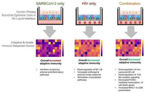




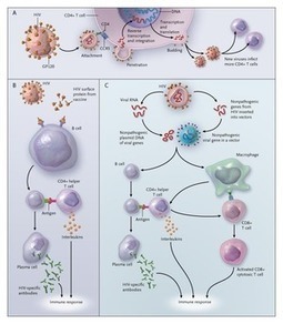

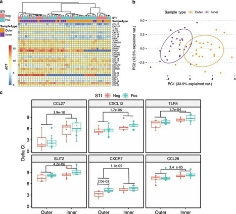
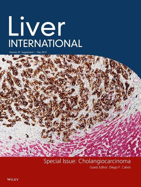

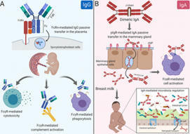

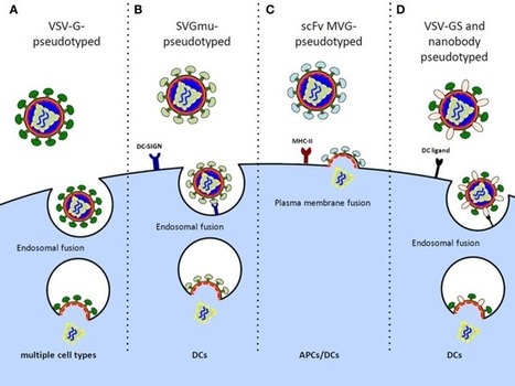

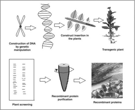

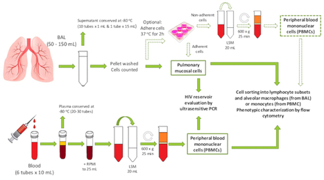






https://www.scoop.it/topic/mucosal-immunity?q=hiv