 Your new post is loading...

|
Scooped by
Gilbert C FAURE
December 22, 2020 3:15 AM
|
|
|
Scooped by
Gilbert C FAURE
December 8, 2020 12:12 PM
|
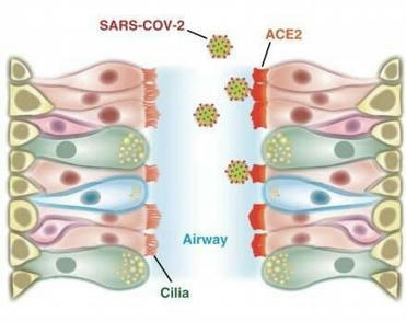
Cells in the nasal passages and upper airways are likely the coronavirus' major point of entry into the body, according to a study by Stanford Medicine researchers.
|
|
Scooped by
Gilbert C FAURE
October 25, 2020 2:50 PM
|
Several of the nasal and oral rinses have been found to have a strong ability to neutralise human coronavirus, which suggests that these products may have the potential to reduce the amount of virus spread by people who are Covid-19 positive.
|
|
Scooped by
Gilbert C FAURE
August 17, 2020 4:09 AM
|
Objectives/Hypothesis Eosinophilic and noneosinophilic chronic rhinosinusitis with nasal polyps (ECRSwNP and NECRSwNP) show distinguished clinical pathology, but their underlying mechanism remains ...
|
|
Rescooped by
Gilbert C FAURE
from Virus World
July 25, 2020 2:54 PM
|
Early and accurate detection is critical for preventing the spread of COVID-19 and providing appropriate care for patients. Nasopharyngeal (NP) swabs, which require inserting a long shaft into the nasal cavity to collect a sample from the back of the nose and throat, are currently the gold standard for collecting a specimen for diagnosis. But the procedure is technically challenging, often uncomfortable for patients and requires personal protective equipment that may be in short supply. Other approaches to collecting specimens—including from an oropharyngeal swab and sputum—have been tested in small studies, but there is uncertainty about which method is best for detecting the virus. In a new study published in EBioMedicine, investigators from Brigham and Women's Hospital conducted a systematic review and meta-analysis, analyzing data from more than 3,000 specimens to compare the three approaches. The team found that sputum testing detected the RNA of the virus that causes COVID-19 at significantly higher rates while oropharyngeal swab testing had lower rates. Regardless of the collection method, the earlier samples were collected after symptoms began, the higher the detection rate. "The accurate diagnosis of COVID-19 has implications for health care, return-to-work, infection control and public health," said corresponding author Jonathan Li, MD, a faculty member in the Division of Infectious Diseases at the Brigham. "Our gold standard in and out of the hospital is the nasopharyngeal swab, but there's a lot of confusion about which sampling modality is best and most sensitive. Our study shows that sputum testing resulted in significantly higher rates of SARS-CoV-2 detection and supports the use of this type of testing as a valuable method for the diagnosis and monitoring of COVID-19 patients." Li and his colleagues scoured the literature—both preprints and published papers—for studies that assessed at least two respiratory sampling sites using an NP swab, oropharyngeal swab or sputum. From more than 1,000 studies, they identified 11 that met their criteria. These studies included results from a total of 3,442 respiratory tract specimens. The team examined how often each collection method produced a positive result. For NP swabs, the rate was 54 percent; for oropharyngeal swabs, 43 percent; for sputum, 71 percent. The rate of viral detection was significantly higher in sputum than either oropharyngeal swabs or NP swabs. Detection rates were highest within one week of symptom onset for all three tests. Study Published in EbioMedicine (July 18, 2020): https://doi.org/10.1016/j.ebiom.2020.102903
Via Juan Lama
|
|
Scooped by
Gilbert C FAURE
July 7, 2020 11:54 AM
|
The human body is colonized by millions of microorganisms named microbiota that interact with our tissues in a cooperative and non-pathogenic manner. These microorganisms are present in the skin, gut, nasal, oral cavities, and genital tract. In fact, it has been described that the microbiota contributes to balancing the immune system to maintain host homeostasis. The gut is a vital organ where microbiota can influence and determine the function of cells of the immune system and contributes to preserve the wellbeing of the individual. Several articles have emphasized the connection between intestinal autoimmune diseases, such as Crohn's disease with dysbiosis or an imbalance in the microbiota composition in the gut. However, little is known about the role of the microbiota in autoimmune pathologies affecting other tissues than the intestine. This article focuses on what is known about the role that gut microbiota can play in the pathogenesis of non-intestinal autoimmune diseases, such as Grave's diseases, multiple sclerosis, type-1 diabetes, systemic lupus erythematosus, psoriasis, schizophrenia, and autism spectrum disorders. Furthermore, we discuss as to how metabolites derived from bacteria could be used as potential therapies for non-intestinal autoimmune diseases.
|
|
Scooped by
Gilbert C FAURE
June 5, 2020 9:36 AM
|
A new study has revealed that SARS-CoV-2 infects the nasal cavity to the greatest degree and infects and replicates progressively less well in cells lower in the respiratory tract. The work, including the development of key laboratory tools, has been accepted by Cell.
|
|
Scooped by
Gilbert C FAURE
March 8, 2020 4:59 AM
|
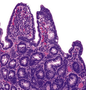
Bovine Coronavirus Related terms: View all Topics Coronaviridae In Fenner's Veterinary Virology (Fifth Edition), 2017 CORONAVIRUSES of Cattle and Horses BOVINE CORONAVIRUS Bovine coronavirus infections are associated with three distinct clinical syndromes in cattle: calf diarrhea, winter dysentery (hemorrhagic diarrhea) in adult cattle, and respiratory infections in cattle of various ages, including the bovine respiratory disease complex (shipping fever) in feedlot cattle. Coronaviruses were first reported as a cause of diarrhea in calves in the United States in 1973, and since then they have been recognized worldwide in association with the three clinical syndromes. The economic impact of respiratory disease and calf diarrhea is considerable. Although many coronaviruses have restricted host ranges, betacoronaviruses such as bovine and SARS coronaviruses (Table 24.1) can infect other animal species, including wildlife. Bovine coronavirus is closely related to the human coronavirus OC43 that causes the common cold; indeed, OC43 has been proposed to represent prior zoonotic transmission of bovine coronavirus. Bovine coronavirus has also been shown to infect dogs subclinically and to infect turkey poults, leading to fecal virus shedding, diarrhea, seroconversion, and transmission to contact controls. Genetically and/or antigenically related bovine coronavirus variants have been isolated from dogs with respiratory disease, humans with diarrhea, and captive or wild ruminants with intestinal disease similar to winter dysentery of cattle. The latter include Sambar deer (Cerous unicolor), waterbuck (Kobus ellipsiprymnus), giraffe (Giraffa camelopardalis), and white-tailed deer (Odocoileus virgineanus). Bovine coronavirus has also been linked to enteric disease in South American camelids. Interestingly, the human enteric coronavirus and wild ruminant coronaviruses both infected and caused diarrhea in experimentally exposed gnotobiotic calves, and the inoculated calves were subsequently immune to infection with bovine coronavirus. Despite the different disease syndromes and apparent interspecies transmission of bovine coronavirus and its variants, only a single serotype of bovine coronavirus is recognized, and there is little sequence diversity between the wild ruminant coronaviruses and coronaviruses associated with the different disease syndromes in cattle. Furthermore, there are few common sequence differences to explain differences in host or tissue tropism. The host cell receptor for bovine coronavirus is sialic acid, which reflects the wide tropism of this virus and explains the presence of a HE gene in the virus. Clinical Features and Epidemiology Coronavirus-induced diarrhea commonly occurs in calves under 3 weeks of age after the decline of passively acquired antibodies, but disease can occur in calves up to 3 months of age. The severity of diarrhea and dehydration depends on the infecting dose as well as the age and immune status of the calf. Coinfections with other enteric pathogens such as rotavirus, torovirus, cryptosporidia, and enterotoxigenic or enteropathogenic E. coli are common; their additive or synergistic effects increase the severity of diarrhea. Calf coronavirus diarrhea is often seasonal, being more common in winter in part because of the increased stability of the virus in the cold. Bovine coronavirus has also been implicated as a cause of winter dysentery, a sporadic, acute enteric disease of adult cattle worldwide that is especially prevalent during winter months, as the name implies. Winter dysentery is characterized by explosive, often bloody diarrhea, accompanied by decreased milk production, depression, anorexia, and frequent respiratory signs. Morbidity rates range from 20% to 100% in affected herds, but mortality rates are usually low (1–2%). A similar winter dysentery syndrome associated with bovine coronaviruses variants occurs in captive and wild ruminants. This finding suggests that certain wild ruminants (deer, elk, caribou, etc.) that share common grazing areas with cattle could be a reservoir for coronavirus strains transmissible to cattle, or vice versa. Bovine coronavirus also causes mild respiratory disease (coughing, rhinitis) or pneumonia in 2–6-month-old calves. An epidemiologic study of calves from birth to 20 weeks of age confirmed both fecal and nasal shedding of coronavirus, with diarrhea prominent upon initial infection. The calves subsequently shed virus intermittently via the respiratory route, with or without signs of disease, suggesting that long-term mucosal immunity in the upper respiratory tract is ineffective in mediating virus clearance. As a consequence, coronavirus may recycle among cattle of all ages and regardless of their immune status, with sporadic nasal or fecal shedding from individual animals. Alternatively, new virus strains may be introduced when cattle from different sources are comingled, or from cohabiting wild ruminants. Since 1993, bovine coronavirus has been incriminated as a precipitating cause of the bovine respiratory disease (shipping fever) complex. Both respiratory and enteric shedding of bovine coronavirus are common in affected feedlot cattle, peaking shortly after arrival at feedlots. Since its discovery, bovine coronavirus repeatedly has been identified in the lungs of feedlot cattle that died with bovine respiratory disease complex. Most feedlot cattle also seroconvert to bovine coronavirus within 3 weeks of arrival. Importantly, studies suggest that cattle arriving at feedlots with high serum titers of bovine coronavirus antibody were less likely to shed virus or to develop shipping fever. This observation suggests a role for serum antibodies in protection, or as an indicator of recent infection and active immunity. Pathogenesis and Pathology Concurrent fecal and nasal virus shedding persists for up to 10 days after coronavirus infection of calves. Coronavirus antigen is commonly detected in epithelial cells of both the upper respiratory and intestinal tracts, and occasionally in the lung. The pathogenesis of coronavirus enteritis in calves is similar to that caused by rotavirus, with the notable exception of extensive involvement of the large intestine by coronavirus. Disease occurs most commonly in calves at about 1–3 weeks of age, when virus exposure increases and antibody titers in the dam’s milk begin to wane. The pathogenesis and consequences of enteric coronavirus infection of calves are similar to those described for transmissible gastroenteritis in piglets. The destruction of the mature absorptive cells lining the intestinal villi and mucosal surface in the large intestine leads to maldigestion and malabsorption, with rapid loss of water and electrolytes. The resultant hypoglycemia, acidosis, and hypovolemia can progress to circulatory failure and death, especially in very young animals. The pathogenesis and lesions of winter dysentery of dairy and beef cattle resemble those of calf diarrhea, but often with marked intestinal hemorrhage and extensive necrosis of cells within the crypts of the large intestinal mucosa. Nasal and fecal shedding is more transient (up to 4–5 days). The anorexia and depression seen in dairy cattle with winter dysentery may explain the precipitous and sometimes prolonged decrease in milk production. The cause of the acute and often voluminous bloody diarrhea in some cattle is unexplained. Both nasal and fecal shedding of bovine coronavirus can occur soon after cattle are transported to feedlots. Coronavirus infection is probably important in predisposing cattle entering feedlots to secondary bacterial infection that results in the characteristic shipping fever pneumonia—a severe, often fatal fibrinous bronchopneumonia caused by Mannheimia haemolytica biotype A, serotype 1 infection. Bovine coronavirus antigen also has been detected in epithelial cells of the upper (trachea, bronchi) and lower (terminal bronchioles and alveoli) respiratory tract of some affected cattle, but the precise role of coronavirus in precipitating the bovine respiratory disease complex awaits definitive characterization. Diagnosis Initially, the diagnosis of enteric bovine coronavirus infections was based on the detection of virus by electron microscopy. Cell culture isolation became a viable option when it was discovered that the virus could be grown when trypsin was added to the medium—virus replication is recognized by hemadsorption or cytopathogenic effects, and the presence of coronavirus is confirmed by diagnostic tests. An array of assays is now available for detection of bovine (or variant) coronaviruses in cell culture or diagnostic specimens such as feces or nasal swabs, including ELISAs that incorporate monoclonal antibodies for antigen capture, immune electron microscopy using hyperimmune antiserum, and RT-PCR using bovine coronavirus or pan-coronavirus-specific primers to detect viral RNA. The use of RT-PCR for detection of bovine coronavirus has significantly increased the detection of this agent, particularly in respiratory samples, and has also substantially increased the recognized period of virus shedding by infected animals. Postmortem diagnosis is performed on acute fresh or fixed respiratory or intestinal tissues using hyperimmune antisera or monoclonal antibodies for immunofluorescence or immunohistochemical tissue staining. Immunity, Prevention, and Control Passive Immunity to Enteric Bovine Coronavirus Infections in Calves Because coronavirus diarrhea occurs in young calves during the nursing period, maternal vaccination is required to provide immediate passive (lactogenic) immunity. Passive immunity to enteric viral infections in calves correlates with high levels of IgG1 antibodies in colostrum and milk. In ruminants, IgG1 antibodies are dominant in colostrum and milk and are selectively transported from serum. Most adult cattle are seropositive for antibodies to bovine coronavirus. Therefore, parenteral vaccination of mothers with adjuvanted inactivated bovine coronavirus vaccines effectively boosts IgG1 antibody titers in serum and mammary secretions, to provide enhanced passive immunity to calves. Immunity to Respiratory Bovine Coronavirus Infections The correlates of immunity to respiratory coronavirus infections in cattle are not clearly defined. The serum antibody titer to bovine coronavirus may be a marker for respiratory protection, as coronavirus-specific antibody titers and isotype (IgG1, IgG2, IgA) were correlated with protection of calves and feedlot cattle against subsequent occurrence of respiratory disease, pneumonia, or coronavirus shedding. However, it can be difficult to distinguish whether serum antibodies are correlates of protection, or whether they merely reflect prior enteric or respiratory coronavirus infection. Intranasal vaccination using live-attenuated enteric coronavirus vaccine has been proposed to reduce the risk of bovine respiratory disease complex (so-called “shipping fever”) in cattle entering feedlots. Respiratory System Jeff L. Caswell, Kurt J. Williams, in Jubb, Kennedy & Palmer's Pathology of Domestic Animals: Volume 2 (Sixth Edition), 2016 Bovine coronavirus. Bovine coronavirus (BCoV) is an important cause of enteric disease in young calves, and the same strains occasionally induce respiratory disease in calves 2-16 weeks of age. BCoV is a less frequent but nonetheless important cause of respiratory disease in calves, and deserves more attention than it has received in the past. Isolates from occurrences of respiratory and diarrheic diseases have similar genotypes, and a single outbreak may include both forms of disease. Respiratory signs include fever, serous nasal discharge, sneezing, and coughing. The virus replicates primarily in the nasal and tracheal epithelium, and occasionally in the lung. Many calves shed BCoV in their nasal secretions and/or seroconvert to BCoV in the first month after arrival in feedlots, but the association between seroconversion or virus shedding and increased risk of respiratory disease has been variable in the studies reported. Bronchiolar necrosis is the typical lesion seen in BCoV-infected calves (eFig. 5-83). Bronchiolar syncytia have been described in feedlot calves with concurrent BCoV infection and bacterial pneumonia, but the contribution of other viruses, such as bovine respiratory syncytial virus, to these lesions is uncertain. BCoV may be demonstrated using immunohistochemistry or RT-PCR, or isolated using specific rectal tumor cell lines. Further reading Bidokhti MR, et al. Tracing the transmission of bovine coronavirus infections in cattle herds based on S gene diversity. Vet J 2012;193:386-390. Fulton RW, et al. Bovine coronavirus (BCV) infections in transported commingled beef cattle and sole-source ranch calves. Can J Vet Res 2011;75:191-199. Park SJ, et al. Dual enteric and respiratory tropisms of winter dysentery bovine coronavirus in calves. Arch Virol 2007;152:1885-1900. Storz J, et al. Isolation of respiratory bovine coronavirus, other cytocidal viruses, and Pasteurella spp from cattle involved in two natural outbreaks of shipping fever. J Am Vet Med Assoc 2000;216:1599-1604. Storz J, et al. Coronavirus and Pasteurella infections in bovine shipping fever pneumonia and Evans' criteria for causation. J Clin Microbiol 2000;38:3291-3298. Diseases of the Respiratory System In Veterinary Medicine (Eleventh Edition), 2017 Bovine Coronavirus Bovine coronavirus (BoCV) is one of the more newly identified viral respiratory pathogens of cattle, being first described in 1993. As a consequence, the clinical significance of BoCV in bovine respiratory disease, and enzootic pneumonia in particular, is still being determined. The current evidence indicates that BoCV plays a primary and important role in enzootic pneumonia. BoCV was the most commonly identified viral pathogen identified in nasal swabs from calves with respiratory disease in Ireland, being present in 23% of calves.3 BoCV was identified throughout the year, but at a much lower rate in summer.3 BoCV was the only virus detected in approximately 75% of respiratory disease outbreaks in two to 3-month calves in Italy.4 Biology and Diseases of Ruminants (Sheep, Goats, and Cattle) Wendy J. Underwood DVM, MS, DACVIM, ... Adam Schoell DVM, DACLAM, in Laboratory Animal Medicine (Third Edition), 2015 Coronavirus Bovine coronavirus, of the family Coronaviridae, produces a more severe, long-lasting disease compared to rotavirus. Clinical signs in lambs and calves are similar to above, although the incubation period tends to be shorter (20–36 h). In addition, mild respiratory disease may be noted (Janke, 1989). Coronavirus infections may be complicated by parasite infestation (e.g., Cryptosporidia, Eimeria) or bacterial infections (e.g., E. coli, Salmonella). Treatment is aimed at correcting dehydration, electrolyte imbalances, and acidosis. Strict hygiene and effective passive transfer by developing good colostrum-management protocols are critical. Bovine vaccines are available both for delivery to pre-partum dams and for the neonate. Rotaviruses, coronavirus, and adenoviruses affect neonatal goats; however, little has been documented on the pathology and significance of these agents in this age group. Unlike calves, it appears that bacteria play a more important role in neonatal kid diarrheal diseases than in neonatal calf diarrheas. Parvovirus and BVDV also may cause diarrhea in neonatal calves. Viral Diseases of the Bovine Respiratory Tract Robert W. Fulton, in Food Animal Practice (Fifth Edition), 2009 BOVINE CORONAVIRUS Bovine coronaviruses (BCVs) were initially associated with neonatal calf diarrhea.15 Then BCVs were identified with “winter dysentery” in adult dairy cattle.7,15 Later BCVs were detected in respiratory secretions of infected calves with subsequent isolation from cattle with BRD signs. This isolation of BCVs from calves with “shipping fever” pneumonias led to the assumption that BCVs were a major etiology for BRD. In some studies other agents such as BRSV, BVDV, and PI-3V along with bacteria were also found in these severely ill cattle. No doubt BCVs are found in conjunction with other respiratory tract infections, yet their sole or primary BRD role has not been clearly established. Including BCV along with other bovine respiratory tract viruses contributing to BRD is best. Clearly, experimental reproduction of detectable and severe respiratory tract disease such as pneumonia would better make the case for BCV as a significant primary pathogen in respiratory tract disease in cattle. Etiology/Epidemiology BCVs are RNA viruses of the viral family Coronaviridae.6,15 They are enveloped viruses, thus sensitive to disinfectants and the environment. It is not unexpected that cattle would have a coronavirus with tropism for the respiratory tract. Coronaviruses infect the respiratory tract of other species including humans, pigs, turkeys, and chickens. BCV infections in cattle are worldwide. Initially implicated in neonatal calf diarrhea, BCVs were also reported with etiology in “winter dysentery” of adult cattle. Subsequently BCVs have been isolated from the nasal samples of cattle undergoing respiratory tract disease.62-66 Thus this virus has a purported role in both respiratory tract disease and enteric diseases. Only one serotype is recognized, but likely there is some antigenic variability.15 The dilemma for working with BCV experimentally and diagnostic laboratories attempting to isolate the virus is that BCV replicates poorly or is quite difficult to isolate in standard cell cultures. A specialized cell line, a human rectal adenocarcinoma line, is permissive for BCV and has been used for virus isolation from feces and nasal swabs by selected laboratories. The BCV is considered relatively common in enteric infections in both beef and dairy operations. The virus has been isolated from cells with disease including calf pneumonias, as well as beef cattle entering feedlots in various U.S. regions. The BCV was isolated from both healthy and sick cattle in these BRD episodes. And BCV was detected by seroconversions during the first month in feedlots in transported cattle. Clinical Disease The association of BCV with BRD has been primarily by the isolation of virus from nasal swabs of cattle with BRD signs and seroconversions to BCV. The virus has been found in healthy calves as well. Likewise, antibody testing has detected seroconversions in cattle in BRD cases. The clinical signs in the BRD cases are not unlike other BRD cases with viral etiologies present such as BVDV; PI-3V; BRSV; and other viruses with fever, nasal and ocular discharges, anorexia, and coughing. Typically these BCV isolations and seroconversions occur soon after arrival to the feedlot. As expected there is often involvement of secondary bacteria such as M. haemolytica and/or P. multocida. Attempts have been made to demonstrate the pathogenicity of BCV for the bovine respiratory tract. After experimental challenge in young calves, the virus could be found in feces of diarrheic calves and nasal swabs for up to 5 days.15 Respiratory disease signs occurred in only a few calves. Lesions of emphysema and interstitial pneumonia were evident in only a few calves.67,68 For other studies, there are mixed reports of BCV detected in lung tissues of cattle with BRD, one report with no BCV detection in lungs of cattle with BRD,6 and another detecting BCV antigen by immunofluorescence in respiratory tissues.69 Diagnosis The virus can be isolated in cell culture provided that a unique cell culture is available to the diagnostic laboratory, the human rectal adenocarcinoma line (HRT-18).15 The nasal swabs collected appear to be the choice of collections from live cattle for testing. An antigen capture ELISA originally used for detecting BCV antigen in fecal samples is also used by some diagnostic laboratories for BCV detection in respiratory disease samples. Also, BCV immunofluorescence is available to detect BCV antigen. Selected diagnostic laboratories and research units have used PCR to detect BCV in diagnostic samples. Use of electron microscopy could detect BCV in respiratory samples similar to the use of EM for fecal samples. Selected research laboratories have used ELISA tests for BCV antibodies, and in some selected studies they found seroconversions when paired samples were available. Clinicians should consult with their respective diagnostic laboratory for their testing for BCV. Prevention and Control Although there are licensed BCV vaccines for enteric disease protection, there are no licensed BCV vaccines in the United States to control respiratory tract disease in cattle. Treatment focuses on the use of antimicrobials to control the bacterial secondary infections. As in prevention of the neonatal enteric disease, it is assumed that adequate colostrum is available to provide protection in the young calf. Diseases of the Alimentary Tract–Ruminant In Veterinary Medicine (Eleventh Edition), 2017 Synopsis Etiology Bovine coronavirus. Epidemiology Northern climates. Adult lactating dairy cows, usually during winter months when housed. Immunity develops and lasts variable periods. High morbidity with outbreaks; low mortality. Transmitted by fecal–oral route. Signs Sudden onset of diarrhea affecting almost entire herd within several days. Mild fever, decline in milk production, inappetence. Recover in few days. Some coughing. Clinical pathology None routinely. Lesions Crypt atrophy on intestinal mucosa; enterocolitis. Diagnostic confirmation Detection of virus in feces. Serology. Treatment None required. Control No specific control measures available. Hygiene. Minimize overcrowding in dairy housing. Alimentary System Francisco A. Uzal, ... Jesse M. Hostetter, in Jubb, Kennedy & Palmer's Pathology of Domestic Animals: Volume 2 (Sixth Edition), 2016 Bovine coronavirus. In neonatal calves, Bovine coronavirus (BCoV) infection is a common cause of diarrhea, either alone or in combination with other agents, particularly Rotavirus and Cryptosporidium. The disease may be severe in combination with BVDV infection. BCoV is capable of infecting absorptive epithelium in the full length of the small intestine, and in the large bowel. Viral antigen is also found in macrophages in the lamina propria of villi and in mesenteric lymph nodes. In field infections, microscopic lesions are found most consistently in the lower small intestine and colon. Calves with BCoV infection usually develop mild depression, but continue to drink milk despite developing profuse diarrhea. With progressive dehydration, acidosis, and hyperkalemia, the animals become weak and lethargic; death can ensue as a result of hypovolemia, hypoglycemia, and potassium cardiotoxicosis. Diarrhea in survivors resolves in 5-6 days. At autopsy, affected animals have the nonspecific lesions of undifferentiated neonatal calf diarrhea. Rarely, mild fibrinonecrotic typhlocolitis is recognized in calves with coronaviral infection. Mesenteric lymph nodes may be somewhat enlarged and wet. Virus replication is cytocidal and initially occurs throughout the length of the villi in all levels of the small intestine, eventually spreading throughout the large intestine up to the end of the large colon and rectum, causing a malabsorptive diarrhea. Large concentration of BCoV can be typically found in the spiral colon. Infected epithelial cells die, slough off, and are replaced by immature cells. The microscopic lesions of coronaviral infection in calves vary with the severity and duration of the infection; villus atrophy in combination with mild colitis is typical (Fig. 1-114). In the calf small intestine, villus atrophy is rarely as severe as that seen in neonatal swine with TGE. Rather, villi are moderately shortened, or have subtotal atrophy with stumpy, club-shaped, or pointed tips, and villus fusion may be common. In the early phase of the clinical disease, villi are often pointed and covered by cuboidal to squamous epithelium. Exfoliation of epithelium and microerosion may be evident. Later, the epithelium is cuboidal to low columnar, basophilic, with irregular nuclear polarity and an indistinct brush border. Cryptal epithelium is hyperplastic. The lamina propria may contain a moderate infiltrate of mainly mononuclear inflammatory cells, some of which may have pyknotic or karyorrhectic nuclei. In the early stages of infection, necrosis of cells in mesenteric lymph nodes is associated with viral replication. Peyer's patches in animals examined after 4-5 days of clinical illness often appear involuted, and are dominated by histiocytic cells. Whether this is the result of viral activity or the effect of endogenous glucocorticoids is unclear. In the colon during the early phase of infection, surface epithelium may be exfoliating, flattened, and squamous or eroded in patchy areas. Some colonic glands may be dilated, lined by flattened epithelium and contain exfoliated cells and necrotic debris. A moderate mixed inflammatory reaction is present in the lamina propria, and neutrophils may be in damaged glands or effusing into the lumen through superficial microerosions. Later in infection, some dilated debris-filled colonic glands will remain, but other glands will be lined by hyperplastic epithelium, and the surface epithelium will be restored to a cuboidal or low columnar cell type. Goblet cells are usually relatively uncommon. Colonic lesions may be recognizable in tissues from animals submitted dead, even though postmortem change has obscured changes in the small intestine. Live calves in the early stages of clinical disease are the best subjects for confirmation of an etiologic diagnosis. In calves becoming ill <7 days of age, enterotoxigenic Escherichia coli is the main alternative diagnosis. Rotavirus, Cryptosporidium, and combined infections must be considered in calves 5-15 days of age. Infectious bovine rhinotracheitis, salmonellosis, and bovine viral diarrhea must also be considered. Both salmonellosis and bovine viral diarrhea may be associated with depletion of Peyer's patches and colitis that can be confused with that of coronaviral infection; neither is common in the strictly neonatal age group (<7-14 days of age). Respiratory tract infection also occurs in calves and feeders infected with BCoV. The virus replicates in the epithelium of the nasal turbinates and tracheobronchial tree, and respiratory infection may precede, be concurrent with, or follow enteric infection. Calf pneumonia caused by BRCoV can be observed in calves 6-9 months of age. Affected animals may develop fever, serous to mucopurulent nasal discharge, coughing, tachypnea, and dyspnea. Respiratory infections may play a role in maintaining the virus within a herd, and significant, but poorly characterized, pneumonia has been reported in some experimentally infected calves. In addition, coronaviral infection may predispose to subsequent respiratory bacterial infections or contribute to more severe respiratory disease as part of the shipping fever syndrome. Virus may be identified in tissue or nasal secretions by immunofluorescence or immunohistochemistry. Winter dysentery is a syndrome in adult cattle that has been associated with BCoV in a number of areas around the world. Animals develop blood-tinged diarrhea, nasolacrimal discharge or cough, anorexia, and drop in milk production. Mortality is rare, but may occur. The disease is characterized by a high morbidity rate ranging from 50-100%, but usually low mortality rate, typically <2%. Winter dysentery outbreaks are predominantly seen in young postpartum dairy cows, which then experience a drop of 25-95% in milk production. Occasional cases are also observed in adult dairy and beef cattle. Despite its name, cases of winter dysentery can be observed, albeit infrequently, during the warmer season. The pathophysiologic characteristics of winter dysentery are mostly attributed to lesions of the colonic mucosa. Grossly, the colon of affected animals has linear congestion and hemorrhage along the crests of mucosal folds and there may be a large amount of blood mixed with colonic contents (Fig. 1-115). The histologic lesions are similar to those seen in calves with classical BCoV diarrhea, although they are mostly restricted to the colon with only occasional lesions seen in the terminal small intestine. Large amount of BCoV can be detected in colonic epithelium by immunohistochemistry. Coronaviruses are commonly demonstrated in the feces of cattle with winter dysentery; seroconversions occur, and seroprevalence increases in affected herds. Coronavirus antigen is found in the colonic glands of affected animals, in which there is necrosis and exfoliation of epithelial cells. Certain management practices, notably housing animals in stanchions and use of equipment that handles both manure and feed, have been associated with the development of winter dysentery. Further reading Blanchard PC. Diagnostics of dairy and beef cattle diarrhea. Vet Clin North Am Food Anim Pract 2012;28:443-464. Boileau MJ, et al. Bovine coronavirus associated syndromes. Vet Clin North Am Food Anim Pract 2010;26:123-146. Cho KO, et al. Detection and isolation of coronavirus from feces of three herds of feedlot cattle during outbreaks of winter dysentery-like disease. J Am Vet Med Assoc 2000;217:1191-1194. Heckert RA, et al. Epidemiologic factors and isotype-specific antibody responses in serum and mucosal secretions of dairy calves with bovine coronavirus respiratory tract and enteric tract infections. Am J Vet Res 1991;52:845-851. Kanno T. Bovine coronavirus infection: pathology and interspecies transmission. J Disast Res 2012;7:293-302. Kapil S, et al. Experimental infection with a virulent pneumoenteric isolate of bovine coronavirus. J Vet Diagn Invest 1991;3:88-89. Natsuaki S, et al. Fatal winter dysentery with severe anemia in an adult cow. J Vet Med Sci 2007;69:957-960. Park SJ, et al. Dual enteric and respiratory tropisms of winter dysentery bovine coronavirus in calves. Arch Virol 2007;152:1885-1900. Saif LJ, et al. Winter dysentery in dairy herds: electron microscopic and serological evidence for an association with coronavirus infection. Vet Rec 1991;128:447-449. Smith DR, et al. Epidemiologic herd-level assessment of causative agents and risk factors for winter dysentery in dairy cattle. Am J Vet Res 1998;59:994-1001. Traven M, et al. Experimental reproduction of winter dysentery in lactating cows using BCV—comparison with BCV infection in milk-fed calves. Vet Microbiol 2001;81:127-151. Zhang Z, et al. Application of immunohistochemistry and in situ hybridization for detection of bovine coronavirus in paraffin-embedded, formalin-fixed tissues. J Clin Microbiol 1997;35:2964-2965. Infectious Diseases of the Gastrointestinal Tract Simon F. Peek, ... Kevin J. Cummings, in Rebhun's Diseases of Dairy Cattle (Third Edition), 2018 Etiology Based on seroprevalence studies, the bovine coronavirus (BCoV) responsible for calf diarrhea is quite prevalent in U.S. cattle herds, as is rotavirus. There is much debate among researchers at this point as to whether BCoV isolates obtained from calf and adult diarrhea cases are the same virus or distinct from those that have been incriminated in respiratory disease outbreaks in feedlot and dairy calves. Whether or not there are antigenic or genomic differences in BCoV strains that mediate different organ tropism is similarly unclear. Winter dysentery in adult cattle has been associated with BCoV, and the same strain that causes diarrhea in calves has been used to experimentally create winter dysentery in adult cattle. Therefore, the upper age limit of susceptibility to infection by this agent is apparently longer than traditionally thought. Although not as common as rotavirus as a cause of viral enteritis in dairy calves, coronavirus has been identified in neonatal calf diarrhea outbreaks, especially in the winter months and with mixed infections. A number of studies indicate that clinical disease associated with BCoV in calves is more severe than rotavirus, with higher mortality rates. Affected calves tend to be slightly older than calves infected with pure ETEC or pure rotavirus. They average 7 to 10 days of age at onset, with some observed as late as 3 weeks of age. The virus causes a severe enterocolitis characterized by villous enterocyte destruction in the small intestine and destruction of both ridges and crypts in the large intestine. Maldigestion, malabsorption, and inflammation all contribute to the pathophysiology of coronavirus diarrhea in calves. The virus is cytolytic, and affected villous enterocytes in the small intestine are replaced by cuboidal cells from the crypts, but the colonic lesions leave denuded mucosa in affected areas of the colon. The severity of this damage helps explain why coronavirus enteritis, unlike rotavirus, may cause some flecks of blood to appear in the stool and can kill calves even in a germ-free isolation facility. Thus, in the natural setting, coronavirus enteritis creates a severe clinical diarrhea and can also be associated with > 50% mortality when combined with other viral, bacterial and C. parvum infections.

|
Suggested by
Société Francaise d'Immunologie
February 4, 2020 5:01 AM
|
Summary Different vaccine strains included in the live attenuated influenza vaccine (LAIV) have variable efficacy. The reasons for this are not clear and may include differences in immunogenicity. ...
|
|
Scooped by
Gilbert C FAURE
November 2, 2019 4:08 PM
|
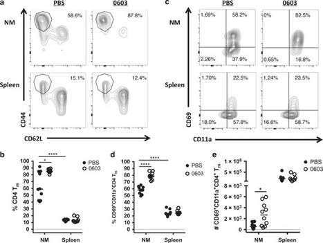
The generation of tissue-resident memory T cells (TRM) is an essential aspect of immunity at mucosal surfaces, and it has been suggested that preferential generation of TRM is one of the principal advantages of mucosally administered vaccines.
|
|
Scooped by
Gilbert C FAURE
September 14, 2019 1:15 PM
|
KEY POINTS Trout have limited IgM and IgT repertoire diversity in NALT. Intranasal vaccination in trout triggers systemic and mucosal Ig response. IgM and IgT respond to i.p. and intranasal bacterin vaccination. Abstract Bony fish represent the most basal vertebrate branch with a dedicated mucosal immune system, which comprises immunologically heterogeneous microenvironments armed with innate and adaptive components. In rainbow trout (Oncorhynchus mykiss), a nasopharynx-associated lymphoid tissue (NALT) was recently described as a diffuse network of myeloid and lymphoid cells located in the olfactory organ of fish. Several studies have demonstrated high levels of protection conferred by nasal vaccines against viral and bacterial pathogens; however, the mechanisms underlying the observed protection are not well understood. We applied 5′RACE and a deep sequencing–based approach to investigate the clonal structure of the systemic and mucosal rainbow trout B cell repertoire. The analysis of Ig repertoire in control trout suggests different structures of IgM and IgT spleen and NALT repertoires, with restricted repertoire diversity in NALT. Nasal and injection vaccination with a bacterial vaccine revealed unique dynamics of IgM and IgT repertoires at systemic and mucosal sites and the remarkable ability of nasal vaccines to induce spleen Ig responses. Our findings provide an important immunological basis for the effectiveness of nasal vaccination in fish and other vertebrate animals and will help the design of future nasal vaccination strategies. Footnotes This work was supported by USDA AFRI Grant 2DN70-2RDN7 (to I.S.). S.M. also received funding from the People Programme (Marie Curie Actions) of the European Union’s Seventh Framework Programme (FP7/2007-2013) under REA Grant Agreement 600391. The dataset presented in this article has been submitted to the National Center for Biotechnology Information (https://www.ncbi.nlm.nih.gov/sra/) under accession number PRJNA551127. The online version of this article contains supplemental material. Abbreviations used in this article: D-NALT diffuse NALT ERM enteric red mouth HSD honestly significant difference HSR highly shared responding IMGT ImMunoGeneTics i.n. intranasally MID molecular identifier NALT nasopharynx-associated lymphoid tissue O-MALT MALT with well-organized SLOs O-NALT organized NALT PBT PBS pH 7.4 with 0.05% Tween 20 SLO secondary lymphoid organ UID unique molecular identifier. Received February 7, 2019. Accepted July 9, 2019. Copyright © 2019 by The American Association of Immunologists, Inc.
|
|
Scooped by
Gilbert C FAURE
August 18, 2019 2:11 PM
|
KEY POINTS Trout have limited IgM and IgT repertoire diversity in NALT. Intranasal vaccination in trout triggers systemic and mucosal Ig response. IgM and IgT respond to i.p. and intranasal bacterin vaccination. Abstract Bony fish represent the most basal vertebrate branch with a dedicated mucosal immune system, which comprises immunologically heterogeneous microenvironments armed with innate and adaptive components. In rainbow trout (Oncorhynchus mykiss), a nasopharynx-associated lymphoid tissue (NALT) was recently described as a diffuse network of myeloid and lymphoid cells located in the olfactory organ of fish. Several studies have demonstrated high levels of protection conferred by nasal vaccines against viral and bacterial pathogens; however, the mechanisms underlying the observed protection are not well understood. We applied 5′RACE and a deep sequencing–based approach to investigate the clonal structure of the systemic and mucosal rainbow trout B cell repertoire. The analysis of Ig repertoire in control trout suggests different structures of IgM and IgT spleen and NALT repertoires, with restricted repertoire diversity in NALT. Nasal and injection vaccination with a bacterial vaccine revealed unique dynamics of IgM and IgT repertoires at systemic and mucosal sites and the remarkable ability of nasal vaccines to induce spleen Ig responses. Our findings provide an important immunological basis for the effectiveness of nasal vaccination in fish and other vertebrate animals and will help the design of future nasal vaccination strategies. Footnotes This work was supported by USDA AFRI Grant 2DN70-2RDN7 (to I.S.). S.M. also received funding from the People Programme (Marie Curie Actions) of the European Union’s Seventh Framework Programme (FP7/2007-2013) under REA Grant Agreement 600391. The dataset presented in this article has been submitted to the National Center for Biotechnology Information (https://www.ncbi.nlm.nih.gov/sra/) under accession number PRJNA551127. The online version of this article contains supplemental material. Received February 7, 2019. Accepted July 9, 2019. Copyright © 2019 by The American Association of Immunologists, Inc.
|
|
Suggested by
Société Francaise d'Immunologie
July 19, 2019 3:52 AM
|
Cell-surface mucins are expressed in apical epithelial cells of the respiratory tract, and contribute a crucial part of the innate immune system. Despite anti-inflammatory or antiviral functions being revealed for certain cell-surface mucins such as MUC1, the roles of other mucins are still poorly...
|
|
|
Scooped by
Gilbert C FAURE
December 21, 2020 8:30 AM
|
The mucosal surfaces represent the major site of entry of many pathogens, and major challenges in vaccine development include safety and stability in …...
|
|
Scooped by
Gilbert C FAURE
November 8, 2020 10:45 AM
|
Chronic rhinosinusitis (CRS) occurs in >10% of the adult population in Europe and the USA and can be differentiated into CRS without nasal polyps and CRS with nasal polyps (CRSwNP). Both phenotypes are characterized by a high disease burden and an overlapping spectrum of symptoms, with facial pain and loss of smell being the most differentiating. Great progress has been made in the understanding of CRS pathophysiology: from the epithelium and epithelial–mesenchymal transition to innate and adaptive immunity pathways and, finally, on the role of eosinophils and Staphylococcus aureus in the persistence of disease. Although clinical manifestations and diagnostic tools (including nasal endoscopy and imaging) have undergone major changes over the past few years, management (including pharmacotherapy, surgery and biologics) has experienced enormous progress based on the growing knowledge of key mediators in severe CRSwNP. The introduction of endotyping has led to a differentiation of ‘tailored’ surgical approaches, focusing on the mucosal concept in those with severe CRSwNP and on the identification of patients eligible for extended surgery and possibly biologics in the future. This Primer on chronic rhinosinusitis summarizes the epidemiology, physiology, diagnosis and management of this disorder and discusses the quality of life of patients with this condition.
|
|
Suggested by
Société Francaise d'Immunologie
September 11, 2020 3:23 PM
|
Official site of The Week Magazine, offering commentary and analysis of the day's breaking news and current events as well as arts, entertainment, people and gossip, and political cartoons.
|
|
Scooped by
Gilbert C FAURE
August 9, 2020 6:48 AM
|
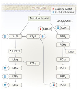
Aspirin-Exacerbated Respiratory Disease About 7% of patients with asthma have mucosal swelling, nasal polyps, and worsening of asthma in response to aspirin and other COX-1 inhibitors. Symptoms may...
|
|
Scooped by
Gilbert C FAURE
July 15, 2020 2:32 AM
|
Some experts say a vaccine puffed in the nose would be better at protecting people from infection. But nasal vaccines won’t be ready right away.
|
|
Scooped by
Gilbert C FAURE
June 25, 2020 2:54 AM
|
L’infection par le SARS-CoV-2 s’accompagne parfois d’une perte d’odorat. La bonne nouvelle ? Celui-ci devrait très probablement être de retour une fois l’infection terminée. Plus ou moins vite.
|
|
Scooped by
Gilbert C FAURE
May 2, 2020 9:21 AM
|
Investigators in a brief communication in Nature Medicine utilised the Covid19 cell atlas database to identify gene expression of the SARS-CoV-2 entry receptor ACE2 and other genes potentially associated with pathogenesis using known single-cell RNA sequencing datasets.
|
|
Scooped by
Gilbert C FAURE
February 8, 2020 9:25 AM
|
Eosinophilic chronic rhinosinusitis (ECRS) is a type of airway disease involving nasal inflammation. Many studies have attempted to understand the molecular-based pathogenesis of recurrent ECRS; none have provided a clear explanation, until now.
|
|
Scooped by
Gilbert C FAURE
January 19, 2020 3:46 AM
|
In mice, a group of nasal epithelial cells called brush cells release pro-inflammatory signals in response to allergens.
|
|
Scooped by
Gilbert C FAURE
October 25, 2019 1:18 PM
|
Chronic rhinosinusitis with nasal polyps (CRSwNP) is characterized by type 2 inflammation with accumulation of activated group 2 innate lymphoid cells (ILC2s) and elevation of thymic stromal lymphopoietin (TSLP).
|
|
Scooped by
Gilbert C FAURE
September 1, 2019 4:34 AM
|
Chronic rhinosinusitis (CRS) is defined as inflammation of the mucosal lining of the nasal cavity and paranasal sinuses that has been present for more than 3 months, causing characteristic symptoms such as nasal blockage, discharge, reduction or loss of smell sensation and facial pressure. It is a broad clinical description that encompasses several phenotypes, most often divided into CRS with polyps and CRS without polyps. CRS may be the endpoint of several different inflammatory pathways and may be secondary to specific conditions such as cystic fibrosis or fungal aetiology, but most often cannot be attributed to a single pathophysiological trigger. CRS places an enormous burden on both physicians and its sufferers. It is common among the general population, affecting approximately 11% of UK adults,1 and causes greater detriment to quality of life (QoL) than angina or chronic lower respiratory tract disease.2 Not only is it a QoL issue, patients with CRS frequently have comorbid asthma, and poorly controlled CRS is associated with more frequent asthma exacerbations and a need for hospitalisation. The socio-economic cost of CRS is significant with 57% of patients reporting absenteeism and 28% experiencing associated anxiety and depression.3 4 One per cent of all UK adults will visit their general practitioner (GP) with CRS symptoms each year and require an average of four appointments each year; despite this, one in three patients in primary care has poorly controlled symptoms.5 CRS that cannot be adequately managed in primary care may require referral to secondary care, and data from Hospital Episode Statistics show that in a single year there were approximately 120 000 outpatient consultations for CRS and 40 000 sinus operations performed in England and Wales.6 However, there remains considerable debate regarding optimum management strategies leading to wide variation in practice.7 Treatment evidence and guidelines The touchstone for treatment of CRS is The European Position Paper on Rhinosinusitis and Nasal Polyps published in 2012 (EPOS 2012).8 EPOS 2012 syntheses the best available evidence and expert opinion into easy to follow treatment flowcharts for both primary care physicians and ear, nose and throat specialists, but acknowledges the large gaps in high-quality trial data on which to base their guidance. In the intervening 7 years since EPOS was published, the International Consensus Statement on Allergy and Rhinology (ICAR)9 was agreed by a multinational working group who undertook a comprehensive overview of the literature. The working group graded the available evidence and used it as the basis for recommendations; where no evidence was available, no recommendation was made. ICAR is therefore an exhaustive insight into the current state of the science in CRS and an invaluable tool for the otolaryngology community; for primary care, however, the EPOS 2012 remains perhaps the more accessible guide. In addition, there have also been several Cochrane reviews10–16 of treatments available for CRS, and these reviews along with the international consensus guidelines have been used to synthesise a National Institute for Health and Care Excellence (NICE) clinical knowledge summary, most recently updated in 2018.17 Intranasal treatment The foundation of CRS treatment is regular use of intranasal saline irrigation and corticosteroids and this strategy finds unanimous favour among the guidelines mentioned above. The evidence is weak but consistent: a Cochrane systematic review studied the effects of saline irrigation in people with chronic sinusitis.12 This identified only two studies that could robustly be included—one which compared the effect of high volume hypertonic saline irrigation to placebo in patients with CRS both with and without polyps, and another which studied only patients with polyps and compared the use of nasal saline irrigation. This Cochrane review concluded that there was weak evidence from these trials to support the use of nasal saline irrigation. In addition, it is a low-cost treatment which with low risk of adverse events. For this reason, it is recommended for virtually all patients with CRS although in practice is usually used in combination with other therapies. Intranasal corticosteroids are similarly favoured by all expert guidelines. There is a greater number of trials on which to base this opinion. A Cochrane systematic review11 identified (search date August 2015) 18 randomised controlled trials which studied the use of intranasal steroids versus placebo in CRS. The majority of these studies included patients with nasal polyps. Regular intranasal corticosteroid use was reported to be superior to placebo in all reported symptoms, although there was no evidence to support an overall improvement in QoL. A further Cochrane review of the different formulations of intranasal corticosteroids and methods of delivery13 did not identify any single agent with convincing evidence of superiority; however, there is an advantage offered by the second-generation agents (eg, mometasone furoate, fluticasone propionate, fluticasone furoate) which have favourable pharmacokinetic characteristics that minimise systemic bioavailability (<1%) compared with older intranasal corticosteroids (eg, beclomethasone diproprionate, betamethasone). Oral corticosteroids Oral corticosteroids are commonly prescribed in the treatment of CRS although there is a distinct lack of evidence for their efficacy. The NICE Clinical Knowledge Summary (CKS) makes no reference to their use. A Cochrane review in 201616 identified no studies of their use in CRS without polyps, a fact which is also reflected in the ICAR guideline.9 Their use in patients with polyps is better established, with several studies demonstrating short-term efficacy of oral corticosteroids in improving CRS-related QoL scores. Treatment courses are short—a typical regime might be 30 mg per day of prednisolone for 2–3 weeks—and thus the risk of side effects such as adrenal suppression and loss of bone mass is minimal. However, there is a risk of side effects such as gastrointestinal disturbance and insomnia even with these short courses. Patients should also be warned of the risk of osteonecrosis—a risk which may occur at doses of steroid previously thought to be safe (lowest reported associated total dose 105 mg, median associated dose 981 mg).18 The benefit of oral corticosteroids typically last 3–6 months, and so are a reasonable choice for patients who have not improved on the first line of saline irrigation and topical corticosteroids. However, an evidence‐based risk analysis of oral corticosteroid use in CRS with polyps performed by Leung et al 19 demonstrated that risk exceeded benefit if used more frequently than once every 2 years. Oral corticosteroid use is therefore restricted to short, infrequent courses, and with full disclosure of the potential serious associated side effects for patients. Antibiotics Where there is consensus recommending the use of nasal douches and corticosteroids, there is controversy regarding the use of antibiotics. Antibiotics are one of the most commonly prescribed medicines for CRS: of the 1% of UK adults with CRS who visit their GP each year, 91% of them will receive an antibiotic prescription.20 They will often receive not just one but repeated courses of antibiotics.21 There is pressure on GPs to use antibiotics as a first-line treatment, as recently some Clinical Commissioning Groups have insisted on a 3-month trial of macrolide antibiotics prior to secondary care referral.22 With the exception of acute infective exacerbations, routine use of antibiotics is controversial for three main reasons: first; the lack of high-quality evidence to support their efficacy; second, the emerging threat of antibiotic resistance and third, the side effects which may accompany some of the most commonly used agents. In 2016, the ICAR guideline noted a paucity of evidence for antibiotic use9; however, on balance it recommended that patients with polyps should not be prescribed short courses of non-macrolide antibiotics, unless in the particular circumstances of suffering an acute exacerbation. No conclusions could be drawn on longer courses, or for patients without polyps. This echoes the findings of a Cochrane review of the use of antibiotics in CRS, which found very little evidence that non-macrolide antibiotics are effective in patients with CRS although only two studies were fit for inclusion in analysis.15 These recommendations guided the NICE CKS17 to make the recommendation to seek specialist advice before initiating long-term antibiotics in primary care. It is, however, recognised that there does remain a role for appropriate use of macrolide antibiotics in specific circumstances. In particular, there is growing interest in immune-modulating and anti-inflammatory effects of macrolides in chronic airway inflammatory disease, with low-dose long-term macrolides being prescribed for its effects on the immune response and not primarily as an anti-bacterial agent.23 The Cochrane review of 2016 noted that there was a moderate quality of evidence of a modest improvement in disease‐specific QoL in patients with CRS without nasal polyps receiving 3 months of a macrolide.15 However, this improvement was short-lived, as 3 months following the cessation of the antibiotics there was no measurable effect of treatment. Resistance Antibiotic resistance is considered one of the most significant threats to patients’ safety in Europe.24 Evaluating effectiveness of antibiotics and promoting appropriate usage is integral to the UK 5-year antimicrobial resistance strategy.25 Given the high prevalence of CRS and the variability in prescribing, overuse of inappropriate antibiotics may represent a public health danger through selective pressure on bacteria and resistance. In particular, macrolide resistance has been noted to have implications in infections secondary to Streptococcus pneumoniae (upper and lower respiratory tract infections) and Mycoplasma genitalium (sexually transmitted infections), leading to longer duration of illness and requirement for multiple antibiotic courses.26 Side effects Antibiotics are commonly associate with wide range of side effects ranging from the mild (rash, nausea, diarrhoea) to the life threatening (anaphylaxis). Specific to the use of macrolides are concerns about potential cardiovascular side effects.27 There have been a number of publications raising concerns about cardiac toxicity with erythromycin in patients with a prolonged QT interval.28 29 A 2016 study which looked at adverse outcomes associated with clarithromycin use found a significant association was found between clarithromycin use and cardiovascular mortality, where longer durations of clarithromycin use were associated with more cardiovascular events.29 By contrast, this association was not found with use of β-lactam antibiotics leading the authors to hypothesise that it was an effect specific to clarithromycin. This hypothesis was unintentionally tested by the lead investigators of the CLARICOR trial (Effect of Clarithromycin on Mortality and Morbidity in Patients With Ischemic Heart Disease) who had planned to measure the efficacy of a 2-week course of 500 mg clarithromycin two times per day to improve the clinical manifestations of stable coronary heart disease.30 The study was halted prematurely due to the finding of excess deaths in the intervention group: all-cause mortality was significantly higher in the group had taken clarithromycin, an effect which persisted for 10 years post-intervention. The use of clarithromycin in patients with known cardiovascular disease is therefore only undertaken with a significant degree of caution. Surgery Patients who are referred to secondary care who are considered to have tried and failed appropriate medical therapy – which is to say, a sufficient trial of nasal douching and topical steroids—are considered candidates for endoscopic sinus surgery (ESS).31 32 ESS is a loosely defined procedure and can range from very simple balloon expansion of the sinus openings, to extensive procedures that open every bony partition within the sinuses. The risks of surgery include epistaxis, breach of the skull base with resultant CSF leak or meningitis and injury to the intraorbital structures which may result in impaired vision. Insufficient evidence to define the role and extent of surgery contributes to a fivefold variation in UK intervention rates,33 with wide variation in surgical practice and very high rates of revision surgery.34 35 Such uncertainty resulted in inclusion of ESS in the NICE Database of Treatment Uncertainties.36 However, there can be no doubt that in appropriately selected patients, surgery offers significant and durable relief from symptoms. The largest UK study of CRS and its outcomes demonstrated that not only was there a significant improvement in patient-reported symptoms following ESS, but that patients who had surgery earlier in their disease course had a greater degree of improvement that lasted longer versus those who underwent delayed surgery.37 In addition to improving nasal symptoms, a systematic review demonstrated that ESS is associated with improved asthma control with significantly fewer attacks, hospitalisations, and a reduced requirement for medication use following sinus surgery in patients with comorbid CRS and asthma.38 Each of the international consensus guidelines referenced above recommends that patients who have tried a course of appropriate medical therapy but do not have control of their symptoms should be considered as a candidate for surgery.8 9 The path forwards In the last two decades, the majority of new treatments that have become available for CRS have in the main been refinements rather than revolutions—for example, new formulations of intranasal steroids or third-generation antihistamines. However, there are some genuinely new and exciting innovations which are on the cusp of integration to clinical practice. Monoclonal antibodies that bind to targets such as IgE, interleukin-4 (IL-4) and IL-5 were developed primarily as treatments for asthma, but clinical trials have demonstrated that several of these agents may also be effective in CRS with polyps.39 At present, they remain expensive and are limited to subsets of patients with difficult to treat asthma, but as prices fall in future and evidence for their efficacy accumulates then in future they may become available for the treatment of CRS. There have also been several promising avenues of research that may unlock further therapeutic targets, such as the role of the microbiome in sinus disease. However, as alluded to in the many international guidelines, there is a paucity of evidence for many of the treatments that are currently used in CRS before even considering new innovations. This has led to a redoubling of efforts in CRS research to produce high-quality evidence for commonly available treatments. Key to these efforts is the need to accurately define the endotypes of disease which will be studied in trials, as broad clinical phenotypes such as ‘polyps’ may encompass a wide range of underlying pathophysiology. Tightly defining the endotypes of disease for trial inclusion will permit robust examination of the response to treatment in each subgroup. Meta-analyses of CRS outcomes have been hampered by the broad clinical phenotypes, and using precise endotypes will enhance our ability to pool data from multiple trials; this will also be enabled by international consensus statements that define key criteria such appropriateness for ESS40 and standardised sets of outcome measures;41 and funding has been secured for one of the largest ever trials of interventions in CRS which will specifically examine the question of the efficacy of both macrolides and ESS (the MACRO trial42) in subgroups of patients defined by precise clinical and pathological parameters. At present, treatment of CRS exists in a time of genuine clinical equipoise where treatment is empirical and even international guidelines depend more heavily on expert opinion than robust scientific statements. Ongoing research endeavours aim to transform the treatment of CRS and usher in the era of precise, personalised medicine for these patients. Main messages Chronic rhinosinusitis (CRS) is a common disease in UK adults and causes significant detriment to life. The best evidence for effective treatment is for topical intranasal corticosteroids and saline irrigation. For patients who do not respond to this first-line treatment, there are many potential avenues for further treatment but there is controversy about which strategy is the most effective Current research questions Is it possible to identify specific endotypes of CRS to replace the broad clinical phenotypes that we currently base our treatment plans on? Are macrolide antibiotics an effective treatment in CRS? Is best medical treatment superior to surgery in the treatment of CRS? Key references Fokkens WJ, et al, European Position Paper on Rhinosinusitis and Nasal Polyps 2012. Rhinolgy 2012 3 p preceding table of contents, 1–298. Orlandi RR, et al, International Consensus Statement on Allergy and Rhinology: Rhinosinusitis. Int Forum Allergy Rhinol 2016. 6 Suppl 1: p. S22-209. NICE. Sinusitis. 2018; Available from: https://cks.nice.org.uk/sinusitis%23!scenario:1 Head K, et al, Systemic and topical antibiotics for chronic rhinosinusitis. Cochrane Database Syst Rev 2016. 4: p. CD011994 Hopkins C, et al, Long-term outcomes from the English national comparative audit of surgery for nasal polyposis and chronic rhinosinusitis. Laryngoscope 2009. 119 : p. 2459–65. Self-assessment questions Which of the following are typical symptoms of chronic rhinosinusitis? (a) Bleeding and crusting (b) Unilateral blood tinged discharge (c) Blockage, discharge, loss of sense of smell, facial pressure (d) Visual disturbance The first-line treatment for chronic rhinosinusitis (CRS) is oral steroids and antibiotics. True or false? A prolonged course of antibiotics (eg, up to 3 months) may occasionally be used in specific circumstances. What group of antibiotics has the best evidence to support their use for prolonged courses in CRS? (a) Penicillins (b) Macrolides (c) Aminoglycosides (d) Fluoroquinolones And what adverse effects should the patient be advised of with these long-term antibiotics? (a) Cardiac comorbidity (b) Osteonecrosis of the femoral head (c) Epistaxis (d) Infertility There is good evidence to support the superiority of surgical treatment of CRS compared with medical treatment. True or false? Answers Answer: (c) These are typical CRS symptoms. Bleeding and crusting should raise the possibility of vasculitis, septal perforations, or recreational cocaine use. Unilateral blood tinged discharge is a red flag sign that should prompt concerns of malignancy. Visual disturbance may be caused by rare complications of CRS, but is not at all typical. Answer: False. The first-line treatment for CRS should be topical steroids and saline irrigation. Answer: Macrolides. Answer: Cardiac comorbidity, particularly in patients with known heart disease. Osteonecrosis of the femoral head is a complication of oral steroids while topical steroids may cause epistaxis. Macrolides are not recognised to cause infertility but their use should be restricted during pregnancy. The Food and Drug Administration of USA recommend that if a macrolide must be used in pregnancy then azithromycin is category B (ie, safe in animal studies) while clarithromycin is category C (ie, shown to cause harm in animal studies). Answer: False. There are very few high-quality trials that compare medical treatment to surgery, and meta-analysis is made difficult by wide variability in study protocols. References ↵ Hastan D , Fokkens WJ , Bachert C , et al . Chronic rhinosinusitis in Europe--an underestimated disease. A GA²LEN study. Allergy 2011;66:1216–23.doi:10.1111/j.1398-9995.2011.02646.x ↵ Gliklich RE , Metson R . The health impact of chronic sinusitis in patients seeking otolaryngologic care. Otolaryngol Head Neck Surg 1995;113:104–9.doi:10.1016/S0194-5998(95)70152-4 ↵ Sahlstrand-Johnson P , Ohlsson B , Von Buchwald C , et al . A multi-centre study on quality of life and absenteeism in patients with CRS referred for endoscopic surgery. Rhinology 2011;49:420–8.doi:10.4193/Rhino11.101 OpenUrlPubMed ↵ Erskine SE , Hopkins C , Clark A , et al . Chronic rhinosinusitis and mood disturbance. Rhin 2017;55:113–9.doi:10.4193/Rhin16.111 OpenUrl ↵ Lange B , Holst R , Thilsing T , et al . Quality of life and associated factors in persons with chronic rhinosinusitis in the general population: a prospective questionnaire and clinical cross-sectional study. Clin Otolaryngol 2013;38:474–80.doi:10.1111/coa.12189 OpenUrlCrossRefPubMed ↵Hospital Episode Statistics Department of Health; 2013. ↵ Vennik J , Eyles C , Thomas M , et al . Management strategies for chronic rhinosinusitis: a qualitative study of GP and ENT specialist views of current practice in the UK. BMJ Open 2018;8:e022643.doi:10.1136/bmjopen-2018-022643 ↵ Fokkens WJ , Lund VJ , Mullol J , et al . EPOS 2012: European position paper on rhinosinusitis and nasal polyps 2012. A summary for otorhinolaryngologists. Rhinology 2012;50:1–12.doi:10.4193/Rhino50E2 OpenUrlCrossRefPubMed ↵ Orlandi RR , Kingdom TT , Hwang PH , et al . International Consensus Statement on Allergy and Rhinology: Rhinosinusitis. Int Forum Allergy Rhinol 2016;6 Suppl 1:S22–S209.doi:10.1002/alr.21695 OpenUrl ↵ Head K , Sharp S , Chong L-Y , et al . Topical and systemic antifungal therapy for chronic rhinosinusitis. Cochrane Database Syst Rev 2018;16.doi:10.1002/14651858.CD012453.pub2 ↵ Chong LY , Head K , Hopkins C , et al . Intranasal steroids versus placebo or no intervention for chronic rhinosinusitis. Cochrane Database Syst Rev 2016;115.doi:10.1002/14651858.CD011996.pub2 ↵ Chong LY , Head K , Hopkins C , et al . Saline irrigation for chronic rhinosinusitis. Cochrane Database Syst Rev 2016;67.doi:10.1002/14651858.CD011995.pub2 ↵ Chong LY , Head K , Hopkins C , et al . Different types of intranasal steroids for chronic rhinosinusitis. Cochrane Database Syst Rev 2016;25(Suppl 2).doi:10.1002/14651858.CD011993.pub2 ↵ Head K , Chong LY , Hopkins C , et al . Short-course oral steroids as an adjunct therapy for chronic rhinosinusitis. Cochrane Database Syst Rev 2016;27.doi:10.1002/14651858.CD011992.pub2 ↵ Head K , Chong LY , Piromchai P , et al . Systemic and topical antibiotics for chronic rhinosinusitis. Cochrane Database Syst Rev 2016;19.doi:10.1002/14651858.CD011994.pub2 ↵ Head K , Chong LY , Hopkins C , et al . Short-course oral steroids alone for chronic rhinosinusitis. Cochrane Database Syst Rev 2016;2.doi:10.1002/14651858.CD011991.pub2 ↵NICE. Sinusitis, 2018. Available: https://cks.nice.org.uk/sinusitis#!scenario:1 ↵ Kennedy P , Bassiouni A , Psaltis A , et al . Avascular necrosis after oral corticosteroids in otolaryngology: Case report and review of the literature. Allergy Rhinol 2016;7:50–4.doi:10.2500/ar.2016.7.0142 OpenUrl ↵ Leung RM , Dinnie K , Smith TL . When do the risks of repeated courses of corticosteroids exceed the risks of surgery? Int Forum Allergy Rhinol 2014;4:871–6.doi:10.1002/alr.21377 OpenUrl ↵ Gulliford MC , Dregan A , Moore MV , et al . Continued high rates of antibiotic prescribing to adults with respiratory tract infection: survey of 568 UK general practices. BMJ Open 2014;4:e006245.doi:10.1136/bmjopen-2014-006245 ↵ Akkerman AE , Kuyvenhoven MM , van der Wouden JC , et al . Prescribing antibiotics for respiratory tract infections by GPs: management and prescriber characteristics. Br J Gen Pract 2005;55:114–8. ↵ Soni-Jaiswal A , Philpott C , Hopkins C . The impact of commissioning for rhinosinusitis in England. Clin Otolaryngol 2015;40:639–45.doi:10.1111/coa.12430 OpenUrl ↵ Cervin A , Wallwork B . Macrolide therapy of chronic rhinosinusitis. Rhinology 2007;45:259–67. ↵ Lodato EM , Kaplan W . Priority Medicines for Europe and the World "A Public Health Approach to Innovation": Background Paper 6.1 Antimicrobial resistance World Health Organisation; 2013. ↵ Davies SC , Gibbens N . UK Five Year Antimicrobial Resistance Strategy 2013 to 2018 Department of Health and Department for Environment, Food & Rural Affairs; 2013. ↵ Gaynor M , Mankin AS . Macrolide antibiotics: binding site, mechanism of action, resistance. Curr Top Med Chem 2003;3:949–60.doi:10.2174/1568026033452159 ↵ Gibbins NE , Theokli C , Hopkins C . Time to reconsider guidelines on clarithromycin in chronic rhinosinusitis? BMJ 2013;346:f2678.doi:10.1136/bmj.f2678 ↵ Wong AYS , Chan EW , Anand S , et al . Managing Cardiovascular Risk of Macrolides: Systematic Review and Meta-Analysis. Drug Saf 2017;40:663–77.doi:10.1007/s40264-017-0533-2 OpenUrlCrossRef ↵ Cheng Y-J , Nie X-Y , Chen X-M , et al . The Role of Macrolide Antibiotics in Increasing Cardiovascular Risk. J Am Coll Cardiol 2015;66:2173–84.doi:10.1016/j.jacc.2015.09.029 OpenUrlFREE Full Text ↵ Winkel P , Hilden J , Hansen JF , et al . Clarithromycin for stable coronary heart disease increases all-cause and cardiovascular mortality and cerebrovascular morbidity over 10years in the CLARICOR randomised, blinded clinical trial. Int J Cardiol 2015;182:459–65.doi:10.1016/j.ijcard.2015.01.020 OpenUrl ↵ Baguley C , Brownlow A , Yeung K , et al . The fate of chronic rhinosinusitis sufferers after maximal medical therapy. Int Forum Allergy Rhinol 2014;4:525–32.doi:10.1002/alr.21315 OpenUrl ↵ Young LC , Stow NW , Zhou L , et al . Efficacy of medical therapy in treatment of chronic rhinosinusitis. Allergy Rhinol 2012;3:8–12.doi:10.2500/ar.2012.3.0027 OpenUrl ↵ Hopkins C . Commissioing Guide: Chronic Rhinosinusitis. Royal College of Surgeons of England London; 2016. ↵ Philpott C , Hopkins C , Erskine S , et al . The burden of revision sinonasal surgery in the UK-data from the Chronic Rhinosinusitis Epidemiology Study (CRES): a cross-sectional study. BMJ Open 2015;5:e006680.doi:10.1136/bmjopen-2014-006680 ↵ Hopkins C , Slack R , Lund V , et al . Long-term outcomes from the English national comparative audit of surgery for nasal polyposis and chronic rhinosinusitis. Laryngoscope 2009;119:2459–65.doi:10.1002/lary.20653 ↵NICE. Database of Uncertainties of Treatments. ↵ Hopkins C , Rimmer J , Lund VJ . Does time to endoscopic sinus surgery impact outcomes in Chronic Rhinosinusitis? Prospective findings from the National Comparative Audit of Surgery for Nasal Polyposis and Chronic Rhinosinusitis. Rhinology 2015;53:10–17.doi:10.4193/Rhin13.217 OpenUrl ↵ Vashishta R , Soler ZM , Nguyen SA , et al . A systematic review and meta-analysis of asthma outcomes following endoscopic sinus surgery for chronic rhinosinusitis. Int Forum Allergy Rhinol 2013;3:788–94.doi:10.1002/alr.21182 OpenUrlCrossRefPubMed ↵ Pauwels B , Jonstam K , Bachert C . Emerging biologics for the treatment of chronic rhinosinusitis. Expert Rev Clin Immunol 2015;11:349–61.doi:10.1586/1744666X.2015.1010517 OpenUrlCrossRefPubMed ↵ Rudmik L , Soler ZM , Hopkins C , et al . Defining appropriateness criteria for endoscopic sinus surgery during management of uncomplicated adult chronic rhinosinusitis: a RAND/UCLA appropriateness study. Int Forum Allergy Rhinol 2016;6:557–67.doi:10.1002/alr.21769 OpenUrl ↵ Hopkins C , Hettige R , Soni-Jaiswal A , et al . CHronic Rhinosinusitis Outcome MEasures (CHROME), developing a core outcome set for trials of interventions in chronic rhinosinusitis. Rhinology 2018;56:22–32.doi:10.4193/Rhin17.247 OpenUrl ↵ Philpott C , le Conte S , Beard D , et al . Clarithromycin and endoscopic sinus surgery for adults with chronic rhinosinusitis with and without nasal polyps: study protocol for the MACRO randomised controlled trial. Trials 2019;20:246.doi:10.1186/s13063-019-3314-7
|
|
Suggested by
Société Francaise d'Immunologie
July 25, 2019 4:00 PM
|
PubMed comprises more than 29 million citations for biomedical literature from MEDLINE, life science journals, and online books. Citations may include links to full-text content from PubMed Central and publisher web sites.
|




 Scoop.it!



















at last....