Your new post is loading...

|
Scooped by
Gilbert C FAURE
May 10, 2024 3:47 AM
|
CD4 T helper cells use the SNARE protein SYNTAXIN-11 to promote B cell differentiation, germinal center formation, and class switching by facilitating CD40
|
|
Scooped by
Gilbert C FAURE
July 20, 2023 2:52 AM
|
Petersone et al. investigate IL-21–dependent germinal center (GC) regulation and reveal key roles for IL-21 in promoting light zone GC B cell selection and cont
|
|
Scooped by
Gilbert C FAURE
January 11, 2023 5:08 AM
|
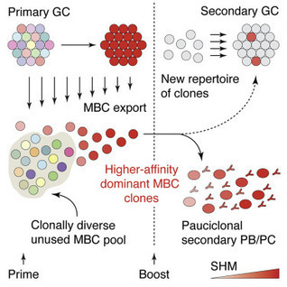
As an immune response progresses over time, germinal center reactions tend to select
for B cells producing antibodies with the highest affinity for the antigen. This work
shows that the ongoing germinal center is replenished with naive B cells that skew
the repertoire toward lower affinity, suggesting that there is a trade-off between
the overall specificity and breadth of the repertoire that can be induced.
|
|
Scooped by
Gilbert C FAURE
June 27, 2022 5:23 AM
|
Abstract T follicular helper cells (TFH) participate in germinal center (GC) development and are necessary for B cell production of high-affinity, isotype-switched antibodies. In a forward genetic screen, we identified a missense mutation in Prkd2, encoding the serine/threonine kinase protein kinase D2, which caused elevated titers of immunoglobulin E (IgE) in the serum. Subsequent analysis of serum antibodies in mice with a targeted null mutation of Prkd2 demonstrated polyclonal hypergammaglobulinemia of IgE, IgG1, and IgA isotypes, which was exacerbated by the T cell–dependent humoral response to immunization. GC formation and GC B cells were increased in Prkd2−/− spleens. These effects were the result of excessive cell-autonomous TFH development caused by unrestricted Bcl6 nuclear translocation in Prkd2−/− CD4+ T cells. Prkd2 directly binds to Bcl6, and Prkd2-dependent phosphorylation of Bcl6 is necessary to constrain Bcl6 to the cytoplasm, thereby limiting TFH development. In response to immunization, Bcl6 repressed Prkd2 expression in CD4+ T cells, thereby committing them to TFH development. Thus, Prkd2 and Bcl6 form a mutually inhibitory positive feedback loop that controls the stable transition from naïve CD4+ T cells to TFH during the adaptive immune response.
|
|
Scooped by
Gilbert C FAURE
April 15, 2021 4:55 AM
|
Positive selection of high-affinity B cells within germinal centers (GCs) drives affinity maturation of antibody responses. Here, we examined the mech…

|
Suggested by
LIGHTING
January 12, 2021 4:08 AM
|
Most of what we know about adaptive immunity has come from inbred mouse studies, using methods that are often difficult or impossible to confirm in humans. In addition, vaccine responses in mice are often poorly predictive of responses to those same vaccines in humans. Here we use human tonsils, readily available lymphoid organs, to develop a functional organotypic system that recapitulates key germinal center features in vitro, including the production of antigen-specific antibodies, somatic hypermutation and affinity maturation, plasmablast differentiation and class-switch recombination. We use this system to define the essential cellular components necessary to produce an influenza vaccine response. We also show that it can be used to evaluate humoral immune responses to two priming antigens, rabies vaccine and an adenovirus-based severe acute respiratory syndrome coronavirus 2 vaccine, and to assess the effects of different adjuvants. This system should prove useful for studying critical mechanisms underlying adaptive immunity in much greater depth than previously possible and to rapidly test vaccine candidates and adjuvants in an entirely human system. An in vitro human tonsil tissue-based system captures key features of a functional germinal center and can be used to study humoral immune responses to vaccines, new antigens and adjuvants.
|
|
Scooped by
Gilbert C FAURE
November 25, 2020 1:29 PM
|
|
|
Scooped by
Gilbert C FAURE
October 14, 2020 2:08 PM
|
Germinal centers : Adaptive immune responses against pathogenic microbes are based on the generation of effector cells and high affinity and specific antibodies that give rise to long lasting immunity and guide appropriate pathogen clearance. The presentation of antigens, with subsequent generation of antibody producing cells primarily takes place in specialized microanatomical sites within lymphoid organs such lymph nodes and spleen, known as germinal centers. These are highly dynamic structures that encompass a milieu of cells such as B and T lymphocytes, dendritic cells that interact with each other in an affinity-based selection process.
We are pleased to present a special collection of articles recently published in JEM highlighting the latest discoveries related to germinal center biology. From immune cell interaction and dynamics, to generation of germinal center immune cell subsets, gene expression regulation and chromatin remodeling and development of malignancies, we hope you will appreciate this collection. If you are interested in submitting work in this field, contact JEM Scientific Editor Montserrat Cols at mcols@rockefeller.edu. We also encourage your comments on Twitter at @JExpMed.
Image © 2020 The Francis Crick Institute
|
|
Scooped by
Gilbert C FAURE
March 17, 2020 3:53 PM
|
Grimsholm et al. show that CD27dull and CD27bright represent sequential MBC developmental
stages. T cell- and germinal center (GC)-independent CD27dull MBCs are the plastic
source of strongly selected and GC-dependent CD27bright MBCs. CD27dull MBCs, able
to expand and differentiate in response to change, ensure stability and flexibility
of human B cell memory.
|
|
Scooped by
Gilbert C FAURE
November 12, 2019 11:56 AM
|
|
|
Scooped by
Gilbert C FAURE
September 13, 2019 8:05 AM
|
The tumor necrosis factor receptor superfamily member HVEM is one of the most frequently mutated surface proteins in germinal center (GC)-derived B ce…...
Via Krishan Maggon

|
Suggested by
Société Francaise d'Immunologie
May 11, 2019 7:05 AM
|
Research ArticleImmunology Free access | 10.1172/jci.insight.126732 Differential transcriptome and development of human peripheral plasma cell subsets Swetha Garimilla,1 Doan C. Nguyen,2 Jessica L. Halliley,3 Christopher Tipton,4,5 Alexander F. Rosenberg,6 Christopher F. Fucile,7 Celia L. Saney,2 Shuya Kyu,2 Denise Kaminski,8 Yu Qian,9 Richard H. Scheuermann,9 Greg Gibson,1 Iñaki Sanz,4,5 and F. Eun-Hyung Lee2,5 First published April 2, 2019 - More info Abstract Human antibody-secreting cells (ASCs) triggered by immunization are globally recognized as CD19loCD38hiCD27hi. Yet, different vaccines give rise to antibody responses of different longevity, suggesting ASC populations are heterogeneous. We define circulating-ASC heterogeneity in vaccine responses using multicolor flow cytometry, morphology, VH repertoire, and RNA transcriptome analysis. We also tested differential survival using a human cell-free system that mimics the bone marrow (BM) microniche. In peripheral blood, we identified 3 CD19+ and 2 CD19– ASC subsets. All subsets contributed to the vaccine-specific responses and were characterized by in vivo proliferation and activation. The VH repertoire demonstrated strong oligoclonality with extensive interconnectivity among the 5 subsets and switched memory B cells. Transcriptome analysis showed separation of CD19+ and CD19– subsets that included pathways such as cell cycle, hypoxia, TNF-α, and unfolded protein response. They also demonstrated similar long-term in vitro survival after 48 days. In summary, vaccine-induced ASCs with different surface markers (CD19 and CD138) are derived from shared proliferative precursors yet express distinctive transcriptomes. Equal survival indicates that all ASC compartments are endowed with long-lived potential. Accordingly, in vivo survival of peripheral long-lived plasma cells may be determined in part by their homing and residence in the BM microniche. Graphical Abstract Introduction High-affinity IgG and IgA antibodies provide serological memory that affords protection against previously encountered pathogens. The serologic protection is mediated by long-lived plasma cells (LLPCs), which have been identified in human bone marrow (BM) and the gastrointestinal tract (1–3). Initial response to vaccination is mounted by proliferative antibody-secreting cells (ASCs), which are highly enriched for antigen-specific cells that undergo a massive expansion for approximately 5–14 days after immunization (4–7). Yet, a fundamental gap in understanding remains regarding whether intrinsic programs of the ASC or extrinsic environmental factors determine survival to become an LLPC. The classic ASC population in blood is based on the relative expression of CD38 and CD27 on CD19+ cells. However, heterogeneity of the circulating ASC populations has been described extensively (8–12). Characterization of the CD19+ ASCs has recognized both CD138+ and CD138– populations in the blood after vaccination (4, 9, 12–14) and during steady state (12). Additional markers such as HLA-DR, Ki-67, CD95, and CD126 demonstrate recent activation of the ASCs in the blood after immunization (13). However, by focusing only on CD19+ ASCs (after excluding CD20+ cells), the complexity of blood ASC subsets in healthy vaccine responses would not have considered the CD19– ASC populations that resemble LLPCs (1). In autoimmune patients, the CD19– ASCs appear in the blood of diseased patients during flares (15), and recently, CD19–ASCs were also described after vaccination of healthy adults (14). Interestingly, contrary to proposed models of the release of old PCs from BM microniches, the CD19– ASC subsets in the blood were shown to have a fraction of new ASCs generated in response to vaccination (14). The identification of CD19–CD38hiCD138+ LLPCs (1) suggests that unique surface markers may play a role in maintaining survival. For example, CD138 was shown to play a direct role in PC survival in mouse models (16). By contrast, the role of CXCR4 in long-lived survival may be related to BM homing rather than intrinsic mechanisms (11). Additionally, loss of markers such as CD19, HLA-DR, and BCR may play a role in survival, although there is little evidence for this observation. Another interpretation for the loss of some markers may actually reflect distinct changes in the intracellular pathways such as G2M checkpoints, metabolism, apoptosis, and autophagy that have been described to sustain LLPCs (1, 17, 18). Nonetheless, it is unclear whether the unique surface markers on heterogeneous ASC populations signify intrinsic differences in cell survival programs. Germinal center responses play a crucial role in LLPC generation. It is thus possible that specific blood ASCs are imprinted during priming in the germinal center by the local milieu consisting of IL-21 from T follicular helper (Tfh) cells, follicular dendritic cells, and other T cell help (19–23). Thus, ASC heterogeneity may have evolved to distinguish particular ASC subsets with unique intrinsic mechanisms that are programmed to become long-lived. In addition to intrinsic mechanisms, extrinsic factors appear to play a critical role in LLPC survival. The BM survival niche plays an important role in the maintenance of LLPCs. The specialized niche that consists of hypoxia, secreted factors from the BM mesenchymal stromal cells (MSCs), and the cytokine APRIL, has recently been shown to maintain human ASCs for over 50 days in culture (24). Whether this environment actually changes the phenotype of the peripheral circulating blood ASCs into LLPCs or merely provides survival factors is still unclear. In this study, we used FLOw Clustering without K (FLOCK), an automated flow cytometry analysis program (4), to identify 5 distinct populations of ASCs that can be consistently isolated from human blood. Our data validate 3 CD19+ and 2 newly described CD19– ASC populations after vaccination. We also show that the majority of circulating CD138+ ASCs (both CD19+ and CD19–) are active participants in new vaccine responses and have undergone recent proliferation. Next-generation sequencing (NGS) analysis of the VH repertoire shows oligoclonality with a large degree of interconnectivity among the 5 subsets, and despite unique RNA signatures distinguishing populations 2 and 3 (CD19+) from population 5 (CD19–), those 3 populations have similar long-lived survival potential. Results Heterogeneity of human ASC subsets in blood. Human antibody responses after vaccination strongly correlate with a transient increase in circulating ASCs characterized by a CD19+CD27hiCD38+ phenotype (4, 25–28). However, the exact contribution of such cells to the long-lived protective antibody production is unclear, in part due to incomplete characterization of the ASC response and over-reliance on the expression of CD19. To address these questions, we performed PBMC fractionation of CD19+ and CD19– populations within CD3–CD14– cells at the peak of the ASC response after tetanus toxoid and influenza vaccination (6–7 days after immunization) (6). Interestingly, CD19– fractions were detected, albeit smaller in frequency than the CD19+ fractions (Figure 1A). Given the high frequency of vaccine-specific ASCs in both fractions, CD19+ and CD19– cells were analyzed using flow cytometry, yielding 5 putative ASC subsets distinguished by their relative expression of CD38 and CD138; these are population (pop) 1 (CD19+CD38+CD138–), pop 2 (CD19+CD38hiCD138–), pop 3 (CD19+CD38hiCD138+), pop 4 (CD19–CD38hiCD138–), and pop 5 (CD19–CD38hiCD138+) (Figure 1A and Table 1). Figure 1 ASC subsets in human blood 7 days after tetanus vaccination. (A) Top panels divide the CD19+ and CD19– fractions. Lower panels represent subsets of CD19–IgD– (left) and CD19+IgD– (right) fractions. (B) Morphology of blood ASC subsets (×100 magnification) by Wright-Giemsa stain. Left column: Sorted blood ASC subsets on day 7 after tetanus vaccination. ASC populations (pops) 1 to 5 and naive B cells are shown. Right column: Percentage of intracellular BLIMP-1 staining per subset is shown in blue histograms (naive controls in red). (C) Percentage of each ASC subset and naive B cells (N) expressing IgG, IgA, or IgM isotypes after peak vaccination. (D) Quantification of each blood ASC subset (pops 1 to 5) in cells/ml (top) and percentage of PBMCs (bottom). (E) Quantitative RNA expression of 5,000 sorted ASC subsets and naive and memory B cells for Pax5 (top), BLIMP-1 (middle), and Xbp-1 (lower), normalized to GAPDH in blood. Relative mRNA expression is expressed in arbitrary units. Table 1 Phenotype of blood ASC subsets 1 to 5 (number, %) We also employed an unbiased automated flow-gating program, FLOCK, with CD19+ and CD19– fractions, and validated these 5 subsets. FLOCK clusters cells in multidimensional hyperdense regions for each of the markers and separates cells into different subsets if they differ from other clusters in at least one marker/dimension. Previously, we identified pop 1 within the CD19+ fractions using FLOCK (4), although it is a relatively minor fraction of the CD19+ ASCs. This unsupervised approach revealed no additional ASC subsets for 6 subjects (Supplemental Figure 1 and Supplemental Table 1; supplemental material available online with this article; https://doi.org/10.1172/jci.insight.126732DS1), demonstrating that our characterization using CD19, CD38, and CD138 is useful and consistent for elaborating the cellular basis of humoral immune responses. Morphology and expression of PC transcription factors. The nature of the ASC subsets identified by flow cytometry was validated by multiple approaches including morphology, expression of transcription factors involved in PC differentiation (BLIMP-1 and Xbp-1), and spontaneous antibody secretion. Morphology was ascertained by cytospin with Wright-Giemsa staining of each sorted ASC subset present in the blood after tetanus vaccination of 2 subjects (ages 50 and 59 years). Pops 1 to 5 had ASC characteristics including large size with big, eccentric nuclei and enlarged cytoplasm, distinct nucleoli, and a prominent Golgi zone (Figure 1B). Nearly all cells in pops 2 to 5 had intracellular BLIMP-1 protein detected by flow cytometry (Figure 1B). Interestingly, IgG isotypes were highest in ASC pops 2 to 5 except for pop 1 (Figure 1C). None were observed in naive B cells. Pop 1 had variable expression of BLIMP-1, suggesting it comprises an early or mixed population of ASCs. Pops 2 and 3 (CD19+ subsets) made up the largest frequencies of all the blood ASC subsets, and pops 4 and 5 (CD19– subsets) were the least abundant (Figure 1D). Additionally, pop 4 was quite heterogeneous; thus, additional backgating of the BLIMP-1–positive pop 4 cells showed no difference in forward or side scatter compared to other ASC populations (Supplemental Figure 2). All peripheral blood ASC populations (pops 1 to 5) were characterized by RNA expression of BLIMP-1 and Xbp-1 and absence of Pax5 (Figure 1E). In contrast, Pax5 expression was higher in naive versus memory B cells, neither of which expressed Xbp-1. BLIMP-1 was undetectable in memory B cells. The lack of Pax5 expression and increased BLIMP-1 expression in pop 1 suggests a process of differentiation from a B cell into an ASC. Xbp-1 is downstream of BLIMP-1 expression; thus, lower Xbp-1 expression observed in pop 1 compared with pops 2 to 5 also suggests that pop 1 may represent an earlier stage in ASC differentiation, or a mixed population of activated B cells that have already downregulated Pax5. A low level of BLIMP-1 expression was observed in naive B cells, which may have been due to a fraction of activated naive cells, or a small number of contaminating B1 cells, since only IgD and CD27 were used to discriminate the naive population, noting that a low level of BLIMP-1 expression has been reported in mouse B1 cells (29). Flow characteristics of blood ASC subsets. As B cells differentiate into ASCs, they undergo massive proliferation and lose features of B cells (such as CD20 and surface Ig), exit cell cycle, gain expression of BLIMP-1, and upregulate receptors for homing to the BM (30–32). Accordingly, we used multiparameter flow cytometry to evaluate these characteristics (Figure 2 and Table 1). As shown in Figure 2A, CD20 was downregulated on nearly every cell in pops 2 to 5, consistent with the loss of CD20 that begins as early as 7 days after vaccination in newly formed ASCs (4). However, pop 1 contained a mixture of cells positive for CD20, suggesting it is the earliest ASC phenotype with downregulation of Pax5 (Figure 1D). In contrast, surface Ig was highest on pop 1 and gradually downregulated in pops 2, 3, and 5 (~34%–45%; only ~8% in pop 4). These proportions of blood ASCs with surface Ig were higher than those found on LLPC (pop D) BM subsets (1). Interestingly, circulating ASC pops 2, 3, and 5 uniformly expressed high levels of CD27, a member of the tumor necrosis factor receptor (TNFR) family that is upregulated during B cell activation and is linked to PC differentiation (33), whereas the lowest frequencies of CD27 expression were found in pop 1 (~17%) (Figure 2A). Figure 2 Phenotype of blood ASC subsets on day 7 after vaccination. (A) CD20, surface Ig (kappa and lamda), and CD27 staining for blood ASC subsets and naive B cells (CD19+IgD+CD27–) illustrated in blue relative to controls in gray (also shown in right-hand panel). (B) HLA-DR and Ki-67 staining for blood ASC subsets. Far right: CD14+ peripheral blood monocytes served as controls for HLA-DR staining and naive B cells for Ki-67. (C and D) Frequency of CXCR4, CD28, IL-6R, FCGR2B, and BCMA in blood ASC subsets (pops 2 to 5) and naive B cells. Respective numbers of subjects are listed in Table 1. HLA-DR, a marker of cell activation previously shown to decrease during PC maturation (9), was highly expressed on nearly all blood ASC subsets (Figure 2B), which contrasts with its near absence on LLPC BM PC subsets (1, 2). We also found that Ki-67, a nuclear protein associated with recent cell division, was expressed by most cells in pops 2 to 5 and at a lower frequency of cells in pop 1 (Figure 2B). Thus, nearly all postvaccination ASCs in peripheral blood expressed Ki-67, consistent with the idea that these cells were recently generated, again in direct contrast to BM PCs (1). The relative expression of molecules involved in LLPC homing and survival were evaluated on blood ASCs (Figure 2C). For example, CXCR4, a chemokine receptor implicated in PC homing and retention in the BM (32, 34), was most commonly expressed in pop 1 and gradually decreased proportionately in pops 2 through 5 (Figure 2C). CD28, a costimulatory molecule associated with LLPC survival (35), which was known to have approximately 20% expression on BM LLPCs (1), was virtually absent on any of the blood ASC subsets. We previously reported that the IL-6R expression was very low on BM PC subsets despite the important role of IL-6 in PC survival (36). However, in the blood, ASC pops 3 to 5 expressed high levels of IL-6R (Figure 2C). Finally, the expression of the inhibitory FCGR2B, which promotes PC apoptosis (37), showed wide variation from subject to subject, but was typically low in blood ASC subsets compared with BM subsets (1) (Figure 2D). Lastly, B cell maturation antigen (BCMA), also known as TNFR superfamily member 17 (TNFRSF17), the receptor for APRIL, was uniformly expressed on pops 2–5 (Figure 2D), demonstrating the importance of this cytokine. Together, these results demonstrate that blood ASC subsets are Ki-67+ and HLA-DR+, suggestive of recent proliferation and activation. ASC subsets before and after vaccination in blood. Overall, compared with day 0 (~0.5%), we found that ASCs represented a larger fraction of circulating PBMCs 7 days after vaccination but with variable input from the different populations, as CD19+ (pops 1, 2, and 3) contributed in excess of 95%. This frequency is in keeping with the postvaccination levels of circulating ASCs previously established by enzyme-linked immunospot (ELISpot) analysis, which typically represent a 5- to 20-fold increase over steady-state levels (28). Therefore, we sought to determine the relative contributions of the different ASC subsets to this substantial increase after tetanus vaccination. We performed multicolor flow cytometry in 8 healthy adults prior to (i.e., steady state) and at peak vaccine responses. We removed pop 1 due to its low abundance and heterogeneity. The average total number of ASCs in each population increased after vaccination as follows: pop 2 increased by 7.6-fold, pop 3 increased 33-fold, and pop 5 increased by 13-fold. Thus, the relative abundance at steady-state and peak vaccine responses were different. During steady state, pop 2 (~60%–80%) dominated the ASC ratios with almost no pop 5; however, vaccination induced relative increases of expansions of pops 3, 4, and 5 in all 8 subjects, with corresponding decreases in pop 2 proportionally (Figure 3A). This appearance of CD19+CD138+ fractions (pop 3) for healthy adults after immunization is consistent with previous observations after tetanus vaccination (13). Figure 3 Ratios of ASC subsets in blood and BM. (A) Pie charts representing proportions of pops 2, 3, 4, and 5 in the blood from 8 different adult subjects at steady state (top) and at peak (days 6–7) ASC response in blood after tetanus vaccination. The proportion of each ASC subset is represented by the corresponding sector size of the pie chart. Kinetics of the ratios of the ASC in the blood after tetanus vaccination is shown for subject 3 (inset). (B) From 8 additional subjects, ratios of pops A, B, C, and D in the BM and blood pops 2, 3, 4, and 5 were matched at the time of the BM aspirate. ASC subsets in blood and BM (pops 1 and Z) are not included. Pops 2 and A, pops 3 and B, pops 4 and C, and pops 5 and D (LLPC subset) are shown in tan, red, blue, and green, respectively. Frequencies of ASC subsets in blood and BM. Direct comparison of the quality and quantity of the ASC subsets in 8 healthy asymptomatic adults (mean age 48.5, range 43 to 56 years) was also performed for matched blood and BM samples from the same subject. Pop A in the BM, which contains surface markers similar to those in ASC pop 2 in the blood, make up a markedly smaller percentage of BM ASC subsets compared with the blood (Figure 3B). The relative frequencies of the different ASCs were remarkably conserved in all 8 BM samples analyzed, with CD138+ (pops B and D) representing the most abundant subsets. Pop 5 could be detected in the blood in some subjects at steady state, but most of these cells were positive by Ki-67 staining (Figure 2B), thereby distinguishing them from BM pop D (Ki-67–) with the same surface markers (1). We found that at steady state, pop 5 had similar Ki-67+ staining that resembled the blood pop 5 during acute immune responses, implying that they are newly generated ASCs and not BM emigrants released in the blood from the BM niches. NGS of the VH antibody repertoire of ASC subsets. NGS was used to assess the complexity and connectivity of the antibody repertoire expressed by the different ASC subsets. Pops 1 to 5 were sorted from a 45-year-old adult after tetanus vaccination and 3,174; 5,000; 5,669; 2,550; and 1,994 cells, respectively, were collected. NGS was performed using Illumina MiSeq Amplicon sequencing with primers positioned in the framework region FR1 of the VH1–VH6 families, and in constant regions corresponding to the IgM, IgG, and IgA isotypes (Figure 4). Sequences were considered part of the same clonal lineage if they shared the same V and J gene segment rearrangement and a CDR3-H of identical length with at least 85% sequence similarity based on our previous studies (38). Total numbers of lineages for pops 1–5 were 984, 943, 825, 83, and 311, respectively. The overall degree of clonality was quantified using D50 or D20 scores calculated as the number of clonotypes accounting for the top 50% or 20%, respectively, of all size-ranked clones within a given population. For ASC pops 1 to 5 and memory B cells, the IgG D20 scores were 14, 2, 2, 1, 2, and 2, and D50 scores were 65, 4, 6, 3, 5, and 9, respectively, demonstrating that each subset at the peak of the vaccine response is dominated by a small number of substantially expanded ASC clones (Figure 4A). Switched memory (SWM) B cells were also oligoclonal, whereas the naive B cells were polyclonal with D20 and D50 scores of 910 and 3,674, respectively. The diversity of the VH-gene repertoire is demonstrated by the Hill diversity score and was highest in pop 1 compared with the other ASC subsets (Figure 4B). Finally, the composition of the isotypes of the VH sequences in the ASC subsets (Figure 4C) were switched between IgG and IgA sequences, with predominantly IgA compared with IgG in pop 1, whereas pops 2 to 5 and SWM B cells were higher in IgG. Very little IgM isotypes were found in the ASC subsets or SWM B cells, whereas naive B cells contained only IgM sequences, as expected. Figure 4 Next-generation sequencing (NGS) repertoire sequencing of blood ASC subsets. NGS was used to analyze the clonal repertoire of the ASC populations, naive B cells, and isotype-switched memory B cells. (A) Diversity of the repertoire is shown by plotting lineage (clone) size versus the cumulative percentage of sequences determined from size-ranked clones. Largest clones are found at the top of the plot and account for a greater area within the subdivided plots. More diverse repertoires, such as the naive population here, only contain small clones in a more even representation. (B) Hill diversity profiles for each population (with different levels of sampling) demonstrate the overall diversity of the repertoire in each of the ASC populations. (C) Relative quantities of IgM, IgG, and IgA sequences in each subset. Naive is predominantly IgM. Blood ASC subsets 1 to 5 and switched memory B cells show mostly IgG and IgA. (D) Circos plot shows interconnectedness of the ASC populations by plotting the sequences from each population in clonal size–ranked order, with the largest clones being in the most clockwise portion of each population segment. The outer-most track shows the isotype makeup of each clone by color. The next track in shows mutation frequency of each sequence, with more mutations represented as more distal from the center of the plot. The next track in shows the number of sequences, followed by the clonality displayed by a circular stacked bar plot. Here, only the largest 50% of the clones are colored to avoid blurring of small clones. The internal connections show clones found in multiple populations. (E) Stacked bar plots again demonstrate the diversity of the repertoire by showing size-ranked clones as segments taking up a percentage of the total repertoire. The largest 10 clones of all populations are colored and like-colors demonstrate the same clone in multiple populations. (F) The Morisita overlap index demonstrates the similarity of repertoires in various populations as a value from 0 (no similarity) to 1 (identical repertoires). The color strength is indicative of interconnectivity. Next, the actively expanded blood IgG, IgA, and IgM ASCs in response to immunization shared similar repertoires, as shown in the outer circos plot tracks (Figure 4D). Connectivity of the repertoires among subsets is also shown by the circos plot (39) (Figure 4D). Blood ASCs were largely oligoclonal with a repertoire dominated by a few clonotypes, many of which in ASC pops 2 to 5 were shared (Figures 4, D and E). The SWM B cells were also highly oligoclonal with many shared clones, in contrast to the naive B cell subset. Many of these same clones were also shared by ASCs in pop 1, but consisted of much smaller clonal populations (Figure 4E). Pops 1 to 5, as well as SWM B cells, were all highly interconnected as quantified by the high Morisita overlap indices (Figure 4F), but this was not the case comparing ASCs with naive B cells. In conclusion, the VH repertoire analysis demonstrates oligoclonality, rich interconnections, and predominately IgG isotypes among the circulating ASC subsets and SWM B cells on day 7. Mutation analysis and VH lineage analyses. On average, all blood ASC pops 1 to 5, which were mostly class switched to IgG or IgA, were highly mutated with similar average mutation frequency, as defined by the number of mutations in each sequence divided by length of that sequence (Supplemental Figure 3A). Intraclonal mutation analysis was also conducted to determine progression of ASC populations within individual clones. In this analysis, average mutation frequencies for each individual population were compared to average mutation frequencies of all populations in that same clone. By examining the mutation frequencies this way, we can get a better picture of which populations tend to have higher or lower mutation frequencies compared with other populations within the same clone. This analysis showed some deviations of the individual ASC populations’ mutation frequencies, namely that pop 4 tended to have the highest mutation frequencies in clones, while SWM and pop 2 tended to have the lowest (Supplemental Figure 3B). To follow the possibility of sequential acquisition of mutations for the different subsets, we analyzed the VH repertoire of 5 of the largest individual clones that shared lineages in pops 1 to 5 using IgTree (40) and PHYLIP phylogenetic analysis. Within the 5 clones, the number of nodes shared among the various populations of ASCs was striking. No apparent progression or sequential differentiation was found, but instead the ASC populations were highly intermixed throughout the phylogenetic tree. An example of one clone shows a deep IgTree structure with high interconnectedness between populations, illustrating the single origin of the multiple blood ASC subsets (Supplemental Figure 3C). Thus, we concluded that sequential increases in progressive accumulation of mutation did not occur from pops 1, 2, 3, 4, and 5, but each clone segregated into independent branches, demonstrating that both CD19+ and CD19– subsets arise from a common B cell progenitor. Tetanus IgG secretion in blood ASC subsets. The secretory function of different ASC subsets was validated by measurement of constitutive antibody secretion as well as participation in antigen-specific responses after tetanus vaccination. Spontaneous total IgG and tetanus-specific IgG without in vitro stimulation were assessed by ELISpot assays in 6 adults (mean age 44 ± 11 years, range 27–59 years) from blood after tetanus boosting. Cells constitutively secreting IgG were detected in all 5 ASC subsets but not every subject had adequate numbers of pops 4 and 5 for FACS analysis. Nonetheless, tetanus-specific IgG ASCs were highly enriched in both CD19+ (pops 1, 2, and 3) and CD19– (pops 4 and 5) ASC fractions when present (mean 19%, 29%, 33%, 33%, and 25% for pops 1 to 5, respectively) (Figure 5, A and B). Thus, all circulating ASCs (including the CD19– and CD138+ subsets) can contribute similarly to the short-term response to tetanus immunization. Figure 5 Vaccine-specific IgG ASC frequencies 7 days after tetanus vaccination. (A) ELISpot of total IgG (top) and tetanus-specific IgG (lower) ASC frequencies from sorted ASC subsets (pops 1 to 5) in the blood. Naive B cells and total PBMCs are also shown. The total number of sorted cells per well is indicated adjacent to each well. (B) Percentage of tetanus-specific IgG/total IgG ASC frequencies from sorted ASC subsets (pops 1–5) in blood ASC populations from 8 adults. Note: some patients had limited frequencies of pops 4 and 5, which could not be sorted. Comparative transcriptome analysis of 3 ASC subsets. The transcriptomes of pops 2, 3, and 5 were contrasted by analysis of variance of RNA sequencing (RNA-seq) profiles of 6 donors (labeled Sub 06 to 11). Two-way hierarchical clustering of 674 genes differentially expressed among the 3 ASC pops shows that pop 5 is significantly different from pops 2 and 3 (Figure 6A). This basic distinction between the CD19– and CD19+ ASCs is supported by principal component analysis (PCA), since PC1, which captures 56% of the gene expression variance, separates pops 2 and 3 from pop 5, while PC2 (7%) distinguishes pop 2 and pop 3 (Supplemental Figure 4). Furthermore, the 464 genes upregulated in both pops 2 and 3, and 210 genes downregulated relative to pop 5, lead to coclustering of the pairs of samples within individuals, implying that the 2 subtypes are transcriptionally closely related within subjects. Figure 6 Transcriptomic analysis of ASC subsets. (A) Heatmap of 672 transcripts differentially expressed among pop 2, pop 3, and pop 5 RNA-seq profiles from 6 subjects. Red color indicates relatively high expression, blue low expression. Two major blocks of genes upregulated or downregulated in pop 5 relative to pops 2 and 3 are indicated. Sample identities to the right show that pairs of samples from the same individual tend to cluster together. (B) Heatmap of PC1 of 29 gene sets found to be enriched in the differentially expressed genes also shows differentiation of pop 5 from pops 2 and 3, with only minor differences between the latter two. (C) Bubble plots show significantly biased gene sets in each pairwise comparison, with the size of the bubble proportional to the negative logarithm of the P value for the normalized enrichment score indicated along the x axis (see Supplemental Table 2 for full list of pathway names). Gene set enrichment analysis (GSEA) reinforces this similarity between pops 2 and 3 in comparison with pop 5 and highlights 29 gene sets that are differentially regulated. These gene sets summarize biological functions that are likely to differ between the pops, and are visualized in 3 different ways since raw summary statistics can misrepresent the relationship between upregulation of transcription and pathway activity. In Figure 6B (and Supplemental Figure 5), the first principal component, which in all cases explains over 45% of the variance in the gene set and has been polarized to ensure that positive values represent a preponderance of upregulated transcripts in the gene set, suggests upregulated pathways of cellular metabolism (adipogenesis, glycolysis, oxidative phosphorylation, fatty acid metabolism, and mTORC1 signaling), stress-induced pathways (DNA repair, UV response, and unfolded protein response), and cell cycle pathways (E2F targets, Myc targets, and G2M checkpoints) in pops 2 and 3. By contrast, several signaling pathways (JAK-STAT3, PI3K-AKT, IFN response, TGF-β signaling, and TNF-α signaling) as well as the hypoxic response appear to be elevated in pop 5. Figure 6C reinforces most of these conclusions by presenting the results of normalized enrichments scores for each pathway, but further reveals a gradient whereby pop 2 is more extreme than pop 3 for several pathways, namely downregulation of TNF-α signaling and upregulation of Myc and E2F target. G2M checkpoint regulators decreased expression with acquisition of CD138 and loss of CD19. This difference in dysregulated pathways suggests different cellular functions for pops 2 and 3 in comparison with pop 5. To visualize differentially expressed genes contributing to pathways, we used spider plots (Figure 7) to contrast the directions of all genes in a gene set whose transcript abundance significantly differed between 2 or more of the ASC subsets. We included some gene sets from which the hallmark pathways are derived and which were of a priori interest. For example, for apoptosis (contained within allograft rejection), hypoxia, TNF-α signaling, and the cell cycle (E2F targets, G2M checkpoints), pop 5 shows very clear upregulation of specific genes resulting in a green polygon that is more expanded along the arcs of the web, whereas the blue-colored pop 3 and pop 2 differential expression is more similar to one another, producing overlapping polygons. This analysis also indicates reduction in expression of all or most of the indicated genes encoding extracellular matrix (ECM) or unfolded protein response (UPR) functions in pop 5. It is not, however, simple to extrapolate transcript abundance to biological functions. For example, the hypoxic response engages 6 genes involved in autophagy; 3 inhibitors of the process (LDHA, PRKCB, and CYBB) are elevated in pop 2, whereas another, MAPK3, is elevated in pop 5, while PIK3CD and MTOR also regulate ER stress and autophagy, yet are expressed in opposite directions. Prediction of the consequences of differential expression awaits systems modeling that is sensitive to the precise nature of which genes are up- or downregulated jointly. Figure 7 Significantly differentially expressed genes in selected pathways. Spider plots of significantly differentially expressed genes in 6 selected pathways showing differences among the 3 ASC populations. Rays of each plot represent transcript abundance for the indicated gene, with low values in the center and high at the periphery. Polygons link observed transcript levels in each cell type, showing how pop 5 differs from pops 2 and 3. ASC survival potential in a human in vitro BM culture system. To discriminate the differential survival potential upon arrival in the BM microniche, we developed an in vitro cell-free culture system that mimics the human BM microenvironment, as previously described (24). For proof of concept using one abundant blood ASC pop 3, we performed long-term cultures and compared survival for 50 days in the BM MSC secretome alone or secretome with the addition of exogenous APRIL in normoxic or hypoxic conditions from a patient (age 23 years) 7 days after hepatitis A vaccination (Figure 8A). Pop 3 survival was barely maintained by day 50 in the secretome alone in normoxia, but a fraction of cells had better survival in secretome and hypoxia. Interestingly, survival was best overall with the secretome with the addition of exogenous APRIL in hypoxic conditions (Figure 8B). With optimal conditions for ASC survival similar to conditions previously shown with total blood ASCs (24), we sorted ASC pops 2, 3, and 5 from 2 subjects (38 and 25 years old) after tetanus toxoid or influenza vaccination, and cultured them in secretome with the addition of exogenous APRIL in hypoxia for 21 or 48 days, respectively (Figures 8, C and D). The low abundance and heterogeneity of pop 4 made them difficult to culture and maintain. The ASC subsets (pops 2, 3, and 5) were easily sustained in culture and pop 2 may have had a slight survival advantage earlier in culture (days 14–21); however, by day 48, pops 2, 3, and 5 had similar survival rates. Thus, both CD19+ (pops 2 and 3) and CD19– (pop 5) ASC subsets could be maintained in culture for nearly 2 months and all subsets have similar fractions of cells with LLPC potential. Figure 8 In vitro human BM microniche systems to measure long-lived survival of the subsets. (A) IgG ELISpots of pop 3 ASCs from a healthy adult after hepatitis A vaccine on days 0, 7, 35, and 50 in the BM MSC secretome (green) or BM MSC secretome with the addition of exogenous APRIL (red) in normoxia and hypoxia (blue open symbol or blue closed symbol). (B) Percentage of IgG ELISpots (relative to the maximal frequency) for pop 3 in MSC secretome in normoxia (green square), MSC secretome in hypoxia (open blue square), MSC secretome with the addition of exogenous APRIL in normoxia (red square), and MSC secretome with the addition of exogenous APRIL in hypoxia (blue square). (C) Percentage of IgG ELISpots from pops 2, 3, and 5 (relative to the maximal frequency for each population) from a healthy adult after tetanus vaccination on days 1, 7, and 21 in MSC secretome with the addition of exogenous APRIL in hypoxia. (D) Percentage of IgG ELISpots from pops 2, 3, and 5 (relative to the maximal frequency) from a healthy adult after influenza vaccination on days 1, 14, 28, and 48 in MSC secretome with the addition of exogenous APRIL in hypoxia. To understand if the ASC maturation required CD138 upregulation, we cultured FACS-isolated CD19+CD27hiCD38hi ASCs from a healthy adult in our BM mimic (MSC secretome, exogenous APRIL, and hypoxic conditions). We performed confocal staining of the ASCs before and 14 days after culture. We show an increased frequency of ASCs with CD138+ staining on day 14 (Supplemental Figure 6). Whether CD138– ASCs upregulated CD138 or CD138+ ASCs preferentially survived in the cultures will need additional studies. Nonetheless, increased frequencies of CD138+ ASCs are shown after culture in the BM microniche and are likely related to enhanced survival. Discussion LLPCs are the cornerstone of vaccinology and lifetime protective mediators of infection. It is widely accepted that during acute-recall immune responses, newly generated, proliferative ASCs with a short lifespan produce a transient burst of antigen-specific antibodies. After the initial response decays, pathogen-specific antibody production is sustained at lower levels by mature, nonproliferating, terminally differentiated PCs capable of surviving for many years in the BM (i.e., LLPCs) in the absence of additional antigenic stimulation. Whether all peripheral blood ASCs eventually evolve into LLPCs upon taking residence in BM survival niches, or if short-lived ASC and LLPC precursors are imprinted with a long lifespan based on the relative strength of antigenic signaling received by the corresponding precursor B cells, represent 2 distinct models (41). Discriminating between these 2 models is dependent on the ability to recognize different subsets of ASCs in the blood and their relationship with BM LLPCs. In this study, we demonstrate the heterogeneity of blood ASC subsets after vaccination by identifying both CD19+ and CD19– subsets with cell surface markers similar to BM PC subsets, as we have previously shown (1). Our results demonstrate significant heterogeneity of human ASC subsets in the blood after vaccination. These heterogeneous subsets include traditional plasmablasts (CD19+CD38hiCD27hi) and 4 newly defined ASC populations distinguished by their relative expression of the surface markers CD19, CD38, and CD138. Discovery and confirmation using FLOCK analysis (4) was essential to comprehensively identify all antibody-producing cells within these subsets. We also show that all circulating ASC subsets are active participants in recent vaccine/immune responses. Interestingly, peripheral CD19–CD138+ ASCs are universally proliferative (>90% Ki-67+) and contain antigen-specific responses with frequencies similar to conventional CD19+CD138– plasmablasts. Also, these antigen-specific ASCs with mature phenotypes are found in the periphery within a few days of immunization, arising at the same time as CD19+ ASCs. Most importantly, CD19– ASCs cannot be the result of displaced CD19– BM LLPCs, since BM LLPCs are almost universally Ki-67 negative (1). This conclusion is further supported by the high frequency of recent vaccine-specific cells and oligoclonality in the blood ASC subsets disclosed by VH repertoire studies within CD19– subsets. In contrast, VH repertoires of the BM PC subsets in healthy adults are highly polyclonal, representing an historical archive of a lifetime of exposures (1). Thus, the CD19–CD138+ ASC subsets in the blood are not displaced LLPCs from the BM microniche, but rather newer clones arising from the ongoing vaccine response. Composition of the antibody repertoire expressed by ASC and B cell subsets provides important clues regarding the origin, diversification, and selection of the compartments in question. Our results portray global repertoires expressed by multiple blood ASC subsets at the peak of vaccination, whereas previous studies had been limited to the analysis of a small number of randomly sampled single cells or global CD19+CD27hiCD38hi ASCs (instead of heterogeneous ASC subsets) (42, 43). The strong connectivity of the ASC subsets with the memory B cell fractions on day 7 may reflect the activated B cell phenotypes previously described as CD19+CD71+IgD–CD38lo/int (43). Consistent with single-cell studies, our global analyses demonstrate that responding ASCs were largely oligoclonal with highly variable rates of somatic hypermutation in the expanded clones, including some with rather low mutational load, a finding suggesting that some ASC clones might derive from recent differentiation of naive B cells. Nevertheless, the majority of expanded ASC clones were shared by all blood ASC populations and SWM B cells irrespective of the degree of mutation, which suggests they all derive from a common ancestral precursor B cell. Consistently, we also observe that, rather than forming a longitudinal gradient of progressive accumulation of mutation from pops 1 to 5 with shared VDJ clonotypes, individual clones within each ASC subset segregated into independent branches. These results indicate that all the circulating ASC subsets, including those with presumed mature phenotypes as indicated by the absence of CD19 and/or expression of CD138, derive from a common B cell progenitor. Then, they appear to develop through parallel differentiation of proliferating cells that acquire the characteristic cell surface phenotypes and divergent patterns of somatic hypermutation. Thus, ASC surface maturation (CD138 or loss of CD19) is likely independent of somatic hypermutation. Intriguingly, pop 1 had many IgA isotypes that predominated this subset with some connectivity to pops 2–5 and SWM B cells, albeit the least of all the subsets (Figure 4, A–F). Pop 1 ASCs were also the most diverse compared with the other ASC and SWM B cell subsets, suggesting that some ASCs in this population may originate differently. It is known that at steady state circulating ASCs are mainly IgA ASCs (44) (our unpublished data); however, the specificities remain unknown. Thus, pop 1 may contain both early vaccine-specific IgG ASCs and IgA ASCs from other sites. Whether they are homeostatic IgA ASCs specific to our microbial flora would be important to ascertain, but this will require further studies. Interestingly, pops 2, 3, and 5 had similar survival rates in our cell-free BM microniche system, implying equal survival potential upon arrival in a supportive niche in the body. Other models using in vitro differentiated ASCs had a massive attrition of ASCs in culture since they did not optimize conditions to mimic the human BM microniche that is the naturally occurring site of LLPC survival. Furthermore, they started with B cells that were differentiated to ASCs in vitro (14), compared with our studies that immediately cultured in vivo–differentiated ASCs, which is more physiologically relevant. Lastly, those models used MSC cell lines that are not likely to be as efficient as our validated human in vitro BM microniche system with primary BM MSC secretomes, exogenous cytokines, and hypoxic conditions to maintain human PCs (14, 24). Mouse models of LLPCs have been described in the BM demonstrating how homing and retention in the BM appears to be a major determinant for the persistence of human LLPCs (25). CXCR4 is important for PC migration and survival in the BM since BM stroma is rich in CXCL12 (45, 46). The CD19+ blood ASCs expressing higher CXCR4 levels may have potential for homing to supportive BM sites. At the same time, BM-resident LLPCs express very high levels of CXCR4, indicating that CXCL12 may also be involved in BM retention (1). Thus, with proper migration and retention, survival may be guaranteed given transcriptional programs intrinsic to the blood PCs. Since the BM-derived MSCs are an abundant source of CXCL12, whether CXCL12 also has a role in survival or just in retention will need further exploration. Our models could not discern a survival advantage of ASCs that may have intrinsic properties for BM homing because our system provides the same BM survival microniche equally to each sorted ASC subset (pops 2, 3, and 5). Therefore, the similar survival advantage of all 3 ASC subsets (Figure 8) may in fact reflect equal survival potential but not necessarily equal homing to or retention in the BM microniche. Recently, CD138, a heparin-sulfate glycoprotein (HSGP), has been shown to potentiate survival of ASCs (16). It was found that the ASCs lacking CD138 were more prone to apoptosis and reduced levels of IL-6 signaling. Evidence of increased ASC expression of surface HSGP is known to be important for the binding of survival factors, such as APRIL, hepatic growth factor (HGF), and epidermal growth factor (EGF), on malignant PCs (47–49); however, it is unclear if it is also important in normal LLPC development. More interestingly, IL-6 and APRIL protect ASCs from apoptosis in experiments using heparin sulfate chains to increase IL-6 and APRIL presentation on the ASC receptors (16). Although both CD138+ (pops 3 and 5) and CD138– (pop 2) ASC subsets had similar survival in our cultures, it was not clear if pop 2 also increased CD138 expression progressively within the in vitro BM microniche with higher concentrations of IL-6 and APRIL. These findings imply that the BM environment may play a role in altering the phenotype of an ASC through additional maturation. CD38+ ASC maturation appears to involve acquisition of CD138 in pop 3 (CD19+CD138+) and pop 5 (CD19–CD138+) cells, suggesting sequential maturation. B cells undergo massive proliferation and then differentiation to become ASCs (50). Our gene expression data indicated engagement of cell cycle, specifically G2M transition, genes among the various subsets, but it remains unclear whether pop 2 is the more proliferative subtype, as would be consistent with a maturation process that starts with pop 2 differentiating into pop 3. Furthermore, pops 2 and 3 showed elevated expression of caspase 3 and so are likely to undergo apoptosis, unless additional ASC maturation is orchestrated. Pop 5 ASCs are quite different from pops 2 and 3, with upregulation of hypoxia, TNF-α, and downregulation of UPR pathways. Because they have downregulated both oxidative phosphorylation and glycolysis, it will be interesting to understand the consequences for their survival, immune-related activity, and potential for ongoing maturation. Recent work by Neu et al. with single-cell transcriptomes of vaccine- versus non–vaccine-specific ASCs showed that the differences between these 2 subsets were in the glycosylation enzymes (51). These findings are interesting but address very different questions from our work. Our studies depict progression of ASC differentiation to a long-lived phenotype with the comparison of CD19+ and CD19– ASCs with upregulation of pathways such as hypoxia, TNF-α, and UPR involved in LLPC generation, whereas Neu et al. compared vaccine- versus non–vaccine-specific ASCs. Interestingly, the differences between vaccine- and non–vaccine-specific ASCs were relatively minor. One reason could be that similar transcriptomes of vaccine- versus non–vaccine-specific ASCs arise with short-lived (influenza) vaccines. Greater differences may have been observed between vaccine- and non–vaccine-specific ASCs in long-lived immunization such as tetanus. Nonetheless, additional studies are needed to further elucidate mechanisms of early ASCs to understand long-lived vaccine durability. Whether the local microenvironment is sufficient for LLPC generation is not known. In this study, we show that both CD19+ and CD19– ASCs have similar potential for longevity, suggesting that both extrinsic factors and intrinsic ASC programming may be essential. However, we also tested ASC subsets from vaccines with relatively long-lived protection such as tetanus (10 years), hepatitis A (25 years), and intermediate longevity for influenza vaccines (52, 53). We have not yet evaluated short-lived vaccines to test the imprinting models with differential strength of antigen signal, or costimulation with Tfh cells, to address this question. Hence, we show that the unique BM locale is necessary, but whether it is sufficient is still unclear. The improved survival in hypoxia within the BM microniche system was quite surprising. Clearly, the circulating ASCs in normoxic peripheral blood must adapt to hostile conditions of 2.5% oxygen. Whether these programs are intrinsic to the blood ASCs as they exit the germinal centers or whether they further differentiate to acclimatize to these microniches is not known. Moreover, mechanisms of how hypoxia provides a survival advantage will also need systematic evaluation of cultured ASCs. In conclusion, we find that significant heterogeneity of ASC subsets resides in the blood during active immune responses in healthy adults. Our results demonstrate that the timing of ASC subsets with markers of maturity (loss of CD19 and acquisition of CD138) are concurrent to CD19+ plasmablasts (CD19+CD27hiCD38hi) in the blood. Furthermore, both CD19+ and CD19– ASC subsets are newly generated, and all are participants of the new vaccine response. On aggregate, our results favor an ongoing evolution of ASCs once released into the peripheral blood and upon arrival to the BM niches. Thus, it is likely that the ASCs have intrinsic mechanisms of maturation (acquisition of CD138 and loss of B cells surface markers, such as CD19), which are further enhanced with extrinsic signals from the BM microniche such as IL-6 and APRIL. The surface surrogate markers may in fact be associated with important inflammatory mediators, such as IL-6, TNF-α, apoptotic pathways, along with metabolic pathway regulation to finally mature into LLPCs. Understanding these mechanisms will be important to study diseases of allergy, transplantation, and autoimmunity and to help develop better long-lived vaccines. Methods Subjects and study approval. Vaccinated and healthy asymptomatic adults (102 healthy adult subjects, 22 to 65 years old, mean 42 ± 11 years, 72 female, 30 male) were enrolled in this study at the University of Rochester Medical Center and Emory University during 2008–2017. Subjects received the tetanus toxoid Td or combination Tdap, influenza, or hepatits A vaccines as a part of routine medical care. PBMCs were isolated before vaccination, and on days 6–7 for all vaccination subjects. All studies were approved by the Institutional Review Boards at the University of Rochester Medical Center and at Emory University. Written informed consent from participants was obtained. BM aspirates. BM aspirates used for isolating ASC subsets were obtained from 8 healthy adults (43 to 56 years old, mean 49 ± 5 years, all female). Mononuclear cells were isolated by density gradient centrifugation. Peripheral blood was also collected from 8 subjects at the time of BM aspiration and PBMCs were isolated. Isolation and expansion of healthy BM-derived MSCs (from 2 unrelated healthy BM donors) and subsequent making of the MSC secretome were performed as previously described (24). Multicolor flow cytometry. PBMCs from peripheral blood or BM were isolated using a Ficoll density gradient and stained with the following anti-human antibody staining reagents: Ki-67–FITC (catalog, MHKI6701), CD3–PE-Cy5.5 (catalog, MHCD0318), CD14–PE-Cy5.5 (catalog, MHCD1418) (Invitrogen); CD20-Cy5 (catalog, 15-0209), CXCR4–PE-Cy5 (catalog, 15-9999), CD27–APC-eFluor 780 (catalog, 47-0279) (eBioscience); CD28-PE (BioLegend, catalog 302907); CD19–PE-Cy7 (catalog, 557835), IgD-PE (catalog, 555779), IL6R-PE (catalog, 561696), kappa- or lambda-PE (kappa, 555792; lambda, 555797), CD38–Pacific Blue (catalog, 561378), HLA-DR–Alexa Fluor 700 (catalog, 560743) (BD Pharmingen); CD138-APC (Miltenyi Biotec, catalog 130-091-250); and FCGR2B–Alexa Fluor 647 (custom conjugated by I. Sanz). The cells were analyzed on an LSRII flow cytometer (BD Biosciences). FLOCK analysis. FLOCK is a web-based program publically available for open use by the immunology research community through the Immunology Database and Analysis Portal—ImmPort (http://www.immport.org) (4). FLOCK is a novel multidimensional automated flow gating program that uses a density-based clustering approach to algorithmically identify cell populations from multiple samples in an unbiased fashion, thereby eliminating operator-dependent variability (4). FLOCK analysis was used for both total CD19+CD3–CD14– and CD19–CD3–CD14– PBMC populations from 6 blood samples to identify ASC subsets in an unsupervised fashion. These were subsequently isolated by sorting with cell-type specific antibodies. ASC subsets sorted by flow cytometry. CD3 and CD14 cells were removed by positive selection (CD3 and CD14 positive selection, Miltenyi Biotec; CD3, 130-050-101; CD14, 130-050-201) from the mononuclear cells isolated from blood or BM. CD3–CD14– cell fractions were stained with the following anti-human antibody staining reagents: IgD-PE (catalog, 555779), CD19–PE-Cy7 (catalog, 557835), CD38–Pacific Blue (catalog, 561378) (BD Pharmingen); CD3–PE-Cy5.5 (catalog MHCD0318), CD14–PE-Cy5.5 (catalog MHCD1418) (Invitrogen); CD138-APC (Miltenyi Biotec, catalog 130-091-250); and CD27–APC-eFluor 780 (eBioscience, catalog 47-0279). Naive and memory B cell fractions as well as multiple ASC subsets were collected (see Figure 1). Approximately 5,000 to 100,000 cells were collected for each population. Cytospins of sorted ASC subsets. Cytospins were performed from sorted ASCs in the blood at 1,300 rpm for 5 minutes on the Cytospin 4 (Thermo Fisher Scientific). Approximately 5,000 sorted cells per subset were dried overnight on albumin-coated slides and stained with Wright stain. Morphology was reviewed by a board certified pathologist and hematologist. In vitro culture systems for ASCs. In vitro cultures of human blood ASCs were performed as previously described (24). Briefly, ASCs were cultured in cell-free MSC secretome media in 96-well flat-bottom cell culture plates (Corning/Sigma) at 37°C in a humid, 5% CO2, 95% air (20% O2) incubator or in hypoxic culture conditions (2.5% O2) at 37°C in a modular incubator chamber (Billups-Rothenberg) that was infused with a preanalyzed gas mixture (AirGas) or in a cell culture incubator programmed for the desired O2 tension. The blood ASC survival and function were assessed by ELISpot assays, and their output values were expressed as the percentage of maximal IgG-secreting ASCs, which typically occurred on days 1–3. Total Ig and antigen-specific ELISpot assays. To assess survival and Ig secretion function of cultured ASCs, ELISpot assay was performed, as previously described (5, 6, 24, 28). Briefly, PBMCs, sorted ASCs, or B cell subsets were added to 96-well ELISpot plates coated with anti–human IgG (5 μg/ml, Jackson Immunoresearch), anti–human IgA (5 μg/ml, Jackson Immunoresearch), or tetanus toxoid (2 μg/ml, EMD Biosciences), and were incubated overnight. Wells were washed and bound antibodies were detected with alkaline phosphatase–conjugated anti–human IgG antibody (1 μg/ml, Jackson Immunoreseach), alkaline phosphatase–conjugated anti–human IgA antibody (1 μg/ml, Jackson Immunoreseach), and developed with a VECTOR Blue Alkaline Phosphatase Substrate Kit III (Vector Laboratories). Spots in each well were counted using the CTL immunospot reader (Cellular Technologies Ltd). Quantitative PCR for expression of select transcription factor genes from mRNA. Five thousand cells were sorted from each population as described above. Total cellular RNA was isolated using the RNeasy Mini Kit (Qiagen) by following the manufacturer’s protocol. Approximately 400 pg of RNA was reverse transcribed using the iScript RT Kit (Bio-Rad). Aliquots of the resulting single-stranded cDNA products were included with BLIMP-1, Pax5, or GAPDH TaqMan assays (assay IDs: Hs00153357_m1, Hs00172003_m1, Hs00231936_m1, and Hs02758991_g1; Life Technologies) using IQ Supermix (Bio-Rad) and amplified using a Bio-Rad CFX96 Real-time PCR Detection System for 50 cycles. Resulting Ct values were normalized to GAPDH levels and relative target mRNA for each population was calculated based on a standard curve created using total RNA from all populations. RNA transcriptome analysis. RNA was isolated from sorted ASC subsets from peripheral blood samples for 6 adult donors. RNA-seq was performed as recommended by the manufacturer (Illumina IIx). Briefly, first-hand cDNA was amplified using random hexamers. Following end repair, addition of adaptor ligation, agarose gel isolation of 200-bp cDNA, and PCR amplification of the 200-bp cDNA, the samples were sequenced on an Illumina HiSeq 2000. Over 25 million single-end sequences were obtained per sample, and aligned to the reference annotated human genome hg38 using STAR (54). Exon-level data for approximately 25,000 known gene sequences per sample were identified and consolidated using HTSeq (55) to yield gene-level expression values, then normalized to total counts per gene per million total aligned reads and subsequently TMM values in edgeR (56) that were converted to the log2 scale. The RNA-seq data have been deposited in the NCBI’s Gene Expression Omnibus database (GSE11697). Differential expression was assessed with the GLM ANOVA routine in JMP Genomics v8.0 (SAS Institute) after further normalization using the SNM procedure in R to remove batch effects with ASC population as the Biological variable and Batch as the adjustment variable with the Rm = True option (57). A lower threshold of 3 log2 units for inclusion was selected by plotting the coefficient of variance against average abundance. All analyses were qualitatively confirmed for the raw TMM values without SNM. Gene expression was visualized by hierarchical clustering using standardization of each gene across individuals and Ward’s method to weight the correlations in JMP Genomics. Pathway analysis. GSEA was performed using the Broad Institute’s preranked hallmark pathway gene lists (58, 59). For each of 29 significantly enriched pathways (FDR < 0.05; Kolgomorov-Smirnov nominal P < 0.05), the first principal component of the genes computed in R was used to visualize the overall regulation of the gene set. Because PC1 is somewhat arbitrarily signed, we compared these values with the direction of normalized enrichment and adjusted the sign to be concordant with an abundance of upregulation, contrasting pop 5 with pops 2 and 3. Because PC1 is dominated by strongly coregulated genes, it does not necessarily give the same results or significance values as normalized enrichment based on all of the genes in the pathway. An additional 6 pathways were excluded from further consideration because PC1 explained less than 20% of the variance, indicating little covariance; the 29 highlighted pathways all have PC1 explaining greater than 45% of the variance of the genes. Supplemental Figure 5 provides details for 12 gene sets (Allograft Rejection, Coagulation, Complement, DNA Repair, Heme Metabolism, IFNG Response, Mitotic Spindle, MTORC1 Signaling, Peroxisome, Spermatogenesis, UV Response Up, and Xenobiotic Metabolism) where there is some discordance between the two modes of analysis. Most cases are because PC1 is computed from both up- and downregulated genes that strongly differ among pops, whereas the enrichment score indicates the significance
|
|
|
Scooped by
Gilbert C FAURE
November 23, 2023 2:46 AM
|
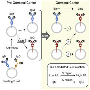
We show in polyclonal settings that GC B cell differentiation into plasma cells is affinity dependent, resulting in the generation of high-affinity serum respon
|
|
Scooped by
Gilbert C FAURE
July 13, 2023 9:38 AM
|
B cells play essential roles in immunity, mainly through the production of high affinity plasma cells (PCs) and memory B (Bmem) cells. The affinity maturation and differentiation of B cells rely on the integration of B-cell receptor (BCR) intrinsic and extrinsic signals provided by antigen binding and the microenvironment, respectively. In recent years, tumor infiltrating B (TIL-B) cells and PCs (TIL-PCs) have been revealed as important players in antitumor responses in human cancers, but their interplay and dynamics remain largely unknown. In lymphoid organs, B-cell responses involve both germinal center (GC)-dependent and GC-independent pathways for Bmem cell and PC production. Affinity maturation of BCR repertoires occurs in GC reactions with specific spatiotemporal dynamics of signal integration by B cells. In general, the reactivation of high-affinity Bmem cells by antigens triggers GC-independent production of large numbers of PC without BCR rediversification. Understanding B-cell dynamics in immune responses requires the integration of multiple tools and readouts such as single-cell phenotyping and RNA-seq, in situ analyses, BCR repertoire analysis, BCR specificity and affinity assays, and functional tests. Here, we review how those tools have recently been applied to study TIL-B cells and TIL-PC in different types of solid tumors. We assessed the published evidence for different models of TIL-B-cell dynamics involving GC-dependent or GC-independent local responses and the resulting production of antigen-specific PCs. Altogether, we highlight the need for more integrative B-cell immunology studies to rationally investigate TIL-B cells as a leverage for antitumor therapies.
|
|
Scooped by
Gilbert C FAURE
July 27, 2022 3:28 AM
|
The formation and maintenance of germinal centers is fundamental to humoral immunity
and requires interleukin 21 (IL-21). Quast et al. show that IL-21 independently regulates germinal center T and B cells and that its effects occur in paracrine, related to amount and outside the confines of the...
|
|
Scooped by
Gilbert C FAURE
April 1, 2022 7:46 AM
|
Common variable immunodeficiency (CVID), the most prevalent symptomatic primary immunodeficiency, displays impaired terminal B-cell differentiation and defective antibody responses. Incomplete genetic penetrance and ample phenotypic expressivity in CVID suggest the participation of additional pathogenic mechanisms. Monozygotic (MZ) twins discordant for CVID are uniquely valuable for studying the contribution of epigenetics to the disease. Here, we generate a single-cell epigenomics and transcriptomics census of naïve-to-memory B cell differentiation in a CVID-discordant MZ twin pair. Our analysis identifies DNA methylation, chromatin accessibility and transcriptional defects in memory B-cells mirroring defective cell-cell communication upon activation. These findings are validated in a cohort of CVID patients and healthy donors. Our findings provide a comprehensive multi-omics map of alterations in naïve-to-memory B-cell transition in CVID and indicate links between the epigenome and immune cell cross-talk. Our resource, publicly available at the Human Cell Atlas, gives insight into future diagnosis and treatments of CVID patients. Common variable immunodeficiency (CVID) is the most prevalent primary immunodeficiency. Here the authors perform single-cell omics analyses in CVID-discordant monozygotic twins and show epigenetic and transcriptional alterations associated with activation in memory B cells.
|
|
Scooped by
Gilbert C FAURE
February 8, 2021 12:40 PM
|
|
|
Scooped by
Gilbert C FAURE
December 17, 2020 12:58 PM
|
|
|
Scooped by
Gilbert C FAURE
November 18, 2020 2:40 PM
|
Abstract Tremendous progress has been made in understanding the role of T cell immunity in acute and convalescent COVID-19 infection. Here we shed light on the “known unknowns” of pre-existing and acquired T cell responses in relation to acute and convalescent SARS-CoV-2 infection. INTRODUCTION The broad clinical spectrum of COVID-19 indicates widespread intraindividual differences in the host immune defense against severe acute respiratory syndrome coronavirus 2 (SARS-CoV-2). The underlying cause of disease heterogeneity is probably multifactorial. However, a rapid early host response is likely critical to generate control of SARS-CoV-2 viremia before spread to the lower respiratory tract and onset of damaging hyperinflammation. In this regard, the literature is full of examples where functional T cell responses can provide early control of acute viral infections, including SARS-CoV and MERS-CoV (1, 2). Although multiple studies have indicated that T cells play a role in the early immune response to SARS-CoV-2 and can generate a functional memory pool, there are still multiple unanswered questions in the field (Box 1). Here, we summarize and speculate on a specific set of questions related to T cell immunity against respiratory viral infections, with a focus on COVID-19 severity, immunity, long-term consequences, and vaccination (Fig. 1). Box 1 Open questions about T cell immunity to SARS-CoV-2. T cells in acute SARS-CoV-2 infection • What do acute SARS-CoV-2-specific T cell responses in the blood tell us about contemporaneous T cell responses in the lung? • Which host and viral factors regulate the strength and efficacy of the early antiviral T cell response? Longevity and memory T cell formation • Do CD4+ T cell responses to the virus predominate over CD8+ responses in the lung as well as the blood? • Do poor CD4+ TFH responses to the virus correlate with reduced longevity of antibody responses? • Is severe COVID 19 linked to an impaired development of SARS-CoV-2-specific memory T cells? Cross-reactive T cells • Do pre-existing memory T cells to seasonal CoV epitopes help or hinder the host response to SARS-CoV-2? SARS-CoV-2-specific TRM • Will establishment of SARS-CoV-2-specific CD4+ and CD8+ TRM in the upper respiratory tract provide durable protection from lower pulmonary disease upon reinfection? Long COVID • Are virus-induced or bystander T cells major inducers of persistent symptoms of inflammation associated with long COVID? T cells in vaccines • What type of T cell response elicited by vaccines will be the best predictor of protection from disease following exposure to the virus? T CELLS IN ACUTE SARS-COV-2 INFECTION T cells are critical to generate early control and clearance of many viral infections of the respiratory system (3). Recent studies in transgenic mouse models provided evidence that T cells are also important for viral clearance and disease resolution after SARS-CoV-2 infection (4). As such, it is not surprising that T cell activation has emerged as a hallmark of acute COVID-19; probably as a consequence of an early SARS-CoV-2-specific cellular immune response (5–9). Although early T cell responses may play a critical role in dampening disease severity, there are also reports describing a dysregulated and unchecked T cell activation pattern in severe cases (10–12). Increased T cell activation in severe cases likely reflects increased antigen levels in the respiratory system, but whether the early T cell response reaches a state of exhaustion in subjects with severe hyperinflammation remains to be determined. Furthermore, given that COVID-19 is a disease of the respiratory tract it will be important to define if early detection of T cell activation in blood correlates with tissue-specific events. For instance, will delayed detection of SARS-CoV-2-specific T cells in blood reflect the later onset of cellular immunity in the respiratory tract or are these two compartments independent of each other in relation to disease severity? If elicitation of an early T cell response would be beneficial to dampen COVID-19 severity, what might be the underlying causes and correlates of an early versus late onset of SARS-CoV-2-specific T cell activity? Old age and male sex are both associated with increased risk of COVID-19 complications. Interestingly, females seem to mount a somewhat stronger T cell activation following SARS-CoV-2 infection (13) and disruption of T and B cell coordination has been implicated in elderly patients with severe COVID-19 (14). On the other end of the age spectrum, decreased frequencies of IFN-γ+CD4+ and CD25+CD4+ T cells have been described in hospitalized pediatric patients, who have shorter lengths of stay compared with their adult counterparts (15). In conjunction with age and sex, host and viral factors probably also play a role in the early immune defense and coordination of the early SARS-CoV-2-specific T cell response. For instance, SARS-CoV-2 has mechanisms to antagonize proinflammatory signals, particularly type I IFN (IFN-I) signaling (16, 17). IFN-I proteins are key inflammatory mediators to initiate antiviral defense, from which viral evasion might lead to a delayed clearance of SARS-CoV-2 (4). This is supported by the observation that inborn errors of immunity and autoantibodies that diminish IFN-I activity are more commonly detected in patients with severe COVID-19 (18, 19). Concordantly, the early expansion and differentiation of antiviral T cells are dependent on the direct action of IFN-I. Given that activated T cells from older individuals exhibit reduced responses to IFN-I, it is tempting to speculate that higher risk elderly persons experience delayed activation of SARS-CoV-2-specific T cells that may lead to reduced clearance of the virus and exacerbated COVID-19 severity. Collectively, more data are needed from mechanistic studies in animal models as well as large cohort studies on males and females in different age groups to identify beneficial and detrimental viral and host factors that have an impact on the early T cell response against SARS-CoV-2. LONGEVITY AND MEMORY T CELL FORMATION Generation of memory T cells can provide lifelong protection against pathogens (20). Previous studies have demonstrated that SARS-CoV- and MERS-CoV-specific T cells can be detected many years after infection (21–23). Likewise, SARS-CoV-2-specific CD4+ and CD8+ T cells are distinguished in a vast majority of convalescent donors (7, 9, 21, 24–27). Studies using peripheral blood have reported stronger SARS-CoV-2-specific CD4+ than CD8+ T cell responses in most subjects. However, it is well established that CD4+ T cells experience a higher propensity to recirculate between tissues and blood than CD8+ T cells. As such, whether SARS-CoV-2-specific CD4+ T cell responses also predominate in tissues, and particularly at barrier sites close to the epithelium, needs to be confirmed through studies on the upper and lower respiratory tract. Similar to the CD4+ T cell polarized response to many other viral infections, SARS-CoV-2-specific CD4+ T cells mainly possess a Th1 or circulating T follicular helper (TFH) cell phenotype (7–9, 14, 28). Circulating TFH differentiation seems to be impaired in certain patients with severe COVID-19 (11, 29) and recent analysis of postmortem lymph nodes and spleen samples showed an absence of germinal centers along with a defect in Bcl6+ TFH differentiation in deceased COVID-19 patients (30). Whether these consequences are due to sampling from postmortem patients remains unknown, but further studies are needed to clarify whether TFH cell formation is impaired by SARS-CoV-2 and could have an impact on declining antibody responses in specific convalescent donors. Furthermore, more mechanistic studies are needed to understand if memory T cells can generate protective immunity to lethal challenge with SARS-CoV-2, as previously demonstrated in SARS-CoV and MERS-CoV models (1, 2), in the presence or absence of high titers of neutralizing antibodies. Likewise, longitudinal human studies will also inform us of whether functional memory T cell responses are present many years after SARS-CoV-2 infection and correlate with protection from reinfection. CROSS-REACTIVE T CELLS Several studies have demonstrated the presence of CD4+ and to a lesser extent CD8+ T cells recognizing SARS-CoV-2 peptides in a significant proportion of unexposed individuals (7, 21, 24, 26, 31). Mapping of SARS-CoV-2 epitopes in unexposed blood donors revealed pre-existing T cell immunity, potentially induced by seasonal human coronaviruses (HCoVs) causing common colds (27, 32). This is supported by a relatively high amino acid similarity between recognized SARS-CoV-2 epitopes and seasonal HCoVs such as HCoV-OC43, -HKU1, -229E and -NL63. The presence of cross-reactive cellular immune responses in the population generates an obstacle to the use of T cell-based assays to track SARS-CoV-2 infection rates in blood donors. Given that antibodies do not result in the same degree of cross-reactivity as T cells and are consequently easier to use in clinical diagnostic settings, serology will likely be a better readout for tracing the infection rate in the society. Nevertheless, more thorough studies are needed to better understand the full spectrum of cross-reactive versus newly-induced SARS-CoV-2-specific CD4+ and CD8+ T cell responses. A key question in the field is whether pre-existing T cell responses influence the severity of COVID-19. Pre-existing SARS-CoV-2-specific T cells are unlikely to provide sterilizing or herd immunity but may allow the host to bypass immune evasion mechanisms, for instance evasion from IFN-I, and generate early pressure on the virus. This concept is supported by studies in mice showing that airway memory CD4+ T cells recognizing a conserved SARS-CoV epitope provided protection from related CoVs (1). Similar scenarios in which pre-existing T cells may provide earlier viral clearance and thus less severe symptoms have been proposed elsewhere (33). Here, the level of conservation between antigens may have a substantial impact on whether pre-existing T cells are beneficial or detrimental for the host. On the other hand, the concept of “original antigenic sin”, in which earlier induced antibody or T cell responses influence the response against future viral infections, needs further evaluation (34). If pre-existing T cells are less effective in clearing viral infection upon activation but contribute to systemic and permanent increase in inflammatory signals, it might lead to increased hyperinflammation and COVID-19 severity. In a first analysis, comparing T cell responses against SARS-CoV-2 and HCoV sequences did not find any evidence of “original antigenic sin” (32). Again, the level of conservation of targeted epitopes is likely to impact the outcome, and further evaluation of this concept is needed. Collectively, further animal studies and human studies done before and after SARS-CoV-2 infection are needed to define the biological relevance of pre-existing T cell responses and their role as friends or foes in host defense against SARS-CoV-2. SARS-COV-2-SPECIFIC TRM Resident memory T cells (TRM) are a distinct memory T cell lineage. These cells reside within tissues, do not recirculate to peripheral blood, and have been defined as local sentinels mediating rapid protection from reinfection (35). In fact, a vast majority of T cells in nonlymphoid tissues, such as the respiratory tract, are considered to be TRM (36). In terms of respiratory infections, there is a growing body of literature demonstrating that TRM can provide protection against severe pulmonary disease (37, 38). Likewise, airway CD4+ T cells can generate cross-reactive immunity between human and bat coronaviruses (1), emphasizing that cross-reactive T cells in the respiratory tract can provide protection from lethal challenge with pathogenic coronaviruses. Whether cross-reactive TRM, induced by seasonal coronaviruses, can block transmission of SARS-CoV-2 from the upper respiratory tract to the lung and thereby attenuate severe COVID-19 remains unanswered. This scenario, where TRM block the spread of viral disease from upper to lower respiratory tract, has been demonstrated in influenza A infection (37) and might account for partial immunity of secondary infection with heterologous strains (39, 40). Furthermore, whether SARS-CoV-2-specific TRM are induced after COVID-19 and whether these cells will provide protection in the long term also remains unknown (41). Although certain studies in mice have suggested that TRM in the lung are short-lived (42), there is evidence that their counterparts in the upper respiratory tract persist with minimal decay (37) and for more than a year in human lung (43). Altogether, there is currently no evidence supporting the provision of “sterilizing immunity” by TRM, but data presented above suggest that TRM could facilitate rapid control of upper respiratory tract SARS-CoV-2 infection, replication, and spread. In this regard, further work in animal models may provide evidence for whether local immunity mediated by TRM can achieve this type of immunity. LONG COVID A substantial number of COVID-19 patients experience heterogeneous symptoms that persist over a month and onward (44–46). This heterogeneous phenomenon is being referred to as “long COVID” and affects around 10% of all COVID-19 patients (44, 45). Many symptoms can be attributed to persistent tissue damage in severe COVID-19. Nevertheless, the fact that many individuals with milder COVID-19 symptoms also experience chronic lingering symptoms, involving the cardiovascular, nervous, and respiratory systems, indicates that persistent immune activation and/or inflammation may play a role in long COVID. Multiple mechanisms are probably involved in this condition and whether T cells play any role in long COVID is unknown. The higher incidence of long COVID in females than males, similar to autoimmune diseases (47), raises the question of whether T cells orchestrate long COVID through similar mechanisms as in autoimmune or inflammatory conditions (48, 49). One hypothetical underlying mechanism behind autoimmune-related conditions after COVID-19 could be molecular mimicry, given that HCoV-specific T cells can cross-react to myelin in multiple sclerosis patients (50). Whether SARS-CoV-2-specific T cells have the ability to react against self-antigens remains to be determined. In line with a possible effect of HLA type on COVID-19 susceptibility/severity (51, 52), we believe that larger genetic studies are needed to clarify if HLA or other immune-related genes are associated with an increased risk of developing long COVID. T CELLS IN VACCINES Based on the uncertainty of whether cross-reactive T cells or antibodies will provide protective or long-lasting immunity to COVID-19, it will become absolutely critical to administrate a safe and effective vaccine to the population to reach broad immunity and break the negative spiral of new infections. Ongoing vaccine efforts mainly target B cells to promote the induction of neutralizing antibodies (nAbs) against SARS-CoV-2 (53, 54). Although the induction of anti-spike nAbs is the key component for an effective SARS-CoV-2 vaccine, it is well-known that T cells, and in particular TFH cells, are critical to generate antibody-producing plasma cells and long-lived memory B cells. In COVID-19 patients, high nAb titers correlated with strong CD4+ T cell responses, and the lack of functional TFH cells reacting against SARS-CoV-2 was shown to be detrimental (11, 29, 30). Preliminary results from the two major mRNA vaccine trials in humans have demonstrated potent Th1 responses (55, 56). However, previous studies have reported strong TFH responses against certain mRNA vaccines (57), and future trials should therefore include other activation induced markers, such as CD40L and/or CD200, in addition to IFN-γ ELISPOT assays to understand if potent B-helper mechanisms are induced by the current vaccine regimens. Other outstanding questions are whether vaccine-induced TFH responses will be equally induced in all age groups and how long these responses will persist in blood and vaccination site-draining lymph nodes. A final issue to consider is whether high quantities of vaccine-induced CD8+ T cells at local sites need to be elicited by future vaccine candidates. If the initial group of vaccines in clinical trials that are primarily focused on generating an effective nAb response provide recipients with long-standing protection, it may not be necessary to invest in such efforts. However, if problems emerge in the vaccinated population with breakthrough infections, waning antibody levels after vaccination, and/or the emergence of new viral strains, it would be wise to reconsider vaccine approaches specifically designed to induce functional CD8+ TRM responses in the upper respiratory tract. SUMMARY Collective efforts have greatly enhanced our scientific understanding of T cell responses against SARS-CoV-2 but many unknowns remain to be resolved. Although it is clear that T cells play a central role in generating early control and clearance of many viral infections, their role in SARS-CoV-2 infection is only starting to be revealed. Specific T cells may even have a detrimental impact on the clinical outcome and contribute to long COVID symptoms. Currently, there is a need for deeper analysis using both animal models and longitudinal follow-up studies of large patient cohorts to define the beneficial versus detrimental aspects of SARS-CoV-2-specific T cells in acute, convalescent and vaccine settings of COVID-19. References and Notes ↵ J. Zhao, J. Zhao, A. K. Mangalam, R. Channappanavar, C. Fett, D. K. Meyerholz, S. Agnihothram, R. S. Baric, C. S. David, S. Perlman, Airway Memory CD4(+) T Cells Mediate Protective Immunity against Emerging Respiratory Coronaviruses. Immunity 44, 1379–1391 (2016). doi:10.1016/j.immuni.2016.05.006pmid:27287409OpenUrlCrossRefPubMed ↵ J. Zhao, J. Zhao, S. Perlman, T cell responses are required for protection from clinical disease and for virus clearance in severe acute respiratory syndrome coronavirus-infected mice. J. Virol. 84, 9318–9325 (2010). doi:10.1128/JVI.01049-10pmid:20610717 ↵ M. E. Schmidt, S. M. Varga, The CD8 T Cell Response to Respiratory Virus Infections. Front. Immunol. 9, 678 (2018). doi:10.3389/fimmu.2018.00678pmid:29686673OpenUrlCrossRefPubMed ↵ J. Sun, Z. Zhuang, J. Zheng, K. Li, R. L.-Y. Wong, D. Liu, J. Huang, J. He, A. Zhu, J. Zhao, X. Li, Y. Xi, R. Chen, A. N. Alshukairi, Z. Chen, Z. Zhang, C. Chen, X. Huang, F. Li, X. Lai, D. Chen, L. Wen, J. Zhuo, Y. Zhang, Y. Wang, S. Huang, J. Dai, Y. Shi, K. Zheng, M. R. Leidinger, J. Chen, Y. Li, N. Zhong, D. K. Meyerholz, P. B. McCray Jr.., S. Perlman, J. Zhao, Generation of a Broadly Useful Model for COVID-19 Pathogenesis, Vaccination, and Treatment. Cell 182, 734–743.e5 (2020). doi:10.1016/j.cell.2020.06.010pmid:32643603OpenUrlCrossRefPubMed ↵ I. Thevarajan, T. H. O. Nguyen, M. Koutsakos, J. Druce, L. Caly, C. E. van de Sandt, X. Jia, S. Nicholson, M. Catton, B. Cowie, S. Y. C. Tong, S. R. Lewin, K. Kedzierska, Breadth of concomitant immune responses prior to patient recovery: A case report of non-severe COVID-19. Nat. Med. 26, 453–455 (2020). doi:10.1038/s41591-020-0819-2pmid:32284614OpenUrlCrossRefPubMed A. J. Wilk, A. Rustagi, N. Q. Zhao, J. Roque, G. J. Martínez-Colón, J. L. McKechnie, G. T. Ivison, T. Ranganath, R. Vergara, T. Hollis, L. J. Simpson, P. Grant, A. Subramanian, A. J. Rogers, C. A. Blish, A single-cell atlas of the peripheral immune response in patients with severe COVID-19. Nat. Med. 26, 1070–1076 (2020). doi:10.1038/s41591-020-0944-ypmid:32514174OpenUrlCrossRefPubMed ↵ T. Sekine, A. Perez-Potti, O. Rivera-Ballesteros, K. Strålin, J.-B. Gorin, A. Olsson, S. Llewellyn-Lacey, H. Kamal, G. Bogdanovic, S. Muschiol, D. J. Wullimann, T. Kammann, J. Emgård, T. Parrot, E. Folkesson, O. Rooyackers, L. I. Eriksson, J.-I. Henter, A. Sönnerborg, T. Allander, J. Albert, M. Nielsen, J. Klingström, S. Gredmark-Russ, N. K. Björkström, J. K. Sandberg, D. A. Price, H.-G. Ljunggren, S. Aleman, M. Buggert; Karolinska COVID-19 Study Group, Robust T Cell Immunity in Convalescent Individuals with Asymptomatic or Mild COVID-19. Cell 183, 158–168.e14 (2020). doi:10.1016/j.cell.2020.08.017pmid:32979941OpenUrlCrossRefPubMed D. Weiskopf, K. S. Schmitz, M. P. Raadsen, A. Grifoni, N. M. A. Okba, H. Endeman, J. P. C. van den Akker, R. Molenkamp, M. P. G. Koopmans, E. C. M. van Gorp, B. L. Haagmans, R. L. de Swart, A. Sette, R. D. de Vries, Phenotype and kinetics of SARS-CoV-2-specific T cells in COVID-19 patients with acute respiratory distress syndrome. Sci. Immunol. 5, eabd2071 (2020). doi:10.1126/sciimmunol.abd2071pmid:32591408 ↵ Y. Peng, A. J. Mentzer, G. Liu, X. Yao, Z. Yin, D. Dong, W. Dejnirattisai, T. Rostron, P. Supasa, C. Liu, C. López-Camacho, J. Slon-Campos, Y. Zhao, D. I. Stuart, G. C. Paesen, J. M. Grimes, A. A. Antson, O. W. Bayfield, D. E. D. P. Hawkins, D.-S. Ker, B. Wang, L. Turtle, K. Subramaniam, P. Thomson, P. Zhang, C. Dold, J. Ratcliff, P. Simmonds, T. de Silva, P. Sopp, D. Wellington, U. Rajapaksa, Y.-L. Chen, M. Salio, G. Napolitani, W. Paes, P. Borrow, B. M. Kessler, J. W. Fry, N. F. Schwabe, M. G. Semple, J. K. Baillie, S. C. Moore, P. J. M. Openshaw, M. A. Ansari, S. Dunachie, E. Barnes, J. Frater, G. Kerr, P. Goulder, T. Lockett, R. Levin, Y. Zhang, R. Jing, L.-P. Ho, R. J. Cornall, C. P. Conlon, P. Klenerman, G. R. Screaton, J. Mongkolsapaya, A. McMichael, J. C. Knight, G. Ogg, T. Dong; Oxford Immunology Network Covid-19 Response T cell Consortium; ISARIC4C Investigators, Broad and strong memory CD4+ and CD8+ T cells induced by SARS-CoV-2 in UK convalescent individuals following COVID-19. Nat. Immunol. 21, 1336–1345 (2020). doi:10.1038/s41590-020-0782-6pmid:32887977OpenUrlCrossRefPubMed ↵ L. Kuri-Cervantes, M. B. Pampena, W. Meng, A. M. Rosenfeld, C. A. G. Ittner, A. R. Weisman, R. S. Agyekum, D. Mathew, A. E. Baxter, L. A. Vella, O. Kuthuru, S. A. Apostolidis, L. Bershaw, J. Dougherty, A. R. Greenplate, A. Pattekar, J. Kim, N. Han, S. Gouma, M. E. Weirick, C. P. Arevalo, M. J. Bolton, E. C. Goodwin, E. M. Anderson, S. E. Hensley, T. K. Jones, N. S. Mangalmurti, E. T. Luning Prak, E. J. Wherry, N. J. Meyer, M. R. Betts, Comprehensive mapping of immune perturbations associated with severe COVID-19. Sci. Immunol. 5, eabd7114 (2020). doi:10.1126/sciimmunol.abd7114pmid:32669287 ↵ D. Mathew, J. R. Giles, A. E. Baxter, D. A. Oldridge, A. R. Greenplate, J. E. Wu, C. Alanio, L. Kuri-Cervantes, M. B. Pampena, K. D’Andrea, S. Manne, Z. Chen, Y. J. Huang, J. P. Reilly, A. R. Weisman, C. A. G. Ittner, O. Kuthuru, J. Dougherty, K. Nzingha, N. Han, J. Kim, A. Pattekar, E. C. Goodwin, E. M. Anderson, M. E. Weirick, S. Gouma, C. P. Arevalo, M. J. Bolton, F. Chen, S. F. Lacey, H. Ramage, S. Cherry, S. E. Hensley, S. A. Apostolidis, A. C. Huang, L. A. Vella, M. R. Betts, N. J. Meyer, E. J. Wherry; UPenn COVID Processing Unit, Deep immune profiling of COVID-19 patients reveals distinct immunotypes with therapeutic implications. Science 369, eabc8511 (2020). doi:10.1126/science.abc8511pmid:32669297 ↵ A. G. Laing, A. Lorenc, I. Del Molino Del Barrio, A. Das, M. Fish, L. Monin, M. Muñoz-Ruiz, D. R. McKenzie, T. S. Hayday, I. Francos-Quijorna, S. Kamdar, M. Joseph, D. Davies, R. Davis, A. Jennings, I. Zlatareva, P. Vantourout, Y. Wu, V. Sofra, F. Cano, M. Greco, E. Theodoridis, J. Freedman, S. Gee, J. N. E. Chan, S. Ryan, E. Bugallo-Blanco, P. Peterson, K. Kisand, L. Haljasmägi, L. Chadli, P. Moingeon, L. Martinez, B. Merrick, K. Bisnauthsing, K. Brooks, M. A. A. Ibrahim, J. Mason, F. Lopez Gomez, K. Babalola, S. Abdul-Jawad, J. Cason, C. Mant, J. Seow, C. Graham, K. J. Doores, F. Di Rosa, J. Edgeworth, M. Shankar-Hari, A. C. Hayday, A dynamic COVID-19 immune signature includes associations with poor prognosis. Nat. Med. 26, 1623–1635 (2020). pmid:32807934OpenUrlCrossRefPubMed ↵ T. Takahashi, M. K. Ellingson, P. Wong, B. Israelow, C. Lucas, J. Klein, J. Silva, T. Mao, J. E. Oh, M. Tokuyama, P. Lu, A. Venkataraman, A. Park, F. Liu, A. Meir, J. Sun, E. Y. Wang, A. Casanovas-Massana, A. L. Wyllie, C. B. F. Vogels, R. Earnest, S. Lapidus, I. M. Ott, A. J. Moore, A. Shaw, J. B. Fournier, C. D. Odio, S. Farhadian, C. Dela Cruz, N. D. Grubaugh, W. L. Schulz, A. M. Ring, A. I. Ko, S. B. Omer, A. Iwasaki; Yale IMPACT Research Team, Sex differences in immune responses that underlie COVID-19 disease outcomes. Nature (2020). doi:10.1038/s41586-020-2700-3pmid:32846427OpenUrlCrossRefPubMed ↵ C. Rydyznski Moderbacher, S. I. Ramirez, J. M. Dan, A. Grifoni, K. M. Hastie, D. Weiskopf, S. Belanger, R. K. Abbott, C. Kim, J. Choi, Y. Kato, E. G. Crotty, C. Kim, S. A. Rawlings, J. Mateus, L. P. V. Tse, A. Frazier, R. Baric, B. Peters, J. Greenbaum, E. Ollmann Saphire, D. M. Smith, A. Sette, S. Crotty, Antigen-Specific Adaptive Immunity to SARS-CoV-2 in Acute COVID-19 and Associations with Age and Disease Severity. Cell 183, 996–1012.e19 (2020). doi:10.1016/j.cell.2020.09.038pmid:33010815OpenUrlCrossRefPubMed ↵ C. A. Pierce, P. Preston-Hurlburt, Y. Dai, C. B. Aschner, N. Cheshenko, B. Galen, S. J. Garforth, N. G. Herrera, R. K. Jangra, N. C. Morano, E. Orner, S. Sy, K. Chandran, J. Dziura, S. C. Almo, A. Ring, M. J. Keller, K. C. Herold, B. C. Herold, Immune responses to SARS-CoV-2 infection in hospitalized pediatric and adult patients. Sci. Transl. Med. 12, eabd5487 (2020). doi:10.1126/scitranslmed.abd5487pmid:32958614 ↵ D. Blanco-Melo, B. E. Nilsson-Payant, W.-C. Liu, S. Uhl, D. Hoagland, R. Møller, T. X. Jordan, K. Oishi, M. Panis, D. Sachs, T. T. Wang, R. E. Schwartz, J. K. Lim, R. A. Albrecht, B. R. tenOever, Imbalanced Host Response to SARS-CoV-2 Drives Development of COVID-19. Cell 181, 1036–1045.e9 (2020). doi:10.1016/j.cell.2020.04.026pmid:32416070OpenUrlCrossRefPubMed ↵ J. Hadjadj, N. Yatim, L. Barnabei, A. Corneau, J. Boussier, N. Smith, H. Péré, B. Charbit, V. Bondet, C. Chenevier-Gobeaux, P. Breillat, N. Carlier, R. Gauzit, C. Morbieu, F. Pène, N. Marin, N. Roche, T.-A. Szwebel, S. H. Merkling, J.-M. Treluyer, D. Veyer, L. Mouthon, C. Blanc, P.-L. Tharaux, F. Rozenberg, A. Fischer, D. Duffy, F. Rieux-Laucat, S. Kernéis, B. Terrier, Impaired type I interferon activity and inflammatory responses in severe COVID-19 patients. Science 369, 718–724 (2020). doi:10.1126/science.abc6027pmid:32661059 ↵ P. Bastard, L. B. Rosen, Q. Zhang, E. Michailidis, H.-H. Hoffmann, Y. Zhang, K. Dorgham, Q. Philippot, J. Rosain, V. Béziat, J. Manry, E. Shaw, L. Haljasmägi, P. Peterson, L. Lorenzo, L. Bizien, S. Trouillet-Assant, K. Dobbs, A. A. de Jesus, A. Belot, A. Kallaste, E. Catherinot, Y. Tandjaoui-Lambiotte, J. Le Pen, G. Kerner, B. Bigio, Y. Seeleuthner, R. Yang, A. Bolze, A. N. Spaan, O. M. Delmonte, M. S. Abers, A. Aiuti, G. Casari, V. Lampasona, L. Piemonti, F. Ciceri, K. Bilguvar, R. P. Lifton, M. Vasse, D. M. Smadja, M. Migaud, J. Hadjadj, B. Terrier, D. Duffy, L. Quintana-Murci, D. van de Beek, L. Roussel, D. C. Vinh, S. G. Tangye, F. Haerynck, D. Dalmau, J. Martinez-Picado, P. Brodin, M. C. Nussenzweig, S. Boisson-Dupuis, C. Rodríguez-Gallego, G. Vogt, T. H. Mogensen, A. J. Oler, J. Gu, P. D. Burbelo, J. I. Cohen, A. Biondi, L. R. Bettini, M. D’Angio, P. Bonfanti, P. Rossignol, J. Mayaux, F. Rieux-Laucat, E. S. Husebye, F. Fusco, M. V. Ursini, L. Imberti, A. Sottini, S. Paghera, E. Quiros-Roldan, C. Rossi, R. Castagnoli, D. Montagna, A. Licari, G. L. Marseglia, X. Duval, J. Ghosn, J. S. Tsang, R. Goldbach-Mansky, K. Kisand, M. S. Lionakis, A. Puel, S.-Y. Zhang, S. M. Holland, G. Gorochov, E. Jouanguy, C. M. Rice, A. Cobat, L. D. Notarangelo, L. Abel, H. C. Su, J.-L. Casanova; HGID Lab; NIAID-USUHS Immune Response to COVID Group; COVID Clinicians; COVID-STORM Clinicians; Imagine COVID Group; French COVID Cohort Study Group; Milieu Intérieur Consortium; CoV-Contact Cohort; Amsterdam UMC Covid-19 Biobank; COVID Human Genetic Effort, Autoantibodies against type I IFNs in patients with life-threatening COVID-19. Science 370, eabd4585 (2020). doi:10.1126/science.abd4585pmid:32972996 ↵ Q. Zhang, P. Bastard, Z. Liu, J. Le Pen, M. Moncada-Velez, J. Chen, M. Ogishi, I. K. D. Sabli, S. Hodeib, C. Korol, J. Rosain, K. Bilguvar, J. Ye, A. Bolze, B. Bigio, R. Yang, A. A. Arias, Q. Zhou, Y. Zhang, F. Onodi, S. Korniotis, L. Karpf, Q. Philippot, M. Chbihi, L. Bonnet-Madin, K. Dorgham, N. Smith, W. M. Schneider, B. S. Razooky, H.-H. Hoffmann, E. Michailidis, L. Moens, J. E. Han, L. Lorenzo, L. Bizien, P. Meade, A.-L. Neehus, A. C. Ugurbil, A. Corneau, G. Kerner, P. Zhang, F. Rapaport, Y. Seeleuthner, J. Manry, C. Masson, Y. Schmitt, A. Schlüter, T. Le Voyer, T. Khan, J. Li, J. Fellay, L. Roussel, M. Shahrooei, M. F. Alosaimi, D. Mansouri, H. Al-Saud, F. Al-Mulla, F. Almourfi, S. Z. Al-Muhsen, F. Alsohime, S. Al Turki, R. Hasanato, D. van de Beek, A. Biondi, L. R. Bettini, M. D’Angio’, P. Bonfanti, L. Imberti, A. Sottini, S. Paghera, E. Quiros-Roldan, C. Rossi, A. J. Oler, M. F. Tompkins, C. Alba, I. Vandernoot, J.-C. Goffard, G. Smits, I. Migeotte, F. Haerynck, P. Soler-Palacin, A. Martin-Nalda, R. Colobran, P.-E. Morange, S. Keles, F. Çölkesen, T. Ozcelik, K. K. Yasar, S. Senoglu, Ş. N. Karabela, C. Rodríguez-Gallego, G. Novelli, S. Hraiech, Y. Tandjaoui-Lambiotte, X. Duval, C. Laouénan, A. L. Snow, C. L. Dalgard, J. D. Milner, D. C. Vinh, T. H. Mogensen, N. Marr, A. N. Spaan, B. Boisson, S. Boisson-Dupuis, J. Bustamante, A. Puel, M. J. Ciancanelli, I. Meyts, T. Maniatis, V. Soumelis, A. Amara, M. Nussenzweig, A. García-Sastre, F. Krammer, A. Pujol, D. Duffy, R. P. Lifton, S.-Y. Zhang, G. Gorochov, V. Béziat, E. Jouanguy, V. Sancho-Shimizu, C. M. Rice, L. Abel, L. D. Notarangelo, A. Cobat, H. C. Su, J.-L. Casanova; COVID-STORM Clinicians; COVID Clinicians; Imagine COVID Group; French COVID Cohort Study Group; CoV-Contact Cohort; Amsterdam UMC Covid-19 Biobank; COVID Human Genetic Effort; NIAID-USUHS/TAGC COVID Immunity Group, Inborn errors of type I IFN immunity in patients with life-threatening COVID-19. Science 370, eabd4570 (2020). doi:10.1126/science.abd4570pmid:32972995 ↵ D. L. Farber, N. A. Yudanin, N. P. Restifo, Human memory T cells: Generation, compartmentalization and homeostasis. Nat. Rev. Immunol. 14, 24–35 (2014). doi:10.1038/nri3567pmid:24336101OpenUrlCrossRefPubMed ↵ N. Le Bert, A. T. Tan, K. Kunasegaran, C. Y. L. Tham, M. Hafezi, A. Chia, M. H. Y. Chng, M. Lin, N. Tan, M. Linster, W. N. Chia, M. I.-C. Chen, L.-F. Wang, E. E. Ooi, S. Kalimuddin, P. A. Tambyah, J. G.-H. Low, Y.-J. Tan, A. Bertoletti, SARS-CoV-2-specific T cell immunity in cases of COVID-19 and SARS, and uninfected controls. Nature 584, 457–462 (2020). doi:10.1038/s41586-020-2550-zpmid:32668444OpenUrlCrossRefPubMed H. S. Shin, Y. Kim, G. Kim, J. Y. Lee, I. Jeong, J.-S. Joh, H. Kim, E. Chang, S. Y. Sim, J.-S. Park, D.-G. Lim, Immune Responses to Middle East Respiratory Syndrome Coronavirus During the Acute and Convalescent Phases of Human Infection. Clin. Infect. Dis. 68, 984–992 (2019). doi:10.1093/cid/ciy595pmid:30060038OpenUrlCrossRefPubMed ↵ F. Tang, Y. Quan, Z.-T. Xin, J. Wrammert, M.-J. Ma, H. Lv, T.-B. Wang, H. Yang, J. H. Richardus, W. Liu, W.-C. Cao, Lack of peripheral memory B cell responses in recovered patients with severe acute respiratory syndrome: A six-year follow-up study. J. Immunol. 186, 7264–7268 (2011). doi:10.4049/jimmunol.0903490pmid:21576510 ↵ A. Grifoni, D. Weiskopf, S. I. Ramirez, J. Mateus, J. M. Dan, C. R. Moderbacher, S. A. Rawlings, A. Sutherland, L. Premkumar, R. S. Jadi, D. Marrama, A. M. de Silva, A. Frazier, A. F. Carlin, J. A. Greenbaum, B. Peters, F. Krammer, D. M. Smith, S. Crotty, A. Sette, Targets of T Cell Responses to SARS-CoV-2 Coronavirus in Humans with COVID-19 Disease and Unexposed Individuals. Cell 181, 1489–1501.e15 (2020). doi:10.1016/j.cell.2020.05.015pmid:32473127OpenUrlCrossRefPubMed L. Ni, F. Ye, M.-L. Cheng, Y. Feng, Y.-Q. Deng, H. Zhao, P. Wei, J. Ge, M. Gou, X. Li, L. Sun, T. Cao, P. Wang, C. Zhou, R. Zhang, P. Liang, H. Guo, X. Wang, C.-F. Qin, F. Chen, C. Dong, Detection of SARS-CoV-2-Specific Humoral and Cellular Immunity in COVID-19 Convalescent Individuals. Immunity 52, 971–977.e3 (2020). doi:10.1016/j.immuni.2020.04.023pmid:32413330OpenUrlCrossRefPubMed ↵ J. Braun, L. Loyal, M. Frentsch, D. Wendisch, P. Georg, F. Kurth, S. Hippenstiel, M. Dingeldey, B. Kruse, F. Fauchere, E. Baysal, M. Mangold, L. Henze, R. Lauster, M. A. Mall, K. Beyer, J. Röhmel, S. Voigt, J. Schmitz, S. Miltenyi, I. Demuth, M. A. Müller, A. Hocke, M. Witzenrath, N. Suttorp, F. Kern, U. Reimer, H. Wenschuh, C. Drosten, V. M. Corman, C. Giesecke-Thiel, L. E. Sander, A. Thiel, SARS-CoV-2-reactive T cells in healthy donors and patients with COVID-19. Nature 587, 270–274 (2020). doi:10.1038/s41586-020-2598-9pmid:32726801OpenUrlCrossRefPubMed ↵ A. Nelde, T. Bilich, J. S. Heitmann, Y. Maringer, H. R. Salih, M. Roerden, M. Lübke, J. Bauer, J. Rieth, M. Wacker, A. Peter, S. Hörber, B. Traenkle, P. D. Kaiser, U. Rothbauer, M. Becker, D. Junker, G. Krause, M. Strengert, N. Schneiderhan-Marra, M. F. Templin, T. O. Joos, D. J. Kowalewski, V. Stos-Zweifel, M. Fehr, A. Rabsteyn, V. Mirakaj, J. Karbach, E. Jäger, M. Graf, L.-C. Gruber, D. Rachfalski, B. Preuß, I. Hagelstein, M. Märklin, T. Bakchoul, C. Gouttefangeas, O. Kohlbacher, R. Klein, S. Stevanović, H.-G. Rammensee, J. S. Walz, SARS-CoV-2-derived peptides define heterologous and COVID-19-induced T cell recognition. Nat. Immunol. (2020). doi:10.1038/s41590-020-00808-xpmid:32999467OpenUrlCrossRefPubMed ↵ G. Chen, D. Wu, W. Guo, Y. Cao, D. Huang, H. Wang, T. Wang, X. Zhang, H. Chen, H. Yu, X. Zhang, M. Zhang, S. Wu, J. Song, T. Chen, M. Han, S. Li, X. Luo, J. Zhao, Q. Ning, Clinical and immunological features of severe and moderate coronavirus disease 2019. J. Clin. Invest. 130, 2620–2629 (2020). doi:10.1172/JCI137244pmid:32217835OpenUrlCrossRefPubMed ↵ J. A. Juno, H.-X. Tan, W. S. Lee, A. Reynaldi, H. G. Kelly, K. Wragg, R. Esterbauer, H. E. Kent, C. J. Batten, F. L. Mordant, N. A. Gherardin, P. Pymm, M. H. Dietrich, N. E. Scott, W.-H. Tham, D. I. Godfrey, K. Subbarao, M. P. Davenport, S. J. Kent, A. K. Wheatley, Humoral and circulating follicular helper T cell responses in recovered patients with COVID-19. Nat. Med. 26, 1428–1434 (2020). doi:10.1038/s41591-020-0995-0pmid:32661393OpenUrlCrossRefPubMed ↵ N. Kaneko, H.-H. Kuo, J. Boucau, J. R. Farmer, H. Allard-Chamard, V. S. Mahajan, A. Piechocka-Trocha, K. Lefteri, M. Osborn, J. Bals, Y. C. Bartsch, N. Bonheur, T. M. Caradonna, J. Chevalier, F. Chowdhury, T. J. Diefenbach, K. Einkauf, J. Fallon, J. Feldman, K. K. Finn, P. Garcia-Broncano, C. A. Hartana, B. M. Hauser, C. Jiang, P. Kaplonek, M. Karpell, E. C. Koscher, X. Lian, H. Liu, J. Liu, N. L. Ly, A. R. Michell, Y. Rassadkina, K. Seiger, L. Sessa, S. Shin, N. Singh, W. Sun, X. Sun, H. J. Ticheli, M. T. Waring, A. L. Zhu, G. Alter, J. Z. Li, D. Lingwood, A. G. Schmidt, M. Lichterfeld, B. D. Walker, X. G. Yu, R. F. Padera Jr.., S. Pillai; Massachusetts Consortium on Pathogen Readiness Specimen Working Group, Loss of Bcl-6-Expressing T Follicular Helper Cells and Germinal Centers in COVID-19. Cell 183, 143–157.e13 (2020). doi:10.1016/j.cell.2020.08.025pmid:32877699OpenUrlCrossRefPubMed ↵ B. J. Meckiff, C. Ramírez-Suástegui, V. Fajardo, S. J. Chee, A. Kusnadi, H. Simon, S. Eschweiler, A. Grifoni, E. Pelosi, D. Weiskopf, A. Sette, F. Ay, G. Seumois, C. H. Ottensmeier, P. Vijayanand, Imbalance of Regulatory and Cytotoxic SARS-CoV-2-Reactive CD4+ T Cells in COVID-19. Cell S0092-8674(20)31307-6 (2020). doi:10.1016/j.cell.2020.10.001pmid:33096020OpenUrlCrossRefPubMed ↵ J. Mateus, A. Grifoni, A. Tarke, J. Sidney, S. I. Ramirez, J. M. Dan, Z. C. Burger, S. A. Rawlings, D. M. Smith, E. Phillips, S. Mallal, M. Lammers, P. Rubiro, L. Quiambao, A. Sutherland, E. D. Yu, R. da Silva Antunes, J. Greenbaum, A. Frazier, A. J. Markmann, L. Premkumar, A. de Silva, B. Peters, S. Crotty, A. Sette, D. Weiskopf, Selective and cross-reactive SARS-CoV-2 T cell epitopes in unexposed humans. Science 370, 89–94 (2020). doi:10.1126/science.abd3871pmid:32753554 ↵ M. Lipsitch, Y. H. Grad, A. Sette, S. Crotty, Cross-reactive memory T cells and herd immunity to SARS-CoV-2. Nat. Rev. Immunol. 20, 709–713 (2020). doi:10.1038/s41577-020-00460-4pmid:33024281OpenUrlCrossRefPubMed ↵ P. Klenerman, R. M. Zinkernagel, Original antigenic sin impairs cytotoxic T lymphocyte responses to viruses bearing variant epitopes. Nature 394, 482–485 (1998). doi:10.1038/28860pmid:9697771OpenUrlCrossRefPubMed ↵ J. M. Schenkel, D. Masopust, Tissue-resident memory T cells. Immunity 41, 886–897 (2014). doi:10.1016/j.immuni.2014.12.007pmid:25526304OpenUrlCrossRefPubMed ↵ E. M. Steinert, J. M. Schenkel, K. A. Fraser, L. K. Beura, L. S. Manlove, B. Z. Igyártó, P. J. Southern, D. Masopust, Quantifying Memory CD8 T Cells Reveals Regionalization of Immunosurveillance. Cell 161, 737–749 (2015). doi:10.1016/j.cell.2015.03.031pmid:25957682OpenUrlCrossRefPubMed ↵ A. Pizzolla, T. H. O. Nguyen, J. M. Smith, A. G. Brooks, K. Kedzieska, W. R. Heath, P. C. Reading, L. M. Wakim, Resident memory CD8+ T cells in the upper respiratory tract prevent pulmonary influenza virus infection. Sci. Immunol. 2, eaam6970 (2017). doi:10.1126/sciimmunol.aam6970pmid:28783656 ↵ P. A. Darrah, J. J. Zeppa, P. Maiello, J. A. Hackney, M. H. Wadsworth 2nd., T. K. Hughes, S. Pokkali, P. A. Swanson 2nd., N. L. Grant, M. A. Rodgers, M. Kamath, C. M. Causgrove, D. J. Laddy, A. Bonavia, D. Casimiro, P. L. Lin, E. Klein, A. G. White, C. A. Scanga, A. K. Shalek, M. Roederer, J. L. Flynn, R. A. Seder, Prevention of tuberculosis in macaques after intravenous BCG immunization. Nature 577, 95–102 (2020). doi:10.1038/s41586-019-1817-8pmid:31894150OpenUrlCrossRefPubMed ↵ J. L. Schulman, E. D. Kilbourne, Induction of Partial Specific Heterotypic Immunity in Mice by a Single Infection with Influenza a Virus. J. Bacteriol. 89, 170–174 (1965). doi:10.1128/JB.89.1.170-174.1965pmid:14255658 ↵ R. B. Effros, P. C. Doherty, W. Gerhard, J. Bennink, Generation of both cross-reactive and virus-specific T-cell populations after immunization with serologically distinct influenza A viruses. J. Exp. Med. 145, 557–568 (1977). doi:10.1084/jem.145.3.557pmid:233901 ↵P. A. Szabo et al., Analysis of respiratory and systemic immune responses in COVID-19 reveals mechanisms of disease pathogenesis. medRxiv, (2020). DOI https://www.medrxiv.org/content/10.1101/2020.10.15.20208041v1 ↵ B. Slütter, N. Van Braeckel-Budimir, G. Abboud, S. M. Varga, S. Salek-Ardakani, J. T. Harty, Dynamics of influenza-induced lung-resident memory T cells underlie waning heterosubtypic immunity. Sci. Immunol. 2, eaag2031 (2017). doi:10.1126/sciimmunol.aag2031pmid:28783666 ↵ M. E. Snyder, M. O. Finlayson, T. J. Connors, P. Dogra, T. Senda, E. Bush, D. Carpenter, C. Marboe, L. Benvenuto, L. Shah, H. Robbins, J. L. Hook, M. Sykes, F. D’Ovidio, M. Bacchetta, J. R. Sonett, D. J. Lederer, S. Arcasoy, P. A. Sims, D. L. Farber, Generation and persistence of human tissue-resident memory T cells in lung transplantation. Sci. Immunol. 4, eaav5581 (2019). doi:10.1126/sciimmunol.aav5581pmid:30850393 ↵ E. Mahase, Long covid could be four different syndromes, review suggests. BMJ 371, m3981 (2020). doi:10.1136/bmj.m3981pmid:33055076OpenUrlFREE Full Text ↵ M. Marshall, The lasting misery of coronavirus long-haulers. Nature 585, 339–341 (2020). doi:10.1038/d41586-020-02598-6pmid:32929257OpenUrlCrossRefPubMed ↵ C. Iadecola, J. Anrather, H. Kamel, Effects of COVID-19 on the Nervous System. Cell 183, 16–27.e1 (2020). doi:10.1016/j.cell.2020.08.028pmid:32882182OpenUrlCrossRefPubMed ↵ C. C. Whitacre, Sex differences in autoimmune disease. Nat. Immunol. 2, 777–780 (2001). doi:10.1038/ni0901-777pmid:11526384 ↵ T. Icenogle, COVID-19: Infection or Autoimmunity. Front. Immunol. 11, 2055 (2020). doi:10.3389/fimmu.2020.02055pmid:33042116OpenUrlCrossRefPubMed ↵ M. Ehrenfeld, A. Tincani, L. Andreoli, M. Cattalini, A. Greenbaum, D. Kanduc, J. Alijotas-Reig, V. Zinserling, N. Semenova, H. Amital, Y. Shoenfeld, Covid-19 and autoimmunity. Autoimmun. Rev. 19, 102597 (2020). doi:10.1016/j.autrev.2020.102597pmid:32535093OpenUrlCrossRefPubMed ↵ A. Boucher, M. Desforges, P. Duquette, P. J. Talbot, Long-term human coronavirus-myelin cross-reactive T-cell clones derived from multiple sclerosis patients. Clin. Immunol. 123, 258–267 (2007). doi:10.1016/j.clim.2007.02.002pmid:17448727 ↵ P. Correale, L. Mutti, F. Pentimalli, G. Baglio, R. E. Saladino, P. Sileri, A. Giordano, HLA-B*44 and C*01 Prevalence Correlates with Covid19 Spreading across Italy. Int. J. Mol. Sci. 21, 5205 (2020). doi:10.3390/ijms21155205pmid:32717807OpenUrlCrossRefPubMed ↵ A. Nguyen, J. K. David, S. K. Maden, M. A. Wood, B. R. Weeder, A. Nellore, R. F. Thompson, Human Leukocyte Antigen Susceptibility Map for Severe Acute Respiratory Syndrome Coronavirus 2. J. Virol. 94, e00510-20 (2020). doi:10.1128/JVI.00510-20pmid:32303592 ↵ F. Krammer, SARS-CoV-2 vaccines in development. Nature 586, 516–527 (2020). doi:10.1038/s41586-020-2798-3pmid:32967006OpenUrlCrossRefPubMed ↵ M. Jeyanathan, S. Afkhami, F. Smaill, M. S. Miller, B. D. Lichty, Z. Xing, Immunological considerations for COVID-19 vaccine strategies. Nat. Rev. Immunol. 20, 615–632 (2020). doi:10.1038/s41577-020-00434-6pmid:32887954OpenUrlCrossRefPubMed ↵ U. Sahin, A. Muik, E. Derhovanessian, I. Vogler, L. M. Kranz, M. Vormehr, A. Baum, K. Pascal, J. Quandt, D. Maurus, S. Brachtendorf, V. Lörks, J. Sikorski, R. Hilker, D. Becker, A.-K. Eller, J. Grützner, C. Boesler, C. Rosenbaum, M.-C. Kühnle, U. Luxemburger, A. Kemmer-Brück, D. Langer, M. Bexon, S. Bolte, K. Karikó, T. Palanche, B. Fischer, A. Schultz, P.-Y. Shi, C. Fontes-Garfias, J. L. Perez, K. A. Swanson, J. Loschko, I. L. Scully, M. Cutler, W. Kalina, C. A. Kyratsous, D. Cooper, P. R. Dormitzer, K. U. Jansen, Ö. Türeci, COVID-19 vaccine BNT162b1 elicits human antibody and TH1 T cell responses. Nature 586, 594–599 (2020). doi:10.1038/s41586-020-2814-7pmid:32998157OpenUrlCrossRefPubMed ↵ L. A. Jackson, E. J. Anderson, N. G. Rouphael, P. C. Roberts, M. Makhene, R. N. Coler, M. P. McCullough, J. D. Chappell, M. R. Denison, L. J. Stevens, A. J. Pruijssers, A. McDermott, B. Flach, N. A. Doria-Rose, K. S. Corbett, K. M. Morabito, S. O’Dell, S. D. Schmidt, P. A. Swanson 2nd., M. Padilla, J. R. Mascola, K. M. Neuzil, H. Bennett, W. Sun, E. Peters, M. Makowski, J. Albert, K. Cross, W. Buchanan, R. Pikaart-Tautges, J. E. Ledgerwood, B. S. Graham, J. H. Beigel; mRNA-1273 Study Group, An mRNA Vaccine against SARS-CoV-2 - Preliminary Report. N. Engl. J. Med. 383, 1920–1931 (2020). doi:10.1056/NEJMoa2022483pmid:32663912OpenUrlCrossRefPubMed ↵ N. Pardi, M. J. Hogan, M. S. Naradikian, K. Parkhouse, D. W. Cain, L. Jones, M. A. Moody, H. P. Verkerke, A. Myles, E. Willis, C. C. LaBranche, D. C. Montefiori, J. L. Lobby, K. O. Saunders, H.-X. Liao, B. T. Korber, L. L. Sutherland, R. M. Scearce, P. T. Hraber, I. Tombácz, H. Muramatsu, H. Ni, D. A. Balikov, C. Li, B. L. Mui, Y. K. Tam, F. Krammer, K. Karikó, P. Polacino, L. C. Eisenlohr, T. D. Madden, M. J. Hope, M. G. Lewis, K. K. Lee, S.-L. Hu, S. E. Hensley, M. P. Cancro, B. F. Haynes, D. Weissman, Nucleoside-modified mRNA vaccines induce potent T follicular helper and germinal center B cell responses. J. Exp. Med. 215, 1571–1588 (2018). doi:10.1084/jem.20171450pmid:29739835 Acknowledgments: Funding. A.C.K. was supported by the Swedish Research Council, the Karolinska Institutet, and The Center for Innovative Medicine. M.B. was supported by the Swedish Research Council, the Karolinska Institutet, the Jeansson Stiftelse, the Åke Wibergs Stiftelse, the Swedish Society of Medicine, the Swedish Cancer Society, the Magnus Bergvalls Stiftelse, the Lars Hiertas Stiftelse, the Swedish Physician against AIDS Foundation, the Jonas Söderquist Stiftelse, and the Clas Groschinskys Minnesfond. Author contributions: A.C.K., M.H. and M.B. contributed to writing and drafting the illustration. A.C.K. and M.B. edited the manuscript. Competing Interests. The authors declare that they have no competing interests. Copyright © 2020, American Association for the Advancement of Science
|
|
Scooped by
Gilbert C FAURE
September 20, 2020 10:31 AM
|
In this Minireview, we discuss basic aspects of germinal center biology in the context
of immunity to influenza infection and speculate on how the simultaneous evolutionary
races of virus and antibody may impact our efforts to design a universal influenza
vaccine.
|
|
Scooped by
Gilbert C FAURE
December 20, 2019 1:35 PM
|
A clonal dynamics analysis of the transition between primary and recall B cell responses
in mice reveals a clonality bottleneck that constricts the breadth of the secondary
antibody response and limits reentry of previously matured B cells into secondary
germinal centers.
|
|
Rescooped by
Gilbert C FAURE
from Cancer Vaccines Collection
September 17, 2019 2:39 PM
|
University of Alabama at Birmingham Department of Medicine research results have identified a regulatory mechanism that controls the generation of germinal center-Tfh cells. The researchers believe their findings have important therapeutic implications.
Via Krishan Maggon
|
|
Scooped by
Gilbert C FAURE
August 14, 2019 4:24 AM
|
In the past 50 years, technological developments and intensive basic research have enabled substantial progress in our understanding of how the normal immune system works, how it participates in the pathophysiology of human diseases such as autoimmunity and immunodeficiencies, and how it can be pharmacologically influenced to combat diseases such as cancer, autoimmunity, inflammation, and infections. Arguably, the most detailed comprehension of the immune system concerns the activation and development of T cells and B cells, which collectively stipulate antigen‐specific immunity, by way of antigen‐specific helper or cytotoxic T cells and antigen‐specific antibody‐producing B cells. Beginning with the identification and gene cloning of the antigen receptors on T cells and B cells, research in this area has led to the characterization of multiple other receptors that diligently fine‐tune the activation and development of T cells and B cells, including co‐receptors, co‐stimulatory receptors, and co‐inhibitory receptors (Figure 1). Similarly, the intracellular signal transduction machineries coupling these receptors to cellular responses have been significantly elucidated. Mechanisms enabling diversification and specification of intracellular signals have been identified. More recent work, which has been facilitated by developments in microscopy technology and biophysical assays, has yielded an increasingly more detailed appreciation of the organization of signaling mechanisms at the molecular level, both in space and in time. A clearer picture of how alterations in these molecular pathways are linked to human diseases has ensued. In this issue of Immunological Reviews, leaders in the field review the literature and provide insights regarding various aspects of signal transduction and diversification in T cells and B cells. First, three reviews deal with the mechanisms by which engagement of the T‐cell antigen receptor (TCR) and other immune receptors leads to signal initiation. Wolgang Schamel (Freiburg, Germany) and collaborators review the main models of TCR conformational regulation. In essence, they converse on the current hypotheses regarding how ligand binding to the TCR changes the structure of the associated CD3‐ζ subunits. A unifying model is proposed. Etienne Gagnon (Montréal, Québec, Canada) and his colleague review the importance of electrostatic interactions in assembly, maintenance of inactive state, triggering and signaling by immune receptors, with a special emphasis on T cells. They discuss how the cytoplasmic domains of TCR subunits interact with charged lipids in the plasma membrane, and how these interactions are regulated in order to influence TCR signal initiation and strength. Furthermore, Enfui Hui (San Diego, CA, USA) and his collaborators review recent inroads into T‐cell signaling that have been provided by membrane reconstitution analyses. Notably, their own work has led to key new insights into how the inhibitory immune checkpoint receptor programmed death‐1 controls T‐cell activation. Next, two reviews focus on the mechanisms involved in amplification of antigen receptor‐driven signals. Claire Hivroz (Paris, France) and her colleagues review how TCR signaling is amplified and diversified in the endocytic compartment. They present data showing that the transmembrane adapter linker for activation of T cells (LAT) is shuttled between endocytic compartments and the immune synapse during T‐cell activation to provide signal amplification. Likewise, Wanli Liu (Beijing, China) and collaborators examine how the phospholipid phosphatidylinositol (PI) 4,5‐bisphosphate (PIP2) amplifies B‐cell antigen receptor (BCR)‐driven signals in B cells. Based on the studies discussed, the authors propose a “gasoline engine model” for amplification of B‐cell activation. They also review data showing the potential broad applicability of this model to other receptor signaling systems. In addition to antigen receptors, multiple other receptors and intracellular signaling molecules play essential roles in T‐cell and B‐cell activation and development. Four reviews cover specific aspects of this topic. Inducible costimulator (ICOS) is a receptor expressed on T cells that plays a critical role in co‐stimulation. Woong‐Kyung Suh (Montréal, Québec, Canada) and his team discuss in detail the roles, mechanisms of action, and signaling mechanisms of ICOS in immune cells, not only in follicular T‐helper (TFH) cells but also in other cell types. The authors also compare the functions and signaling mechanisms of ICOS with those of the related receptor CD28. Ion channels, including the calcium release‐activated channel (CRAC), have been broadly implicated in lymphocyte functions. Bebhinn Treanor (Toronto, Ontario, Canada) and colleagues review how these channels regulate B‐cell activation and development. They discuss not only calcium channels such as CRAC, but also chloride, potassium, and other types of ion channels. The Cbl proteins are ubiquitin ligases implicated in ubiquitination and degradation of several components of the antigen receptor signaling machinery in T cells and B cells. Hua Gu (Montréal, Québec, Canada) and his colleagues review accumulating data showing the broad range of molecular targets for the Cbl proteins in T cells and B cells, allowing the Cbl proteins to be gatekeepers of T‐cell activation, B‐cell development, and the germinal center reaction. Lastly, Jeroen Roose (San Francisco, CA, USA) and his collaborators review recent data showing how more downstream effectors of T‐cell activation, such as mammalian target of rapamycin and other kinases, are regulated to control T‐cell activation. The involvement of these kinases in mRNA translation, metabolic adaptation, and tonic signaling is also discussed. Last, three reviews deal with how alterations in components of the antigen receptor signaling machinery in T cells, B cells, or both have been broadly implicated in the pathophysiology of human diseases. Pamela Schwartzberg (Bethesda, MA, USA) and her team show that activating mutations in PI 3′ kinase in humans result in altered T‐cell and B‐cell functions, thereby causing a range of clinical manifestations including immunodeficiency and autoimmunity. In a related way, Sylvain Latour (Paris, France) and his collaborators examine how mutations in a broad array of components of the T‐cell signaling apparatus can cause immunodeficiencies in humans, especially upon infection by Epstein‐Barr virus. Some of these mutations affect proteins required for T‐cell cytotoxicity, while others involve polypeptides needed for T‐cell proliferation. Finally, Louis Staudt (Bethesda, MD, USA) and colleagues underscore how activating mutations in the BCR signaling machinery are implicated in human B‐cell malignancies. They discuss compelling clinical data showing that inhibitors of the mutated molecules can be used to treat effectively these diseases. Altogether, this monograph by leading scientists underscores the extensive progress that has been made through basic research in understanding how activation and development of T cells and B cells is controlled at the molecular level. It also clearly shows that alterations in the T‐cell or B‐cell signaling apparatus are implicated in human diseases. Furthermore, it indicates that dysregulated components of this machinery can be targeted to treat human pathologies. Nonetheless, while progress in this research area has been enormous over the past 50 years, it is clear that much more work in the future is needed to comprehend fully the processes involved, in order to use best the information provided for understanding and treating human diseases.
|
|
Scooped by
Gilbert C FAURE
June 23, 2019 4:06 AM
|
Summary Lymphoid organs guarantee productive immune cell interactions through the establishment of distinct microenvironmental niches that are built by fibroblastic reticular cells (FRC). These specialized immune‐interacting fibroblasts coordinate the migration and positioning of lymphoid and myeloid cells in lymphoid organs and provide essential survival and differentiation factors during homeostasis and immune activation. In this review, we will outline the current knowledge on FRC functions in secondary lymphoid organs such as lymph nodes, spleen and Peyer's patches and will discuss how FRCs contribute to the regulation of immune processes in fat‐associated lymphoid clusters. Moreover, recent evidence indicates that FRC critically impact immune regulatory processes, for example, through cytokine deprivation during immune activation or through fostering the induction of regulatory T cells. Finally, we highlight how different FRC subsets integrate innate immunological signals and molecular cues from immune cells to fulfill their function as nexus between innate and adaptive immune responses. 1 FIBROBLASTIC RETICULAR CELLS UNDERPIN THE STRUCTURE OF LYMPHOID ORGANS The activation of adaptive immune responses depends on the interaction of professional antigen‐presenting cells (APC) with T and B cells in specialized compartments of lymphoid organs. Secondary lymphoid organs (SLO) are strategically positioned at routes of pathogen invasion and thereby increase the likelihood of lymphocytes to encounter their cognate antigens at a particular location and during a certain time window. Lymph nodes are found at convergence points of afferent lymph vessels and surveil extracellular fluids from separate areas of peripheral tissue. Other classical SLO such as Peyer′s patches are located right below the area of the intestinal surface they surveil, while the splenic white pulp provides specialized niches for the development of immune responses against blood‐borne pathogens.1 Nonclassical SLO such as fat‐associated lymphoid clusters (FALC) serve as surveillance hubs of the body cavities by sampling the fluids that are secreted by mesothelial cells.2 Transient lymphoid aggregates that display T‐ and B‐cell zone segregation and that appear in inflamed tissues are referred to as tertiary lymphoid structures (TLS, also known as tertiary lymphoid organs).3 Fluid flow in lymphoid organs is sustained by endothelial stromal cells with blood endothelial cells facilitating the delivery of oxygen and nutrients via blood vessels and lymphatic endothelial cells granting drainage of extracellular liquids via lymphatic vessels. The structural integrity of all lymphoid organs is determined by fibroblastic stromal cells that build, for example, the capsule of lymph nodes or the spleen. In addition, specialized immune‐interacting fibroblasts, generally termed fibroblastic reticular cells (FRC), form the scaffold structures that underpin the distinct microenvironments required for efficient immune cell interactions (Figure 1). FRC crucially contribute to the functioning of the immune system through the secretion of homeostatic chemokines to coordinate the interaction between APC and lymphocytes and provide growth and survival factors that nurture both innate and adaptive lymphocytes.4, 5 Moreover, FRC are equipped with a large range of pattern recognition receptors6-8 that play a crucial role in the control innate immune reactions. The detection of pathogen‐associated molecular signatures by germline‐encoded receptors is a key determinant for the immune system to distinguish harmful molecules of foreign origin from inoccuous self‐molecules.9, 10 Sensing of microbial products by pattern recognition receptors expressed by APC such as dendritic cells and macrophages has been considered as a key step for the activation of the adaptive immune system.9, 11 However, recent studies have revealed that FRC in classical and nonclassical SLO actively shape adaptive immune responses through the integration of innate immunological signals.8, 12 In this review, we will highlight the ability of FRC to generate tissue‐specific microenvironmental niches that orchestrate complex immunological processes and discuss recent work that has revealed the role of FRC as an important nexus of innate and adaptive immune responses. 2 DIVERSITY OF FIBROBLASTIC RETICULAR CELLS IN LYMPHOID ORGANS The most commonly used distinction of endothelial and fibroblastic stromal cells of SLO relies on the expression of the endothelial marker CD31 (platelet endothelial cell adhesion molecule‐1, PECAM‐1) and the fibroblast marker podoplanin (PDPN).13 While PDPN expression captures the majority of FRC in lymph nodes, the expression of this marker in the spleen is restricted mainly to FRC in the T‐cell zone.14 Moreover, PDPN is a surface molecule that is expressed by fibroblasts in nonlymphoid tissues and in tumors15 rendering this marker unsuitable to specifically track lymphoid tissue FRC. In contrast, the promoter activity of the FRC signature genes Ccl19 and Cxcl13 is well‐suited to capture the complexity of the immune‐interacting fibroblasts in SLO.16, 17 Indeed, the Ccl19‐Cre model facilitates targeting of FRC in all relevant microenvironments in lymph nodes,16, 18, 19 in Peyer's patches12 and in the white pulp of the spleen.20 Likewise, the Cxcl13‐Cre/tdTomato transgene targets the majority of FRC in all SLO.17 The combination of such advanced transgenic mouse models with single‐cell RNA‐seq‐based analyses of lymph node7, 21 and splenic white pulp22 FRC will enable a series of novel studies to further explore the functional complexity of FRC in lymphoid organs. 2.1 The many shapes of FRC in classical secondary lymphoid organs While the differentiation trajectories of splenic white pulp FRC from perivascular progenitors have been delineated recently using Ccl19 promoter‐based cell fate mapping22 and lineage tracing,20 the origin of lymph node FRC has not yet been fully elucidated. Nevertheless, the aggregation of Ccl19‐Cre+ and Cxcl13‐Cre+ cells in the vicinity of blood vessels of the lymph node anlage16, 17 strongly suggests that lymph node FRC originate from myofibroblastic progenitors in the perivascular space. It appears that these precursor cells are able to generate the various FRC subsets that underpin the major compartments of the lymph node (Figure 1A). The lymph fluid from peripheral tissues that arrives to the lymph node via the afferent lymph vessels is drained through the subcapsular sinus which is lined by lymphatic endothelial cells and, beneath the subcapsular sinus floor, by several layers of marginal reticular cells (MRC).23 These cells can be distinguished from other FRC subsets in lymph nodes by the expression of mucosal vascular addressin cell adhesion molecule 1 (MAdCAM‐1), receptor activator of nuclear factor kappa‐Β ligand (RANKL, TNFSF11), and CXCL13.23 In the spleen, MRC support immune responsiveness by supporting the capture and delivery of antigens from the marginal zone to B‐cell follicles.24 The expression of RANKL by Ccl19‐Cre+ lymph node MRC has been shown to be critical for the integrity of lymphatic endothelial cells in the subcapsular sinus25 indicating that MRC actively participate in shaping the cellular environment of the lymph node. Likewise, splenic white pulp MRC impact the phenotype of their interacting immune cell partners. For example, Notch 2‐driven differentiation of marginal zone B cells and of Esam+ dendritic cells requires the expression of the Notch ligand Delta‐like (DL)1 in splenic Ccl19‐Cre+ cells,26 while RANK has been shown to maintain myeloid cell populations in the marginal zone to secure initiation of antiviral immune responses.27 As an extension from the MRC layer unfolds the conduit system through the lymph node parenchyma as a fibrillary network formed by FRC.28 Conduits both in lymph nodes and in the splenic white pulp are ensheathed by FRC and consist of a collagen‐rich core that is surrounded by a microfibrillar zone and a basement membrane.29, 30 The conduit system conveys low‐molecular‐weight substances such as chemokines and antigens from the lymph node subcapsular sinus through the T‐cell area toward high endothelial venules.30 A recent report revealed that IgM transiently gains access to the luminal side of the lymph node conduit system to facilitate rapid export of early‐response IgM antibodies out of the lymph node parenchyma.31 In sum, MRC of lymph nodes and the splenic white pulp shape the border region of lymphoid tissues through the dedicated interaction with immune cells and other stromal cells and by maintaining communication channels for rapid distribution of immunologically relevant information. It will be important to fully exploit existing and novel reticular cell targeting approaches (such as the CollagenVI‐Cre model32) to characterize the function of MRC in Peyer's patches. T‐cell zone reticular cells (TRC), ie, the FRC that co‐localize predominantly with T cells and dendritic cells, express PDPN and the signature chemokines CCL19 and CCL21.12-14, 22 It is important to reiterate that PDPN expression in lymph nodes can be found in almost all FRC subsets and that high PDPN expression is associated with maturation into fully immunocompetent FRC,16 while PDPN expression both in Peyer's patches and in the splenic white pulp is mainly found in TRC.12, 22 Both T cells and dendritic cells are in close contact with TRC and move along their projections within the T‐cell area.33 The mobility of dendritic cells is boosted by the ligation of the C‐type lectin‐like receptor 2 (CLEC‐2) with PDPN on TRC.34 Moreover, lymph node TRC are thought to function as the major source of the cytokine IL‐7 to promote T‐cell homeostasis.13 However, since IL‐7 can also be produced by lymphatic endothelial cells within lymph nodes35 and in the afferent lymphatics,36 the specific function of TRC‐derived IL‐7 for T‐cell sustenance and activation remains to be demonstrated. B‐cell follicles harbor distinct FRC subsets that produce CXCL13, the B cell‐attracting chemokine that binds to CXCR5.37 As mentioned above, CXCL13‐expressing MRC extend into the B cell follicle, while the most prominent reticular cell subset that underpins the B‐cell area has been named follicular dendritic cell (FDC).38 Due to their dendritic morphology, FDC have been assumed to be related to conventional dendritic cells, ie, to originate from bone marrow progenitors. Only recently, the progenitor cell of FDC has been revealed as perivascular myofibroblast that is characterized by the expression of platelet‐derived growth factor receptor β (Pdgfrb, CD140b).39 FDC in the B cell follicle can be identified by the expression the complement receptors CD21 and CD35 and can be found both in primary and secondary follicles.40 The main functions of FDC are the retention and presentation of particulate antigen on their surface to B cells using a broad range of FCγ‐receptors (ie, CD16, CD32)41 and the promotion of affinity maturation and somatic hypermutation of B cells in the germinal center reaction.42 FDC and probably other FRC subsets43 in the B‐cell follicle control the survival of B cells via B cell activation factor (BAFF) or transmembrane activator and CAML interactor (TACI).44 It has been suggested that the dark zone of the germinal center harbors a subset of CXCL12‐expressing reticular cells (CRC) that express PDPN and can be lineage‐traced using the Ccl19‐Cre and CD21‐Cre model.18 However, a unique phenotype of CRC could not be revealed using single‐cell RNA‐seq analysis7 indicating that the phenotypical distinction of FRC/FDC subsets underpinning the germinal center is still unclear. A main fraction of FRC express markers that are indicative for their perivascular location such as Itga7 (integrin α7), Pdgfrb (CD140b), and Acta2 (αSMA) in lymph nodes6, 7 and Ly6a (Sca‐1), Pdgfra (CD140a), and Vcam1 (CD106) in the spleen.22 It is likely that the perivascular reticular cell (PRC) fraction harbors the adult progenitor of all FRC subsets.22, 39 Other regions of the lymph node such as the deep cortical area appear to harbor a subset of FRC that is characterized by the expression of CCL21a, CXCL12, and LepR.19 This area of the lymph node is occupied by T cells, dendritic cells, and B cells suggesting that FRC acquire distinct phenotypical properties when they interact with multiple cell types. Indeed, FRC attain yet other properties when they co‐localize in medullary cords with macrophages, NK cells, and plasma cells.19 In this location, medullary reticular cells (medRC) express high levels of CXCL12, IL‐6, and BAFF and facilitate thereby the formation of dedicated niches for plasma cells.45 Single‐cell RNA‐seq analysis has confirmed the existence of at least two FRC subsets that localize in the medullary region indicating that medRC also promote the maintenance of NK cells in this region.7 Clearly, further studies are required to unveil the molecular properties and function of FRC subsets not only in the lymph node B‐cell niches but also in the different microenvironments of classical SLO. 2.2 Limited FRC heterogeneity in nonclassical SLO and TLS While the formation of classical SLO, ie, lymph nodes, splenic white pulp and Peyer's patches, is fully dependent on the presence of the lymphotoxin‐β receptor,46 the generation of nonclassical SLO (eg, FALC) or TLS (eg, inducible bronchus‐associated lymphoid tissue [BALT]) is largely independent of this pathway.2 For example, the formation of FALC requires the activation of stromal cells via the production of inflammatory cytokines such as the tumor necrosis factor (TNF), which are induced through the presence of microbiota in the intestine.47 Interestingly, the highly activated milieu of the intestinal lamina propria does not provide sufficient cytokine‐mediated stimulation to override the dependence of cryptopatch and isolated follicle formation on lymphotoxin‐β receptor signaling,48 indicating that the pathways employed in the generation of nonclassical SLOs are organ‐dependent. Likewise, TLS, which are locally inducible leukocytic aggregates that form in chronically inflamed nonlymphoid tissues,49 can form in different organs in a context‐dependent manner through triggering of inflammatory circuits involving IL‐17, IL‐6, IL‐1β, and/or IL‐22.50-53 In terms of structural organization and FRC content, both nonclassical SLO (Figure 1B) and TLS (Figure 1C) exhibit a reduced complexity when compared to the classical SLO. We will focus our review here on FALC and inducible BALT as examples of nonclassical SLOs and TLS, respectively, to highlight the few knowns and many unknowns of FRC biology in these compartments. FALC are located beneath the mesothelium and are surrounded by adipose tissues. A clear structural segregation of lymphocytes is not recognizable with a dense cluster of B cells being intermingled with CD4+ T cells and CD11b+ myeloid cells.54, 55 The main B cell population within FALC are B1 B cells that patrol body cavities and are the source of natural, low‐affinity immunoglobulin M (IgM) antibodies that bind to pathogenic bacteria.56 FALC also contain innate lymphoid cells (ILC), particularly type 2 ILCs and NKT cells.47, 55 The production of CCL19 and CCL21 by FALC FRC most likely mediates the attraction and retention of naive T cells.54 PDPN‐expressing FRC that underpin FALC structures are highlighted by the Ccl19‐Cre transgene, express PDPN and occupy perivascular niches with a surface marker profile that resembles splenic PRC (ie, PDGFRα+VCAM‐1+ICAM‐1+).8 Although these cells do not display the general phenotype of MRC or FDC, FALC FRC have been shown to produce the B cell‐attractant CXCL13.57 Hence, it appears that the somewhat random mixture of T and B cells in FALC is due to a high versatility of FALC FRC which permits attraction and interaction with B cells, T cells, and myeloid cells. Clearly, FALC FRC—and probably as well the FRC underpinning intestinal isolated lymphoid follicles—can steer both innate and adaptive immune responses without forming distinct microenvironments such as germinal centers. The formation of TLS is frequently associated with chronic inflammatory and autoimmune diseases.3, 58 Importantly, in the context of cancer, the presence of TLS correlates with improved survival in a growing list of human cancers including breast cancer,59 lung cancer,60 oral squamous cell61 and Merkel cell carcinomas,62 and melanoma.63 Hence, it is tempting to speculate that TLS serve as inducible and transient outposts of the immune system to locally cope with ongoing immunological threats. Moreover, it appears that the coordination of immune cell interaction within these structures relies on organizational principles that are comparable to those in the classical SLO (Figure 1C). During tumor formation, TLS undergo a maturation process that has been suggested to start with the segregation of T‐cell and B‐cell areas in the perivascular space and progresses by the appearance of germinal centers.64, 65 The subsequent development of germinal centers in tumor TLS is accompanied by the appearance of CXCL13‐producing, CD21+ FDC networks both in human colorectal cancer65 and in squamous cell carcinoma of the lung.64 The presence of chemokine‐secreting FRC that underpin TLS has been demonstrated in a variety of models of chronic organ inflammation.52, 53, 66 The formation of inducible BALT in the lung has revealed that the activation of the innate immune system via lipopolysaccharide instillation can drive the formation of local TLS in an IL‐17‐dependent fashion,50 while this cytokine is not necessary to induce BALT formation following intranasal infection with a propagation‐deficient virus.67 Nevertheless, viral infection appears to be sufficient to induce highly organized BALT structures that contain B‐cell follicles underpinned by a network of CXCL13‐expressing FDC as well as CXCL12‐producing, yet undefined, reticular stromal cells.66 Furthermore, the bacterial infection with Pseudomonas aeruginosa triggers BALT formation in a TLR‐dependent manner leading to the emergence of CXCL12+ reticular cells that is dependent on γδ T cell‐derived IL‐17, while FDC fail to develop under these conditions.66 Overall, the emerging view is that remodeling and maturation of immune‐stimulating FRC is one of the initiating events in the establishment of an immune‐competent niche capable of recruiting and retaining disease‐relevant lymphocytes in TLS.3 Hence, targeted modulation of FRC differentiation processes within TLS may lead to treatment modalities that either attenuate TLS formation during chronic inflammatory diseases or foster the development of such immune‐activating structures in malignant diseases. 3 IMMUNE CELL‐FRC INTERACTIONS The main function of secondary lymphoid organs is to preempt68 and to deal with1 the encounter of microbial agents and tumor cells. Pathogens and other antigenic material arrive at the antigen‐sampling zone of SLO, eg, the subcapsular sinus region of lymph nodes, where dedicated macrophage/dendritic cell populations take up and transfer antigen to B cells.69, 70 Consequently, disruption of the subcapsular sinus structure results in defective immune responses during secondary infection in mice.71 The cellular infrastructure of the murine splenic marginal zone functions in the same manner and efficiently retains infectious agents.72 Interestingly, it appears that the function of antigen capture and innate immunological sensing in the marginal zone of murine spleens is assigned to CD169‐positive macrophages and/or dendritic cells,69, 72 while in the human spleen MAdCAM1‐positive MRC operate as coordinators of immune cell interaction and drivers of subsequent immune activation.24 Antigens are further dispersed in the lymph node through the lymphatic sinuses that pervade the lymph node parenchyma, and are taken up by distinct dendritic cell subsets for the delivery to CD8+ or CD4+ T cells.73 The interaction of dendritic cells and T cells depends on the infrastructure provided by TRC74 and is regulated by TRC‐derived factors such as CCL2175 or lysophosphatidic acid.76 The efficient interaction of T and B cells at the T‐B border during viral infection depends on the presence of BAFF‐producing FRC.44 Other FRC subsets contribute during the subsequent steps of B‐cell activation in lymph nodes including antigen presentation in the germinal center,42 regulating B‐cell migration during the germinal center reaction,18, 42 and establishing plasma cell competent microenvironments in the medulla.45 FRC can perform these multiple functions because they are able to integrate a variety of signals through sensing of innate immunological stimuli and the differentiation of cellular signals in their immediate environment (Figure 2A). In the following sections, we will summarize how FRC detect and process pathogen‐derived innate immunological signals and illustrate the molecular pathways employed by FRC to regulate adaptive immune responses in lymph nodes, Peyer's patches and FALC. 3.1 Innate immunological sensing by FRC FRC can directly recognize pathogens and their immune‐activating signals using various pattern recognition receptors and particular sets of immune‐activating molecules.6, 7, 77 The activation of in vitro cultivated FRC from SLO12 or FALC8 with various TLR ligands including lipopolysaccharide, poly(I:C), and zymosan leads to upregulation of adhesion molecules ICAM‐1 and VCAM‐1 and the secretion of inflammatory mediators such as CCL2, IL‐6, and TNF. In vivo, lymph node FRC react rapidly to systemic application of lipopolysaccharide with the activation of antigen presentation and type 1 interferon (IFN) pathways and alterations in the generation of extracellular matrix proteins including matrix metalloproteinase‐9, periostin, collagen type VI, and laminin α2.6 Direct ligation of TLR4 on FDC by lipopolysaccharide leads to increased expression of adhesion molecules and promotes the production of antigen‐specific antibodies when FDC are co‐cultured with B cells in vitro.78 Moreover, the activation of FDC by oxidized phospholipids, which function as endogenous TLR4 ligands, can foster the germinal center reaction by promoting higher rates of class‐switch recombination and somatic hypermutation in B cells.79 The formyl peptide receptor 2 that binds microbial products derived from Escherichia coli or Listeria, interacts with the endogenous ligand LL‐37 to enhance CXCL13 and BAFF production by FDC and thereby promotes B‐cell proliferation.80 Viruses can directly infect FRC as demonstrated for the lymphocytic choriomeningitis virus in mice14, 81 and human viruses including Chikungunya virus82 or Ebola virus.83 Complex immune cell interactions are triggered when intracellular viral RNA is recognized by FRC via TLR712 (Figure 2B). The transcriptomic analysis of lymph node FRCs after subcutaneous infection with herpes simplex virus‐1 (HSV‐1) revealed a pronounced activation of type‐I IFN pathway in FRC.77 Virus‐induced inflammation results in FRC proliferation and the induction of a substantial remodeling of the FRC landscape.35, 77 However, excessive activation of TLR7 ligands, eg, through internalization of ribonucleotide proteins complexes via CD21, can result in type 1 IFN production by FDC which supports the long‐lasting maintenance of the germinal center response and sustained production of antinuclear antibodies with perpetuation of a lupus‐like disease in mice.84 Hence, FRC activation through innate immunological pathways requires dedicated control mechanisms to avoid immunopathological sequela. Innate immunological recognition circuits are integrated intracellularly via particular molecular switches such as the myeloid differentiation primary response 88 (MYD88) protein85 and can be amplified by cellular receptors such as the type‐I IFN receptor (IFNAR).86 During infection with the murine cytomegalovirus, blockade of the type‐I IFN pathway leads to a change in viral tropism with a shift from subcapsular macrophages to MRC as the main target cells. The elevated infection rate of MRC leads to the activation and recruitment of NK cells, which efficiently reduce the viral burden in the subcapsular sinus but consequently destroy the reticular cell network of lymph nodes leading to systemic distribution of the virus.87 Overall, it appears that IFNAR signaling in the stromal cell compartment is important to contain viral replication in a broad range of experimental models. However, whether and to which extent IFNAR signaling in FRC directly contributes to the control of a viral infection has not been determined yet. Further FRC innate activation signals can be derived from immune cells that populate their particular microenvironmental niches. For example, in murine FALC, both FRC and hematopoietic cells attracted by FRC can serve as source for TNF8 (Figure 2C). Likewise, human tonsillar FRC respond in vitro to TNF exposure with increased expression of adhesion molecules and enhanced production of inflammatory cytokines.88 Further consequences of innate immunological sensing by FRC include the production of T cell‐activating factors such as IL‐33, which is produced by splenic PDPN+ FRC during vaccination with a recombinant viral vector.89 Single‐cell RNA‐seq analysis has revealed a population of FRC in naive lymph nodes that express high levels of Cxcl9.7 It appears that CXCL9 is mainly provided by stromal cells while CXCL10 is mainly express by murine myeloid cells DC following immunization of mice with dendritic cells that are pulsed with ovalbumin protein and activated with lipopolysaccharide and poly(I:C).90 In sum, FRC actively participate in the earliest phases of developing immune responses through their function as recipients of innate immunological signals and as coordinators of subsequent immune reactions through autocrine and paracrine signal amplification. 3.2 FRC control adaptive immune responses in lymph nodes and Peyer's patches Molecules of the TNF receptor superfamily are not only crucial for the formation of lymphoid organs2, 46, 68, 91 but also profoundly impact the maturation of myofibroblastic progenitors into fully immunocompetent FRC. For example, genetic ablation of lymphotoxin‐β receptor expression on Ccl19‐Cre+ FRC leads to defective FRC maturation with reduced expression of key molecules such as Ccl19, Ccl21, and Il7, that precipitates high susceptibility to viral infection due to impaired activation of T cells.16 While the lymphotoxin‐β receptor appears to affect the maturation of all FRC subsets in lymph nodes, other molecules from the TNF receptor superfamily appear to be required for subset specification. For example, FDC differentiation depends on TNF produced by B cells,92 while CD30 contributes to proper B‐ and T‐cell zone segregation that is associated with reduced PDPN expression on yet undefined FRC subsets.93 The intricate regulatory circuits of FRC‐immune cell interaction that found the basis of protective immune responses is highlighted in a recent study on intestinal Listeria monocytogenes infection in mice.94 Listeria infection induces intestinal epithelial cell proliferation and depletion of goblet cells, while CX3CR1+ myeloid cells in Peyer's patches produce IL‐23 and thereby activate IL‐17‐secreting γδ T cells. The resulting IL‐11 production by PDPN‐expressing cells in Peyer's patches facilitates the activation of enterocytes and limits intestinal villus invasion by Listeria.94 However, such protective FRC‐immune cell interactions may precipitate immunopathological consequences such as a decreased thickness of the mucus barrier that eventually fosters intestinal inflammation.94 Thus, FRC can function both as activating and regulating cellular entities during immune responses. The ability of FRC to negatively impact T‐cell responses has been first noted by the Turley group who demonstrated that FRC in lymph nodes can present peripheral tissue antigens to T cells and thereby attenuate self‐reactivity.95 It has been proposed that the generation of nitric oxide by lymph nodes FRC regulates the expansion and activity of T cells.96, 97 However, since nitric oxide can be produced by other lymph node stromal cells such as lymphatic endothelial cells,96 it remains to be determined, for example, through FRC‐specific ablation of the inducible nitric oxide synthase gene, to which extent FRC regulate T‐cell activity through this mechanism. It is also possible that FRC regulate global immune responsiveness by impacting regulatory T‐cell differentiation. It is generally assumed that dendritic cells are the main cell population that control the differentiation pathway of CD4+ T cells toward the FoxP3+ regulatory T‐cell phenotype.98, 99 However, a recent study suggests that FRC in mesenteric lymph nodes can modulate resident dendritic cells via a bone morphogenic protein‐2‐dependent pathway to foster the induction of regulatory T cells.21 Further studies are warranted to elaborate such direct and indirect pathways of FRC‐dependent immune regulation. Innate lymphoid cells (ILC) regulate immune responsiveness by bridging innate and adaptive immunity. In contrast to T and B cells, ILC lack rearranged antigen receptors and their development and activation is therefore mainly steered via soluble factors and their receptors. Since ILC are particularly abundant at mucosal sites, they are considered as the main cell population that maintains tissue integrity and homeostasis via innate immune mechanisms.100 ILC accumulation at mucosal surfaces relies on the provision of survival factors such as IL‐7 and IL‐15 that can be produced by FRC13, 101 However, ILC also reside in SLO where they are involved in the regulation of adaptive immunity.102, 103 Recently, it has been shown that group 1 ILC are localized in the T cell zones of Peyer's patches and that Ccl19‐Cre+ FRC generate an essential niche for these cells through the provision of IL‐15.12 However, IL‐15 production by PDPN+ FRC in Peyer's patches is rapidly abrogated under excessive inflammatory conditions such as infection with a cytopathic virus. Importantly, the swift cessation of IL‐15 production by FRC in Peyer's patches and mesenteric lymph nodes is dependent on MyD88 signaling which prevents an overshooting activation of NK1.1+ ILC and immunopathological overstimulation of IFN‐γ‐producing Th1 cells (Figure 2B). As a consequence, unrestrained Peyer's patch FRC lacking MyD88 expression permit the rapid clearance of a cytopathic viral infection through boosted ILC1 and Th1 responses which is accompanied by impaired intestinal integrity, bacterial dysbiosis, and chronical intestinal inflammation.12 This study shows that FRC in lymph nodes and Peyer's patches can act as immune rheostats through the regulation of group 1 ILC activity. It will be important to further elaborate the mechanisms that grant FRC in lymphoid organs control over innate and adaptive immune responses. 3.3 FRC‐dependent immune responses in FALC In case of a breach of pathogenic microorganisms into one of the body cavities, protective immunity needs to be mounted swiftly to prevent harm to the internal organs. The adipose tissue underlying the mesothelial surface of the pericardial, pleural and peritoneal cavities harbors variable numbers of FALC.104 In the omentum, a mesothelium‐covered tissue flap that connects stomach, pancreas, colon, and spleen, FALC have been shown to collect fluids and particles from the peritoneal cavity.105 The uptake of inflammation‐inducing substances from the peritoneal cavity such as lipopolysaccharide106 or the lipid‐based antigen zymosan47 induces an increase of omental FALC size and numbers. It has been shown that opsonization of bacterial antigens by natural, low‐affinity IgM antibodies generated by peritoneal B1 B cells can promote the uptake and elimination of bacteria by myeloid cells.107 Moreover, the presence of viral particles in the peritoneal cavity increases the cellularity and size of FALC with concomitant attraction of macrophages from the peritoneal cavity to these structures.108 Other immune cells in the peritoneal cavity such as B‐2 B cells109 or neutrophils110 reach FALC via the blood vasculature. Although FALC lack the compartmentalization observed in classical SLO, their structural foundations support the rapid generation of both T‐ and B‐cell immune responses. For example, intraperitoneal application of antigens such as ovalbumin leads to the initial activation of antigen‐specific CD4+ and CD8+ T cells in omental FALC.54 Similarly, T‐dependent and T‐independent B‐cell responses are generated first in FALC following intraperitoneal antigen application.47 The role of FRC in the initiation of adaptive immune responses in FALC has only recently been clarified. It appears that CXCL13‐expressing FRC in FALC not only support the attraction and retention of B cells,54, 57 but that Ccl19‐Cre+ FALC FRC can directly sense the presence of microbial products via TLR4 and initiate a MyD88‐dependent immune‐amplifying cascade8 (Figure 2C). Following exposure to TLR2 and TLR4 ligands FALC FRC secrete inflammatory cytokines and chemokines including CCL2 to attract CCR2+ inflammatory monocytes from the circulation.8 Inflammatory monocytes establish a crucial crosstalk with FALC FRC that leads to the rapid growth and remodeling of FALC. TNF functions as the main factor that regulates the reciprocal communication between FRC and the myeloid cells and eventually facilitates the generation of humoral immunity within FALC8, 47 (Figure 2C). Interestingly, although FALC lack FDC, the microenvironmental remodeling provided by FRC is sufficient to promote germinal center‐like B‐cell responses with class‐switch recombination and a discrete somatic hypermutation.47, 54 Currently, it remains unknown whether CXCL13+ and CCL19+ FRC represent two distinct cell types or whether a variable expression of the two chemokines represents different functional states. It will be possible to clarify this question by using appropriate cell fate mapping models to track the lineage commitments of FRC subsets within nonclassical SLO. Beyond their prominent function in the initiation and coordination of innate and adaptive immune responses in FALC, FRC present in visceral adipose tissues may also contribute to the maintenance of immune homeostasis and the regulation of the immune‐suppressive environment of adipose tissues under steady‐state conditions. Adipose tissues, including the omental fat favor the accumulation of IL‐10‐producing B cells111 and regulatory T cells.112 Regulatory T cells in adipose tissues are characterized by high expression of the IL‐33 receptor ST2,112 with IL‐33 being one of the main tissue factors that impacts the development and maintenance of regulatory T cells in this compartment.113 Since IL‐33 is mainly produced by FRC‐like cells in FALC,114 it is tempting to speculate that this circuit not only controls B1 B‐cell activation and local IgM production during lung infection and inflammation,114 but that FALC FRC equilibrate immune‐activating and ‐suppressive circuits in these tissues. The regulation of physiological functions by FRC‐derived IL‐33 in the adipose tissue may even extend to other body functions such a thermogenesis.115 Further studies will be required to better understand the molecular processes underlying FRC‐dependent immune activation in FALC. Moreover, it will be important to dissect the relation of FALC FRC to other PDPN expression fibroblasts in visceral adipose tissues and to elaborate the mechanisms employed by FRC that contribute to the maintenance of tissue homeostasis. 4 CONCLUDING REMARKS The translation of innate immune signals into activating or regulating processes that steer adaptive immune responses has been regarded as one of the main roles of professional APC such as dendritic cells. FRC as dedicated immune‐interacting fibroblasts have now entered the stage to be recognized as cells that crucially contribute to the decision‐making within the immune system. The many functions of FRC are accomplished through the generation of specific microenvironmental niches for various types of immune cells. Distinct subsets of FRC form these niches to support immune cell migration, survival, and differentiation. Interestingly, it seems that particular genetic programs are imprinted in the immune‐interacting fibroblasts depending on their anatomical location. The main differentiation switches of FRC differentiation have been identified with the lymphotoxin‐β receptor representing a necessary “signal 1” for FRC maturation in classical SLO.16 However, further research needs to dissect the intercellular communication pathways between FRCs and the various immune cells that impact the differentiation trajectories of FRC in lymphoid and nonlymphoid organs. Indeed, it appears that the intestinal lamina propria of patients suffering from inflammatory bowel disease harbors at least one mesenchymal stromal cell population that closely resembles lymphoid organ FRC and is highlighted by the expression of IL‐33 and Lysyl oxidases.116 Thus, understanding the mechanisms that govern FRC differentiation in SLO and TLS will, for example, open novel avenues to target drugable FRC differentiation pathways in TLS that form during human chronic inflammatory diseases such as rheumatoid arthritis117, 118 or Sjören's syndrome.119 A major challenge that should to be addressed in the future is the lineage relationship of FRC with other mesenchymal cell types such as adipocytes, chondrocytes, or regular tissue fibroblasts. The definition of mesenchymal cell types needs to be coupled to their development origin and their function within a tissue. Hence, a combination of single‐cell transcriptome analysis and faithful in vivo lineage tracing needs to be employed120 to obtain consistent definitions of the cell types that are currently covered by the broad terms “stromal cells” or “mesenchymal cells”. ACKNOWLEDGEMENTS We thank Dr. Natalia Pikor for critical reading of the manuscript. This study received financial support from the Swiss National Science Foundation (grants 166500 and 177208 to BL). The funders had no role in study design, data collection and analysis, decision to publish, or preparation of the manuscript. CONFLICT OF INTEREST The authors declare no conflict of interest. REFERENCES
|