Your new post is loading...

|
Scooped by
Gilbert C FAURE
June 30, 2025 8:01 AM
|
Tear duct M cells exacerbate allergic conjunctivitis by facilitating germinal-center reactions
|
|
Scooped by
Gilbert C FAURE
January 31, 2023 5:22 AM
|
Most studies of adaptive immunity to SARS-CoV-2 infection focus on peripheral blood, which may not fully reflect immune responses at the site of infection. Using samples from 110 children undergoing tonsillectomy and adenoidectomy during the COVID-19 pandemic, we identified 24 samples with evidence of previous SARS-CoV-2 infection, including neutralizing antibodies in serum and SARS-CoV-2-specific germinal center and memory B cells in the tonsils and adenoids. Single-cell B cell receptor (BCR) sequencing indicated virus-specific BCRs were class-switched and somatically hypermutated, with overlapping clones in the two tissues. Expanded T cell clonotypes were found in tonsils, adenoids and blood post-COVID-19, some with CDR3 sequences identical to previously reported SARS-CoV-2-reactive T cell receptors (TCRs). Pharyngeal tissues from COVID-19-convalescent children showed persistent expansion of germinal center and antiviral lymphocyte populations associated with interferon (IFN)-γ-type responses, particularly in the adenoids, and viral RNA in both tissues. Our results provide evidence for persistent tissue-specific immunity to SARS-CoV-2 in the upper respiratory tract of children after infection. Manthiram and colleagues analyze the peripheral blood, tonsils and adenoids in children undergoing tonsillectomy or adenoidectomy and find evidence of continued tissue-specific immunity to SARS-CoV-2 and viral RNA persistence weeks to months after acute infection.
|
|
Scooped by
Gilbert C FAURE
November 14, 2020 1:35 PM
|
In situ hybridization and immunohistochemical examinations of surgically resected specimens of human hyperplastic tonsils showed that macrophages in the germinal center and mast cells in the parafo...
|
|
Scooped by
Gilbert C FAURE
November 12, 2019 1:54 PM
|
Abstract B cells in germinal centers (GCs) cycle between light zone (LZ) and dark zone (DZ). The cues in the GC microenvironment that regulate the transition from LZ to DZ have not been well characterized. In Peyer’s patches (PPs), transforming growth factor-β (TGFβ) promotes IgA induction in activated B cells that can then differentiate into GC B cells. We show here that TGFβ signaling occurs in B cells in GCs and is distinct from signaling that occurs in activated B cells in PPs. Whereas in activated B cells TGFβ signaling is required for IgA induction, in the GC it was instead required for the transition from LZ to DZ. In the absence of TGFβ signaling, there was an accumulation of LZ GC B cells and reduced antibody affinity maturation likely due to reduced activation of Foxo1. This work identifies TGFβ as a microenvironmental cue that is critical for GC homeostasis and function. Submitted: 1 November 2018 Revision received 18 June 2019 Accepted: 1 August 2019 http://www.rupress.org/terms/ https://creativecommons.org/licenses/by-nc-sa/4.0/ This article is distributed under the terms of an Attribution–Noncommercial–Share Alike–No Mirror Sites license for the first six months after the publication date (see http://www.rupress.org/terms/). After six months it is available under a Creative Commons License (Attribution–Noncommercial–Share Alike 4.0 International license, as described at https://creativecommons.org/licenses/by-nc-sa/4.0/).
|
|
Scooped by
Gilbert C FAURE
April 8, 2019 2:27 AM
|
Research ArticleGastroenterologyImmunology Free access | 10.1172/JCI98929 Intestinal development and homeostasis require activation and apoptosis of diet-reactive T cells Alexander Visekruna,1 Sabrina Hartmann,1 Yasmina Rodriguez Sillke,2 Rainer Glauben,2 Florence Fischer,1 Hartmann Raifer,3 Hans Mollenkopf,4 Wilhelm Bertrams,5 Bernd Schmeck,5 Matthias Klein,6 Axel Pagenstecher,7 Michael Lohoff,1 Ralf Jacob,8 Oliver Pabst,9 Paul William Bland,10 Maik Luu,1 Rossana Romero,1 Britta Siegmund,2 Krishnaraj Rajalingam,11 and Ulrich Steinhoff1 First published April 2, 2019 - More info Abstract The impact of food antigens on intestinal homeostasis and immune function is poorly understood. Here, we explored the impact of dietary antigens on the phenotype and fate of intestinal T cells. Physiological uptake of dietary proteins generated a highly activated CD44+Helios+CD4+ T cell population predominantly in Peyer patches. These cells are distinct from regulatory T cells and develop independently of the microbiota. Alimentation with a protein-free, elemental diet led to an atrophic small intestine with low numbers of activated T cells, including Tfh cells and decreased amounts of intestinal IgA and IL-10. Food-activated CD44+Helios+CD4+ T cells in the Peyer patches are controlled by the immune checkpoint molecule PD-1. Blocking the PD-1 pathway rescued these T cells from apoptosis and triggered proinflammatory cytokine production, which in IL-10–deficient mice was associated with intestinal inflammation. In support of these findings, our study of patients with Crohn’s disease revealed significantly reduced frequencies of apoptotic CD4+ T cells in Peyer patches as compared with healthy controls. These results suggest that apoptosis of diet-activated T cells is a hallmark of the healthy intestine. Introduction Immunological tolerance to environmental factors such as intestinal bacteria and food antigens is an active, multilayered process, and its breakdown leads to hyperactivation of mucosal T and B cells and subsequent development of intestinal pathologies (1–3). Successful dietary intervention therapies in patients suffering from celiac disease, food allergies, and inflammatory bowel disease (IBD) demonstrate that diet contains immunologically relevant antigens (4–6). Yet, our understanding of diet-specific immune reactivity is largely derived from T cell receptor (TCR) transgenic mice and model antigens where tolerance mechanisms comprising clonal anergy, deletion, and induction of regulatory T cells (Tregs) have been shown (7–9). These data reveal that the contribution of clonal anergy/deletion versus active suppression by Tregs depends on type, dose, and frequency of applied antigens. Even though the vast majority of peripheral Tregs (pTregs) in the lamina propria (LP) of the small intestine is induced by normal food, oral tolerance is still intact if functional Treg levels are strongly reduced (10, 11). Although the LP is not considered a typical site of immune induction, Peyer patches (PPs) are specialized lymphoid follicles known for effective stimulation of B and T cells by luminal antigens. Surprisingly, little attention has been paid to their role in tolerance against dietary antigens. This might be due to controversial findings showing, on the one hand, that tolerance occurs in PP-deficient mice and, on the other hand, that PPs are required for oral tolerance (12–15). Here, we investigated the occurrence of diet-activated T cells in various tissues and characterized their phenotype and fate. We observed that recognition of dietary antigens is essential for the cellular and functional maturation of the small intestine, including generation of Tfh cells and IgA production. In the healthy murine and human gut, food-activated CD4+ T cells in PPs exhibited a proapoptotic phenotype characterized by the transcription factor Helios and the programmed cell death protein 1 (PD-1). In contrast, low Helios expression and reduced apoptosis were observed in PP CD4+ T cells of patients with IBD, suggesting that activation and subsequent death of food-reactive T cells is a hallmark of intestinal homeostasis. Results PPs harbor a population of activated Helios+Foxp3–CD4+T lymphocytes. First, we addressed the question of whether normal dietary antigens leave a signature of immune activation in nontransgenic animals. To differentiate between stimulation by microbial and dietary antigens, intestinal and nonintestinal tissues of specific pathogen–free (SPF) and germ-free (GF) mice were screened for activated T cells. In contrast to spleen and mesenteric lymph nodes (mLNs), the majority of CD4+ T cells in PPs and small intestinal lamina propria (siLP) exhibited an antigen-experienced phenotype with high expression of CD44. In line with previous reports, GF mice revealed lower frequencies of activated CD44+CD62L– T cells in PPs and colonic LP (cLP), indicating that this population includes both microbiota-dependent and -independent T cells (Figure 1A) (16). Figure 1 Helios+Foxp3–CD4+ T cells accumulate in Peyer patches. (A) Percentage of CD44+CD62L–CD4+ T cells in spleen, mLNs, inguinal lymph nodes (iLNs), PPs, siLP, and cLP of SPF and GF mice (n = 6, 1 of 2 experiments is shown). (B) Representative dot plots of CD4+ T cells from SPF mice expressing Helios and Foxp3 in indicated organs (n = 6). (C) Expression of Helios and Foxp3 on gated CD4+ T cells of PPs from GF mice (n = 5). (D) Distribution of Helios+Foxp3–CD4+ T cells in indicated tissues of SPF and GF mice (n = 6, 1 of 2 experiments is shown). (E) Expression of Helios, CD62L, CD44, and CD69 in PP CD4+ T cells from SPF mice (n = 6). (F) TCR Vβ repertoire of Helios+Foxp3–CD4+ T cells (white) and Tregs (black) derived from PPs of SPF mice (n = 5, 1 of 2 experiments is shown). (G) Analysis of CD25, CTLA-4, and IL-10 in Helios+Foxp3–CD4+ T cells (red) and Tregs (black) from PPs of SPF mice (n = 3). (H) In vitro suppression assay of CD4+ T responder cells (Teff) in the presence of in vitro–generated Tregs or CD44+CD62L–Helios+Foxp3–CD4+ T cells (THel) enriched from PPs of DEREG mice. Data are representative of 2 (A–D and F) or 3 (E, G, and H) independent experiments. Error bars indicate mean ± SD. Data were analyzed using the Student’s t test; *P < 0.05, ***P < 0.001. Activated T lymphocytes, including Foxp3+ and Foxp3– T cell subsets, were shown to express the transcription factor Helios (17). Markedly increased frequencies (25%–35%) of Helios+Foxp3–CD4+ T cells were found in PPs compared with low frequencies (<10%) in other organs (Figure 1B). Microbiota-independent Helios+ CD4 T cells, as shown in GF or antibiotic-treated (ATB-treated) SPF mice, were mainly found in PPs and to a lesser extent in the siLP (Figure 1, C and D, and Supplemental Figure 1; supplemental material available online with this article; https://doi.org/10.1172/JCI98929DS1) and exhibited an antigen-experienced phenotype with high expression of CD44, CD69, and a low level of CD45RB and CD62L (Figure 1E and Supplemental Figure 2). As the role of Helios is controversial, we tested PP-derived Helios+Foxp3–CD4+ T cells for their regulatory function (18, 19). Although the usage of TCR Vβ chains was broad and similar to Tregs, these cells lacked expression of CD25, CTLA-4, and IL-10, all typically expressed by Tregs (Figure 1, F and G). Moreover, Helios+Foxp3–CD4+ PP T cells were not able to suppress proliferation of effector T cells (Figure 1H), did not secrete IL-4, IFN-γ, or IL-17 after polyclonal stimulation, and were distinct from a subgroup of latency-associated peptide–expressing (LAP-expressing) Tregs, shown to produce IL-10 and TGF-β (Supplemental Figure 3) (20). Helios+Foxp3–CD4+ T cells in PPs are activated by dietary antigens. As food is the major source of antigens in GF mice, and thus might explain the high frequency of Helios+Foxp3–CD4+ T cells, we assumed that this population should be affected if animals were kept on an antigen-free, elemental diet (ED). Low proliferation of PP CD4+ T cells in ED mice resulted in a vastly decreased frequency and number of Helios+Foxp3–CD4+ T cells as compared with animals kept on conventional diet (ConvD). Much less pronounced, but still affected by ED, was the number of Tregs in PPs despite unaltered frequencies (Figure 2, A–D). Figure 2 Helios+Foxp3–CD4+ T cells are food antigen–dependent. (A) Proliferation of PP CD4+ T cells (BrdU+) of mice kept on ConvD or ED was determined by flow cytometry (n = 3). (B) Frequency of Helios+Foxp3–CD4+ T cells in PPs of ED mice. (C) Absolute cell numbers of Helios+Foxp3–CD4+ T cells (left) and Tregs (right) in PPs of ConvD and ED mice (n = 5). (D) ConvD of SPF mice was switched to ED for 27 days and then back to ConvD. Frequencies of Tregs and Helios+Foxp3–CD4+ T cells in PPs refer to ConvD mice, set at 100% (n = 6). (E and F) SPF mice were fed ConvD or ED 27 days prior to analysis of Foxp3 and Helios (E) or total CD44+ T cell numbers within the PP CD4+ T cell population (F) (n = 6). Data are representative of 2 (D–F) or 3 (A–C) independent experiments. Error bars indicate mean ± SD. Data were analyzed using the Student’s t test; **P < 0.001, ***P < 0.001. With exception of the suckling period, the occurrence of Helios+Foxp3–CD4+ T cells in ConvD and their decline in ED mice was age independent (Supplemental Figure 4, A–C). To study kinetics of food antigen–dependent expansion of Foxp3–Helios+ and Foxp3+CD4+ T cells, ConvD was replaced by ED for 4 weeks and then switched to ConvD again. A gradual decline in the frequencies of Helios+Foxp3–CD4+ T cells in PPs was observed with the duration of antigen-free alimentation. These levels returned to normal after ED was replaced by ConvD. In contrast, frequencies of Foxp3+ Tregs were not altered by a change of diet (Figure 2D). Flow cytometric analysis of PP CD4+ T cells indicated that the frequency of activated CD44+Helios+Foxp3– T cells was affected by ED (Figure 2, E and F). The effect of dietary antigens on total PP CD4+ T cells was also seen in GF animals. GF mice fed ED showed further declined frequencies and total numbers of Helios+Foxp3–CD4+ T cells in the PPs, as compared with GF mice kept on ConvD. Of note, despite the unaltered frequency of the Foxp3+ population, the low intestinal cellularity in GF mice affected total numbers of Tregs, but not as much as the Helios+ non-Treg population (Supplemental Figure 5, A–C). We conclude that the intestinal cellularity is influenced by both microbiota and nutrition, but the expansion of Helios+Foxp3–CD4+ T cells is only dependent on dietary proteins. Dietary antigens mediate immunological maturation of the small intestine. We next investigated the long-term effects that lack of food antigens has on organ development. Macroscopic examination of animals fed ED showed a reduced diameter of the small intestinal tube with marginal PPs, whereas the spleen and colon were normal (Figure 3A). Immunohistological and FACS analysis of PPs revealed reduced numbers of CD4+ T cells in germinal centers (GCs) of ED mice, including PD-1+ Helios+ CXCR5+ Tfh cells (Figure 3, B–E and Supplemental Figure 6, A–C). Approximately one-third of Helios+ Foxp3– T cells expressed the Tfh marker CXCR5, whereas two-thirds remained CXCR5 negative (Supplemental Figure 6D). Figure 3 Food antigens are required for immunological maturation of the small intestine. (A) Macroscopic appearance of organs from animals kept on ConvD or ED for 4 generations. (B) Immunohistology of PPs from ConvD and ED mice stained for CD4 (red) and the germinal center marker GL7 (green) (n = 4). Germinal centers are outlined with dashed line. (C) Total numbers of CD4+ T and B cells in indicated organs of ConvD and ED mice (n = 6). (D) FACS analysis showing percentages of CXCR5hiPD-1hi Tfh cells within the PP CD4+ T cells of ConvD and ED mice. (E) Frequencies and total cell number of CXCR5hiPD-1hi Tfh cells in PPs of ConvD and ED mice (n = 6, 1 of 2 experiments is shown). (F) Frequencies of B220+ IgA+ cells in PPs of ConvD and ED mice (n = 4). (G) Small intestinal IgA concentrations of ConvD and ED mice were measured by ELISA (n = 4). Data are representative of 2 (A–G) independent experiments. Error bars indicate mean ± SD. Data were analyzed using the Student’s t test; *P < 0.05, **P < 0.01, ***P < 0.001. Despite unaltered frequencies of B and CD4+ T cells, lack of dietary antigens resulted in reduced absolute numbers of CD4+ T and B cells in PPs, mLNs, and siLP, but not in colon or spleen (Figure 3C and Supplemental Figure 7). In addition, PPs of ED animals contained considerably reduced numbers of activated B cells in the GC and diminished frequencies of IgA+ B lymphocytes. This explains the low amounts of secretory IgA in the small intestines of ED mice (Figure 3, F and G, Supplemental Figure 8). Importantly, normal thymic T cell development, together with unaltered T and B cell numbers in the spleen, demonstrated that food antigen deficiency does not disturb systemic lymphocyte development (Supplemental Figure 9) but specifically affects expansion and survival of lymphocytes within the small intestine. Helios induction by sustained TCR-restricted antigen stimulation. To investigate whether dietary antigens are recognized in a TCR-specific manner, we analyzed RAG-sufficient OT-II mice in which the vast majority of T cells expresses the OVA-specific Vα2+ TCR and a minor population of T cells, including diet-reactive T cells, expresses an endogenous Vα2–, polyclonal TCR that preferentially accumulated in PPs (Figure 4A). In the absence of OVA, most Vα2+ T cells were Helios–, whereas more than 10% of the Vα2– CD4+ T cells revealed a signature of food antigen activation in PPs but not in spleen or mLNs (Figure 4B). Correspondingly, OVA-supplemented drinking water triggered the expansion of Helios+ Foxp3– Vα2+ T cells in PPs (Figure 4C), but also in mLNs and spleen (data not shown). OVA-stimulated Vα2+ PP T cells differentiated into Helios+Foxp3– or Helios+Foxp3+ phenotypes, demonstrating that the majority of Vα2+ T cells expressed Helios independently of Foxp3 and only a small fraction differentiated into Helios+Foxp3+ Tregs (Figure 4D). In vitro studies further confirmed that Foxp3-independent induction of Helios expression requires recognition of cognate antigen (Supplemental Figure 10). Figure 4 Induction of Helios is TCR-specific. (A) Frequencies of Ova-specific TCR tg Vα2+ (white bars) and endogenous Vα2– (black bars) CD4+ T cells in indicated organs of naive OT-II RAG+/+ mice. (B) Frequencies of Helios+Foxp3–CD4+ T cells within the Vα2+ and Vα2– populations in indicated organs of naive OT-II RAG+/+ mice (n = 5). (C) Expression of Helios and Foxp3 in Vα2+ PP T cells without or after stimulation of OVA (1 mg/ml) for 5 days. (D) Kinetics of Helios and Foxp3 expression in Vα2+CD4 PP T cells of OT-II mice given 1 mg/ml OVA in drinking water for 5 days. Expression was analyzed by FACS at days 2, 10, and 23 after withdrawal of OVA (n = 4). Data are representative of 2 (C and D) or 3 (A and B) independent experiments. Error bars indicate mean ± SD. Data were analyzed using the Student’s t test; ***P < 0.001. These results show that induction of the Helios+Foxp3– phenotype requires TCR-mediated recognition of dietary antigens, with Vα2+ T cells being specifically activated by OVA but not by other dietary antigens, whereas Vα2– T cells react in the opposite way. Dietary antigen impact on the transcriptional profile of PP CD4+ T cells. Since stimulation with food antigens is crucial for the maturation of the intestinal immune system, we investigated the mRNA profile of PP CD4+ T cells in response to dietary antigens and their activation state. We wondered whether continuous exposure to dietary antigens leads to a phenotype of exhaustion similar to that described for chronic viral infections. In contrast to CD4+ T cells from chronic viral infection, the transcriptional signature of ConvD-activated CD4+ T cells was slightly skewed toward exhaustion. CD4+ T cells from ConvD mice expressed increased mRNA for Lag3, Icos, Pdcd1, and Tigit as compared with ED mice (Figure 5A). Figure 5 Transcriptional profiling of food antigen–dependent CD4+ T cells in PPs. (A) CD4+ T cells from PPs were sorted and analyzed by RNA sequencing. The gene expression signature of T cell exhaustion was compared between ConvD and ED mice. Each dot represents mean transcripts per million (TPM) ± SD for 6 mice per group. (B) Key gene TPM associated with Tfh function were transformed to Z scores and compared between ConvD and ED mice (6 mice per group). ASCL2 and Bcl6 target genes were investigated by Gene Set Enrichment Analysis (GSEA). (C and D) Genes identified by RNA sequencing of CD44+CD62L–Foxp3–CD4+ and CD44–CD62L+Foxp3–CD4+ T cells from PPs of ConvD mice were investigated by GSEA for enrichment of the oxidative phosphorylation pathway and apoptosis as defined in the KEGG database. NES, normalized enrichment score; FDR, false discovery rate. In accordance with our phenotypic analyses, the expression pattern of ConvD animals was markedly biased toward a Tfh cell signature, including transcripts for IL-21, Cxcr5, Bcl6, Pdcd1, and Ascl2. While Tfh cell differentiation may be controlled by a Bcl6- or Ascl2 gene-centered network, the latter network seems to apply to Tfh cell polarization by dietary antigens, as illustrated by positive gene association upon ConvD treatment (Figure 5B). Finally, we compared the transcriptional profile of activated (CD44+CD62L–Foxp3–) versus naive (CD44–CD62L+Foxp3–) CD4+ T cells in the PPs of ConvD mice. Gene enrichment analysis revealed a marked upregulation of genes that are associated with apoptosis and oxidative phosphorylation in activated CD4+ T cells as compared with their naive controls (Figure 5, C and D). In summary, these gene signatures show that not only the microbiota but also food antigens induce Tfh cells and further suggest that PP CD4+ T cells activated by dietary antigens are more prone to apoptosis than to exhaustion. Food-reactive PP T cells undergo apoptosis and trigger IL-10 production. To further examine the fate of food antigen–reactive Helios+Foxp3–CD4+ T cells, we first analyzed the expression of molecules required for cell survival. While the majority of CD4+ T cells in spleen and mLNs expressed the IL-7Rα (CD127), a substantial population of PP T cells, including most food protein–reactive T cells, downregulated this survival receptor (Figure 6, A and B). In addition, OX40 and Bcl-2, both essential for long-term survival of CD4+ T cells, were almost absent in food-reactive T cells but were strongly expressed in Tregs of PPs (Figure 6, C and D) (21). As lack of survival molecules is common in cells prone to apoptosis, we performed annexin V/PI staining for T and B cells of SPF and GF mice. Strong staining of annexin V was exclusively seen within CD4+ T but not B cells in PPs of SPF and GF mice (Figure 6E and Supplemental Figure 11, A and B). The apoptotic signature was mainly confined to food-activated CD44hiHelios+Foxp3–CD4+ T cells (Figure 6, F and G) and was significantly decreased in animals fed ED (Figure 6H). Figure 6 Food antigen–activated T cells undergo apoptosis in PPs. (A) Expression of CD127 on CD4+ T cells derived from mLNs (black) and PPs (red) of ConvD mice. (B) Flow cytometric analysis of CD127 and Helios in viable Foxp3–CD4+ T cells from PPs of ConvD mice. (C and D) Expression OX40 and Bcl-2 in Helios+Foxp3–CD4+ T cells (red) or Tregs (black) from PPs of Conv mice (n = 4). The filled area shows isotype control staining. (E) Frequencies of annexin V/PI+ CD4+ T cells in spleen, mLNs, siLP, and PPs of ConvD mice. (F) Frequencies of Helios+Foxp3– cells in annexin V+ and annexin V– CD4+ T cells sorted from PPs of ConvD mice (n = 4). (G) Annexin V staining of CD4+CD44hi and CD4+CD44lo CD4+ T cells from PPs of ConvD mice (n = 5). (H) Annexin V+ CD4+ T cells derived from PPs of ConvD and ED mice (n = 5). (I) Immunoblot analysis of sorted CD44hi and CD44lo CD4+ T cells from PPs of ConvD mice for cleavage of PARP and activation of caspases. Data are representative of 2 (F–I) or 3 (A–E) independent experiments. Results are expressed as mean ± SD. Data were analyzed using the Student’s t test; *P < 0.05; ***P < 0.001. To define the pathway of apoptosis, sorted CD44hiCD4+ and CD44loCD4+ T cells from PPs of ConvD animals were analyzed for cleavage of poly (ADP-ribose) polymerase (PARP) and caspase activation. Strong cleavage of PARP together with activation of caspase-3 and -8 but not caspase-9 were seen in CD44hi but not in CD44lo T cells (Figure 6I), suggesting that programmed cell death is the fate of diet-activated T cells in PPs of the healthy intestine. Ingestion of apoptotic cells has been shown to induce IL-10 and to inhibit proinflammatory cytokine production, thus preventing chronic inflammation or autoimmunity (22, 23). Thus, we tested whether apoptotic cells are linked with the induction of IL-10 in PPs. In contrast to spleen and mLNs, substantial amounts of IL-10 were produced by cultured cells of PPs. Importantly, IL-10 secretion by PP-derived cells was dependent on dietary antigens, as only marginal amounts of IL-10 were detectable in ED animals (Supplemental Figure 12, A and B). Further, cocultures of macrophages with lymphocytes from various organs revealed that uptake of T lymphocytes from PPs but not spleen or mLNs triggered macrophage-derived IL-10 synthesis. Secretion of IL-10 correlated with the amount of dead cells, as staurosporine pretreatment further enhanced this effect (Supplemental Figure 12C). Apoptosis of Helios+Foxp3–CD4+ T cells is controlled by PD-1. PD-1 is known to be an important regulator of T cell activation and apoptosis (24). We thus tested the expression of PD-1 on both Helios+ and Helios– T cell populations in the PPs. Flow cytometric analysis revealed selective expression of PD-1 in Helios+ but not Helios– PP T cells (Figure 7, A and B). Further, PD-1 ligands PDL-1 and PDL-2 were found to be predominantly expressed on CD11b+ and CD11c+ APCs of the PPs (Figure 7C). Figure 7 Anti–PD-1 treatment activates proinflammatory T cell responses in PPs. (A and B) Expression of PD-1 in Helios–Foxp3– and Helios+Foxp3–CD4+ PP T lymphocytes (n = 6). (C) Expression of PD-L1 and PD-L2 in Cd11b+ (green) and Cd11c+ (red) APCs as well as in CD4+ T cells (blue) was analyzed by FACS. One representative of 3 similar experiments is shown. Numbers indicate the geometric mean fluorescence intensity (MFI) of PD-L1 and PD-L2. (D and E) Impact of in vivo anti–PD-1 treatment on IFN-γ+ and annexin V+ CD4+ T cells in PPs of ConvD mice. (F) Effects of in vitro anti–PD-1 treatment on IFN-γ expression in PP CD4+ T cells. PP cell cultures were treated as described in Methods and the frequency of IFN-γ+ CD4+ T cells was analyzed by FACS. Data are representative of 3 experiments (A–F). Error bars indicate mean ± SD. Data were analyzed using the Student’s t test; ***P < 0.001. Next, we determined whether PD-1 signaling controls the response of PP CD4+ T cells. To this end, we either injected PD-1–blocking antibodies or isotype controls into normal C57BL/6 mice i.p. or added anti–PD-1 antibodies directly into PP cell cultures. Polyclonal stimulation of PP cell suspensions revealed that anti–PD-1 treatment but not isotype control rescues PP CD4+ T cells from apoptosis to become IFN-γ–secreting effector cells (Figure 7, D–F). In inflammatory bowel disease, such as Crohn’s disease (CD), a variety of models have been used, including the IL-10–KO mouse which develops inflammatory reactions closest to CD (25). We wondered whether modulation of the PD-1 pathway leads to disease progression in IL-10–deficient mice. In contrast to WT controls, IL-10–KO mice treated with anti–PD-1 mAb developed severe colitis, accompanied by enhanced cellularity of T cells and increased numbers of IFN-γ+CD4+ T cells in the PPs (Supplemental Figure 13, A–D). Although these data suggest that PD-1 immune checkpoint blockade in the absence of IL-10 favors colitis development, the precise contribution of PP Helios+CD4+ T cells remains unclear. Reduced apoptosis of PP T cells in patients with IBD. Finally, we investigated the apoptotic behavior of human PP CD4+ T cells in healthy controls and patients with CD. Biopsies from PPs of the terminal ileum were taken with written consent from persons who had undergone routine intestinal health surveillance or patients with clinically diagnosed CD. The latter group was selected for discrete inflammation of the colon and/or ileum, whereas none of the controls showed signs of inflammation (Supplemental Table 1). To assure that T cells were derived from PPs, lymphocytes were stained and gated for expression of the germinal center marker GL7 prior to further analysis. While approximately 40% of the CD4+ GL7 + PP T cells from healthy controls were annexin V+, patients with CD revealed significantly lower frequencies of apoptotic PP T cells (approximately 22%) (Figure 8, A and B). Increased survival of PP T cells in CD was reflected by enhanced expression of Bcl-2 in viable cells (Figure 8C). Consistent with the finding that Helios marks CD4+ T cells undergoing programmed cell death with subsequent downregulation of Bcl-2 (26, 27), the expression of Helios in Foxp3–CD4+ T cells from PPs of patients with CD was significantly reduced as compared with healthy controls (Figure 8D). These data suggest that high apoptosis of PP CD4+ T cells is a homeostatic mechanism of the healthy gut. Figure 8 Analysis of CD4+ T cells from human Peyer patches. (A) Representative histogram of annexin V staining for GL7+CD4+ T cells derived from PPs of patients with Crohn’s disease (CD) and healthy controls. (B) Frequencies of annexin V+/PI+GL7+ CD4+ T cells in PPs. (C) Expression of Bcl2 on PP CD4+ T cells. (D) Percentages of Helios+Foxp3–PP CD4+ T cells. Results are expressed as mean ± SEM; n = 9 per healthy group and n = 8–9 per CD group. Data were analyzed using Mann-Whitney U test; **P < 0.01, ***P < 0.001. Discussion Although the value of nutritional treatment in patients with IBD was first reported over 30 years ago, a poor understanding of immunological mechanisms hinders both acceptance and use of medical diets (28, 29). From an immunological point of view, diet contains components of potential antigenicity that the immune system should not actively challenge. Accordingly, several studies have demonstrated that exposure to dietary antigens results in the generation of Tregs. However, the phenotype and fate of food-reactive T cells in the inductive tissue sites have received little attention. Antigen sampling of the luminal content of the gut is a prerequisite for monitoring both harmless and harmful antigens, and for responding appropriately. In the small intestine, efficient encounter between APCs and antigen-specific T cells takes place in specialized secondary lymphoid organs, that is, mLNs and PPs. Particularly, M cells and the follicle-associated epithelium (FAE) of PPs have been shown to efficiently take up and transport antigens from the gut lumen to immune-inductive sites of PPs (30, 31). Our search for diet-activated T cells in gut-associated and systemic lymphoid tissues revealed an unusually high frequency of activated CD44+Helios+Foxp3–CD4+ T cells exclusively in PPs of SPF and GF mice, indicating that activation was mediated by dietary rather than microbial antigens. This finding is in accordance with a previous observation that antigen-experienced T cells naturally accumulate in PPs but not mLNs with age, suggesting that these cells received antigenic stimulation directly in PPs and not in other sites of the intestine. Only a small proportion of these PP-resident CD62L–CD4+ cells were follicular helper T cells (Tfh) or Tregs (32). In contrast, Tregs became a prominent population in the siLP after weaning of GF animals to ConvD, and alimentation with ED prevented the development of Tregs in the siLP (10). Preferential expansion of pTregs in the siLP and Helios+Foxp3–CD4+ T cells in PPs seems to reflect the distinct morphology of both tissue sites. In PPs, the majority of DCs express CCR6 and recruit antigen from the subepithelial dome (SED) region before clustering with antigen-specific CD4+ T cells in the PPs but not in LP, supporting their selective role in intestinal-inductive sites (33). In contrast, the majority of LP DCs express CX3CR1, are CD103+, and capture antigen from the intestinal lumen via transepithelial dendrites (34). In the presence of TGF-β and retinoic acid, LP-derived CD103+CD11b+ DCs have been shown to efficiently induce Foxp3+ Tregs (35, 36). Surprisingly, their role in mediating oral tolerance still remains enigmatic, as oral tolerance was intact in CD11b– DC-deficient animals with strongly reduced pTregs (11). We here describe a population of ConvD-activated CD44+Helios+Foxp3–CD4+ T cells in PPs that is distinct from pTregs, although they express Helios, a marker recently used for differentiation between natural and inducible Foxp3+ Tregs (37). Since transient expression of Foxp3 has been described (38), there is a possibility that food-reactive CD44+Helios+Foxp3–CD4+ PP T cells may have originated from an “exFoxp3” population. Such reprogramming is highly unlikely as the frequency of Foxp3+ Tregs in PPs was not affected by the type of diet, whereas the Helios+Foxp3– population expanded or shrank in the presence or absence of dietary antigens, respectively. The mechanism of Foxp3-independent Helios expression and its function in activated PP CD4+ T cells remains unresolved. Autonomous expression of Helios has been described during differentiation and activation of CD4+ T cells, for Tfh cells and autoreactive T cells undergoing apoptosis (27, 39). It is notable that oral antigen uptake preferentially induces IL-27 and IL-10 in PP DCs, both cytokines exhibiting antiinflammatory properties (40, 41). Correspondingly, we observed that lack of dietary protein vastly decreased the number of Helios+CD4+ T cells and the amount of IL-10 in PPs. Although we were unable to directly track food-reactive T cells in normal mice, analysis of OT-II animals revealed TCR-specific recognition of dietary antigens. Oral uptake of OVA induced the Helios+Foxp3– phenotype within the TCR-transgenic Vα2+ population, whereas normal chow induced Helios in the endogenous Vα2–CD4+ T cell population within PPs. An important consequence of food antigen recognition by the intestinal immune system is the cellular and functional maturation of the small intestine. In the absence of food antigens, a low amount of IgA together with reduced numbers of B and T cells, including Tfh cells, assigns a new and essential aspect to dietary proteins aside from nutrition; that is, their antigenicity is required for normal development and function of the small intestine. Because the accumulation of diet-specific T cells in PPs might cause severe immunopathology if their activity and lifespan remain uncontrolled, we investigated the fate of these cells. In accordance with the downregulation of survival molecules, an exceptionally high level of apoptotic cells was found within the PPs. This increase was confined to the activated CD44+Helios+Foxp3–CD4+ T cell population and was decreased in ED animals, suggesting that caspase-3–mediated apoptosis of diet-activated T cells is a default mechanism, which warrants intestinal homeostasis. Helios+Foxp3–CD4+ PP T cells expressed high levels of PD-1, a molecule known to fine tune the fate and the function of T cells (42). We observed that anti–PD-1 treatment of PP T cells triggered IFN-γ production and simultaneously reduced the frequencies of apoptotic CD4+ T cells, indicating that PD-1 blockade rescues dysfunctional T cells from cell death. Interestingly, in the absence of IL-10, blocking of PD-1 favored development of colitis in these mice. Numerous reports show that colitis development remains a complication after treatment of cancer patients with an anti–PD-1 checkpoint inhibitor (43–45). In line with our murine data, we found that PP CD4+ T cells from healthy donors but not patients with CD revealed a substantially increased frequency of apoptotic T cells with high expression of Helios and low expression of Bcl2. Although we have not yet identified the precise mechanisms for enhanced survival of PP CD4+ T cells in patients with IBD, disturbed apoptosis might explain food antigen–specific immune responses in patients with CD, which were ameliorated by restricting dietary antigens (3). In this context, clinical observations have suggested lymphoid follicles as the initial site of inflammation in patients with CD, and that peak frequency of CD correlates with peak number of PPs at an age between 15 and 25 years (46–48). Here, we present an approach that combines murine and human studies for characterization of intestinal immune responses against dietary antigens. Our data suggest that apoptosis of PP lymphocytes is a hallmark of the healthy intestine, which eliminates potentially harmful food-reactive T cells and triggers the production of local IL-10. Reduced apoptosis of PP T cells in patients with CD might explain the beneficial effects of ED by depriving these T cells of their antigenic stimulus. These questions underline the importance of further investigating the molecular and cellular mechanisms of increased survival of PP T cells in patients with CD. Methods Animals. C57BL/6 mice were purchased from Charles River Laboratories and kept under specific pathogen–free (SPF) conditions. DEREG mice were provided by Tim Sparwasser (Institute of Infection Immunology, Hannover, Germany). IL10–/– mice, OT-II, and GF C57BL/6 mice were bred at the animal facilities at the University of Marburg. GF mice generated via rederivation through cesarean section were a gift of Petra Kirsch (University of Ulm). GF animals were kept in plastic isolators (Metall and Plastik) with autoclaved food, bedding, and water. Sterility of animals was checked biweekly by culturing feces in thioglycollate medium under aerobic and anaerobic conditions for at least 10 days. All handling procedures for GF mice were conducted in a laminar flow hood under sterile conditions. Antibiotic sterilization of mice. To deplete the commensal gut microbiota, 6- to 8-week-old ConvD mice were transferred to sterile cages and orally treated with antibiotic cocktail (1 g/l ampicillin, 0.5 g/l vancomycin, 1 g/l metronidazole, 0.25 g/l imipenem, and 0.2 g/l ciprofloxacin) for 8 weeks according to a standard protocol (49). Bottles with antibiotic-containing drinking water were replaced once per week. The intestinal microbial status was tested weekly by culturing fecal samples under aerobic and anaerobic conditions in thioglycollate broth. Animal nutrition. C57BL/6 mice (SPF and GF) at 6–20 weeks of age were used. The ConvD group were kept on a conventional chow (LASQCdietRod16, LASvendi) and food antigen–free, ED mice were kept on an amino acid–containing diet for up to 5 generations. Pellets of ED diet (ssniff, S7242-E014/-E714) contained all essential vitamins, minerals, trace elements, fat, dextrin, sucrose, and free amino acids equimolar to the protein content of normal rodent chow. In summary, the ED pellets contained 20% corn starch, 44% sucrose, 7% cellulose, 17.5% amino acid mixture, 1% vitamin premix w/o choline, 5% mineral trace element premix, 0.2% choline Cl, 02% sodium bicarbonate, 0.02% butylated hydroxytoluene, 5% corn oil. Oral administration of OVA. The drinking water was supplemented with 1 mg/ml OVA (Sigma-Aldrich) for low-dose OVA application. OVA-supplemented water was given to OT-II animals for 5 or 7 days prior to analysis. Isolation of lamina propria mononuclear cells. Single-cell suspensions were performed from spleen, mLNs, and PPs by mechanical disruption and passage through filter (Milteny Biotec). Lamina propria mononuclear cells (LPMCs) of the colon and small intestine were isolated after enzymatic digestion of intestinal tissue pieces with collagenase D and collagenase VIII (MilliporeSigma, 0.4 mg/ml each). Cells were purified by Percoll gradient (40%/70 %). LPMCs were collected from the interphase, washed, and resuspended in complete medium. Small intestinal IgA. The complete small intestine was flushed with 5 ml PBS/2% milk powder, and the intestinal liquid was collected, homogenized, and centrifuged (850g, 30 minutes). Supernatants were used for IgA ELISA. In brief, flat-bottom 96-well plates (NUNC) were coated with rabbit–anti-mouse IgA (Rockland) overnight. After blocking with PBS/10% FCS, 50 μl intestinal liquid was incubated for 3 hours at 4°C, washed twice, and IgA was detected by adding the secondary rabbit–anti-mouse IgA HRP and TMB (R&D Systems). The enzymatic reaction was stopped by the addition of sulphuric acid and absorption was measured with an ELISA Reader (FLUOstar Omega, BMG Labtech). IL-10 ELISA. Single-cell suspensions from PPs, mLNs, and spleen of Conv and ED mice were cultured at a density of 2 × 10⁶ cells/200 μl RPMI medium in round-bottom 96-well tissue-culture plates for 24 hours. The next day, cell-culture supernatants were collected and IL-10 secretion was measured using a murine IL-10 ELISA kit (BD Bioscience) according to the manufacturer’s instructions. Antibodies, reagents, and staining methods. The following antibodies were obtained from eBioscience unless otherwise stated: anti-CD4 (RM4-5, BioLegend), anti-CD127 (A7R34), anti-CD62L (MEL-14), anti-CD44 (IM7, BD Biosciences), anti-CD25 (PC61.5), anti-Vα2 (B20.1), anti-B220 (RA3-6B2, BD Biosciences), anti-CD69 (H1.2F3), anti–CTLA-4 (UC10-4F10-11, BD Biosciences), anti-OX40 (OX-86), anti-CXCR5 (2G8, BD Biosciences), TCR Vβ screening panel (BD Biosciences), anti-Bcl2 (10C4), anti–PD-L1 (MIH7, BioLegend), and anti–PD-L2 (catalog 122). Anti-CD279 (anti–PD-1, clone J43) for in vivo application was purchased from Höltzel diagnostika. Detection of apoptotic cells was performed with annexin-V–FLUOS staining kit (Roche Applied Science). For intracellular cytokine staining, PP cells were stimulated for 4 hours with PMA (50 ng/ml) and ionomycin (750 ng/ml) in the presence of Brefeldin A (5 μg/ml, MilliporeSigma). Subsequently, cells were stained with anti-CD4, fixed, and permeabilized in 0.3 % saponine buffer before staining for cytokines. Intracellular staining was performed for anti–IL-10 (JES5-16E3). Foxp3- and Helios-expressing cells were routinely detected by Foxp3 staining kit (eBioscience), anti-Foxp3 (FJK-16s), and anti-Helios (22F6). Cells were analyzed by flow cytometry using the FlowJo software (Tree Star). RNA-Seq and bioinformatical analysis. RNA was purified from sorted PP CD4+ T cells of ConvD and ED mice with the RNeasy Plus Micro Kit according to the manufacturer’s protocol (Qiagen). RNA was quantified with a Qubit 2.0 fluorometer (Invitrogen) and the quality was assessed on a Bioanalyzer 2100 using an RNA 6000 Pico chip (both from Agilent). Samples with an RNA integrity number (RIN) of greater than 8 were used for library preparation. Barcoded mRNA-Seq cDNA libraries were prepared from 10 ng total RNA using NEBNext Poly(A) mRNA Magnetic Isolation Module and NEBNext Ultra II RNA Library Prep Kit for Illumina (New England Biolabs) according to the manual. Quantity was assessed using Invitrogen’s Qubit HS assay kit and library size was determined using Agilent’s 2100 Bioanalyzer HS DNA assay. Barcoded RNA-Seq libraries were onboard clustered using HiSeq Rapid SR Cluster Kit v2 using 8 pM, and 59 bps were sequenced on the Illumina HiSeq2500 using HiSeq Rapid SBS Kit v2 (59 cycle). The raw output data of the HiSeq were preprocessed according to the Illumina standard protocol. After initial quality assessment with the FastQC software (available online at http://www.bioinformatics.babraham.ac.uk/projects/fastqc/), raw output fastq sample files were trimmed and mapped with the Qiagen CLC Workbench v. 10.0.1, using the murine genome version GRCm38. Total read counts were further processed in R, using the DeSeq2 package to compute differential gene expression and adjusted P values. Normalized read counts were ranked and investigated for pathway-specific enrichment by GSEA (available online at http://software.broadinstitute.org/gsea/index.jsp) and were used for graphic representation of differential expression. The RNA-Seq microarray data were deposited in the NCBI’s Gene Expression Omnibus database (GEO GSE124286) and are available at https://www.ncbi.nlm.nih.gov/geo/query/acc.cgi?acc=GSE124286 Anti–PD-1 treatment of mice and cell cultures. WT and IL-10–deficient mice (C57BL/6 background) were administered 100 μg anti–PD-1 Ab (clone J43, Hölzel diagnostika) i.p. on days 0, 4, 8, and 12. As isotype control, animals were injected with 100 μg polyclonal Armenian hamster IgG (Hölzel diagnostika). On day 15, mice were analyzed for signs of inflammation and intestinal CD4+ T cells were collected and analyzed for apoptosis and cytokine expression by flow cytometry. For in vitro studies, 3 μg/ml anti–PD-1 mAb together with 2.5 μg/ml anti-CD3/CD28 were added to single-cell suspensions from PPs. After 3 days of culture, cells were stimulated for 4 hours with PMA/ionomycin in the presence of Brefeldin A and analyzed by FACS. Human Peyer patch T cells. Cells from PP biopsies were isolated by mechanical dissociation through 100-μm and 40-μm cell strainers. The strainers were washed with RPMI 1640 plus 1% Pen/Strep plus 10% FCS (Life Technologies, Gibco) and cells were centrifuged at 400g for 10 minutes at 4°C and resuspended in PBS (Life Technologies, Gibco) for flow cytometric analyses. For surface staining, fluorochrome-conjugated antibodies CD4 (RPA-T4) and GL7 were added and incubated for 15 minutes at 4°C. For the annexin V staining, cells were resuspended in binding buffer and stained with annexin V and 7-AAD (Annexin V Apoptosis Detection Kit, eBioscience). For intracellular staining of cytokines and transcription factors (Bcl2 [100/D5+124], Foxp3 [PCH101], and Helios [22F6]), cells were fixed, permeabilized, and stained using the Foxp3 staining kit (eBioscience). Flow cytometry analysis was performed on a FACS Canto II (BD Bioscience). Data were analyzed with FlowJo analysis software. Treg suppression assay. Suppressive properties of Tregs and CD4+Helios+Foxp3– cells from PPs were compared. CD4+GFP– responder T cells from spleen and enriched Helios-expressing CD4+GFP–CD44+CD62L– T cells were sorted from PPs of DEREG mice. As positive control, iTregs were generated from sorted naive CD4+ T cells of DEREG mice. In brief, cells were activated by plate-bound anti-CD3 (5 μg/ml; 145-2C11) and soluble anti-CD28 (1 μg/ml; 37.51) in the presence of IL-2 (50 U/ml, Novartis), TGF-β1 (2 ng/ml, R&D Systems), anti–IL-4 (10% culture supernatant of clone 11B11), and anti–IFN-γ (5 μg/ml, XMG1-2) for 3 days. Responder T cells were labeled with CFSE and cocultured for 3 days with Tregs or Helios-expressing T cells from PPs in the presence of 1 μg/ml soluble anti-CD3 (clone 145-2C11) and irradiated T cell–depleted splenocytes. Proliferative responses of responder cells were analyzed by FACS. BrdU incorporation assay. To label proliferating cells in vivo, 1 mg BrdU (Sigma-Aldrich) in 200 μl PB was injected i.p. and animals were subsequently supplied with BrdU (0.8 mg/ml) in drinking water for 5 days. Cell suspensions were stained with anti-CD4 and anti-BrdU (eBioscience). The percentage of BrdU+ T cells was assessed by FACS. Western blot analysis. CD44+ and CD44– T lymphocytes were sorted from PPs of C57BL/6 mice and lysed directly in 4× Laemmli sample buffer (with 5% ß-mercaptoethanol). Proteins were separated by SDS-PAGE and transferred to nitrocellulose membrane. The following antibodies were used for immunoblot analysis: anti-GAPDH as loading control, mouse anti-PARP, rabbit anti–caspase-3, mouse anti–caspase-8, and rabbit anti–caspase-9 (Cell Signaling Technology). Immunofluorescence analysis. Tissue samples were snap frozen in methylbutane cooled with liquid nitrogen. Cryosections of about 5 μm were made with a Frigocut 2800E (Leica), air-dried, and incubated for 20 minutes in ice-cold acetone and stored at –20°C or washed 3 times with PBST for immunostaining. Following incubation in antibody diluent (Dako), samples were incubated with primary antibodies for 2 hours at room temperature followed by washing 3 times in PBST. The following primary antibodies were used: anti-mouse CD4–Alexa Fluor 646 (BioLegend) and anti-mouse GL7-FITC (BD Biosciences). Confocal images of cryosections were acquired by fluorescence microscopy on a Leica TCS SP2 microscope (Leica Microsystems). Statistics. Statistical analysis was performed using Prism software (GraphPad Software). Results are generally expressed as the mean ± SEM. Data were analyzed using the Student’s t test or Mann-Whitney U test as indicated. Values with P less than or equal to 0.05 were considered statistically significant. Study approval. All patients gave written consent after approval by the Charité ethics committee (EA4/097/14). All animal experiments were conducted in accordance with German animal protection laws and were approved by the RP-Giessen (MR 20/6 1/2018 and EX7-2015). Author contributions US and AV directed and designed the experiments and analyzed the data. PWB, OP, ML, and BS gave scientific advice and critically read the manuscript. AV, SH, ML, HR, RR, and FF performed the animal experiments and in vitro studies. RJ and AP performed the histological analysis. HM, WB, and MK performed and evaluated RNA-Seq experiments. KR performed the molecular and biochemical studies. YRS, RG, and BS conducted the human studies. US and AV wrote the manuscript. Supplemental material View Supplemental data Acknowledgments We thank Anne Hellhund and Carrie Anderson for expert technical help and Guido Schemken, Hosam Shams-Eldin, and Claudia Trier for the animal care. We thank Allan Mowat and Ayse Kilic for discussion and critical reading of the manuscript. Parts of this work were funded by BMBF (Federal Ministry of Education and Research, JPI-AMR – FKZ 01Kl1702; ERACoSysMed2 – SydMed-COPD – FKZ 031L0140) and DFG (German Research Foundation, SFB/TR-84 TP C01) to BS. This study was supported by the von Behring-Röntgen-Stiftung (66-0008) and the DFG Priority Programm to US (STE 776/3-1) and BS (SI 749/9-1). Footnotes Conflict of interest: The authors have declared that no conflict of interest exists. Copyright: © 2019, American Society for Clinical Investigation. Reference information: J Clin Invest. https://doi.org/10.1172/JCI98929. References Brandtzaeg P. Food allergy: separating the science from the mythology. Nat Rev Gastroenterol Hepatol. 2010;7(7):380–400. View this article via: PubMed CrossRef Google Scholar Pabst O, Mowat AM. Oral tolerance to food protein. Mucosal Immunol. 2012;5(3):232–239. View this article via: PubMed CrossRef Google Scholar Kawaguchi T, et al. Food antigen-induced immune responses in Crohn’s disease patients and experimental colitis mice. J Gastroenterol. 2015;50(4):394–405. View this article via: PubMed CrossRef Google Scholar Nakahigashi M, Yamamoto T, Sacco R, Hanai H, Kobayashi F. Enteral nutrition for maintaining remission in patients with quiescent Crohn’s disease: current status and future perspectives. Int J Colorectal Dis. 2016;31(1):1–7. View this article via: PubMed CrossRef Google Scholar Assor E, Marcon MA, Hamilton N, Fry M, Cooper T, Mahmud FH. Design of a dietary intervention to assess the impact of a gluten-free diet in a population with type 1 Diabetes and Celiac Disease. BMC Gastroenterol. 2015;15:181. View this article via: PubMed Google Scholar Groetch M, Nowak-Wegrzyn A. Practical approach to nutrition and dietary intervention in pediatric food allergy. Pediatr Allergy Immunol. 2013;24(3):212–221. View this article via: PubMed CrossRef Google Scholar Chen Y, Kuchroo VK, Inobe J, Hafler DA, Weiner HL. Regulatory T cell clones induced by oral tolerance: suppression of autoimmune encephalomyelitis. Science. 1994;265(5176):1237–1240. View this article via: PubMed CrossRef Google Scholar Chen Y, Inobe J, Marks R, Gonnella P, Kuchroo VK, Weiner HL. Peripheral deletion of antigen-reactive T cells in oral tolerance. Nature. 1995;376(6536):177–180. View this article via: PubMed CrossRef Google Scholar Friedman A, Weiner HL. Induction of anergy or active suppression following oral tolerance is determined by antigen dosage. Proc Natl Acad Sci U S A. 1994;91(14):6688–6692. View this article via: PubMed CrossRef Google Scholar Kim KS, et al. Dietary antigens limit mucosal immunity by inducing regulatory T cells in the small intestine. Science. 2016;351(6275):858–863. View this article via: PubMed CrossRef Google Scholar Esterházy D, Loschko J, London M, Jove V, Oliveira TY, Mucida D. Classical dendritic cells are required for dietary antigen-mediated induction of peripheral T(reg) cells and tolerance. Nat Immunol. 2016;17(5):545–555. View this article via: PubMed CrossRef Google Scholar Fujihashi K, et al. Peyer’s patches are required for oral tolerance to proteins. Proc Natl Acad Sci U S A. 2001;98(6):3310–3315. View this article via: PubMed CrossRef Google Scholar Kraus TA, et al. Induction of mucosal tolerance in Peyer’s patch-deficient, ligated small bowel loops. J Clin Invest. 2005;115(8):2234–2243. View this article via: JCI PubMed CrossRef Google Scholar Mucida D, Kutchukhidze N, Erazo A, Russo M, Lafaille JJ, Curotto de Lafaille MA. Oral tolerance in the absence of naturally occurring Tregs. J Clin Invest. 2005;115(7):1923–1933. View this article via: JCI PubMed CrossRef Google Scholar Song F, et al. The Peyer’s patch is a critical immunoregulatory site for mucosal tolerance in experimental autoimmune encephalomylelitis (EAE). J Autoimmun. 2008;30(4):230–237. View this article via: PubMed CrossRef Google Scholar Chung H, et al. Gut immune maturation depends on colonization with a host-specific microbiota. Cell. 2012;149(7):1578–1593. View this article via: PubMed CrossRef Google Scholar Akimova T, Beier UH, Wang L, Levine MH, Hancock WW. Helios expression is a marker of T cell activation and proliferation. PLoS One. 2011;6(8):e24226. View this article via: PubMed CrossRef Google Scholar Bilate AM, Lafaille JJ. Induced CD4+Foxp3+ regulatory T cells in immune tolerance. Annu Rev Immunol. 2012;30:733–758. View this article via: PubMed CrossRef Google Scholar Gottschalk RA, Corse E, Allison JP. Expression of Helios in peripherally induced Foxp3+ regulatory T cells. J Immunol. 2012;188(3):976–980. View this article via: PubMed CrossRef Google Scholar Scurr M, et al. Highly prevalent colorectal cancer-infiltrating LAP+ Foxp3+ T cells exhibit more potent immunosuppressive activity than Foxp3+ regulatory T cells. Mucosal Immunol. 2014;7(2):428–439. View this article via: PubMed CrossRef Google Scholar Rogers PR, Song J, Gramaglia I, Killeen N, Croft M. OX40 promotes Bcl-xL and Bcl-2 expression and is essential for long-term survival of CD4 T cells. Immunity. 2001;15(3):445–455. View this article via: PubMed CrossRef Google Scholar Chung EY, et al. Interleukin-10 expression in macrophages during phagocytosis of apoptotic cells is mediated by homeodomain proteins Pbx1 and Prep-1. Immunity. 2007;27(6):952–964. View this article via: PubMed CrossRef Google Scholar Voll RE, Herrmann M, Roth EA, Stach C, Kalden JR, Girkontaite I. Immunosuppressive effects of apoptotic cells. Nature. 1997;390(6658):350–351. View this article via: PubMed CrossRef Google Scholar Sharpe AH, Pauken KE. The diverse functions of the PD1 inhibitory pathway. Nat Rev Immunol. 2018;18(3):153–167. View this article via: PubMed Google Scholar Scheinin T, Butler DM, Salway F, Scallon B, Feldmann M. Validation of the interleukin-10 knockout mouse model of colitis: antitumour necrosis factor-antibodies suppress the progression of colitis. Clin Exp Immunol. 2003;133(1):38–43. View this article via: PubMed CrossRef Google Scholar Rebollo A, Schmitt C. Ikaros, Aiolos and Helios: transcription regulators and lymphoid malignancies. Immunol Cell Biol. 2003;81(3):171–175. View this article via: PubMed CrossRef Google Scholar Daley SR, Hu DY, Goodnow CC. Helios marks strongly autoreactive CD4+ T cells in two major waves of thymic deletion distinguished by induction of PD-1 or NF-κB. J Exp Med. 2013;210(2):269–285. View this article via: PubMed CrossRef Google Scholar O’Moráin C, Segal AW, Levi AJ. Elemental diet as primary treatment of acute Crohn’s disease: a controlled trial. Br Med J (Clin Res Ed). 1984;288(6434):1859–1862. View this article via: CrossRef Google Scholar Hunter J. Elemental diet and the nutritional treatment of Crohn’s disease. Gastroenterol Hepatol Bed Bench. 2015;8(1):4–5. View this article via: PubMed Google Scholar Kunkel D, Kirchhoff D, Nishikawa S, Radbruch A, Scheffold A. Visualization of peptide presentation following oral application of antigen in normal and Peyer’s patches-deficient mice. Eur J Immunol. 2003;33(5):1292–1301. View this article via: PubMed CrossRef Google Scholar Nagata S, et al. Human Peyer’s patch T cells are sensitized to dietary antigen and display a Th cell type 1 cytokine profile. J Immunol. 2000;165(9):5315–5321. View this article via: PubMed CrossRef Google Scholar Ugur M, Schulz O, Menon MB, Krueger A, Pabst O. Resident CD4+ T cells accumulate in lymphoid organs after prolonged antigen exposure. Nat Commun. 2014;5:4821. View this article via: PubMed Google Scholar Salazar-Gonzalez RM, et al. CCR6-mediated dendritic cell activation of pathogen-specific T cells in Peyer’s patches. Immunity. 2006;24(5):623–632. View this article via: PubMed CrossRef Google Scholar Niess JH, et al. CX3CR1-mediated dendritic cell access to the intestinal lumen and bacterial clearance. Science. 2005;307(5707):254–258. View this article via: PubMed CrossRef Google Scholar Coombes JL, et al. A functionally specialized population of mucosal CD103+ DCs induces Foxp3+ regulatory T cells via a TGF-beta and retinoic acid-dependent mechanism. J Exp Med. 2007;204(8):1757–1764. View this article via: PubMed CrossRef Google Scholar Sun CM, et al. Small intestine lamina propria dendritic cells promote de novo generation of Foxp3 T reg cells via retinoic acid. J Exp Med. 2007;204(8):1775–1785. View this article via: PubMed CrossRef Google Scholar Thornton AM, et al. Expression of Helios, an Ikaros transcription factor family member, differentiates thymic-derived from peripherally induced Foxp3+ T regulatory cells. J Immunol. 2010;184(7):3433–3441. View this article via: PubMed CrossRef Google Scholar Miyao T, et al. Plasticity of Foxp3(+) T cells reflects promiscuous Foxp3 expression in conventional T cells but not reprogramming of regulatory T cells. Immunity. 2012;36(2):262–275. View this article via: PubMed CrossRef Google Scholar Serre K, et al. Helios is associated with CD4 T cells differentiating to T helper 2 and follicular helper T cells in vivo independently of Foxp3 expression. PLoS One. 2011;6(6):e20731. View this article via: PubMed CrossRef Google Scholar Diveu C, et al. IL-27 blocks RORc expression to inhibit lineage commitment of Th17 cells. J Immunol. 2009;182(9):5748–5756. View this article via: PubMed CrossRef Google Scholar Fitzgerald DC, et al. Suppression of autoimmune inflammation of the central nervous system by interleukin 10 secreted by interleukin 27-stimulated T cells. Nat Immunol. 2007;8(12):1372–1379. View this article via: PubMed CrossRef Google Scholar Keir ME, Butte MJ, Freeman GJ, Sharpe AH. PD-1 and its ligands in tolerance and immunity. Annu Rev Immunol. 2008;26:677–704. View this article via: PubMed CrossRef Google Scholar Bertha M, Bellaguara E, Kuzel T, Hanauer S. Checkpoint inhibitor-induced colitis: a new type of inflammatory bowel disease? ACG Case Rep J. 2017;4:e112. View this article via: PubMed Google Scholar Chen JH, Pezhouh MK, Lauwers GY, Masia R. Histopathologic features of colitis due to immunotherapy with anti-PD-1 antibodies. Am J Surg Pathol. 2017;41(5):643–654. View this article via: PubMed CrossRef Google Scholar Kim J, et al. Anti-PD-1 induced colitis: a case series of 25 patients. Gastroenterology. 2017;152(5):S811. Fujimura Y, Kamoi R, Iida M. Pathogenesis of aphthoid ulcers in Crohn’s disease: correlative findings by magnifying colonoscopy, electron microscopy, and immunohistochemistry. Gut. 1996;38(5):724–732. View this article via: PubMed CrossRef Google Scholar Fujimura Y, Hosobe M, Kihara T. Ultrastructural study of M cells from colonic lymphoid nodules obtained by colonoscopic biopsy. Dig Dis Sci. 1992;37(7):1089–1098. View this article via: PubMed CrossRef Google Scholar Van Kruiningen HJ, Ganley LM, Freda BJ. The role of Peyer’s patches in the age-related incidence of Crohn’s disease. J Clin Gastroenterol. 1997;25(2):470–475. View this article via: PubMed CrossRef Google Scholar Rakoff-Nahoum S, Paglino J, Eslami-Varzaneh F, Edberg S, Medzhitov R. Recognition of commensal microflora by toll-like receptors is required for intestinal homeostasis. Cell. 2004;118(2):229–241. View this article via: PubMed CrossRef Google Scholar Version history Version 1 (April 2, 2019): Electronic publication
|
|
Scooped by
Gilbert C FAURE
March 6, 2019 8:46 AM
|
Shulman and colleagues show that in contrast to events in lymph nodes and spleen, in Peyer’s patches, T cells promote B cell expansion without clonal selection during pre-germinal center events.
|
|
Scooped by
Gilbert C FAURE
February 6, 2019 8:56 AM
|
![Figure][1]
Ectopic lymphoid structures form in a wide range of inflammatory conditions, including infection, autoimmune disease, and cancer. In the context of infection, this response can be beneficial for the host: influenza A virus infection–induced pulmonary ectopic germinal centers give rise to more broadly cross-reactive antibody responses, thereby generating cross-strain protection. However, despite the ubiquity of ectopic lymphoid structures and their role in both health and disease, little is known about the mechanisms by which inflammation is able to convert a peripheral tissue into one that resembles a secondary lymphoid organ. Here, we show that type I IFN produced after viral infection can induce CXCL13 expression in a phenotypically distinct population of lung fibroblasts, driving CXCR5-dependent recruitment of B cells and initiating ectopic germinal center formation. This identifies type I IFN as a novel inducer of CXCL13, which, in combination with other stimuli, can promote lung remodeling, converting a nonlymphoid tissue into one permissive to functional tertiary lymphoid structure formation.
[1]: pending:yes
|
|
Scooped by
Gilbert C FAURE
January 10, 2018 7:25 AM
|
Generation of Ag-specific humoral responses requires the orchestrated development and function of highly specialized immune cells in secondary lymphoid organs. We used a multiparametric approach combining flow cytometry, confocal microscopy, and histocytometry to analyze, for the first time to our knowledge in children, tonsils from seasonal influenza–vaccinated children. We used these novel imaging assays to address the mucosal immune dynamics in tonsils investigating the spatial positioning, frequency, and phenotype of immune cells after vaccination. Vaccination was associated with a significantly higher frequency of follicular helper CD4 T cells compared with the unvaccinated control group. The imaging analysis revealed that potential suppressor (FOXP3hi) CD4 T cells are mainly located in extrafollicular areas. Furthermore, a significantly reduced frequency of both follicular and extrafollicular FOXP3hi CD4 T cells was found in the vaccine group compared with the control group. Levels of circulating CXCL13 were higher in those vaccinated compared with controls, mirroring an increased germinal center reactivity in the tonsils. Notably, a strong correlation was found between the frequency of tonsillar T follicular helper cells and tonsillar Ag-specific Ab-secreting cells. These data demonstrate that influenza vaccination promotes the prevalence of relevant immune cells in tonsillar follicles and support the use of tonsils as lymphoid sites for the study of germinal center reactions after vaccination in children.
|
|
Scooped by
Gilbert C FAURE
December 13, 2016 1:00 PM
|
The TNF family member a proliferation-inducing ligand (APRIL; also known as TNFSF13), produced by myeloid cells, participates in the generation and survival of antibody–producing plasma cells. We studied the potential role of APRIL in the pathogenesis of IgA nephropathy (IgAN). We found that a significant proportion of germinal centers (GCs) in tonsils of patients with IgAN contained cells aberrantly producing APRIL, contributing to an overall upregulation of tonsillar APRIL expression compared with that in tonsils of control patients with tonsillitis. In IgAN GC, antigen–experienced IgD–CD38+/–CD19+ B cells expressing a switched IgG/IgA B cell receptor produced APRIL. Notably, these GC B cells expressed mRNA encoding the common cleavable APRIL-α but also, the less frequent APRIL-/ mRNA, which encodes a protein that lacks a furin cleavage site and is, thus, the uncleavable membrane-bound form. Significant correlation between TLR9 and APRIL expression levels existed in tonsils from patients with IgAN. In vitro, repeated TLR9 stimulation induced APRIL expression in tonsillar B cells from control patients with tonsillitis. Clinically, aberrant APRIL expression in tonsillar GC correlated with greater proteinuria, and patients with IgAN and aberrant APRIL overexpression in tonsillar GC responded well to tonsillectomy, with parallel decreases in serum levels of galactose-deficient IgA1. Taken together, our data indicate that antibody disorders in IgAN associate with TLR9–induced aberrant expression of APRIL in tonsillar GC B cells.
|
|
|
Scooped by
Gilbert C FAURE
February 26, 2025 10:16 AM
|
|
|
Scooped by
Gilbert C FAURE
December 9, 2020 9:09 AM
|
Citation: Langel SN, Otero CE, Martinez DR, Permar SR (2020) Maternal gatekeepers: How maternal antibody Fc characteristics influence passive transfer and infant protection. PLoS Pathog 16(3): e1008303. https://doi.org/10.1371/journal.ppat.1008303 Editor: Matthew J. Evans, Mount Sinai School of Medicine, UNITED STATES Published: March 26, 2020 Copyright: © 2020 Langel et al. This is an open access article distributed under the terms of the Creative Commons Attribution License, which permits unrestricted use, distribution, and reproduction in any medium, provided the original author and source are credited. Funding: SNL is supported by an NIH National Institute of Allergy and Infectious Diseases (NIAID: https://www.niaid.nih.gov) Ruth L. Kirschstein National Research Service Award T32 AI007392. CEO is supported by an NIH NIAID Ruth L. Kirschstein National Research Service Award T32 CA009111. DRM is supported by an NIH NIAID Ruth L. Kirschstein National Research Service Award T32 AI007151 and a Burroughs Wellcome Postdoctoral Enrichment Award (www.bwfund.org). SRP is supported by NIH NIAID R01 AI122909, P01 AI129859, and P01 AI117915. The funders had no role in study design, data collection, analysis, decision to publish, or preparation of the manuscript. The content is solely the view of the authors and does not necessarily represent the official views of the National Institutes of Health. Competing interests: I have read the journal's policy and have the following conflicts: SRP serves as a consultant for Pfizer, Sanofi, Moderna, and Merck vaccines and has a sponsored program on preclinical cytomegalovirus vaccine development with Merck and Moderna. All other authors declare no competing interests. Introduction Maternal antibodies (MatAbs) passively transferred across the placenta and into breast milk are critical for protection against infectious disease and immune development during the first year of life [1]. Passive transfer in the placenta and mammary gland (MG) is dependent on MatAbs binding to crystallizable fragment (Fc) receptors (FcRs) on polarized epithelial cells. For example, immunoglobulin G (IgG) transfers through the placenta by Fc domain binding to the Fc receptor neonatal (FcRn) on syncytiotrophoblasts [2], providing the fetus with a systemic source of protective IgG antibodies [3]. Additionally, maternal dimeric immunoglobulin A (dIgA) antibodies transfer into breast milk by binding to the polymeric immunoglobulin receptor (pIgR) on MG epithelial cells through the antibody joining chain (J-chain) [4] and provide immune protection in the gut while shaping microbiota colonization [4,5]. Yet MatAbs can interfere with the neonatal immune response, particularly after vaccination [6]. This Pearl explores the role of monomeric IgG, the only antibody isotype to cross the placenta, and polymeric IgA, the major antibody species in breast milk, and their Fc domain characteristics on passive transfer to and functional activity in the newborn. The IgG Fc domain and its effector functions in the context of MatAb passive transfer Antibodies contain 2 domains that exert a wide range of effector functions. The antigen-binding fragment (Fab) domain binds foreign antigens and drives antibody diversity [7], whereas the Fc is responsible for initiating innate immune cell activation and passive antibody transfer [8]. The classical FcRn-driven IgG transport mechanism is responsible for shuttling IgG within acidified endosomes across the syncytiotrophoblast cell barrier from maternal to fetal circulation (Fig 1A) [2]. Once in the neonate, the IgG Fc domain can engage classical type I Fc gamma (Fcγ) receptors (activating [FcγRI, FcγRIIa, FcγRIIc, FcγRIIIa, FcγRIIIb]; inhibitory [FcγRIIb]) or complement to mediate nonneutralizing functions like antibody-dependent cell-mediated cytotoxicity (ADCC) and antibody-dependent cellular phagocytosis (ADCP), or complement-dependent cytotoxicity (CDC), respectively (Fig 1A) [9]. Nonclassical type II FcRs are C-type lectin receptors, including CD209 (DC-SIGN) and CD23, which bind IgG to facilitate immune complex formation [9]. Considering each family of Fc receptors initiates distinct effector functions, the diversity of the Fc domain allows tailoring of nonneutralizing Fc-mediated activity to protect against viruses like HIV, influenza, and cytomegalovirus [10–12]. Alternatively, pathogens such as dengue virus utilize complement and FcR pathways for antibody-dependent enhancement of disease [13]. Download: PPT PowerPoint slide PNG larger image TIFF original image Fig 1. Maternal antibody passive transfer and functional activity in the neonate. (A) IgG passive transfer in the placenta influences FcγR-mediated cell cytotoxicity, phagocytosis, and complement activation in the developing fetus/newborn. (B) IgA passive transfer in the mammary gland results in FcαR- and IgA-mediated cell activation and microbiota regulation, respectively. Fab, antigen-binding fragment; Fc, crystallizable fragment; FcαR, Fc alpha receptor; FcRn, Fc receptor neonatal; FcγR, Fc gamma receptor; IgA, immunoglobulin A; IgG, immunoglobulin G; J-chain, joining chain; pIgR, polymeric immunoglobulin receptor. https://doi.org/10.1371/journal.ppat.1008303.g001 The IgG Fc domain mediates considerable heterogeneity of its effector functions depending on the subclass and glycan profile. For example, each IgG subclass (IgG1-4) has one N-glycosylation site in each CH2 domain, an important binding site for FcγRs (Fig 2). Interestingly, there are up to 36 possible antibody glycan profiles that could theoretically be present on each CH2 domain. This allows for combinatorial diversity of the Fc region with 144 different potential functional states for the 4 IgG subclasses [14]. This is relevant in the context of maternal–fetal immunity, as FcRn has different binding affinities to each IgG subclass, which may reflect their placental transfer efficiency [15]. Additionally, recent data suggest that Fc glycan profiles create antibody transfer hierarchies in the placenta of both healthy and HIV-infected pregnant women. For example, in healthy pregnant women, there is a shift toward IgG galactosylated antibodies, which have higher FcRn-binding affinity, are more efficiently transferred across the placenta, and enhance natural killer (NK) cell degranulation and chemokine secretion [16]. Additionally, binding of tetanus toxoid–specific IgG to placental FcγRIIa H131, FcγRIIa R131, and FcγRIIIa F158 (but not canonical FcRn) was positively associated with placental IgG transfer efficiency in HIV-infected women, suggesting that noncanonical placental FcRs may also play a role in IgG placental transfer [17,18]. Fc-mediated differential selection of IgG antibodies in the placenta is likely an adaptive evolutionary mechanism to passively transfer the most effective antibodies to the infant, which can be altered by disease status. Download: PPT PowerPoint slide PNG larger image TIFF original image Fig 2. Schematic representation of IgA and IgG glycosylation. N-linked glycosylation is depicted as yellow circles, whereas O-linked glycosylation is depicted as green stars. IgA, immunoglobulin A; IgG, immunoglobulin G; sIgA2, secretory IgA. https://doi.org/10.1371/journal.ppat.1008303.g002 Do IgA Fc region characteristics influence IgA passive transfer or effector function in breast milk? IgA antibodies bind their own unique Fc receptors that facilitate epithelial cell transcytosis and innate immune cell activation. dIgA antibodies are composed of 2 monomers, linked by a 15-kDa J-chain. Transport of dIgA into breast milk is dependent on C-terminal binding of the J-chain to a portion of pIgR, known as the secretory component, on the basolateral surface of MG epithelial cells [19]. Without the J-chain, IgA antibodies are secreted as monomers and are not actively transported across the mucosal epithelium [20]. After transport of the J-chain/pIgR complex to the apical portion of the cell, pIgR is cleaved, releasing secretory IgA (sIgA) into breast milk and other mucosal fluids (Fig 1B) [21]. IgA also binds to Fc alpha receptor (FcαR) [22] on the surface of myeloid cells. Monomeric serum IgA induces inhibitory signals, whereas IgA immune complexes have increased avidity to and cross-link FcαRI, resulting in proinflammatory responses [23]. The dominant immunoglobulin class in breast milk sIgA has decreased affinity for FcαRI likely due to steric hindrance from the attached secretory component [24]. Although the opsonic activity of sIgA is poor compared with monomeric and dIgA [25], sIgA can initiate macrophage phagocytosis and neutrophil respiratory burst [26,27]. Further defining the anti- and proinflammatory effects of IgA subclass–FcR interactions would allow fine tuning of breast milk immunity and may represent an attractive therapeutic strategy. Considering breast milk sIgA protects from pathogenic insult and facilitates maturation of the microbiota in early life [28], understanding breast milk antibody Fc–mediated effector functions is integral to neonatal intestinal health (Fig 1B). This is further supported by the fact that bacteria have evolved mechanisms to block IgA and FcαR interactions [29]. Additionally, it is likely that the more complex and extensive glycosylation pattern of IgA antibodies (Fig 2) impacts effector function in milk. Indeed, mucosal secretions, including breast milk, contain mostly IgA2 [30], which has 2 [IgA2m(1)] or 3 [IgA2m(2)] additional conserved N-glycans compared with IgA1 [31], which dominates in serum [32]. Recent evidence demonstrates that IgA glycan–bacteria interactions regulate gut microbiota composition and metabolism as well as retrograde transport of sIgA immune complexes back to the lamina propria independent of antibody–epitope interactions [33–35]. Additionally, the sialic acid in IgA antibody’s C-terminal tail competes with receptor binding to some viruses, providing an innate line of defense against infection [36]. This is relevant to breast milk IgA passive transfer, considering that the leading causes of severe pediatric gastroenteritis worldwide (rotavirus and norovirus) both utilize sialic acid receptors for intestinal infection [37,38]. Studies are needed to define the mechanisms of Fc-mediated IgA effector functions in breast milk, including their interactions with the developing infant microbiome and protection against intestinal viral infections. MatAb interference is influenced by MatAb Fc domain–receptor interactions Despite the well-documented benefits of MatAbs on early life immunity [3], a mounting body of evidence indicates that MatAbs can inhibit immune responses to certain infant vaccinations [6,39]. A recent meta-analysis demonstrated that MatAbs acquired transplacentally inhibited antibody responses to priming vaccinations and these effects were not overcome by administration of a booster dose [40]. This highlights the “window of susceptibility” that exists for infants when MatAbs are not high enough for seroprotection yet still interfere with infant vaccine responses. Multiple mechanisms have been proposed to describe both Fab- and Fc-mediated MatAb interference. These include live virus vaccine neutralization, inhibition of B-cell responses by epitope masking [39], and IgG Fc binding to FcγRIIB [41]. Kim and colleagues demonstrated that B-cell responses to a live attenuated measles vaccine were inhibited by passively transferred measles-specific IgG antibodies in a FcγRIIB-dependent manner, suggesting that IgG Fc region characteristics contribute to suppression of the immune response [41]. Additionally, removing the glycans from IgG2b abolished its immunosuppressive activity both in vitro and in vivo [42,43]. Considering that the mechanisms of MatAb interference likely differ depending on vaccine type (live attenuated, inactivated, subunit), route of delivery (oral, subcutaneous [SQ], intramuscular [IM]), and adjuvant formulation, defining glycan-dependent passive transfer of maternal IgG antibody subclasses in the placenta is crucial for developing effective maternal immunization strategies. Although less studied, IgA antibodies in breast milk may also contribute to interference of immune responses to oral infant vaccines such as rotavirus and poliovirus [44,45]. Indeed, the 2 oral rotavirus vaccines Rotarix and Rotateq demonstrate lower efficacy and immunogenicity in infants from some low- and middle-income countries (LMICs) [46,47] where women tend to have higher titers of antirotavirus IgA antibodies and neutralizing activity in milk [48,49]. The high rotavirus neutralizing activity in breast milk of women from LMICs may partially explain the decrease in rotavirus vaccine efficacy; however, IgA MatAb interference is not well defined [49]. Although infant CD4+ T-cell responses are mostly unaffected by MatAb interference [6,39], recent work demonstrated that MatAbs dampen mucosal T-cell responses against commensal bacteria [50] and limit the expansion of T follicular helper (TFH) cells in the germinal center (GC) [39]. The premature decline in GC TFH cells resulted in the reduction or prevention of plasma cell and memory B-cell generation in a MatAb- and antigen dose–dependent manner [39]. Interestingly, at low or intermediate titers, MatAbs did not prevent the induction of memory B cells, suggesting a gradient effect of MatAbs on infant immune responses [39]. Defining the functional consequences of MatAb gradients will be essential for infant vaccine design and immunization timing. For example, and in addition to previously discussed mechanisms, there is evidence that preexisting antibodies can promote higher affinity antibody responses due to competitive binding in the GC [51] and increased uptake and antigen presentation through immune complexes (ICs) [52] in a Fc glycosylation–dependent manner [53,54]. However, more research is needed to determine the effects of these mechanisms in the setting of passively transferred MatAbs and infant GC responses. Harnessing MatAb Fc region characteristics and receptor interactions to fine-tune maternal immunizations that maximize infant protection Passive transfer of MatAbs is central to pathogen protection and immune system development in early life. However, MatAb-mediated interference dampens antibody responses to vaccinations, leaving children more susceptible to infections while increasing transmission rates to unvaccinated cohorts. Recent work demonstrated that maternal IgG antibodies are differentially transferred across the placenta in an Fc glycan–dependent manner [16,17]. Considering vaccination strategies could direct antigen-specific antibody glycosylation [55], defining MatAb glycan profiles represents an adaptable and powerful mechanism to fine-tune maternal immunizations that maximize infant protection while limiting MatAb interference. Future studies are needed to determine (1) how maternal vaccination and their distinct adjuvant mixtures alter IgG Fc domain glycosylation and whether certain glycan profiles are associated with IgG Fc-mediated MatAb interference and (2) whether or not the IgA Fc domain regulates passive transfer in the MG or effector function in breast milk. Defining the molecular mechanisms of Fc-mediated functional activity at the maternal–fetal/neonatal interface is critical for developing next-generation maternal vaccines and antibody-based therapeutics to improve the health of the mother–neonatal dyad. References 1. Hurley WL, Theil PK. Perspectives on immunoglobulins in colostrum and milk. Nutrients. 2011 Apr;3(4): 442–474. pmid:22254105; PubMed Central PMCID: PMC3257684. View Article PubMed/NCBI Google Scholar 2. Simister NE, Story CM, Chen HL, Hunt JS. An IgG-transporting Fc receptor expressed in the syncytiotrophoblast of human placenta. Eur J Immunol. 1996 July;26(7): 1527–1531. pmid:8766556 View Article PubMed/NCBI Google Scholar 3. Fouda GG, Martinez DR, Swamy GK, Permar SR. The Impact of IgG transplacental transfer on early life immunity. Immunohorizons. 2018 Jan 1;2(1): 14–25. pmid:29457151 View Article PubMed/NCBI Google Scholar 4. Rogier EW, Frantz AL, Bruno MEC, Wedlund L, Cohen DA, Stromberg AJ, et al. Secretory antibodies in breast milk promote long-term intestinal homeostasis by regulating the gut microbiota and host gene expression. Proc Natl Acad Sci U S A. 2014 Feb 25;111(8):3074–3079. pmid:24569806 View Article PubMed/NCBI Google Scholar 5. Sadeharju K, Knip M, Virtanen SM, Savilahti E, Tauriainen S, Koskela P, et al. Maternal antibodies in breast milk protect the child from enterovirus infections. Pediatrics. 2007 May;119: 941–946. pmid:17473095. View Article PubMed/NCBI Google Scholar 6. Niewiesk S. Maternal antibodies: clinical significance, mechanism of interference with immune responses, and possible vaccination strategies. Front Immunol. 2014 Sep 16;5: 446. pmid:25278941; PubMed Central PMCID: PMC4165321. View Article PubMed/NCBI Google Scholar 7. Li Z, Woo CJ, Iglesias-Ussel MD, Ronai D, Scharff MD. The generation of antibody diversity through somatic hypermutation and class switch recombination. Genes Dev. 2014 Jan 1;18(1): 1–11. pmid:14724175. View Article PubMed/NCBI Google Scholar 8. Lu LL, Suscovich TJ, Fortune SM, Alter G. Beyond binding: antibody effector functions in infectious diseases. Nat Rev Immunol. 2018 Jan;18(1):46–61. pmid:29063907 View Article PubMed/NCBI Google Scholar 9. Pincetic A, Bournazos S, DiLillo DJ, Maamary J, Wang TT, Dahan R, et al. Type I and type II Fc receptors regulate innate and adaptive immunity. Nat Immunol. 2014 Aug;15(8):707–16. pmid:25045879 View Article PubMed/NCBI Google Scholar 10. Horwitz JA, Bar-On Y, Lu CL, Fera D, Lockhart AAK, Lorenzi JCC, et al. Non-neutralizing Antibodies Alter the Course of HIV-1 Infection In Vivo. Cell. 2017 Aug 10;170(4):637–648.e10. pmid:28757252 View Article PubMed/NCBI Google Scholar 11. Henry Dunand CJ, Leon PE, Huang M, Choi A, Chromikova V, Ho IY, et al. Both Neutralizing and Non-Neutralizing Human H7N9 Influenza Vaccine-Induced Monoclonal Antibodies Confer Protection. Cell Host Microbe. 2016 Jun 8;19(6):800–813. pmid:27281570 View Article PubMed/NCBI Google Scholar 12. Nelson CS, Huffman T, Jenks JA, Cisneros de la Rosa E, Xie G, Vandergrift N, et al. HCMV glycoprotein B subunit vaccine efficacy mediated by nonneutralizing antibody effector functions. Proc Natl Acad Sci U S A. 2018 Jun 12;115(24):6267–6272. pmid:29712861 View Article PubMed/NCBI Google Scholar 13. Katzelnick LC, Gresh L, Halloran ME, Mercado JC, Kuan G, Gordon A, et al. Antibody-dependent enhancement of severe dengue disease in humans. 2017 Nov 17;358(6365):929–932. pmid:29097492 View Article PubMed/NCBI Google Scholar 14. Jennewein MF, Alter G. The Immunoregulatory Roles of Antibody Glycosylation. Trends Immunol. 2017 May;38(5):358–372. pmid:28385520. View Article PubMed/NCBI Google Scholar 15. Palmeira P, Quinello C, Silveira-Lessa AL, Zago CA, Carneiro-Sampaio M (2012) IgG placental transfer in healthy and pathological pregnancies. Clin Dev Immunol. 2012;2012: 985646. pmid:22235228; PubMed Central PMCID: PMC3251916. View Article PubMed/NCBI Google Scholar 16. Jennewein MF, Goldfarb I, Dolatshahi S, Cosgrove C, Noelette FJ, Krykbaeva M, et al. Fc Glycan-Mediated Regulation of Placental Antibody Transfer. Cell. 2019 Jun 27;178(1):202–215.e14. pmid:31204102; PubMed Central PMCID: PMC6741440. View Article PubMed/NCBI Google Scholar 17. Martinez DR, Fong Y, Li SH, Yang F, Jennewein MF, Weiner JA, et al. Fc Characteristics Mediate Selective Placental Transfer of IgG in HIV-Infected Women. Cell. 2019 Jun 27;178(1):190–201.e11. pmid:31204101; PubMed Central PMCID: PMC6727200. View Article PubMed/NCBI Google Scholar 18. Martinez DR, Fouda GG, Peng X, Ackerman ME, Permar SR. Noncanonical placental Fc receptors: What is their role in modulating transplacental transfer of maternal IgG? PLoS Pathog. 2018 Aug 30;14(8):e1007161. pmid:30161231; PubMed Central PMCID: PMC6117057. View Article PubMed/NCBI Google Scholar 19. Johansen FE, Kaetzel CS. Regulation of the polymeric immunoglobulin receptor and IgA transport: new advances in environmental factors that stimulate pIgR expression and its role in mucosal immunity. Mucosal Immunol. 2011 Nov;4(6):598–602. pmid:21956244; PubMed Central PMCID: PMC3196803. View Article PubMed/NCBI Google Scholar 20. Johansen FE, Braathen R, Brandtzaeg P. Role of J chain in secretory immunoglobulin formation. Scand J Immunol. 2000 Sep;52(3): 240–248. pmid:10972899 View Article PubMed/NCBI Google Scholar 21. Johansen F-E, Braathen R, Brandtzaeg P. The J Chain Is Essential for Polymeric Ig Receptor-Mediated Epithelial Transport of IgA. J Immunol. 2001 Nov 1;167(9):5185–5192. pmid:11673531. View Article PubMed/NCBI Google Scholar 22. Aleyd E, Heineke MH, van Egmond M. The era of the immunoglobulin A Fc receptor FcalphaRI; its function and potential as target in disease. Immunol Rev. 2015 Nov;268(1):123–138. pmid:26497517. View Article PubMed/NCBI Google Scholar 23. Hansen IS, Baeten DLP, den Dunnen J. The inflammatory function of human IgA. Cell Mol Life Sci. 2019 Mar;76(6):1041–1055. pmid:30498997; PubMed Central PMCID: PMC6513800 View Article PubMed/NCBI Google Scholar 24. Bonner A, Furtado PB, Almogren A, Kerr MA, Perkins SJ (2008) Implications of the near-planar solution structure of human myeloma dimeric IgA1 for mucosal immunity and IgA nephropathy. J Immunol. 2008 Jan 15;180(2):1008–1018. pmid:18178841. View Article PubMed/NCBI Google Scholar 25. Bakema JE, van Egmond M. Immunoglobulin A: A next generation of therapeutic antibodies? mAbs. 2011 Jul 1;3(4): 352–361. pmid:21691145 View Article PubMed/NCBI Google Scholar 26. Stewart WW, Kerr MA. The specificity of the human neutrophil IgA receptor (Fc alpha R) determined by measurement of chemiluminescence induced by serum or secretory IgA1 or IgA2. Immunology. 1990 Nov;71(3): 328–334. pmid:2269470; PubMed Central PMCID: PMC1384427. View Article PubMed/NCBI Google Scholar 27. Gorter A, Hiemstra PS, Leijh PC, van der Sluys ME, van den Barselaar MT, van Es LA, et al. IgA- and secretory IgA-opsonized S. aureus induce a respiratory burst and phagocytosis by polymorphonuclear leucocytes. Immunology. 1987 Jul; 61(3): 303–309. pmid:3610212; PubMed Central PMCID: PMC1453393. View Article PubMed/NCBI Google Scholar 28. Fadlallah J, El Kafsi H, Sterlin D, Juste C, Parizot C, Dorham, K, et al. Microbial ecology perturbation in human IgA deficiency. Sci Transl Med. 2018 May 2;10(439): eaan1217. pmid:29720448 View Article PubMed/NCBI Google Scholar 29. Woof JM. The human IgA-Fc alpha receptor interaction and its blockade by streptococcal IgA-binding proteins. Biochem Soc Trans. 2002 Aug;30(4):491–494. pmid:12196121. View Article PubMed/NCBI Google Scholar 30. Kett K, Brandtzaeg P, Radl J, Haaijman JJ. Different subclass distribution of IgA-producing cells in human lymphoid organs and various secretory tissues. J Immunol. 1986 May 15;136(10):3631–3635. pmid:3517160. View Article PubMed/NCBI Google Scholar 31. Mattu TS, Pleass RJ, Willis AC, Kilian M, Wormald MR, Lellouch AC, et al. The glycosylation and structure of human serum IgA1, Fab, and Fc regions and the role of N-glycosylation on Fcα receptor interactions. J Biol Chem. 1998 Jan 23;273(4):2260–2272. pmid:9442070. View Article PubMed/NCBI Google Scholar 32. Delacroix DL, Dive C, Rambaud JC, Vaerman JP. IgA subclasses in various secretions and in serum. Immunology. 1982 Oct;47(2):383–375. pmid:7118169; PubMed Central PMCID: PMC1555453. View Article PubMed/NCBI Google Scholar 33. Nakajima A, Vogelzang A, Maruya M, Miyajima M, Murata M, Son A, et al. IgA regulates the composition and metabolic function of gut microbiota by promoting symbiosis between bacteria. J Exp Med. 2018 Aug 6;215(8):2019–2034. pmid:30042191; PubMed Central PMCID: PMC6080902. View Article PubMed/NCBI Google Scholar 34. Rochereau N, Drocourt D, Perouzel E, Pavot V, Redelinghuys P, et al. Dectin-1 is essential for reverse transcytosis of glycosylated SIgA-antigen complexes by intestinal M cells. PLoS Biol. 2013 Sep;11(9):e1001658. pmid:24068891; PubMed Central PMCID: PMC3775721. View Article PubMed/NCBI Google Scholar 35. Mathias A, Corthesy B. Recognition of gram-positive intestinal bacteria by hybridoma- and colostrum-derived secretory immunoglobulin A is mediated by carbohydrates. J Biol Chem. 2011 May 13;286(19):17239–17247. pmid:21454510; PMCID: PMC3089566. View Article PubMed/NCBI Google Scholar 36. Maurer MA, Meyer L, Bianchi M, Turner HL, Le NPL, et al. Glycosylation of Human IgA Directly Inhibits Influenza A and Other Sialic-Acid-Binding Viruses. Cell Rep. 2018 Apr 3;23(1):90–99. pmid:29617676; PMCID: PMC5905402. View Article PubMed/NCBI Google Scholar 37. Graziano VR, Wei J, Wilen CB (2019) Norovirus Attachment and Entry. Viruses. 2019 May 30;11(6). pii: E495. pmid:31151248; PMCID: PMC6630345. View Article PubMed/NCBI Google Scholar 38. Stencel-Baerenwald JE, Reiss K, Reiter DM, Stehle T, Dermody TS. The sweet spot: defining virus-sialic acid interactions. Nat Rev Microbiol. 2014 Nov;12(11):739–749. pmid:25263223; PMCID: PMC4791167. View Article PubMed/NCBI Google Scholar 39. Vono M, Eberhardt CS, Auderset F, Mastelic-Gavillet B, Lemeille S, Christensen D, et al. Maternal Antibodies Inhibit Neonatal and Infant Responses to Vaccination by Shaping the Early-Life B Cell Repertoire within Germinal Centers. Cell Rep. 2019 Aug 13;28(7):1773–1784.e5. pmid:31412246. View Article PubMed/NCBI Google Scholar 40. Voysey M, Kelly DF, Fanshawe TR, Sadarangani M, O'Brien KL, Perera R, et al. The Influence of Maternally Derived Antibody and Infant Age at Vaccination on Infant Vaccine Responses: An Individual Participant Meta-analysis. JAMA Pediatr. 2017 Jul 1;171(7):637–646. pmid:28505244; PMCID: PMC5710349. View Article PubMed/NCBI Google Scholar 41. Kim D, Huey D, Oglesbee M, Niewiesk S. Insights into the regulatory mechanism controlling the inhibition of vaccine-induced seroconversion by maternal antibodies. 2011 Jun 9;117(23):6143–51. pmid:21357766; PMCID: PMC3122939. View Article PubMed/NCBI Google Scholar 42. Heyman B, Nose M, Weigle WO. Carbohydrate chains on IgG2b: a requirement for efficient feedback immunosuppression. J Immunol. 1985 Jun;134(6):4018–4023. pmid:3872908. View Article PubMed/NCBI Google Scholar 43. Heyman B, Wigzell H. Immunoregulation by monoclonal sheep erythrocyte-specific IgG antibodies: suppression is correlated to level of antigen binding and not to isotype. J Immunol. 1984 Mar;132(3):1136–1143. pmid:6363534. View Article PubMed/NCBI Google Scholar 44. Chan J, Nirwati H, Triasih R, Bogdanovic-Sakran N, Soenarto Y, Hakimi M, et al. Maternal antibodies to rotavirus: could they interfere with live rotavirus vaccines in developing countries? Vaccine. 2011 Feb 1;29(6):1242–1247. pmid:21147127. View Article PubMed/NCBI Google Scholar 45. Plotkin SA, Katz M, Brown RE, Pagano JS. Oral poliovirus vaccination in newborn African infants. The inhibitory effect of breast feeding. Am J Dis Child. 1966 Jan;111(1):27–30. pmid:5900282. View Article PubMed/NCBI Google Scholar 46. Ruiz-Palacios GM, Perez-Schael I, Velazquez FR, Abate H, Breuer T, Clemens SC, et al. Safety and efficacy of an attenuated vaccine against severe rotavirus gastroenteritis. N Engl J Med. 2006 Jan 5;354(1):11–22. pmid:16394298. View Article PubMed/NCBI Google Scholar 47. Vesikari T, Uhari M, Renko M, Hemming M, Salminen M, et al. Impact and effectiveness of RotaTeq(R) vaccine based on 3 years of surveillance following introduction of a rotavirus immunization program in Finland. Pediatr Infect Dis J. 2013 Dec;32(12):1365–1373. pmid:24051998. View Article PubMed/NCBI Google Scholar 48. Moon SS, Tate JE, Ray P, Dennehy PH, Archary D, Coutsoudis A, et al. Differential profiles and inhibitory effect on rotavirus vaccines of nonantibody components in breast milk from mothers in developing and developed countries. Pediatr Infect Dis J. 2013 Aug;32(8):863–870. pmid:23584581; PMCID: PMC4610365. View Article PubMed/NCBI Google Scholar 49. Moon SS, Wang Y, Shane AL, Nguyen T, Ray P, Dennehy P, et al. Inhibitory effect of breast milk on infectivity of live oral rotavirus vaccines. Pediatr Infect Dis J. 2010 Oct;29(10):919–23. pmid:20442687; PMCID: PMC3704726. View Article PubMed/NCBI Google Scholar 50. Koch MA, Reiner GL, Lugo KA, Kreuk LS, Stanbery AG, Ansaldo E, et al. Maternal IgG and IgA Antibodies Dampen Mucosal T Helper Cell Responses in Early Life. Cell. 2016 May 5;165(4):827–41. pmid:27153495; PMCID: PMC4866587. View Article PubMed/NCBI Google Scholar 51. Zhang Y, Meyer-Hermann M, George LA, Figge MT, Khan M, et al. Germinal center B cells govern their own fate via antibody feedback. J Exp Med. 2013 Mar 11;210(3):457–464. pmid:23420879; PMCID: PMC3600904. View Article PubMed/NCBI Google Scholar 52. Garg AK, Desikan R, Dixit NM. Preferential Presentation of High-Affinity Immune Complexes in Germinal Centers Can Explain How Passive Immunization Improves the Humoral Response. Cell Rep. 2019 Dec 17;29(12):3946–3957.e5. pmid:31851925. View Article PubMed/NCBI Google Scholar 53. Heesters Balthasar A, Chatterjee P, Kim Y-A, Gonzalez Santiago F, Kuligowski Michael P, et al. Endocytosis and Recycling of Immune Complexes by Follicular Dendritic Cells Enhances B Cell Antigen Binding and Activation. Immunity. 2013 Jun 27;38(6):1164–75. pmid:23770227; PMCID: PMC3773956. View Article PubMed/NCBI Google Scholar 54. Lofano G, Gorman MJ, Yousif AS, Yu W-H, Fox JM, Dugast AS, et al. Antigen-specific antibody Fc glycosylation enhances humoral immunity via the recruitment of complement. Sci Immunol. 2018 Aug 17;3(26). pii: eaat7796. pmid:30120121; PMCID: PMC6298214. View Article PubMed/NCBI Google Scholar 55. Mahan AE, Jennewein MF, Suscovich T, Dionne K, Tedesco J, et al. Antigen-Specific Antibody Glycosylation Is Regulated via Vaccination. PLoS Pathog. 2016 Mar 16;12(3):e1005456. pmid:26982805; PMCID: PMC4794126. View Article PubMed/NCBI Google Scholar
|
|
Scooped by
Gilbert C FAURE
December 17, 2019 2:58 AM
|
Our understanding of how class-switch recombination (CSR) to IgA occurs in the gut is still incomplete. Earlier studies have indicated that Tregs are important for IgA CSR and these cells were thought to transform into follicular helper T cells (Tfh), responsible for germinal center formation in the Peyer’s patches (PP). Following adoptive transfer of T-cell receptor-transgenic (TCR-Tg) CD4 T cells into nude mice, we unexpectedly found that oral immunization did not require an adjuvant to induce strong gut IgA and systemic IgG responses, suggesting an altered regulatory environment in the PP. After sorting of splenic TCR-Tg CD4 T cells into CD25+ or CD25− cells we observed that none of these fractions supported a gut IgA response, while IgG responses were unperturbed in mice receiving the CD25− cell fraction. Hence, while Tfh functions resided in the CD25− fraction the IgA CSR function in the PP was dependent on CD25+ Foxp3+ Tregs, which were found to be Helios+ neuropilin-1+ thymus-derived Tregs. This is the first study to demonstrate that Tfh and IgA CSR functions are indeed, unique, and separate functions in the PP with the former being TCR-dependent while the latter appeared to be antigen independent.
|
|
Scooped by
Gilbert C FAURE
November 2, 2019 4:08 PM
|
KEY POINTS Inhibitory receptors are upregulated on peripheral Tregs of DC-deficient mice. Tregs from DC-deficient mice are functionally impaired. Tregs require DCs for inhibition of T cell transfer-induced colitis. Abstract Dendritic cells (DCs) together with regulatory T cells (Tregs) are essential mediators of immune homeostasis. Disruption of function or frequency of either cell type can lead to fatal autoimmunity. We previously described that mice constitutively lacking DCs (∆DC) develop autoimmunity characterized by reduced body weight, autoantibodies, and pronounced intestinal inflammation. In this study, we show that lack of DCs leads to an altered gene expression profile in peripheral but not thymic Tregs with increased expression of inhibitory receptors. The suppressive function of Tregs from ΔDC mice was impaired in T cell cocultures. In a model of transfer colitis, Tregs from ∆DC mice were only functional in the presence of DCs in recipient mice. Lack of MHC class II on DCs also resulted in upregulation of inhibitory receptors on Tregs, reduced body weight, and elevated serum IgA levels. Further analysis of the IgA response revealed an expansion of IgA+ germinal center B cells and plasma cells in mesenteric lymph nodes and more IgA-coated commensal bacteria in feces of ∆DC mice. Thus, we show a critical role for DCs to establish intestinal homeostasis by regulating Treg function for prevention of spontaneous inflammation. Footnotes This work was supported by Deutsche Forschungsgemeinschaft Grants CRC643_B15 (to D.V.), CRC1181_A02 (to D.V.), CRC1181_C04 (to J.M.), and CRC1181_A07 (to D.D.), the Staedler Stiftung (to J.M.), and an Interdisciplinary Center for Clinical Research intramural Grant IZKF_J54 (to C.H.K.L.). The dataset presented in this article has been submitted to the Gene Expression Omnibus database under accession number GSE120690. The online version of this article contains supplemental material. Received March 18, 2019. Accepted September 25, 2019. Copyright © 2019 by The American Association of Immunologists, Inc.
|
|
Scooped by
Gilbert C FAURE
March 19, 2019 4:36 PM
|
Microfold (M) cells residing in the follicle-associated epithelium (FAE) of the gut-associated lymphoid tissue are specialized for antigen uptake to initiate mucosal immune responses. The molecular machinery and biological significance of M cell differentiation, however, remain to be fully elucidated. Here, we demonstrate that Sox8, a member of the SRY-related HMG box transcription factor family, is specifically expressed by M cells in the intestinal epithelium. The expression of Sox8 requires activation of RANKL-RelB signaling. Chromatin immunoprecipitation and luciferase assays revealed that Sox8 directly binds the promoter region of Gp2 to increase Gp2 expression, which is the hallmark of functionally mature M cells. Furthermore, genetic deletion of Sox8 causes a marked decrease in the number of mature M cells, resulting in reduced antigen uptake in Peyer’s patches. Consequently, juvenile Sox8 -deficient mice showed attenuated germinal center reactions and antigen-specific IgA responses. These findings indicate that Sox8 plays an essential role in the development of M cells to establish mucosal immune responses.
|
|
Scooped by
Gilbert C FAURE
February 28, 2019 1:24 PM
|
It is well accepted that antigen-induced B cell differentiation often results in the generation of exceptionally long-lived plasma cells. Much of the work supporting this viewpoint stems from studies focused on germinal center-derived plasma cells secreting ...
|
|
Scooped by
Gilbert C FAURE
June 14, 2018 2:30 PM
|
Antigen-specific immunoglobulin (Ig) A plays a major role in host defense against infections in gut mucosal tissue. Follicular helper T (Tfh) cells are located in germinal centers and promote IgA production via interactions with germinal center B cells.
|
|
Scooped by
Gilbert C FAURE
December 18, 2017 1:47 PM
|
Generation of Ag-specific humoral responses requires the orchestrated development and function of highly specialized immune cells in secondary lymphoid organs. We used a multiparametric approach combining flow cytometry, confocal microscopy, and histocytometry to analyze, for the first time to our knowledge in children, tonsils from seasonal influenza–vaccinated children. We used these novel imaging assays to address the mucosal immune dynamics in tonsils investigating the spatial positioning, frequency, and phenotype of immune cells after vaccination. Vaccination was associated with a significantly higher frequency of follicular helper CD4 T cells compared with the unvaccinated control group. The imaging analysis revealed that potential suppressor (FOXP3hi) CD4 T cells are mainly located in extrafollicular areas. Furthermore, a significantly reduced frequency of both follicular and extrafollicular FOXP3hi CD4 T cells was found in the vaccine group compared with the control group. Levels of circulating CXCL13 were higher in those vaccinated compared with controls, mirroring an increased germinal center reactivity in the tonsils. Notably, a strong correlation was found between the frequency of tonsillar T follicular helper cells and tonsillar Ag-specific Ab-secreting cells. These data demonstrate that influenza vaccination promotes the prevalence of relevant immune cells in tonsillar follicles and support the use of tonsils as lymphoid sites for the study of germinal center reactions after vaccination in children.
|
|
Scooped by
Gilbert C FAURE
May 16, 2016 1:05 PM
|
|


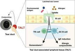

 Scoop.it!