Immunology
189.5K views | +454 today
Follow
Immunology
Teaching and Learning Immunology. Information you never would have searched for!
Your new post is loading...
Scooped by Gilbert C FAURE
October 28, 2021 3:07 AM
Scoop.it!

Des scientifiques identifient la super immunité naturelle du SARS-CoV-2 contre 23 variants - forum.chaudiere.ca

Des scientifiques identifient la super immunité naturelle du SARS-CoV-2 contre 23 variants - forum.chaudiere.ca | Immunology | Scoop.it
5 juillet 2021 Une équipe de scientifiques internationaux a récemment identifié des anticorps ultrapuissants anti-coronavirus 2 (SRAS-CoV-2) anti-syndrome respiratoire aigu sévère provenant de donneurs convalescents. Les anticorps sont capables de neutraliser une large gamme de variantes du SRAS-CoV-2, même à des concentrations sous-nanomolaires. De plus, les combinaisons de ces anticorps réduisent le risque de générer des mutants d'échappement in vitro. L'étude a été publiée le 13 août 2021 dans la revue Science. https://www.science.org/doi/full/10.112 ... ce.abh1766 Sur le fond Le coronavirus 2 du syndrome respiratoire aigu sévère (SRAS-CoV-2), l'agent pathogène responsable de la maladie à coronavirus 2019 (COVID-19), est un virus à ARN simple brin enveloppé de sens positif appartenant à la famille des bêta-coronavirus humains. La glycoprotéine de pointe sur l'enveloppe virale est composée de deux sous-unités S1 et S2. Dont, la sous-unité S1 se lie directement au récepteur de l'enzyme de conversion de l'angiotensine 2 (ACE2) de la cellule hôte via le domaine de liaison au récepteur (RBD) pour initier le processus d'entrée virale. La majorité des anticorps thérapeutiques contre le SRAS-CoV-2 ont été conçus sur la base de la séquence de protéine de pointe native trouvée dans la souche Wuhan originale du SRAS-CoV-2. Ainsi, de nouvelles variantes virales avec de multiples mutations de la protéine de pointe peuvent probablement développer une résistance contre ces anticorps. Dans ce contexte, des études ont montré que les anticorps développés en réponse aux vaccins COVID-19 actuellement disponibles ont moins d'efficacité pour neutraliser les nouvelles variantes préoccupantes (COV) du SRAS-CoV, notamment B.1.1.7, B.1.351, P1 et B.1.617.2. Dans la présente étude, les scientifiques ont isolé et caractérisé des anticorps anti-pic RBD de patients guéris du COVID-19. Identification des anticorps Les anticorps ont été isolés de quatre donneurs convalescents infectés par la souche Washington-1 (WA-1) du SRAS-CoV-2. La séquence de pointe dans la souche WA-1 est similaire à la séquence de pointe dans la souche originale de Wuhan. Les cellules B isolées à partir d'échantillons de sang provenant de donneurs ont été triées pour l'identification des anticorps. Cela a conduit à l'identification de quatre anticorps neutralisants puissants ciblant le pic RBD. Ces anticorps ont montré une forte affinité pour le pic SRAS-CoV-2 même à des concentrations nanomolaires. Pour déterminer si les anticorps hautement puissants pouvaient bloquer l'ACE2 - la liaison aux pointes, des tests d'interférométrie par compétition ACE2 et de liaison à la surface cellulaire ont été effectués. Les résultats ont révélé que sur 4 anticorps, deux liés aux RBD en « position haute » et deux liés aux RBD en « position basse ». De plus, trois anticorps sur quatre bloquaient directement l'interaction RBD - ACE2, et un inhibait indirectement l'interaction par encombrement stérique - le ralentissement des réactions chimiques dû à l'encombrement stérique. Neutralisation médiée par les anticorps Tous les anticorps expérimentaux ont montré une puissance significativement plus élevée dans la neutralisation des variants contenant la mutation D614G que la souche WA-1. Une analyse plus poussée avec des particules lentivirales pseudotypées avec des variantes à pointes a indiqué que les anticorps maintiennent une puissance élevée pour neutraliser un ensemble diversifié de 10 variantes à pointes. Il est important de noter que trois des quatre anticorps expérimentaux ont montré une grande efficacité pour neutraliser 13 variantes circulantes préoccupantes/intéressantes du SRAS-CoV-2, notamment B.1.1.7, B.1.351, B.1.427, B.1.429, B.1.526, P.1, P.2, B.1.617.1 et B.1.617.2. Analyse structurale et fonctionnelle des anticorps Des analyses au microscope cryoélectronique des structures du complexe anticorps-antigène ont révélé que deux anticorps avec le pouvoir de neutralisation le plus élevé se lient à la protéine de pointe avec tous les RBD en « position haute ». D'autres analyses structurelles ont révélé que les modes de liaison à l'épitope des anticorps sont responsables d'un pouvoir neutralisant élevé contre les COV du SRAS-CoV-2. La capacité de liaison et de neutralisation des anticorps a été affectée négativement par trois mutations de pointe, dont F486R, N487R et Y489R. Résistance aux anticorps Une pression de sélection d'anticorps a été appliquée à la souche WA-1 pour identifier les mutations potentielles d'échappement qui peuvent apparaître au cours de l'infection virale. La pression de sélection positive a été appliquée en incubant le virus avec des concentrations croissantes des anticorps pour déclencher la résistance aux anticorps. Dans deux des anticorps les plus puissants, l'un était affecté négativement par une seule mutation F486S et l'autre était affecté par les mutations F486L, N487D et Q493R. Cependant, la mutation Q493R a montré un impact négligeable sur la liaison et la neutralisation. Une analyse plus poussée a révélé que ces mutations d'échappement sont principalement absentes dans les variantes virales circulantes, indiquant l'absence de pression de sélection. En effectuant plusieurs cycles de sélection à l'aide de traitements combinés avec deux anticorps, il a été observé que les combinaisons d'anticorps pourraient réduire le risque d'acquisition de mutations d'échappement et le développement ultérieur de variantes virales résistantes. Source: News Medical et merci à QcLiLi, dans Twitter En référence à "Wang L. 2021. Anticorps ultrapuissants contre des variantes diverses et hautement transmissibles du SRAS-CoV-2. Sciences". -- -- --
No comment yet.
Scooped by Gilbert C FAURE
October 13, 2021 10:57 AM
Scoop.it!

Lessons in self-defence: inhibition of virus entry by intrinsic immunity

Lessons in self-defence: inhibition of virus entry by intrinsic immunity | Immunology | Scoop.it
Virus entry, consisting of attachment to and penetration into the host target cell, is the first step of the virus life cycle and is a critical ‘do or die’ event that governs virus emergence in host populations. Most antiviral vaccines induce neutralizing antibodies that prevent virus entry into cells. However, while the prevention of virus invasion by humoral immunity is well appreciated, considerably less is known about the immune defences present within cells (known as intrinsic immunity) that interfere with virus entry. The interferon-induced transmembrane (IFITM) proteins, known for inhibiting fusion between viral and cellular membranes, were once the only factors known to restrict virus entry. However, the progressive development of genetic and pharmacological screening platforms and the onset of the COVID-19 pandemic have galvanized interest in how viruses infiltrate cells and how cells defend against it. Several host factors with antiviral potential are now implicated in the regulation of virus entry, including cholesterol 25-hydroxylase (CH25H), lymphocyte antigen 6E (LY6E), nuclear receptor co-activator protein 7 (NCOA7), interferon-γ-inducible lysosomal thiol reductase (GILT), CD74 and ARFGAP with dual pleckstrin homology domain-containing protein 2 (ADAP2). This Review summarizes what is known and what remains to be understood about the intrinsic factors that form the first line of defence against virus infection. Besides neutralizing antibodies, viruses face a range of cell-intrinsic inhibitors that are specialized to limit virus entry into host cells. Majdoul and Compton describe the mechanisms of action of the cellular factors providing this important first line of defence against virus infection, including infection by severe acute respiratory syndrome coronavirus 2 (SARS-CoV-2).
No comment yet.
Scooped by Gilbert C FAURE
September 14, 2021 5:17 AM
Scoop.it!

Early cross-coronavirus reactive signatures of humoral immunity against COVID-19

Early cross-coronavirus reactive signatures of humoral immunity against COVID-19 | Immunology | Scoop.it
The introduction of vaccines has inspired new hope in the battle against SARS-CoV-2. However, the emergence of viral variants, in the absence of potent antivirals, has left the world struggling wit...
No comment yet.
Scooped by Gilbert C FAURE
August 24, 2021 5:31 AM
Scoop.it!

X-linked recessive TLR7 deficiency in ~1% of men under 60 years old with life-threatening COVID-19 | Science Immunology

X-linked recessive TLR7 deficiency in ~1% of men under 60 years old with life-threatening COVID-19 | Science Immunology | Immunology | Scoop.it
No comment yet.
Rescooped by Gilbert C FAURE from Virus World
June 24, 2021 3:01 AM
Scoop.it!

Long COVID Symptoms Likely Caused by Epstein-Barr Virus Reactivation

Long COVID Symptoms Likely Caused by Epstein-Barr Virus Reactivation | Immunology | Scoop.it

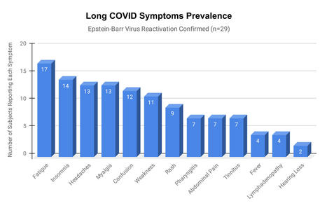
Epstein-Barr virus (EBV) reactivation resulting from the inflammatory response to coronavirus infection may be the cause of previously unexplained long COVID symptoms—such as fatigue, brain fog, and rashes—that occur in approximately 30% of patients after recovery from initial COVID-19 infection. The first evidence linking EBV reactivation to long COVID, as well as an analysis of long COVID prevalence, is outlined in a new long COVID study published in the journal Pathogens.  "We ran EBV antibody tests on recovered COVID-19 patients, comparing EBV reactivation rates of those with long COVID symptoms to those without long COVID symptoms," said lead study author Jeffrey E. Gold of World Organization. "The majority of those with long COVID symptoms were positive for EBV reactivation, yet only 10% of controls indicated reactivation." The researchers began by surveying 185 randomly selected patients recovered from COVID-19 and found that 30.3% had long term symptoms consistent with long COVID after initial recovery from SARS-CoV-2 infection. This included several patients with initially asymptomatic COVID-19 cases who later went on to develop long COVID symptoms.


The researchers then found, in a subset of 68 COVID-19 patients randomly selected from those surveyed, that 66.7% of long COVID subjects versus 10% of controls were positive for EBV reactivation based on positive EBV early antigen-diffuse (EA-D) IgG or EBV viral capsid antigen (VCA) IgM titers. The difference was significant (p < 0.001, Fisher's exact test). "We found similar rates of EBV reactivation in those who had long COVID symptoms for months, as in those with long COVID symptoms that began just weeks after testing positive for COVID-19," said coauthor David J. Hurley, Ph.D., a professor and molecular microbiologist at the University of Georgia. "This indicated to us that EBV reactivation likely occurs simultaneously or soon after COVID-19 infection." The relationship between SARS-CoV-2 and EBV reactivation described in this study opens up new possibilities for long COVID diagnosis and treatment. The researchers indicated that it may be prudent to test patients newly positive for COVID-19 for evidence of EBV reactivation indicated by positive EBV EA-D IgG, EBV VCA IgM, or serum EBV DNA tests. If patients show signs of EBV reactivation, they can be treated early to reduce the intensity and duration of EBV replication, which may help inhibit the development of long COVID. "As evidence mounts supporting a role for EBV reactivation in the clinical manifestation of acute COVID-19, this study further implicates EBV in the development of long COVID," said Lawrence S. Young, Ph.D., a virologist at the University of Warwick, and Editor-in-Chief of Pathogens. "If a direct role for EBV reactivation in long COVID is supported by further studies, this would provide opportunities to improve the rational diagnosis of this condition and to consider the therapeutic value of anti-herpesvirus agents such as ganciclovir."


Original findings published in Pathogens (June 10, 2021):

https://doi.org/10.3390/pathogens10060763


Via Juan Lama
Clarisse Staehlé's curator insight, December 5, 2022 11:11 AM
L’infection par le virus d’Epstein-Barr peut être une des causes de longs symptômes de COVID tes que : la fatigue, les éruptions cutanées …
Cela permettrait de traiter tôt les patients afin de réduire l’intensité et la durée de la réplication de l’EBV, ce qui peut aider à inhiber le développement d'un long COVID.
Suggested by LIGHTING
June 14, 2021 8:14 AM
Scoop.it!

Frontiers | Neutrophil Extracellular Traps Induce the Epithelial-Mesenchymal Transition: Implications in Post-COVID-19 Fibrosis | Immunology

Frontiers | Neutrophil Extracellular Traps Induce the Epithelial-Mesenchymal Transition: Implications in Post-COVID-19 Fibrosis | Immunology | Immunology | Scoop.it
The release of neutrophil extracellular traps (NETs), a process termed NETosis, avoids pathogen spread but may cause tissue injury. NETs have been found in severe COVID-19 patients, but their role in disease development is still unknown. The aim of this study is to assess the capacity of NETs to drive epithelial-mesenchymal transition (EMT) of lung epithelial cells and to analyze the involvement of NETs in COVID-19. Bronchoalveolar lavage fluid of severe COVID-19 patients showed high concentration of NETs that correlates with neutrophils count; moreover, the analysis of lung tissues of COVID-19 deceased patients showed a subset of alveolar reactive pneumocytes with a co-expression of epithelial marker and a mesenchymal marker, confirming the induction of EMT mechanism after severe SARS-CoV2 infection. By airway in vitro models, cultivating A549 or 16HBE at air-liquid interface, adding alveolar macrophages (AM), neutrophils and SARS-CoV2, we demonstrated that to trigger a complete EMT expression pattern are necessary the induction of NETosis by SARS-CoV2 and the secretion of AM factors (TGF-β, IL8 and IL1β). All our results highlight the possible mechanism that can induce lung fibrosis after SARS-CoV2 infection.
Scooped by Gilbert C FAURE
June 8, 2021 3:08 PM
Scoop.it!

Kinetics of peripheral blood neutrophils in severe coronavirus disease 2019 - Metzemaekers - 2021 - Clinical & Translational Immunology

Kinetics of peripheral blood neutrophils in severe coronavirus disease 2019 - Metzemaekers - 2021 - Clinical & Translational Immunology | Immunology | Scoop.it
In this study, we found that patients suffering from severe COVID-19 presented with immature, activated blood neutrophils and were characterized by elevated plasma levels of G-CSF and CXCL8 tha
Scooped by Gilbert C FAURE
June 1, 2021 1:24 PM
Scoop.it!

JCI - Is complement the culprit behind COVID-19 vaccine-related adverse reactions?

Viewpoint Free access | 10.1172/JCI151092 Is complement the culprit behind COVID-19 vaccine-related adverse reactions? Dimitrios C. Mastellos,1 Panagiotis Skendros,2 and John D. Lambris3 Published May 4, 2021 - More info View PDF Rare thrombotic reactions to adenovirus-based COVID-19 vaccines Undeniably, a global and coordinated COVID-19 vaccination effort is a prerequisite for taming the spread of emerging SARS-CoV-2 variants and achieving population-wide immunity that can thwart future viral surges. Ongoing vaccination programs have relied on both mRNA and adenoviral-based formulations eliciting potent humoral responses and showing safety in the majority of the population vaccinated so far. However, rare vaccine-related adverse events were recently reported in individuals who received the adenoviral-encoded ChAdOx1 nCov-19 and Ad26.COV2.S vaccines (1–3). Most of these cases developed acute thrombotic complications (cerebral venous thrombosis or disseminated intravascular coagulation) and severe thrombocytopenia following the first dose of the vaccine, without any prior history of heparin exposure or known risk factors for thrombophilia. This rare syndrome, termed vaccine-induced immune thrombotic thrombocytopenia (VITT), clinically resembles the hallmarks of autoimmune heparin-induced thrombocytopenia (autoimmune HIT) and is linked to the consistent presence of circulating autoantibodies against platelet factor 4 (PF4), which can directly activate platelets in the absence of heparin (2). The prothrombotic action of anti-PF4 antibodies likely involves antibody-mediated platelet activation through IgG-FcγR interactions and similar FcR-mediated engagement of immune effector cells such as monocytes/macrophages and neutrophils (Figure 1). Since VITT is a newly described syndrome, its clinical management mostly relies on its similarities with HIT and non–heparin-dependent autoimmune thrombotic thrombocytopenias. While nonheparin anticoagulants and intravenous immunoglobulin (IVIg) have been proposed as first-line treatments for patients with VITT, these approaches are not fail-safe and entail drawbacks that may curtail their effectiveness (e.g., risk of promiscuous FcR activation, acute anaphylaxis, or even thromboembolic events in the case of intravenous immunoglobulin) (4). Several second-line therapies for HIT have also been suggested, including plasmapheresis, especially when nonheparin anticoagulation is contraindicated because of major bleeding events (5). Thus, more insight into the precise pathophysiology of VITT is needed to enable more personalized medicine approaches. Figure 1 A schematic diagram of the plausible mechanisms by which complement may contribute to the VITT-associated prothrombotic response. Vaccine-induced immune thrombotic thrombocytopenia (VITT) has been described in individuals presenting with high titers of platelet-activating anti–platelet factor 4 (PF4) autoantibodies. These antibodies recognize large multiantigenic complexes comprising PF4 and polyanionic structures similar to heparin, and resemble autoimmune heparin-induced thrombocytopenia (HIT). Complement activation can be triggered in multiple ways in patients presenting with VITT and anti-PF4 antibodies. First, the formation of complexes between PF4 and certain vaccine constituents (e.g., adenoviral capsid proteins or DNA) (23) may serve as a scaffold for C3 activation, similarly to what has been observed with PF4-heparin ultra-large complexes. In addition, complement activation can be triggered by the binding of C1q to the anti-PF4 immune complexes that are deposited on the endothelium, monocyte, or platelet surface via binding to polyanionic structures such as glycosaminoglycans (GAGs). Classical pathway activation leads to C3 cleavage, amplification of complement responses via the alternative pathway, downstream generation of proinflammatory C3a and C5a anaphylatoxins, and the formation of the C5b-9 complex. Complement activation fragments mediate a broad range of thromboinflammatory reactions by interacting with complement receptors on platelets, monocytes, and neutrophils. These interactions can induce or enhance FcR-mediated platelet activation, neutrophil-platelet aggregation, and release of TF-loaded neutrophil extracellular traps (NETs) from activated neutrophils. All these processes are fueled by complement activation and can collectively contribute to a prothrombotic environment that may lead to VITT. Moreover, C3-opsonized immune complexes can enhance FcγR-dependent effector responses on platelets further promoting thrombotic responses. Abbreviations: CP, classical pathway; AP, alternative pathway; TF, tissue factor. A proposed role for complement Complement is a host innate immune sentinel comprising over 50 proteins that can be swiftly activated through any of three initiating pathways (classical, lectin, or alternative) that sense microbial invaders or other inflammatory cues, such as immune complexes. In fact, complement activation has been implicated as a perpetrator of immune complex–driven thromboinflammation in autoimmune pathologies such as the antiphospholipid syndrome, a systemic disorder that increases risk for blood clots (6). Prompted by the striking findings in patients who developed VITT after a single dose of adenoviral-associated vaccine, we posit here that the observed thrombotic complications may partly reflect immune complex–triggered complement activation through the classical pathway. PF4-containing immune complexes can be recognized by C1q, a soluble pattern recognition molecule of the classical pathway that binds to the Fc portion of IgG molecules. Anti-PF4 complex–driven classical pathway activation can lead to C3 activation, alternative pathway–mediated amplification of complement responses, and downstream generation of potent proinflammatory mediators and effectors (C3a, C5a and membrane attack complex) that can potentiate thromboinflammation (7). In fact, the thrombogenic capacity of distinct complement activation fragments has been linked to a broad spectrum of immune-related mechanisms that underpin the immunothrombosis of severe COVID-19 (7, 8). Considering that certain allelic combinations of complement gene polymorphisms within the population confer differential susceptibility to deregulated complement activity, it is tempting to speculate that these vaccinated individuals may represent cases where complement deregulation occurs due to genetic alterations that result in ineffective host regulatory control (9). Deregulated complement responses can perpetuate a vicious cycle of inflammatory tissue damage that renders the vascular endothelium and platelet/monocyte compartment more thrombogenic. In this regard, circulating anti-PF4 immune complexes may well serve as a first hit that ignites complement activation, in a way similar to complement-mediated thrombotic microangiopathies (10). The fact that a fraction of these anti-PF4 autoantibodies may recognize antigenic complexes between PF4 and polyanionic structures (such as GAGs or heparan sulfate) raises the possibility that these immune complexes may skew the binding of endogenous complement regulators to host polyanionic surfaces, thereby altering the capacity of these surfaces to withstand autologous complement attack. C3 activation may play a pivotal pathogenic role in this vaccine-induced immunothrombotic phenotype. C3-opsonized anti-PF4 immune complexes can enhance the phagocytic and inflammatory properties of monocytes and neutrophils through the synergistic engagement of FcγR and complement receptors, thereby increasing the thrombogenic milieu in the vasculature (Figure 1). C3 fragment deposition on the endothelium and on platelets/neutrophils can potentiate thromboinflammatory responses by promoting CR3-dependent platelet-neutrophil interactions that favor platelet adhesion and clotting (11). Also, C3aR signaling on platelets can further drive thrombotic responses (12). Of note, it has been shown that PF4–heparin complexes, a hallmark of HIT pathology, can potently activate C3 in the fluid phase and C3 inhibitors such as Cp40 can abrogate C3 activation triggered by HIT-associated PF4–heparin complexes (13). In line with this, the Cp40-based drug candidate AMY-101 has been shown to effectively decrease both neutrophils and neutrophil extracellular trap (NET) formation, two major drivers of HIT-associated thrombosis, in patients with COVID-19 (7, 14–16). Potential treatment approaches In an analogy to HIT, clinical-stage complement C3 inhibitors may represent a novel therapeutic strategy for ameliorating the thrombotic complications elicited in VITT. On one hand, C3 inhibition may quench anti-PF4 antibody production by reducing antigen uptake or processing in the lymphoid tissue. On the other hand, it can broadly suppress multiple paths of immune complex–driven thromboinflammation in VITT (Figure 1). Moreover, C3 inhibition may offer broader therapeutic control of thromboembolic reactions mediated by pathogenic autoantibodies. In this regard, C3 activation, but not C5, was shown to be indispensable for antiphospholipid antibody-induced tissue factor (TF) activation and thrombosis in antiphospholipid syndrome (17). The recently reported resolution of vaccine-induced thrombotic microangiopathy in two patients following combined treatment with anticoagulants and the C5 inhibitor eculizumab further corroborates our hypothesis that therapeutic complement modulation may offer significant clinical benefits to patients with VITT (18). In this regard, C3 inhibitors may exert a broader therapeutic effect than anti-C5 agents by suppressing both terminal pathway activation and thrombogenic pathways operating upstream of C5 (14, 19). Because this newly described immune syndrome is still poorly defined, it would be of great benefit to patients with VITT if the algorithm for VITT clinical management considered diagnostic monitoring of complement biomarkers as a means of validating the use of specific complement inhibitors. As a cautionary note, we should stress that plasma levels of complement proteins reflect both turnover and synthesis. Therefore, the accurate monitoring of complement activity should rely on markers of ongoing activation combined with corroborative functional assays that gauge the impact of complement activation on the tissue or cellular level. mRNA vaccine reactions In addition to these cases of VITT in individuals receiving adenovirus vectored vaccines, the rollout of COVID-19 mRNA vaccines has brought to the spotlight rare adverse events involving a maladaptive immune reaction to certain vaccine constituents. Rare cases of severe allergic reactions (anaphylaxis) were reported following the first dose of the Pfizer–BioNTech or Moderna SARS-CoV-2 mRNA vaccines (20). Such allergic reactions are likely triggered by the PEG or lipid moieties of these mRNA vaccines. They involve complement and mast cell activation in an IgE-independent manner and closely resemble the complement mediated pseudo-allergy reaction (CARPA) that has been described in the case of liposomal carriers (21). Complement activation in this case may be initiated by IgG or IgM directed against the PEGylated lipids of these vaccine formulations. Concluding remarks Taken together, these paradigms of vaccine-related adverse reactions highlight the potential contribution of complement activation to vaccine-related pathology, raise the importance of incorporating complement diagnostic monitoring in the early clinical management of such patients, and endorse complement inhibitors as potential treatment options. In addition to the complement-activating capacity of anti-PF4 antibodies, we cannot rule out the possibility that complement activation in VITT may also be fueled by preformed (natural IgM) or induced IgG antibodies against adenoviral capsid antigens or direct interaction of pattern recognition molecules with capsid elements (22). The relevance of these plausible mechanisms to VITT pathology warrants further investigation. Footnotes Conflict of interest: JDL is the founder of Amyndas Pharmaceuticals, which is developing complement inhibitors for therapeutic purposes. He is the inventor on patents or patent applications that describe the use of complement inhibitors for therapeutic purposes, some of which are developed by Amyndas Pharmaceuticals: US patents 8946145/9371365 (Modified compstatin with peptide backbone and C-terminal modifications), 9630992 (Compstatin analogs with improved pharmacokinetic properties), 9579360 (Methods of treating or preventing periodontitis and diseases associated with periodontitis). JDL is also the inventor of the compstatin technology licensed to Apellis Pharmaceuticals, i.e., 4(1MeW)7W/POT-4/APL-1 and PEGylated derivatives such as APL-2/pegcetacoplan/empaveli and APL-9: US patents 6319897 (Peptides which inhibit complement activation), 7989589 (Compstatin analogs with improved activity), 7888323 (Potent compstatin analogs). Copyright: © 2021, American Society for Clinical Investigation. Reference information: J Clin Invest. 2021;131(11):e151092. https://doi.org/10.1172/JCI151092. References Muir K-L, et al. Thrombotic thrombocytopenia after Ad26.COV2.S vaccination. [published online April 14, 2021]. N Engl J Med. https://doi.org/10.1056/NEJMc2105869. View this article via: PubMed Google Scholar Scully M, et al. Pathologic antibodies to platelet factor 4 after ChAdOx1 nCoV-19 vaccination. [published online April 16, 2021]. N Engl J Med. https://doi.org/10.1056/NEJMoa2105385. View this article via: PubMed Google Scholar Schultz NH, et al. Thrombosis and thrombocytopenia after ChAdOx1 nCoV-19 vaccination. [published online April 9, 2021]. N Engl J Med. https://doi.org/10.1056/NEJMoa2104882. View this article via: PubMed Google Scholar Kapoor M, et al. Thromboembolic risk with IVIg: incidence and risk factors in patients with inflammatory neuropathy. Neurology. 2020;94(6):e635–e638. View this article via: PubMed CrossRef Google Scholar Marchetti M, et al. Heparin-induced thrombocytopenia: a review of new concepts in pathogenesis, diagnosis, and management. J Clin Med. 2021;10(4):683. View this article via: PubMed CrossRef Google Scholar Chaturvedi S, et al. Complement in the pathophysiology of the antiphospholipid syndrome. Front Immunol. 2019;10:449. View this article via: PubMed Google Scholar Skendros P, et al. Complement and tissue factor-enriched neutrophil extracellular traps are key drivers in COVID-19 immunothrombosis. J Clin Invest. 2020;130(11):6151–6157. View this article via: JCI PubMed CrossRef Google Scholar Risitano AM, et al. Complement as a target in COVID-19? Nat Rev Immunol. 2020;20(6):343–344. View this article via: PubMed CrossRef Google Scholar Heurich M, et al. Common polymorphisms in C3, factor B, and factor H collaborate to determine systemic complement activity and disease risk. Proc Natl Acad Sci U S A. 2011;108(21):8761–8766. View this article via: PubMed CrossRef Google Scholar Gavriilaki E, Brodsky RA. Complementopathies and precision medicine. J Clin Invest. 2020;130(5):2152–2163. View this article via: JCI PubMed CrossRef Google Scholar Hamad OA, et al. Contact activation of C3 enables tethering between activated platelets and polymorphonuclear leukocytes via CD11b/CD18. Thromb Haemost. 2015;114(6):1207–1217. View this article via: PubMed Google Scholar Sauter RJ, et al. Functional relevance of the anaphylatoxin receptor C3aR for platelet function and arterial thrombus formation marks an intersection point between innate immunity and thrombosis. Circulation. 2018;138(16):1720–1735. View this article via: PubMed CrossRef Google Scholar Khandelwal S, et al. Novel immunoassay for complement activation by PF4/heparin complexes. Thromb Haemost. 2018;118(8):1484–1487. View this article via: PubMed CrossRef Google Scholar Mastellos DC, et al. Complement C3 vs C5 inhibition in severe COVID-19: early clinical findings reveal differential biological efficacy. Clin Immunol. 2020;220:108598. View this article via: PubMed Google Scholar Gollomp K, et al. Neutrophil accumulation and NET release contribute to thrombosis in HIT. JCI Insight. 2018;3(18):99445. View this article via: JCI Insight PubMed Google Scholar Perdomo J, et al. Neutrophil activation and NETosis are the major drivers of thrombosis in heparin-induced thrombocytopenia. Nat Commun. 2019;10(1):1322. View this article via: PubMed CrossRef Google Scholar Müller-Calleja N, et al. Complement C5 but not C3 is expendable for tissue factor activation by cofactor-independent antiphospholipid antibodies. Blood Adv. 2018;2(9):979–986. View this article via: PubMed CrossRef Google Scholar Tiede A, et al. Prothrombotic immune thrombocytopenia after COVID-19 vaccine. [published online April 30, 2021]. Blood. https://doi.org/10.1182/blood.2021011958. View this article via: PubMed Google Scholar Hillmen P, et al. Pegcetacoplan versus eculizumab in paroxysmal nocturnal hemoglobinuria. N Engl J Med. 2021;384(11):1028–1037. View this article via: PubMed CrossRef Google Scholar Castells MC, Phillips EJ. Maintaining safety with SARS-CoV-2 vaccines. N Engl J Med. 2021;384(7):643–649. View this article via: PubMed CrossRef Google Scholar Klimek L, et al. Allergenic components of the mRNA-1273 vaccine for COVID-19: possible involvement of polyethylene glycol and IgG-mediated complement activation. [published online March 3, 2021]. Allergy. https://doi.org/10.1111/all.14794. View this article via: PubMed Google Scholar Chéneau C, Kremer EJ. Adenovirus-extracellular protein interactions and their impact on innate immune responses by human mononuclear phagocytes. Viruses. 2020;12(12):1351. View this article via: PubMed CrossRef Google Scholar Greinacher A, et al. Towards understanding ChAdOx1 nCov-19 vaccine-induced immune thrombotic thrombocytopenia (VITT) [preprint]. https://doi.org/10.21203/rs.3.rs-440461/v1 Posted on Research Square April 20, 2021. Version history Version 1 (May 4, 2021): In-Press Preview Version 2 (June 1, 2021): Electronic publication
No comment yet.
Scooped by Gilbert C FAURE
May 27, 2021 12:16 PM
Scoop.it!

Had COVID? You’ll probably make antibodies for a lifetime

Had COVID? You’ll probably make antibodies for a lifetime | Immunology | Scoop.it
People who recover from mild COVID-19 have bone-marrow cells that can churn out antibodies for decades, though viral variants could dampen some of the protection they offer.
No comment yet.
Scooped by Gilbert C FAURE
May 19, 2021 7:22 AM
Scoop.it!

The COVID-19 puzzle: deciphering pathophysiology and phenotypes of a new disease entity - ScienceDirect

The zoonotic SARS-CoV-2 virus that causes COVID-19 continues to spread worldwide, with devastating consequences. While the medical community has gaine…
No comment yet.
Scooped by Gilbert C FAURE
May 13, 2021 12:41 PM
Scoop.it!

Interplay between inflammation and thrombosis in cardiovascular pathology

Interplay between inflammation and thrombosis in cardiovascular pathology | Immunology | Scoop.it
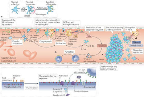
Thrombosis is the most feared complication of cardiovascular diseases and a main cause of death worldwide, making it a major health-care challenge. Platelets and the coagulation cascade are effectively targeted by antithrombotic approaches, which carry an inherent risk of bleeding. Moreover, antithrombotics cannot completely prevent thrombotic events, implicating a therapeutic gap due to a third, not yet adequately addressed mechanism, namely inflammation. In this Review, we discuss how the synergy between inflammation and thrombosis drives thrombotic diseases. We focus on the huge potential of anti-inflammatory strategies to target cardiovascular pathologies. Findings in the past decade have uncovered a sophisticated connection between innate immunity, platelet activation and coagulation, termed immunothrombosis. Immunothrombosis is an important host defence mechanism to limit systemic spreading of pathogens through the bloodstream. However, the aberrant activation of immunothrombosis in cardiovascular diseases causes myocardial infarction, stroke and venous thromboembolism. The clinical relevance of aberrant immunothrombosis, referred to as thromboinflammation, is supported by the increased risk of cardiovascular events in patients with inflammatory diseases but also during infections, including in COVID-19. Clinical trials in the past 4 years have confirmed the anti-ischaemic effects of anti-inflammatory strategies, backing the concept of a prothrombotic function of inflammation. Targeting inflammation to prevent thrombosis leaves haemostasis mainly unaffected, circumventing the risk of bleeding associated with current approaches. Considering the growing number of anti-inflammatory therapies, it is crucial to appreciate their potential in covering therapeutic gaps in cardiovascular diseases. In this Review, Stark and Massberg discuss how the interplay between innate immunity, platelet activation and coagulation, known as immunothrombosis, functions as a host defence mechanism to limit pathogen spreading, yet its aberrant activation, termed thromboinflammation, results in thrombotic complications, highlighting the therapeutic potential of anti-inflammatory strategies in cardiovascular pathologies.


No comment yet.
Scooped by Gilbert C FAURE
April 25, 2021 7:05 AM
Scoop.it!

Plusieurs formes « d’orages cytokiniques » sont associés à la sévérité et la mortalité dans la Covid-19

Plusieurs formes « d’orages cytokiniques » sont associés à la sévérité et la mortalité dans la Covid-19 | Immunology | Scoop.it
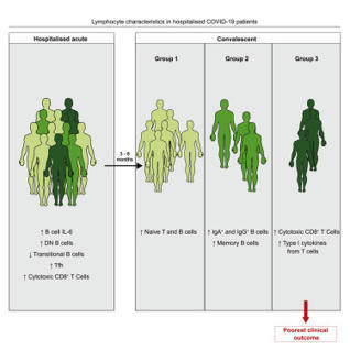
Une étude montre qu’il existe au moins deux profils de réponse cytokinique associée à la Covid-19, ce qui impliquerait donc la nécessité d’une prise en charge hautement personnalisée des patients.
No comment yet.
Scooped by Gilbert C FAURE
April 12, 2021 12:25 PM
Scoop.it!

Multilevel proteomics reveals host perturbations by SARS-CoV-2 and SARS-CoV

The global emergence of SARS-CoV-2 urgently requires an in-depth understanding of molecular functions of viral proteins and their interactions with the host proteome. Several individual omics studies have extended our knowledge of COVID-19 pathophysiology1–10. Integration of such datasets to obtain a holistic view of virus-host interactions and to define the pathogenic properties of SARS-CoV-2 is limited by the heterogeneity of the experimental systems. We therefore conducted a concurrent multi-omics study of SARS-CoV-2 and SARS-CoV. Using state-of-the-art proteomics, we profiled the interactome of both viruses, as well as their influence on transcriptome, proteome, ubiquitinome and phosphoproteome in a lung-derived human cell line. Projecting these data onto the global network of cellular interactions revealed crosstalk between the perturbations taking place upon SARS-CoV-2 and SARS-CoV infections at different layers and identified unique and common molecular mechanisms of these closely related coronaviruses. The TGF-β pathway, known for its involvement in tissue fibrosis, was specifically dysregulated by SARS-CoV-2 ORF8 and autophagy by SARS-CoV-2 ORF3. The extensive dataset (available at
https://covinet.innatelab.org

) highlights many hotspots that can be targeted by existing drugs and it can guide rational design of virus- and host-directed therapies, which we exemplify by identifying kinase and MMPs inhibitors with potent antiviral effects against SARS-CoV-2.
Gilbert C FAURE's insight:

vous avez dit simple?

No comment yet.
Scooped by Gilbert C FAURE
October 25, 2021 1:51 PM
Scoop.it!

Clues that natural killer cells help to control COVID

Clues that natural killer cells help to control COVID | Immunology | Scoop.it
A defence response malfunctions in people with severe COVID-19.
No comment yet.
Scooped by Gilbert C FAURE
September 18, 2021 4:49 AM
Scoop.it!

How does SARS-CoV-2 cause COVID-19?

How does SARS-CoV-2 cause COVID-19? | Immunology | Scoop.it
The viral receptor on human cells plays a critical role in disease progression
Gilbert C FAURE's insight:

You should know where virus attacks to fight against it

Kaupang's comment, October 8, 2021 9:32 AM
nice
Scooped by Gilbert C FAURE
August 27, 2021 6:36 AM
Scoop.it!

Biology | Free Full-Text | Type I Interferons in COVID-19 Pathogenesis

Biology | Free Full-Text | Type I Interferons in COVID-19 Pathogenesis | Immunology | Scoop.it
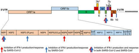
Among the many activities attributed to the type I interferon (IFN) multigene family, their roles as mediators of the antiviral immune response have emerged as important components of the host response to Severe Acute Respiratory Syndrome Coronavirus 2 (SARS-CoV-2) infection.
No comment yet.
Scooped by Gilbert C FAURE
August 10, 2021 5:12 AM
Scoop.it!

More than 50 long-term effects of COVID-19: a systematic review and meta-analysis

More than 50 long-term effects of COVID-19: a systematic review and meta-analysis | Immunology | Scoop.it
COVID-19 can involve persistence, sequelae, and other medical complications that last weeks to months after initial recovery. This systematic review and meta-analysis aims to identify studies assessing the long-term effects of COVID-19. LitCOVID and Embase were searched to identify articles with original data published before the 1st of January 2021, with a minimum of 100 patients. For effects reported in two or more studies, meta-analyses using a random-effects model were performed using the MetaXL software to estimate the pooled prevalence with 95% CI. PRISMA guidelines were followed. A total of 18,251 publications were identified, of which 15 met the inclusion criteria. The prevalence of 55 long-term effects was estimated, 21 meta-analyses were performed, and 47,910 patients were included (age 17–87 years). The included studies defined long-COVID as ranging from 14 to 110 days post-viral infection. It was estimated that 80% of the infected patients with SARS-CoV-2 developed one or more long-term symptoms. The five most common symptoms were fatigue (58%), headache (44%), attention disorder (27%), hair loss (25%), and dyspnea (24%). Multi-disciplinary teams are crucial to developing preventive measures, rehabilitation techniques, and clinical management strategies with whole-patient perspectives designed to address long COVID-19 care.
No comment yet.
Scooped by Gilbert C FAURE
June 19, 2021 10:26 AM
Scoop.it!

A series of COVID‐19 autopsies with clinical and pathologic comparisons to both seasonal and pandemic influenza - McMullen - - The Journal of Pathology: Clinical Research

A series of COVID‐19 autopsies with clinical and pathologic comparisons to both seasonal and pandemic influenza - McMullen - - The Journal of Pathology: Clinical Research | Immunology | Scoop.it
Abstract Autopsies of patients who have died from COVID-19 have been crucial in delineating patterns of injury associated with SARS-CoV-2 infection. Despite their utility, comprehensive autopsy stu...
Gilbert C FAURE's insight:

at last....

No comment yet.
Scooped by Gilbert C FAURE
June 11, 2021 3:10 AM
Scoop.it!

VARIANTS DU COVID - Le Péril du Delta | lepetitjournal.com

VARIANTS DU COVID - Le Péril du Delta | lepetitjournal.com | Immunology | Scoop.it
Les variants qui apparaissent toujours plus différenciés et plus tenaces sont-ils un péril pour la sortie du tunnel ?
No comment yet.
Rescooped by Gilbert C FAURE from Veille Coronavirus - Covid-19
June 2, 2021 10:49 AM
Scoop.it!

A Systematic Review of COVID - 19 Induced Myocarditis - Symptomatology, Prognosis, and Clinical Findings | medRxiv

medRxiv - The Preprint Server for Health Sciences

Via HAS-veille
No comment yet.
Scooped by Gilbert C FAURE
June 1, 2021 10:31 AM
Scoop.it!

COVID-19 Immunology 101 for Non-immunologists by Akiko Iwasaki, Ph.D.

In collaboration with BioRender, Akiko Iwasaki, Ph.D., Professor of Immunobiology at Yale University School of Medicine, explores COVID-19 Immunology 101 for Non-immunologists.

Acknowledgements:
Dr. Akiko Iwasaki, PhD (Collaborator, narrator, content expert)
Dr. Yanet Valdez Tejeira, PhD (Spanish subtitle contributor)
Mahadi B. Alyami (@RTKase) (Arabic subtitle contributor)
Nick Atanelov (@Nick_atanelov) (Georgian subtitle contributor)
Anonymous (Turkish subtitle contributor)

You can find and customize the figures used in this video by visiting the BioRender template library (http://app.biorender.com/biorender-templates) and search for "tweetorial".

The video is also available in Japanese: https://youtu.be/XAE7U9lCNjg

About BioRender:
BioRender is the easy-to-use science illustration tool that’s quickly becoming a staple in academic institutions and labs around the world! You can access the program for free at https://biorender.com/

BioRender COVID-19 Vaccine & Therapeutics Tracker
https://biorender.com/covid-vaccine-tracker
Suggested by LIGHTING
May 20, 2021 3:00 AM
Scoop.it!

How T Cells Recognize SARS-CoV-2

How T Cells Recognize SARS-CoV-2 | Immunology | Scoop.it
Part 1 video in how the immune system recognizes the virus that causes COVID-19
No comment yet.
Scooped by Gilbert C FAURE
May 15, 2021 12:16 PM
Scoop.it!

Innate immune and inflammatory responses to SARS-CoV-2: implications for COVID-19

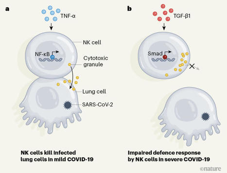
Lowery et al. review the mechanisms by which SARS-CoV-2 activates and antagonizes
the interferon and inflammatory response following infection, how a dysregulated cytokine
and cellular response contributes to immune-mediated pathology in COVID-19, and therapeutic
strategies that target elements of the innate response.
No comment yet.
Scooped by Gilbert C FAURE
May 2, 2021 4:42 AM
Scoop.it!

UK-CIC Public Webinar: COVID-19 and your immune system | UK-CIC

UK-CIC Public Webinar: COVID-19 and your immune system | UK-CIC | Immunology | Scoop.it
Title: COVID-19 and your immune system Date/Time: Tuesday 4 May 2021 – 18:00 to 19:00 Location: GoToWebinar Register now Description:  The UK Coronavirus Immuno...
Scooped by Gilbert C FAURE
April 15, 2021 10:36 AM
Scoop.it!

Alterations in T and B cell function persist in convalescent COVID-19 patients - ScienceDirect

Alterations in T and B cell function persist in convalescent COVID-19 patients - ScienceDirect | Immunology | Scoop.it
Emerging studies indicate that some coronavirus disease 2019 (COVID-19) patients suffer from persistent symptoms, including breathlessness and chronic…
No comment yet.