


From WebMD, an overview of psoriasis, an skin condition that forms thick, red patches.

AUTOIMMUNITY
20.9K views |
+0 today

 Your new post is loading...
 Scoop.it! 
Sepsis is a major cause of death for hospitalized patients, and is characterized by massive overreaction of immune responses to invading pathogens which is mediated by cytokines. For decades, there has been no effective treatment for sepsis. Sialic acid-binding, Ig-like lectin-9 (Siglec-9), is an immunomodulatory receptor expressed primarily on hematopoietic cells which is involved in various aspects of inflammatory responses and is a potential target for treatment of sepsis. The aim of the present study was to develop a human anti-Siglec-9 Fab fragment, which was named hS9-Fab03, and investigated its immune activity in human macrophages. We began by constructing the hS9-Fab03 prokaryotic expression vector from human antibody library and phage display. Then we utilized a multitude of assays, including SDS-PAGE, Western blotting, ELISA, affinity and kinetics assay to evaluate the binding affinity and specificity of hS9-Fab03. Results demonstrated that hS9-Fab03 specifically bind to Siglec-9 antigen with high affinity, and pre-treatment with hS9-Fab03 could attenuated LPS-induced TNF-α, IL-6, IL-1β, IL-8 and IFN-β production in human PBMCs derived macrophages, but slightly increased IL-10 production in an early time point. We also observed similar results in human THP-1 differentiated macrophages. Collectively, we prepared the hS9-Fab03 with efficient activity for blocking LPS-induced proinflammatory cytokines production in human macrophages. These results indicated that ligation of Siglec-9 with hS9-Fab03 might be a novel anti-inflammatory therapeutic strategy for sepsis. 
Scoop.it!
Clinical & Translational Immunology - Primate autoimmune disease models; lost for translation[quest]
Clinical & Translational Immunology focuses on the general functioning of the immune system in its broadest sense, with a particular emphasis on its cell biology. Areas that are covered include but are not limited to: Cellular immunology, Innate and adaptive immunity, Immune responses to pathogens,Tumour immunology,Immunopathology, Immunotherapy, Immunogenetics, Immunological studies in humans and model organisms (including mouse, rat, Drosophila etc)
Via Krishan Maggon
Scoop.it!
Lactoferrin (Lf) is one of the antigens of anti-neutrophil cytoplasmic antibodies (ANCA) and functions as an endogenous suppressor of neutrophil extracellular trap (NET) formation. However, the prevalence and pathogenicity of anti-lactoferrin antibodies (aLf) in ANCA-associated vasculitis (AAV) remain unrevealed. This study aimed to examine the significance of aLf in AAV, initially. Sixty-five sera from AAV patients, including 41 microscopic polyangiitis (MPA), 5 granulomatosis with polyangiitis (GPA), and 19 eosinophilic granulomatosis with polyangiitis (EGPA) patients, were subjected to aLf detection using enzyme-linked immunosorbent assay. Clinical characteristics were compared between aLf-positive and aLf-negative patients. Neutrophils from healthy donors were exposed to suboptimal dose (10 nM) of phorbol myristate acetate (PMA) with aLf followed by evaluation of NET formation. Results demonstrated that four out of 65 AAV sera (6.2%) were positive for aLf. All of them were EGPA sera (4/19, 21.1%). In EGPA, the frequency of renal involvement, serum CRP levels, and Birmingham Vasculitis Activity Score (BVAS) in the aLf-positive patients were significantly higher than those in the aLf-negative patients, and the aLf titer correlated positively with the serum CRP level and BVAS. The NET formation was particularly enhanced by combined stimulation of 10 nM PMA and 1 µg/mL aLf. IgG isolated from sera of the aLf-positive EGPA patients (250 µg/mL) enhanced NET formation induced by 10 nM of PMA, and the effect was abolished completely by absorption of the aLf. This pilot study suggests that aLf enhance NET formation induced by PMA and are associated with disease activity of EGPA.
Gilbert C FAURE's insight:
MPO, PR3, BPI, Lysozyme, lactoferrin... as targets...
Scoop.it!

Rheumatology (Oxford). 2016 Dec 24. pii: kew407. doi: 10.1093/rheumatology/kew407. [Epub ahead of print]
Scoop.it!

T cells are important for fighting infectious agents, but T cells that recognize the body’s own cells are often central to the development of autoimmune disease, leading Borroto et al . to develop a compound that hampers T cell signaling without completely blocking it. Treatment with this compound prevented or treated autoimmune disease in multiple mouse models, and the compound was demonstrated to skew human T cell differentiation toward less inflammatory subsets. Treatment with the compound did not prevent T cell pathogen responses in mice, suggesting that it would not leave patients susceptible to infection.
Scoop.it!

New research may help explain why dietary changes are often effective when treating inflammatory conditions.
Gilbert C FAURE's insight:
two days ago on scoop.it autoimmunity
Scoop.it!

Case study of eye injury and decreased vision - investigation, diagnosis and treatment of Sympathetic ophthalmia plus the immunology of the disease.
Scoop.it!

Scurfy mice are deficient in regulatory T cells (Tregs), develop a severe, generalized autoimmune disorder that can affect almost every organ and die at an early age. Some of these manifestations resemble those found in systemic lupus erythematosus (SLE). In addition, active SLE is associated with low Treg numbers and reduced Treg function, but direct evidence for a central role of Treg malfunction in the pathophysiology of lupus-like manifestations is still missing. In the present study, we characterize the multiorgan pathology, autoantibody profile and blood count abnormalities in scurfy mice and show their close resemblances to lupus-like disease. Scurfy mice have dysfunctional Tregs due to a genetic defect in the transcription factor Forkhead box protein 3 (Foxp3). We analyzed skin, joints, lung and kidneys of scurfy mice and wild-type (WT) controls by conventional histology and immunofluorescence (IF) performed hematological workups and tested for autoantibodies by IF, immunoblotting and enzyme-linked immunosorbent assay. We also analyzed the intestines, liver, spleen and heart, but did not analyze all organs known to be affected in scurfy mice (such as the testicle, the accessory reproductive structures, the pancreas or the eyes). We transferred CD4+ T cells of scurfy or WT mice into T cell-deficient B6/nude mice. We confirm previous reports that scurfy mice spontaneously develop severe pneumonitis and hematological abnormalities similar to those in SLE. We show that scurfy mice (but not controls) exhibited additional features of SLE: severe interface dermatitis, arthritis, mesangioproliferative glomerulonephritis and high titers of anti-nuclear antibodies, anti-double-stranded DNA antibodies, anti-histone antibodies and anti-Smith antibodies. Transfer of scurfy CD4+ T cells (but not of WT cells) induced autoantibodies and inflammation of lung, skin and kidneys in T cell-deficient B6/nude mice. Our observations support the hypothesis that lupus-like autoimmune features develop in the absence of functional Tregs.
Gilbert C FAURE's insight:
missed it
Scoop.it!
Int Immunol. 2016 Apr;28(4):173-9. doi: 10.1093/intimm/dxv079. Epub 2015 Dec 29. Review
Scoop.it!
Clin Transl Immunology. 2016 Nov 2;5(11):e112. eCollection 2016. Review
Scoop.it!

Researchers from ETH Zurich have developed artificial beta cells that can measure blood glucose concentration and respond by producing insulin. The team’s work was published this week in Science. The ETH researchers used the natural glucose transport proteins and potassium channels found in the membrane of human kidney cells and modified them with a voltage-dependent calcium channel, […]
Scoop.it!
Explore the connection between the #oralmicrobiome and #autoimmunity https://t.co/lVBF0WgsSy
|
Scoop.it!

Here are 12 celebrities with psoriasis, an autoimmune skin condition that causes scaly, red skin patches.
Gilbert C FAURE's insight:
how laypeople gain access to information
Scoop.it!
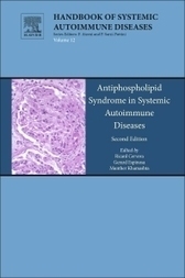
Elsevier Store: Antiphospholipid Syndrome in Systemic Autoimmune Diseases, 2nd Edition from Ricard Cervera, Gerard Espinosa, Munther Khamashta. ISBN-9780444636553, Printbook , Release Date: 2016
Scoop.it!

Scientists at the Immunology Frontier Research Center (IFReC), Osaka University, Japan, report a new molecular mechanism that could explain the cause of some autoimmune diseases.
Scoop.it!
Autoimmunity. 2016 Dec 23:1-16. doi: 10.1080/08916934.2016.1261834. [Epub ahead of print]
Scoop.it!
Sci Rep. 2016 Dec 21;6:39378. doi: 10.1038/srep39378.
Scoop.it!
To the Editor: Vitiligo is an autoimmune disease caused by autoreactive CD8+ T lymphocytes that target melanocytes, and interferon-γ-induced CXCL10 plays an important role.1 Simvastatin inhibits interferon-γ signaling by blocking activation of STAT12 and prevented and reversed disease in our mouse model.3 A case report described a patient with vitiligo who repigmented with simvastatin.4 We conducted a small, randomized, double-blind, placebo-controlled, phase II clinical trial to test simvastatin as a treatment for vitiligo.
Scoop.it!

Monocytes are key players in atherosclerotic. Human monocytes display a considerable heterogeneity and at least three subsets can be distinguished.
Scoop.it!

Regulatory T (T reg) cell deficiency causes lethal, CD4+ T cell–driven autoimmune diseases. Stem cell transplantation is used to treat these diseases, but this procedure is limited by the availability of a suitable donor. The intestinal microbiota drives host immune homeostasis by regulating the differentiation and expansion of T reg, Th1, and Th2 cells. It is currently unclear if T reg cell deficiency–mediated autoimmune disorders can be treated by targeting the enteric microbiota. Here, we demonstrate that Foxp3+ T reg cell deficiency results in gut microbial dysbiosis and autoimmunity over the lifespan of scurfy (SF) mouse. Remodeling microbiota with Lactobacillus reuteri prolonged survival and reduced multiorgan inflammation in SF mice. L. reuteri changed the metabolomic profile disrupted by T reg cell deficiency, and a major effect was to restore levels of the purine metabolite inosine. Feeding inosine itself prolonged life and inhibited multiorgan inflammation by reducing Th1/Th2 cells and their associated cytokines. Mechanistically, the inhibition of inosine on the differentiation of Th1 and Th2 cells in vitro depended on adenosine A2A receptors, which were also required for the efficacy of inosine and of L. reuteri in vivo . These results reveal that the microbiota–inosine–A2A receptor axis might represent a potential avenue for combatting autoimmune diseases mediated by T reg cell dysfunction.
Gilbert C FAURE's insight:
treks in autoimmunity
Scoop.it!
Nature Reviews Rheumatology | doi:10.1038/nrrheum.2016.200
Gilbert C FAURE's insight:
full text available?
Scoop.it!

Tolerance is required to prevent rejection of intrahepatic islet allografts as a potential treatment for type 1 diabetes.
|





