Your new post is loading...

|
Scooped by
Dr. Stefan Gruenwald
July 8, 2016 5:19 PM
|
Proteins mostly do not work in isolation but rather make up larger complexes like the molecular machines that enable cells to communicate with each other, move cargo around in their interiors or replicate their DNA. Our ability to observe and track each individual protein within these machines is crucial to our ultimate understanding of these processes. Yet, the advent of super-resolution microscopy that has allowed researchers to start visualizing closely positioned molecules or molecular complexes with 10-20 nanometer resolution is not powerful enough to distinguish individual molecular features within those densely packed complexes. A team at Harvard’s Wyss Institute for Biologically Inspired Engineering led by Core Faculty member Peng Yin, Ph.D., has, for the first time, been able to tell apart features distanced only 5 nanometers from each other in a densely packed, single molecular structure and to achieve the so far highest resolution in optical microscopy. Reported on July 4 in a study in Nature Nanotechnology, the technology, also called "discrete molecular imaging" (DMI), enhances the team’s DNA nanotechnology-powered super-resolution microscopy platform with an integrated set of new imaging methods. DNA-PAINT technologies, developed by Yin and his team are based on the transient binding of two complementary short DNA strands, one being attached to the molecular target that the researchers aim to visualize and the other attached to a fluorescent dye. Repeated cycles of binding and unbinding create a very defined blinking behavior of the dye at the target site, which is highly programmable by the choice of DNA strands and has now been further exploited by the team’s current work to achieve ultra-high resolution imaging. "By further harnessing key aspects underlying the blinking conditions in our DNA-PAINT-based technologies and developing a novel method that compensates for tiny but extremely disruptive movements of the microscope stage that carries the samples, we managed to additionally boost the potential beyond what has been possible so far in super-resolution microscopy," said Mingjie Dai, who is the study’s first author and a Graduate Student working with Yin.
|
|
Scooped by
Dr. Stefan Gruenwald
July 8, 2016 4:47 PM
|
People with intermittent explosive disorder (IED), or impulsive aggression, have a weakened connection between regions of the brain associated with sensory input, language processing and social interaction. In a new study published in the journal Neuropsychopharmacology, neuroscientists from the University of Chicago show that white matter in a region of the brain called the superior longitudinal fasciculus (SLF) has less integrity and density in people with IED than in healthy individuals and those with other psychiatric disorders. The SLF connects the brain’s frontal lobe–responsible for decision-making, emotion and understanding consequences of actions–with the parietal lobe, which processes language and sensory input. “It’s like an information superhighway connecting the frontal cortex to the parietal lobes,” said Royce Lee, MD, associate professor of psychiatry and behavioral neuroscience at the University of Chicago and lead author of the study. “We think that points to social cognition as an important area to think about for people with anger problems.” Lee and his colleagues, including senior author Emil Coccaro, MD, Ellen C. Manning Professor and Chair of Psychiatry and Behavioral Neuroscience at University of Chicago, used diffusion tensor imaging, a form of magnetic resonance imaging (MRI) that measures the volume and density of white matter connective tissue in the brain. Connectivity is a critical issue because the brains of people with psychiatric disorders usually show very few physical differences from healthy individuals. “It’s not so much how the brain is structured, but the way these regions are connected to each other,” Lee said. “That might be where we’re going to see a lot of the problems in psychiatric disorders, so white matter is a natural place to start since that’s the brain’s natural wiring from one region to another.”
|
|
Scooped by
Dr. Stefan Gruenwald
July 8, 2016 4:41 PM
|
Rhesus monkeys are aware of the limits of their knowledge, new research shows. They exhibit metacognition. "We know when we need to Google something," said researcher Laurie Santos. New research suggests monkeys do, too. According to scientists at Harvard and Yale, the monkeys realized when they didn't know something and needed outside expertise. "Metacognition -- the ability to think about our own thoughts -- has long been considered a hallmark of being human," Yale University psychologist Laurie Santosexplained in a news release. "We all know the difference between feeling like we know something for sure and feeling like we're not all that certain. We know when we need to Google something." Previous studies have shown animals exhibit signs of uncertainty when exposed to a novel problem over a period of time, but researchers wanted definitive evidence of metacognition -- proof that monkeys quickly recognize, on the spot, when a new problem requires information they don't have. To isolate this type of thinking, researchers enticed free ranging monkeys by depositing food into one of two cylinders, arranged in a V shape. When monkeys could see which cylinder the food went into, they immediately rushed to the proper opening to retrieve their treat. When they couldn't, they went to a specific vantage point where the contents of both cylinders could be viewed -- the bottom of the V, where the two openings joined. The rhesus monkeys rarely rushed to the center of the V, where they were unable to tell which cylinder contained food. The findings show that monkeys are aware of when they have the knowledge they need, and when they need to gain new perspective. Santos and her research partner from Harvard University, evolutionary biologist Alexandra Rosati, detailed their findings in the journal Psychological Science.
|
|
Scooped by
Dr. Stefan Gruenwald
July 8, 2016 4:34 PM
|
Since its detection in 2014, the brown dwarf known as WISE 0855 has fascinated astronomers. Only 7.2 light-years from Earth, it is the coldest known object outside of our solar system and is just barely visible at infrared wavelengths with the largest ground-based telescopes. Now, a team led by astronomers at UC Santa Cruz has succeeded in obtaining an infrared spectrum of WISE 0855 using the Gemini North telescope in Hawaii, providing the first details of the object's composition and chemistry. Among the findings is strong evidence for the existence of clouds of water or water ice, the first such clouds detected outside of our solar system. "We would expect an object that cold to have water clouds, and this is the best evidence that it does," said Andrew Skemer, assistant professor of astronomy and astrophysics at UC Santa Cruz. Skemer is first author of a paper on the new findings to be published in Astrophysical Journal Letters and currently available online. A brown dwarf is essentially a failed star, having formed the way stars do through the gravitational collapse of a cloud of gas and dust, but without gaining enough mass to spark the nuclear fusion reactions that make stars shine. With about five times the mass of Jupiter, WISE 0855 resembles that gas giant planet in many respects. Its temperature is about 250 degrees Kelvin, or minus 10 degrees Fahrenheit, making it nearly as cold as Jupiter, which is 130 degrees Kelvin. "WISE 0855 is our first opportunity to study an extrasolar planetary-mass object that is nearly as cold as our own gas giants," Skemer said. Previous observations of the brown dwarf, published in 2014, provided tentative indications of water clouds based on very limited photometric data. Skemer, a coauthor of the earlier paper, said obtaining a spectrum (which separates the light from an object into its component wavelengths) is the only way to detect an object's molecular composition. WISE 0855 is too faint for conventional spectroscopy at optical or near-infrared wavelengths, but thermal emission from the deep atmosphere at wavelengths in a narrow window around 5 microns offered an opportunity where spectroscopy would be "challenging but not impossible," he said. The team used the Gemini-North telescope in Hawaii and the Gemini Near Infrared Spectrograph to observe WISE 0855 over 13 nights for a total of about 14 hours. "It's five times fainter than any other object detected with ground-based spectroscopy at this wavelength," Skemer said. "Now that we have a spectrum, we can really start thinking about what's going on in this object. Our spectrum shows that WISE 0855 is dominated by water vapor and clouds, with an overall appearance that is strikingly similar to Jupiter." The researchers developed atmospheric models of the equilibrium chemistry for a brown dwarf at 250 degrees Kelvin and calculated the resulting spectra under different assumptions, including cloudy and cloud-free models. The models predicted a spectrum dominated by features resulting from water vapor, and the cloudy model yielded the best fit to the features in the spectrum of WISE 0855. Comparing the brown dwarf to Jupiter, the team found that their spectra are strikingly similar with respect to water absorption features. One significant difference is the abundance of phosphine in Jupiter's atmosphere. Phosphine forms in the hot interior of the planet and reacts to form other compounds in the cooler outer atmosphere, so its appearance in the spectrum is evidence of turbulent mixing in Jupiter's atmosphere. The absence of a strong phosphine signal in the spectrum of WISE 0855 implies that it has a less turbulent atmosphere.
|
|
Scooped by
Dr. Stefan Gruenwald
July 6, 2016 1:44 PM
|
A team of researchers working at South Korea's National Institute of Science and Technology has found a way to make building flexible pressure sensors easier—by mimicking the suction cups on octopus's tentacles. In their paper published in the journal Advanced Materials, the researchers describe how they studied the octopus's parts and then used what they learned to develop a new type of suction based adhesive material.
|
|
Scooped by
Dr. Stefan Gruenwald
July 6, 2016 1:38 PM
|
The dinosaurs’ long reign was not ended swiftly but by torturous millennia of climate change before and after a giant asteroid slammed into Earth, scientists have said. The impact at Chicxulub in modern-day Mexico certainly contributed to the disappearance of the dinosaurs, but was by no means the sole cause, a team concluded in a study published in Nature Communications. Ten of the 24 species which disappeared at one Antarctic island did so long before the comet or an asteroid hit the planet some 66m years ago. The other 14 disappeared in a second extinction wave that started with the impact contributing to the second-biggest mass extinction in history. Nearly half of life on Earth, including all non-avian dinosaurs, were wiped out. The dinosaurs’ demise, according to US-based researchers in the new paper, was caused by two periods of global warming – the first sparked by monster volcanic eruptions in what is India today, and the second by the asteroid’s impact. Both sets of calamities would have emitted ash and dust with short-term Sun-blocking and cooling effects, but also massive ejections of planet-heating greenhouse gases that would have caused “warming episodes” in the longer term. “We find that the end-Cretaceous mass extinction was caused by a combination of the volcanism and asteroid impact, delivering a theoretical ‘one-two punch’,” said study co-author Sierra Petersen of the University of Michigan, using boxing terminology.
|
|
Scooped by
Dr. Stefan Gruenwald
July 5, 2016 2:21 PM
|
In 2002, a group of plant researchers studying legumes at the Max Planck Institute for Plant Breeding Research in Cologne, Germany, discovered that a 679-nucleotide RNA believed to function in a noncoding capacity was in fact a protein-coding messenger RNA (mRNA).1 It had been classified as a long (or large) noncoding RNA (lncRNA) by virtue of being more than 200 nucleotides in length. The RNA, transcribed from a gene called early nodulin 40 (ENOD40), contained short open reading frames (ORFs)—putative protein-coding sequences bookended by start and stop codons—but the ORFs were so short that they had previously been overlooked. When the Cologne collaborators examined the RNA more closely, however, they found that two of the ORFs did indeed encode tiny peptides: one of 12 and one of 24 amino acids. Sampling the legumes confirmed that these micropeptides were made in the plant, where they interacted with a sucrose-synthesizing enzyme. Five years later, another ORF-containing mRNA that had been posing as a lncRNA was discovered inDrosophila.2,3 After performing a screen of fly embryos to find lncRNAs, Yuji Kageyama, then of the National Institute for Basic Biology in Okazaki, Japan, suppressed each transcript’s expression. “Only one showed a clear phenotype,” says Kageyama, now at Kobe University. Because embryos missing this particular RNA lacked certain cuticle features, giving them the appearance of smooth rice grains, the researchers named the RNA “polished rice” (pri). Turning his attention to how the RNA functioned, Kageyama thought he should first rule out the possibility that it encoded proteins. But he couldn’t. “We actually found it was a protein-coding gene,” he says. “It was an accident—we are RNA people!” The pri gene turned out to encode four tiny peptides—three of 11 amino acids and one of 32—that Kageyama and colleagues showed are important for activating a key developmental transcription factor.4 Since then, a handful of other lncRNAs have switched to the mRNA ranks after being found to harbor micropeptide-encoding short ORFs (sORFs)—those less than 300 nucleotides in length. And given the vast number of documented lncRNAs—most of which have no known function—the chance of finding others that contain micropeptide codes seems high. Genomes contain countless sORFs, but most do not produce functional proteins. To help identify the true protein-coding needles in the nonsense haystacks, scientists have devised methods and metrics to calculate sORFs’ coding potential based on their sequences and ribosome profiling characteristics.
Ribosome Release Score (RSS): After a ribosome reaches the stop codon of a true protein-coding mRNA, the ribosome’s association with the transcript ceases. The distribution of ribosome-bound fragments for those RNAs would thus show a dramatic reduction following the putative stop codon. (Cell, 154:240-51, 2013)
Fragment Length Organization Similarity Score (FLOSS):This metric distinguishes RNAs that have ribosome profiling fragment sizes clustered tightly in the 30–32 nucleotide range—the size protected by a eukaryotic ribosome—from those that have more varied fragment sizes, which might indicate protection by contaminating nonribosomal proteins. (Cell Rep, 8:1365-79, 2014)
ORF Regression Algorithm for Translation Evaluation of RPFs (ribosome-protected mRNA fragments) (ORF-RATER):This algorithm determines the likelihood that an ORF is translated based on its similarity to known protein-coding ORFs in terms of ribosome-occupancy pattern—that is, the distribution of ribosome profiling fragments across the ORF. For example, true protein-coding ORFs tend to exhibit peaks in the number of fragments at the start and stop codons where ribosomes are built and dismantled, and their fragments show a three-nucleotide periodicity in the expected reading frame—the ribosome appears to jump along three nucleotides (one codon) at a time. (Mol Cell, 60:816-27, 2015)
Phylogenetic Conservation Score of a sORF (PhyloCSF): This metric examines conservation of a sORF across species. (Bioinformatics, 27:i275-i282, 2011)
|
|
Rescooped by
Dr. Stefan Gruenwald
from DNA and RNA research
July 5, 2016 2:17 PM
|
Researchers have identified a gene that kickstarts the male-specific genetic program in the African malaria mosquito Anopheles gambiae. When expressed in genetically female mosquito embryos, the gene, called Yob, is lethal. The results, published today (June 30) in Science, highlight a way toward genetic approaches to propagate male-only mosquitoes that could help kill malaria parasite–carrying females in the wild. “This is a breakthrough in the field and potentially very useful for control of the malaria-transmitting mosquito,” said Steven Sinkins, who studies mosquito-borne diseases at Lancaster University in the U.K. “There is also an interesting evolutionary story that could be revealed as a result of this work,” added Sinkins, who penned an accompanying editorial but was not involved in the work. “This is an important step forward in understanding the biology of this malaria vector and also has potential for applications to control malaria,” Zhijian “Jake” Tu, who studies sex determination and embryonic development of mosquitos at Virginia Tech, toldThe Scientist. Last year, Tu’s team identified the “maleness” gene in Aedes aegypti, the mosquito vector of dengue, Zika, and other viruses. Insect species employ a variety of different genetic mechanisms to confer either the female or male sex. While the downstream components are evolutionarily well conserved, the initiating gene that instructs male-specific development of the embryo has been elusive in most species. Among the insects with a Y chromosome, only the maleness gene of A. aegypti had previously been determined. To try to find the maleness gene in A. gambiae, Jaroslaw Krzywinski of the Pirbright Institute in the U.K. and colleagues isolated the minute amount of messenger RNA (mRNA) in early male and female mosquito embryos and performed transcriptome analyses to identify those transcripts found in male—but not female—embryos. The researchers found transcripts that mapped to the Yob gene on the Y chromosome, which overlapped with a previously described YG2 gene, a candidate male-determining gene. Krzywinski’s team indeed found that this gene had the characteristics of a male-determining gene: the transcripts were present from two hours after eggs were laid and continued for the duration of a mosquito’s life. Yob also controlled the synthesis of male-specific gene products in the sex determination pathway, the researchers found.
Via Integrated DNA Technologies
|
|
Scooped by
Dr. Stefan Gruenwald
July 5, 2016 1:59 PM
|
MIT engineers have developed a new type of easily customizable vaccine that can be manufactured in one week, allowing it to be rapidly deployed in response to disease outbreaks. So far, they have designed vaccines against Ebola, H1N1 influenza, and Toxoplasma gondii (a relative of the parasite that causes malaria), which were 100 percent effective in tests in mice.
|
|
Scooped by
Dr. Stefan Gruenwald
July 5, 2016 1:50 PM
|
A new algorithm clusters the millions of peptide mass spectra in the PRIDE Archive public database, making it easier to detect millions of consistently unidentified spectra across different datasets. Published inNature Methods, the new tool is an important step towards fully exploiting data produced in discovery proteomics experiments. On average, almost three quarters of spectra measured in discovery proteomics experiments remain unidentified, regardless of the quality of the experiment, as they cannot be interpreted by standard sequence-based search engines. Alternative approaches to improve the rate of identification exist, but are fraught with disadvantages including ambiguous results. In today's study, researchers working on the PRIDE Archive public repository of proteomics data present a large-scale 'spectrum clustering' solution that takes advantage of the growing number of mass spectrometry (MS) datasets to systematically study millions of unidentified spectra. "MS experiments produce huge amounts of data, but identifying meaningful sequences that could be assigned to specific biological functions can be troublesome," says Johannes Griss, formerly at EMBL-EBI in the UK and now at the Medical University of Vienna, Austria. "Discovery proteomics is a mature technology, and it's crucial that we are able to exploit the data efficiently." One of the challenges with these technologies is that a large proportion of the data generated can't be interpreted, as they correspond to peptides that have not yet been observed and are not available in databases. Such spectra could correspond to peptide variants derived from individual generic variation, or to peptides containing post-translational modifications, which are essential for the biological functions of proteins.
|
|
Scooped by
Dr. Stefan Gruenwald
July 2, 2016 11:39 AM
|
Today’s radio technology has almost nothing in common with its counterpart from a century ago. Countless advances have produced gear that is fantastically smaller, more reliable, and more efficient in its use of power and bandwidth. And yet, one enormous limitation remains from long ago: Radios still can’t transmit and receive on the same frequency at the same time. Such a capability, called full duplex, would be a great advance for wireless. It would in one fell swoop double a network’s capacity, the physical ability to carry data. At a time when unused radio-frequency spectrum has already been mined to near extinction and demands for data are increasing—and with emerging 5G networks targeting a 1,000-fold increase in data flows—full-duplex wireless has become a holy grail in the search for a way to ease the spectrum crunch. Now, at last, several groups have demonstrated that a full-duplex wireless system can be practical and reliable. Research in our own labs at Columbia University under the FlexICoN project and in Europe under the DUPLO initiative has demonstrated full-duplex operation within CMOS integrated circuits, the kind that are ubiquitous in today’s computing and communications gear. This work follows the first demonstrations of this technique, which were accomplished only a few years ago in separate projects at Rice and Stanford universities using benchtop laboratory equipment. The Stanford research has since been spun off into a startup company, Kumu Networks, that is using discrete components to bring full-duplex capability to base stations and infrastructure, where cost and size constraints are more relaxed than in handsets.
|
|
Scooped by
Dr. Stefan Gruenwald
June 30, 2016 9:55 PM
|
Use of copper as a fluorescent material allows for the manufacture of inexpensive and environmentally compatible organic light-emitting diodes (OLEDs). Thermally activated delayed fluorescence (TADF) ensures high light yield. Scientists of Karlsruhe Institute of Technology (KIT), CYNORA, and the University of St Andrews have now measured the underlying quantum mechanics phenomenon of intersystem crossing in a copper complex. The results of this fundamental work are reported in the Science Advances journal and contribute to enhancing the energy efficiency of OLEDs. Organic light-emitting diodes are deemed tomorrow's source of light. They homogeneously emit light in all observation directions and produce brilliant colors and high contrasts. As it is also possible to manufacture transparent and flexible OLEDs, new application and design options result, such as flat light sources on window panes or displays that can be rolled up. OLEDs consist of ultra-thin layers of organic materials, which serve as emitter and are located between two electrodes. When voltage is applied, electrons from the cathode and holes (positive charges) from the anode are injected into the emitter, where they form electron-hole pairs. These so-called excitons are quasiparticles in the excited state. When they decay into their initial state again, they release energy. Excitons may assume two different states: Singlet excitons decay immediately and emit light, whereas triplet excitons release their energy in the form of heat. Usually, 25 percent singlets and 75 percent triplets are encountered in OLEDs. To enhance energy efficiency of an OLED, also triplet excitons have to be used to generate light. In conventional light-emitting diodes heavy metals, such as iridium and platinum, are added for this purpose. But these materials are expensive, have a limited availability, and require complex OLED production methods. It is cheaper and environmentally more compatible to use copper complexes as emitter materials. Thermally activated delayed fluorescence (TADF) ensures high light yields and, hence, high efficiency: Triplet excitons are transformed into singlet excitons which then emit photons. TADF is based on the quantum mechanics phenomenon of intersystem crossing (ISC), a transition from one electronic excitation state to another one of changed multiplicity, i.e. from singlet to triplet or vice versa. In organic molecules, this process is determined by spin-orbit coupling. This is the interaction of the orbital angular momentum of an electron in an atom with the spin of the electron. In this way, all excitons, triplets and singlets, can be used for the generation of light. With TADF, copper luminescent material reaches an efficiency of 100 percent. Stefan Bräse and Larissa Bergmann of KIT's Institute of Organic Chemistry (IOC), in cooperation with researchers of the OLED technology company CYNORA and the University of St Andrews, United Kingdom, for the first time measured the speed of intersystem crossing in a highly luminescent, thermally activated delayed fluorescence copper(I) complex in the solid state. The results are reported in the Science Advances journal. The scientists determined a time constant of intersystem crossing from singlet to triplet of 27 picoseconds (27 trillionths of a second). The reverse process -- reverse intersystem crossing -- from triplet to singlet is slower and leads to a TADF lasting for an average of 11.5 microseconds. These measurements improve the understanding of mechanisms leading to TADF and facilitate the specific development of TADF materials for energy-efficient OLEDs.
|
|
Scooped by
Dr. Stefan Gruenwald
June 30, 2016 9:33 PM
|
Because Volkswagen has committed large-scale fraud with the software in diesel engines, nine million fraudulent cars, sold in Europe and the US from 2009 to 2015, have emitted a cumulative amount of 526 kilo tons of nitrogen oxides more than was legally allowed. The Volkswagen fraud has had an even larger environmental impact in Europe than in the US: more Volkswagens were sold and the population density is higher. Environmental scientists from Radboud University in the Netherlands have provided an estimate of the public health consequences caused by this fraud in the scientific magazineEnvironmental Pollution. According to the calculations of Rik Oldenkamp, Rosalie van Zelm and Mark Huijbregts, 45,000 life years were lost due to ill-health or early death, of which over 44,000 in Europe (and almost 700 in the United States) are associated with inhalation of fine dust due to the extra nitrogen oxides emitted by the diesel cars that where tampered with. If Volkswagen does not recall the cars that were tampered with, another 72,000 healthy life years will be lost in Europe due to the above-mentioned emissions. Aside from 'healthy life years', there is another commonly used way to calculate damage, based on the value of statistical life (VSL). This measure of costs reflects the rate at which an individual would trade consumption of other goods and services for small changes in her own mortality risk, given her preferences and budget constraints. Researchers from Nijmegen have calculated these specific health costs due to mortality in Europe and the United States to be at least 39 billion US dollars, which is significantly more than the 7.3 billion US dollars that Volkswagen Group has set aside to cover worldwide costs related to the diesel emissions scandal. Using this method of damage calculation, one should take into account another 62 billion US dollars in damage if Volkswagen does not recall their cars.
|
|
|
Scooped by
Dr. Stefan Gruenwald
July 8, 2016 4:50 PM
|
Since the 1970s, U.S. doctors have prescribed lithium to treat patients with bipolar disorder. While the drug has a good success rate, scientists are still not sure about the exact mechanism. Lithium’s ability to act as a tranquilizer for people suffering from mania and bipolar disorder was discovered in 1949 by the Australian psychiatrist John Cade, but the drug was not approved by the U.S. Food and Drug Administration until 1970. Lithium interacts with many proteins and other molecules in the brain, so it has been difficult for scientists to determine which of these interactions produce mood stabilization. Some of the hypothesized targets are an enzyme that produces inositol, a simple sugar involved in cell signaling, and an enzyme called GSK3, which inactivates other proteins. However, no studies have conclusively linked these targets to lithium’s effects on bipolar patients. An MIT team did not set out to study lithium but fell upon it while exploring interactions between the round worm Caenorhabditis elegans and its microbial environment. This worm has a simple nervous system consisting of 302 neurons, most of which occur in pairs. In a paper published in 2014, Meisel and Kim discovered that a pair of neurons known as ASJ neurons are necessary for the worm’s avoidance of harmful bacteria. Previous studies from other labs had shown that the ASJ neurons are also required for reawakening from a starvation-induced hibernation state. This reactivation, known as the dauer exit, occurs when food becomes more plentiful. As a follow-up to that study, the researchers performed a genetic screen in which they looked for mutated genes that disrupt ASJ neurons. To their surprise, one of the genes implicated by this screen was one that codes for a protein called BPNT1, which was already known to be inhibited by lithium. BPNT1 is a protein that removes phosphate groups from a compound known as PAP, a process that is critical to maintaining normal cell function. When the researchers knocked out the gene for BPNT1, they found that the ASJ neurons entered a dormant state and the worms could no longer execute either avoidance behavior or the dauer exit. They also found the same behavioral effects in worms treated with lithium.
|
|
Scooped by
Dr. Stefan Gruenwald
July 8, 2016 4:43 PM
|
New generations of microscopy have opened up a dazzling world that exists in the interior of new cells. But even the best of the new technology has had a trouble of recording the depth of cellular structures – until now. Yale University researchers, employing some tricks of powerful astronomy telescopes, have discovered a way to view in three dimensions tiny structures within cells such as mitochondria, the cellular power packs, and nuclear membranes that envelope DNA. In accompanying movie, researchers recorded three-dimensional representations of 19 paternal and maternal mouse chromosomes by using colored fluorescent tags attached to proteins that bind them together. The research paper was published online July 7, 2016, in the journal Cell.
|
|
Scooped by
Dr. Stefan Gruenwald
July 8, 2016 4:37 PM
|
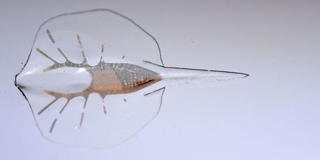
This soft robotic stingray is made of rat heart muscle. Yeah, it's just as crazy as it sounds. "I THINK WE'VE GOT A BIOLOGICAL LIFE-FORM HERE."
"Roughly speaking, we made this thing with a pinch of rat cardiac cells, a pinch of breast implant, and a pinch of gold. That pretty much sums it up, except for the genetic engineering," says Kit Parker, the bio-engineer at Harvard who led the team that developed the strange robot.
Parker's robotic stingray is tiny—a bit more than half an inch long—and weighs only 10 grams. But it glides through liquid with the very same undulating motion used by fish like real stingrays and skates. The robot is powered by the contraction of 200,000 genetically engineered rat heart-muscle cells grown on the underside of the bot. Even stranger, Parker's team developed the robot to follow bright pulses of light, allowing it to smoothly twist and turn through obstacle courses. The fascinating robot was unveiled today in the journalScience. "By using living cells they were able to build this robot in a way that you just couldn't replicate with any other material," says Adam Feinberg, a roboticist at Carnegie Mellon University who has worked with Parker's team before, but was not involved in developing this new robot. "You shine a light, and it triggers the muscles to swim. You couldn't replicate this movement with on-board electronics and actuators while keeping it lightweight and maneuverable. And it really is remote controlled, like a TV set."
|
|
Scooped by
Dr. Stefan Gruenwald
July 6, 2016 1:47 PM
|
Researchers at MIT and elsewhere have developed a new combination of methods that can provide detailed information about the microstructure of polycrystalline metals. Such materials—composed of a random matrix of multiple small crystals rather than one single large crystal—are widely used for such applications as nuclear reactors, civil infrastructure, and aircraft. However understanding the details of their crystal structure and the boundaries between the crystal areas has been difficult. The new findings are published in the journal Nature Computational Materials, in a paper by Matteo Seita, an MIT postdoc; Michael Demkowicz, a professor of materials science and engineering; Christopher Schuh, the Danae and Vasilis Salapatas Professor of Metallurgy, and five others. "This is a unique combination of different technologies," Seita explains. The new approach he and the team developed addresses "one of the most common problems in materials science: How do we quantify the characteristics of materials in a high-throughput fashion?" Some techniques offer a great deal of detail about structures, but they take time to carry out and can't reveal rapid changes within the material. Others work rapidly but provide much less structural detail, and still other methods provide both spatial and temporal detail but are prohibitively expensive or only available in limited places. The new combination of techniques, Seita says, can help resolve these limitations by providing fast, high-resolution, and low-cost imaging of the materials. In polycrystalline metals, which are composed of many small crystal grains, it is important to know the location, dimensions, angles of contact, and other characteristics of the different grains making up the material. In particular, the interfaces between the crystal grains, called grain boundaries, "happen to be critical," Seita says, "to many individual properties of the material—its strength, radiation tolerance, hardness, electrical resistance, and so on—but they are very difficult to characterize experimentally, because they are very complex." There are five basic characteristics about these grain boundaries that researchers would like to be able to quantify, but most tools for studying the materials can only yield some subset of two or three of those. One method for getting all five characteristics at once is high-energy synchrotron radiation, which is only available in a few facilities that are expensive and tend to be oversubscribed.
|
|
Scooped by
Dr. Stefan Gruenwald
July 6, 2016 1:41 PM
|
Located some 23 light years away, the brown dwarf designated WISEP J060738.65+242953.4 or W0607+24 for short, turns out to be a source of radio emission. According to a research paper published July 4, this substellar object showcases quiescent radio emission, making it one of the most radio-faint, ultra-cool dwarfs yet detected. Brown dwarfs like W0607+24 are objects that are too large to be called planets and too small to be stars. With a mass below that necessary to maintain hydrogen-burning nuclear fusion reactions, they are much cooler and dimmer than main sequence stars. These objects could be also sources of radio emission, but the cause of this process is still not completely understood. Due to its proximity, W0607+24 is an excellent target for studying the nature of brown dwarfs. It is the nearest known late-L dwarf in the northern hemisphere, and the third-nearest in the whole sky, allowing researchers to study phenomena occurring in brown dwarfs. That's why a team of astronomers, led by John Gizis of the University of Delaware, Newark, carried out a series of mid-infrared, radio, and optical observations of W0607+24 to study any activity in this curious substellar object. Motivated by the results from NASA's prolonged Kepler mission, named K2, which obtained long time-series photometry of W0607+24, they observed this object simultaneously with the Karl G. Jansky Very Large Array (VLA) in New Mexico and NASA's Spitzer Space Telescope. The most important results were provided by VLA, as the team found that W0607+24 is a source of quiescent, non-bursting, radio emission. The scientists noted that the discovery of this process in this nearby object could be crucial for our understanding of this process in ultracool dwarfs in general. "W0607+24 is a valuable test case for understanding the mechanisms of radio emission in ultracool dwarfs. Some of the radio-detected objects emit rapid, intense bursts with high circular polarization, often found to occur periodically and attributed to coherent emission due to the electron cyclotron maser instability," the paper reads. The astronomers also suggested that the relative radio faintness of W0607+24 may be due to its orientation if the emission is concentrated at the poles. However, follow-up observations are needed to better characterize the detected radio emission and to eliminate all the uncertainties. The team also hopes for more significant findings when studying this nearby substellar object in the future.
|
|
Scooped by
Dr. Stefan Gruenwald
July 5, 2016 2:27 PM
|
More than 100 Nobel laureates have signed a letter urging Greenpeace to end its opposition to genetically modified organisms (GMOs). The letter asks Greenpeace to cease its efforts to block introduction of a genetically engineered strain of rice that supporters say could reduce Vitamin-A deficiencies causing blindness and death in children in the developing world. "We urge Greenpeace and its supporters to re-examine the experience of farmers and consumers worldwide with crops and foods improved through biotechnology, recognize the findings of authoritative scientific bodies and regulatory agencies, and abandon their campaign against 'GMOs' in general and Golden Rice in particular," the letter states. The letter campaign was organized by Richard Roberts, chief scientific officer of New England Biolabs and, with Phillip Sharp, the winner of the 1993 Nobel Prize in physiology or medicine for the discovery of genetic sequences known as introns. The campaign has a website, supportprecisionagriculture.org, that includes a running list of the signatories, and the group plans to hold a news conference Thursday morning at the National Press Club in Washington. “We’re scientists. We understand the logic of science. It's easy to see what Greenpeace is doing is damaging and is anti-science," Roberts told The Washington Post. “Greenpeace initially, and then some of their allies, deliberately went out of their way to scare people. It was a way for them to raise money for their cause." Roberts said he endorses many other activities of Greenpeace, and said he hopes the group, after reading the letter, would "admit that this is an issue that they got wrong and focus on the stuff that they do well."
|
|
Rescooped by
Dr. Stefan Gruenwald
from DNA and RNA research
July 5, 2016 2:18 PM
|
University of Pennsylvania engineers have developed a rapid, low-cost genetic test for the Zika virus. University of Pennsylvania engineers have developed a rapid, low-cost genetic test for the Zika virus. The $2 testing device, about the size of a soda can, does not require electricity or technical expertise to use. A patient would simply provide a saliva sample. Color-changing dye turns blue when the genetic assay detects the presence of the virus. Rapid, accurate diagnosis is especially important for pregnant women who may be infected. However, the only approved tests for the virus currently require highly sensitive laboratory equipment. Diagnostic tools that can be used in the field, while the patient waits, would be a critical tool for fighting the Zika epidemic. The engineers demonstrated the design and efficacy of their test in a study published in the journal Analytical Chemistry. It was conducted by Research Assistant Professor Changchun Liu and Professor Haim Bau of the Department of Mechanical Engineering and Applied Mechanics in Penn’s School of Engineering and Applied Science, along with members of the Bau lab, Jinzhao Song and Michael Mauk. They collaborated with Sara Cherry, associate professor of microbiology in Penn’s Perelman School of Medicine, and Brent Hackett, a member of her lab. Assays that detect genetic material from the Zika virus itself are considered the gold standard in diagnostics. Alternatives, such as tests that look for antibodies the body creates in response to the virus, are insufficient as they may produce false negatives from people who are infected but haven’t yet produced enough antibodies, or false positives from people who have antibodies for a different disease that is similar enough to trigger the test. Tests that look for RNA sequences from the virus itself, known as reverse transcriptase polymerase chain reaction, or RT-PCR, avoid both problems. However, RT-PCR requires delicate laboratory work. Viral gene sequences in a patient’s sample must be amplified, or repeatedly copied, to levels where they can be detected, a process that normally involves multiple precise temperature changes. “The CDC has approved, on an emergency basis, only these kinds of laboratory-based molecular tests for the Zika virus,” said Liu. “Generally, lateral flow tests, which directly change the color of a test strip based on the presence of Zika antibodies, suffer from low sensitivity. And since antibodies to the Zika virus cross-react with other similar viruses prevalent in Zika-endemic areas, lateral flow tests for Zika also suffer from low specificity.” With the amplification step as the main hurdle to a portable genetic test, the Penn researchers investigated the possibility of using an alternative technique known as RT-LAMP, or reverse transcription loop-mediated isothermal amplification, which only requires the sample to be kept at a specific temperature, not cycled through multiple precise temperature changes as in RT-PCR. As a trade-off for this simplified amplification process, RT-LAMP requires even more specialized “primers,” short gene sequences that are designed to match the regions of the virus’ DNA targeted by the test. “Although Zika primers for RT-PCR have been published in the literature, RT-LAMP primers have not,” Bau said. “So, using data mining, we identified highly conserved regions of the Zika virus genome that are divergent from other known pathogens. We then designed appropriate primers to recognize this sequence.
Via Integrated DNA Technologies
|
|
Scooped by
Dr. Stefan Gruenwald
July 5, 2016 2:01 PM
|
Materials researchers at the Swiss Paul Scherrer Institute PSI in Villigen and the ETH Zurich have developed a very simple and cost-effective procedure for significantly enhancing the performance of conventional Li-ion rechargeable batteries. The procedure is scalable in size, so the use of rechargeable batteries will be optimized in all areas of application-whether in wristwatches, smartphones, laptops or cars. Battery storage capacity will be significantly extended, and charging times reduced. The researchers reported on their results in the latest issue of the research journal Nature Energy.
|
|
Scooped by
Dr. Stefan Gruenwald
July 5, 2016 1:54 PM
|
A new use of chemotherapy followed by autologous hematopoietic stem cell transplantation (aHSCT) has fully halted clinical relapses and development of new brain lesions in 23 of 24 patients with multiple sclerosis (MS) for a prolonged period without the need for ongoing medication, according to a new phase 2 clinical trial, published in The Lancet. Eight of the 23 patients had a sustained improvement in their disability 7.5 years after treatment. This is the first treatment to produce this level of disease control or neurological recovery from MS, but treatment related risks limit its widespread use. MS is among the most common chronic inflammatory diseases of the central nervous system, with around 2 million people affected worldwide. It is caused when the immune system attacks the body, known as autoimmunity. Some specialist centres offer aHSCT for MS, which involves harvesting bone marrow stem cells from the patient, using chemotherapy to suppress the patient's immune system, and reintroducing the stem cells into the blood stream to "reset" the immune system to stop it attacking the body. However, many patients relapse after these treatments, so more reliable and effective methods are needed. Dr Harold L Atkins and Dr Mark S Freedman from The Ottawa Hospital and the University of Ottawa, Ottawa, Canada, and colleagues tested whether complete destruction, rather than suppression, of the immune system during aHSCT would reduce the relapse rate in patients and increase long-term disease remission. They enrolled 24 patients aged 18-50 from three Canadian hospitals who had all previously undergone standard immunosuppressive therapy which did not control the MS. All patients had poor prognosis and their disability ranged from moderate to requiring a walking aid to walk 100m, according to their Expanded Disability Status Scale (EDSS) scores [1]. The researchers used a similar method of aHSCT as is currently used, but instead of only suppressing the immune system before transplantation, they destroyed it completely using a chemotherapy regimen of busulfan, cyclophosphamide and rabbit anti-thymocyte globulin. Dr Atkins explains that this treatment is "similar to that used in other trials, except our protocol uses stronger chemotherapy and removes immune cells from the stem cell graft product. The chemotherapy we use is very effective at crossing the blood-brain barrier and this could help eliminate the damaging immune cells from the central nervous system." The primary outcome of the study was multiple sclerosis activity-free survival at 3 years (as measured by relapses of MS symptoms, new brain lesions, and sustained progression of EDSS scores) which occurred in 69.6% of patients after transplantation. Out of the 24 patients, one (4%) died from hepatic necrosis and sepsis caused by the chemotherapy. Prior to the treatment, patients experienced 1.2 relapses per year on average. After treatment, no relapses occurred during the follow up period (between 4 and 13 years) in the surviving 23 patients. These clinical outcomes were mirrored by freedom from detectable new disease activity on MRI images taken after the treatment. The initial 24 MRI scans revealed 93 brain lesions, and after the treatment only one of the 327 scans showed a new lesion. Furthermore, progressive brain deterioration typical of MS slowed to a rate associated with normal aging in 9 patients with the longest follow-up, and 8 (35%) of 23 patients had a sustained improvement in their EDSS score at 7.5 years after treatment. At 3 years, 6 patients (37%) were able to reduce or stop receiving disability insurance and return to work or school. Eight (33%) of the 24 patients had a moderate toxic effect and 14 (58%) patients had only a mild toxic effect related to transplantation. Dr Freedman highlights the need to interpret the results with caution: "The sample size of 24 patients is very small, and no control group was used for comparison with the treatment group. Larger clinical trials will be important to confirm these results. Since this is an aggressive treatment, the potential benefits should be weighed against the risks of serious complications associated with aHSCT, and this treatment should only be offered in specialist centers experienced both in multiple sclerosis treatment and stem cell therapy, or as part of a clinical trial. Future research will be directed at reducing the risks of this treatment as well as understanding which patients would best benefit from the treatment."[
|
|
Scooped by
Dr. Stefan Gruenwald
July 2, 2016 11:41 AM
|
A million-face benchmark test shows that even Google's facial recognition algorithm suffers in accuracy. Facial recognition algorithms that had previously performed with more than 95 percent accuracy on a popular benchmark test involving 13,000 faces saw significant drops in accuracy when faced with the new MegaFace Challenge involving one million faces. The best performer on one test, Google’s FaceNet algorithm, dropped from near-perfect accuracy on five-figure datasets to 75 percent on the million-face test. Other top algorithms dropped from above 90-percent accuracy on the small datasets to below 60 percent on the MegaFace Challenge. Some algorithms made the proper identification as seldom as 35 percent of the time. “Megaface's key idea is that algorithms should be evaluated at large scale,” says Ira Kemelmacher-Shlizerman, an assistant professor of computer science at the University of Washington in Seattle and the project’s principal investigator. “And we make a number of discoveries that are only possible when evaluating at scale.” The huge drops in accuracy when scanning a million faces matters because facial recognition algorithms inevitably face such challenges in the real world. People increasingly trust facial recognition algorithms to correctly identify them in security verification scenarios for automatically unlocking smartphones or entering workplaces. Law enforcement officials may also rely on facial recognition algorithms to find the correct match to a single photo of a suspect among hundreds of thousands of faces captured on surveillance cameras. The most popular benchmark test until now has been the Labeled Faces in the Wild (LFW) test created in 2007. LFW includes 13,000 images of just 5,000 people. Many facial recognition algorithms have been fine-tuned to the point that they scored near-perfect accuracy when picking through that smaller set of images, which may have created a false sense of confidence about the state of facial recognition. “The big disadvantage is that [the field] is saturated, i.e. there are many, many algorithms that perform above 95 percent on LFW,” Kemelmacher-Shlizerman explains. “This gives the impression that face recognition is solved and working perfectly.” With that in mind, University of Washington researchers raised the bar by creating the MegaFace Challenge using one million Flickr images that are publicly available under a Creative Commons license. The MegaFace dataset includes one million images featuring 690,000 unique faces. The MegaFace Challenge forces facial recognition algorithms to doverification and identification, two separate but related tasks. Verification involves trying to correctly determine whether two faces presented to the facial recognition algorithm belong to the same person. Identification involves trying to find a matching photo of the same person among a million “distractor” faces. Initial results appear in a paper that was presented at the IEEE Conference on Computer Vision and Pattern Recognition (CVPR 2016) on 30 June.
|
|
Scooped by
Dr. Stefan Gruenwald
June 30, 2016 10:02 PM
|
Robots so small they can enter the bloodstream and perform surgeries are one step closer, a research team from Monash University has discovered. Led by Dr Zhe Liu, the Monash Engineering team has focused on graphene oxide – which is a single atom thick – as an effective shape memory material. Graphene has captured world scientific and industrial interest for its miracle properties, with potential applications across energy, medicine, and even biomedical nano-robots. Until now shape memory effects have only been observed in materials larger than approximately 10nm. Graphene Oxide is approximately 1nm thick. In contrast to other shape memory materials, the Monash team discovered that subjecting certain forms of graphene oxide to an electric field caused shape changes almost instantaneously and retained the new form until stretched back to its original shape. PhD candidate and first author Zhenyue Chang, said the shape memory effect arises from an "atomic switch" enabling a super-fast response. "Aside from being able to transform at high speeds, graphene oxide has many other advantages over existing shape memory materials. It is incredibly light, has a high density to strain ratio, is very stable, and is able to perform a relative size change of 15 per cent, compared to shape memory alloys, which only change by four per cent. "Like the science fiction movie, Fantastic Voyage, our research brings one step closer intelligent biomedical nano-robots that can be delivered into a living cell for future cellular surgery," Dr Liu said. Dr Liu's team, from the Monash Centre for Atomically Thin Materials (MCATM) and Mechanical and Aerospace Engineering department at Monash University, made their discovery through computer simulations. "We are excited now to use our research to create the compound we have predicted. That would be amazing," Dr Liu said.
|
|
Scooped by
Dr. Stefan Gruenwald
June 30, 2016 9:36 PM
|
Scientists from Moscow Institute of Physics and Technology (MIPT), the Institute for Theoretical and Experimental Physics, and the National Research University Higher School of Economics have devised a method of distinguishing black holes from compact massive objects that are externally indistinguishable from one another. The method involves studying the energy spectrum of particles moving in the vicinity -- in one case it will be continuous and in the other it will be discrete. The findings have been published in Physical Review D. Black holes, which were predicted by Einstein's theory of general relativity, have an event horizon -- a boundary beyond which nothing, even light, can return to the outside world. The radius of this boundary is called the Schwarzschild radius, in physical terms it is the radius of an object for which the escape velocity is greater than the speed of light, which means that nothing is able to overcome its gravity. Black holes of stellar mass are the result of gravitational collapse which occurs at the time when a star "burns out" all its thermonuclear fuel and the force of the gas pressure can no longer resist gravity. If the star is massive enough, it collapses to a size smaller than the Schwarzschild radius and turns into a black hole. However, time on the event horizon slows down so much that for an outside observer the collapsing process almost stops (if a ship falls into a black hole, for example, to an outside observer it will appear to be continually falling toward the horizon), therefore all the black holes we see are objects that are eternally collapsing. Astrophysicists have not yet been able to "see" a black hole directly, but there are many objects that are "suspected" of being black holes. Most scientists are sure that in the centerof our galaxy there is a supermassive black hole; there are binary systems where one of the components is most likely a black hole. However, some astrophysicists believe that there may be compact massive objects that fall very slightly short of black hole status; their range is only a little larger than the Schwarzschild radius. It may be the case that some of the "suspects" are in fact objects such as these. From the outside, however, they are not distinguishable from black holes. Emil Akhmedov, Fedor Popov, and Daniil Kalinov devised a method to tell the difference between them, or more precisely the difference between compact massive objects and collapsing objects. "We examined the scalar quantum field around a black hole and a compact object and found that around the collapsing object -- the black hole, there are no bound states, but around the compact object there are," explains FedorPopov, a member of staff at MIPT's Laboratory of High Energy Physics. He and his colleagues examined the behavior of scalar particles (the spin of these particles is zero -- an example of this could be the Higgs boson) in the vicinity of black holes and massive compact objects. The scientists derived analytical expressions for the energy spectrum of the particles. It was found that near the surface of an ultra-compact star with a radius slightly larger than the Schwarzschild radius there is a "potential hole" -- an area of space where particles fall into a gravitational "trap." The problem in this case is then similar to a simple task in quantum mechanics where the spectrum of the particles in the potential hole needs to be found. This spectrum is discrete, i.e. it has energy values where there are no particles. In simpler terms, the potential hole does not release particles of certain energies, and an "empty space" appears in the spectrum.
|