Your new post is loading...

|
Scooped by
Dr. Stefan Gruenwald
October 6, 2016 1:58 PM
|
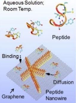
Engineers at the University of Washington have created genetically engineered peptides that self-assemble into arrays of nanowires on two-dimensional nanosheets (single-layer graphene and molybdenum disulfide) to relay information across a bio-nano interface — a first step towards fully self-assembled future biomedical and electro-optical bionanoelectronic devices. Arrays of peptides could provide organized scaffolds for functional biomolecules, enabling nanoscale bioelectronics interfaces. And designed peptides could be incorporated with metal ions or nanoparticles with specific physical characteristics, thus fine-tuning 2D device performance for chemical and biological sensors. “Bridging this divide would be the key to building the genetically engineered biomolecular solid-state devices of the future,” said UW professor Mehmet Sarikaya in the Departments of Materials Science & Engineering, senior author ofan open-access paper published Sept. 22 in Scientific Reports. The UW team is also planning to develop genetically engineered peptides with specific chemical and structural properties. Their ideal peptide would change the physical properties of synthetic materials and respond to that change. That way, it would transmit “information” from the synthetic material to other biomolecules — bridging the chemical divide between biology and technology. The peptides function through molecular recognition — the same principles that underlie biochemical interactions such as an antibody binding to its specific antigen or protein binding to DNA.
|
|
Scooped by
Dr. Stefan Gruenwald
October 6, 2016 1:54 PM
|
The Nobel Prize in Chemistry 2016 was awarded today to Jean-Pierre Sauvage, PhD, Sir J. Fraser Stoddart,PhD, and Bernard L. Feringa, PhD, for their design and production of molecular machines. They have developed molecules with controllable movements, which can perform a task when energy is added. The first step towards a molecular machine was taken by Jean-Pierre Sauvage in 1983, when he succeeded in linking two ring-shaped molecules together to form a chain, called a catenane. Normally, molecules are joined by strong covalent bonds in which the atoms share electrons, but in the chain they were instead linked by a freer mechanical bond. For a machine to be able to perform a task it must consist of parts that can move relative to each other. The two interlocked rings fulfilled exactly this requirement. The second step was taken by Fraser Stoddart in 1991, when he developed a rotaxane. He threaded a rotaxane cyclophane molecular ring onto a thin molecular axle and demonstrated that the ring was able to move along the axle — the start of applying topological entanglement in the development of molecular machinery. Among his other developments based on rotaxanes are a molecular lift, a molecular muscle and a molecule-based computer chip. Bernard Feringa was the first person to develop a molecular motor; in 1999 he got a molecular rotor blade to spin continually in the same direction. Using molecular motors, he has also rotated a glass cylinder that is 10,000 times bigger than the motor and also designed a nanocar. 2016′s Nobel Laureates in Chemistry have taken molecular systems out of equilibrium’s stalemate and into energy-filled states in which their movements can be controlled. In terms of development, the molecular motor is at the same stage as the electric motor was in the 1830s, when scientists displayed various spinning cranks and wheels, unaware that they would lead to electric trains, washing machines, fans and food processors. Molecular machines will most likely be used in the development of things such as new materials, sensors and energy storage systems.
|
|
Scooped by
Dr. Stefan Gruenwald
October 4, 2016 4:42 PM
|
A star known by the unassuming name of KIC 8462852 in the constellation Cygnus has been raising eyebrows both in and outside of the scientific community for the past year. In 2015 a team of astronomers announced that the star underwent a series of very brief, non-periodic dimming events while it was being monitored by NASA’s Kepler space telescope, and no one could quite figure out what caused them. A new study from Carnegie’s Josh Simon and Caltech’s Ben Montet has deepened the mystery. Simon and Montet’s findings caused a stir in August, when they were posted on a preprint server while their paper was being reviewed. Now their work is now accepted for publication by The Astrophysical Journal. The researchers analyzed further Kepler observations of the puzzling star and showed that in addition to its rapid unexplained brightness changes, the star also faded slowly and steadily during the four years it was watched by Kepler. Speculation to explain KIC 8462852’s dips in brightness has ranged from an unusually large group of comets orbiting the star to an alien megastructure. In general, stars can appear to dim because a solid object like a planet or a cloud of dust and gas passes between it and the observer, eclipsing and effectively dimming its brightness for a time. But the erratic pattern of abrupt fading and re-brightening in KIC 8462852 is unlike that seen for any other star. Spurred by a controversial claim that the star’s brightness gradually decreased by 14 percent from 1890 to 1989, Montet and Simon decided to investigate its behavior in a series of Kepler calibration images that had not previously been used for scientific measurements. “We thought that these data could confirm or refute the star’s long-term fading, and hopefully clarify what was causing the extraordinary dimming events observed in KIC 8462852,” explained Simon. Simon and Montet found that, over the first three years of the Kepler mission, KIC 8462852 dimmed by almost 1 percent. Its brightness then dropped by an extraordinary 2 percent over just six months, remaining at about that level for the final six months of the mission. The pair then compared this with more than 500 similar stars observed by Kepler and found thata small fraction of them showed fading similar to that seen in KIC 8462852 over the first three years of Kepler images. However, none exhibited such a dramatic dimming in just six months, or a total change in brightness of 3 percent. “The steady brightness change in KIC 8462852 is pretty astounding,” said Montet. “Our highly accurate measurements over four years demonstrate that the star really is getting fainter with time. It is unprecedented for this type of star to slowly fade for years, and we don’t see anything else like it in the Kepler data.” "This star was already completely unique because of its sporadic dimming episodes. But now we see that it has other features that are just as strange, both slowly dimming for almost three years and then suddenly getting fainter much more rapidly,” Simon added. Astronomers were already running short of good ideas to account for the dips in KIC 8462852’s brightness, and the new results will make that task even harder. Simon and Montet think that the best proposal so far for explaining the star’s drastic six month dimming might be a collision or breakup of a planet or comet in the star’s system, creating a short-term cloud of dust and debris that blocks some starlight. However, this wouldn’t explain the longer-term dimming observed during the first three years of Kepler and suggested by measurements of the star dating back to the nineteenth century. “It’s a big challenge to come up with a good explanation for a star doing three different things that have never been seen before,” Montet said. “But these observations will provide an important clue to solving the mystery of KIC 8462852.”
|
|
Scooped by
Dr. Stefan Gruenwald
October 3, 2016 4:50 PM
|
Scientists from Japan have created touchable holograms which are named as Fairy lights. This development could well lead to human beings able to interact with holograms. In particular, this technology could do very well in medicine industry as it can be used to demonstrate complex procedures. According to the research paper presented by the scientists, the holograms used during the research were made from femtosecond lasers, “a laser that can excite physical matter to emit light in 3D form” explained by the paper. Furthermore, the lasers can product anywhere between 1000 to 200,000 pulses per second. The rate for frequency depends on the type of lasers used. During tests, pulses responded to the human touch which made it possible for them to be disrupted in the air.
|
|
Scooped by
Dr. Stefan Gruenwald
October 3, 2016 4:39 PM
|
In a recent post about biohacking, I wrote about people who have implanted chips into their bodies to benefit their health, simplify their lives, or connect themselves to an external network. Though some have been quick to adopt it, biohacking is still a relatively new and bizarre trend that makes many people wary. The thought of burying chips in our arms is unsettling, and most of us would only do it if it was medically necessary. But for those who are curious yet not quite ready to take the chip-implantation plunge, there’s now another way to join the biohacking party: temporary tattoos. Created by MIT PhD student Cindy Hsin-Liu Kao in conjunction with Microsoft Research, the Duoskintattoos transfer onto your skin with water, and they can be customized for both aesthetic and functional purposes. Hsin-Liu Kao presented her paper about the tattoos at the International Symposium on Wearable Computers in Heidelberg, Germany last week. The first step in creating a tattoo is to make a tiny circuit board using graphic design software. A stencil of the circuit is created by applying a layer of vinyl film onto tattoo paper, then gold leaf is layered over the stencil to act as conductive material. The last step is to surface-mount electronics. All tattoos except those with an NFC chip connect to a microcontroller that processes sensor data, supplies power, and links devices through Bluetooth. The total cost of creating a three by four centimeter squared NFC tag is less than $2.50. In trials, the team tested conductive thread and copper tape as alternatives to gold leaf, but found gold leaf to be the most durable and the most skin-friendly.
|
|
Scooped by
Dr. Stefan Gruenwald
October 2, 2016 10:11 PM
|
The Internet of Things-infecting malware that launched a historic DDoS attack is now free for anyone to use. It’s only been a few weeks since hackers hijacked more than 1 million Internet of Things (IoT) devices to launch a record-breaking Distributed Denial of Service (DDoS) attack against the website of cybersecurity reporter Brian Krebs. Now, the malware that powered the unprecedented attack has been released online for anyone to use. A link to the malware code, first spotted by Krebs, was posted in the criminal hacker site Hackforum by a user named “Anna-senpai,” who dubbed the malware “Mirai.” The malware is designed to infect Internet of Things (IoT) devices that haven’t changed their default usernames and passwords—a common occurrence in the frighteningly poor security used by IoT products like web cams, “smart” refrigerators, and other internet-connected home appliances. Once assembled, these massive armies of zombie devices can be controlled from a central server, where they are typically leased out to other criminal hackers to launch DdoS attacks against target websites. “When I first go in DDoS industry, I wasn’t planning on staying in it long. I made my money, there’s lots of eyes looking at IOT now, so it’s time to GTFO,” Anna-senpai wrote. “So today, I have an amazing release for you. With Mirai, I usually pull max 380k bots from telnet alone. However, after the Kreb DDoS, ISPs been slowly shutting down and cleaning up their act. Today, max pull is about 300k bots, and dropping.” “So, I am your senpai, and I will treat you real nice, my hf-chan,” Anna-senpai added, cheekily using the Japanese honorific for a fellow classmate. It’s unclear why the malware’s authors chose to dump the code online, since the ability to grow botnets can be a big moneymaking asset in the criminal hacking world. But Anna-chan’s Hackforums post seems to suggest that spreading the malware code around is a way for its creators to confuse attribution attempts, now that the Krebs DDoS attack has brought increased attention. "Miscreants who develop malicious software often dump their source code publicly when law enforcement investigators and security firms start sniffing around a little too close to home,” Krebs writes. “Publishing the code online for all to see and download ensures that the code’s original authors aren’t the only ones found possessing it if and when the authorities come knocking with search warrants.”
|
|
Scooped by
Dr. Stefan Gruenwald
October 1, 2016 11:24 PM
|
Single photons are ideally suited for sending information in digital form so that each photon encodes a 0 or a 1. In that case, it’s easy to imagine that this is all the data that a single photon can hold. Not so! In theory, there is no limit to the amount of information a single photon can encode. And that raises an interesting question. How much information can physicists pack into a single photon in practice? What does current technology allow? Today we get an answer thanks to the work of Tristan Tentrup and pals at the University of Twente in the Netherlands. They have packed more than 10 bits into a single photon for the first time. Their method is straightforward, in theory. The approach is to associate a single photon with a unique member of an alphabet. When the alphabet contains lots of members, the photon carries lots of information. It’s not hard to see why. When an alphabet contains only two members, such as binary code, each member encodes one bit of information. This is the amount of information needed to describe each symbol in the alphabet. But when the alphabet is bigger, it takes more information to uniquely describe each member. So each member can encode that amount of data. The actual amount of information is given by the log to base 2 of the number of members. For example, in an alphabet of 10 symbols, such as each decimal number, each symbol encodes about 3.3 bits. In an alphabet of 26 symbols, such as the English alphabet, each symbol encodes 4.7 bits. And so on. Tentrup and co achieve their goal by creating an alphabet with 9,072 symbols. In that case, each symbol encodes more than 13 bits of information. Creating this alphabet is simple. Tentrup and co do it by defining a 112 x 81 grid of pixels—that’s 9,072 of them. Each pixel represents a different symbol of the alphabet. To encode a photon with one of these symbols, all they have to do is point the photon toward that part of the grid. So when a specific pixel registers the arrival of a photon, it registers that symbol. The tricky part is doing this accurately with single photons. One way to steer photons is with a tilting mirror that simply reflects them in a specific, controllable direction. But Tentrup and co use a more flexible device called a spatial light modulator which modifies a photon’s wavefront as it reflects it. This uses diffraction effects to steer the photon toward its target. Detecting single photons is also a potential banana skin, since any stray light can overwhelm the signal. Tentrup and co have a handy trick for preventing this. Instead of creating single photons, they create them in pairs and encode just one of them with information using this steering mechanism. They look out for the other as a warning that the first is about to arrive at the pixel. This allows them to switch on the pixel at the very instant the first photon arrives. And this dramatically reduces the chances of a stray photon swamping the signal. Nevertheless, noise still has an impact and the photons end up carrying slightly less information than the theoretical maximum.
|
|
Scooped by
Dr. Stefan Gruenwald
October 1, 2016 11:20 PM
|
In an unexpected discovery, researchers have found that the complex eyes of mantis shrimp are equipped with optics that generate ultraviolet (UV) color vision. Mantis shrimp's six UV photoreceptors pick up on different colors within the UV spectrum based on filters made from an ingredient other animals depend on as built-in biological sunscreen, according to research reported in the Cell Press journal Current Biology on July 3, 2016. "The mantis shrimp visual system contains six types of photoreceptors functioning completely outside the visual range of humans," says Michael Bok of the University of Maryland Baltimore County. "Surprisingly, they produce their six UV photoreceptors using only two types of visual pigments by pairing one visual pigment with one of four UV filters. The UV filters block certain wavelengths of light from reaching the photoreceptors, chromatically shifting their sensitivity." The filters are composed of so-called mycosporine-like amino acids (or MAAs), which are commonly found in the skin or exoskeleton of marine organisms, where they absorb damaging UV rays. They do the same thing in mantis shrimp eyes, but for an entirely novel purpose. "The effect is akin to putting red-tinted glasses over your eyes that block other wavelengths of light, except this is being done at the photoreceptor cellular level in shrimp," Bok explains. Exactly why mantis shrimp need such a sophisticated visual system remains mysterious, Bok says. Mantis shrimp use their eyes to navigate and spot predators and prey on the vibrant reef that is their home. Mantis shrimp also have complex social interactions that are likely mediated by distinct visual signals on their bodies. Their complex eyes, which include 16 or more types of photoreceptors in all, may provide them with a complex color and polarization visual system without a big brain to post-process lots of information. In other words, their eyes may sense and respond to complex visual inputs without the need to think very hard about it, Bok explains. Despite the new discovery, the researchers say, it's still tough to imagine the reef as mantis shrimp see it. "The way their eyes are built and how visual information is processed in their brains is so fundamentally different [from] humans that is very difficult to conceptualize what the world actually looks like to them," Bok says.
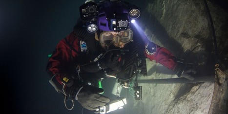
Czech abyss is so deep the Empire State Building could fit inside. A team of explorers has found the deepest underwater cave yet discovered, with a bottom that lies at least 1,325 feet below the water's surface. Polish explorer Krzysztof Starnawski, who led the team, first explored the cave -- named Hranická Propast and located near the Czech town of Hranice -- in 1999, and instantly knew it was an unusual find. He told National Geographic, which sponsored his most recent expedition, that hot water saturated with carbon dioxide bubbled up like a volcano, and made his exposed skin itch. A series of dives over the years hinted at the abyss' depth. In 2014, he reached 656 feet, thinking he had found the bottom, only to discover a very narrow opening that led to a vertical tunnel. The following year, some of the rock in the cave had crumbled, widening the opening and making it possible for him to squeeze through. He reached a depth of 869 feet and released a probe, which at 1,214 feet landed on what was probably a pile of debris from the collapsed passage above.
Via Kathy Bosiak
Carbon dioxide levels often hit lows in September, but now remain above a crucial benchmark. In the centuries to come, history books will likely look back on September 2016 as a major milestone for the world’s climate. At a time when atmospheric carbon dioxide is usually at its minimum, the monthly value failed to drop below 400 parts per million. That all but ensures that 2016 will be the year that carbon dioxide officially passed the symbolic 400 ppm mark, never to return below it in our lifetimes, according to scientists. Because carbon pollution has been increasing since the start of the Industrial Revolution and has shown no signs of abating, it was more a question of “when” rather than “if” we would cross this threshold. The inevitability doesn’t make it any less significant, though. September is usually the month when carbon dioxide is at its lowest after a summer of plants growing and sucking it up in the northern hemisphere. As fall wears on, those plants lose their leaves, which in turn decompose, releasing the stored carbon dioxide back into the atmosphere. At Mauna Loa Observatory, the world’s marquee site for monitoring carbon dioxide, there are signs that the process has begun but levels have remained above 400 ppm.
Via Kathy Bosiak
|
|
Scooped by
Dr. Stefan Gruenwald
September 30, 2016 8:31 PM
|
Watching another episode on Netflix, reading the Guardian online and downloading apps are not obvious ways to pollute the atmosphere. But collectively, our growing appetite for digital services means the data centers that power them are now responsible for about 2% of global greenhouse gas emissions, a similar share to aviation. Varying from a small room with servers to vast farms with a floor area of 150,000 square meters, data centers are big energy users. As well as requiring power to run the equipment that stores and serves us cloud computing and on-demand music, films and entertainment, those servers also generate a lot of heat and require huge amounts of energy to keep them cool. That’s why big data users such as Facebook are siting their centers in cool climates such as northern Sweden. Individually, our everyday browsing has a relatively minuscule impact. Google, in response to claims that each search on its site generated as much CO2 as boiling half the water for a cup of coffee (7g), calculated the true figure was much lower, at 0.2g. Watching a YouTube video of cats was higher – 1g for every 10 minutes of viewing – while using Gmail for a year produced about 1.2kg a user. Not to be outdone, Facebook put a figure on its average user’s annual footprint – 269g of CO2, roughly equivalent to the carbon footprint of a cup of coffee. But with the technology giants’ billions of users worldwide, those numbers scale up quickly. Google’s carbon footprint was 1,766,014 tons CO2 equivalent in 2013, and the bulk of that was from data centers. As with emissions from air travel, the real issue with data centers is the rate at which they are growing. “It’s an exponential growth in data,” said Sophia Flucker, director at Operational Intelligence, a UK-based consultancy that advises data centers on their energy use. “Although IT efficiency is improving, and we can do more with less power, the demand is still there,” she said. “Any efficiency gains are being eaten up by demand. It’s very much an upward trajectory.”
|
|
Scooped by
Dr. Stefan Gruenwald
September 30, 2016 8:26 PM
|
A study led by researchers at the University of Washington and the National Oceanic and Atmospheric Administration connects the unprecedented West Coast toxic algal bloom of 2015 that closed fisheries from Southern California. "We have toxic algae events that result in shellfish closures off the Washington and Oregon coast every three to five years or so, but none of them have been as large as this one," said lead author Ryan McCabe, a research scientist at the UW's Joint Institute for the Study of the Atmosphere and Ocean, a collaborative center with NOAA. "This one was entirely different, and our results show that it was connected to the unusual ocean conditions." The study is now online in Geophysical Research Letters, a journal of the American Geophysical Union. "This paper is significant because it identifies a link between ocean conditions and the magnitude of the toxic bloom in 2015 that resulted in the highest levels of domoic acid contamination in the food web ever recorded for many species," said co-author Kathi Lefebvre, a marine biologist at NOAA's Northwest Fisheries Science Center. "This is an eye-opener for what the future may hold as ocean conditions continue to warm globally." The authors found that the 2015 harmful algal bloom, which set records for its spatial extent and the level of toxicity, was dominated by a single species of diatom, Pseudo-nitzschia australis, normally found farther south off California. Warm water not only allowed this species to survive, it also created an environment favoring its growth. By early 2015 the warm "blob" had moved toward shore and spread all along the West Coast. Warmer water creates less dense surface water that is more likely to stay floating on the surface, where it can become depleted in nutrients. Previous laboratory studies by co-author William Cochlan of San Francisco State University showed that P. australis can take up nitrogen very quickly from a variety of sources, and appear to outcompete other, nontoxic phytoplankton in nutrient-depleted warm water.
|
|
Scooped by
Dr. Stefan Gruenwald
September 30, 2016 6:02 PM
|
Light, when strongly concentrated, is enormously powerful. Now, a team of physicists led by Professor Jörg Schreiber from the Institute of Experimental Physics – Medical Physics, which is part of the Munich-Centre for Advanced Photonics (MAP), a Cluster of Excellence at LMU Munich, has used this energy source with explosive effect. The researchers focus high-power laser light onto beads of plastic just a few micrometers in size. The concentrated energy blows the nanoparticles apart, releasing radiation made up of positively charged atoms (protons). Such proton beams could be used in future for treating tumors, and in advanced imaging techniques. Their findings appear in the journal Physical Review E.
|
|
|
Scooped by
Dr. Stefan Gruenwald
October 6, 2016 1:55 PM
|
A new safer catheter design that allows cardiologists to see inside arteries for the first time and remove plaque from only diseased tissue has been used by interventional cardiologists at UC San Diego Health. The new image-guided device, Avinger’s Pantheris, allows doctors to see and remove plaque simultaneously during an atherectomy — a minimally invasive procedure that involves cutting plaque away from the artery and clearing it out to restore blood flow. The new technology treats patients suffering from the painful symptoms of peripheral artery disease (PAD), a condition caused by a build-up of plaque that blocks blood flow in the arteries of the legs and feet, preventing oxygen-rich blood from reaching the extremities. Patients with PAD frequently develop life threatening complications, including heart attack and stroke; in some severe cases, patients may even face amputation. PAD affects nearly 20 million adults in the United States and more than 200 million globally. “Peripheral artery disease greatly impacts quality of life, with patients experiencing cramping, numbness and discoloration of their extremities,” said Mitul Patel, MD, cardiologist at UC San Diego Health. “This new device is a significant step forward for the treatment of PAD with a more efficient approach for plaque removal and less radiation exposure to the doctor and patient.”
|
|
Scooped by
Dr. Stefan Gruenwald
October 6, 2016 1:52 PM
|
In a scientific first, a team of researchers from Macquarie University and the University of Vienna have developed a new technique to measure molecular properties – forming the basis for improvements in scientific instruments like telescopes, and with the potential to speed up the development of pharmaceuticals.
The study, titled "Quantum Optical Rotatory Disperson" and published in interdisciplinary journal Science Advances today, developed a new technique to allow chemists to learn about the position of atoms in a molecule – called quantum optical rotatory dispersion. This quantum method measures the chilarity of molecules, which is essentially how asymmetric the molecules are and is used across fields like biology, biomechemistry and physics, and has a practical application in pharmaceuticals among other sectors. The team found that quantum methods can allow for a more precise analysis of properties of molecules – meaning molecules can be examined even with a low concentration of the molecule or with less light. This new technique enables a more gentle analysis of samples that, for example, may be damaged by light. "We've found a way to analyse delicate samples by using less light," said lead author Nora Tischler, who carried out the research as part of her PhD, which she completed at Macquarie University between 2012 and 2016. "We hope to see this proof of concept built upon to eventually see efficiencies in the pharmaceutical sector, to help develop new medicines more productively." Macquarie's Associate Professor Gabriel Molina-Terriza from the Department of Physics and Astronomy co-authored the study, alongside postdoctoral fellow Xavier Vidal and researchers from the Austrian Academy of Sciences and University of Vienna. The lead author was jointly enrolled at the University of Vienna and Macquarie University as part of a cotutelle PhD program, spurring the international partnership between the two teams.
|
|
Scooped by
Dr. Stefan Gruenwald
October 4, 2016 12:18 PM
|
Blood testing is the standard option for checking glucose levels, but a new technology could allow non-invasive testing via a contact lens that samples glucose levels in tears. “There’s no noninvasive method to do this,” said Wei-Chuan Shih, a researcher with the University of Houston who worked with colleagues at UH and in Korea to develop the project, described in the high-impact journal Advanced Materials. “It always requires a blood draw. This is unfortunately the state of the art.” But glucose is a good target for optical sensing, and especially for what is known as surface-enhanced Raman scattering spectroscopy, said Shih, an associate professor of electrical and computer engineering whose lab, the NanoBioPhotonics Group, works on optical biosensing enabled by nanoplasmonics. This is an alternative approach, in contrast to a Raman spectroscopy-based noninvasive glucose sensor Shih developed as a Ph.D. student at the Massachusetts Institute of Technology. He holds two patents for technologies related to directly probing skin tissue using laser light to extract information about glucose concentrations. The paper describes the development of a tiny device, built from multiple layers of gold nanowires stacked on top of a gold film and produced using solvent-assisted nanotransfer printing, which optimized the use of surface-enhanced Raman scattering to take advantage of the technique’s ability to detect small molecular samples. Surface-enhanced Raman scattering – named for Indian physicist C.V. Raman, who discovered the effect in 1928 – uses information about how light interacts with a material to determine properties of the molecules that make up the material. The device enhances the sensing properties of the technique by creating “hot spots,” or narrow gaps within the nanostructure which intensified the Raman signal, the researchers said.
|
|
Scooped by
Dr. Stefan Gruenwald
October 3, 2016 4:44 PM
|
Companies like GeoCosmo and Terra Seismic are working on earthquake prediction algorithms that integrate lithosphere-atmosphere-ionosphere coupling data with related indicators like changes in ground water level and ground conductivity. GeoCosmo has accurately forecast twenty different earthquakes in North and South America up to seven days before they happened. The company is integrating a cellphone app into its system; called the Cellphone Sensor Project, the app sends users lightweight bursts of data that allow phones’ magnetometers to collect and send data about the strength of magnetic fields in their locations. Their goal is to be able to leverage one billion cellphone magnetometers in the most seismically active regions of the world. When the Tohoku earthquake hit, Tokyo residents received a one-minute warning via Japan's earthquake early warning system. High-speed trains and factory assembly lines were stopped and buildings were evacuated. Nonetheless, the death toll according to Japan's National Police Agency was 15,891. How might this number have shrunk if the Japanese people had had three days or a week to prepare instead of one minute? Granted, in this case, a large portion of the death toll was caused by the tsunami, and even if we can forecast an earthquake we won’t be able to predict all of its byproducts. But many more lives will be saved and disasters like Fukushima averted as physics and artificial intelligence refine their ability to accurately predict earthquakes. GeoCosmo Chairman and Chief Scientist Friedemann Freund says, “The total energy released during a magnitude 9.0 earthquake is equivalent to the explosion of two million atomic bombs of the Hiroshima class simultaneously. I find it incomprehensible from a physics perspective that a process that releases so much energy in the moment of rupture will not express itself in some recognizable way before the rupture occurs.”
|
|
Scooped by
Dr. Stefan Gruenwald
October 2, 2016 10:30 PM
|
Hokkaido University researchers have revealed that key sex-determining genes continue to operate in a mammalian species that lacks the Y chromosome, taking us a step further toward understanding sex differentiation. In most placental mammals, the Y chromosome induces male differentiation during development, whereas embryos without it become female. The sex-determining gene SRY is present on the Y chromosome and induces other regulatory genes that suppress female differentiation. The Amami spiny rat (Tokudaia osimensis) is exceptional as it lacks a Y chromosome and thus the SRY gene, raising the question of why male differentiation can still occur. Tomofumi Otake and Asako Kuroiwa of Hokkaido University in Japan performed gene mapping to determine the chromosomal locations of sex-related genes in the T. osimensis genome. They then compared its nucleotide and amino acid sequences with those of the mouse and rat. Furthermore, using cultured cells, they examined how the sex-related genes were regulated. SRY has been well-investigated in previous research and is known to turn on a range of regulatory genes such as Sox9 and AMH that play an important role in male differentiation. The team's results suggest that, even though there is no SRY gene in T. osimensis, the regulatory genes that normally turns on are present and operate as they do in other placental mammals. "We speculate that there is an unknown gene that acts as a substitute for SRY in T. osimensis," says Professor Kuroiwa. "The mammalian Y chromosome has been shrinking through an evolutionary process by reducing the number of its genes, and some scientists think that it will completely disappear at some point. I hope our research will help in the understanding of the sex determination mechanism that is independent on the Y chromosome and its evolutionary aspect."
|
|
Rescooped by
Dr. Stefan Gruenwald
from Popular Science
October 1, 2016 11:48 PM
|
Viruses that infected our ancestors provided the genetic foundations for many traits that define us. The rise of the mammals may be feel like a familiar tale, but there’s a twist you likely don’t know about: If it wasn’t for a virus, it might not have happened at all. One of the few survivors of the asteroid impact 65 million years ago was a small, furry, shrew-like creature that lived in underground burrows and only ventured out at night, when predators weren’t active. The critter—already the product of some 100 million years of evolution—looked like a modern mammal, with body hair and mammary glands, except for one tiny detail: according to a recent genetic study, it didn’t have a placenta. And its kind might never have evolved one if not for a chance encounter with a retrovirus. Unlike most viruses, which infect, replicate, and then leave their host, retroviruses elbow their way into their host’s genome where they are copied and passed on to daughter cells for the life of the host. This retrovirus, however, managed to sneak its way into one of our ancestor’s sperm or egg cells, able to be passed on to every cell in every subsequent generation. Virus and host had become one.
Via Neelima Sinha
|
|
Scooped by
Dr. Stefan Gruenwald
October 1, 2016 11:21 PM
|
MIT researchers have designed nanosensors that can profile tumors and may yield insight into how they will respond to certain therapies. Once adapted for humans, this type of sensor could be used to determine how aggressive a tumor is and help doctors choose the best treatment, says Sangeeta Bhatia, the John and Dorothy Wilson Professor of Health Sciences and Technology and Electrical Engineering and Computer Science and a member of MIT's Koch Institute for Integrative Cancer Research. "This approach is exciting because people are developing therapies that are protease-activated," Bhatia says. "Ideally you'd like to be able to stratify patients based on their protease activity and identify which ones would be good candidates for these therapies." Once injected into the tumor site, the nanosensors are activated by a magnetic field that is harmless to healthy tissue. After interacting with and being modified by the target tumor proteins, the sensors are secreted in the urine, where they can be easily detected in less than an hour. Bhatia and Polina Anikeeva, the Class of 1942 Associate Professor of Materials Science and Engineering, are the senior authors of the paper, which appears in the journal Nano Letters. The paper's lead authors are Koch Institute postdoc Simone Schurle and graduate student Jaideep Dudani. Tumors, especially aggressive ones, often have elevated protease levels. These enzymes help tumors spread by cleaving proteins that compose the extracellular matrix, which normally surrounds cells and holds them in place. In 2014, Bhatia and colleagues reported using nanoparticles that interact with a type of protease known as matrix metalloproteinases (MMPs) to diagnose cancer. In that study, the researchers delivered nanoparticles carrying peptides, or short protein fragments, designed to be cleaved by the MMPs. If MMPs were present, hundreds of cleaved peptides would be excreted in the urine, where they could be detected with a simple paper test similar to a pregnancy test. In the new study, the researchers wanted to adapt the sensors so that they could report on the traits of tumors in a known location. To do that, they needed to ensure that the sensors were only producing a signal from the target organ, unaffected by background signals that might be produced in the bloodstream. They first designed sensors that could be activated with light once they reached their target. That required the use of ultraviolet light, however, which doesn't penetrate very far into tissue.
|
|
Rescooped by
Dr. Stefan Gruenwald
from DNA and RNA research
October 1, 2016 10:58 PM
|
A new method allows researchers to rapidly map the routes of thousands of neurons in the brain1. The technique, described 7 September in Neuron, could reveal the arrangement of individual neurons in networks that underlie behaviors altered in autism. Neurons send information through long projections called axons. These projections can be short, connecting neighboring cells, or long, linking distant brain regions. Researchers can visualize axons by infecting neurons with a virus that emits a colorful glow under a microscope. But visually tracing each cell’s projection through large swaths of brain tissue takes months, and having a limited palette of fluorescent proteins means researchers can track only 5 to 10 cells in a single experiment. With the new tool, called MAPseq, researchers can quickly — in less than a week — deduce the paths of thousands of neurons by swapping color codes for genetic barcodes. Viruses deliver the barcodes, which are random RNA sequences, into neurons. The viruses also carry the code for a protein designed to transport RNA molecules to the end of an axon. When the virus infects a neuron, the cell produces the protein, which shuttles the RNA down the axon. In addition, the viruses express a green fluorescent protein, allowing the researchers to see which cells have picked up a barcode.
Via Integrated DNA Technologies
The Americas has become the first region in the world to be free of measles, following a 22-year vaccination drive against the disease which continues to infect tens of thousands of people globally, the Pan American Health Organization said late September, 2016. The milestone was confirmed after no cases of the highly contagious disease originating in the Americas were recorded in at least three years, the PAHO said. "This is truly a historic deed," said Carissa Etienne, director of the PAHO, which serves as the World Health Organization's (WHO) regional office for the Americas. Globally, measles remains a leading cause of death among young children in the developing world. About 250,000 people were infected with measles last year, most in Africa and Asia, the PAHO said. According to the WHO, the virus that can lead to deadly complications like diarrhea, dehydration, respiratory infection and encephalitis kills an estimated 314 people every day. The last outbreak of measles that originated in the Americas occurred in Venezuela in 2002, PAHO said. But the region was only declared free of measles this year.
Via Kathy Bosiak
|
|
Scooped by
Dr. Stefan Gruenwald
September 30, 2016 8:35 PM
|
Double-helix molecules are frequently encountered in biological and synthetic organic systems, where they typically provide improved strength and better electrical properties relative to materials containing linear chains or single helices. DNA is the defining example. A purely inorganic double helix has been hard to come by, until now. A team of some 20 researchers led by Tom Nilges of the Technical University of Munich has prepared the first completely inorganic substance, SnIP, featuring a well-defined double-helix structure. This semiconducting material consists of a twisted tin iodide (SnI+) chain intertwined with a twisted phosphide (P–) chain. The team prepared gram amounts of SnIP by heating tin, red phosphorus, and tin tetra-iodide together (Adv. Mater.2016, DOI: 10.1002/adma.201603135). Chemists have been seeking out inorganic double helices for decades. Researchers have reported X-ray crystal structures of bulk LiP and LiAs containing spiral and coaxial chains, but it remained unclear as to whether they should be called double-helix structures. More recently, researchers have attempted making metal or metal salt double-helix materials using nanotubes or DNA as templates. But a non-templated, carbon-free example with a fully characterized double-helix structure had remained elusive.
|
|
Scooped by
Dr. Stefan Gruenwald
September 30, 2016 8:28 PM
|
The Food and Drug Administration approved a so-called artificial pancreas Wednesday. The first-of-its-kind device, the size of a cell phone, monitors and treats patients with type 1 diabetes, also known as juvenile diabetes. In those with type 1 diabetes, the pancreas does not produce enough insulin, a hormone people need to get energy from food. The Medtronic MiniMed 670G system continuously monitors glucose (blood sugar) levels and delivers needed insulin to patients. "This is a revolutionary day for the treatment of diabetes. We've been long awaiting the artificial pancreas, and it's exciting to see it," said Dr. Robert Courgi, an endocrinologist at Northwell Health's South Side Hospital in Bay Shore, New York. The device, which requires a prescription and will become available during the spring, is intended for patients 14 or older, according to the company's website. The Medtronic system includes a glucose meter (an electrode under the skin), an insulin pump strapped to the body and an infusion patch connected to the pump, with a tiny catheter for delivering insulin. The system measures a patient's glucose levels every five minutes and either administers or withholds insulin as needed, helping patients maintain glucose levels within the normal range the majority of the time.
|
|
Scooped by
Dr. Stefan Gruenwald
September 30, 2016 6:05 PM
|
We normally think of an emotion as the internal awareness of a feeling, but there’s more to it than that, says Clint Perry at Queen Mary University of London. Physical changes to your body and shifts in your behavior accompany sensations of happiness or sadness. “Many of these things actually cause the subjective feeling that we have,” says Perry. “Those are all necessary parts of emotion.” Researchers can measure those adjustments in behavior when they’re studying emotions in animals, he says. When humans feel happy, we’re more likely than normal to respond to an ambiguous situation with optimism. If you’re happily eating a chocolate bar, for example, you might view a newcomer as a friend instead of a possible foe, says Perry. Happiness also makes us quicker to shrug off a negative experience, such as getting cut off in traffic. To see whether bumblebee behavior follows similar patterns, Perry and his colleagues trained 24 bees to associate particular locations and colors in the lab with cylinders of sugar water or plain water. They then closed these cylinders off, gave half the insects a dollop of water spiked with sugar – the bee equivalent of a bit of chocolate – and measured the time it took them to enter a separate container. This was located midway between the two closed cylinders and had an intermediate color, to make the bees unsure whether it contained rewarding sugar water or not. Bumblebees that received the sugar jolt were faster to enter the ambiguous middle station than those that didn’t, as if they were more optimistic about the possibility that it contained food. And it wasn’t simply that they were more active because of the energy boost. Sugar-dosed bees and their sugarless peers were equally quick to visit a container that the insects knew contained food, and equally slow to visit a station with just plain water.
|