Your new post is loading...

|
Scooped by
Gilbert C FAURE
June 19, 2021 10:32 AM
|
Click on the article title to read more.
|
|
Scooped by
Gilbert C FAURE
May 29, 2021 10:26 AM
|
Increased IgE is a typical feature of allergic rhinitis. Local class-switch recombination has been intimated but B cell precursors and mechanisms remain elusive. Here we describe the dynamics underlying the generation of IgE-antibody secreting cells (ASC) in human nasal polyps (NP), mucosal...
|
|
Scooped by
Gilbert C FAURE
November 8, 2020 10:45 AM
|
Chronic rhinosinusitis (CRS) occurs in >10% of the adult population in Europe and the USA and can be differentiated into CRS without nasal polyps and CRS with nasal polyps (CRSwNP). Both phenotypes are characterized by a high disease burden and an overlapping spectrum of symptoms, with facial pain and loss of smell being the most differentiating. Great progress has been made in the understanding of CRS pathophysiology: from the epithelium and epithelial–mesenchymal transition to innate and adaptive immunity pathways and, finally, on the role of eosinophils and Staphylococcus aureus in the persistence of disease. Although clinical manifestations and diagnostic tools (including nasal endoscopy and imaging) have undergone major changes over the past few years, management (including pharmacotherapy, surgery and biologics) has experienced enormous progress based on the growing knowledge of key mediators in severe CRSwNP. The introduction of endotyping has led to a differentiation of ‘tailored’ surgical approaches, focusing on the mucosal concept in those with severe CRSwNP and on the identification of patients eligible for extended surgery and possibly biologics in the future. This Primer on chronic rhinosinusitis summarizes the epidemiology, physiology, diagnosis and management of this disorder and discusses the quality of life of patients with this condition.
|
|
Scooped by
Gilbert C FAURE
August 9, 2020 6:48 AM
|
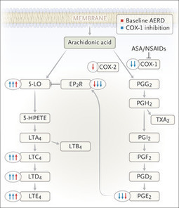
Aspirin-Exacerbated Respiratory Disease About 7% of patients with asthma have mucosal swelling, nasal polyps, and worsening of asthma in response to aspirin and other COX-1 inhibitors. Symptoms may...
|
|
Scooped by
Gilbert C FAURE
September 1, 2019 4:34 AM
|
Chronic rhinosinusitis (CRS) is defined as inflammation of the mucosal lining of the nasal cavity and paranasal sinuses that has been present for more than 3 months, causing characteristic symptoms such as nasal blockage, discharge, reduction or loss of smell sensation and facial pressure. It is a broad clinical description that encompasses several phenotypes, most often divided into CRS with polyps and CRS without polyps. CRS may be the endpoint of several different inflammatory pathways and may be secondary to specific conditions such as cystic fibrosis or fungal aetiology, but most often cannot be attributed to a single pathophysiological trigger. CRS places an enormous burden on both physicians and its sufferers. It is common among the general population, affecting approximately 11% of UK adults,1 and causes greater detriment to quality of life (QoL) than angina or chronic lower respiratory tract disease.2 Not only is it a QoL issue, patients with CRS frequently have comorbid asthma, and poorly controlled CRS is associated with more frequent asthma exacerbations and a need for hospitalisation. The socio-economic cost of CRS is significant with 57% of patients reporting absenteeism and 28% experiencing associated anxiety and depression.3 4 One per cent of all UK adults will visit their general practitioner (GP) with CRS symptoms each year and require an average of four appointments each year; despite this, one in three patients in primary care has poorly controlled symptoms.5 CRS that cannot be adequately managed in primary care may require referral to secondary care, and data from Hospital Episode Statistics show that in a single year there were approximately 120 000 outpatient consultations for CRS and 40 000 sinus operations performed in England and Wales.6 However, there remains considerable debate regarding optimum management strategies leading to wide variation in practice.7 Treatment evidence and guidelines The touchstone for treatment of CRS is The European Position Paper on Rhinosinusitis and Nasal Polyps published in 2012 (EPOS 2012).8 EPOS 2012 syntheses the best available evidence and expert opinion into easy to follow treatment flowcharts for both primary care physicians and ear, nose and throat specialists, but acknowledges the large gaps in high-quality trial data on which to base their guidance. In the intervening 7 years since EPOS was published, the International Consensus Statement on Allergy and Rhinology (ICAR)9 was agreed by a multinational working group who undertook a comprehensive overview of the literature. The working group graded the available evidence and used it as the basis for recommendations; where no evidence was available, no recommendation was made. ICAR is therefore an exhaustive insight into the current state of the science in CRS and an invaluable tool for the otolaryngology community; for primary care, however, the EPOS 2012 remains perhaps the more accessible guide. In addition, there have also been several Cochrane reviews10–16 of treatments available for CRS, and these reviews along with the international consensus guidelines have been used to synthesise a National Institute for Health and Care Excellence (NICE) clinical knowledge summary, most recently updated in 2018.17 Intranasal treatment The foundation of CRS treatment is regular use of intranasal saline irrigation and corticosteroids and this strategy finds unanimous favour among the guidelines mentioned above. The evidence is weak but consistent: a Cochrane systematic review studied the effects of saline irrigation in people with chronic sinusitis.12 This identified only two studies that could robustly be included—one which compared the effect of high volume hypertonic saline irrigation to placebo in patients with CRS both with and without polyps, and another which studied only patients with polyps and compared the use of nasal saline irrigation. This Cochrane review concluded that there was weak evidence from these trials to support the use of nasal saline irrigation. In addition, it is a low-cost treatment which with low risk of adverse events. For this reason, it is recommended for virtually all patients with CRS although in practice is usually used in combination with other therapies. Intranasal corticosteroids are similarly favoured by all expert guidelines. There is a greater number of trials on which to base this opinion. A Cochrane systematic review11 identified (search date August 2015) 18 randomised controlled trials which studied the use of intranasal steroids versus placebo in CRS. The majority of these studies included patients with nasal polyps. Regular intranasal corticosteroid use was reported to be superior to placebo in all reported symptoms, although there was no evidence to support an overall improvement in QoL. A further Cochrane review of the different formulations of intranasal corticosteroids and methods of delivery13 did not identify any single agent with convincing evidence of superiority; however, there is an advantage offered by the second-generation agents (eg, mometasone furoate, fluticasone propionate, fluticasone furoate) which have favourable pharmacokinetic characteristics that minimise systemic bioavailability (<1%) compared with older intranasal corticosteroids (eg, beclomethasone diproprionate, betamethasone). Oral corticosteroids Oral corticosteroids are commonly prescribed in the treatment of CRS although there is a distinct lack of evidence for their efficacy. The NICE Clinical Knowledge Summary (CKS) makes no reference to their use. A Cochrane review in 201616 identified no studies of their use in CRS without polyps, a fact which is also reflected in the ICAR guideline.9 Their use in patients with polyps is better established, with several studies demonstrating short-term efficacy of oral corticosteroids in improving CRS-related QoL scores. Treatment courses are short—a typical regime might be 30 mg per day of prednisolone for 2–3 weeks—and thus the risk of side effects such as adrenal suppression and loss of bone mass is minimal. However, there is a risk of side effects such as gastrointestinal disturbance and insomnia even with these short courses. Patients should also be warned of the risk of osteonecrosis—a risk which may occur at doses of steroid previously thought to be safe (lowest reported associated total dose 105 mg, median associated dose 981 mg).18 The benefit of oral corticosteroids typically last 3–6 months, and so are a reasonable choice for patients who have not improved on the first line of saline irrigation and topical corticosteroids. However, an evidence‐based risk analysis of oral corticosteroid use in CRS with polyps performed by Leung et al 19 demonstrated that risk exceeded benefit if used more frequently than once every 2 years. Oral corticosteroid use is therefore restricted to short, infrequent courses, and with full disclosure of the potential serious associated side effects for patients. Antibiotics Where there is consensus recommending the use of nasal douches and corticosteroids, there is controversy regarding the use of antibiotics. Antibiotics are one of the most commonly prescribed medicines for CRS: of the 1% of UK adults with CRS who visit their GP each year, 91% of them will receive an antibiotic prescription.20 They will often receive not just one but repeated courses of antibiotics.21 There is pressure on GPs to use antibiotics as a first-line treatment, as recently some Clinical Commissioning Groups have insisted on a 3-month trial of macrolide antibiotics prior to secondary care referral.22 With the exception of acute infective exacerbations, routine use of antibiotics is controversial for three main reasons: first; the lack of high-quality evidence to support their efficacy; second, the emerging threat of antibiotic resistance and third, the side effects which may accompany some of the most commonly used agents. In 2016, the ICAR guideline noted a paucity of evidence for antibiotic use9; however, on balance it recommended that patients with polyps should not be prescribed short courses of non-macrolide antibiotics, unless in the particular circumstances of suffering an acute exacerbation. No conclusions could be drawn on longer courses, or for patients without polyps. This echoes the findings of a Cochrane review of the use of antibiotics in CRS, which found very little evidence that non-macrolide antibiotics are effective in patients with CRS although only two studies were fit for inclusion in analysis.15 These recommendations guided the NICE CKS17 to make the recommendation to seek specialist advice before initiating long-term antibiotics in primary care. It is, however, recognised that there does remain a role for appropriate use of macrolide antibiotics in specific circumstances. In particular, there is growing interest in immune-modulating and anti-inflammatory effects of macrolides in chronic airway inflammatory disease, with low-dose long-term macrolides being prescribed for its effects on the immune response and not primarily as an anti-bacterial agent.23 The Cochrane review of 2016 noted that there was a moderate quality of evidence of a modest improvement in disease‐specific QoL in patients with CRS without nasal polyps receiving 3 months of a macrolide.15 However, this improvement was short-lived, as 3 months following the cessation of the antibiotics there was no measurable effect of treatment. Resistance Antibiotic resistance is considered one of the most significant threats to patients’ safety in Europe.24 Evaluating effectiveness of antibiotics and promoting appropriate usage is integral to the UK 5-year antimicrobial resistance strategy.25 Given the high prevalence of CRS and the variability in prescribing, overuse of inappropriate antibiotics may represent a public health danger through selective pressure on bacteria and resistance. In particular, macrolide resistance has been noted to have implications in infections secondary to Streptococcus pneumoniae (upper and lower respiratory tract infections) and Mycoplasma genitalium (sexually transmitted infections), leading to longer duration of illness and requirement for multiple antibiotic courses.26 Side effects Antibiotics are commonly associate with wide range of side effects ranging from the mild (rash, nausea, diarrhoea) to the life threatening (anaphylaxis). Specific to the use of macrolides are concerns about potential cardiovascular side effects.27 There have been a number of publications raising concerns about cardiac toxicity with erythromycin in patients with a prolonged QT interval.28 29 A 2016 study which looked at adverse outcomes associated with clarithromycin use found a significant association was found between clarithromycin use and cardiovascular mortality, where longer durations of clarithromycin use were associated with more cardiovascular events.29 By contrast, this association was not found with use of β-lactam antibiotics leading the authors to hypothesise that it was an effect specific to clarithromycin. This hypothesis was unintentionally tested by the lead investigators of the CLARICOR trial (Effect of Clarithromycin on Mortality and Morbidity in Patients With Ischemic Heart Disease) who had planned to measure the efficacy of a 2-week course of 500 mg clarithromycin two times per day to improve the clinical manifestations of stable coronary heart disease.30 The study was halted prematurely due to the finding of excess deaths in the intervention group: all-cause mortality was significantly higher in the group had taken clarithromycin, an effect which persisted for 10 years post-intervention. The use of clarithromycin in patients with known cardiovascular disease is therefore only undertaken with a significant degree of caution. Surgery Patients who are referred to secondary care who are considered to have tried and failed appropriate medical therapy – which is to say, a sufficient trial of nasal douching and topical steroids—are considered candidates for endoscopic sinus surgery (ESS).31 32 ESS is a loosely defined procedure and can range from very simple balloon expansion of the sinus openings, to extensive procedures that open every bony partition within the sinuses. The risks of surgery include epistaxis, breach of the skull base with resultant CSF leak or meningitis and injury to the intraorbital structures which may result in impaired vision. Insufficient evidence to define the role and extent of surgery contributes to a fivefold variation in UK intervention rates,33 with wide variation in surgical practice and very high rates of revision surgery.34 35 Such uncertainty resulted in inclusion of ESS in the NICE Database of Treatment Uncertainties.36 However, there can be no doubt that in appropriately selected patients, surgery offers significant and durable relief from symptoms. The largest UK study of CRS and its outcomes demonstrated that not only was there a significant improvement in patient-reported symptoms following ESS, but that patients who had surgery earlier in their disease course had a greater degree of improvement that lasted longer versus those who underwent delayed surgery.37 In addition to improving nasal symptoms, a systematic review demonstrated that ESS is associated with improved asthma control with significantly fewer attacks, hospitalisations, and a reduced requirement for medication use following sinus surgery in patients with comorbid CRS and asthma.38 Each of the international consensus guidelines referenced above recommends that patients who have tried a course of appropriate medical therapy but do not have control of their symptoms should be considered as a candidate for surgery.8 9 The path forwards In the last two decades, the majority of new treatments that have become available for CRS have in the main been refinements rather than revolutions—for example, new formulations of intranasal steroids or third-generation antihistamines. However, there are some genuinely new and exciting innovations which are on the cusp of integration to clinical practice. Monoclonal antibodies that bind to targets such as IgE, interleukin-4 (IL-4) and IL-5 were developed primarily as treatments for asthma, but clinical trials have demonstrated that several of these agents may also be effective in CRS with polyps.39 At present, they remain expensive and are limited to subsets of patients with difficult to treat asthma, but as prices fall in future and evidence for their efficacy accumulates then in future they may become available for the treatment of CRS. There have also been several promising avenues of research that may unlock further therapeutic targets, such as the role of the microbiome in sinus disease. However, as alluded to in the many international guidelines, there is a paucity of evidence for many of the treatments that are currently used in CRS before even considering new innovations. This has led to a redoubling of efforts in CRS research to produce high-quality evidence for commonly available treatments. Key to these efforts is the need to accurately define the endotypes of disease which will be studied in trials, as broad clinical phenotypes such as ‘polyps’ may encompass a wide range of underlying pathophysiology. Tightly defining the endotypes of disease for trial inclusion will permit robust examination of the response to treatment in each subgroup. Meta-analyses of CRS outcomes have been hampered by the broad clinical phenotypes, and using precise endotypes will enhance our ability to pool data from multiple trials; this will also be enabled by international consensus statements that define key criteria such appropriateness for ESS40 and standardised sets of outcome measures;41 and funding has been secured for one of the largest ever trials of interventions in CRS which will specifically examine the question of the efficacy of both macrolides and ESS (the MACRO trial42) in subgroups of patients defined by precise clinical and pathological parameters. At present, treatment of CRS exists in a time of genuine clinical equipoise where treatment is empirical and even international guidelines depend more heavily on expert opinion than robust scientific statements. Ongoing research endeavours aim to transform the treatment of CRS and usher in the era of precise, personalised medicine for these patients. Main messages Chronic rhinosinusitis (CRS) is a common disease in UK adults and causes significant detriment to life. The best evidence for effective treatment is for topical intranasal corticosteroids and saline irrigation. For patients who do not respond to this first-line treatment, there are many potential avenues for further treatment but there is controversy about which strategy is the most effective Current research questions Is it possible to identify specific endotypes of CRS to replace the broad clinical phenotypes that we currently base our treatment plans on? Are macrolide antibiotics an effective treatment in CRS? Is best medical treatment superior to surgery in the treatment of CRS? Key references Fokkens WJ, et al, European Position Paper on Rhinosinusitis and Nasal Polyps 2012. Rhinolgy 2012 3 p preceding table of contents, 1–298. Orlandi RR, et al, International Consensus Statement on Allergy and Rhinology: Rhinosinusitis. Int Forum Allergy Rhinol 2016. 6 Suppl 1: p. S22-209. NICE. Sinusitis. 2018; Available from: https://cks.nice.org.uk/sinusitis%23!scenario:1 Head K, et al, Systemic and topical antibiotics for chronic rhinosinusitis. Cochrane Database Syst Rev 2016. 4: p. CD011994 Hopkins C, et al, Long-term outcomes from the English national comparative audit of surgery for nasal polyposis and chronic rhinosinusitis. Laryngoscope 2009. 119 : p. 2459–65. Self-assessment questions Which of the following are typical symptoms of chronic rhinosinusitis? (a) Bleeding and crusting (b) Unilateral blood tinged discharge (c) Blockage, discharge, loss of sense of smell, facial pressure (d) Visual disturbance The first-line treatment for chronic rhinosinusitis (CRS) is oral steroids and antibiotics. True or false? A prolonged course of antibiotics (eg, up to 3 months) may occasionally be used in specific circumstances. What group of antibiotics has the best evidence to support their use for prolonged courses in CRS? (a) Penicillins (b) Macrolides (c) Aminoglycosides (d) Fluoroquinolones And what adverse effects should the patient be advised of with these long-term antibiotics? (a) Cardiac comorbidity (b) Osteonecrosis of the femoral head (c) Epistaxis (d) Infertility There is good evidence to support the superiority of surgical treatment of CRS compared with medical treatment. True or false? Answers Answer: (c) These are typical CRS symptoms. Bleeding and crusting should raise the possibility of vasculitis, septal perforations, or recreational cocaine use. Unilateral blood tinged discharge is a red flag sign that should prompt concerns of malignancy. Visual disturbance may be caused by rare complications of CRS, but is not at all typical. Answer: False. The first-line treatment for CRS should be topical steroids and saline irrigation. Answer: Macrolides. Answer: Cardiac comorbidity, particularly in patients with known heart disease. Osteonecrosis of the femoral head is a complication of oral steroids while topical steroids may cause epistaxis. Macrolides are not recognised to cause infertility but their use should be restricted during pregnancy. The Food and Drug Administration of USA recommend that if a macrolide must be used in pregnancy then azithromycin is category B (ie, safe in animal studies) while clarithromycin is category C (ie, shown to cause harm in animal studies). Answer: False. There are very few high-quality trials that compare medical treatment to surgery, and meta-analysis is made difficult by wide variability in study protocols. References ↵ Hastan D , Fokkens WJ , Bachert C , et al . Chronic rhinosinusitis in Europe--an underestimated disease. A GA²LEN study. Allergy 2011;66:1216–23.doi:10.1111/j.1398-9995.2011.02646.x ↵ Gliklich RE , Metson R . The health impact of chronic sinusitis in patients seeking otolaryngologic care. Otolaryngol Head Neck Surg 1995;113:104–9.doi:10.1016/S0194-5998(95)70152-4 ↵ Sahlstrand-Johnson P , Ohlsson B , Von Buchwald C , et al . A multi-centre study on quality of life and absenteeism in patients with CRS referred for endoscopic surgery. Rhinology 2011;49:420–8.doi:10.4193/Rhino11.101 OpenUrlPubMed ↵ Erskine SE , Hopkins C , Clark A , et al . Chronic rhinosinusitis and mood disturbance. Rhin 2017;55:113–9.doi:10.4193/Rhin16.111 OpenUrl ↵ Lange B , Holst R , Thilsing T , et al . Quality of life and associated factors in persons with chronic rhinosinusitis in the general population: a prospective questionnaire and clinical cross-sectional study. Clin Otolaryngol 2013;38:474–80.doi:10.1111/coa.12189 OpenUrlCrossRefPubMed ↵Hospital Episode Statistics Department of Health; 2013. ↵ Vennik J , Eyles C , Thomas M , et al . Management strategies for chronic rhinosinusitis: a qualitative study of GP and ENT specialist views of current practice in the UK. BMJ Open 2018;8:e022643.doi:10.1136/bmjopen-2018-022643 ↵ Fokkens WJ , Lund VJ , Mullol J , et al . EPOS 2012: European position paper on rhinosinusitis and nasal polyps 2012. A summary for otorhinolaryngologists. Rhinology 2012;50:1–12.doi:10.4193/Rhino50E2 OpenUrlCrossRefPubMed ↵ Orlandi RR , Kingdom TT , Hwang PH , et al . International Consensus Statement on Allergy and Rhinology: Rhinosinusitis. Int Forum Allergy Rhinol 2016;6 Suppl 1:S22–S209.doi:10.1002/alr.21695 OpenUrl ↵ Head K , Sharp S , Chong L-Y , et al . Topical and systemic antifungal therapy for chronic rhinosinusitis. Cochrane Database Syst Rev 2018;16.doi:10.1002/14651858.CD012453.pub2 ↵ Chong LY , Head K , Hopkins C , et al . Intranasal steroids versus placebo or no intervention for chronic rhinosinusitis. Cochrane Database Syst Rev 2016;115.doi:10.1002/14651858.CD011996.pub2 ↵ Chong LY , Head K , Hopkins C , et al . Saline irrigation for chronic rhinosinusitis. Cochrane Database Syst Rev 2016;67.doi:10.1002/14651858.CD011995.pub2 ↵ Chong LY , Head K , Hopkins C , et al . Different types of intranasal steroids for chronic rhinosinusitis. Cochrane Database Syst Rev 2016;25(Suppl 2).doi:10.1002/14651858.CD011993.pub2 ↵ Head K , Chong LY , Hopkins C , et al . Short-course oral steroids as an adjunct therapy for chronic rhinosinusitis. Cochrane Database Syst Rev 2016;27.doi:10.1002/14651858.CD011992.pub2 ↵ Head K , Chong LY , Piromchai P , et al . Systemic and topical antibiotics for chronic rhinosinusitis. Cochrane Database Syst Rev 2016;19.doi:10.1002/14651858.CD011994.pub2 ↵ Head K , Chong LY , Hopkins C , et al . Short-course oral steroids alone for chronic rhinosinusitis. Cochrane Database Syst Rev 2016;2.doi:10.1002/14651858.CD011991.pub2 ↵NICE. Sinusitis, 2018. Available: https://cks.nice.org.uk/sinusitis#!scenario:1 ↵ Kennedy P , Bassiouni A , Psaltis A , et al . Avascular necrosis after oral corticosteroids in otolaryngology: Case report and review of the literature. Allergy Rhinol 2016;7:50–4.doi:10.2500/ar.2016.7.0142 OpenUrl ↵ Leung RM , Dinnie K , Smith TL . When do the risks of repeated courses of corticosteroids exceed the risks of surgery? Int Forum Allergy Rhinol 2014;4:871–6.doi:10.1002/alr.21377 OpenUrl ↵ Gulliford MC , Dregan A , Moore MV , et al . Continued high rates of antibiotic prescribing to adults with respiratory tract infection: survey of 568 UK general practices. BMJ Open 2014;4:e006245.doi:10.1136/bmjopen-2014-006245 ↵ Akkerman AE , Kuyvenhoven MM , van der Wouden JC , et al . Prescribing antibiotics for respiratory tract infections by GPs: management and prescriber characteristics. Br J Gen Pract 2005;55:114–8. ↵ Soni-Jaiswal A , Philpott C , Hopkins C . The impact of commissioning for rhinosinusitis in England. Clin Otolaryngol 2015;40:639–45.doi:10.1111/coa.12430 OpenUrl ↵ Cervin A , Wallwork B . Macrolide therapy of chronic rhinosinusitis. Rhinology 2007;45:259–67. ↵ Lodato EM , Kaplan W . Priority Medicines for Europe and the World "A Public Health Approach to Innovation": Background Paper 6.1 Antimicrobial resistance World Health Organisation; 2013. ↵ Davies SC , Gibbens N . UK Five Year Antimicrobial Resistance Strategy 2013 to 2018 Department of Health and Department for Environment, Food & Rural Affairs; 2013. ↵ Gaynor M , Mankin AS . Macrolide antibiotics: binding site, mechanism of action, resistance. Curr Top Med Chem 2003;3:949–60.doi:10.2174/1568026033452159 ↵ Gibbins NE , Theokli C , Hopkins C . Time to reconsider guidelines on clarithromycin in chronic rhinosinusitis? BMJ 2013;346:f2678.doi:10.1136/bmj.f2678 ↵ Wong AYS , Chan EW , Anand S , et al . Managing Cardiovascular Risk of Macrolides: Systematic Review and Meta-Analysis. Drug Saf 2017;40:663–77.doi:10.1007/s40264-017-0533-2 OpenUrlCrossRef ↵ Cheng Y-J , Nie X-Y , Chen X-M , et al . The Role of Macrolide Antibiotics in Increasing Cardiovascular Risk. J Am Coll Cardiol 2015;66:2173–84.doi:10.1016/j.jacc.2015.09.029 OpenUrlFREE Full Text ↵ Winkel P , Hilden J , Hansen JF , et al . Clarithromycin for stable coronary heart disease increases all-cause and cardiovascular mortality and cerebrovascular morbidity over 10years in the CLARICOR randomised, blinded clinical trial. Int J Cardiol 2015;182:459–65.doi:10.1016/j.ijcard.2015.01.020 OpenUrl ↵ Baguley C , Brownlow A , Yeung K , et al . The fate of chronic rhinosinusitis sufferers after maximal medical therapy. Int Forum Allergy Rhinol 2014;4:525–32.doi:10.1002/alr.21315 OpenUrl ↵ Young LC , Stow NW , Zhou L , et al . Efficacy of medical therapy in treatment of chronic rhinosinusitis. Allergy Rhinol 2012;3:8–12.doi:10.2500/ar.2012.3.0027 OpenUrl ↵ Hopkins C . Commissioing Guide: Chronic Rhinosinusitis. Royal College of Surgeons of England London; 2016. ↵ Philpott C , Hopkins C , Erskine S , et al . The burden of revision sinonasal surgery in the UK-data from the Chronic Rhinosinusitis Epidemiology Study (CRES): a cross-sectional study. BMJ Open 2015;5:e006680.doi:10.1136/bmjopen-2014-006680 ↵ Hopkins C , Slack R , Lund V , et al . Long-term outcomes from the English national comparative audit of surgery for nasal polyposis and chronic rhinosinusitis. Laryngoscope 2009;119:2459–65.doi:10.1002/lary.20653 ↵NICE. Database of Uncertainties of Treatments. ↵ Hopkins C , Rimmer J , Lund VJ . Does time to endoscopic sinus surgery impact outcomes in Chronic Rhinosinusitis? Prospective findings from the National Comparative Audit of Surgery for Nasal Polyposis and Chronic Rhinosinusitis. Rhinology 2015;53:10–17.doi:10.4193/Rhin13.217 OpenUrl ↵ Vashishta R , Soler ZM , Nguyen SA , et al . A systematic review and meta-analysis of asthma outcomes following endoscopic sinus surgery for chronic rhinosinusitis. Int Forum Allergy Rhinol 2013;3:788–94.doi:10.1002/alr.21182 OpenUrlCrossRefPubMed ↵ Pauwels B , Jonstam K , Bachert C . Emerging biologics for the treatment of chronic rhinosinusitis. Expert Rev Clin Immunol 2015;11:349–61.doi:10.1586/1744666X.2015.1010517 OpenUrlCrossRefPubMed ↵ Rudmik L , Soler ZM , Hopkins C , et al . Defining appropriateness criteria for endoscopic sinus surgery during management of uncomplicated adult chronic rhinosinusitis: a RAND/UCLA appropriateness study. Int Forum Allergy Rhinol 2016;6:557–67.doi:10.1002/alr.21769 OpenUrl ↵ Hopkins C , Hettige R , Soni-Jaiswal A , et al . CHronic Rhinosinusitis Outcome MEasures (CHROME), developing a core outcome set for trials of interventions in chronic rhinosinusitis. Rhinology 2018;56:22–32.doi:10.4193/Rhin17.247 OpenUrl ↵ Philpott C , le Conte S , Beard D , et al . Clarithromycin and endoscopic sinus surgery for adults with chronic rhinosinusitis with and without nasal polyps: study protocol for the MACRO randomised controlled trial. Trials 2019;20:246.doi:10.1186/s13063-019-3314-7

|
Suggested by
Société Francaise d'Immunologie
January 21, 2019 1:44 PM
|
Background It is hypothesized that uncontrolled inflammation is responsible for many of the manifestations and symptoms of chronic rhinosinusitis (CRS). Although earlier work has demonstrated an association between olfactory loss and mucus cytokines, the impact on other symptoms is unknown. In this study we investigated the relationship between cytokines, inflammatory cell counts, and patient‐reported outcomes measures to better understand how the inflammatory microenvironment correlates with CRS symptomatology. Methods The 22‐item Sino‐Nasal Outcome Test (SNOT‐22) and 8‐item Short Form Health Survey (SF‐8) were administered to 76 patients undergoing endoscopic sinus surgery for CRS. Mucus was collected intraoperatively from the middle meatus and tested for 17 cytokines using a multiplex flow cytometric bead assay. Eosinophil/neutrophil counts were obtained from histopathologic slide review. Spearman correlations between cytokines, cell counts, and quality‐of‐life subdomain scores were assessed without multiple comparisons correction due to the small sample size. Results Interleukin‐4 (IL‐4) correlated significantly with the Rhinologic domain (Rs = 0.25, p = 0.03), whereas eosinophil and neutrophil counts were inversely correlated with the Extranasal Rhinologic domain (Rs = −0.32, p = 0.01; and Rs = −0.27, p = 0.03). Subgroup analysis for nasal polyposis (CRSwNP) showed significant correlations between IL‐6 and Total SF‐8 (Rs = 0.35, p = 0.02), General Health (Rs = 0.34, p = 0.03), and Emotional (Rs = 0.47, p = 0.002) scores. In patients without polyps (CRSsNP), IL‐21 correlated positively with Extranasal Rhinologic Symptoms domain (Rs = 0.41, p = 0.01). Conclusion This pilot study identifies possible pairwise correlations between mucus cytokine levels and baseline quality‐of‐life measures that need confirmation in larger, targeted studies. Due to the exploratory methodology, positive results may be spurious and should only be used as a starting point for future confirmatory work.
|
|
Scooped by
Gilbert C FAURE
December 8, 2018 7:21 AM
|
Chronic rhinosinusitis (CRS) is a heterogeneous inflammatory disease with an as-yet-undefined etiology. The management of CRS has historically been phenotypically driven, and the presence or absence of nasal polyps has frequently guided diagnosis, prognosis, and treatment algorithms.
|
|
Scooped by
Gilbert C FAURE
June 17, 2017 8:26 AM
|
Rationale Interleukin (IL)-33, a new member of the IL-1 family, is constitutively expressed in epithelial tissues and lymphoid organs and plays an important role in the pathogenesis of allergic disease. However, the role of IL-33 in chronic rhinosinusitis with nasal polyps (CRSwNP) remains unclear.
Objective To investigate the role of IL-33 in the pathophysiology of CRSwNP.
Methods We investigated IL-33 expression and its cellular origins in the nasal polyps (NPs) of human subjects by immunohistochemistry (IHC), quantitative reverse transcription PCR (qRT-PCR), and multiplex cytokine assays. Correlations between IL-33 expression and other inflammatory markers were also explored. To investigate the role of IL-33 in CRSwNP, anti-IL-33 antibody was used in a murine model of CRS.
Results Uncinate process tissues from control (19), CRSsNP (61), CRSwNP (69) and NP tissues (71) were used in this study. Increased expression of IL-33 mRNA and protein in patients with CRSwNP compared with controls was observed. The concentration of IL-33 protein in CRSwNP was positively correlated with the number of neutrophils and the expression of several Th1 and Th17 inflammatory markers, including interferon (IFN)-γ, IL-1β, tumour necrosis factor (TNF)-α, IL-17A, IL-22, and various markers for neutrophil recruitment. However, protein levels of IL-5 and quantity of eosinophils were inversely correlated with levels of IL-33. The expression of tissue inhibitor of metalloproteinase (TIMP)-1 was negatively correlated with IL-33 protein levels, while the expression of matrix metalloproteinase (MMP)-2 and MMP-9 was positively correlated with IL-33 protein levels. In animal studies, IL-33 expression was upregulated in the CRSwNP group compared with controls. Anti-IL-33 treatment reduced the thickness of oedematous mucosa, subepithelial collagen deposition, and infiltration of neutrophils, but infiltration of eosinophils was not reduced. This treatment also inhibited the expression of neutrophilic inflammatory cytokines, but not IL-4. In addition, the expression of intracellular adhesion molecule 1, vascular adhesion molecule 1 and CXCL-2 in the nasal mucosa was suppressed in mice treated with anti-IL-33 antibody.
Conclusions Our data suggest a role for IL-33 in the pathogenesis of CRSwNP via neutrophil recruitment. Therefore, anti-IL-33 may provide a new treatment strategy to target infiltrating neutrophils in CRSwNP.
|
|
Scooped by
Gilbert C FAURE
February 5, 2016 2:30 PM
|
NewsResearchers decided to test dupilumab, a human antibody which already demonstrated clinical efficacy for asthma and atopic dermatitis, on nasal polyps.Contributed Author: Ghent UniversityTopics: Drug Development...
|
|
Scooped by
Gilbert C FAURE
March 4, 2015 2:03 PM
|
Interleukin-25 secretions generated from the sinonasal epithelia and infiltrating mast cells play a crucial role in the pathogenesis of chronic rhinosinusitis among Asian patients with nasal polyps, according to study results.“IL-25 production by ...
|
|
Scooped by
Gilbert C FAURE
October 3, 2014 2:56 PM
|
Allergy Immunology | Sanofi and Regeneron Pharmaceuticals announced that a phase 2a study of dupilumab met all endpoints in patients with chronic sinusitis with nasal polyps who did not respond to intranasal corticosteroids, according to a press...
|
|
Scooped by
Gilbert C FAURE
March 6, 2014 1:20 PM
|
Chronic rhinosinusitis with nasal polyps more severe in women Family Practice News Digital Network "We thought we would find that CRSwNP was more prevalent and severe in women, especially because these patients have asthma and some features of...
|
|
|
Scooped by
Gilbert C FAURE
June 19, 2021 10:31 AM
|
Pathogenesis of Nasal polyp has been primarily investigated on the basis of sinonasal mucosal immunity. Several inflammatory responses have been reported in nasal polyp pathogenesis to date. In this study the researchers have summarised new research on pathways for developing chronic nasal polyp rhinosinusitis (CRSwNP), which focuses on Th2 inflammation. The th2 inflammation may be driven by Staphylococcus aureus by the production of enterotoxins and a serine protease-like protein. Furthermore, S. aureus affected the activity of the mucosal obstruction directly and increased synthesis of Th2 cytokines by the rapid activation of endogenous epithelial cytokines. The synthesis of innate lymphoid epithelial-derived cytokines, including TSLP, IL-25, and IL-33, promotes th2 reactions. Mast cell produces IL-5, IL-13 and periostin and has a function to play in the pathogenesis of nasal polyps by orchestrating infiltration of eosinophils. The formation of extracellular eosinophil traps and Charcot-Leyden crystals is closely linked with the seriousness of disease and development of viscous mucus plugs. The role of S. aureus in innate and adaptive immunity is increasingly evident and contributes to the inflammation of Th2 in CRSwNP. Innate immunity, like epithelial cytokines, is critical in the production of CRSwNP, inducing different pathways, and must be further examined as Th2-controlled biomarkers. Neutrophilic inflammation has recently begun to be investigated but still remains uncertain. For latest news and updates By signing up, you will receive emails about Physician's Weekly products and you agree to our Terms & Conditions and Privacy Policy. Reference: https://journals.lww.com/co-allergy/Abstract/2020/02000/Th2_inflammatory_responses_in_the_development_of.2.aspx
|
|
Scooped by
Gilbert C FAURE
January 31, 2021 3:46 AM
|
Click on the article title to read more.
|
|
Scooped by
Gilbert C FAURE
August 17, 2020 4:09 AM
|
Objectives/Hypothesis Eosinophilic and noneosinophilic chronic rhinosinusitis with nasal polyps (ECRSwNP and NECRSwNP) show distinguished clinical pathology, but their underlying mechanism remains ...
|
|
Scooped by
Gilbert C FAURE
October 25, 2019 1:18 PM
|
Chronic rhinosinusitis with nasal polyps (CRSwNP) is characterized by type 2 inflammation with accumulation of activated group 2 innate lymphoid cells (ILC2s) and elevation of thymic stromal lymphopoietin (TSLP).
|
|
Suggested by
Société Francaise d'Immunologie
July 25, 2019 4:00 PM
|
PubMed comprises more than 29 million citations for biomedical literature from MEDLINE, life science journals, and online books. Citations may include links to full-text content from PubMed Central and publisher web sites.
|
|
Scooped by
Gilbert C FAURE
December 13, 2018 6:11 AM
|
|
|
Rescooped by
Gilbert C FAURE
from Immunology and Biotherapies
November 12, 2018 4:36 AM
|
Abstract United airways disease (UAD) is the concept that the upper and lower airways, which are anatomically and immunologically related, form a single organ. According to this concept, upper and lower airway diseases are frequently comorbid because they reflect manifestations of a single underlying disease at different sites of the respiratory tract. Allergic asthma‐allergic rhinitis is the archetypal UAD, but emerging data indicate that UAD is a heterogeneous condition and consists of multiple phenotypes (observable clinical characteristics) and endotypes (pathobiologic mechanisms). The UAD paradigm also extends to myriad sinonasal diseases (eg, chronic rhinosinusitis with or without nasal polyps) and lower airway diseases (eg, bronchiectasis, chronic obstructive pulmonary disease). Here, we review currently known phenoendotypes of UAD and propose a “treatable traits” approach for the classification and management of UAD, wherein pathophysiological mechanisms and factors contributing to disease are identified and targeted for treatment. Treatable traits in UAD can be analyzed according to a framework comprising airway inflammation (eosinophilic, neutrophilic), impaired airway mucosal defense (impaired mucociliary clearance, antibody deficiency), and exogenous cofactors (allergic sensitizers, tobacco smoke, microbes). Appreciation of treatable traits is necessary in advancing the effort to deliver precise treatments and achieve better outcomes in patients with UAD. 1 INTRODUCTION The respiratory tract is divided into the upper and lower airways at the level of the vocal cords. Despite this artificial distinction, they are anatomically contiguous and immunologically related. United airways disease (UAD) is the concept that upper and lower airways form a single organ, with upper and lower airway diseases co‐occurring frequently because they reflect different manifestations of a single underlying disease process.1 Possible mechanisms linking the upper and lower airways include aspiration of nasal inflammatory cells and mediators into the tracheobronchial tree, hematogenous propagation of inflammatory mediators from one part of the airway to another, stimulation of bone marrow progenitor cells, or the nasobronchial reflex.2 The archetypal UAD is allergic asthma‐rhinitis, but emerging data indicate that UAD is complex and heterogenous, and may present with other clinical phenotypes of sinonasal disease (eg, chronic rhinosinusitis with or without nasal polyps) and lower airway disease (eg, bronchiectasis, chronic obstructive pulmonary disease [COPD]). Even within a given clinical phenotype, there is further variability based on the underlying pathobiologic mechanisms, known as endotypes. Current guideline‐based management approaches (summarized in Table 1) stratify patients according to symptoms, disease control, future risk, and occasionally broad clinical phenotypes, but do not sufficiently consider complexity and variability in other important dimensions such as airway inflammation, airway mucosal defense, and exogenous cofactors. Failure to recognize this complexity or treat both upper and lower compartments of the airways inevitably leads to suboptimal outcomes for patients. Site Disease Approach to stratification of patients and selection of therapies Patient stratification groups Recommended treatments according to patient stratification Other possible treatments Upper airway Allergic rhinitis Depends on symptoms, time course, severity, control, patient preferences, availability of treatment, and cost. One approach is a step‐up/step‐down approach based on assessment of control Step 1: mild symptoms Intranasal or oral non‐sedating H1‐antihistamines Leukotriene antagonists Allergen and irritant avoidance Allergen immunotherapy Nasal filters or saline Step 2: moderate‐to‐severe symptoms or persistent AR Intranasal corticosteroids Step 3: uncontrolled at step 2 (current or historical) Combination intranasal corticosteroids and intranasal H1‐antihistamines Step 4: uncontrolled at step 3 Short course oral steroids Chronic rhinosinusitis Based on clinical phenotype CRSsNP Intranasal corticosteroids Antibiotics (second line) Consider short course of oral steroids Saline irrigation Sinus surgery for medically refractory disease CRSwNP Intranasal corticosteroids Short course of oral steroids Antibiotic if suspicion of infection Consider leukotriene receptor for acetylsalicylic acid sensitivity Lower airway Asthma Assessment of symptoms, exacerbations, and lung function followed by stepwise escalation/de‐escalation of therapy Step 1 Consider low‐dose ICS Leukotriene receptor antagonists Theophylline Allergen avoidance Allergen immunotherapy for patients with allergic rhinitis and sensitized to house dust mites, frequent exacerbations, and FEV1 > 70% Vaccinations Weight reduction for obese asthma patients Step 2 Low‐dose ICS Step 3 Low‐dose ICS + LABA Step 4 Medium‐/high‐dose ICS + LABA Step 5 Low‐dose oral corticosteroids Add‐on treatments: tiotropium, anti‐IgE, anti‐IL5, bronchial thermoplasty Patients may benefit from phenotyping and endotyping at Step 5 Chronic obstructive pulmonary disease Assessment of symptoms (CAT, mMRC), exacerbations, airflow obstruction (FEV1) Group A (CAT < 10, mMRC < 2, infrequent exacerbations) Bronchodilator Smoking cessation (including pharmacologic treatment) Education, self‐management, and pulmonary rehabilitation Pneumococcal and influenza vaccinations Long‐term oxygen therapy in the presence of severe resting hypoxemia Long‐term noninvasive ventilation for patients with severe chronic hypercapnia and a history of hospitalization for acute respiratory failure Bronchoscopic or surgical lung volume reduction surgery for selected patients Group B (CAT ≥ 10, mMRC ≥ 2, infrequent exacerbations) LABA or LAMA Increase to LAMA + LABA if persistent symptoms Group C (CAT < 10, mMRC < 2, frequent exacerbations) LAMA Increase to LAMA + LABA or change to LABA + ICS if further exacerbations Group D (CAT ≥ 10, mMRC ≥ 2, frequent exacerbations) LAMA + LABA Increase to LAMA + LABA + ICS if persistent symptoms/further exacerbations Consider macrolides Consider roflumilast if FEV1 < 50% predicted and patient has chronic bronchitis Bronchiectasis Assessment of microbial infection, exacerbations and airway symptoms Pseudomonas aeruginosa infection with frequent exacerbations Long‐term inhaled antibiotic treatment Escalate to combined oral and inhaled antibiotic treatment if inadequate response Physiotherapy, pulmonary rehabilitation, and/or airway clearance Pneumococcal and influenza vaccinations Long‐acting bronchodilators for patients with significant breathlessness Non‐Pseudomonas aeruginosa infection with frequent exacerbations Long‐term macrolide treatment Escalate to combined oral and inhaled antibiotic treatment if inadequate response Multiple sequential airway treatments Short‐ or long‐acting bronchodilators → Mucolytic/physiotherapy adjuncts → airway clearance → inhaled antibiotics AR, allergic rhinitis; CAT, COPD Assessment Tool; CRSsNP, chronic rhinosinusitis without nasal polyps; CRSwNP, chronic rhinosinusitis with nasal polyps; FEV1, forced expiratory volume in one second; ICS, inhaled corticosteroids; ICS + LABA, combination inhaled corticosteroids and long‐acting beta agonist; LABA, long‐acting beta agonist; LAMA, long‐acting muscarinic antagonist; mMRC, modified Medical Research Council dyspnea score. The treatable traits approach has been described for lower airway diseases,3 and in this review, we extend its application to combined upper and lower airway disease. We summarize the current evidence for known clinicopathobiologic subtypes (“phenoendotypes”) of UAD. We have grouped the clinical manifestations of UAD according to the “treatable traits” of airway inflammation, airway mucosal defense dysfunction, and exogenous cofactors (Figure 1 and Table 2). These traits are not mutually exclusive and multiple traits may contribute to disease in an individual at any one point in time. Treatable traits may also vary over time. It is with the intention of drawing the readers’ attention to traits that are common to both airway compartments that we have artificially classified the upper and lower airway conditions as presented in this review. We believe that targeting common disease processes, when used in combination with current patient stratification approaches, is likely to lead to clinical improvements at both sites of the respiratory tract. Trait Endotype/pathophysiology Method of assessment/biomarker Clinical phenotype Targeted treatment Upper airway Lower airway Inflammation Eosinophilic, high type 2/TH2‐cytokines IL‐4, IL‐5, IL‐13 Blood periostin, high FeNO, sputum eosinophilia, blood eosinophilia CRSwNP Eosinophilic asthma Corticosteroids (systemic, inhaled, intranasal), anti‐IL‐5, IL‐4, IL‐13, anti‐TSLP, ?CRTh2 antagonist Allergic, systemic CD4+ TH2 cells Blood total IgE, blood specific IgEs, skin prick tests Allergic rhinitis Allergic asthma Allergen avoidance, corticosteroids, antileukotrienes, anti‐IgE, immunotherapy Allergic, local Local nasal and bronchial mucosa specific IgE production Blood total IgE not raised, negative blood specific IgEs, negative skin prick tests, local IgE and specific IgE production, nasal allergen provocation test Local allergic rhinitis Intrinsic asthma ?Omalizumab, ?immunotherapy Non‐allergic eosinophilic IL‐25, IL‐33, TSLP, group 2 innate lymphoid cells Blood periostin, high FeNO, sputum eosinophilia, blood eosinophilia, non‐reactive skin prick tests, absent specific IgE in blood Non‐allergic rhinitis with eosinophilia syndrome (NARES) Adult onset eosinophilic asthma Anti‐TSLP, anti‐IL‐4, IL‐5, IL‐13, ?CRTh2 antagonist Neutrophilic TH17 cells, IL‐17, CXCL1, CXCL8 IL‐8, sputum neutrophilia CRSsNP COPD, neutrophilic asthma Smoking cessation, macrolides Impaired airway mucosal defense Cystic fibrosis Autosomal recessive disorder in CFTR gene leading to improper salt balance and viscous airway surface liquid Genetic testing, sweat chloride test CRSwNP (acid mucin) Bronchiectasis CFTR modulators Primary ciliary dyskinesia Autosomal recessive mutations causing dynein and radial spoke defects Low nasal nitric oxide, ciliary motion and ultrastructure CRSsNP Bronchiectasis No validated PCD‐specific therapies Primary antibody deficiency Deficient immunoglobulins or functional antibody response Quantitative serum immunoglobulin levels, functional antibody responses CRS Bronchiectasis Immunoglobulins, vaccinations Exogenous cofactors Occupational exposures High molecular weight sensitizers: IgE‐mediated type 2 inflammation Low molecular weight sensitizers: haptens Irritant‐induced: no immunological reaction Total IgE, skin prick tests Peak flow monitoring Specific bronchoprovocation challenge Occupational rhinitis, reactive upper airways dysfunction syndrome Occupational asthma, reactive airways dysfunction syndrome Exposure avoidance, respiratory protection devices, ?anti‐IgE Aspirin/non‐steroidal anti‐inflammatories Constitutive overexpression of 5‐lipoxygenase and leukotriene C4 synthase Aspirin/NSAID challenge, urinary leukotriene E4 CRSwNP Severe asthma Avoidance, desensitization, antileukotrienes Staphylococcus aureus enterotoxin Superantigens elicit Th1, Th17 and Th2 cytokines, including IL‐4, IL‐5, IL‐13 Staph aureus enterotoxin IgE's CRSwNP Severe asthma ?anti‐IgE Aspergillus/fungal hypersensitivity TH2 cells, IL‐4, IL‐5, IL‐13, IgE, eosinophilia Aspergillus skin test positivity, Aspergillus specific IgE, IgE > 1000 IU/mL, Blood eosinophil >500/μL, precipitating antibodies to Aspergillus Allergic fungal rhinosinusitis Allergic bronchopulmonary aspergillosis (asthma, bronchiectasis) Steroids, antifungal, ?anti‐IgE Upper respiratory tract infection Variable depending on microbe and underlying disease Respiratory cultures and nucleic acid tests Exacerbation of rhinitis/rhinosinusitis Exacerbation of asthma, COPD or bronchiectasis Antivirals, antibiotics, vaccinations Typical upper and lower airway clinical phenotypes associated with underlying treatable traits are listed. Taxonomy based on clinical phenotypes has significant limitations: each phenotype may represent comprise treatable traits, for example, CRSwNP and asthma may be neutrophilic or eosinophilic inflammation depending on the population studied. Hence, a more precise approach is needed to identify contributory treatable trait(s) in each case, which can aid in selection of targeted treatments. Question mark (?) indicates targeted therapies under investigation. Treatable traits and targeted therapies may be used to complement the current approaches for patient stratification and treatment selection—including non‐targeted treatments—shown in Table 1. 2 AIRWAY INFLAMMATION Airway inflammation is important in the pathogenesis of many upper and lower airway diseases. It is a dynamic trait which varies over time and is modulated by factors such as treatment, infection, environmental exposures, and natural history of disease.4 Clinicians commonly assume that asthma and chronic rhinosinusitis with nasal polyps (CRSwNP) are stereotypically eosinophilic in nature, and that chronic rhinosinusitis without nasal polyps (CRSsNP) or COPD is stereotypically neutrophilic. As discussed below, while these generalizations may hold true for many cases there is substantial variability in the underlying pathophysiological mechanisms for each clinical phenotype or disease. Identification and quantification of airway inflammation can help to direct appropriate treatment. Current methods to assess upper and lower airway inflammation are summarized in Table 3. Method Comments Strengths Limitations Nasal lavage Luminal proteins, cytokines and cells Simple and safe Correlates with, but less invasive than induced sputum Variable fluid recovery and dilution with lavages No sampling of the epithelium Nasal brush or scraping Samples the nasal epithelium Simple and minimally invasive Does not sample deeper layers of the mucosa or the nasal lumen Nasal biopsy Identifies tissue inflammatory cellular subtypes of rhinitis or rhinosinusitis Samples the total nasal mucosa Technically difficult No information about nasal lumen Blood periostin Marker of Th2, eosinophilic airway inflammation Predicts severity of chronic rhinosinusitis Predicts chronic rhinosinusitis with nasal polyps phenotype Noninvasive Not widely available Conflicting evidence on whether periostin is associated with sputum eosinophilia Sputum induction Neutrophil count ≥65% or >500 × 104/mL identifies neutrophilic airway inflammation Eosinophil count ≥3% identifies eosinophilic airway inflammation Noninvasive Well‐validated Not widely available Logistically, technically and practically difficult High failure rate Risk of inducing bronchoconstriction Peripheral blood eosinophils Eosinophil count ≥2% identifies eosinophilic airway inflammation Noninvasive, minimal risk Widely available, cheap Predicts with important clinical outcomes such as exacerbation risk and response to treatment Wide diurnal variation of readings (40%) within the same subject May not be applicable when only local mucosal inflammation occurs Fractional exhaled nitric oxide (FeNO) FeNO < 25 ppb indicates low likelihood of airway eosinophilia FeNO > 47 ppb suggests eosinophilic airway inflammation and corticosteroid responsiveness Noninvasive Quick, easy, and safe to perform Readings may be affected by smoking, respiratory tract infections Sputum eosinophilia and FeNO are not always concordant Urinary leukotriene E4 (LTE4) Marker of cysteinyl leukotriene activity Higher levels observed in aspirin‐exacerbated respiratory disease Collection of urine is simple and of no risk Special equipment and training are required to measure LTE4 2.1 Allergic asthma‐rhinitis The link between allergic asthma and allergic rhinitis (AR) is well established. Up to 40% of AR patients have asthma and almost all patients with asthma have AR.5 Many patients with AR also exhibit nonspecific bronchial hyperresponsiveness.6 IgE‐mediated hypersensitivity to aeroallergens results in type 2/eosinophilic inflammation in the respiratory tract, manifesting as rhinitis in the upper airways and asthma in the lower airways. Upon allergen exposure, sensitization occurs whereby specific IgE (sIgE) are generated, circulate in the blood, and attach to the surface of mast cells and basophils, including those that reside in the airway mucosa. Subsequent exposure to allergen triggers mast cells degranulation, release of chemical mediators, and synthesis of leukotrienes and cytokines, resulting in an immediate allergic airway reaction. The late phase response involves Th2 lymphocyte recruitment with production of inflammatory cytokines such as IL‐4, IL‐5, and IL‐13, as well as activation of eosinophils. The central role of sIgE in allergic asthma‐rhinitis is highlighted by a population‐based birth cohort study showing that presence of peripheral blood sIgE directed against common allergens is associated with increased odds of asthma and rhinitis in childhood.7 In adults with AR, experimental nasal provocation with house dust mite allergens induces not only nasal eosinophilia but also bronchial eosinophilic inflammation, bronchoconstriction, and bronchial hyperresponsiveness,8 suggesting inflammatory crosstalk between upper and lower airways. The allergic asthma‐rhinitis phenoendotype usually begins in childhood and can be identified by measuring peripheral blood total IgE which is elevated, plus demonstration of atopy by the presence of raised sIgE levels in blood or skin prick test positivity. Omalizumab, an anti‐IgE monoclonal antibody, is efficacious for both severe allergic asthma9 and uncontrolled AR.10 Therapeutic response to omalizumab is predicted by higher baseline periostin levels and reduced free serum IgE levels during period of treatment.11 In some cases, specific causative allergens can be identified and targeted with allergen immunotherapy. In both AR12 and allergic asthma,13 immunotherapy improves symptoms and reduces the need for pharmacotherapy in the short term, albeit with limited evidence for longer‐term efficacy. Treatment of AR patients with grass pollen sublingual immunotherapy tablets was associated with slower AR progression, less frequent asthma onset, and slower asthma progression.14 Candidate biomarkers that can identify and/or predict response to allergen immunotherapy include the sIgE:total IgE ratio, specific IgG4 and IgE‐FAB.15 2.2 Local allergic rhinitis‐non‐allergic asthma: local mucosal IgE production A subset of asthma and rhinitis patients demonstrate type 2/eosinophilic allergic airway responses mediated by sIgE which is produced and sequestered in the airway mucosa, with skin prick test non‐reactivity and no detectable circulating sIgE in peripheral blood. In the upper airway, this phenoendotype is known as local allergic rhinitis (LAR), a distinct entity characterized by local production of sIgE in the nasal mucosa without evidence of systemic atopy. LAR does not appear to represent a forme fruste of AR,16 and is associated with increased risk of asthma compared to healthy individuals without LAR.17 The lower airway analog of LAR may correspond to a subtype of non‐allergic asthma which is currently not well defined, but whose existence is supported by studies (Supporting Information: References S1‐S4). In the aforementioned entity, intramucosal IgE synthesis is observed in the lower airways, and functional sIgE can be found in the sputum despite negative skin prick tests and blood sIgE levels. Bronchial inhalational challenge with allergen induces a clinical lower airway allergic response characterized by sputum eosinophilia and bronchial hyperresponsiveness. It is not known why individuals with allergic asthma‐rhinitis demonstrate positive skin prick tests and elevated sIgE in blood, whereas non‐allergic asthmatics and patients with LAR do not although local mucosal IgE synthesis occurs in all those conditions. One possibility is that local mucosal IgE synthesis may not reach sufficient quantities to spill over into peripheral blood. Alternatively, IgE may be sequestered within the bronchial mucosa. Regardless, efficacy of omalizumab has been shown in non‐atopic asthma with evidence of local mucosal IgE production. Pillai et al. (Supporting Information: References S5) randomized 18 such subjects and obtained bronchial biopsies from subjects before and after treatment with 12‐14 weeks of omalizumab. The patients subsequently underwent supervised dose reduction of their regular asthma therapy. Compared to placebo, omalizumab therapy significantly reduced total bronchial mucosal IgE+ cells. Lung function declined in the placebo‐treated group but improved in the omalizumab‐treated group. This small but important study serves as proof‐of‐concept that anti‐IgE can target disease associated with local mucosal IgE production. 2.3 Non‐allergic rhinitis with eosinophilia syndrome‐non‐allergic eosinophilic asthma Type 2 (eosinophilic) inflammation can occur in UAD without evidence of IgE‐mediated hypersensitivity. In the upper airways, this endotype has been termed non‐allergic rhinitis with eosinophilia syndrome (NARES) and is characterized by persistent rhinitis and marked eosinophilia of the nasal mucosa in the absence of local or systemic IgE production, suggesting allergen‐independent induction of eosinophilic airway inflammation.18 Almost half of patients with NARES who do not report asthmatic symptoms nevertheless demonstrate bronchial hyperresponsiveness and sputum eosinophilia, supporting a link between NARES and asthma.19 The corresponding asthma phenoendotype is non‐allergic eosinophilic asthma, in which eosinophilic inflammation and type 2 cytokine production are not driven by allergen‐activated Th2 lymphocytes, but is instead postulated to be initiated by type 2 innate lymphoid cells (ILC2).20 ILC2s are a recently discovered population of lymphocytes capable of producing large amounts of type 2 cytokines (IL‐5 and IL‐13) and orchestrate eosinophilic inflammation in the absence of adaptive immunity, including Th2 cells, B cells, and IgE antibody.21 ILC2s are activated by epithelium‐derived cytokines IL‐25, IL‐33, and thymic stromal lymphopoietins (TSLP), which are released by epithelial cells in response to epithelial damage caused by a wide range of stimuli including microbiota and pollutants. ILC2s are increased in peripheral blood in eosinophilic asthmatics compared to non‐eosinophilic asthmatics and their numbers in sputum correlate with eosinophil counts.22 Increased ILC2s has also been observed in eosinophilic nasal polyps,23 but evidence for the involvement of ILC2 cells in NARES and non‐allergic eosinophilic asthma is lacking and should be the focus of future studies. Potential targeted treatments for this pathway include monoclonal antibodies targeting any of the type 2 cytokines, discussed in the section below. TSLP, targeted by the monoclonal antibody tezepelumab, was shown in a recent dose‐ranging proof‐of‐concept study to reduce exacerbations in patients with uncontrolled asthma, including in those with low IgE (<100 IU/mL).24 Antagonists of CRTH2, the receptor for prostaglandin D2 on ILC2 cells, are currently undergoing evaluation to assess clinical efficacy (Supporting Information: References S6, S7). 2.4 Asthma‐chronic rhinosinusitis Chronic rhinosinusitis (CRS) is broadly defined as an inflammatory disorder of the paranasal sinuses and mucosa of the nasal passages that lasts 12 weeks or longer. The condition is heterogeneous, with 2 broad clinical phenotypes, with nasal polyps (CRSwNP) and without nasal polyps (CRSsNP), as well as diverse endotypes.25 Epidemiologic studies have demonstrated higher prevalence of CRS in asthmatics compared to non‐asthmatics across different populations.26, 27 However, the inflammatory endotype of CRS varies according to the population being studied. In Caucasian subjects, CRSwNP is predominantly eosinophilic and CRSsNP is predominantly neutrophilic, whereas in Chinese subjects CRSwNP is mainly neutrophilic, with fewer Chinese subjects demonstrating eosinophilic nasal polyps.28 Various studies have elucidated the detailed phenoendotypes of either CRS or asthma separately, but there is a paucity of data about phenoendotypes of combined asthma‐CRS. Despite this, phenoendotypes of asthma and CRS are remarkably analogous to each other. Based on sputum inflammatory cells, asthma can be eosinophilic, neutrophilic, mixed eosinophilic and neutrophilic, or paucigranulocytic.29 Based on bronchoepithelial cell gene expression profiles, asthma inflammatory signatures can be partitioned into “Th2 high” (associated with elevated IL‐4, IL‐5, IL‐13, sputum, and blood eosinophilia) or “Th2 low”.30 CRS endotypes can be similarly classified into “IL‐5 high” and “IL‐5 low” clusters.31 Subjects with IL‐5 high CRS have high eosinophilic cationic protein and IgE from nasal specimens, and present clinically with CRSwNP and asthma. Low IL‐5 clusters clinically exhibit the CRSsNP phenotype. “IL‐5 high” CRS appears to be the upper airway analog of “Th2‐high” asthma. The inflammatory pattern of the upper and lower airways in patients with nasal polyps and comorbid asthma was evaluated in a recent study using nasal polyp biopsies and exhaled nitric oxide.32 Approximately, two‐thirds of patients were eosinophilic and one‐third was non‐eosinophilic, with concordance demonstrated between the upper and lower airways in individual subjects. More studies are needed to clarify concordance between upper and lower airway phenoendotypes across the spectrum of inflammatory endotypes in asthma and CRS. Systemic, inhaled, or intranasal corticosteroids are cost‐effective treatments for eosinophilic upper and lower airway inflammation. In cases that are refractory to corticosteroids or in patients who are unable to tolerate high doses of corticosteroids, several biologics targeting eosinophilic inflammation can be considered, such as anti‐IL‐13 (tralokinumab), anti‐IL‐5 (mepolizumab and reslizumab), antibodies against alpha chain of IL‐5 receptor (benralizumab), and alpha subunit of the IL‐4 receptor (dupilumab). Mepolizumab is efficacious in both eosinophilic nasal polyposis and severe eosinophilic asthma,33, 34 reducing polyp burden and need for surgery in the former and reducing exacerbations in the latter. Similarly, dupilumab was demonstrated in small phase 2 trials to reduce polyp burden in patients with chronic sinusitis and nasal polyposis,35 and was found to decrease exacerbations and improve forced expiratory volume in one second (FEV1) in patients with severe eosinophilic asthma.36 Patients with asthma and nasal polyps show greater improvement in Asthma Control Questionnaire score with reslizumab compared to asthmatics without nasal polyps.37 2.5 Chronic obstructive pulmonary disease‐chronic rhinosinusitis Chronic obstructive pulmonary disease is a disease characterized by chronic respiratory symptoms and persistent airflow limitation caused by significant exposure to noxious particles or gases, most commonly tobacco smoke. Although COPD has traditionally been thought to be a purely lower airway disease, there is increasing evidence to suggest frequent concomitant involvement of the upper airways. Multiple epidemiologic studies have shown a high prevalence of nasal symptoms among COPD patients. An early population‐based epidemiologic study conducted in the Swedish general population in 1992 found that 40.1% of individuals with self‐reported chronic bronchitis or emphysema had symptoms of rhinosinusitis, compared with 32.7% in the total study sample.38 In a follow‐up study in 2000, those who reported thick, yellow, nasal discharge and nasal blockage in 1992 had a higher incidence of COPD diagnosis over an 8‐year follow‐up period.39 Another cross‐sectional population‐based Canadian study also found an association between CRS and COPD, both self‐reported.40 Self‐report may lead to misclassification errors of COPD and sinonasal disease but are an inherent limitation of large‐scale epidemiologic studies. Medical record diagnoses, which are considered more reliable than self‐reported medical history, were used in one study from Taiwan which extracted International Classification of Diseases diagnostic codes from a national insurance database and found an increased risk of CRSsNP among COPD patients.41 Recent studies have utilized more stringent and objective criteria for classifying COPD and rhinosinusitis. Bergqvist et al.42 used spirometry for identifying COPD, defined as FEV1/forced vital capacity (FVC) ratio <0.7, and found that the 5‐year incidence of nasal symptoms without having a cold was higher in subjects who demonstrated obstruction on spirometry compared to subjects without baseline spirometric evidence of airflow limitation. Hansen et al43 evaluated the prevalence of paranasal sinus opacification at magnetic resonance imaging in COPD and asthma patients compared to a control group. COPD was defined using similar spirometric criteria as the Bergqvist et al study, whereas asthma was self‐reported. COPD and asthma patients were, respectively, found to be 6 and 2 times more likely than healthy controls to demonstrate paranasal sinus opacification. These findings support the link between upper and lower airway diseases in both asthma and COPD, but we cannot conclude from this study that the association with sinonasal disease is stronger for COPD than for asthma because misclassification bias may have occurred among asthma patients, who were diagnosed based on self‐report instead of objective evidence of variable airflow limitation. Similarly, uncontrolled COPD cohort studies have also found a high prevalence of up to 75% of sinonasal disease, when using spirometry for COPD diagnosis and CT paranasal sinuses for diagnosis of CRS.44, 45 An important limitation of all the aforementioned studies is the use of prebronchodilator, instead of postbronchodilator, FEV1/FVC ratio for identifying cases of COPD. Such a definition could result in other obstructive lung diseases (such as asthma or bronchiectasis) being misclassified as COPD. Only one small study thus far has evaluated COPD using postbronchodilator spirometry and found higher nasal symptoms scored on a visual analogue scale compared to healthy age‐matched controls.46 Although prevalence of sinonasal disease does not increase with declining FEV1,44, 45 the presence of sinonasal disease has profound effects on other important COPD‐related outcomes such as symptom morbidity and exacerbations, which are known to be relatively independent of FEV1. Upper airway symptoms worsen over the natural history of COPD,47 and are associated with lower airway symptoms44 and impaired quality of life.48 Sinonasal symptoms also feature prominently during COPD exacerbations, and are associated with frequent exacerbations47 as well as treatment failure of acute exacerbation of COPD.49 Tobacco smoke forms the common link between COPD and CRS,50 likely by inducing neutrophilic inflammation in the nasal and bronchial mucosa. Compared with non‐smoking healthy controls, smokers without COPD present with nasal inflammation characterized by increased neutrophils and CD8 T lymphocytes51 although this has not been consistently demonstrated.52 Nasal and bronchial IL‐8 levels, neutrophil counts, and CD8 Th1 cells are even higher in smoking COPD patients than in smoking “healthy” controls matched for pack‐year smoking history.51, 53 Nasal and sputum IL‐8 concentrations correlate in COPD patients,53 indicating concordance of neutrophilic inflammation between upper and lower airways in keeping with the UAD hypothesis. Release of neutrophil‐derived proteases such as elastase and matrix metalloproteinase‐9 causes alveolar wall destruction and mucous hypersecretion characteristic of COPD, and may contribute to tissue destruction and remodeling of the sinonasal mucosa seen in CRS. Neutrophilic inflammation results in airflow limitation in the lower airways in COPD, which is mirrored in the upper airways by reduced nasal patency54 and higher nasal resistance.55 Severity of lower airway obstruction, as measured by FEV1, correlates with severity of CRS symptoms.45 Smoking cessation alters the progressive clinical course of COPD by reducing lung function decline and mortality, hence remaining a cornerstone of COPD treatment in addition to other medical therapies. Additionally, in CRS, smoking cessation improves symptoms, quality of life and decreases CRS‐related antibiotic use.56Neutrophilic airway inflammation per se may potentially be targeted for treatment using macrolides, which exert a repertory of effects that suppress neutrophilic inflammation, including downregulation of IL‐8, elastase and matrix metalloproteinase‐9, and reduction of neutrophil chemotaxis and survival.57 Macrolides have proven efficacy in decreasing the frequency of COPD exacerbations,58 and in a small double‐blind randomized controlled trial was associated with improvement in symptoms, endoscopic scores, and nasal lavage IL‐8 levels specifically in Th1, neutrophilic CRS patients.59 Further studies are needed to confirm the efficacy of macrolides. In addition, long‐term macrolides should only be used in carefully selected patients after optimization of other standard medical therapies because of potential cardiac and otological adverse effects, as well as risk of inducing antibiotic resistance. 3 IMPAIRED AIRWAY MUCOSAL DEFENCE Major components of the airway mucosal defense mechanism include the mucociliary escalator, immunoglobulins, antimicrobial peptides, the epithelial barrier, and cells of the innate and adaptive immune system. Impaired mucosal airway defense has a causal role in several phenoendotypes of UAD. The predominant lower airway phenotype in UAD due to impaired mucosal defense is bronchiectasis, which is characterized by abnormal and irreversible dilatation of the lower airways. Frequent coexistence of bronchiectasis and sinonasal disease has long been recognized.60 Patients with bronchiectasis have a high prevalence of CRS when assessed using European Position Paper on Rhinosinusitis and Nasal Polyps criteria, that is, presence of symptoms and positive findings on either nasoendoscopy or CT of paranasal sinuses.61, 62 Conversely, patients with CRS more frequently demonstrate CT evidence of dilated airways compared to individuals without CRS.63 3.1 Cystic fibrosis: bronchiectasis‐chronic rhinosinusitis due to airway surface liquid viscosity Airway surface liquid (ASL) viscosity is regulated in part by the cystic fibrosis transmembrane regulator (CFTR) protein, which mediates ion transport at the airway epithelial surface. Pathological processes which impair CFTR activity can result in abnormal ion flux at the epithelial surface, increased sodium reabsorption, periciliary liquid depletion, and increased mucous viscosity, thereby impairing mucociliary clearance.64 Loss of CFTR‐mediated bicarbonate secretion in the airway epithelium also results in acidification of the ASL, which impairs the action of antimicrobial peptides in the ASL, rendering the airway more susceptible to colonization and infection by microbes.64 Cystic fibrosis (CF) is due to heterogeneous autosomal recessive mutations in the CFTR gene and affects mucociliary clearance at the sinoepithelial lining, lower airways, gastrointestinal tract, and exocrine organs. Sinonasal disease is ubiquitous in CF and characterized by neutrophilic polyps with acid mucin glands, mucoceles, or pseudomucoceles.65 Lower airway disease is characterized by intense neutrophilic inflammation resulting in bronchiectasis, which occurs in 70%‐75% of children by 5 years of age and represents the major cause of morbidity and death in this condition. Treatment of CF has been transformed by the discovery of novel CFTR modulators aimed at addressing the specific defects causing reduced CFTR activity. The most common gene mutation in CF is F508del which causes a processing defect that severely reduces CFTR levels at the epithelial surface and also disrupts channel opening. The latter gating defect is also seen in other mutations, such as the G551D mutation. Lumacaftor and tezacaftor are both CFTR correctors, which improve trafficking of CFTR to the epithelial cell surface. Ivacaftor is a CFTR potentiator and acts by increasing the probability of the channel remaining open. Randomized controlled trials have proven that treatment with these compounds is associated with improved lung function in CF,66-69 and improvement of CF‐associated CRS following treatment with CFTR modulators has been reported anecdotally.70, 71 There is emerging evidence that CFTR dysfunction is a trait in other UADs and may be a target for treatment. CF carrier mutations are implicated in CRS patients who do not have CF.72 Constituents in tobacco smoke such as cadmium, arsenic, and acrolein can cause CFTR dysfunction,73 either by reducing CFTR expression or by inducing gating defects. CFTR decrements are also seen in COPD, including reduced nasal and lower airway potential difference74 and elevated sweat chloride,75 and the degree of CFTR impairment correlates with severity of symptoms and airflow limitation.74, 75 Ivacaftor reverses the acute reduction of CFTR activity after exposure to cigarette smoke in human bronchoepithelial cells,76 and has been demonstrated to ameliorate chronic bronchitis symptoms and improve CFTR activity in a preliminary study.77 Roflumilast, a phosphodiesterase‐4 inhibitor, can stimulate CFTR by increasing cyclic adenosine monophosphate and reverses smoke‐induced inhibition of CFTR activity.78 Roflumilast is approved for reducing exacerbations in severe COPD associated with chronic bronchitis. 3.2 Primary ciliary dyskinesia (PCD): bronchiectasis‐chronic rhinosinusitis due to ciliary dysfunction Chronic rhinosinusitis and bronchiectasis are universal in adults with PCD. The underlying pathophysiology is absent or impaired ciliary motility due to heterogeneous recessive mutations affecting the biogenesis of cilia lining the upper and lower airways.79 In turn, this leads to impaired mucociliary clearance manifesting in the neonatal period as difficulty feeding due to nasal blockage/rhinitis, neonatal respiratory distress syndrome and later in life as recurrent sinopulmonary infections, and bronchiectasis. Nasal nitric oxide is an excellent screening test for patients with a history suggestive of PCD.80 Other tests to assess for ciliary dyskinesia include analysis of ciliary ultrastructure by electron microscopy, assessment of ciliary beat frequency and pattern by high resolution, high‐speed video microscopy, and immunofluorescent staining of ciliary proteins. Ciliary dysfunction occurs in other airway diseases but is usually acquired, for example, due to smoking or infection. In CRS, blunted ciliary response to environmental stimuli has been demonstrated, and this is reversed once the tissue is removed from infected/inflamed environment.81 In COPD, cigarette smoke is associated with ciliary abnormalities in both bronchial82 and nasal mucosae.83 Sputum from atopic and non‐atopic asthmatics have ciliary inhibitory effect on human bronchoepithelial cell cultures, which was reversible upon removal of the sputum.84 Bronchial biopsies from asthma patients demonstrate reduced ciliary number which recovers after treatment with inhaled corticosteroids.85 There are currently no validated effective therapies for primary ciliary dyskinesia and this should be a focus for future research. In acquired cases, removal of the causative factor may reverse ciliary impairment. 3.3 Primary antibody deficiency The most common immunodeficiencies associated with CRS and bronchiectasis are primary antibody deficiencies,86 of which common variable immunodeficiency (CVID), characterized by reduced serum IgG concentrations, is the most common in adults. One‐third and two‐thirds of patients with CVID have bronchiectasis and recurrent sinusitis, respectively.87 Specific antibody deficiency, which is associated with normal immunoglobulin and IgG subclass levels but impaired antibody response to encapsulated respiratory pathogens such as Streptococcus pneumoniae and Hemophilus influenza, is also associated with a high prevalence of CRS and bronchiectasis, accounting for as many as 50% of idiopathic bronchiectasis.88 Patients with primary antibody deficiency most likely require lifelong immunoglobulin replacement, vaccinations, and prophylactic antibiotics. There is also some evidence that antibody deficiency may be a treatable underlying contributor in other airway diseases. For example, low circulating IgG levels are associated with higher rate of exacerbations and shorter time to next exacerbation in two COPD cohorts.89 Treatment with immunoglobulin replacement is associated with significant reduction in exacerbations and rescue antibiotic use in a small case series of 14 COPD patients (Supporting Information: References S8), warranting future prospective, randomized clinical trials to assess the efficacy of IVIG in preventing exacerbations in selected COPD patients with antibody deficiency. A potential contributing role of antibody deficiency in asthma has also been suggested by a small case‐control study which found that asthma patients are more likely to have a diagnosis of CVID or selective IgA deficiency than non‐asthmatic individuals, potentially accounting for the increased risk of bacterial infections in some individuals with asthma.90 A preliminary proof‐of‐concept study found that IVIG significantly reduces the frequency of asthma exacerbations and improved asthma control in adults with IgG subclass deficiency.91 4 EXOGENOUS COFACTORS The respiratory tract is continuously in contact with air from the environment which contains particulates such as microbes, pollutants, and other noxious agents that can cause or aggravate UAD. Aeroallergens such as house dust mites, pollen, and animal dander are the archetypal examples of how specific exposures can cause and worsen allergic asthma‐rhinitis. There are several other well‐delineated phenoendotypes of UAD in which specific inciting or aggravating cofactors are implicated. 4.1 Occupational asthma‐rhinitis More than 90% of individuals with occupational asthma (OA) also have occupational rhinitis (OR).92 Different occupational exposures can cause OA‐OR by different mechanisms. High molecular weight sensitizers, such as proteins and glycopeptides, often cause symptoms after a latency period via IgE‐mediated hypersensitivity.93 In contrast, the mechanism by which low molecular weight agents induces OA‐OR is poorly understood. Single or multiple exposures to high concentrations of irritants can lead to nonspecific inflammation in both upper and lower airways, clinically recognized as reactive upper airways dysfunction syndrome and reactive airways dysfunction syndrome, respectively. In addition to usual care for asthma and rhinitis, the management of OA‐OR is centered around exposure avoidance and use of appropriate respiratory protection equipment. The efficacy of omalizumab for sensitizing agents with IgE‐dependent reactions requires future studies. 4.2 Aspirin‐exacerbated respiratory disease The clinical phenotype of aspirin‐exacerbated respiratory disease (AERD) consists of a triad of asthma, CRSwNP, and exacerbation of upper and lower respiratory tract symptoms within 30 minutes to 3 hours after exposure to aspirin and other cyclooxygenase‐1‐inhibiting drugs. Nasal polyps in AERD can be distinguished from CRSwNP in individuals without AERD by the presence of enhanced eosinophilia, GM‐CSF, and decreased tissue plasminogen activator.94 The endotype of AERD is not fully understood but current available data suggest an underlying dysregulation of arachidonic acid metabolism, with constitutive overproduction of proinflammatory cysteinyl leukotrienes and decreased levels of anti‐inflammatory mediators such as prostaglandin E2.95 This dysregulation is aggravated by cyclooxygenase inhibition following exposure to aspirin and other non‐steroidal anti‐inflammatory drugs in susceptible individuals. The gold standard for diagnosing AERD is aspirin challenge, but challenge testing carries the risk of severe bronchospasm. Urinary leukotriene E4, the end‐product of arachidonic acid metabolism, is raised in AERD and may have a role in diagnosing AERD.96 Treatment options include avoidance of aspirin and NSAIDs, desensitization, and leukotriene receptor antagonists. Omalizumab and mepolizumab may be helpful in AERD as suggested by small trials involving subsets of patients with AERD (Supporting Information: References S9, S10). Whether the observed clinical efficacy of these biologics can be attributed to target AERD‐specific endotype above and beyond their effect on type‐2 eosinophilic inflammation is unclear. 4.3 Asthma‐CRS: hypersensitivity to Staphylococcus aureus enterotoxins Staphylococcus aureus is consistently found in nasal tissues of CRS patients, and evidence supports a role for S. aureus in the pathogenesis of CRS‐asthma. S. aureus colonizes nasal polyps and secretes proteins, including enterotoxins and serine protease‐like proteins in the nasal mucosa, which have the ability to elicit a polyclonal IgE response resulting in an intense type 2, IL‐5 high, eosinophilic inflammation which is likely responsible for chronic severe inflammation of the airways. Presence of sIgE to S. aureus enterotoxins in nasal polyp tissue is strongly associated with asthma in patients with CRSwNP,31, 97 providing an etiological link between asthma‐CRS. In a small placebo‐controlled, proof‐of‐concept study, omalizumab was found to ameliorate nasal polyps and airway symptoms in a small cohort of patients with asthma‐CRSwNP who demonstrated sIgE toward S. aureus enterotoxins.98 4.4 Allergic bronchopulmonary aspergillosis‐allergic aspergillus rhinosinusitis: fungal hypersensitivity IgE‐mediated hypersensitivity to Aspergillus hyphae is the underlying endotype for allergic bronchopulmonary aspergillosis (ABPA) and allergic aspergillus rhinosinusitis (AARS). Upper airway disease is characterized by pansinusitis, passage of nasal plugs, and recurrent nasal polyposis. Lower airway disease is characterized by mucus inspissation within the bronchi and severe asthma, central bronchiectasis, mucous plugging, and fleeting pulmonary infiltrates. In a case series, 22/95 patients of ABPA had also concomitant AARS. All these patients had positive skin tests, positive precipitins, and raised total and sIgE (Supporting Information: References S11). This combination of ABPA and AAS has been termed “sinobronchial allergic mycosis” to recognize their shared etiology and association. The optimal treatment of ABPA and AAS is not known but includes the use of steroids, antifungals, or omalizumab. 5 CONCLUSION The evidence for UAD is strongest for asthma and AR/CRS. However, close associations between upper airway diseases and other lower respiratory tract disorders can also be appreciated. UAD is complex and heterogeneous but a “treatable traits” paradigm, based on assessment of airway inflammation, mucosal defense dysfunction, and exogenous cofactors can help to identify pathophysiological mechanisms involved in the development of disease and potential targets for intervention in individuals presenting with combined upper and lower airway disease. A given individual with disease can demonstrate multiple traits which may vary over time. We have summarized the various treatable traits in UAD and their proven or notional targeted treatments in Table 2. Appreciation of these treatable traits is necessary toward advancing the precision medicine approach in UAD. CONFLICT OF INTEREST The authors declare that they have no conflicts of interest. Supporting Information REFERENCES Notes : AR, allergic rhinitis; CAT, COPD Assessment Tool; CRSsNP, chronic rhinosinusitis without nasal polyps; CRSwNP, chronic rhinosinusitis with nasal polyps; FEV1, forced expiratory volume in one second; ICS, inhaled corticosteroids; ICS + LABA, combination inhaled corticosteroids and long‐acting beta agonist; LABA, long‐acting beta agonist; LAMA, long‐acting muscarinic antagonist; mMRC, modified Medical Research Council dyspnea score. Typical upper and lower airway clinical phenotypes associated with underlying treatable traits are listed. Taxonomy based on clinical phenotypes has significant limitations: each phenotype may represent comprise treatable traits, for example, CRSwNP and asthma may be neutrophilic or eosinophilic inflammation depending on the population studied. Hence, a more precise approach is needed to identify contributory treatable trait(s) in each case, which can aid in selection of targeted treatments. Question mark (?) indicates targeted therapies under investigation. Treatable traits and targeted therapies may be used to complement the current approaches for patient stratification and treatment selection—including non‐targeted treatments—shown in Table 1. Citing Literature Number of times cited: 2 Ana Margarida Pereira, Cristina Jácome, Rute Almeida and João Almeida Fonseca, How the Smartphone Is Changing Allergy Diagnostics, Current Allergy and Asthma Reports, 10.1007/s11882-018-0824-4, 18, 12, (2018). Crossref Umut Can Kucuksezer, Cevdet Ozdemir, Mubeccel Akdis and Cezmi A. Akdis, Precision/Personalized Medicine in Allergic Diseases and Asthma, Archivum Immunologiae et Therapiae Experimentalis, 10.1007/s00005-018-0526-6, (2018). Crossref
Via Krishan Maggon
|
|
Scooped by
Gilbert C FAURE
January 9, 2017 9:00 AM
|
Am J Respir Cell Mol Biol. 2017 Jan 6. doi: 10.1165/rcmb.2016-0024OC. [Epub ahead of print]
|
|
Scooped by
Gilbert C FAURE
June 7, 2015 6:00 AM
|
IL-25 as a novel therapeutic target in nasal polyps of patients with chronic rhinosinusitis http://t.co/Otg3t3ni52
|
|
Suggested by
Société Francaise d'Immunologie
January 9, 2015 2:51 PM
|
Significance of CYCLOOXYGENASE - 2(COX - 2), PERIOSTIN (POSTN) and INTERLEUKIN - 4(IL - 4) gene expression in ... http://t.co/169NzHzzvX
|
|
Scooped by
Gilbert C FAURE
March 17, 2014 9:30 AM
|
Dr. Hamilos summarizes the knowledge of host-microbial interactions in regards to normal sinus physiology and pathology, patients with CRS with and without polyps, and allergic fungal rhinosinusitis (AFRS) (J Allergy Clin Immunol 2014; 133(3 ):...
|
|
Scooped by
Gilbert C FAURE
August 1, 2012 1:29 PM
|
Takabayashi et al. present novel findings in this issue on the distribution and protease secretion of mast cells (MC) found in nasal polyps from subjects with chronic rhinosinusitis (CRSwNP) (J Allergy Clin Immunol 2012; ...
|


 Scoop.it!