Your new post is loading...

|
Scooped by
Gilbert C FAURE
February 23, 4:27 AM
|
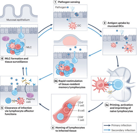
A very insightful review from the Akiko Iwasaki's group on the potential to harness mucosal immunity in next-generation vaccine development.
While the rapid deployment of intramuscular mRNA vaccines was a landmark achievement in preventing severe COVID-19, intramuscular shots often fall short of providing sterilizing immunity.
Mucosal immunity represents a particularly promising avenue for improving vaccines against respiratory viruses, as it enables immune protection to be established directly at the site of viral entry and early replication.
Mucosal tissues, such as the respiratory tract, host locally regulated specialized immune cells that are functionally and spatially distinct. Reduction of infection and transmission requires engaging the mucosal immune response: a coordinated process beginning with epithelial pathogen sensing and culminating in the establishment of tissue-resident memory T (TRM) and B (BRM) cells, alongside robust local secretory IgA (SIgA) production. Unlike systemic IgG, nasal SIgA has demonstrated superior virus-neutralizing activity and greater breadth against antigenically drifted variants.
A promising strategy for advancing vaccine design is the heterologous prime-boost approach. Research suggests that intramuscular priming (to establish peripheral memory pools) followed by an intranasal boost can effectively "pull" memory cells to the respiratory mucosa.
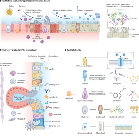
However, the so-called 'mucosal' vaccines requires navigating complex physiological constraints, such as the mucociliary clearance system and the anionic mucus layer.
Moreover, the regulatory path for mucosal vaccines is primarily hindered by the lack of validated correlates of protection, making it difficult to predict efficacy and guide clinical trial designs. Additionally, the anatomical proximity of the nasal mucosa to the central nervous system necessitates rigorous safety evaluations to prevent neuro-olfactory spillover or unintended neuro-inflammation.
https://lnkd.in/eb5ZUMY4
|
|
Scooped by
Gilbert C FAURE
June 7, 2025 4:29 AM
|
|
|
Scooped by
Gilbert C FAURE
January 2, 2023 8:02 AM
|
MIT researchers have identified molecules found in mucus that can block cholera infection by interfering with the genes that cause the microbe to switch into a harmful state.These protective molecules, known as glycans, are a major constituent of muc...
|
|
Scooped by
Gilbert C FAURE
December 17, 2022 1:53 AM
|
Less known to the general public, mucosal vaccination via the mucus membranes could provide robust protection against SARS-CoV-2 infections. Immune cells in the nose and lungs are considered better prepared to encounter and block the virus that causes COVID-19.
|
|
Scooped by
Gilbert C FAURE
January 13, 2022 1:16 PM
|
While the gut and lungs are both mucosa-lined luminal organs with a shared embryological origin, their gross and micro-anatomical features are quite distinct, yielding marked differences in the composition and population dynamics of their microbiota. In the absence of vomiting or esophageal reflux, migration of microbes in the digestive tract is unidirectional (from the mouth to the anus), and is serially interrupted by widely varying physical and chemical barriers. In order for an orally introduced microbe to immigrate into the cecum, it must endure the acidic pH of the stomach (~2.0) and the alkaline pH of the duodenum (~8.0) and compete for resources with a densely populated resident microbiome. By contrast, movement of air, mucus, and microbes in the lung is bidirectional, with no physical barrier between the larynx and the most distal alveolus. Thus the microbiome of the lungs is more dynamic and transient than that of the lower gastrointestinal tract. While the gastrointestinal tract is of uniform temperature (37°C) throughout its entire 9 meters of length, the mucosa of the respiratory tract (a short half-meter in length) represents a gradient from ambient temperature at the point of inhalation to core body temperature in the alveoli [8]. Unlike the gut, the lung environment is oxygen-rich. Though the trachea and bronchi are, like the gut, lined with the heavily glycosylated proteins of secreted mucus, the vast majority of the lung’s surface area is lined with lipid-rich surfactant, which has bacteriostatic effects against select bacterial species [9]. Bacterial density in the airways is quite modest, comparable to that of the duodenum [7], orders of magnitude less than that of the large intestine; thus inter-bacterial metabolic interactions are markedly different. Finally, the gut and lungs differ in the character of host–bacterial interactions. Luminal IgA levels are far higher in the gut, while the lungs exhibit far more extraluminal interactions between bacteria and host leukocytes (alveolar macrophages). Together, these markedly divergent environmental conditions result in correspondingly divergent microbial communities.
|
|
Scooped by
Gilbert C FAURE
June 19, 2021 10:31 AM
|
Pathogenesis of Nasal polyp has been primarily investigated on the basis of sinonasal mucosal immunity. Several inflammatory responses have been reported in nasal polyp pathogenesis to date. In this study the researchers have summarised new research on pathways for developing chronic nasal polyp rhinosinusitis (CRSwNP), which focuses on Th2 inflammation. The th2 inflammation may be driven by Staphylococcus aureus by the production of enterotoxins and a serine protease-like protein. Furthermore, S. aureus affected the activity of the mucosal obstruction directly and increased synthesis of Th2 cytokines by the rapid activation of endogenous epithelial cytokines. The synthesis of innate lymphoid epithelial-derived cytokines, including TSLP, IL-25, and IL-33, promotes th2 reactions. Mast cell produces IL-5, IL-13 and periostin and has a function to play in the pathogenesis of nasal polyps by orchestrating infiltration of eosinophils. The formation of extracellular eosinophil traps and Charcot-Leyden crystals is closely linked with the seriousness of disease and development of viscous mucus plugs. The role of S. aureus in innate and adaptive immunity is increasingly evident and contributes to the inflammation of Th2 in CRSwNP. Innate immunity, like epithelial cytokines, is critical in the production of CRSwNP, inducing different pathways, and must be further examined as Th2-controlled biomarkers. Neutrophilic inflammation has recently begun to be investigated but still remains uncertain. For latest news and updates By signing up, you will receive emails about Physician's Weekly products and you agree to our Terms & Conditions and Privacy Policy. Reference: https://journals.lww.com/co-allergy/Abstract/2020/02000/Th2_inflammatory_responses_in_the_development_of.2.aspx
|
|
Scooped by
Gilbert C FAURE
January 10, 2021 12:58 PM
|
From a general structural perspective, a mucosal tissue is constituted by two main matrices: the tissue and the secreted mucus. Jointly, they fulfil a wide range of functions including the protection of the epithelial layer.
|
|
Scooped by
Gilbert C FAURE
August 27, 2020 7:43 AM
|
Nutr Clin Pract. Author manuscript; available in PMC 2016 Dec 1. Published in final edited form as: Published online 2015 Oct 8. doi: 10.1177/0884533615609899 PMCID: PMC4838018 NIHMSID: NIHMS777222 PMID: 26449893 The Gut Microbiome: What we do and don’t know Gail A. Cresci, PhD, RD, LD, CNSC, Associate Staff and Emmy Bawden, RD The publisher's final edited version of this article is available at Nutr Clin Pract See other articles in PMC that cite the published article. Introduction Interest in and knowledge of the gut microbiome has increased exponentially in the past decade. An internet search using key words such as “gut microbiome” or “gut microbiota” generates between 1 and 2.2 million results. Once overlooked, this component of the gastrointestinal (GI) tract is now gaining appreciation for its importance in optimal health. The food industry has taken this to full heights and a plethora of food and supplement products touting “probiotic” or “fermented foods” populate store shelves; and advertisements for them can be found in magazines, the internet and television commercials. Never before has the discussion of one’s bowel habits and GI symptoms been more mainstream. Interestingly, this rapid growth has left both consumers and many clinicians confused as the data, while growing, still leave many unanswered questions. Dating back to the early 1900’s Eli Metchnikoff, a Russian scientist of the Pasteur Institute in Paris, associated longevity of rural Bulgarians to their consumption of fermented milk products. He postulated that the lactic acid bacteria in the fermented milk products ingested by these peasants living in harsh climate and poverty provided an anti-aging effect which contributed to their greatly outliving wealthier Europeans. He named the organism “Lactobacillus bulgaricus.” As Metchnikoff researched, he hypothesized that seeding the gut with healthy bacteria by drinking fermented milk products could fight off harmful bacteria and prolong life. He was the first scientist to suggest that it was possible to modify the gut microbiome by replacing bad bacteria with good bacteria, and he earned a Nobel Prize in 1908 for his work in immunity. The discovery of penicillin in 1928 by Sir Alexander Fleming, a Scottish biologist, turned the attention of researchers away from using bacteria to assist with healing to that of using soil fungi derivatives to kill bacteria. During the golden age of antibiotic discovery from 1940–1960, most antibiotics were originally isolated by screening soil-derived actinomycetes (1). However as returns diminished on this discovery platform, new discoveries were based upon synthetic compounds. However this target-focused screening process of large libraries also failed, partly due to the inability of these synthetic compounds to penetrate the bacterial envelope (1). Unfortunately the deceleration of antibiotic discovery is accompanied with the spread of resistant bacteria, and a major public health threat of untreatable infections. With advances in micro- and molecular biology techniques, the culture is poised to revisit past successes with bacterial therapy for gut health and immunity in order to develop future treatments. One of several international efforts, the Human Microbiome Project (HMP) utilizes high through multi ‘omics analyses to identify and study the microbiome in human health (2,3). Funded by the NIH common fund in 2008, the HMP has resulted in the isolation and sequencing of over 1,300 reference strains thus far from the human body (2,3). The HMP Consortium has reported the structure and function of the human microbiome in 300 healthy adults (18–40 yrs) at 18 body sites from a single time point. This has led to an unprecedented amount of data about the complexity of the human microbiome allowing for a baseline for further research into the impacts of the microbiome on health and disease. As a precursor to the Human Microbiome Project, the Human Gut Microbiome Project has widened our appreciation for the bacterial ecosystem that resides within the human intestinal tract. This system is comprised of microorganisms such as bacteria, archaea, fungus and viruses that are distributed throughout the entire gastrointestinal tract (4). Ongoing investigations are revealing the importance of the gut microorganisms in exerting beneficial effects on human health. The typical healthy person is inhabited with trillions of microbes (Figure 1). But to confuse matters, two healthy people may have very different microbiomes. Analysis of HMP samples with lifestyle and history questionnaires has revealed associations between life-history characteristics and microbiome composition (5). Ding and Schloss analyzed a 16S rRNA gene sequence data set from the HMP Consortium. As a significant difficulty in analyzing microbiome data has been the considerable intra- and interpersonal variation in the composition of the human microbiome, this study took the approach of clustering samples into bins based on their taxonomic similarity. Using bacterial community structures collected from18 body sites and up to three time points, they applied community typing analysis to better understand the factors that affect the structure of the microbiome and contribute to human health. Using Dirichlet multinomial mixture models to partition the data into community types for each body site, their analysis of simulated data and the HMP data suggest that the community types represent clusters of relative abundance profiles. Three important observations were made with this approach. Firstly, there were strong associations between whether someone was breastfed, their gender and education level with their community types at several body sites. Secondly, the community types of the oral cavity and gut microbiome were predictive of each other even though the specific taxonomic compositions were different. Lastly, stability of community types was least stable in the oral cavity and most stable in the vagina and gut communities over the course of the sampling period. Therefore the authors concluded that despite substantial intra- and interpersonal variation in the human microbiome, by partitioning this variation intocommunity types, the types became predictive of each other and are likely the result of life-history characteristics (5). The gut microbiome is influenced by multiple factors including mode of infant delivery and feeding, aging process, diet composition, geography, medications, and stress (Figure 2). The following sections provide a brief overview of current knowledge regarding variances in the gut microbiota related to these factors. Our first colonization In utero It was previously believed that the GI tract was sterile in utero, and that during the birthing process was when first colonization and initiation of the gut microbiome occurred (6). However, recent studies have challenged this belief by demonstrating the presence of microorganisms in the placenta, amniotic fluid and umbilical cord (7–9). This work is still preliminary but deserves further study as it highlights the importance of maternal gut microbiota health. It is hypothesized that by swallowing amniotic fluid and its bacteria in utero, the fetus begins to colonize the developing GI tract (10). The meconium, the first infant stool sample, has also been shown to contain microorganisms (11). Notably, the meconium from preterm infants shows a different microbiota composition from that seen in a sample acquired after the first week of life, demonstrating how normal gut colonization takes place throughout the course of fetal development (10). Absence of this progressive colonization could place preterm infants at risk for various gastrointestinal infections, including necrotizing enterocolitis (NEC) (10). As a means to mimic the natural inoculation of the gut, multiple studies have evaluated probiotic supplementation as a means to influence the incidence and severity of NEC. A recent Cochrane review, which included 24 randomized controlled trials (n=5529), assessed the role of probiotic supplementation in preterm neonates. The review confirmed previous findings that probiotic supplementation significantly reduces the risk of stage II or greater necrotizing enterocolitis and all cause mortality in this patient population (12). As probiotics are known to have potentially beneficial effects on gut function and maturity, Athalye-Jape et al conducted a meta-analysis that included 25 randomized controlled trials (n=5895) to determine if probiotic supplementation influenced the time to establish first full enteral feeds in preterm neonates (13). Overall, the subjects supplemented with probiotics took less time to achieve full feeds (mean difference: −1.54 d; 95% CI: −2.75, −0.32d; p=0.01; I2 = 93%). The type and/or number of probiotic strains (e.g., Bifidobacterium or non-Bifidobacterium) did not influence this improvement. Additionally, probiotic supplementation reduced hospital length of stay, feeding intolerance, and duration of indirect hyperbilirubinemia as well as increased weight gain and growth velocity. None of the trials reported adverse effects of probiotics on outcomes. Delivery Mode The birthing process exposes newborns to a wide range of microorganisms that also contribute to the colonization of the gut microbiome. The mode of delivery affects the composition of the infant’s gut microbiota, and interestingly, the gut microbiota of the newborn will closely resemble the microbiota with which they encountered during birth. During a vaginal birth, the infant is inoculated with vaginal microbiota, which differs from skin microbiota encountered during a caesarian section (14). At the phylum level, studies have shown that infants delivered vaginally have a larger population of Bacteroidetes than Firmicutes when compared to infants delivered via cesarean section (15). Despite exposure in utero, the majority of microbes that will colonize the infant gut are acquired postpartum. The initial colonization pattern is thought to be chaotic, and increasing evidence suggests that environmental exposures early in life, including diet, are responsible for these variations (16). Gut colonization in an infant occurs in a succession of stages. Early on, the gut becomes colonized by primarily aerobic organisms, such as enterobacteria, staphylococci, and streptococci, many of which have the potential to be pathogenic. These early colonizers begin to change the gut environment, paving the way for colonization by an increasingly anaerobic community of microbes (17). The structure of the gut community continues to change over the first year of life and thereafter in response to external factors such as diet and antibiotic use (18, 19). Weaning, breastfeeding status, and the successive introduction of different types of food all correspondingly affect the infant gut microbiome and immune system (20). Infant Feeding Method Breast milk is the optimal food for infants as it meets all their nutritional and physiologic requirements. Human milk contains protein, fat and carbohydrate, as well as immunoglobulins and endocannabinoids (21). Breast milk is not sterile as it contains as many as 600 different species of bacteria including beneficial Bifidobacterium breve, B. adolescentis, B. longum, B. bifidum, and B. dentium (22). In addition to lactose, the carbohydrate component of human milk also contains oligosaccharides which comprise the third largest solid component (23). Human milk oligosaccharides are indigestible polymers formed by a small number of different monosacchrides that serve as prebiotics by selectively stimulating growth of members of the genus Bifidobacterium(23). Some of the beneficial effects of breast feeding over formula feeding are attributed to the effects of oligosaccharides on the beneficial bacteria as an increased proportion of Bifidobacteria is noted in breast-fed infants compared to those fed formula (24). Bifidobacteria have been linked with strengthening gut mucosal protection through activities against pathogens (25). Bifidobacteria have been shown to increase the production of immunoglobulin A which is correlated with modulation of the intestinal immune system (26). Schwartz et al studied breast-fed and formula-fed infants and their mothers. Stool samples from each infant were collected, and microbial DNA was extracted and sequenced; and mRNA was isolated from stool containing host gut exfoliated epithelial cells and processed for microarray analysis (20). They found that the microbiome of breast-fed infants to be significantly enriched in genes associated with virulence functionality, and demonstrated a multivariate correlation between the gut flora genes associated with bacterial pathogenicity and the expression of host genes associated with immune and defense mechanisms. Interestingly, the operational taxonomic unit (OTU) composition and genetic potential of the microbiota differed between breast-fed and formula-fed infants. The researchers suggested their findings indicate human milk promotes the mutualistic crosstalk between the mucosal immune system and the microbiome in the maintenance of intestinal homeostasis. More research is needed to better determine the mechanisms by which Bifidobacteria produce these effects. Aerotolerance of the intestinal microbiota also seems to differ between breast-fed and formula-fed infants. Aerobic organisms are more common in the feces of breast-fed infants, whereas anaerobic and facultatively anaerobic organisms, which preferentially use anaerobic glycolysis, are more frequently identified in feces of formula-fed infants (27). Bacteroides and Clostridia colonization differs between the two types of feeding, with breast-fed infants characterized with lower concentrations of both (28–30). With a better understanding of the composition of human breast milk, developments of complex infant formulas have attempted to mimic its nutritional value making them an acceptable alternative for mothers unable to breast-feed. Infant formulas are not a perfect substitute for human milk since bioactive compounds contained in breast milk known to affect nutrient absorption and digestion, immune protection, and defense against potentially pathogenic microbes are lacking (21). Unfortunately it is difficult to mimic the actions of these bioactive compounds. While infants fed a formula enriched with oligosaccharides have been shown to harbor more Bididobacteria in the feces, more evidence is needed for affirmation that infant formulas designed to mimic breast milk are beneficial (31). Changes in the Gut Microbiome with Age Revealed by metagenomic analysis, gut microbiota composition transforms throughout early stages of human development and is influenced by the diet (32). As an infant’s diet comprises breast milk and formula, this is reflective in that the microbiome has minimal diversity and is enriched in genes to facilitate lactate utilization (33). A shift in the functional capacity to preferentially utilize plant-derived glycans occurs prior to the introduction of solid foods. Around 3-years of age, the bacterial composition resembles that of an adult and remains stable until old age when variability in community composition increases (34). In terms of ecological succession, the Bifidobacterium-dominated microbiota of the infant changes over time into the Bacteroidetes- and Firmicutes-dominated microbiota of the adult (35). This distribution remains fairly stable throughout adulthood in the absence of perturbations, such as long-term dietary changes or repeated antibiotic usage. Declines in dentition, salivary function, digestion and intestinal transit time may affect the gut microbiota upon ageing (36). There are notable differences in the microbiota in elderly people compared to young adults, with relative proportions of Bacteroidetes predominating in the elderly compared to higher proportions of Firmicutes in young adults (37). The elderly are also noted to have significant decreases in Bifidobacteria, Bacteriodes, and Clostridium cluster IV (38). Variability varies greatly among individuals ranging from 3 to 92% for Bacteroidetes and 7 to 94% for Firmicutes (34). However the microbiota are less variable within individual subjects (34). Alterations in gut microbiome are associated with health concerns pertaining to the elderly, such as frailty. A significant reduction in microbial diversity with reduced composition of lactobacilli, Bacteroides/Prevotella and Faecalibacterium prausnitzii, and increased proportions of Ruminococcus, Atopobium and Enterobacteriaceae was observed in people with high frailty scores (39). Claesson et al investigated the links between diet, environment, health and microbiota in 178 older people (>65 years) subjects and found an association between gut microbiome diversity and functional independence (39). Decreased microbial diversity was noted in individuals living in short- or long-term residential care compared to those living in the community, and this difference was associated with increased frailty, decreased diet diversity and health parameters as well as increased inflammatory markers (serum TNF-α, IL-6, IL-8 and C-reactive protein) (38). Dietary patterns in residential location correlated with separations based on microbiota composition with the most discriminating food types being vegetables, fruit and meats. Complete linkage clusterings revealed four dietary groupings: low fat/high fiber and moderate fat/high fiber included 98% of the community and day hospital subjects; and moderate fat/low fiber and high fat/low fiber included 83% of long-stay subjects (39).Significant associations between several health/frailty measurements were found with minimum variability amongst community dwellers, but within the long-stay subjects the most significant associations were related to functional independence, Barthel index (functional assessment) and nutrition, followed by blood pressure and calf circumference. The authors speculated that the later may be attributable to the influence of diet and/or the microbiota on muscle mass and sarcopenia and hence frailty. Influences on the Gut Microbiome Geography Geographical location and ethnicity have been shown to be determinants of the diversity and overall composition of microbiota. A 2013 study performed by Prideaux (40) et al looked at Caucasian and Chinese subjects in the United States and Hong Kong and found that microbial composition differed between countries and between ethnicities within the same country. In an elegant study, Yatsunenko et al characterized bacterial species in fecal samples from 531 individuals comprised of healthy children and adults from the Amazonas of Venezuela, rural Malawi and US metropolitan areas and included mono- and dizygotic twins (41). All populations studied showed shared features of gut microbiome development during the first three years of life including age-associated changes in the genes involved in vitamin biosynthesis and metabolism. Phylogenetic composition of fecal microbiota was significantly altered between individuals living in the different countries, with pronounced separation occurring between the US and the Malawian and Amerindian gut communities; this was true for individuals aged 0–3 years, 3–17 years, and for adults. However there was also separation between the non-US populations. Bacterial diversity increased with age in all three populations with the microbiome of the US population having the least diversity. Microbiome datasets from breastfed children (n=110: (24 babies (0.6–5 months old), 60 children and adolescents (6 months to 17 years old) and 26 adults) were analyzed to determine which bacterial taxa changed monotonically with increasing age within and between the three sampled populations. In all babies 16S rRNA sequences mapped to members of the Bifidobacterium genus, and while Bifidobacteria dominated fecal communities during the first year of life, they proportionally declined during this period. Also noted were age and population-related changes in metabolism. A total of 476 enzymes were identified as being significantly different in US versus Malawian and Amerindian breastfed babies (p<.0.001). The most prominent differences involved pathways related to vitamin biosynthesis and carbohydrate metabolism. Malawian and Amerindian babies, but not adults, had higher representation of enzymes that were components of the vitamin B2 (riboflavin) biosynthetic pathway. Compared with adults, baby microbiomes were enriched in enzymes involved in the scavenging of glycans represented in breastmilk and the intestinal mucosa (mannans, sialylated glycans, galactose and fucosyloligosaccharides), with several of these genes significantly overrepresented in Amerindian and Malawian baby microbiomes compared with US. Interestingly, urease gene representation was significantly higher but decreased with age in Malawian and Amerindian baby microbiomes, as opposed to the United States, where it was low from infancy to adulthood. Urea, which comprises up to 15% of the nitrogen present in human breast milk, is broken down by urease to ammonia which can then be used for microbial biosynthesis of essential and nonessential amino acids. Urease also has a crucial role in nitrogen recycling, particularly when diets are deficient in protein making urease potentially advantageous to both the microbiome and host when dietary nitrogen supply is suboptimal (42). A typical US diet is rich in protein, whereas diets in Malawian and Amerindian populations are dominated by corn and cassava. The differences between US and Malawian/Amerindian microbiomes can be related to these differences in diet. The enzymes that were the most significantly enriched in US fecal microbiomes parallel differences observed in carnivorous versus herbivorous mammals (43). Interestingly, several enzymes involved in the degradation of amino acids were overrepresented in adult US fecal microbiomes including aspartate, proline, ornithine and lysine as were enzymes involved in the catabolism of simple sugars (glucose-6-phosphate dehydrogenase and 6-phosphofructokinase), sugar substitutes (L-iditol 2-dehydrogenase, which degrades sorbitol), and host glycans (α-mannosidase, β-mannosidase and α-fucosidase). By contrast Malawian and Amerindian microbiomes had an over-representation of α-amylase which participates in the degradation of starch reflecting their corn-rich diet. Perhaps reflecting a fat-rich diet, the US microbiomes had an over-representation of enzymes involved with vitamin biosynthesis (cobalamin, biotin and lipoic acid), in the metabolism of xenobiotics (phenylacetate CoA ligase), which participates in the metabolism of aromatic compounds, and mercury reductase, and in bile salt metabolism (choloylglycine hydrolase). To obtain insights into commonalities and differences between gut microbiomes across different populations, Arumugam et al sequenced 22 European metagenomes from Danish, French, Italian and Spanish individuals that were selected for diversity and combined them with previous sequencing results from Japanese and Americans to total 39 individuals (44). They found that despite the vast number of species residing in the gut and their inter-individual variability, the microbiota composition can be classified into at least 3 distinct groups, or enterotypes. The enterotypes contain functional markers that correlate with individual features such as age and body mass index (BMI). For example, starch degradation enzymes such as glycosidases and glucan phosphorylases increase with age, which could be a reaction to decreased efficiency of host breakdown of dietary carbohydrates with age (45). Three marker modules, two of which were ATPase complexes, correlated strongly with the hosts’ BMI supporting the link found between the gut microbiota’s capacity for energy harvest and obesity in the host (46). The authors concluded that these functional markers might be utilizable for diagnostic and perhaps even prognostic tools for numerous human disorders, for instance colorectal cancer and obesity-linked co-morbidities such as metabolic syndrome, diabetes and cardiovascular pathologies. Food Supply Diet has emerged as one of the most relevant factors in influencing the gut microbiome. Significant and meaningful changes in the gut microbiota have been associated with dietary alterations, primarily consumption of dietary fiber from fruits, vegetables and other plants. A diet that is varied and complex is associated with a more diversified microbiome. Globally the microbiome composition is noted to be different amongst different populations and cultures. De Filippo et al compared the gut microbiota of children aged 1–6 years living in a village of rural Africa in an environment that still resembles that of Neolithic subsistence farmers with the gut microbiota of western European children (Florence, Italy) of the same age, with dietary habits and living conditions typical of the developed world (47). Traditionally the African children are breast-fed until the age of 2 after which their diet is predominantly vegetarian; low in fat and animal protein and rich in starch, plant polysaccharides and fiber. All food resources are completely produced locally, cultivated and harvested nearby the village. The dietary content of carbohydrate, fiber and non-animal protein is very high. Animal protein intake is low and consists of mainly chicken and termites during the rainy season. The European children were breast-fed for up to 1 y of age after which they ate a typical Western diet high in animal protein, sugar, starch, and fat and low in fiber. Average caloric intake differed between the two populations (African children: 1–2 y old, 672 kcal/d; 2–6 y old, 996 kcal/d; European children: 1–2 y old, 1,068 kcal/d; 2–6 y old, 1,512 kcal/d). Multiplex pyrosequencing of the 16S rRNA gene revealed that 94.2% of the sequences in all of the African and European samples were found to belong to the four most populated bacterial phyla, namely Actinobacteria, Bacteroidetes, Firmicutes,and Proteobacteria, in agreement with previous studies describing such phyla as those contributing to the majority of human gut microbiota (48). Relevant differences were found in the proportions of four phyla: Actinobacteria and Bacteroidetes were more represented in African than in European children’s microbiota (10.1% versus 6.7% and 57.7% versus 22.4%, respectively), whereas Firmicutes and Proteobacteria were more abundant in European than in African children (63.7% versus 27.3% and 6.7% versus 0.8%, respectively). Firmicutes were twice as abundant in the European children as evidenced by the different ratio between Firmicutes and Bacteroidetes (F/B ratio ± SD, 2.8 ± 0.06 in European and 0.47 ± 0.05 in African). While short-chain fatty acid (SCFA)-producing bacteria were found in both populations, bacteria (Xylanibacter, Prevotella, Butyrivibrio, and Treponema) which use xylane, xylose, and carboxymethylcellulose to produce high levels of SCFAs and have a protective role against gut inflammation (49) were found exclusively in the African children. Notably, the African children had higher levels of total SCFA than the European children, with 4 times higher levels of butyrate and propionate. The authors concluded a correlation exists between polysaccharide-degrading microbiota and the calories that the host can extract from his/her diet, potentially influencing the survival and fitness of the host, suggesting that the microbiome of the African children co-evolved with their diet to assist with energy harvest by producing high levels of SCFA. Microbial enrichment has been associated with diets high in fruits, vegetables and fiber compared to a Western diet rich in fat, sugars and animal protein and depleted of fiber. Zimmer et al analyzed the fecal flora of a large group of healthy volunteers on a strict vegetarian or vegan diet with classical microbiological culture and compared them with age and gender matched subjects consuming an omnivorous diet (50). The faecal microbiota of vegetarian and vegan subjects showed significantly lower microbial counts of Bifidobacterium, Bacteroides, E. coli and Enterobacteriaceae species and lower stool pH compared with omnivores. Compared to an omnivore diet, a vegetarian/vegan diet is associated with a higher carbohydrate and fiber content in which the undigestible polysaccharides can be fermented into SCFA by the gut microbiota. Production of SCFA is associated with decreasing luminal pH. The fact that E. coli and Enterobacteriacea do not thrive in lower pH ranges (5.5–6.5) and that they prefer proteins as their energy source, may explain the lower counts in the subjects consuming a vegan/vegetarian diet. Depleted microbial biodiversity of the gut microbiota in people consuming a Western diet is associated with increasing incidence of obesity, coronary vascular disease, metabolic syndrome and certain malignancies. Detailed discussion of each of these associations is beyond the scope of this paper. At a minimum, the diversity of the gut microbiome may be a future biomarker of long-term consumption of a “healthy” versus “unhealthy” diet which may be linked to potential for disease development. Stress Stress is defined as an organism’s total response to environmental demands or pressures. There are several different types of stressors, such as acute or chronic, acute on chronic, or repetitive acute. Stress can be predictable and controllable as well as unpredictable and uncontrollable, mild or severe, and occur in or out of context (51). Both the perception of stress and the persistence of its consequences vary between people. Stress contributes to susceptibility to disease and disabilities, therefore represents a severe economic burden. Psychological and physical stressors activate the hypothalamic-pituitary-adrenal (HPA) axis. This results in a series or hormonal responses including release of corticotrophin-releasing hormone which induces the release of adrenocorticotrophic hormone systemically with then stimulates glucocorticoid synthesis (cortisol) in the adrenal cortex (52). Additionally, catecholamines (noradrenaline and adrenaline) are also released following psychological and physical stressors. The GI tract, and more recently known, the gut microbiota, is sensitive to stress and stress mediators. Enteric bacteria respond to the release of stress-related neurochemical mediators by the host which can influence the response to a bacterial infection (53). Recent theory suggests that bacteria act as delivery vehicles for neuroactive compounds, and therefore can affect host physiology by providing neurochemicals (54). Physiological Stress-Exercise High intensity exercise is a physiological stressor that can lead to gastrointestinal distress. There are some reports that between 30–90% of distance runners have experienced intestinal problems related to exercise (55). The degree of intestinal distress can range from mild to severe, and symptoms include nausea, vomiting, abdominal angina, and bloody diarrhea. High intensity training has been associated with reduced GI blood flow, tissue hyperthermia and hypoxia leading to possible alterations of microbiota and gut barrier. Athletes often have different dietary pattern from non-athletes depending upon the type of activity they in which they participate. In a study of professional rugby players during a regulated environment of preseason training, Clarke et al demonstrated the impact of exercise and associated dietary changes on the gut microbiota (56). Enhanced gut microbial diversity was significantly higher and positively correlated with increased exercise and dietary protein intake in athletes as compared with size matched (high Body Mass Index – BMI ~30 kg/m2) and age/gender matched (lower BMI <25) non-athletic control groups, with fewer differences seen between the two control groups. Interestingly, when compared to controls, the athletes also exhibited lower inflammatory markers and improved metabolic markers. Athletes consumed more calories, with a higher percentage of protein, and grazed eating throughout the day compared to non-athletes. Microbiota diversity measures were positively correlated with protein intake and plasma creatine kinase levels, a marker of extreme exercise, suggesting that both diet and exercise were influencing changes in microbial diversity. The athlete’s increased microbial diversity was reflected by the presence of representatives of 22 phyla of bacteria in contrast to 11 and 9 phyla in the low and high BMI controls, respectively. There are several potential mechanisms by which physical activity and fitness might modify the microbiota. Abrupt exercise produces multiple metabolites and inflammatory mediators whereas habitual exercise and fitness leads to suppression of basal inflammatory cytokines suggesting a regulatory loop between exercise biology and host immunity (57). Regular exercise has an anti-inflammatory effect which improves the immunological profile in type 2 diabetes mellitus, coronary artery disease, peripheral arterial disease and obesity (58–61). In animal models, repeated exercise results in reduced pro-inflammatory cytokine expression and increased anti-inflammatory IL-10 expression (62, 63). Regular exercise also decreases colonic oxidative insult in a rat model of colitis (64). Contrary to the benefits of regular exercise, prolonged excessive exercise can negatively affect intestinal function. As high-intensity exercise can lead to prolonged intestinal hypo-perfusion, intestinal ischemia may result. Increased intestinal permeability can result making the gut susceptible to endotoxin translocation (65–67). Probiotic supplements, in conjunction with other dietary strategies, have been studied in athletes as a means to improve gut health, as characterized by decreased symptoms of nausea, cramping, bloating and diarrhea,as well as improved immunity. Primarily supplements containing Lactobacillus and/or Bifidobacterium species have been utilized. Products have been provided from 1 to 6 months, before and/or after exercise or a competition, and at varying doses (109–1012 CFU/day). Some studies report clinical outcomes of improved upper respiratory tract illness (64–69), gastrointestinal illness (70–72)), as well as immunological measures and outcomes (67, 68, 73–76)). Physiological Stress-Critical Illness As seen in high intensity exercise, intestinal hypoperfusion resulting from redistribution of splanchnic circulation severe enough to cause gut ischemia and mucosal injury, often occurs in the critically ill. In the critically ill, the gut has an important role in promoting infectious complications and multiple organ dysfunction syndrome (MODS). This is due to deteriorated intestinal epithelium, altered gut immune system, and dysfunctional metabolic activities of commensal bacteria (77). Shimizu et al, evaluated the gut microbiota and gut environment (fecal pH and presence of organic acids) in patients with systemic inflammatory response syndrome (SIRS) (78). In comparison to healthy controls, patients with severe SIRS had significantly lower total anaerobic bacterial counts (especially 2–4 log fewer commensal Bifidobacterium and Lactobacillus) and 2 log higher potentially pathogenic Staphylococcus and Pseudomonas group counts. Concentrations of total organic acids, in particular the short-chain fatty acids (SCFA) acetate, propionate and butyrate, were significantly decreased in the patients, whereas pH was markedly increased. This group of researchers further investigated the impact of fecal pH in critical illness in 138 trauma patients (79). Patients with acidic or alkaline feces were noted to have decreased Bacteroide and Bifidobacterium species. The incidence of bacteremia in patients with an acidic or alkaline fecal pH was significantly higher than those with a fecal pH in the normal range (P < 0.05 versus normal range). The incidence of both bacteremia and mortality were associated with an increased pH of 6.6. When the pH level was increased or decreased by one, the incidence of bacteremia more than tripled and mortality more than doubled. Total SCFA (propionate, butyrate) concentrations decreased with pH >6.6; lactic, succinic, and formic acids were increased in acidic feces which is notable as these are produced by Enterobacteriaceae (80). Whether these changes are a cause or consequence of SIRS is yet to be determined. While this study lends potential that fecal pH could be a risk factor marker, it does have limitations because these data were made with the use of culture-based interrogation of the microbiota and because the pH was not specified by gastrointestinal region. Alterations in the gut microbiome have been shown to occur within 6 hours of a metabolic insult and the microbiome fails to return to microbial patterns seen in healthy controls (81). Numerous studies attempting to modify the gut microbiome and improve clinical outcomes in the critically ill by providing probiotics, prebiotics or synbiotics have been performed (82–89). Details of these studies are beyond the scope of this review. Methods of dosing, supplement duration and supplements utilized vary amongst studies making general recommendations difficult. Several meta-analyses and systematic reviews have been performed evaluating probiotics in the critically ill (89–91)). The choice of the studies included in the analysis will determine the overall outcome of the study. Generally, it seems probiotic supplementation in the critically ill favorably improves outcomes. However, the issue of heterogeneity and lack of good quality studies is a highlight of systematic reviews of probiotic use in critically ill patients. Knowing that unrecoverable gut dysbiosis occurs early after metabolic insult and knowing the potential mechanisms of action of certain probiotic strains, further rigorous study on probiotic provision in the critically ill deserves attention. Future studies should focus on 1) clarification of viable probiotic strain(s) provided, and their dosage, route and timing of delivery; 2) clear definitions for the outcomes attempting to modify (ventilator associated pneumonia, diarrhea); and 3) focus effect of probiotic supplementation on meaningful clinical endpoints (mortality, duration of mechanical ventilation, other infections, length of stay). Psychological Stress The discovery of hormonal regulation of digestion initiated the concept of the gut-brain axis (92). The interaction of psychological factors and altered gut physiology via the gut-brain axis, where brain and gut symptoms are reciprocally influencing each other’s expression, is a conceptual framework of functional GI disorders (irritable bowel syndrome, functional dyspepsia). Early life stressors (e.g., psychological, sexual and/or physical abuse) have been suggested as important contributors to the pathogenesis of functional GI disorders (93–96)). This is a crucial developmental period when the gut microbiota are diversifying, thus making it particularly vulnerable to these stressors. Animal models of maternal separation, a model commonly used for early life stress, induces prolonged HPA-axis hyperactivity (97–103), anxiety-like behavior (100,,104–106), visceral hypersensitivity (107, 108–110), and altered cholinergic activity in the gut (111, 112), along with increased intestinal permeability (107, 113–116). This model results in a dysfunctional gut-brain axis and produces phenotypes found in irritable bowel syndrome patients (106, 116– 118). This animal model also yields gut dysbiosis in addition to other physiological and behavioral features of irritable bowel syndrome (118–120)). Various means to restore gut dysbiosis with probiotics have been investigated and appear to improve the negative stress-induced effects (107, 111, 120– 122).). Morphologic changes also result from maternal separation with increased goblet cells in the crypts of proximal colon with a subsequent increase in mucus secretion and a thinner mucosal layer (112). Thus changes in the gut microbiota in maternally separated animals could be resultant from changes to gut physiology (111– 114)) and morphology (112). Although the exact mechanisms are unknown, data thus suggest that stress, whether acute or chronic, creates a dysbiotic gut microbiome which then may induce anxiety and depression (52). It seems that metabolites produced by the gut microbiota might modulate brain biochemistry and behavior (54, 123–125). Through regulation of the vagus nerve, behavior can be altered by the gut microbiota which then affects neurotransmitter metabolism (126, 127)) or other undefined pathways (128)). Thus futuristic therapy for patients with irritable bowel syndrome with co-morbid depression or anxiety may be targeted probiotic and/or synbiotic either alone or as an adjuvant to traditional therapy. Pharmaceuticals There is substantial variation in the pharmacokinetics of most commonly used drugs, and large differences in the responses of ill persons to therapeutics. The gut microbiota assists in the conversion of inactive therapeutics (e.g., prodrugs) and dietary bioactives into their active forms (129–131). For example, a medication prescribed for ulcerative colitis, sulfasalazine remains inactive until it reaches the distal gut. This prodrug consists of an ant-iinflammatory 5-aminosalicylic acid (5-ASA) molecule connected to a sulfapyridine molecule through an N-N double bond. Gut microbiota encoded azoreductases cleave the N-N double bond to release active 5-ASA (131). Likewise, foods such as fruits, vegetables, cereals and coffee contain conjugated hydroxycinnamates, antioxidant and anti-inflammatory compounds which are activated following microbial biotransformation (132). Therefore, a question arises whether variation in medication response amongst people is due to alterations in their gut microbiome? The host possesses many protective mechanisms to protect against ingested pathogens including an acidic gastric environment, optimal bile flow, peristalsis and the gut microbiota. Current data suggest that the gut microbiota protects the host from pathogens by competing for binding sites, competing for requirements, and by direct inhibition through release of inhibitory molecules (133). When these protective mechanisms are disrupted, an imbalance in the gut microbiota can occur. Gastric Acid Suppression Several conditions such as intestinal dysmotility, altered GI anatomy, immune deficiencies, and hypochlorhydria (134) are predisposing factors for the development of small intestinal bacterial overgrowth (SIBO). While hypochlorhydria can be a result of Helicobacter pylori colonization and aging, many people also take medications to reduce their gastric acidity for stress ulcer prophylaxis or gastric esophageal reflux disease. The pH of gastric acid in the stomach lumen is normally 1.5–3.5; maintaining intragastric pH above 3.5–5.0 prevents gastric mucosal injury and may be facilitated with histamine 2 receptor antagonists (H2RAs), sucralfate, and proton pump inhibitors (PPIs) (135). Previous studies show that PPIs are known to alter the GI bacterial population in 50% of patients on long-term treatment with any type of effective antisecretory drug (135, 136). Lombardo et al, conducted a study in 200 patients with gastroesophageal reflux disease (GERD) receiving PPI treatment, 200 patients with irritable bowel syndrome (IBS) not receiving PPI treatment, and 50 healthy control subjects not receiving PPI treatment greater than 10 years and showed that SIBO occurs more frequently in long-term PPI users than patients with IBS or control subjects (137). They found the incidence of SIBO was significantly different (P< .001) in patients using PPIs (50%) as compared to patients with IBS not using PPIs (24.5%), and healthy control subjects (6%). According to a multivariate analysis including 5,387 elderly subjects, the presence of diarrhea was significantly associated with use of PPIs (odds ratio [OR] 2.97, 95% confidence interval [CI] 2.03–4.35) and antibiotics (OR 4.58, 95% CI 1.95–10.73) (138). Theisen et al, found that the suppression of gastric acid with omeprazole led to a high prevalence of SIBO, leading to a markedly increased concentration of unconjugated bile acids (139). Conversely a retrospective chart review completed in 1,191 patients with glucose hydrogen breath testing (GHBT) evaluated whether this value positively correlated with patients on PPI therapy (134). Interestingly, GHBT was positively associated with older age and antidiarrheal use, but not with PPI use. Therefore, the authors concluded that PPI use was not associated with the presence of SIBO as measured by GHBT. However, Lombardo (140) challenged these results making a point that PPI use has become very common, both prescription and over-the-counter, and many patients may not consider non-prescription use as a medication and not disclose it to their physician. The authors were unable to confirm actual use of PPI due to the nature of a retrospective chart review. Additionally, as PPI duration is related to incidence of SIBO, the lack of knowledge regarding the duration the patients were taking a PPI leads one to speculate that if they only took it for a short time would it have an influence in GHBT results? Knowing the protective role gastric acidity has in regards to protecting the host against ingested pathogens, it is plausible that prolonged use of gastric acid suppressants contributes to the incidence of SIBO and that clinicians should be judicious in recommending their use. Antibiotics Antibiotic therapies not only target pathogenic microorganisms, but also the host-associated microbial communities in the gut. Most antibiotics have broad-spectrum activity so they can be used to treat many diseases. Thus, although antibiotics are designed to target pathogenic organisms, related members of the microbiota are also affected, leaving a lasting negative effect on the gut microbial community long after the antibiotics have ceased (141). Antibiotics can also promote the expansion of antibiotic-resistant strains which can act as a reservoir for resistance genes in the gut microenvironment (142). Decreased diversity in the microbiome typically follows antibiotic treatment, and even though most of the microbiota return to pretreatment levels, some members are lost from the community indefinitely (142). As microorganisms can be dependent upon other colonizers for the provision of nutrition, secondary metabolites or removal of toxic waste products, alterations in microbial co-dependence can leave detrimental effects (143). The antibiotic spectrum of activity will influence the shift in gut microbiota composition. Additionally, the administration dose of the antibiotic is also important in determining the ecological impact it will have on the microbiota. While the sub-therapeutic antibiotic dosing often used by the agriculture industry prophylactically and to promote animal growth does not reduce the total bacterial mass, this use is criticized as it shifts the composition of the microbiota and promotes dangerous levels of antibiotic resistance (144). Similarly in humans, antibiotic therapeutic doses are designed to minimize these effects, but despite these efforts, subsets of the microbiota can shift to that of increased colonization by opportunistic pathogens such as Clostridium difficile and Candida albicans. Noteworthy is that these effects of antibiotic delivery are not limited to oral delivery. Intravenously delivered antibiotics can have an effect on the gut microbiota as they become incorporated into bile and secreted into the intestine via the biliary system (144). Gut microbial community alterations can result in dysregulation of host immune homeostasis and an increased susceptibility to disease. Host-microbial interactions are very specific and antibiotic therapy causes alterations or loss of highly co-evolved processes (145, 146). Antibiotic-induced changes that are important to microbial regulation of host immunity include loss of bacterial ligands that are recognized by the host, loss of specific bacterial signals, and alterations in the metabolites produced by the microbiota such as short-chain fatty acids (147). Short-chain fatty acids (SCFA) are beneficial for gut health as they serve as a primary fuel source for the colonocyte, are involved with water and electrolyte absorption, help to maintain the intestinal barrier, and modulate cell proliferation, differentiation, growth and apoptosis (147). Provision of butyrate during broad-spectrum antibiotic therapy mitigated the negative effects of antibiotics on gut health, notably the mRNA and protein expression for several anion exchangers, butyrate receptor and transports as well as protein expression of intestinal tight junction proteins (148). Changes in gut immunity increase host susceptibility to infection by pathogens. For instance, treatment with metronidazole, an antibiotic that targets anaerobic bacteria, reduces the integrity of the mucus layer and accelerates mucosal attachment of C. rodentium (149). Changes in gut microbiota structure and function after antibiotic treatment create a metabolic environment that favors C. difficile germination and colonization and associated infectious diarrhea (150). While antibiotic therapy is the major risk factor for C. difficile colonization, the accepted treatment for C. difficile infection is further antibiotic therapy, initially with metronidazole and then vancomycin, if C. difficile persists (151). Repeated antibiotic use is often necessary in the treatment of recurrent Clostridium difficile infections which unfortunately causes further disruption of an imbalanced microbiota (152). Chang et al, identified that patients with recurrent C. difficile infection possessed a microbiota characterized by markedly reduced overall diversity compared to controls and to patients with an initial episode of the infection (152). C. difficile infection is debilitating to patients and extremely costly, with symptoms ranging from diarrhea to fulminant colitis, toxic megacolon and death (153). Fecal Microbiota Transplantation (FMT) is the process by which a homogenized stool sample from a healthy donor is administered into the GI tract of an individual. This therapy is now becoming a sought-out treatment to eradicate C. difficile colonization and correct gut dysbiosis. FMT has been shown to be a successful treatment option to eliminate recurrent and refractory C. difficile infection and to re-establish a healthy colony in certain patients (154). With its 91% primary and 98% secondary cure rates, FMT is gaining increased interest among researchers as a treatment option for other gastrointestinal disorders (154). Perspective and future directions With technological advances, knowledge regarding the gut microbiome has expanded from earlier beliefs (155, 156). The topics covered in this review are examples of what is currently understood regarding how the human gut microbiome is first established and how certain life encounters alter the composition, but they are by no means inclusive. The influence of the gut microbiome on health and disease is currently one of the most exciting areas of science. Learning more about how breaches upon the gut microbiome early in life coupled with further insults throughout life influences health may provide new insights for treatments of many challenging syndromes and diseases (e.g., fibromyalgia, irritable bowel disease, IBD, autism). We are just beginning to uncover the impact of how things we ingest can influence pathology. Future research should direct its focus to the gut microbiome as a biomarker for dietary intake and disease development, the effect of probiotic supplementation on clinical endpoints, and bacterial therapy for gut health, immunity and therapeutic treatments. New available and developing technologies to uncover the metabolon opens the path of exploration to this exciting frontier. The horizons are broad with exciting developments sure to come. Supplementary Material Copyright Click here to view.(14K, docx) Contributor Information Gail A. Cresci, Department of Gastroenterology/Hepatology, Cleveland Clinic, Cleveland, OH 44195, Ph: 216-445-8317, Fax: 216-444-9401. Emmy Bawden, Dietetic Intern, Cleveland Clinic, Cleveland, OH 44195. References 1. Lewis K. Platforms for antibiotic discovery. Nature Reviews. 2013;12:371–387. [PubMed] [Google Scholar] 2. Human Microbiome Project Consortium. A framework for human microbiome research. Nature. 2012;486:215–221. [PMC free article] [PubMed] [Google Scholar] 3. Human Microbiome Project Consortium. Structure, function and diversity of the healthy human microbiome. Nature. 2012;86:207–214. [PMC free article] [PubMed] [Google Scholar] 4. Backhed F, Ley RE, Sonnenburg JL, Peterson DA, Gordon JI. Host-bacterial mutualism in the human intestine. Nature. 2005;307:1915–1920. [PubMed] [Google Scholar] 5. Ding T, Schloss PD. Dynamics and associations of microbial community types across the human body. Nature. 2014;509(7500):357–60. [PMC free article] [PubMed] [Google Scholar] 6. Salazar N, Arboleya S, Valdés L, et al. The human intestinal microbiome at extreme ages of life. Frontiers in Genetics. 2014;5:1–9. [PMC free article] [PubMed] [Google Scholar] 7. DiGiulio DB. Diversity of microbes in amniotic fluid. Semin Fetal Neonatal Med. 2012;17:2–11. [PubMed] [Google Scholar] 8. Jiménez E, Fernándes L, Marín M, et al. Isolation of commensal bacteria from umbilical cord blood of healthy neonates born by cesarean section. Curr Microbiol. 2005;51:270–274. [PubMed] [Google Scholar] 9. Aagard K, Ma J, Antony K, et al. The placenta harbors a unique microbiome. Sci Transl Med. 2014;6:237–265. [Google Scholar] 10. Moles L, Gomez M, Heilig H, Bustos G, Fuentes S, de Vos W, Fernández L, Rodríguez JM, Jiménez E. Bacterial diversity in meconium of preterm neonates and evolution of their fecal microbiota during the first months of life. PLoS ONE. 2013;8:1–13. [PMC free article] [PubMed] [Google Scholar] 11. Jimenez E, Marín ML, Martín R, et al. Is meconium from healthy newborns actually sterile? Res Microbiol. 2008;159:187–193. [PubMed] [Google Scholar] 12. Alfaleh K, Anabrees J. Probiotics for prevention of necrotizing enterocolotis in preterm infants. The Cochrane Database Syst Rev. 2014;4:CD005496. [PubMed] [Google Scholar] 13. Athalye-Jape G, Deshpande G, Rao S, Patole S. Benefits of probiotics on enteral nutrition in preterm neonates: a systematic review. Am J Clin Nutr. 2014;100:1508–19. [PubMed] [Google Scholar] 14. Clemente JC, Ursell LK, Parfrey LW, et al. The impact of the gut microbiota on human health: An integrated view. Cell. 2012;148:1258–1270. [PMC free article] [PubMed] [Google Scholar] 15. Jakobsson HE, Abrahamsson TR, Jenmalm MC, et al. Decreased gut microbiota diversity, delayed Bacteriodetes colonisation and reduced Th1 responses in infants delivered by caesarean section. Gut. 2014;63:559–566. [PubMed] [Google Scholar] 16. Koenig JE, Spor A, Scalfone N, et al. Succession of microbial consortia in the developing infant gut microbiome. Proc Natl Acad Sci U S A. 2011;108(Suppl 1):4578–85. [PMC free article] [PubMed] [Google Scholar] 17. Pop M. We are what we eat: how the diet of infants affects their gut microbiome. Genome Biology. 2012;13:152. [PMC free article] [PubMed] [Google Scholar] 18. Palmer C, Bik EM, DiGiulio DB, Relman DA, Brown PO. Development of the human infant intestinal microbiota. PLoS Biol. 2007;5:e177. [PMC free article] [PubMed] [Google Scholar] 19. Morowitz MJ, Denef VJ, Costello EK, Thomas BC, Poroyko V, Relman DA, Banfield JF. Strain-resolved community genomic analysis of gut microbial colonization in a premature infant. Proc Natl Acad Sci U S A. 2011;108:1128–1133. [PMC free article] [PubMed] [Google Scholar] 20. Schwartz S, Friedberg I, Ivanov I, Davidson LA, Goldsby JS, Dahl DB, et al. A metagenomic study of diet-dependent interaction between gut microbiota and host in infants reveals differences in immune response. Genome Biology. 2012;13:r32. [PMC free article] [PubMed] [Google Scholar] 21. Le Huerou-Luron I, Blat S, Boudry G. Breast- v. formula-feeding: impacts on the digestive tract and immediate and long-term health effects. Nutr Res Rev. 2010;23:23–36. [PubMed] [Google Scholar] 22. Martín R, Jiménez E, Heilig H, Fernández L, Marín ML, Zoetendal EG, Rodríguez JM. Isolation of Bifidobacteria from breast milk and assessment of the bifidobacterial population by pcr-denaturing gradient gel electrophoresis and quantitative real-time pcr. Applied and Environmental Microbiology. 2009;75:965–969. [PMC free article] [PubMed] [Google Scholar] 23. German JB, Freeman SL, Lebrilla CB, Mills DA. Human Milk Oligosaccharides: Evolution, Structures and Bioselectivity as Substrates for Intestinal Bacteria. Nestle Nutr Workshop Ser Pediatr Program. 2008;62:205–222. [PMC free article] [PubMed] [Google Scholar] 24. Balmer SE, Wharton BA. Diet and faecal flora in the newborn: breast milk and infant formula. Arch Dis Child. 1989;64:1672–7. [PMC free article] [PubMed] [Google Scholar] 25. Fukuda S, Toh H, Hase K, et al. Bifidobacteria can protect from enteropathogenic infection through production of acetate. Nature. 2011;469:543–7. [PubMed] [Google Scholar] 26. Ouwehand A, Isolauri E, Salminen S. The role of the intestinal microflora for the development of the immune system in early childhood. Eur J Nutr. 2002;41(Suppl 1):I32– 7. [PubMed] [Google Scholar] 27. Stark PL, Lee A. The microbial ecology of the large bowel of breast-fed and formula-fed infants during the first year of life. J Med Microbiol. 1982;15:189–203. [PubMed] [Google Scholar] 28. Harmsen HK, Wildeboer-Veloo AC, Raangs GC, Wagendorp AA, Klijn N, Bindels JG, Welling GW. Analysis of intestinal flora development in breast-fed and formula-fed infants by using molecular identification and detection methods. J Pediatr Gastroenterol Nutr. 2000;30:61–7. [PubMed] [Google Scholar] 29. Stark PL, Lee A, Parsonage BD. Colonization of the large bowel by Clostridium difficile in healthy infants: quantitative study. Infect Immun. 1982;35:895–9. [PMC free article] [PubMed] [Google Scholar] 30. Marcobal A, Barboza M, Sonnenburg ED, et al. Bacteroides in the infant gut consume milk oligosaccharides via mucus-utilization pathways. Cell Host Microbe. 2011;10:507–14. [PMC free article] [PubMed] [Google Scholar] 31. Beereman-Wauters G, Staelens S, Van de Broek H, et al. Physiological and bifidogenic effects of prebiotic supplements in infant formulae. J Pediatr Gastroenterol Nutr. 2011;52:763–71. [PubMed] [Google Scholar] 32. Koenig JE, Spor A, Scalfone N, Fricker AD, Stombaugh J, Knight R, Angenent LT, Ley RE. Succession of microbial consortia in the developing infant gut microbiome. Proc Natl Acad Sci USA. 2011;108(Suppl 1):4578–4585. [PMC free article] [PubMed] [Google Scholar] 33. Salazar N, Arboleya S, Valdés L, et al. The human intestinal microbiome at extreme ages of life. Frontiers in Genetics. 2014;5:1–9. [PMC free article] [PubMed] [Google Scholar] 34. Claesson MJ, Cusack S, O’Sullivan O, Greene-Diniz R, de Weerd H, Flannery E, Marchesi JR, Falush D, Dinan T, Fitzgerald G, et al. Composition, variability, and temporal stability of the intestinal microbiota of the elderly. Proc Natl Acad Sci USA. 2011;108(Suppl 1):4586– 4591. [PMC f
|
|
Scooped by
Gilbert C FAURE
October 22, 2019 2:51 AM
|
More than 200 square meters of our bodies — including the digestive tract, lungs, and urinary tract — are lined with mucus. Far from being a gross waste product, this slippery secretion produced by, and covering, the mucous membranes serves an important physiological purpose.
|
|
Scooped by
Gilbert C FAURE
July 12, 2019 2:58 PM
|
What's the point of mucus? The slimy stuff has a surprisingly wide array of beneficial biological functions.
|
|
Scooped by
Gilbert C FAURE
June 12, 2019 9:29 AM
|
Significance Functional loss of gut barrier integrity with subsequent increased antigen trafficking and occurrence of low-grade intestinal inflammation precede the onset of type 1 diabetes (T1D) in patients and preclinical models, thus suggesting that these events are mechanistically linked to the autoimmune pathogenesis of the disease. However, a causal relationship between increased intestinal permeability and autoimmune diabetes was never demonstrated. Our data show that breakage of gut barrier continuity leads to activation of islet-reactive T cells in the intestine, thus triggering autoimmune diabetes. An important implication of our findings is that restoration of a healthy gut barrier through microbiota and diet modulation in diabetes-prone individuals could reduce intestinal activation of islet-reactive T cells and prevent T1D occurrence. Abstract Low-grade intestinal inflammation and alterations of gut barrier integrity are found in patients affected by extraintestinal autoimmune diseases such as type 1 diabetes (T1D), but a direct causal link between enteropathy and triggering of autoimmunity is yet to be established. Here, we found that onset of autoimmunity in preclinical models of T1D is associated with alterations of the mucus layer structure and loss of gut barrier integrity. Importantly, we showed that breakage of the gut barrier integrity in BDC2.5XNOD mice carrying a transgenic T cell receptor (TCR) specific for a beta cell autoantigen leads to activation of islet-reactive T cells within the gut mucosa and onset of T1D. The intestinal activation of islet-reactive T cells requires the presence of gut microbiota and is abolished when mice are depleted of endogenous commensal microbiota by antibiotic treatment. Our results indicate that loss of gut barrier continuity can lead to activation of islet-specific T cells within the intestinal mucosa and to autoimmune diabetes and provide a strong rationale to design innovative therapeutic interventions in “at-risk” individuals aimed at restoring gut barrier integrity to prevent T1D occurrence. Footnotes ↵1To whom correspondence may be addressed. Email: falcone.marika{at}hsr.it. Author contributions: M.F. designed research; C.S., I.C., M.L.C., L.D.G., F.F., R.L., M.R., and R.F. performed research; C.S., R.F., F.S., F.C., and M.F. analyzed data; and C.S. and M.F. wrote the paper. The authors declare no conflict of interest. This article is a PNAS Direct Submission. This article contains supporting information online at www.pnas.org/lookup/suppl/doi:10.1073/pnas.1814558116/-/DCSupplemental. Copyright © 2019 the Author(s). Published by PNAS. This open access article is distributed under Creative Commons Attribution-NonCommercial-NoDerivatives License 4.0 (CC BY-NC-ND). References ↵ J. A. Todd, Etiology of type 1 diabetes. Immunity 32, 457–467 (2010).OpenUrlCrossRefPubMed ↵ F. Dotta et al., Coxsackie B4 virus infection of beta cells and natural killer cell insulitis in recent-onset type 1 diabetic patients. Proc. Natl. Acad. Sci. U.S.A. 104, 5115–5120 (2007). ↵ M. C. Honeyman et al., Association between rotavirus infection and pancreatic islet autoimmunity in children at risk of developing type 1 diabetes. Diabetes 49, 1319–1324 (2000).OpenUrlAbstract ↵ O. Vaarala et al., Cow’s milk formula feeding induces primary immunization to insulin in infants at genetic risk for type 1 diabetes. Diabetes 48, 1389–1394 (1999).OpenUrlAbstract ↵ J. M. Norris et al., Timing of initial cereal exposure in infancy and risk of islet autoimmunity. JAMA 290, 1713–1720 (2003).OpenUrlCrossRefPubMed ↵ L. Wen et al., Innate immunity and intestinal microbiota in the development of type 1 diabetes. Nature 455, 1109–1113 (2008).OpenUrlCrossRefPubMed ↵ A. Giongo et al., Toward defining the autoimmune microbiome for type 1 diabetes. ISME J. 5, 82–91 (2011).OpenUrlCrossRefPubMed ↵ C. T. Brown et al., Gut microbiome metagenomics analysis suggests a functional model for the development of autoimmunity for type 1 diabetes. PLoS One 6, e25792 (2011).OpenUrlCrossRefPubMed ↵DIABIMMUNE Study Group T. Vatanen et al.; DIABIMMUNE Study Group, Variation in microbiome LPS immunogenicity contributes to autoimmunity in humans. Cell 165, 842–853 (2016).OpenUrlCrossRefPubMed ↵ E. Badami et al., Defective differentiation of regulatory FoxP3+ T cells by small-intestinal dendritic cells in patients with type 1 diabetes. Diabetes 60, 2120–2124 (2011). ↵ T. Vatanen et al., The human gut microbiome in early-onset type 1 diabetes from the TEDDY study. Nature 562, 589–594 (2018).OpenUrlCrossRef ↵ J. B. Meddings, J. Jarand, S. J. Urbanski, J. Hardin, D. G. Gall, Increased gastrointestinal permeability is an early lesion in the spontaneously diabetic BB rat. Am. J. Physiol. 276, G951–G957 (1999).OpenUrlPubMed ↵ S. Graham et al., Enteropathy precedes type 1 diabetes in the BB rat. Gut 53, 1437–1444 (2004). ↵ F. Maurano et al., Small intestinal enteropathy in non-obese diabetic mice fed a diet containing wheat. Diabetologia 48, 931–937 (2005).OpenUrlCrossRefPubMed ↵ A. D. Mooradian, J. E. Morley, A. S. Levine, W. F. Prigge, R. L. Gebhard, Abnormal intestinal permeability to sugars in diabetes mellitus. Diabetologia 29, 221–224 (1986).OpenUrlCrossRefPubMed ↵ R. Carratù et al., Altered intestinal permeability to mannitol in diabetes mellitus type I. J. Pediatr. Gastroenterol. Nutr. 28, 264–269 (1999).OpenUrlCrossRefPubMed ↵ M. Westerholm-Ormio, O. Vaarala, P. Pihkala, J. Ilonen, E. Savilahti, Immunologic activity in the small intestinal mucosa of pediatric patients with type 1 diabetes. Diabetes 52, 2287–2295 (2003). ↵ M. Secondulfo et al., Ultrastructural mucosal alterations and increased intestinal permeability in non-celiac, type I diabetic patients. Dig. Liver Dis. 36, 35–45 (2004).OpenUrlCrossRefPubMed ↵ E. Bosi et al., Increased intestinal permeability precedes clinical onset of type 1 diabetes. Diabetologia 49, 2824–2827 (2006).OpenUrlCrossRefPubMed ↵ J. Neu et al., Changes in intestinal morphology and permeability in the biobreeding rat before the onset of type 1 diabetes. J. Pediatr. Gastroenterol. Nutr. 40, 589–595 (2005).OpenUrlCrossRefPubMed ↵ O. Vaarala, M. A. Atkinson, J. Neu, The “perfect storm” for type 1 diabetes: The complex interplay between intestinal microbiota, gut permeability, and mucosal immunity. Diabetes 57, 2555–2562 (2008). ↵ J. Sun et al., Pancreatic β-cells limit autoimmune diabetes via an immunoregulatory antimicrobial peptide expressed under the influence of the gut microbiota. Immunity 43, 304–317 (2015).OpenUrlCrossRefPubMed ↵ S. J. Turley, J. W. Lee, N. Dutton-Swain, D. Mathis, C. Benoist, Endocrine self and gut non-self intersect in the pancreatic lymph nodes. Proc. Natl. Acad. Sci. U.S.A. 102, 17729–17733 (2005). ↵ A. Sapone et al., Zonulin upregulation is associated with increased gut permeability in subjects with type 1 diabetes and their relatives. Diabetes 55, 1443–1449 (2006). ↵ T. Pelaseyed et al., The mucus and mucins of the goblet cells and enterocytes provide the first defense line of the gastrointestinal tract and interact with the immune system. Immunol. Rev. 260, 8–20 (2014).OpenUrlCrossRefPubMed ↵ M. E. Johansson, H. Sjövall, G. C. Hansson, The gastrointestinal mucus system in health and disease. Nat. Rev. Gastroenterol. Hepatol. 10, 352–361 (2013).OpenUrlCrossRefPubMed ↵ P. M. Smith et al., The microbial metabolites, short-chain fatty acids, regulate colonic Treg cell homeostasis. Science 341, 569–573 (2013). ↵ N. Arpaia et al., Metabolites produced by commensal bacteria promote peripheral regulatory T-cell generation. Nature 504, 451–455 (2013).OpenUrlCrossRefPubMed ↵ R. L. Gallo, L. V. Hooper, Epithelial antimicrobial defence of the skin and intestine. Nat. Rev. Immunol. 12, 503–516 (2012).OpenUrlCrossRefPubMed ↵ P. Chairatana, E. M. Nolan, Defensins, lectins, mucins, and secretory immunoglobulin A: Microbe-binding biomolecules that contribute to mucosal immunity in the human gut. Crit. Rev. Biochem. Mol. Biol. 52, 45–56 (2017).OpenUrl ↵ M. Shan et al., Mucus enhances gut homeostasis and oral tolerance by delivering immunoregulatory signals. Science 342, 447–453 (2013). ↵ Y. H. Sheng et al., MUC1 and MUC13 differentially regulate epithelial inflammation in response to inflammatory and infectious stimuli. Mucosal Immunol. 6, 557–568 (2013).OpenUrlCrossRefPubMed ↵ P. Courtois, C. Jurysta, A. Sener, F. W. Scott, W. J. Malaisse, Quantitative and qualitative alterations of intestinal mucins in BioBreeding rats. Int. J. Mol. Med. 15, 105–108 (2005).OpenUrlPubMed ↵ I. Jaakkola, S. Jalkanen, A. Hänninen, Diabetogenic T cells are primed both in pancreatic and gut-associated lymph nodes in NOD mice. Eur. J. Immunol. 33, 3255–3264 (2003).OpenUrlCrossRefPubMed ↵ J. D. Katz, B. Wang, K. Haskins, C. Benoist, D. Mathis, Following a diabetogenic T cell from genesis through pathogenesis. Cell 74, 1089–1100 (1993).OpenUrlCrossRefPubMed ↵ K. Haskins, M. Portas, B. Bradley, D. Wegmann, K. Lafferty, T-lymphocyte clone specific for pancreatic islet antigen. Diabetes 37, 1444–1448 (1988). ↵ R. Mueller, L. M. Bradley, T. Krahl, N. Sarvetnick, Mechanism underlying counterregulation of autoimmune diabetes by IL-4. Immunity 7, 411–418 (1997).OpenUrlCrossRefPubMed ↵ M. S. Horwitz et al., Diabetes induced by Coxsackie virus: Initiation by bystander damage and not molecular mimicry. Nat. Med. 4, 781–785 (1998).OpenUrlCrossRefPubMed ↵ M. E. Johansson et al., Bacteria penetrate the inner mucus layer before inflammation in the dextran sulfate colitis model. PLoS One 5, e12238 (2010).OpenUrlCrossRefPubMed ↵ C. Hernández-Chirlaque et al., Germ-free and antibiotic-treated mice are highly susceptible to epithelial injury in DSS colitis. J. Crohns Colitis 10, 1324–1335 (2016).OpenUrlCrossRefPubMed ↵ N. Tai et al., Microbial antigen mimics activate diabetogenic CD8 T cells in NOD mice. J. Exp. Med. 213, 2129–2146 (2016). ↵ F. R. Costa et al., Gut microbiota translocation to the pancreatic lymph nodes triggers NOD2 activation and contributes to T1D onset. J. Exp. Med. 213, 1223–1239 (2016). ↵ X. Li, M. A. Atkinson, The role for gut permeability in the pathogenesis of type 1 diabetes—a solid or leaky concept? Pediatr. Diabetes 16, 485–492 (2015).OpenUrlPubMed ↵ O. Vaarala, Leaking gut in type 1 diabetes. Curr. Opin. Gastroenterol. 24, 701–706 (2008).OpenUrlCrossRefPubMed ↵ O. Vaarala, Is the origin of type 1 diabetes in the gut? Immunol. Cell Biol. 90, 271–276 (2012).OpenUrlCrossRefPubMed ↵ A. Hänninen et al., Akkermansia muciniphila induces gut microbiota remodelling and controls islet autoimmunity in NOD mice. Gut 67, 1445–1453 (2018). ↵ H. Brauner et al., Markers of innate immune activity in patients with type 1 and type 2 diabetes mellitus and the effect of the anti-oxidant coenzyme Q10 on inflammatory activity. Clin. Exp. Immunol. 177, 478–482 (2014).OpenUrlCrossRefPubMed ↵ D. L. Kaufman et al., Spontaneous loss of T-cell tolerance to glutamic acid decarboxylase in murine insulin-dependent diabetes. Nature 366, 69–72 (1993).OpenUrlCrossRefPubMed ↵ K. Robinson, Z. Deng, Y. Hou, G. Zhang, Regulation of the intestinal barrier function by host defense peptides. Front. Vet. Sci. 2, 57 (2015).OpenUrl ↵ L. M. Loonen et al., REG3γ-deficient mice have altered mucus distribution and increased mucosal inflammatory responses to the microbiota and enteric pathogens in the ileum. Mucosal Immunol. 7, 939–947 (2014).OpenUrlCrossRefPubMed ↵ H. W. Koon et al., Cathelicidin signaling via the Toll-like receptor protects against colitis in mice. Gastroenterology 141, 1852–1863.e1-3 (2011).OpenUrlCrossRefPubMed ↵ A. S. Lee et al., Gut barrier disruption by an enteric bacterial pathogen accelerates insulitis in NOD mice. Diabetologia 53, 741–748 (2010).OpenUrlCrossRefPubMed ↵ K. Berer et al., Commensal microbiota and myelin autoantigen cooperate to trigger autoimmune demyelination. Nature 479, 538–541 (2011).OpenUrlCrossRefPubMed ↵ B. D. Stadinski et al., Chromogranin A is an autoantigen in type 1 diabetes. Nat. Immunol. 11, 225–231 (2010).OpenUrlCrossRefPubMed ↵ C. M. Gleeson, W. J. Curry, C. F. Johnston, K. D. Buchanan, Occurrence of WE-14 and chromogranin A-derived peptides in tissues of the human and bovine gastro-entero-pancreatic system and in human neuroendocrine neoplasia. J. Endocrinol. 151, 409–420 (1996). ↵ W. J. Curry et al., Chromogranin A and its derived peptides in the rat and porcine gastro-entero-pancreatic system. Expression, localization, and characterization. Adv. Exp. Med. Biol. 482, 205–213 (2000).OpenUrlPubMed ↵ C. Sorini, M. Falcone, Shaping the (auto)immune response in the gut: The role of intestinal immune regulation in the prevention of type 1 diabetes. Am. J. Clin. Exp. Immunol. 2, 156–171 (2013).OpenUrlPubMed ↵ M. Knip, H. Siljander, The role of the intestinal microbiota in type 1 diabetes mellitus. Nat. Rev. Endocrinol. 12, 154–167 (2016).OpenUrl ↵ J. D. Taurog et al., The germfree state prevents development of gut and joint inflammatory disease in HLA-B27 transgenic rats. J. Exp. Med. 180, 2359–2364 (1994). ↵ C. King, N. Sarvetnick, The incidence of type-1 diabetes in NOD mice is modulated by restricted flora not germ-free conditions. PLoS One 6, e17049 (2011).OpenUrlCrossRefPubMed ↵ M. W. Sadelain, H. Y. Qin, J. Lauzon, B. Singh, Prevention of type I diabetes in NOD mice by adjuvant immunotherapy. Diabetes 39, 583–589 (1990). ↵ T. C. Martins, A. P. Aguas, Changes in B and T lymphocytes associated with mycobacteria-induced protection of NOD mice from diabetes. J. Autoimmun. 9, 501–507 (1996).OpenUrlPubMed ↵ D. Elias, D. Markovits, T. Reshef, R. van der Zee, I. R. Cohen, Induction and therapy of autoimmune diabetes in the non-obese diabetic (NOD/Lt) mouse by a 65-kDa heat shock protein. Proc. Natl. Acad. Sci. U.S.A. 87, 1576–1580 (1990). ↵ P. Saï, A. S. Rivereau, Prevention of diabetes in the nonobese diabetic mouse by oral immunological treatments. Comparative efficiency of human insulin and two bacterial antigens, lipopolysacharide from Escherichia coli and glycoprotein extract from Klebsiella pneumoniae. Diabetes Metab. 22, 341–348 (1996).OpenUrlPubMed ↵ R. Horai et al., Microbiota-dependent activation of an autoreactive T cell receptor provokes autoimmunity in an immunologically privileged site. Immunity 43, 343–353 (2015).OpenUrlCrossRefPubMed ↵ T. C. Fung et al., Lymphoid-tissue-resident commensal bacteria promote members of the IL-10 cytokine family to establish mutualism. Immunity 44, 634–646 (2016).OpenUrlCrossRefPubMed ↵ D. K. Cole et al., Hotspot autoimmune T cell receptor binding underlies pathogen and insulin peptide cross-reactivity. J. Clin. Invest. 126, 3626 (2016).OpenUrl ↵ S. Wirtz, C. Neufert, B. Weigmann, M. F. Neurath, Chemically induced mouse models of intestinal inflammation. Nat. Protoc. 2, 541–546 (2007).OpenUrlCrossRefPubMed ↵ R. L. Brown, R. P. Sequeira, T. B. Clarke, The microbiota protects against respiratory infection via GM-CSF signaling. Nat. Commun. 8, 1512 (2017).OpenUrl ↵ K. Atarashi et al., Induction of colonic regulatory T cells by indigenous Clostridium species. Science 331, 337–341 (2011). ↵ B. Weigmann et al., Isolation and subsequent analysis of murine lamina propria mononuclear cells from colonic tissue. Nat. Protoc. 2, 2307–2311 (2007).OpenUrlCrossRefPubMed ↵ L. Lefrancois, N. Lycke, Isolation of mouse small intestinal intraepithelial lymphocytes, Peyer’s patch, and lamina propria cells. Curr. Protoc. Immunol. Chapter 3, Unit 3.19 (2001).OpenUrl ↵ S. Ghosh et al., Colonic microbiota alters host susceptibility to infectious colitis by modulating inflammation, redox status, and ion transporter gene expression. Am. J. Physiol. Gastrointest. Liver Physiol. 301, G39–G49 (2011).OpenUrlCrossRefPubMed
|
|
Scooped by
Gilbert C FAURE
May 8, 2019 11:05 AM
|
Although several mouse models of inflammatory bowel disease exist, Crohn’s disease has been particularly difficult to model. Here, Caruso et al. report a mouse model where they can recapitulate multiple hallmarks of intestinal inflammation seen in patients with Crohn’s disease. Mice lacking nucleotide-binding oligomerization domain–containing protein 2 (NOD2) and the cytochrome b-245 beta chain (CYBB) subunit of phagocyte NADPH oxidase develop spontaneous colitis that is driven by a Gram-negative pathobiont, Mucispirillum schaedleri . NOD2/CYBB-deficient mice developed colitis only after weaning because maternal antibodies protected them from it before weaning. In addition to developing a model for studying Crohn’s disease, the study also highlights the importance of maternal antibodies in regulating immune homeostasis in early life.
Bacterial dysbiosis is associated with Crohn’s disease (CD), a chronic intestinal inflammatory disorder thought to result from an abnormal immune response against intestinal bacteria in genetically susceptible individuals. However, it is unclear whether dysbiosis is a cause or consequence of intestinal inflammation and whether overall dysbiosis or specific bacteria trigger the disease. Here, we show that the combined deficiency of NOD2 and phagocyte NADPH oxidase, two CD susceptibility genes, triggers early-onset spontaneous TH1-type intestinal inflammation in mice with the pathological hallmarks of CD. Disease was induced by Mucispirillum schaedleri , a Gram-negative mucus-dwelling anaerobe. NOD2 and CYBB deficiencies led to marked accumulation of Mucispirillum , which was associated with impaired neutrophil recruitment and killing of the bacterium by luminal neutrophils. Maternal immunoglobulins against Mucispirillum protected mutant mice from disease during breastfeeding. Our results indicate that a specific intestinal microbe triggers CD-like disease in the presence of impaired clearance of the bacterium by innate immunity.
|
|
Scooped by
Gilbert C FAURE
March 6, 2019 8:47 AM
|
Mucus represents a major barrier precluding sustained and targeted drug delivery to mucosal epithelium. Ideal drug carriers should not only rapidly diffuse across mucus, but also bind the epithelium. Unfortunately, ligand‐conjugated particles often exhibit poor penetration across mucus. Here, we explored a two‐step “pretargeting” approach through engineering a bispecific antibody that binds both cell‐surface ICAM‐1 and polyethylene glycol (PEG) on the surface of nanoparticles, effectively decoupling cell targeting from particle design and formulation. When tested in a mucus‐coated Caco‐2 culture model that mimics the physiological process of mucus clearance, pretargeting increased the amount of PEG‐particles binding to cells by ~2‐fold or more compared to either non‐targeted or actively targeted PEG‐particles. Pretargeting also markedly enhanced particle retention in mouse intestinal tissues. Our work underscores pretargeting as a promising strategy to improve the delivery of therapeutics to mucosal surfaces.
|
|
|
Scooped by
Gilbert C FAURE
October 13, 2025 4:57 AM
|
Airway mucus has a crucial role in protecting against inhaled pathogens and regulating water homeostasis, but it can also diminish the efficacy of therapeutic pulmonary delivery. Recent development in inhalable materials and biologics has introduced strategies to modify mucus properties, strengthening mucosal protection, advancing drug delivery and targeting and supporting effective water regulation. In this Review, we thoroughly examine the structure and function of airway mucus, along with the challenges and opportunities it presents for inhaled treatments. We explore new methods that enhance the protective role of mucus through physical reinforcement, pathogen neutralization, muco-trapping and rehydration, as well as strategies that overcome the mucus barrier to improve drug delivery, including physical modulation, mucoadhesive design, muco-penetrating design, mucolytics and active targeting. Finally, we discuss the clinical implications of these promising strategies, emphasizing the need to balance mucosal function with optimized therapeutic delivery. We seek to explore prospective ways to improve inhalation therapies for both infectious and chronic lung diseases by reviewing recent progress in inhalable materials and biologics. Airway mucus complicates treatment of respiratory disease by both defending the lungs and hindering inhaled drugs to cross the barriers. This Review explores translational advances in inhalable materials and biologics that enhance mucus protection or drug penetration.
|
|
Scooped by
Gilbert C FAURE
February 22, 2025 7:35 AM
|
The human gut includes plasma cells (PCs) expressing immunoglobulin A1 (IgA1) or IgA2, two structurally distinct IgA subclasses with elusive regulation, function, and reactivity. We show here that intestinal IgA1+ and IgA2+ PCs co-emerged early in life, comparably accumulated somatic mutations, and were enriched within short-lived CD19+and long-lived CD19− PC subsets, respectively. IgA2+ PCs were extensively clonally related to IgA1+ PCs and a subset of them presumably emerged from IgA1+ precursors. Of note, secretory IgA1 (SIgA1) and SIgA2 dually coated a large fraction of mucus-embedded bacteria, including Akkermansia muciniphila. Disruption of homeostasis by inflammatory bowel disease (IBD) was associated with an increase in actively proliferating IgA1+plasmablasts, a depletion in long-lived IgA2+ PCs, and increased SIgA1+SIgA2+ gut microbiota. Such increase featured enhanced IgA1 reactivity to pathobionts, including Escherichia coli, combined with depletion of beneficial A. muciniphila. Thus, gut IgA1 and IgA2 emerge from clonally related PCs and show unique changes in both frequency and reactivity in IBD.
|
|
Scooped by
Gilbert C FAURE
December 26, 2022 5:52 AM
|
The nasal cavity is a primary checkpoint for the invasion of respiratory pathogens. Numerous pathogens, including SARS-CoV-2, S. pneumoniae, S. aureus, etc., can adhere/colonize nasal lining to trigger an infection. Secretory IgA (sIgA) serves as the first line of immune defense against foreign pathogens. sIgA facilitates clearance of pathogenic microbes by intercepting their access to epithelial receptors and mucus entrapment through immune exclusion. Elevated levels of neutralizing IgA at the mucosal surfaces are associated with a high level of protection following intranasal immunizations. This review summarizes recent advances in intranasal vaccination technology and challenges in maintaining nominal IgA levels at the mucosal surface. Overall, the review emphasizes the significance of IgA-mediated nasal immunity, which holds a tremendous potential to mount protection against respiratory pathogens.
|
|
Scooped by
Gilbert C FAURE
September 5, 2022 2:35 PM
|
The epithelium lining of the conducting airways plays a pivotal role in protecting the lungs from environmental insults and shaping the innate and adaptive immunity in the lungs. Patients with chronic obstructive pulmonary disease (COPD) often exhibit structural changes in the airway epithelium (1), which can affect its innate immune function, including its secretion of IgA. On the basis of the role of secretory IgA (SIgA) in gut mucosal immunity, it is speculated that SIgA may play a similar role in the airways, which are constantly bombarded with airborne pathogens. The data regarding SIgA in COPD airway fluid is controversial, with one study showing reduction (2), a second demonstrating no difference between normal and COPD (3), and a third reporting higher concentrations of SIgA in smokers and patients with COPD with mild to moderate disease (4). However, there appears to be variability in the concentrations of SIgA within the same patient on the basis of the airway remodeling that can range from normal-appearing to goblet cell hyperplasia, basal cell metaplasia, incompletely differentiated areas, and squamous metaplasia (2). SIgA was more abundant in normal-appearing than in remodeled zones, indicating a localized loss of SIgA. Importantly, loss of SIgA in small airways correlated with severe airway obstruction in COPD (5). Furthermore, mice lacking SIgA developed spontaneous COPD-like lung disease and showed the presence of bacteria in the affected airways (6).SIgA concentrations in the airways depend on IgA expression by plasma cells in the lamina propria, polymeric Ig receptor (PIGR)-dependent transcytosis of IgA from basolateral to apical surface of the epithelium (7), and degradation of SIgA by a protease. IgA production by plasma cells in the lamina propria appears to be normal in COPD (8). In COPD airways, protease concentrations and their activity is increased because of infiltrating inflammatory cells and bacterial pathogens (9), which can potentially reduce the concentrations of SIgA in the airway lumen. Depending on the extent of airway remodeling, expression of PIGR varies, and epithelium with squamous metaplasia shows a total loss of PIGR (2). Normal airway epithelium has a variety of cell types, and it is not known which cell types express and transport SIgA. In this issue of the Journal, Blackburn and colleagues (pp. 334–345) report that secretory cells positive for SCGB1A, SCGB3A2, or both, are the primary source of PIGR in human small airways, whereas ciliated cells, which are more abundant, showed relatively lower expression (10). Interestingly, single-cell RNA sequencing analysis of peripheral lung revealed significantly higher concentrations of PIGR mRNA expression in both secretory and ciliated cells. But, by immunofluorescence, they observed localized loss of PIGR protein, which coincided with the loss of luminal SIgA. The authors note that most of the normal cohort in this study were light smokers, whereas all the COPD subjects were former smokers. Previous studies have demonstrated increased expression of PIGR in smokers (11). Therefore, it is unclear from these studies whether the observed increase in PIGR protein is because of smoking status or dysregulation of protein translation because of an altered state of differentiation in club cells in COPD (12). Another limitation of this paper is that the authors do not consider the contribution of goblet cells to PIGR expression or SIgA concentrations. Although goblet cells are absent in the small airways of normal subjects, they are abundant in COPD small airways. In fact, mucus secreted by these goblet cells is the major cause of small airway obstruction in COPD.Another hypothesis is that loss of SIgA may be because of increased proteolytic activity in COPD airways. Interestingly, the authors show that some airways, despite the expression of PIGR, show loss of SIgA (13). These airways were positive for eubacterial 16S RNA and contained infiltrated neutrophils, macrophages, and other inflammatory cells, all of which contribute to increased concentrations of proteases. In addition, they demonstrate the degradation of IgA by proteases elaborated by nontypeable Haemophilus influenzae, a major bacterial pathogen in COPD. If this hypothesis is true, treatment with antiproteinases or protease inhibitors should increase the concentrations of SIgA and improve the mucosal immunity in COPD. However, because of different classes of proteases in the COPD airways, this treatment strategy is not considered for patients with COPD unless the patients have alpha-1 antitrypsin deficiency.The authors confirmed their findings that secretory cells are the major cell type that transports SIgA. They generated mice lacking PIGR specifically in either ciliated cells or SCGB1A1-positive secretory (club) cells. Club cell-specific PIGR knockout (K/O) mice showed significantly lower concentrations of SIgA than the ciliated cell-specific PIGR K/O mice or wild-type mice. Previously, these authors have demonstrated that the global PIGR K/O mice, which display no SIgA in mucosal secretions (14), spontaneously develop age-dependent COPD-like lung disease such as emphysema and increased airway wall thickness and show microbial invasion in the airways (6). In addition, these PIGR K/O mice were more prone to develop emphysema and airway remodeling when exposed to cigarette smoke. Because both ciliated and club cells express PIGR, and ciliated cells are more abundant in the small airways, authors tested whether PIGR expressed by club cells is sufficient to prevent the age-dependent development of COPD-like disease. They show that mice lacking PIGR in ciliated cells do not develop age-dependent lung disease, confirming that SIgA transported by club cells is sufficient to protect the lungs. The relevance of these studies should be confirmed in mice that display COPD-like lung disease, including airway epithelial remodeling. Nevertheless, these studies provide important insight into the cell types that express PIGR and the protective role of SIgA in the lungs.ReferencesSection:ChooseTop of pageReferences <<1. Lapperre TS, Sont JK, van Schadewijk A, Gosman MM, Postma DS, Bajema IM, et al.; GLUCOLD Study Group. Smoking cessation and bronchial epithelial remodelling in COPD: a cross-sectional study. Respir Res 2007;8:85. Crossref, Medline, Google Scholar2. Polosukhin VV, Cates JM, Lawson WE, Zaynagetdinov R, Milstone AP, Massion PP, et al. Bronchial secretory immunoglobulin a deficiency correlates with airway inflammation and progression of chronic obstructive pulmonary disease. Am J Respir Crit Care Med 2011;184:317–327. Abstract, Medline, Google Scholar3. Atiş S, Tutluoğlu B, Salepçi B, Ocal Z. Serum IgA and secretory IgA levels in bronchial lavages from patients with a variety of respiratory diseases. J Investig Allergol Clin Immunol 2001;11:112–117. Medline, Google Scholar4. Ohlmeier S, Mazur W, Linja-Aho A, Louhelainen N, Rönty M, Toljamo T, et al. Sputum proteomics identifies elevated PIGR levels in smokers and mild-to-moderate COPD. J Proteome Res 2012;11:599–608. Crossref, Medline, Google Scholar5. Pilette C, Godding V, Kiss R, Delos M, Verbeken E, Decaestecker C, et al. Reduced epithelial expression of secretory component in small airways correlates with airflow obstruction in chronic obstructive pulmonary disease. Am J Respir Crit Care Med 2001;163:185–194. Abstract, Medline, Google Scholar6. Richmond BW, Brucker RM, Han W, Du RH, Zhang Y, Cheng DS, et al. Airway bacteria drive a progressive COPD-like phenotype in mice with polymeric immunoglobulin receptor deficiency. Nat Commun 2016;7:11240. Crossref, Medline, Google Scholar7. Johansen FE, Kaetzel CS. Regulation of the polymeric immunoglobulin receptor and IgA transport: new advances in environmental factors that stimulate pIgR expression and its role in mucosal immunity. Mucosal Immunol 2011;4:598–602. Crossref, Medline, Google Scholar8. Li Y, Jin L, Chen T. The effects of secretory IgA in the mucosal immune system. BioMed Res Int 2020;2020:2032057. Medline, Google Scholar9. Dey T, Kalita J, Weldon S, Taggart CC. Proteases and their inhibitors in chronic obstructive pulmonary disease. J Clin Med 2018;7:244. Crossref, Medline, Google Scholar10. Blackburn JB, Schaff JA, Gutor S, Du R-H, Nichols D, Sherrill T, et al. Secretory cells are the primary source of pIgR in small airways. Am J Respir Cell Mol Biol 2022;67:334–345. Abstract, Google Scholar11. Gohy ST, Detry BR, Lecocq M, Bouzin C, Weynand BA, Amatngalim GD, et al. Polymeric immunoglobulin receptor down-regulation in chronic obstructive pulmonary disease. Persistence in the cultured epithelium and role of transforming growth factor-β. Am J Respir Crit Care Med 2014;190:509–521. Abstract, Medline, Google Scholar12. Rostami MR, LeBlanc MG, Strulovici-Barel Y, Zuo W, Mezey JG, O’Beirne SL, et al. Smoking shifts human small airway epithelium club cells toward a lesser differentiated population. NPJ Genom Med 2021;6:73. Crossref, Medline, Google Scholar13. Owen CA. Roles for proteinases in the pathogenesis of chronic obstructive pulmonary disease. Int J Chron Obstruct Pulmon Dis 2008;3:253–268. Crossref, Medline, Google Scholar14. Shimada S, Kawaguchi-Miyashita M, Kushiro A, Sato T, Nanno M, Sako T, et al. Generation of polymeric immunoglobulin receptor-deficient mouse with marked reduction of secretory IgA. J Immunol 1999;163:5367–5373. Medline, Google Scholar Supported by National Institutes of Health grant HL15725801 (U.S.). Originally Published in Press as DOI: 10.1165/rcmb.2022-0210ED on June 15, 2022 Author disclosures are available with the text of this article at www.atsjournals.org.

|
Suggested by
LIGHTING
June 26, 2021 11:39 AM
|
Scientists use a protein found in mucus membranes to ferry vaccines to the lymph nodes.
|
|
Rescooped by
Gilbert C FAURE
from Virus World
January 17, 2021 2:14 AM
|
Compared with nasal swabs, saliva tests may better reflect infection deep in the lungs. To the known risk factors for developing severe COVID-19—age, male sex, or any of a series of underlying conditions—a new study adds one more: high levels of the virus in your saliva. Standard COVID-19 tests sample the nasal passage. But several new tests look for SARS-CoV-2, the pandemic coronavirus, in saliva, and the new work finds a striking correlation between high virus levels there and later hospitalization or death. If the results are confirmed, saliva tests could help doctors prioritize which patients in the early stages of the disease should receive medicines that drive down levels of the virus. “I thought it was pretty striking,” says Shane Crotty, a virologist at the La Jolla Institute for Immunology, who was not involved with the research. Crotty notes the results suggest virus levels in saliva reflect viral load deep in the lungs, where the disease does much of its damage in severe cases. “That is a fundamentally valuable insight,” Crotty says. The new work isn’t the first to link the body’s coronavirus load and disease outcome. Several research groups have found a correlation between high viral levels in the nasal passages at the time of a patient’s hospital admission and ultimate disease severity. But other groups have failed to find that same link. The standard test to detect SARS-CoV-2 samples nasal mucus using nasopharyngeal (NP) swabs. The procedure is unpleasant, but it is the customary way to sample respiratory pathogens. In recent months, however, several research groups have developed and received emergency use authorization from the U.S. Food and Drug Administration for tests detecting SARS-CoV-2 in saliva. Yale University researchers were among the first, and the university’s hospitals have been using both saliva and NP swab tests. In both cases, labs analyze the samples using quantitative reverse transcription polymerase chain reaction tests, which can detect genetic material from SARS-CoV-2 and quantify the number of viral particles in each milliliter of sample. Researchers led by Akiko Iwasaki, an immunologist at Yale, compared viral loads in saliva and NP swabs from 154 patients and 109 people without the virus. They divided the patients into groups that had low, medium, and high viral loads as determined by both types of test. Then they compared those results with the severity of symptoms the patients developed later. They found that patients who developed severe disease, were hospitalized, or died were more likely to have had high virus loads in their saliva tests, but not in their NP swabs. Viral load in both saliva and nasal mucus declined over time in patients who recovered, but not in those who died. When Iwasaki and her colleagues reviewed patients’ electronic medical records for markers of disease in the blood, they found that high saliva viral loads correlated with high levels of immune signals such as cytokines and chemokines, nonspecific molecules that ramp up in response to viral infections and have been linked to tissue damage. People with more virus in their saliva also gradually lost certain cells that mount an immune response against viral targets, had lower levels of antibodies targeting the spike protein that the virus uses to enter cells, and were slower to develop the strong immune response needed to knock down the virus in cases where they recovered. The team’s results appeared on 10 January in a preprint that has not been peer reviewed. Iwasaki and her colleagues argue that saliva may be a better predictor of disease outcome than nasal mucus because the latter comes from the upper respiratory tract, whereas severe disease is associated with damage deep in the lungs. “Saliva may better represent what is going on in the lower respiratory tract,” Iwasaki says, because cilia lining the respiratory tract naturally move mucus up from the lungs into the throat, where it mixes with saliva; coughs have the same effect. The results don’t have enough statistical power to reveal how much more likely a person with a high saliva viral load is to develop severe COVID-19, Iwasaki says. She is also eager for other groups to replicate the results, especially because efforts to link high NP swab viral loads with disease progression have had mixed results. If other research confirms the finding, “it would clear away a lot of the fog” around this disease, Crotty says. Monica Gandhi, an infectious disease expert at the University of California, San Francisco, adds that if saliva tests are predictive, they could help doctors identify patients to treat early with either antibodies to reduce viral load or steroids to tamp down overactive nonspecific immune responses. Saliva tests are cheaper and easier than NP tests, but much less widely available. So confirmation of the new results could bolster efforts to make saliva tests more readily available, says Sri Kosuri, CEO of Octant, Inc., a biotech company. “If this study happened in March, we’d be talking about whether we should be doing NP testing at all,” Kosuri says. Preprint available in medRxiv (Jan. 10, 2021): https://doi.org/10.1101/2021.01.04.21249236
Via Juan Lama
|
|
Scooped by
Gilbert C FAURE
October 24, 2020 2:18 PM
|
Mucus is a viscoelastic gel that traps pathogens and other foreign particles to limit their penetration into the underlying epithelium.Dosage forms c…...
|
|
Scooped by
Gilbert C FAURE
November 29, 2019 3:10 PM
|
The colonic epithelium facilitates host–microorganism interactions to control mucosal immunity, coordinate nutrient recycling and form a mucus barrier. Breakdown of the epithelial barrier underpins inflammatory bowel disease (IBD). However, the specific contributions of each epithelial-cell subtype to this process are unknown. Here we profile single colonic epithelial cells from patients with IBD and unaffected controls. We identify previously unknown cellular subtypes, including gradients of progenitor cells, colonocytes and goblet cells within intestinal crypts. At the top of the crypts, we find a previously unknown absorptive cell, expressing the proton channel OTOP2 and the satiety peptide uroguanylin, that senses pH and is dysregulated in inflammation and cancer. In IBD, we observe a positional remodelling of goblet cells that coincides with downregulation of WFDC2—an antiprotease molecule that we find to be expressed by goblet cells and that inhibits bacterial growth. In vivo, WFDC2 preserves the integrity of tight junctions between epithelial cells and prevents invasion by commensal bacteria and mucosal inflammation. We delineate markers and transcriptional states, identify a colonic epithelial cell and uncover fundamental determinants of barrier breakdown in IBD. Profiling of single epithelial cells in healthy and inflamed colons identifies specialized cellular subpopulations, including a type of goblet cell that secretes the antibacterial protein WFDC2, which preserves the integrity of the epithelial barrier layer.
|
|
Scooped by
Gilbert C FAURE
September 14, 2019 1:13 PM
|
Abstract Lectins are found in most living organisms, providing immune surveillance by binding to carbohydrate ligands. In fishes, C-type lectins were isolated from mucus of respiratory organs (skin and gills), where they aid the mucosal immune response in regulating microbiota and suppressing pathogens. In shrimp, however, no mucosal immunity or any form of gill-specific immune defense has been reported, and most identified C-type lectins are associated with hemocyte cellular and humoral responses. Interestingly, our microarray analysis revealed the localization of highly expressed novel biodefense genes in gills, among which is Marsupenaeus japonicus gill C-type lectin (MjGCTL), which we previously reported. Gill mucus collected from M. japonicus displayed similar bacterial agglutination ability as observed with recombinant MjGCTL. This agglutinating ability can be attributed to endogenous MjGCTL (nMjGCTL) detected in gill mucus, which was confirmed with an agglutination assay using purified nMjGCTL from gills. In addition, nMjGCTL also promoted in vivo bacterial phagocytosis by hemocytes. In vivo knockdown of MjGCTL resulted in a compromised immune system, which was manifested by impaired agglutination capacity of gill mucus and downregulation of the gill antimicrobial peptides, crustin and penaeidin. Shrimp immunocompromised by MjCGTL knockdown, apparently lost the ability to respond to attaching and penetrating bacteria. This was evident as increased total bacteria and Vibrio counts in both gills and hemolymph, which were correlated with low survival during a bacterial challenge. These results reveal immune defense by shrimp gills resembling a primitive form of mucosal immunity.
|
|
Scooped by
Gilbert C FAURE
July 12, 2019 2:37 AM
|
The lungs are continuously subjected to environmental insults making them susceptible to infection and injury. They are protected by the respiratory epithelium, which not only serves as a physical barrier but also a reactive one that can release cytokines, chemokines and other defense proteins in response to danger signals, and can undergo conversion to protective mucus-producing goblet cells. The lungs are also guarded by a complex network of highly specialized immune cells and their mediators to support tissue homeostasis and resolve integrity deviation. This review focuses on specialized innate-like lymphocytes present in the lung that act as key sensors of lung insults and direct the pulmonary immune response. Included amongst these tissue-resident lymphocytes are innate lymphoid cells (ILCs), which are classified into five distinct subsets (natural killer, ILC1, ILC2, ILC3, lymphoid tissue-inducer cells), and unconventional T cells including natural killer T (NKT) cells, mucosal-associated invariant T (MAIT) cells and γδ-T cells. While ILCs and unconventional T cells together comprise only a small proportion of the total immune cells in the lung, they have been found to promote lung homeostasis and are emerging as contributors to a variety of chronic lung diseases including pulmonary fibrosis, allergic airway inflammation and chronic obstructive pulmonary disease (COPD). A particularly intriguing trait of ILCs that has recently emerged is their plasticity and abilit

|
Suggested by
Société Francaise d'Immunologie
May 13, 2019 2:43 PM
|
PLoS Pathog. 2012 Apr; 8(4): e1002619. Published online 2012 Apr 26. doi: 10.1371/journal.ppat.1002619 PMCID: PMC3343118 PMID: 22577358 The Role of Mast Cells in the Defence against Pathogens Joseph Heitman, Editor This article has been cited by other articles in PMC. Although mast cells are best known for their role in mediating allergic diseases, recent studies have highlighted the important role that these cells play in the protection against infection with a variety of organisms. What Are Mast Cells? Mast cells are leukocytes that are derived from haematopoietic progenitor cells. They circulate in the blood in an immature form before migrating to vascularised tissues, where they undergo final differentiation and maturation with the help of stem-cell factor and other cytokines secreted by endothelial cells and fibroblasts. Mast cells are found in most tissues of the body, particularly in locations that are in close contact with the external environment, such as skin, airways, and intestines. They are, therefore, ideally placed to participate in the early recognition of pathogens. Activation of mast cells results in the release of a variety of soluble factors. Within seconds of stimulation, mast cells can undergo degranulation, rapidly releasing pre-formed mediators present within cytoplasmic granules, including histamine, the proteases tryptase and chymase, and pre-formed tumour necrosis factor-alpha (TNF-α; reviewed in [1], [2]). Shortly after the initiation of degranulation, mast cells can produce lipid-derived eicosanoids such as prostaglandin D2 and leukotriene C4 (LTC4). Finally, over the course of hours, the transcriptional up-regulation of cytokines and chemokines, including TNF-α and interleukin-4, can be observed. Importantly, each of these responses may occur alone or in combination depending on the stimulus. Because of their location, their plasticity, and the various mediators they produce, mast cells are important immune effector and modulatory cells that help link innate and adaptive immunity in the fight against pathogens. How Do Pathogens Activate Mast Cells? The best studied mechanism for the activation of mast cells is via stimulation of the high-affinity immunoglobulin E (IgE) receptor FcεRI (reviewed in [3]). Binding of an antigen by FcεRI-bound specific IgE leads to FcεRI clustering, which in turn induces downstream signalling events and ultimately the release of mediators. Although initially described in the context of allergy, this response is important in the response to parasites, including nematodes and malaria. Mast cells also express Fc receptors that bind IgG and a variety of complement receptors, and therefore can potentially respond to opsonised organisms. The role of these receptors in mast cell activation during infection remains less well defined. As with other leukocytes, mast cells can also be activated by directly interacting with pathogens through pattern recognition receptors (PRRs), including the Toll-like receptors (TLRs), Nod-like receptors, C-type lectins such as Dectin-1, and the glycosylphosphatidylinositol-anchored protein CD48. Selective engagement of PRRs is also an important mechanism in governing the type of mast cell response. For example, while peptidoglycan stimulation of bone marrow-derived mast cells via TLR2 leads to both cytokine release and degranulation, lipopolysaccharide (LPS) stimulation through TLR4 results in cytokine release alone [4]. Furthermore, Dectin-1 binding of fungal β-glucan induces the release of LTC4 by mast cells [5] while CD48 binds to the Escherichia coli adhesin FimH, and induces the release of TNF-α [6]. How Do Mast Cells Contribute to Host Defence? Mast cells are well placed to serve as immune sentinel cells to both respond directly to pathogens and send signals to other tissues to modulate both innate and adaptive immune responses (Figure 1). Mast cells can participate in direct killing of organisms by phagocytosis and reactive oxygen species production [7], and can produce antimicrobial peptides, such as cathelicidins, both constitutively and in response to LPS or lipoteichoic acid exposure [8]. These peptides were found to mediate killing of Group A streptococci (GAS) in vitro and in vivo [8]. Additionally, similar to neutrophils, mast cells have been found to produce extracellular traps that encompass and kill organisms, such as GAS, in vitro [9]. Although these microbicidal responses may be important in some infections, the relatively small number of mast cells in tissues suggests that indirect effects of mast cells in coordinating host innate and adaptive responses may be more important in the balance of host defence. Future studies defining the relative contributions of direct and indirect antimicrobial effects are required. Mast cells can modulate host innate immune responses through the release of granular and secreted mediators (reviewed in [1], [2]). The release of histamine and other vasoactive mediators increases vascular permeability and local blood flow, and can act on smooth muscle to increase the expulsion of mucosal parasites. In addition, histamine enhances epithelial cell mucus production, which may aid in pathogen immobilisation and cytoprotection. Finally, mast cell production of chemotactic factors can enhance the recruitment of multiple inflammatory cells including eosinophils (eotaxin), natural killer (NK) cells (IL-8), and neutrophils (IL-8 and TNF-α). Mast cell products have also been implicated in the regulation of adaptive immune responses (reviewed in [1], [2]). Mast cell–derived cytokines and chemokines can enhance the migration of dendritic cells (DCs; TNF-α and CCL20) and effector T cells (CXCL10/IP10 and CCL5/RANTES) to the site of infection and to draining lymph nodes. Mast cells can also function directly as antigen-presenting cells, particularly for CD8+ T cells. In addition, mast cell products can enhance the maturation of immature DCs, and up-regulate antigen presentation and the expression of co-stimulatory molecules. Interestingly, while mast cell–derived histamine was observed to favour the polarization of naive T cells towards a Th2 phenotype by reducing DC production of IL-12 and increasing IL-10 secretion in response to LPS [10], direct contact with mast cells can prime DCs to promote Th17 and Th1 polarization in vitro [11]. Although these findings require confirmation in vivo, they suggest that mast cells may act to promote the development of different immune responses depending on environmental and other cues. Importantly, while mast cell responses may act to increase host defence locally at the site of infection, it is also possible that mast cell–mediated enhancement of inflammation could induce damage of host tissues and worsen outcome during some infections. In support of this hypothesis, a recent study observed that while intraperitoneal mast cells were found to be protective in a model of experimental polymicrobial intra-abdominal sepsis, extraperitoneal mast cells in the same system produced pro-inflammatory IL-6, which was associated with increased mortality [12]. This increase in mortality was associated with increased circulating histamine, suggesting that severe sepsis led to the induction of systemic mast cell degranulation distal to the site of infection, and subsequent overproduction of pro-inflammatory mediators. Mast Cells Have Been Implicated in the Defence against Which Pathogens? Recent studies have demonstrated that mast cells play a protective role against many pathogens. Significant advances have been made using mast cell–deficient mice, although the specific mechanisms by which mast cells inhibit most pathogens remain relatively undefined. The first evidence supporting the protective role of mast cells against pathogens came from studies of parasitic infections including helminths, nematodes, and protozoa (reviewed in [1], [2]). Experiments using mast cell–deficient mice have found that mast cells accelerate hookworm expulsion from the gut in association with the production of mast cell protease 2. Similar studies in models of Trichinella spiralis and Strongyloides infection have found that mast cells mediate gut expulsion of nematodes and limit the parasite tissue burden. Moreover, mast cell–deficient mice develop increased parasite burden and larger lesions during infection with Leishmania major in association with a reduction in inflammation and IL-12 production at the site of infection. Finally, a critical role for mast cell–derived TNF in limiting parasitaemia in a murine model of malaria has been demonstrated by reconstituting mast cell–deficient mice with mast cells derived from wild-type and TNF-deficient mice [13]. More recently, the contributions of mast cells to antibacterial immunity have also been established, particularly with respect to gram-negative bacteria (reviewed in [1], [2]). Mast cells have been found to attenuate experimental pulmonary infection with Klebsiella pneumoniae [14] and Mycoplasma pneumoniae; Pseudomonas aeruginosa and GAS skin infection; Haemophilus influenzae otitis media; and E. coli peritoneal and urinary infections, as well as polymicrobial intra-abdominal sepsis [15]. Evidence that mast cells mediate antiviral immunity is more limited. Mast cell activation by synthetic viral dsRNA led to the recruitment of CD8+ T cells to the site of infection that was absent in mast cell–deficient mice [16]. Dengue infected mast cell–deficient mice had an increased viral burden within draining lymph nodes due to the lack of recruitment of NK and NK T cells to the site of infection [17]. Conversely, however, in HIV infection, mast cells may serve as a viral reservoir during latent infection [18]. The role of mast cells in the pathogenesis of fungal infection is even less well understood. In vitro studies have found that mast cells released LTC4 in response to zymosan, a Saccharomyces cerevisiae cell wall preparation [5]. A single study examining the interaction of live fungi and mast cells in vitro found that Aspergillus fumigatus hyphae induced degranulation of mast cells via an IgE-independent mechanism [19]. Extending these studies in vivo will be critical for understanding the role of mast cells in fungal infections, as there may be important differences between the role of mast cells in the defence against fungi and other eukaryotic pathogens such as parasites. For example, while the induction of a mast cell–associated Th2 response is classically protective in parasitic infection, a Th2 response is usually detrimental during fungal infection [20]. Could Enhancing Mast Cell Function Protect against Infection? In allergic diseases, mast cells are seen as harmful triggers of chronic inflammation, and mast cell stabilizing agents and inhibitors are frequently used as treatment. However, emerging data suggest that mast cells are crucial in protecting the host from many infections. Although substantial effort has been directed towards defining and reversing the effects of corticosteroids and other immunosuppressive agents on neutrophil, macrophage, and dendritic cell function, similar studies are lacking for mast cells. Failure of mast cells to function as immune sentinels early in infection may play an important role in mediating susceptibility to infection in patients receiving corticosteroids or other mast cell–suppressing agents. Future studies will be required to understand the effects of these agents on specific aspects of mast cell function and subsequent susceptibility to specific infections. New strategies focused on enhancing the beneficial roles of mast cells may facilitate the early response to pathogens when the microbial burden is low. Footnotes The authors have declared that no competing interests exist. This work was supported by an operating grant from the Canadian Cystic Fibrosis Foundation to DCS and federal funds from the National Institute of Allergy and Infectious Diseases, under contract no. N01-AI-30041 1. DCS is supported by Chercheur-Clinicien Award from the Fonds de Recherche du Québec -Santé. The funders had no role in study design, data collection and analysis, decision to publish, or preparation of the manuscript. References 1. Abraham SN, St John AL. Mast cell-orchestrated immunity to pathogens. Nat Rev Immunol. 2010;10:440–452. [PMC free article] [PubMed] [Google Scholar] 2. Dawicki W, Marshall JS. New and emerging roles for mast cells in host defence. Curr Opin Immunol. 2007;19:31–38. [PubMed] [Google Scholar] 3. Gilfillan AM, Tkaczyk C. Integrated signalling pathways for mast-cell activation. Nat Rev Immunol. 2006;6:218–230. [PubMed] [Google Scholar] 4. Supajatura V, Ushio H, Nakao A, Okumura K, Ra C, et al. Protective roles of mast cells against enterobacterial infection are mediated by Toll-like receptor 4. J Immunol. 2001;167:2250–2256. [PubMed] [Google Scholar] 5. Olynych TJ, Jakeman DL, Marshall JS. Fungal zymosan induces leukotriene production by human mast cells through a dectin-1-dependent mechanism. J Allergy Clin Immunol. 2006;118:837–843. [PubMed] [Google Scholar] 6. Malaviya R, Gao Z, Thankavel K, van der Merwe PA, Abraham SN. The mast cell tumor necrosis factor alpha response to FimH-expressing Escherichia coli is mediated by the glycosylphosphatidylinositol-anchored molecule CD48. Proc Natl Acad Sci U S A. 1999;96:8110–8115. [PMC free article] [PubMed] [Google Scholar] 7. Malaviya R, Ross EA, MacGregor JI, Ikeda T, Little JR, et al. Mast cell phagocytosis of FimH-expressing enterobacteria. J Immunol. 1994;152:1907–1914. [PubMed] [Google Scholar] 8. Di Nardo A, Vitiello A, Gallo RL. Cutting edge: mast cell antimicrobial activity is mediated by expression of cathelicidin antimicrobial peptide. J Immunol. 2003;170:2274–2278. [PubMed] [Google Scholar] 9. von Kockritz-Blickwede M, Goldmann O, Thulin P, Heinemann K, Norrby-Teglund A, et al. Phagocytosis-independent antimicrobial activity of mast cells by means of extracellular trap formation. Blood. 2008;111:3070–3080. [PubMed] [Google Scholar] 10. Mazzoni A, Young HA, Spitzer JH, Visintin A, Segal DM. Histamine regulates cytokine production in maturing dendritic cells, resulting in altered T cell polarization. J Clin Invest. 2001;108:1865–1873. [PMC free article] [PubMed] [Google Scholar] 11. Dudeck A, Suender CA, Kostka SL, von Stebut E, Maurer M. Mast cells promote Th1 and Th17 responses by modulating dendritic cell maturation and function. Eur J Immunol. 2011;41:1883–1893. [PubMed] [Google Scholar] 12. Seeley EJ, Sutherland RE, Kim SS, Wolters PJ. Systemic mast cell degranulation increases mortality during polymicrobial septic peritonitis in mice. J Leukoc Biol. 2011;90:591–597. [PMC free article] [PubMed] [Google Scholar] 13. Furuta T, Kikuchi T, Iwakura Y, Watanabe N. Protective roles of mast cells and mast cell-derived TNF in murine malaria. J Immunol. 2006;177:3294–3302. [PubMed] [Google Scholar] 14. Malaviya R, Ikeda T, Ross E, Abraham SN. Mast cell modulation of neutrophil influx and bacterial clearance at sites of infection through TNF-alpha. Nature. 1996;381:77–80. [PubMed] [Google Scholar] 15. Echtenacher B, Mannel DN, Hultner L. Critical protective role of mast cells in a model of acute septic peritonitis. Nature. 1996;381:75–77. [PubMed] [Google Scholar] 16. Orinska Z, Bulanova E, Budagian V, Metz M, Maurer M, et al. TLR3-induced activation of mast cells modulates CD8+ T-cell recruitment. Blood. 2005;106:978–987. [PubMed] [Google Scholar] 17. St John AL, Rathore AP, Yap H, Ng ML, Metcalfe DD, et al. Immune surveillance by mast cells during dengue infection promotes natural killer (NK) and NKT-cell recruitment and viral clearance. Proc Natl Acad Sci U S A. 2011;108:9190–9195. [PMC free article] [PubMed] [Google Scholar] 18. Sundstrom JB, Ellis JE, Hair GA, Kirshenbaum AS, Metcalfe DD, et al. Human tissue mast cells are an inducible reservoir of persistent HIV infection. Blood. 2007;109:5293–5300. [PMC free article] [PubMed] [Google Scholar] 19. Urb M, Pouliot P, Gravelat FN, Olivier M, Sheppard DC. Aspergillus fumigatus induces immunoglobulin E-independent mast cell degranulation. J Infect Dis. 2009;200:464–472. [PubMed] [Google Scholar] 20. Cenci E, Perito S, Enssle KH, Mosci P, Latge JP, et al. Th1 and Th2 cytokines in mice with invasive aspergillosis. Infect Immun. 1997;65:564–570. [PMC free article] [PubMed] [Google Scholar] Articles from PLoS Pathogens are provided here courtesy of Public Library of Science
|
|
Scooped by
Gilbert C FAURE
March 7, 2019 3:00 AM
|
PubMed ID: 29716793 Allaire JM, Crowley SM, Law HT, Chang SY, Ko HJ, Vallance BA Trends Immunol. Sep 2018 COMMENT: The intestinal mucosa, a single layer of cells, interacts with and, at the same time, is a barrier to luminal microbiota. The communication between microbes and intestinal epithelial cells (IECs) and how these cells interacts with the immune system is a crucial matter and the main topic of this review. This review illustrates the cellular and functional complexity of the intestinal mucosa: Various differentiated cell types are found within the gut epithelium, and each carries out unique and specialized functions. The distribution of these cell types is also different between the small and large bowel These cell types include: enterocytes, the most prominent cell type of the intestinal epithelium that is responsible for nutrient and water absorption, various secretory cells such as goblet cells that secrete mucins, enteroendocrine cells that secrete hormones, and Paneth cells that release antimicrobial factors to protect nearby stem cells at the base of small intestinal crypts. Finally, there are the chemosensory tuft cells which play a key role in defense against helminths, and M cells that are integral to the uptake and eventual presentation of luminal antigens to the immune system Regarding the well known and close relationship between microbiome and Intestinal mucosa, the authors indicate: The intestinal mucosa of germ- free mice is very thin, displaying reduced IEC proliferation as well as impaired production of mucins and other IEC-derived mediators. This thinning of the protective mucin layer makes germ-free mice highly susceptible to the direct toxic effects of the colitogenic agent dextran sodium sulfate (DSS), reinforcing the importance of gut microbes in promoting intestinal tissue protection and repair It seems that the presence of continous antigenic stimulus at the mucosa is an important factor to maintain a normal mucosa and that this depends on an intact innate MyD88 signalling pathway. Another important factor is the metabolic products of microbiota: Obligate anaerobes such as Clostridium clusters IV and IXa, Faecalibacterium prausnitzii, and Bacteroides thetaiotaomicron produce the majority of butyrate, a short-chain fatty acid (SCFA) that is found in the lumen of the colon and is mostly converted through fermentation of dietary fiber Luminal butyrate is largely consumed by differentiated colonocytes residing at the top of the crypts. The architecture of the intestinal crypt shields the stem cells located at the crypt base from this metabolite. Disruption/damage of the crypt architecture (as occurs in DSS colitis) exposes stem cells to higher butyrate levels, which in turn suppresses their proliferative capacity by inhibiting histone deacetylase (HDAC) enzymes and inducing transcription factor Forkhead box O3 (FoxO3) Other microbial metabolites have been found to modulate IEC function. Lactate is an important energy source for small intestinal stem cells to sustain their proliferation and differentiation capacity. Acetate, an SCFA produced by Bifidobacterium, has also been shown to influence goblet cell differentiation. The presence of acetate-producing microbes as well as acetate itself increased goblet cell secretion of mucin, and also promoted the terminal decoration of mucin glycans with sialic acid, whereas the presence of an acetate-consumer (Fecalibacterium) reduced these effects. Germ-free mice, which lack these bacterial metabolites, displayed shorter Muc2 O-glycans, and this correlated with a decrease in the expression of their respective glycosyltransferase enzymes by IECs In summary: Bacterial metabolites within the intestinal environment are important in maintaining normal IEC physiology in the small intestine as well as in the colon. Crosstalk between the microbiota and IECs developed as a means to prevent IEC dysfunction, and recent studies have shown that disruption of this crosstalk can also lead to aberrant changes in the resident microbial populations, termed ‘dysbiosis’. There is substantial evidence that the resident intestinal microbiota works in concert with colonocytes to prevent enteric microbial dysbiosis such as that observed in IBD patients Butyrate is an important metabolic factor modulating intestinal immune cells functions: Butyrate can also epigenetically regulate gene expression through the inhibition of histone deacetylases. This appears to be the mode of action for the modulating influence of commensal microbiota on peripheral regulatory T cell (Treg) differentiation. In butyrate-treated naïve CD4+ T cells has been reported an increased histone H3 acetylation of Foxp3 at its promoter region and at the intronic enhancer ‘intragenic enhancer elements conserved noncoding DNA sequence 1’ (CNS1) and CNS3 . This increased acetylation of Foxp3 resulted in increased gene expression. CNS1-deficient mice were unresponsive to the pres- ence of butyrate and did not induce FOXP3 expression in naïve CD4+ T cells Overall, butyrate influences the balance between intestinal pro- and anti-inflammatory mechanisms Colonization resistance is an important feature of a healthy microbiome: Invading microbes must compete for limited space and nutrients against entrenched microbiota–host symbiotic relationships as well as resist against potent antimicrobial molecules produced by some resident gut microbes. For example, Bacteroides thuringiensis, a bacterium found in human feces, produces a narrow-spectrum bacteriocin that is highly potent against the nosocomial pathogen Clostridium difficile. This bacteriocin, named thucin CD, is a two-component antimicrobial that acts synergistically to disrupt the cell membrane of its target Innate Immune System molecules and IECs: Situated at the interface between the luminal microbiota and the underlying immune system, the intestinal epithelium plays a crucial role in the detection of microbes under homeostatic as well as pathologic conditions. IECs express a variety of innate receptors that detect microbes and endogenous danger signals, including the TLRs Enterocytes are known to express TLR2, TLR3, TLR4, TLR5, and TLR9 The polarized nature of IECs facilitates the anatomical distribution of TLRs, segregating the majority to the basolateral membrane, while TLR2 and TLR9 are also expressed at the apical surface Once activated at the basolateral membrane, TLR signaling initiates a signaling cascade cumulating in the nuclear translocation of NF-kB. This leads to the expression and secretion of various cytokines and chemokines, including TNF-a, IL-6, IL-8, IL-18, and CCL20, which signal and prime underlying immune cells By contrast, apical stimulation of TLR9 results in a net immune inhibitory effect through the stabilization of IkB, demonstrating the unique ability of IECs to differentially respond to microbial signals using the same receptors but at varying spatiotemporal positions TLRs expressed by IECs play a key role in recognizing nearby microbes and their products, the responses they elicit are primarily aimed at keeping microbes at a distance (mucin secretion and antimicrobial production) or recruiting immune cells (via chemokines) to the site of infection. Upon direct invasion of IECs by intracellular pathogens, a more vigorous means of host defense is triggered, by which enterocytes can physically expel themselves from the intestinal epithelial lining while still maintaining mucosal barrier function. This process prevents intracellular pathogens from breaching the epithelial barrier, and instead they are extruded into the fecal stream, along with the sloughed enterocyte. This expulsion is independent from the homeostatic process of continuous enterocyte renewal. and instead involves activation of the inflammatory caspases (caspase-1, -4, and -11) as well as caspase-8 Not only enterocytes and colonocytes but also enterochromaffin cells make a crosstalk with microbiota: Some members of the intestinal microbiota, particularly spore-forming Clostridia spp., can stimulate colonic entero- chromaffin cells to upregulate their production of the neurotransmitter 5-HT In the context of an enteric infection, a limited number of studies suggest that production of 5-HT can aid in preventing incursion by microbial (S. Typhimurium) and helminthic (Trichuris muris) pathogens, possibly through the modulation of enterocyte function (such as antimicrobial peptide secretion and IL-13 receptor a1 signaling). Recent findings revealed that L cells, another type of enteroendocrine cell, can secrete glucagon-like peptide (GLP) 1 in a TLR4-dependent manner upon exposure to bacteria-derived LPS during gut injury or chemical-induced colitis in mice And also Paneth cells with a potential very interesting relationship with Crohn’s disease: Paneth cells, found specifically in the small intestine, have received the most attention based on their specialized role in secreting large granules filled with antimicrobial peptides and enzymes such as a-defensins, RegIIIg, sPLA2, and lysozyme. The baseline release of these antimicrobials is thought to promote normal microbial composition within the intestinal lumen. This theory is based on recent studies linking the presence of structurally and functionally abnormal Paneth cells in Crohn’s disease (CD) patients to the development of microbial dysbiosis within the ileum. Significant reductions in barrier promoting microbes such as Faecalibacterium prausnitzii were also observed, emphasizing the crucial role played by Paneth cells in regulating intestinal microbial homeostasis. In this regard, single-nucleotide polymorphisms (SNPs) in several IBD-associated genes such as NOD2, XBP1, ATG16L1, and IRGM have been linked to Paneth cell dysfunction in IBD patients. These Paneth cell abnormalities include structurally irregular granules as well as impaired autophagic secretory pathways and failure in autophagy-based removal of damaged organelles. Two different hypomorphic Atg16l mouse models suggest that ATG16L1 controls the shape and distribution of Paneth cell granules as well as their production of lysozyme. Interestingly, biopsies of CD patients possessing the relevant ATG16L1 polymorphisms, but with quiescent disease, show an increased incidence of intestinal colonization by the microbial pathobiont adherent-invasive Escherichia coli (AIEC). This suggests that defects in autophagy, presumably within Paneth cells, may increase the susceptibility of CD patients to colonization or infection by specific microbes such as AIEC, and potentially predispose the patients to disease progression that requires intestinal surgery Although innate signaling plays an important role in the function of Paneth cells, presumably there are other pathways involved in controlling their ability to release their secretory granules, such as would occur in response to an infection of the small intestine. Interestingly, both in vivo and in vitro studies have demonstrated that exposure to IFN-g causes rapid and complete degranulation of Paneth cells, accompanied by their luminal extrusion and apoptosis And Globet cells: Intestinal goblet cells are best known for their production and secretion of the glycosylated mucin protein Muc2 which forms the protective mucus barrier atop the intestinal epithelium. In its absence, Muc2�-/-� mice suffer exaggerated bacterial contact with their colonic epithelium, provoking inflammatory responses that eventually develop into spontaneous colitis. While Muc2 is normally released at a baseline level, in response to noxious luminal stimuli goblet cells undergo compound exocytosis, thereby releasing all of their mucin granules and other contents into the lumen to protect the epithelial barrier from direct contact with these threats. Intestinal goblet cells exposed to bacterial products or cytokines such IL-22 and IFN-g undergo this compound exocytosis IECs communicate with the Immune System: Early in the 1990s enterocytes were found to secrete an array of chemokines and cytokines, particularly in response to direct contact with bacteria or their products. Many of these secreted molecules are known to recruit and/or activate neutrophils, macrophages, dendritic cells, and T cells. The ability of enterocytes, and presumably other IEC types, to direct these cells and the local immune response is thought to be key to the resolution of infections that have crossed the gut barriers, as well as to the promotion mucosal repair following intestinal damage. Tuft cells: Renewed interest in tuft cells has been prompted recently owing to their newly discovered roles in detecting helminth parasites and as drivers of ILC2 expansion. Luminal helminths such as T. muris can be sensed by tuft cells through their GTP-binding protein a-gustducin that activates the non-selective cation channel transient receptor potential melastatin-like subtype 5 channel (Trpm5). Interestingly, in the oral cavity Trpm5 is responsible for the transduction of bitter, sweet, and umami tastes, and when colonic tuft cells are subjected to the bitter substrate denatonium this correlated with a sharp increase in intracellular calcium. This may denote chemosensors as a new class of pathogen recognition receptors if tuft cells are truly responding to chemicals secreted by invading parasites. Tuft cells constitutively secrete IL-25 to maintain ILC2 homeostasis, but upon recognizing helminths they increased their secretion of IL-25, which directly acts on ILC2 cells to release IL-13 This in turns feeds forward, with IL- 13 acting on epithelial progenitors in an IL-13Ra1/IL-4aR-dependent manner to promote tuft cell and goblet cell hyperplasia, cumulating in a dramatic increase in bulk mucin release that drives the expulsion of the parasite from the intestinal tract In summary, this review highlights the importance and complexity of IECs for the microbiome, defense against pathogens and local modulation of the Immune System Contributor Eduardo Pareja
|