Your new post is loading...

|
Scooped by
Gilbert C FAURE
February 5, 2019 6:52 AM
|
Scientists working on the gut microbiome have discovered and isolated more than 100 completely new species of bacteria from healthy people's intestines. The study has created the most comprehensive collection of human intestinal bacteria to date.

|
Suggested by
Société Francaise d'Immunologie
February 3, 2019 7:14 AM
|
Lung cancer development is associated with increased bacterial burden and altered
bacterial composition in the lung. Depletion of microbiota or blockade of the downstream
cellular or molecular immune mediators significantly suppress lung tumor growth.
|
|
Scooped by
Gilbert C FAURE
February 3, 2019 3:46 AM
|
The body’s microbial community may influence the brain and behavior, perhaps even playing a role in dementia, autism and other disorders.
|
|
Scooped by
Gilbert C FAURE
February 2, 2019 1:18 PM
|
Columbia researchers have discovered that the human intestine has a reservoir of blood-forming stem cells and that the cells play a central role in the success of organ transplantation.
|
|
Suggested by
Société Francaise d'Immunologie
January 30, 2019 1:08 PM
|
Martínez-López et al. explore host signaling pathways linking recognition of commensal
microbes and Th17 differentiation. They find that the Mincle-Syk axis in Peyer’s patch DCs detects mucosal-resident bacteria, inducing IL-6 and IL-23p19 and stimulating IL-17 and IL-22 production by intestinal...
|
|
Scooped by
Gilbert C FAURE
January 29, 2019 1:45 PM
|
|
|
Scooped by
Gilbert C FAURE
January 28, 2019 1:38 PM
|
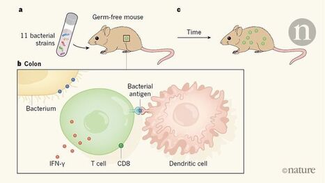
Summary The immune system is essential for maintaining a delicate balance between eliminating pathogens and maintaining tolerance to self‐tissues to avoid autoimmunity. An enormous and complex community of gut microbiota provides essential health benefits to the host, particularly by regulating immune homeostasis. Many of the metabolites derived from commensals can impact host health by directly regulating the immune system. Many autoimmune diseases arise from an imbalance between pathogenic effector T cells and regulatory T (Treg) cells. Recent interest has emerged in understanding how cross‐talk between gut microbiota and the host immune system promotes autoimmune development by controlling the differentiation and plasticity of T helper and Treg cells. At the molecular level, our recent study, along with others, demonstrates that asymptomatic colonization by commensal bacteria in the gut is capable of triggering autoimmune disease by molecular mimicking self‐antigen and skewing the expression of dual T‐cell receptors on T cells. Dysbiosis, an imbalance of the gut microbiota, is involved in autoimmune development in both mice and humans. Although it is well known that dysbiosis can impact diseases occurring within the gut, growing literature suggests that dysbiosis also causes the development of gut‐distal/non‐gut autoimmunity. In this review, we discuss recent advances in understanding the potential molecular mechanisms whereby gut microbiota induces autoimmunity, and the evidence that the gut microbiota triggers gut‐distal autoimmune diseases. Introduction The mammalian gastrointestinal tract is home to an enormous and complex community of commensal bacteria.1-3 This gut microbial community (microbiota) has co‐evolved with its host over millennia, suggested by the fact that out of 52 bacterial phyla, two dominant phyla, Bacteroidetes and Firmicutes, represent 98% of the bacteria in the gut.4, 5 These gut microbes provide benefits to their host in many ways, including, but not limited to, digestion, production of nutrients, detoxification, protection against pathogens and regulation of the immune system.1-3, 6, 7 As a result of its collective metabolic activity, and necessity for human health, the gut microbiota is often referred to as the ‘forgotten’ organ.8 Interestingly, many of the metabolites derived from commensal bacteria have recently been found to directly impact the immune system. Commensal‐mediated immunomodulation includes the promotion of T helper cell subset differentiation and T helper cell plasticity, the ability of a differentiated CD4+ T cell to take on characteristics of other T‐cell subsets simultaneously or at different times.9 Plasticity is an especially important subject for mucosal immunity, as mucosal tissues are major sites where T‐cell plasticity has been observed.10-14 Therefore, the mucosa harbors the frontline immune response to commensal bacteria, and their interactions may subsequently control health versus disease status through T‐cell differentiation and plasticity. An autoimmune condition develops when the immune system mistakenly attacks our own self‐tissues. A long‐standing question in the field of microbe–host communication is how interaction of microbes with T cells promotes autoimmune development. Molecular mimicry is one prominent hypothesis, theorizing that microbes trigger autoimmunity by a shared immunogenic epitope between microbes and a self‐peptide, which leads to the activation of autoreactive T cells.15, 16 Another less well‐known theory is that expression of dual T‐cell receptors (TCRs) on T cells promotes autoimmunity by allowing autoreactive T cells to escape thymic clonal deletion.17-19 Alterations in the composition of the gut microbiome, known as dysbiosis, have been implicated in many diseases and disease models, including those that show clear association with the gut such as inflammatory bowel disease, colorectal cancer, enteric infections, and obesity.20-27 Dysbiosis has also been implicated in immune disorders that occur outside the gut, such as asthma, eczema, allergies, and gut‐distal autoimmune diseases (autoimmune arthritis, type 1 diabetes, experimental autoimmune encephalomyelitis).20, 28-37 Although it is easy to see how the gut microbiota can influence immune responses within the gut, over the last decade an appreciation for the impact of the gut microbiome on gut‐distal autoimmunity disease has developed. For example, a strong interest has emerged to characterize the impact of gut microbiota on health and disease status in the lung and brain, namely ‘gut–lung axis’ and ‘gut–brain axis’, respectively.38, 39 This article aims to review the influence of microbiota on the immune system by producing key commensal‐derived metabolites as well as controlling T‐cell subsets and plasticity. Additionally, we discuss the recent findings on how gut commensals affect autoimmunity by molecular mimicry and skewing the expression of dual TCRs. Finally, we discuss the role of dysbiosis in the pathogenesis of common autoimmune diseases. As dysbiosis in inflammatory bowel disease has been extensively reviewed elsewhere (see ref. 40-42), this review will focus on the impact of gut microbiota dysbiosis in the development of gut‐distal autoimmune diseases. Understanding the mechanisms of how gut commensals affect the immune system will be crucial for us to elucidate autoimmune pathogenesis and to generate novel therapies. This is an urgent subject, as dysbiosis‐related diseases have emerged as new epidemics in the industrialized world.43-45 Commensal bacteria‐mediated metabolites and immunity Gut commensals are well‐known for their function in digestion, a process involving extraction and synthesis of many metabolites, some of which are produced only by commensal bacteria and are crucial for host health.46 Recently, several advancements have been made determining how the metabolites generated by commensal bacteria can directly influence the development and function of the immune system, directly impacting health and diseases (Table 1). Here, we review the most recent reports in this field. Commensal Metabolite Disease phenotype Immunomodulation References Clusters IV and XIVa of Clostridium ↑ IDO, enzyme involved in tryptophan catabolism, on IEC ↓ Colitis ↑ Colon Treg ↓ Th1 63, 66 Lactobacillus reuteri ↑ Indole derivatives by metabolizing tryptophan ↑ Differentiation of DP IEL 69 Clostridium sporogenes ↑ IPA derivatives by metabolizing tryptophan, phenylalanine, and tyrosine ↓ Permeable intestines ↓ Neutrophils, monocytes, and memory T cells 72 General microbiota signal through MyD88 ↑ RALDH, enzyme able to catalyze the synthesis of RA from Vitamin A ↓ EAE ↓ Autoimmune arthritis ↑ Treg ↓ Th17 ↑ Gut‐homing integrin α4β7 ↑ Maintenance of peripheral lymph nodes 78, 79, 92 Ruminococcaceae eubacterium ↑ SCFAs signal through GPR43 ↓ EAE ↓ Type 1 diabetes ↑ Lamina propria Treg ↑ Antimicrobial peptides 57, 53, 55 Abbreviations: DP IEL, double‐positive intraepithelial T lymphocytes; EAE, experimental autoimmune encephalomyelitis; IDO, indoleamine 2,3‐dioxygenase; IEC, intestinal epithelial cells; IPA, indolepropionic acid; RA, rheumatoid arthritis; RALDH, retinal dehydrogenase; SCFA, short‐chain fatty acids; Th1, T helper type 1; Treg, regulatory T. Short‐chain fatty acids Short‐chain fatty acids (SCFAs) including butyric acid, propionic acid, and acetic acid are the main metabolic products of undigested carbohydrates by gut commensal bacteria and have broad effects on the host immune system.47 Among the fatty acid receptors, two orphan G protein‐coupled receptors, GPR41 and GPR43, are activated by SCFAs.48, 49 SCFAs are significantly reduced in the colon of germ‐free mice indicating that the gut microbiota is essential for their production.50 Recently, one group reported that long‐chain fatty acids enhanced the differentiation and proliferation of T helper type 1 (Th1) and/or Th17 cells, whereas SCFAs expanded gut regulatory T (Treg) cells.51, 52 Using experimental autoimmune encephalomyelitis (EAE) as a model for multiple sclerosis, Haghikia et al. showed that long‐chain fatty acids decreased SCFAs in the gut, leading to exacerbated EAE by expanding pathogenic Th1 and/or Th17 cells in the small intestine.51 Treatment with SCFAs ameliorated EAE by inducing lamina‐propria‐derived Treg cells. Consistent with the anti‐inflammatory properties of SCFAs, treatment of germ‐free mice with acetate, a type of SCFA, is sufficient to reduce intestinal inflammation in the dextran sulfate sodium model of colitis.53 The protective effect of acetate was lost in GPR43−/− mice, indicating that signaling of acetate through GPR43 is necessary for the anti‐inflammatory effect of SCFAs. Remarkably, administration of the SCFA butyrate ameliorates inflammation in patients with ulcerative colitis.54 SCFAs also indirectly control type 1 diabetes through the production of antimicrobial peptides. Mechanistically, SCFAs produced by the gut microbiota have been shown to stimulate the production of antimicrobial peptides by pancreatic beta cells.55 Furthermore, systemic administration of these antimicrobial peptides induce Treg cells in the pancreatic islets of pre‐diabetic mice, reducing the incidence of autoimmune diabetes. Additionally, it has been recently demonstrated that administration of the SCFA propionate significantly attenuated HLA‐B27‐associated intestinal inflammation in Fischer 344 HLA‐B27/β2m transgenic rats.56 Interestingly, this propionate‐mediated inhibition of inflammation is independent of its role in regulating the Treg cell response. Amino acids An early study gives an indication that gut bacteria may play an important role in host amino acid homeostasis and health by showing that germ‐free mice had an altered distribution of free amino acids along the gastrointestinal tract compared with conventionalized mice.58 More recent reports suggest that the most abundant amino‐acid‐fermenting bacteria in the human small intestine are bacteria belonging to the Clostridia, the Bacillus–Lactobacillus–Streptococcus groups, Proteobacteria, and Peptostreptococcus.59-61 These bacteria are therefore likely to be important for amino acid absorption in the gastrointestinal tract. Among the 20 amino acids, tryptophan belongs to one of the nine ‘essential’ amino acids that humans and higher vertebrates cannot synthesize, and that must be supplied in their diet.62 Numerous studies suggest that tryptophan metabolites generated by gut commensal bacteria serve as important signaling molecules in host–microbe cross‐talk. For example, the colonic intestinal epithelial cells from mice colonized with clusters IV, XIVa, and XVIII of Clostridium express high levels of indoleamine 2,3‐dioxygenase,63 an enzyme involved in the initial and rate‐limiting step of tryptophan catabolism, which has been implicated in Treg cell induction.64 Specifically, it has been demonstrated that kynurenine, the first product in the indoleamine 2,3‐dioxygenase‐dependent tryptophan degradation pathway, activates the aryl hydrocarbon receptor, leading to aryl hydrocarbon receptor‐dependent Treg cell generation.65 Indeed, an induction of colonic Treg cells was observed in mice colonized with Clostridium species.63 The small intestinal epithelium contains a unique population of CD4+ CD8αα+ double‐positive intraepithelial T lymphocytes (DP IELs) which exhibit anti‐inflammatory properties.67, 54 It has been shown that upon migration to the epithelium, Treg cells lose Foxp3 and convert to DP IELs in a microbiota‐dependent manner, as microbiota depletion by treating mice with broad‐spectrum antibiotics prevents Foxp3 loss.68 Whereas all Treg cells express the CD4‐lineage transcription factor T helper‐inducing POZ/Krüppel‐like factor (ThPOK), CD8αα+ CD4+ and > 50% of Foxp3– CD8α– CD4+ cells in the small intestinal epithelium lack ThPOK expression. Later, it was found that loss of ThPOK corresponds to an IEL‐like behavior in CD4+ T cells. Interestingly, supplement of TCR stimulation can overcome the microbiota requirement for the DP IEL differentiation, as demonstrated by the relatively normal DP IEL differentiation in antibiotics‐treated Rag1−/− OT‐II (ova‐specific) mice exposed to oral ovalbumin. By comparing the microbiota profile between mice housed at two vivaria, with mice at one vivarium displaying significant numbers and another displaying negligible or absent DP IELs, Cervantes‐Barragan et al. discovered that the commensal Lactobacillus reuteri was responsible for the differentiation of DP IELs.69 Lactobacillus reuteri generated indole derivatives by metabolizing tryptophan, which activated the aryl‐hydrocarbon receptor in CD4+ T cells, leading to their ThPOK down‐regulation and differentiation into DP IELs. Hence, L. reuteri together with a tryptophan‐rich diet can promote gut immune homeostasis by allowing the differentiation of intraepithelial CD4+ T cells into anti‐inflammatory DP IELs. Only a few species of gut commensals, including Clostridium sporogenes, can break down tryptophan and produce the metabolite indolepropionic acid (IPA), a deamination product of tryptophan.70 Bacterial tryptophanase catalyzes the conversion of dietary tryptophan to indole and subsequently to IPA.71 A recent study identified a total of 12 compounds that can be produced in this process, nine of which can accumulate in the blood and three of which are produced exclusively by bacteria.72 Specifically, the C. sporogenes‐expressed gene fldC is necessary for the production of IPA. In fact, fldC is essential for the reductive metabolism of all three aromatic amino acids (tryptophan, phenylalanine, and tyrosine). Germ‐free mice receiving wild‐type C. sporogenes exhibit high levels (~80 μm) of serum IPA whereas germ‐free mice receiving the mutated version of C. sporogenes lacking fldC, had undetectable serum IPA. Importantly, mice with undetectable IPA had higher levels of immune cells, including neutrophils, classical monocytes, and memory T cells. In addition, the mice with the engineered version of C. sporogenes had increased intestinal permeability, a defect that is often seen in inflammatory bowel disease. In addition to having a direct impact on the immune system, undigested amino acids have the potential to become supplemental precursors for SCFA generation by the gut microbiota, in addition to indigestible carbohydrates.73 Therefore, amino acids could have indirect impact on the immune system through SCFAs as we discussed earlier. Numerous amino acids including glycine, threonine, glutamate, and ornithine can be metabolized by anaerobic bacteria to generate acetate, whereas threonine, lysine, and glutamate can be used for butyrate synthesis.74 Moreover, at the molecular level, it has been shown that intracellular leucine concentrations can be sensed by the multiprotein complex leucyl‐tRNA synthetase,75, 76 which activates the mechanistic target of rapamycin kinase, proving to be a vital link between immune function and metabolism.77 Retinoic acid Retinoic acid, a metabolite of vitamin A, is one of the most active physiological retinoid metabolites and has a wide range of biological activity including regulating immune responses.78 A major part of retinoic acid's anti‐inflammatory effects depends on the inhibition of Th17 cells and promotion of Foxp3+ Treg cell responses.78, 79 In addition, retinoic acid has been shown to be important for the expression of the gut homing receptor integrin α4β7 on T cells.80-82 The α4β7 integrin receptors are imprinted on lymphocytes by dendritic cells (DCs) from Peyer's patches (PPs), and mesenteric lymph nodes.82, 83 In the absence of microbial toll‐like receptor signaling in Myd88−/− mice, gut DCs express low levels of retinal dehydrogenase (RALDH) required for retinoic acid biosynthesis, so these Myd88−/− mice are unable to generate gut‐homing lymphocytes.84 AM80 is a synthetic retinoic acid that is characterized by higher stability and fewer potential adverse effects compared with retinoic acid.85, 86 It has been reported that both retinoic acid and AM80 ameliorate many autoimmune responses, including experimental autoimmune myositis, experimental autoimmune encephalitis, and collagen‐induced arthritis.87-90 We recently showed that oral administration of AM80 inhibits autoimmune disease in the joints as well as in the lung.91 We elucidated a novel mechanism whereby AM80 suppresses the autoimmune pathology in both the lung and joints by inhibiting T follicular helper (Tfh) cells in addition to inhibiting Th17 responses. Specifically, AM80 increased the expression of the gut‐homing integrin α4β7 on Tfh cells, which diverted Tfh cells from systemic (non‐gut) inflamed sites such as the lung draining lymph nodes into the gut (the non‐immunopathological site) and so reduced systemic autoantibody production. Moreover, the impact of retinoic acid can go beyond the cellular level and impact the development of whole lymphoid tissues.92 It has been reported that cellular expansion in peripheral lymphoid tissues is controlled by gut microbiota in a retinoic acid‐dependent manner.93 The mucosal addressin MAdCAM‐1 is the receptor for the gut‐homing integrin α4β7, and peripheral node addressin PNAd is the receptor for CD62L.94 In neonatal mice, MAdCAM‐1 expression in lymph nodes is elevated shortly after birth,95 followed by a switch to a PNAd over a course of 2–3 weeks.93 Zhang et al. demonstrated that commensal fungi drive a wave of RALDH+ DCs to migrate to the peripheral lymph nodes after birth.92 The arrival of these cells increases the amounts of retinoic acid in situ, mediates the neonatal MAdCAM‐1 to adult PNAd addressin switch on endothelial cells, and directs the homing of lymphocytes to gut‐associated lymphoid tissues. Finally, the authors found that a diet deficient in vitamin A causes reduced homing of RALDH+ DCs into peripheral lymph nodes and a lack of maintenance of peripheral lymph node structures, suggesting a dependence on retinoic acid signaling for structural and functional maintenance of peripheral immune tissues. Microbiota and T‐cell subset determination Understanding the role of microbiota in T‐cell subset commitment and plasticity holds the key for unveiling the pathogenesis and therapeutic options for autoimmune diseases, as well as diseases associated with unbalanced immune responses such as cancers and infections. Not surprisingly, addressing this question has become a focused area of research in recent years. One study using transgenic mice expressing a limited but diverse TCR repertoire, through fixed TCR‐β expression, showed that the TCR repertoire of colonic Treg cells is unique compared with thymically derived Treg cells.96 Furthermore, these colonic Treg cells were shown to react with bacterial isolates, suggesting that encountering gut microbes in the intestines leads to peripheral Treg cell induction, and hence to tolerance to the gut microbiota.96 A more recent study, also using transgenic mice expressing a constrained TCR repertoire, through fixed TCR‐β expression and limited TCR‐α rearrangement (TCRmini mice97) showed that thymic and intestinal Treg cells expressed overlapping TCR repertoires, many of which recognize microbial antigens.98 Alteration of the gut microbiota through treatment of TCRmini mice with a cocktail of antibiotics concurrently altered the colonic and thymic Treg TCR repertoire.98 This suggests that the repertoire of thymically derived Treg cells is heavily influenced by the microbiota. Although these studies have differing results regarding the initial site of gut microbiota influence, in either case it is clear that direct recognition of microbial antigens by T cells can promote Treg cell lineage commitment. Beyond Treg cells, the gut microbiota can also influence T helper subsets. In the presence of a normal microbiota, CX3CR1+ intestinal antigen‐presenting cells act to limit Th1 cell development and expansion.99 However, antibiotic‐induced dysbiosis leads to a shift in the function of CX3CR1+ antigen‐presenting cells, allowing them to promote pathogenic Th1 cell development.99 Our laboratory and others have also demonstrated that a single type of commensal bacteria, segmented filamentous bacteria (SFB), can promote Th17 cell responses. In C57BL/6 mice, SFB specifically induce intestinal Th17 cells.25 More recent studies, however, have demonstrated that SFB can also influence T helper subsets outside the gut. In 2011, Lee et al. discovered that SFB alone was able to promote Th17 cells specifically in the spinal cord of EAE mice.33 Similarly, we have shown that in the K/BxN mouse model of autoimmune arthritis, SFB specifically promotes Th17 cell responses in the lung.100 Additionally, SFB can also drive the differentiation of Tfh cells and promote their migration to systemic sites, leading to exacerbated autoimmune arthritis in K/BxN mice.101 However, the exact molecule(s) employed by SFB to promote the differentiation of T helper cell subsets remain elusive. Although many microbiome studies focus on the microbes residing in the intestines, the entire digestive tract is colonized with bacteria. Hence, the oral microbiome has recently become an area of interest with regards to mucosal immunity. Data from Dutzan et al. showed that oral homeostatic Th17 responses, unlike gut Th17 responses, were unaffected by shifts in the oral or gut microbiomes.25, 102 This suggests that not all mucosal T cells are influenced by the microbiome, and that other environmental cues such as chewing activity in the oral cavity and chewing‐associated damage can maintain the Th17 cell population. Interestingly, a more recent study from this same group found that inflammatory Th17 cells isolated directly from periodontal lesions were in fact heavily influenced by oral microbiome dysbiosis.103 By focusing on Th17 isolated from periodontal lesions of both mice and humans, they found that increased abundance of Th17 cells and enhanced production of inflammatory cytokine IL‐17 was associated with shifts in the oral microbiome. Specific outgrowth of Enterococcus and Actinobacteria along with loss of Streptococcus was associated with periodontitis and enhanced Th17 cell responses in the ligature‐induced periodontitis mouse model. Together the studies suggest that only pathogenic but not homeostatic Th17 cell responses are influenced by dysbiosis. We recently addressed how both age and gut microbiota affect T‐cell subsets in the context of autoimmunity.104 Our results show an augmented autoimmune disease phenotype in both the joints and the lung of middle‐aged compared with young mice. Mechanistically, we saw a soaring accumulation of Tfh, but surprisingly not Th17 cells with age. Our data suggest exposure to immunomodulatory commensals such as SFB may allow the young host to develop an overactive immune system similar to that found in middle‐aged hosts. Our study also shows that age can independently increase the Tfh cell response without the help of immunomodulatory commensals. CD4 T helper cell plasticity and the microbiota The development of specific transcription factor reporter mice has proven to be an invaluable tool in dissecting the plasticity of T‐cell subsets. An early study using adoptive transfer of cells from Foxp3eGFP reporter mice showed that 80% of Treg cells lost GFP expression in the PPs compared with 50% in spleen.12 Within the GFP‐negative PP population, > 60% of the cells expressed CXCR5, consistent with a Tfh phenotype,12 suggesting that PP, but not spleen, is the preferred site for a Treg to Tfh reprogramming. Two other studies found that retinoic‐acid‐receptor‐related orphan receptor γ t (Rorγt), the hallmark Th17 transcription factor, was preferentially expressed by colonic Treg cells, and furthermore, that in the absence of gut bacteria, the Rorγt+ Treg cell population was substantially diminished.10, 14 Interestingly, several bacterial species were able to induce Rorγt+ Treg cells, which were indispensable for maintaining gut homeostasis as determined by enhanced IL‐17, interferon‐γ (IFN‐γ), and colitis in their absence.10 Another study showed that the pathogenic bacterium Helicobacter hepaticus promoted expansion of Rorγt+ Treg cells in a c‐MAF‐dependent manner, which selectively suppressed pro‐inflammatory Th17 cells in the intestines.105 However, in the absence of Treg cells (in Il10−/− mice), H. hepaticus predominantly induced Th17 cells, suggesting that pathobionts can have an impact on T‐cell plasticity by expanding potent suppressive T cells, which in turn, inhibit the inflammatory T helper cells necessary for pathogen elimination. Plasticity among T helper subsets has also been observed. Using cell fate reporter mice in which Th17 cells are permanently marked by YFP (Il17creR26eYFP), Hirota et al. found that selectively in the PP, but not spleen, Th17 cells were reprogramed to a Tfh (PD‐1hi CXCR5hi) phenotype.13 Furthermore, these ex‐Th17‐Tfh cells localized in germinal centers where they assisted in T‐cell‐dependent IgA production. In a Th17 adoptive transfer model of colitis, it was demonstrated that Th17 cell conversion to a Th1 phenotype was necessary for colitis development as Th17 cells unable to produce IFN‐γ (Ifng−/−) were incapable of inducing colitis.106 Interestingly, not only did Th17 cells convert to Th1 and contribute to colitis development directly, but also Th17 were capable of providing help to naive T cells for Th1 lineage commitment. Together these studies suggest that gut microbiota is able to influence T‐cell plasticity within the gut microenvironment and influence the development of autoimmunity and other inflammatory diseases. Gut microbiota induces autoimmunity by TCR‐mediated mechanisms Central to T‐cell function is the TCR, through which T cells recognize their cognate antigen and become activated. An important feature of the TCR is its ability to recognize multiple antigens, known as TCR cross‐reactivity. Sequencing‐based methods estimate that humans and mice express 107 and 106 productive unique αβ TCRs, respectively, yet this is insufficient to cover the possible foreign antigen repertoire.107, 108 Therefore, TCRs possess the ability to recognize multiple antigens in order to recognize the plethora of possible foreign antigens (see ref. 109). One potential mechanism through which pathogenic autoreactive T cells may become activated is through weak recognition of a microbial antigen for which their TCR is cross‐reactive. This is also known as molecular mimicry, a phenomenon in which a foreign antigen shares significant structural or sequence similarities with a self‐antigen.15, 16 Later, this now activated autoreactive T cell can home to the tissue where its cognate self‐antigen is expressed and elicit autoimmune pathology. In fact, cross‐reactive TCR recognition of microbial peptides by autoreactive T cells has been seen in several autoimmune settings (Fig. 1). In 2015, Horai et al. showed that T cells restricted to expressing only the retina‐specific TCR (R161H), became activated in the intestines in response to an unidentified microbial antigen.110 T‐cell activation was in response to TCR‐signaling to a non‐cognate microbial antigen, as T cells in the small intestines of R161H‐Tcrα−/− mice had clear co‐localization of phosphorylated Zap‐70 with CD4, known to be downstream of TCR signaling.110-112 Later, it was shown that Islet‐specific glucose‐6‐phosphate catalytic subunit‐related protein (IGRP)‐specific CD8+ T cells in non‐obese diabetic (NOD) mice were activated through recognition of a peptide from Fusobacteria and elicited autoimmune diabetes.113 Interestingly, the Fusobacteria peptide shared significant homology with the IGRP peptide targeted by the IGRP‐specific transgenic TCR,113 suggesting that molecular mimicry in microbial peptides may pose a threat because they can activate pathogenic autoreactive T cells. Most recently, it was shown that gut commensal bacteria expressing an ortholog to human Ro60, a nuclear RNA‐binding protein and primary targeted self‐antigen in lupus, were able to activate human Ro60‐specific CD4+ T cells through cross‐reactive TCR recognition.114 Furthermore, germ‐free mice monocolonized with Ro60‐ortholog‐expressing commensal bacteria spontaneously developed lupus‐like disease, as indicated by glomerular immune complex deposits. Together, these studies suggest that molecular mimicry is a mechanism through which the intestinal microbiota is able to propagate autoimmune responses leading to disease. In addition to molecular mimicry, it has long been thought that pathogenic autoreactive T cells may arise from a subset of thymocytes expressing two unique TCRs, one of which allows them to evade negative selection along with one that has a high affinity for a self‐antigen.115-117 Approaching from a different angle, outside the central immune system, we found that in peripheral tissues, SFB selectively expand dual TCR‐expressing T cells, leading to the augmentation of lung autoimmune pathology (Fig. 1).100 This is the first study to show that dual TCR T cells can be activated through the recognition of the cognate commensal‐derived antigen and migrate to elicit autoimmune pathology in the lung, a gut‐distal site. At the molecular level, we demonstrate that SFB selectively expand autoimmune T cells co‐expressing SFB‐specific TCRs in addition to their self‐reactive TCRs. This additional SFB‐specific TCR provides a proliferative advantage for autoreactive Th17 cells in SFB(+) hosts. Our data suggest that the induction of robust SFB‐specific Th17 response by transferring SFB(+) recipients with SFB‐specific 7B8 T cells could not rescue the autoimmune activity of Rag−/−.KRN T cells through bystander activation. Instead, our data suggest that the same T cells need to specifically recognize self‐ as well as SFB‐antigen to induce IL‐17‐expressing pathogenic T cells to cause autoimmunity. This is supported by previous data showing that in healthy mice, cognate TCR recognition of SFB‐antigen is crucial for SFB‐mediated Th17 induction.118, 119 Taken together, our study suggests that the same T cell needs to recognize both self‐antigen and be activated by SFB to undergo preferential Th17 cell expansion and so enhance autoimmunity in an SFB‐dependent manner. Beyond molecular mimicry, our study suggests an alternative, dual TCR‐based mechanism for commensal‐mediated autoimmunity. Role of gut microbiota in human and murine gut‐distal autoimmunity Autoimmune disorders have been sharply increasing worldwide in recent decades.26 The dramatic changes in the disease onset rate cannot be explained by genetic basis as it occurs in such a short period of time. On the contrary, these data suggest that environmental factors play a key role in the recent surge of autoimmune diseases. In recent years, we have begun to appreciate that the gut microbiota provides environmental cues controlling human health and disease, including its potential impact on autoimmune responses. Under healthy conditions, the human intestinal microbiota is composed primarily of bacteria of the phylum Firmicutes, with Bacteroidetes and Actinobacteria also represented.120 On the family and species level, the gut microbiota varies widely among individuals, although it remains largely stable within an individual over the course of several years.121 Studies using mouse models or samples from human subjects transplanted into mice have shown that age, gender, diet, smoking, autoimmunity, and other factors can alter the microbiota, and that the altered microbiota can be transferred and exert influence over the metabolism and immune system of the recipient.122 These studies demonstrate that the host–microbiota relationship is not a one‐way street, but rather a dynamic conversation that can have long‐lasting impacts. Many studies have investigated a correlation between gut microbiome composition and autoimmune disease in humans, but these studies are limited in their ability to determine causality. Exploring this field using animal models allows for perturbation of the gut microbiome to better distinguish cause from effect, permit mechanistic dissection and allow pre‐clinical evaluation of suggested therapeutic strategies. Throughout this section, we will highlight the important observations made in both human patients and mouse models. Multiple sclerosis (MS) is an autoimmune disease in which pathogenic CD4+ T cells penetrate the blood–brain barrier and cause damage to the central nervous system.123 Numerous studies have shown that patients with MS display intestinal microbiome dysbiosis.124-126 Two independent studies comparing patients with relapsing–remitting MS and healthy controls found significant alterations in the composition of the microbiome from patients with MS.124, 127 Perhaps even more intriguing, when compared with treatment‐naive patients, those receiving treatment displayed some restoration in their microbiome composition.127 Untreated MS patients displayed increased relative abundance of Methanobrevibacter and Akkermansia, which were lower in healthy controls and treated MS patients. MS patients receiving treatment had increased Sutterella, which has been previously observed to be increased in healthy controls compared with treatment‐naive patients with inflammatory bowel disease (Fig. 2).128 This suggests that although dysbiosis may contribute to a predisposition for developing MS, perhaps treatment may act to normalize pro‐inflammatory microbiota. A recent study by Cekanaviciute et al. showed that intestinal microbiome samples from MS patients displayed specific outgrowth of Acinetobacter and Akkermansia.129 Importantly, they further demonstrated that in the presence of Acinetobacter calcoaceticus, human peripheral blood mononuclear cells preferentially shifted towards an inflammatory phenotype (IFN‐γ) and away from a regulatory phenotype (Foxp3+ CD25+). This suggests that the specific dysbiosis observed in patients with MS favors an inflammatory T‐cell response. More definitive studies using the EAE mouse model of MS have shown that in germ‐free mice lacking gut microflora, EAE disease severity is reduced to an almost undetectable level, due to a more than twofold reduction in IFN‐γ and IL‐17 production from CD4+ T cells.33 Importantly, if CD4+ T cells from germ‐free mice were transferred to specific pathogen‐free mice harboring a diverse intestinal microflora, these cells gained the ability to mediate EAE disease development, suggesting that the gut microbiota is necessary to elicit the pathogenic Th1 and Th17 responses seen in EAE and perhaps MS.33 Importantly, it was recently demonstrated that colonization of germ‐free Myelin oliodendrocyte glycoprotein (MOG)‐specific TCR transgenic mice (RR mice) with gut microbiota from patients with MS resulted in increased incidence of spontaneous EAE development.130 This suggests that the microbiota of patients is sufficient to precipitate autoimmune disease. Together these studies suggest a gut–brain axis of communication that allows gut microbes to elicit autoimmune responses in the brain. Rheumatoid arthritis (RA) is an autoimmune disease that primarily affects joints but may also affect other parts of the body including the lungs and heart.131-133 Several studies have suggested that many patients with RA display antigen–antibody complexes with complement fixation in joints that can cause tissue damage and tumor necrosis factor‐α production.134-139 Recent studies also suggest that anti‐citrullinated protein antibodies are involved in RA pathogenesis.140, 141 Tfh cells are a T effector cell type specialized in helping B cells.142-144 Over‐reactive Tfh responses pose a threat of triggering excessive autoantibody production and autoimmune disease.145-149 In that regard, the presence of circulating CXCR5+ Tfh (cTfh) cells in the blood has been observed in patients with different autoimmune diseases, including RA.20 In a recent study assessing 77 treatment‐naive patients with RA, 21 treated patients with RA, and 80 healthy controls, the gut microbiome was found to be altered in patients with RA.150 In particular, Haemophilus spp. were depleted in individuals with RA and negatively correlated with serum autoantibodies titers, whereas Lactobacillus salivarius was over‐represented in individuals with RA and abundance correlated with disease severity (Fig. 2). Interestingly, similar to what has been observed in patients with MS, patients with RA undergoing treatment displayed partial normalization of their intestinal microflora compared with treatment‐naive patients. By using a photo‐labeling mouse model to track cell migration, our laboratory has demonstrated that a commensal bacterium, SFB, is able to promote the differentiation and migration Tfh cells from the PPs in the small intestine to systemic sites where they elicit autoimmune arthritis development.101 At the molecular level, SFB induce PP Tfh cell differentiation by limiting the access of IL‐2 to PP CD4+ T cells and DCs are required for SFB‐mediated IL‐2Rα suppression and up‐regulation of Bcl‐6, a master regulator of Tfh cells, in PPs. Furthermore, we have recently shown that SFB can also promote RA‐associated autoimmune lung disease through propagation and mobilization of gut‐derived Th17 cells.100 Importantly, this highlights a gut‐lung axis of communication through which specific gut commensal bacteria are able to promote pathogenic T‐cell responses leading to gut‐distal autoimmune pathology. In the collagen‐induced arthritis model of RA, dysbiosis characterized by outgrowth of Lachnospiraceae and Lactobacillaceae was detected before arthritis development.151 Furthermore, antibiotic‐induced depletion of the microbiota before induction of collagen‐induced arthritis reduced disease severity by 40% and was accompanied by reduction in the inflammatory cytokines IL‐17, IL‐22, and IL‐23 in the intestines along with reduced anti‐collagen antibodies. This suggests that the gut microbiota is necessary for autoimmune disease development and that alterations in the composition of the gut microbiota precipitate autoimmune disease. Systemic lupus erythematosus (SLE) is an autoimmune disease affecting almost all organs of the body, in which both autoreactive T cells and autoantibody‐producing B cells contribute to the pathogenesis of SLE.152 Like many gut‐distal autoimmune diseases, recent studies have linked gut microbiota dysbiosis with SLE.153 In a 2016 study from Lopez et al., it was shown that intestinal microbiota isolates from patients with SLE promoted naive T‐cell differentiation into Th17 cells.154 Furthermore, peripheral blood mononuclear cells from patients with SLE were enriched for both Th17 and Foxp3+ IL‐17+ cells compared with healthy controls. As described earlier, due to T cell plasticity, Treg cells are able to shift between regulatory and potentially pathogenic phenotypes. Hence, the presence of Foxp3+ IL‐17+ cells in the peripheral blood of patients with SLE suggests that the shift observed in the gut microbiome of these patients may promote inflammatory T‐cell responses and may negatively impact Treg cell stability. More recently, in a study by Luo et al. which assessed the intestinal microbiota dynamics of both SLE patients and the NZB/W F1 mouse model of SLE, it was shown that alteration in the gut microbiota resulted in decreased diversity with increased representation of Gram‐negative bacteria (Fig. 2).155 Interestingly, as NZB/W F1 mice progressed to overt disease, the relative abundance of Lactobacillaceae increased from 0·1% to 10%. Furthermore, there was a positive correlation between Lactobacillaceae abundance and disease severity in female NZB/W F1 mice. This suggests that reduction in α diversity, a measure of the number of different species present in a given site, and outgrowth of specific commensal microbes may promote autoreactive immune responses in susceptible individuals. Another report found a high frequency of Th17 cells in the kidneys of patients with glomerulonephritis, a form of SLE.156 By using Kaede mice to track intestinal T‐cell migration during glomerulonephritis induction, they observed that Th17 cells egress from the intestine and subsequently migrate to the kidney through CCR6 recognition of CCL20. Furthermore, depletion of gut Th17 cells in germ‐free and antibiotics‐treated mice ameliorated the autoimmune glomerulonephritis, suggesting that targeting the intestinal Th17 cells may offer a therapeutic strategy for autoimmune diseases. Hidradenitis suppurativa (HS) is a chronic inflammatory skin condition of the hair follicles thought to be mediated in large part by Th17 cells (Fig. 2).157 With respect to perturbations in the microbiota, most studies assess changes in the cutaneous microbiota as this is the site of the disease manifestation. Recently, HS has been identified to have a strong concurrence rate with inflammatory bowel disease, with over 17% of Crohn's patients also having HS.158 Hence, while there are definite changes in the cutaneous microbiome of patients with HS,159 taking a closer look at the gut microbiome may prove fruitful in the quest for identifying a causal mechanism. Type 1 diabetes (T1D) is a T‐cell‐mediated autoimmune disease characterized by the selective destruction of insulin secreting β‐cells in the pancreatic islets.160 Many patients with T1D present with increased intestinal permeability or ‘leaky gut’, which has been shown to precede the onset of clinical disease.161 In addition, patients with T1D also display an altered intestinal microbiome characterized by decreased α diversity and increased abundance of inflammatory species compared with healthy controls (Fig. 2).162 Hence, it is tempting to assume that in combination with increased intestinal permeability, dysbiosis could promote an inflammatory environment in the gut–proximal pancreatic tissue leading to activation of an autoimmune response. In a 2016 study from Costa et al., it was determined that autoimmune diabetes development in the streptozotocin‐induced model of T1D is dependent on translocation of gut microbiota to the pancreatic lymph node.163 Interestingly, islet‐infiltrating T cells seem to be of gut origin as they often express α4β7 integrin.83, 164, 165 We now know that the effect of commensal bacteria on autoimmune disease can be model dependent. For example, SFB is pathogenic in animal models of arthritis and multiple sclerosis32, 33 but protective in the NOD mouse model of type1 diabetes.166, 167 The beneficial and detrimental effects of SFB could depend on whether commensal‐mediated immunomodulation is enhancing or inhibiting the pathogenesis of each disease. For example, autoantibodies are a key pathogenic factor and diseases can be induced by passive transfer of autoantibodies in the K/BxN and EAE models.168-170 In contrast, type 1 diabetes in NOD mice is a T‐cell‐mediated autoimmune disease, and although B cells of NOD mice produce autoantibodies, these are not thought to play a diabetogenic role.171, 172 Hence, SFB, with their Tfh and autoantibody boosting effect, are more likely to play a pathogenic role in the K/BxN model and other antibody‐mediated autoimmune diseases than in T‐cell‐mediated autoimmune diseases such as in NOD mice. One feature that many of the autoimmune diseases have in common is evidence of gut‐derived T cells participating in the autoimmune response.83, 101, 156, 164, 165 This would suggest that perhaps one mechanism through which the gut microbiota modulates autoimmune responses is through mobilization of T cells from the tolerogenic environment of the gut to systemic sites where alleviated tolerogenic pressure allows these cells to become active and initiate autoimmune disease development.173-175 However, more studies are required to determine whether the altered microbiome drives changes in immunity or conversely the onset of immunological disease induces changes in the gut microflora in human patients. It is likely a combination, as changes in the gut microbiota precede onset of clinical autoimmune manifestations,162 yet some evidence suggests that perhaps T cells, Foxp3+ Treg cells in particular, regulate gut microbiota diversity.176 Conclusions and future directions There is a surge of recent studies investigating the molecular mechanisms of dysbiosis‐related autoimmune diseases. In both molecular mimicry and dual TCR theories, until recently, only infectious pathogens including viruses and bacteria have been implicated as the primary culprits,15-17 and little is known about the molecular mechanism by which asymptomatic commensal colonization could trigger autoimmunity. Our recent study along with others demonstrates a previously unknown condition that asymptomatic colonization by commensal bacteria in the gut is capable of triggering systemic autoimmune disease by molecular mimicking self‐antigen and skewing the dual TCR expression in the host. Notably, dual TCR expression is not limited to transgenic mouse models. In wild‐type mice and in humans, up to 15% and 33%, respectively, of peripheral T cells have been reported to express dual TCRs, due to incomplete allelic exclusion at the Tcra locus.177, 178 Future investigation is required to determine whether dual TCR expression is involved in dysbiosis‐related immune disorders in human patients. Another important, but poorly understood, aspect of microbiota–T‐cell interaction is how different microbial signals impact T‐cell stability and plasticity at the mucosal frontline. Understanding the plasticity and stability of T‐cell subsets in mucosal tissues is highly relevant for future therapeutic strategies as we will not be able to manipulate T cells for therapeutic purposes without understanding the mechanisms by which T cells shift between alternative programs because of exposure to gut microbes. As illustrated in this review, the use of animal models provides a powerful tool for mechanistic studies as it allows us to establish the causative effect of microbiota in disease development. By using germ‐free and specific pathogen‐free mouse models, numerous studies unravel the crucial roles of gut commensals in immune regulation in the context of health and disease. Although these studies provide promising clinical implications, various challenges need to be overcome by the field to harness these findings for future diagnostic and therapeutic approaches. One of the challenges is that the reductionist experiments in mice often do not reflect important features of the immune system in the adult human. In this regard, Beura et al. demonstrated that laboratory mice – like neonates, but not adult humans – lack effector‐differentiated and mucosal memory T cells.179 These cell populations are present in feral mice and pet store mice with constant and diverse microbial exposure. Furthermore, laboratory mice co‐housed with pet store mice display profound changes in immune systems, resulting in an immune signature that is more closely reflected by adult humans than by neonates. Hence, the ‘dirty’ mouse model may provide a unique advantage over the current reductionist mouse models and should be considered for studies with translational purposes in human disease. One of the biggest dilemmas facing the field of gut microbiota research is how to apply laboratory findings to clinical therapies. Currently, microbiota‐based therapy relies on three major strategies: complete fecal microbiota transplants (FMTs), administering one or more species of bacteria orally (probiotics), or administering substrates to favor the expansion of certain kinds of bacteria or a shift in metabolite production, a strategy known as prebiotics.180 Each of these approaches has limitations; for example, probiotics often colonize only transiently because of ineffective competition with the existing microbiota.181 Additionally, despite the focus on taxonomic composition analyzed by 16S rRNA sequencing of microbial communities in many studies, the functional relevance of microbial communities is more likely to be revealed by their metagenomic gene expression and metabolomics profiles. This is further proved by the fact that bacterial isolates of the same strain can display very different immunoregulatory capabilities.182 FMTs depend on severe depletion of the constituent microbiota through antibiotics and bowel flushing before administration of the transfer.183, 184 Other researchers are working to find alternative methods to circumvent the difficulties of FMTs, such as filtering donor material to the point of removing intact cells, leaving only bacterial byproducts such as metabolites.185 An additional approach is to identify bacterial metabolites able to modulate human immune responses and artificially administer the identified metabolites as novel therapeutics.186 This approach would be appealing from a pharmaceutical perspective, because it would allow the development of drugs that would need to be taken regularly. It would also minimize the factors that need to be controlled, since administering metabolites would not depend on the engraftment of a foreign bacterial species. However, a careful determination on how to achieve a comparable delivery of the metabolite that mimics the one produced by the intestinal microbiota will be crucial for the effectiveness of the treatment. In conclusion, the gut microbiota is a complex community that engages in significant cross‐talk with the host. A better understanding of host–microbe interaction and the underlying microbiota‐derived molecules that modulate the immune system and disease development may help to pave the way for better patient‐tailored interventions and microbial molecule‐based therapies for immune disorders. Disclosures The authors have no competing interests to declare. References
|
|
Scooped by
Gilbert C FAURE
January 28, 2019 5:31 AM
|
|
|
Scooped by
Gilbert C FAURE
January 27, 2019 5:42 AM
|
FASEB J. 2019 Jan 10:fj201801748R. doi: 10.1096/fj.201801748R.[Epub ahead of print]...
|
|
Suggested by
Société Francaise d'Immunologie
January 26, 2019 6:51 AM
|
<b><i>Background:</i></b> The gastrointestinal tract is one of the most microbiologically active ecosystems that plays a crucial role in the working of the mucosal immune system (MIS).In this ecosyst...
|
|
Scooped by
Gilbert C FAURE
January 25, 2019 1:50 PM
|
|
|
Scooped by
Gilbert C FAURE
January 24, 2019 12:06 PM
|
Human noroviruses are responsible for approximately 200,000 deaths worldwide each
year. In 2012, the GII.4 Sydney strain emerged and became the major circulating norovirus
strain associated with human disease. Our understanding of the human norovirus-specific
antibody response is limited because few human monoclonal antibodies (mAbs) to noroviruses
have been described, and there are no functional assays to measure virus neutralization.
We studied the antibody-mediated response to the genogroup (G) II.4 strain by isolating
mAbs to GII.4 from infected patients and developing virus neutralization assays.
|
|
Suggested by
Société Francaise d'Immunologie
January 23, 2019 8:56 AM
|
Research ArticleGastroenterologyOncology Free access | 10.1172/JCI98554 Graft-versus-host disease propagation depends on increased intestinal epithelial tight junction permeability Sam C. Nalle,1 Li Zuo,2,3 Ma. Lora Drizella M. Ong,2 Gurminder Singh,1,2 Alicia M. Worthylake,2 Wangsun Choi,2 Mario Cabrero Manresa,2 Anna P. Southworth,2 Karen L. Edelblum,1,4 Gregory J. Baker,5 Nora E. Joseph,1 Peter A. Savage,1 and Jerrold R. Turner1,2 First published January 22, 2019 - More info Abstract Graft-versus-host disease (GVHD) is a complication of hematopoietic stem cell transplantation (HSCT) that affects multiple organs. GVHD-associated intestinal damage can be separated into two distinct phases, initiation and propagation, which correspond to conditioning-induced damage and effector T cell activation and infiltration, respectively. Substantial evidence indicates that intestinal damage induced by pretransplant conditioning is a key driver of GVHD initiation. Here, we aimed to determine the impact of dysregulated intestinal permeability on the subsequent GVHD propagation phase. The initiation phase of GVHD was unchanged in mice lacking long MLCK (MLCK210), an established regulator of epithelial tight junction permeability. However, MLCK210-deficient mice were protected from sustained barrier loss and exhibited limited GVHD propagation, as indicated by reduced histopathology, fewer CD8+ effector T cells in the gut, and improved overall survival. Consistent with these findings, intestinal epithelial MLCK210 expression and enzymatic activity were similarly increased in human and mouse GVHD biopsies. Intestinal epithelial barrier loss mediated by MLCK210 is therefore a key driver of the GVHD propagation. These data suggest that inhibition of MLCK210-dependent barrier regulation may be an effective approach to limiting GVHD progression. Graphical Abstract Introduction Allogeneic hematopoietic stem cell transplantation (HSCT) is used in the treatment of neoplastic and immune disorders. Graft-versus-host disease (GVHD), in which donor-derived T cells target recipient alloantigens to cause tissue pathology, is a common, and frequently severe, complication of HSCT (1). Tissue damage that occurs as a result of pretransplant conditioning is an important factor in GVHD initiation (2, 3), and we have demonstrated that this primarily reflects a requirement for intestinal injury and associated barrier loss (4). Thus, although conditioning-induced damage typically resolves before GVHD is clinically apparent, the two processes are closely linked. Although efforts to reduce conditioning-associated intestinal damage for prevention of GVHD initiation have shown some efficacy, their utility is limited by the fact that intense antineoplastic therapies and bone marrow ablation are necessary for successful HSCT (2, 3). We therefore focused on the less-well-defined mechanisms by which barrier loss promotes ongoing attack of recipient tissues, i.e., GVHD propagation, which is associated with local T cell recruitment and apoptosis of intestinal crypt epithelial cells (5, 6). Identification of these mechanisms and means to prevent GVHD propagation could have tremendous therapeutic potential (7–9). Sustained intestinal barrier loss has been proposed as a contributor to GVHD propagation based on the observation that the degree of intestinal barrier loss correlates with disease severity in patients and mice with GVHD (4, 6, 8–13). However, it remains unclear whether this increased permeability is a consequence of disease or a primary driver of disease propagation. This difficulty in separating cause from effect has prevented detailed characterization of intestinal barrier dysfunction as a contributor to GVHD pathogenesis. To resolve this, we focused on defining mechanisms and consequences of intestinal barrier loss during GVHD propagation. Here, our analysis of human GVHD biopsies revealed elevated expression and enzymatic activity of long myosin light chain kinase (MLCK210), a well-defined regulator of tight junction permeability (2, 14–16), suggesting that MLCK210-dependent alterations in barrier function may drive GVHD propagation. Consistent with this hypothesis, mechanistic studies in mice revealed that MLCK210 deficiency in recipient mice reduced GVHD propagation. Our studies demonstrate a requirement for MLCK210-dependent intestinal permeability increases during the propagation phase and indicate that intestinal epithelial MLCK210 may be an accessible target for limiting GVHD incidence and severity. Results MLCK210 expression and activity are increased in human GVHD. In an effort to understand GVHD propagation, we analyzed intestinal biopsies from patients with GVHD for common hallmarks of intestinal damage and altered intestinal permeability. Although patients with GVHD have increased intestinal permeability (17), epithelial damage is limited to scattered apoptotic crypt epithelial cells in most cases (Figure 1A) (18–20). Our analyses therefore focused on patients with epithelial apoptosis but without greater degrees of epithelial damage. Tight junction and epithelial structure within intestinal biopsies from these GVHD patients were generally intact, as indicated by unaltered distribution of the tight junction protein zonula occludens–1 (ZO-1) and actin relative to healthy control subjects (Figure 1B). We therefore asked whether expression of MLCK210, a principal regulator of the tight junction leak pathway, was increased in small intestinal biopsies from patients with GVHD relative to healthy controls (Figure 1C). GVHD biopsies exhibited increased MLCK210 expression, and enhanced enzymatic activity, as indicated by increased myosin II regulatory light chain (MLC) phosphorylation (Figure 1D). These data are consistent with the hypothesis that MLCK210-mediated changes in tight junction permeability are associated with GVHD pathogenesis in human subjects. Figure 1 MLCK210 expression and myosin light chain phosphorylation are increased within the intestinal epithelium of patients with GVHD. (A) Representative histopathology of human small intestinal biopsies. Arrowhead denotes an apoptotic cell. Scale bars: 50 μm (top), 10 μm (bottom). (B) Human small intestinal biopsies from healthy controls (left) or GVHD patients (right) were immunostained for ZO-1 (red), β-actin (white), and Hoechst 33342 (blue). Scale bar: 10 μm. Representative images are shown. (C) Biopsies from healthy controls (left) or GVHD patients (right) were immunostained for MLCK210 (brown), counterstained with hematoxylin, and scored from 0 to 3. Each point represents an individual patient. Representative images from n = 9 patients and n = 8 controls are shown. *P < 0.05, Mann-Whitney U test. Scale bar: 10 μm. (D) Biopsies from healthy controls (left) or GVHD patients (right) were immunostained for phosphorylated myosin light chain (pMLC, green) and E-cadherin (ECAD, red). Arrowhead denotes MLC phosphorylation at the perijunctional actomyosin ring. Stains were scored from 0 to 3, with each point representing an individual patient. Representative images from n = 9 patients and n = 8 controls are shown. *P < 0.05, Mann-Whitney U test. Scale bar: 10 μm. Intestinal epithelial MLCK210 expression and activity are increased after allogeneic bone marrow transplantation in mice. We previously developed a minor antigen mismatch GVHD model that recapitulates human GVHD (4). In this bone marrow transplant (BMT) model, transfer of donor bone marrow cells together with mature splenocytes from mice of the 129S6 strain (herein, 129) drives GVHD following infusion into irradiated C57BL/6J (B6) recipient mice. Of note, the 129 and B6 mouse strains are MHC matched (H-2b haplotype), but harbor minor antigen mismatches that trigger allorecognition of recipient B6 cells by donor 129 T cells. This system recapitulates many features of human GVHD after HSCT, which is commonly HLA matched. For example, GVHD propagation is relatively slow in this mouse model, with appearance of clinical features on day 21 (d21) after BMT. This allows unequivocal separation of GVHD propagation from initiation-inducing damage caused by conditioning, as the latter is healed by d14 after irradiation (4). Histological examination on d14 after BMT showed that epithelial damage was limited to rare apoptotic crypt cells after allogeneic BMT (129→B6) but not syngeneic BMT (B6→B6) (Figure 2A). As in human GVHD, jejunal epithelial MLCK expression (Figure 2, B and D) and activity (Figure 2, C and D) were increased after allogeneic BMT. Disease initiation in this model is characterized by IFN-γ, IL-1β, and TNF upregulation within jejunal mucosa after allogeneic, but not syngeneic, BMT (Figure 2E), consistent with previous work showing that TNF and IL-1β signaling induce MLCK210 upregulation (21–23). Thus, this model recapitulates MLCK210 upregulation in human disease and can be utilized to assess the impact of MLCK210-dependent barrier regulation on GVHD pathogenesis. Figure 2 MLCK210 expression and activity as well as cytokines associated with MLCK210 upregulation are elevated in minor mismatch experimental GVHD. B6 WT recipients were lethally irradiated followed by a syngeneic (B6) or allogeneic (129) BMT. Mice were sacrificed 14 days after BMT. (A) Representative histopathology of the small intestine. Arrowheads denote apoptotic epithelial cells. Scale bars: 50 μm (top), 10μm (bottom). (B) Jejunal segments were immunostained for MLCK210 (green) and E-cadherin (red). Arrowheads indicate the location of the perijunctional actomyosin ring. Images are representative of >3 independent experiments. Quantitative analysis is shown in D. Scale bar: 10 μm. (C) Jejunal segments were immunostained for phosphorylated myosin light chain (pMLC, green) and E-cadherin (red). Arrowheads indicate the location of the perijunctional actomyosin ring. Images are representative of >3 independent experiments. Quantitative analysis is shown in D. Scale bars: 50 μm (top), 10 μm (bottom). (D) MLCK210 expression and MLC phosphorylation were determined morphometrically. Each point represents an average of 4 fields from one segment of tissue from a single mouse. Two segments were analyzed per mouse. Data are normalized to the mean of mice that did not receive BMT. MLCK210 mRNA was determined by quantitative PCR (qPCR) in purified epithelial cells. Each point represents an individual mouse. Data are normalized to the mean of mice that did not receive BMT. *P < 0.05, 2-tailed t test (B6→WT vs. 129→WT). (E) Jejunal cytokines were determined by ELISA. Each point represents an individual mouse. *P < 0.05, 2-tailed t test (B6→WT vs. 129→WT). Experimental GVHD-associated barrier loss requires intestinal epithelial MLCK210. Epithelial MLCK210 regulates tight junction permeability in two distinct ways. Modest MLCK210 activation, such as that induced by physiological stimuli, increases paracellular permeability to molecules with diameters up to ~8 Å (24, 25). In contrast, activation of MLCK210 by pathologic stimuli, such as TNF, increases tight junction permeability to larger molecules, including those with diameters of 28 Å and 70 Å (2, 14, 26–28). To assess these distinct modes of MLCK210-dependent barrier loss, we measured intestinal permeability of fluorescein and 4-kDa dextran (Figure 3A), which have diameters of 8 Å and 28 Å, respectively, in WT and MLCK210-deficient (MLCK–/–) mice on the B6 background that received a BMT from WT 129 donors. The MLCK–/– mice lack MLCK210 but display normal expression of the shorter, smooth muscle MLCK protein that is derived from the same Mylk gene (2, 29). Figure 3 Allogeneic BMT fails to induce barrier dysfunction in MLCK–/– mice. (A) Probes used to measure MLCK210-dependent barrier loss. (B) Intestinal permeability to fluorescein was evaluated on d14 after syngeneic (B6→WT) or allogeneic (129→WT or MLCK–/–) BMT. Values are normalized to the mean of WT mice that did not receive BMT. Each point represents an individual mouse. *P < 0.05, 2-tailed t test. (C and D) Intestinal permeability to 4-kDa dextran on d14 (C) or d35 (D) after BMT, normalized to the mean of WT mice without BMT. Each point represents an individual mouse. *P < 0.05, 2-tailed t test. (E and F) Jejunum harvested on d35 and immunostained for phosphorylated myosin light chain (pMLC, green) and E-cadherin (ECAD, red). Arrowheads denote perijunctional actomyosin ring. Note the preservation of myosin light chain phosphorylation within villus smooth muscle in MLCK–/– mice. Each point represents the average of 4 fields from one segment of tissue; 2 segments were analyzed per mouse. Data are normalized to the mean of WT mice without BMT. Scale bars: 50 μm (top), 10 μm (bottom). **P < 0.01, ANOVA with Bonferroni’s correction (vs. all other conditions). (G and H) 35 days after BMT, mice were injected with Alexa Fluor 647–BSA and sacrificed 30 minutes later. Confocal imaging demonstrates bright intravascular signal, indicative of retained dye, in WT and MLCK–/– mice without BMT or after syngeneic (B6→B6) BMT. In contrast, intravascular signal is markedly diminished in both WT and MLCK–/– mice after allogeneic (129→B6) BMT. Note the sharp paracellular pattern in MLCK–/– mice, consistent with preservation of the tight junction barrier, relative to the diffuse signal over the epithelium of WT mice (arrowheads). Scale bar: 25 μm. Each point represents an average of 6 regions of lamina propria from one mouse. Data are normalized to mean of WT mice without BMT. **P < 0.01, ANOVA with Bonferroni’s correction (vs. other conditions for each genotype; matched conditions across genotypes were not significantly different). On d14 after BMT, increased intestinal fluorescein permeability was relatively muted in MLCK–/– mice compared with WT controls (Figure 3B). Increased permeability to 4-kDa dextran was not detected on d14, but it was significantly reduced in MLCK–/–, relative to WT, on d35 (Figure 3, C and D). These permeability changes corresponded with increased intestinal epithelial MLC phosphorylation between d14 and d35 after BMT in WT mice (Figure 3, E and F). This was particularly evident within the apical perijunctional actomyosin ring, which interfaces directly with the tight and adherens junctions (30). Increased epithelial MLC phosphorylation was not detected in MLCK–/– mice (Figure 3, E and F). Thus, although several kinases, e.g., Rho and citron kinases, can phosphorylate MLC in other cell types (31), MLCK210 is responsible for increased intestinal epithelial MLC phosphorylation during GVHD progression. Notwithstanding previous studies indicating a role for MLCK210 in endothelial contraction and regulation of microvascular permeability (32, 33), there were no differences between WT and MLCK–/– mice in terms of tissue permeability to intravenously injected albumin 35 days after BMT (Figure 3, G and H). Therefore, the genetic MLCK210 inhibition in this GVHD model limited MLC phosphorylation and paracellular permeability in intestinal epithelia but did not prevent increased microvascular permeability. MLCK210-dependent processes contribute to GVHD progression. Intestinal epithelial tight junction barrier preservation in MLCK–/– mice provides an opportunity to define the contributions of tight junction dysregulation to GVHD progression during the propagation phase. Weight loss (Figure 4A) and disease activity (Figure 4B) were similar in WT and MLCK–/– mice during the first week after minor mismatch BMT, indicating that radiation-induced damage in the initiation phase was not affected by MLCK210 deficiency. Weight loss of MLCK–/– and WT recipients diverged on d28 after allogeneic BMT, and by d35 weight loss in WT mice was 40% greater than that in MLCK–/– mice (Figure 4A). Disease activity of MLCK–/– and WT mice differed within 10 days of BMT, and this discrepancy increased as disease progressed (Figure 4B). Serum TNF levels were also markedly lower in MLCK–/– mice (Figure 4C). Further, long-term survival was improved in MLCK–/–, compared with WT, after allogeneic BMT (Figure 4D). As an additional measure of disease progression, we assessed intestinal mucosal infiltration by CD8+ T cells, the major effectors of tissue damage in GVHD. There were fewer CD8+ T cells within the intestinal mucosa as well as in biliary and skin epithelium of MLCK–/– minor mismatch (129) BMT recipients relative to WT (Figure 4, E and F), in line with the reduced histopathology at these sites in MLCK–/– mice. Further, CD3+ T cell infiltrate density in the small intestine was markedly reduced in MLCK–/–, relative to WT (Figure 4G). Subset analyses failed to show significant differences in numbers of CD4+Foxp3+ regulatory or CD4+T-bet+ Th1-polarized skewed T cells in MLCK–/– versus WT BMT recipients (Figure 4G). Figure 4 GVHD severity is markedly reduced in MLCK–/– mice. B6 WT or MLCK–/– recipients were lethally irradiated, followed by a syngeneic (B6) or allogeneic (129) BMT. (A) Relative weight and (B) disease activity scores (n = 8–12/group). *P < 0.05, 2-tailed t tests on d35 (129→WT vs. 129→MLCK–/–). (C) Serum TNF as determined by ELISA, 35 days after BMT. Each point represents an individual mouse. **P < 0.01, 2-tailed t test (129→WT vs. 129→MLCK–/–). (D) Survival (n = 6–8/group). **P < 0.01, Kaplan-Meier log-rank test (129→WT vs. 129→MLCK–/–). (E and F) Liver, skin, and jejunum were harvested 35 days after BMT and sections immunostained for CD8 (green) and E-cadherin (ECAD, red). (E) Representative images of biliary and squamous (skin) epithelium from WT (left panels) and MLCK–/– (right panels) mice are shown. Arrowheads denote infiltration by CD8+ cells. Scale bars: 20μm. (F) Representative images of jejunum from WT (left) and MLCK–/– (right) mice. Arrowheads denote direct contact of CD8+ cells with the epithelium. Scale bar: 50 μm. (G) Jejunum harvested 35 days after BMT was immunostained for CD3, CD4, Foxp3, and T-bet. Graphs show CD3+, CD4+Foxp3+, or CD4+T-bet+ T cells. Each point represents an individual mouse. **P < 0.01, 2-tailed t test. Cumulatively, these data demonstrate that MLCK210 contributes significantly to systemic GVHD propagation but has no measurable effect on the initiation phase. Although a role for MLCK210 in non-epithelial cell types, e.g., endothelia, cannot be completely ruled out (29, 34), the use of donor cells from WT mice in these studies excludes a role for MLCK210-dependent changes in immune and other bone marrow–derived cells as a potential explanation for reduced disease in MLCK–/– recipients. Intestinal epithelial MLCK210 drives barrier loss and GVHD propagation. To verify that the disease amelioration observed in MLCK–/– BMT recipients was due to deletion of MLCK210 within intestinal epithelia, we asked whether the protection observed in MLCK–/– mice could be reversed by crossing MLCK–/– mice with transgenic mice specifically expressing MLCK within the intestinal epithelium. The MLCKTg mice used express constitutively active MLCK under control of the intestinal epithelium–specific 9-kb villin promoter (35). Weight loss (Figure 5A), disease activity (Figure 5B), survival (Figure 5C), cytolytic CD8+granzyme B+ T cell infiltration (Figure 5D), and histopathology (Figure 5, E and F) of both MLCKTgMLCK–/– and MLCKTg mice were all similar to those of WT mice. In contrast, each of these features of GVHD was significantly ameliorated in MLCK–/– mice relative to WT, MLCKTgMLCK–/–, or MLCKTg mice (Figure 5, A–F). In addition, the hallmark skin ulceration observed in advanced GVHD was similar in MLCKTg and WT mice, while this characteristic was absent and fur texture markedly less perturbed in MLCK–/– mice (Figure 5G). Figure 5 GVHD severity is modulated by intestinal epithelial MLCK210. B6 recipients of the indicated genotypes were lethally irradiated, followed by a 129 BMT. (A) Relative weight and (B) disease activity scores (n = 6–9/group). *P < 0.05, ANOVA with Bonferroni’s correction vs. all other conditions. (C) Survival (n = 8–15/group). *P < 0.05, Kaplan-Meier log-rank test, MLCK–/– compared with all other groups. (D) Sections of jejunum were immunostained for CD8 (green) and granzyme B (red) 35 days after BMT. Representative images are shown. Arrowheads indicate colocalization. Scale bar: 20 μm. Each point represents an individual mouse. **P < 0.01, ANOVA with Bonferroni’s correction for MLCK210-KO mice vs. all other conditions (except syngeneic BMT) on d35. (E) Histopathology of jejunum (arrowheads denote apoptotic epithelial cells), liver (arrowheads denote lymphocytes infiltrating biliary epithelium), and skin (arrowheads denote apoptotic squamous cells, asterisks indicate preserved pilosebaceous units) on d35. Scale bars: intestine, 100 μm, 20 μm; liver, 300 μm, 50 μm; skin, 300 μm. (F) Total pathology scores (sum of jejunum, liver, and skin scores) 35 days after BMT. Each point represents an individual mouse. **P < 0.01, ANOVA with Bonferroni’s correction (vs. all other conditions). (G) Gross photos of 2 mice/group 35 days after BMT. Arrowheads point to hair loss and skin ulcers. Scale bar: 1 cm. (H) Correlation between intestinal permeability to fluorescein at 2 weeks after BMT and GVHD severity 5 weeks after BMT. Each point represents an individual mouse. Colors correspond to those for each condition shown in F. r = 0.75, P < 0.01 by Pearson’s correlation coefficient and degrees of freedom. To better define the impact of MLCK-dependent barrier regulation on GVHD propagation, we assessed overall disease severity on d35 as a function of barrier loss on d14 after BMT. When all 4 genotypes that received allogeneic BMT as well as mice that received syngeneic BMT were considered, there was a strong correlation between ultimate disease severity and permeability increases that occurred much earlier (Figure 5H; r = 0.75, P < 0.01). As a whole, these results indicate that disease propagation and cumulative GVHD severity are driven by MLCK210-dependent regulation of intestinal epithelial permeability. MLCK210 inhibition reduces GVHD in a major antigen mismatch model. In order to test the role of MLCK210 in a separate GVHD model, we injected irradiated B6 recipients (H-2b haplotype) with bone marrow and splenocytes from BALB/c donors (H-2d haplotype). In this pairing the donor and recipient are MHC mismatched, leading to substantial allorecognition of recipient antigen by donor immune cells. This results in a robust graft-versus-host response and rapid GVHD progression. As in minor mismatch GVHD, skin ulcerations (Figure 6A), histopathology (Figure 6, B and C), and disease activity (Figure 6D) were all improved in MLCK–/–, relative to WT, recipients. Despite protection, d35 disease activity scores of MLCK–/– mice receiving major mismatch BMT were substantially greater than those of MLCK–/– mice after minor mismatch BMT. The greater severity of major mismatch GVHD was also apparent in survival of WT mice; ~40% survived to d35 after BALB/c→B6 BMT relative to ~70% after 129→B6 BMT. Nevertheless, survival of MLCK–/– mice on day 35 after major mismatch BMT was more than twice that of WT mice (Figure 6E). Figure 6 GVHD is diminished in MLCK–/– mice following major mismatch BMT. B6 WT or MLCK–/– recipients were lethally irradiated, followed by BALB/c BMT. (A) Gross photos of 3 mice/group 35 days after BMT. Arrowhead points to hair loss and skin ulcers. Scale bar: 0.5 cm. (B) Representative histopathology of jejunum (arrowheads denote apoptotic epithelial cells; arrows indicate intraepithelial lymphocytes), liver (arrowhead indicates intraepithelial lymphocyte; asterisk denotes neutrophils, i.e., active pericholangitis), and skin (asterisk is adjacent to a damaged pilosebaceous gland) on d35. Scale bars: intestine, 200 μm, 20 μm; liver, 200 μm, 20 μm; skin, 100 μm. (C) Total pathology scores (jejunum, liver, and skin) 35 days after BMT. Each point represents an individual mouse. **P < 0.01, 2-tailed t test. (D) Disease activity scores at 35 days after BMT. Each point represents an individual mouse. *P < 0.01, 2-tailed t test. (E) Survival (n = 9 per group). *P < 0.05, Kaplan-Meier log-rank test, MLCK–/– compared with WT. (F) MLCK210 mRNA was analyzed by qPCR in purified jejunal epithelial cells. Each point represents an individual mouse. Data are normalized to the mean of mice that did not receive BMT. **P < 0.01, 2-tailed t test. (G and H) Jejunum was harvested 35 days after BMT and sections immunostained for phosphorylated myosin light chain (pMLC, green) and E-cadherin (ECAD, red). Arrowheads denote perijunctional actomyosin ring. **P < 0.01, 2-tailed t test. Scale bar: 25 μm. (I) Jejunum was harvested 35 days after BMT and immunostained for CD8, granzyme B, and E-cadherin. Left: quantification of CD8+granzyme B+ cells/mm2 of tissue. **P < 0.01, 2-tailed t test. Right: representative images of CD8 (green) infiltration. Arrowheads in low-power views denote intraepithelial CD8+ cells. Scale bars: 50 μm, 10 μm. Similar to minor mismatch disease, major mismatch GVHD was associated with marked increases in intestinal epithelial MLCK210 expression (Figure 6F) and intestinal epithelial MLC phosphorylation in WT mice (Figure 6, G and H). In contrast, no increases in MLC phosphorylation were detected in intestinal epithelia of MLCK–/– mice (Figure 6, G and H). Finally, lamina propria infiltration by CD8+granzyme B+ T cells was significantly greater in WT, relative to MLCK–/–, mice after major mismatch BMT (Figure 6I). Taken together, these results demonstrate that MLCK210 deletion limits propagation of major antigen mismatch GVHD and provides further evidence that intestinal epithelial MLCK210 is a critical regulator of GVHD pathogenesis. MLCK210-dependent intestinal epithelial tight junction barrier loss is associated with polyclonal cytolytic T expansion within mesenteric lymph nodes. While the data above indicate a key role for MLCK210-dependent intestinal paracellular permeability increases in GVHD propagation, they do not explain how intestinal barrier loss is translated to systemic disease. To better assess immune aspects of disease progression, we developed an antigen-specific GVHD model. We used OVA as a model antigen, as the minor antigen(s) responsible for allorecognition have not been defined in the 129→B6 model (36). WT and MLCK–/– B6 mice expressing membrane-bound OVA (mOVATg and mOVATgMLCK–/–) were used as BMT recipients. For these studies, WT B6 bone marrow and splenocytes were supplemented with a minor fraction of splenocytes from congenically disparate (CD90.1) B6 OT-I mice, which express a transgenic T cell receptor reactive to the OVA peptide SIINFEKL in the context of H-2Kb (37). GVHD developed in mOVATg BMT recipients, but not in WT B6 recipients lacking membrane-bound OVA expression, confirming that the OVA peptide serves as a minor mismatch antigen rendering recipient mice susceptible to GVHD (Figure 7, A and B). When mOVATg and mOVATgMLCK–/– BMT recipients were compared, the kinetics with which weight and disease activity scores diverged were similar to that in the 129 B6 allogeneic BMT model. Characteristic histopathological features of GVHD were evident in mOVATg BMT recipients but were significantly decreased in mOVATgMLCK–/– recipients (Figure 7C). Finally, consistent with the results in 129→B6 allogeneic BMT, the density of CD3+ T cells within the small intestine of mOVATg BMT recipients was far greater than that in mOVATgMLCK–/– recipients (Figure 7D). Thus, MLCK210 deletion in this antigen-specific model limits GVHD propagation and recapitulates the effects seen in minor and major antigen mismatch models. Figure 7 Genetic MLCK210 inhibition reduces effector CD8+ T cell accumulation within mesenteric, but not systemic, lymph nodes. B6 mOVATg (n = 14), B6 mOVATgMLCK–/– (n = 11), and B6 WT controls (n = 4) were lethally irradiated, followed by an OT-I BMT consisting of 5 million WT bone marrow cells, 10 million WT splenocytes, and 2 million OT-I splenocytes. (A) Weight and (B) disease activity scores. **P < 0.01, 2-tailed t tests on d35. (C) Pathology scores of GVHD target organs (jejunum, liver, and skin) and representative small intestinal histopathology 35 days after BMT. Each point represents an individual mouse. *P < 0.05, 2-tailed t test. Arrows denote intraepithelial lymphocytes; arrowheads denote apoptotic bodies. Scale bars: 50 μm; inset: 10 μm. (D) Small intestine samples from day 35 after BMT were immunostained for CD3 (green), ZO-1 (red), E-cadherin (ECAD, blue), and Hoechst 33342 (white). Representative images are shown. Scale bar: 50μm. Graph shows CD3+ infiltration in the small intestine 35 days after BMT; each point represents an individual mouse. *P < 0.05, 2-tailed t test. (E) Flow cytometry analysis of cells isolated from spleen, mesenteric lymph nodes (MLN), and axillary/brachial lymph nodes (A/B) on d35 after BMT. The proportions of CD8+ T cells relative to live cells are shown. (F and G) CD8+ T cell intracellular granzyme B expression as evaluated by flow cytometry 35 days after BMT. (F) Representative flow cytometry plots and (G) quantification. Each point represents an individual sample. *P < 0.05, 2-tailed t test. The major advantage of the mOVATg GVHD model is that it allows direct analysis of the CD90.1+ OT-I T cells that drive disease. Based on reduced numbers of tissue-infiltrating T cells in MLCK–/– BMT recipients (Figure 4G and Figure 7D), we expected that the proportion of CD90.1+ T cells within lymph nodes and spleen would be similarly reduced in MLCK–/– recipients. This was not, however, the case. The fractions of CD3+ T cells that were CD90.1+ in spleen, axillary/brachial lymph nodes, and mesenteric lymph nodes of mOVATg and mOVATgMLCK–/– BMT recipients were similar (Figure 7E). This suggested that some other characteristic of the CD90.1+ T cells might explain their enhanced pathogenicity in MLCK210-expressing BMT recipients. Consistent with this, the fraction of CD90.1+CD8+ T cells expressing granzyme B was significantly greater in mesenteric lymph nodes of mOVATg BMT recipients relative to mOVATgMLCK–/– BMT recipients (Figure 7, F and G). Remarkably, this was also the case for polyclonal CD90.1–CD8+ T cells within mesenteric lymph nodes of mOVATg and mOVATgMLCK–/– BMT recipients (Figure 7, F and G). In contrast, the proportions of axillary/brachial lymph nodes and spleen CD90.1+ and CD90.1– CD8+ T cells expressing granzyme B were unaffected by MLCK210 deletion (Figure 7G). These data indicate that MLCK210 expression within the intestinal epithelium specifically enhances the cytolytic potential of CD8+ effector T cells within regional lymph nodes and suggest that this may be a principal mechanism by which increased intestinal epithelial tight junction permeability drives systemic GVHD propagation. Discussion We show here that preventing MLCK-dependent intestinal epithelial tight junction barrier dysfunction can limit GVHD propagation, as defined by reduced tissue damage, cytolytic T cell differentiation, and mortality. The idea that tight junction preservation could be beneficial has long been proposed (4, 13, 38, 39), but the hypothesis has never been tested by selectively preventing disease-associated barrier loss during the propagation phase. Our use of mice lacking MLCK210 allowed us to determine the selective impact of cytoskeletally mediated intestinal epithelial tight junction permeability on GVHD and to distinguish this effect from pretransplant conditioning-associated damage during disease initiation. Our data demonstrate a causal role for MLCK210-dependent tight junction regulation, rather than gross epithelial damage, in driving GVHD propagation after recovery from conditioning-induced damage. The results further suggest that MLCK210-dependent changes in intestinal tight junction permeability promote disease propagation by establishing a local environment that fosters cytolytic effector CD8+ T cell expansion. We also observed reduced CD8+ T cell infiltration in the distant target organs of the liver and skin in MLCK–/– mice after allogeneic BMT compared with WT recipients. We speculate that the decrease in CD8+ T cells in systemic target organs in MLCK–/– mice reflects quelling of the cytokine storm propagated by tight junction–mediated barrier dysfunction. In addition to regulating intestinal epithelial tight junctions, MLCK210 has been reported to regulate endothelial retraction and apoptosis (40–43). Moreover, endothelial damage and increased microvascular permeability have been linked to GVHD pathogenesis (44–46). We therefore considered the alternative hypothesis that the protective effect of MLCK210 deletion could reflect preservation of vascular, rather than intestinal, barrier function. We tested this in two ways. First, we directly assessed vascular permeability to fluorescently tagged albumin. WT and MLCK–/– mice demonstrated similar loss of vascular albumin after BMT. The one qualitative difference was that albumin released from vessels within the villus highlighted sharply defined lateral intercellular spaces with a bright spot just beneath the apical junctional complex in MLCK–/–, but not WT, mice. This suggests that albumin was able to leak out of vessels and across the epithelial basement membrane, but that diffusion was limited by the epithelial tight junction. Although vascular albumin was similarly lost in WT mice, fluorescent albumin within the intestinal epithelium of these mice created wider profiles of lateral intercellular spaces. These data indicate that vascular albumin leakage and barrier loss after BMT are similar but that intestinal epithelial barrier function is preserved during GVHD in MLCK–/–, relative to WT, mice. A potential contribution of endothelial MCLK210 was further probed using a genetic approach. Because tissue-specific MLCK210-knockout mice are not available, we took advantage of MLCKTg mice that express constitutively active MLCK under control of the intestinal epithelium–specific 9-kb villin promoter (14, 15, 47). Expression of this transgene has been shown to restore colitis-associated MLC phosphorylation within intestinal epithelia of MLCK210-deficient mice (15). The transgene also restored GVHD in MLCK–/– mice; the magnitude of disease was similar in WT, MLCKTgMLCK–/–, and MLCKTg mice; and severity in each was greater than that in MLCK–/– mice. Thus, intestinal epithelium–specific MLCK activation and consequent MLC phosphorylation are able to complement MLCK210 deletion. Since the transgene is not expressed in endothelium, the contrast between MLCKTgMLCK–/– and MLCK–/– mice indicates that endothelial MLCK210 is not required for GVHD propagation. This might be surprising, given the ability of MLCK210 to regulate endothelial permeability (29, 48–52). However, our data demonstrate that GVHD-associated increases in microvascular permeability are comparable in WT and MLCK–/– recipients. We must therefore conclude that other effectors (53–57) or direct endothelial damage (40, 44, 46, 58, 59) are responsible for increased microvascular permeability in GVHD. The primary clinical approach to treating GVHD after allogeneic BMT is immunosuppression (7). However, immunosuppression also increases risk of infection; compromises graft viability; and, in the case of HSCT as part of cancer therapy, can dampen graft-versus-tumor activity (60–66). Thus, there is an urgent need for alternate approaches to prevent GVHD development or reduce its severity. The current study identifies MLCK210 as a critical regulator of GVHD propagation and indicates that MLCK210 may be a promising therapeutic target. This will, however, require defining mechanisms to avoid toxicities resulting from inhibition of non-barrier functions of epithelial MLCK210 as well as smooth muscle MLCK, which has an identical catalytic domain (15, 67, 68). These studies clearly distinguish between GVHD initiation and propagation and indicate that MLCK210-dependent increases in intestinal epithelial tight junction permeability are central to propagation. In contrast, previous work from our group and others indicates that tight junction–independent barrier loss, due to epithelial damage, is necessary for GVHD initiation (4). Thus, while barrier loss is critical to both initiation and propagation of GVHD, the mechanisms and impact of barrier loss differ markedly. Finally, these studies demonstrate that the magnitude of increases in intestinal permeability shortly after GVHD initiation, i.e., on d14, correlates with disease severity at later times. Noninvasive analyses of intestinal permeability are relatively easy to accomplish in patients (69, 70). It would therefore be of interest to determine whether intestinal permeability has similar prognostic value in patients. If so, one could envision developing barrier-restorative therapies that are used selectively in patients with increased intestinal permeability at early times after HSCT. In summary, these data show that MLCK210-mediated intestinal epithelial barrier dysfunction is a primary contributor to systemic GVHD propagation. The results also suggest that targeting non-hematopoietic functions, such as epithelial barrier integrity, may provide an alternative to immunosuppression for the treatment of GVHD. Methods Immunohistochemistry and immunofluorescence. Deidentified patient biopsies were obtained from the archives of the University of Chicago Department of Pathology under an IRB-approved protocol. Duodenal biopsies from subjects with GVHD were included only if (i) they were collected 2 or more weeks after HSCT, in order to exclude effects of pretransplant conditioning or other interventions in the peritransplant period; and (ii) clinical and histological features of disease were present. Biopsies from patients who had not undergone HSCT and did not have duodenal disease served as controls. Formalin-fixed mouse tissues were embedded in paraffin and assembled into tissue microarrays before staining. After deparaffinization, 5-μm sections were rehydrated, and antigen was unmasked by boiling in 0.01 citrate (pH 6.0) or 0.01 M Tris/EDTA (pH 9.0), both with 0.05% Tween-20. Slides were stained with rat anti–human ZO-1 (clone 6B6E4), rat anti–mouse ZO-1 (clone R40.76), mouse anti–E-cadherin (Abcam, clone M168), rabbit anti–β-actin (Abcam, ab8227), rabbit anti–phosphorylated MLC (Cell Signaling Technology, catalog 3671), mouse anti-MLCK (MilliporeSigma, clone K36), rat anti-CD3 (Bio-Rad, clone CD3-12), rabbit anti-CD3 (Abcam, clone SP7), biotinylated rat anti-CD8α (Invitrogen, clone 4SM15), biotinylated rat anti-CD4 (Invitrogen, clone 4SM95), rabbit anti-CD4 (Abcam, clone EPR19514), rabbit anti–granzyme B (LifeSpan Biosciences, catalog LS‑B7602), biotinylated rat anti-Foxp3 (Invitrogen, clone FJK-16s), or mouse anti–T-bet (Invitrogen, clone 4B10). MLCK detection in human samples used donkey anti-mouse IgG conjugated to HRP and the Envision+ System-HRP (DAB) kit (Dako). All other primary antibodies were detected using Alexa Fluor–conjugated streptavidin or highly purified secondary Fab′2 fragments (Jackson ImmunoResearch Laboratories Inc.) with Hoechst 33342 (Invitrogen). Stained sections were mounted in Permount for peroxidase stains and ProLong Diamond (Invitrogen) for fluorescent stains. H&E-stained and immunoperoxidase-stained tissues were imaged using a DMLB microscope (Leica) with 10× HC FL PLAN NA0.25 and 40× HC FL PLAN NA0.65 objectives and a MicroPublisher 3.3 CCD camera (QImaging) controlled by QCapture Pro 6. Post-acquisition processing was performed in Adobe Photoshop Lightroom. Fluorescence-stained tissues were imaged using either (i) a DM4000 epifluorescence microscope (Leica) equipped with 63× NA1.32 HCX PlanApo and 20× NA0.70 HC PlanApo objectives and Retiga Exi (QImaging) or Coolsnap HQ2 (Photometrics) cameras or (ii) an Axioplan 2 microscope (Zeiss) with 20× NA0.8 Plan-Apochromat and 100× NA1.30 Plan-Neofluar objectives and a Coolsnap HQ (Photometrics) camera. All fluorescence microscopes were controlled by Metamorph 7 (Molecular Devices). Images of larger tissue areas, e.g., for quantitative analysis of T cell infiltrates, were collected as tiled images using a DM4000, 20× NA0.70 HC PlanApo objective, and Coolsnap HQ2 controlled by custom journals with MetaMorph. Post-acquisition processing, including stitching of tiled images and quantitative analysis of signal intensity, was performed using Metamorph. T cell infiltrates were counted manually on tiled images. Mice. Donor female 129S6 and BALB/c mice were purchased from Taconic and the Jackson Laboratory, respectively. All mice were on a C57BL/6J (B6) background, and were bred and maintained at the University of Chicago, Brigham and Women’s Hospital, or Boston Children’s Hospital. B6 mice that express membrane-bound OVA on the surface of all cells (mOVA) were purchased from the Jackson Laboratory. B6 CD90.1/OT-I mice were generated from mice purchased from the Jackson Laboratory. MLCK210–/– and MLCKTg mice were developed as previously described (14, 29). MLCK210–/– mice (29) were backcrossed to B6 mice for 10 generations, and background was confirmed by high density SNP analyses. Transplantations. For allogeneic BMT, 5 × 10⁶ bone marrow cells and 30 × 106 million splenocytes (minor mismatch BMT) or 5 × 10⁶ bone marrow cells and 10 × 106 million splenocytes (major mismatch BMT) were given to age-matched, 7- to 12-week-old B6 recipients 24 hours after lethal irradiation (11 Gy). Antigen-specific GVHD was induced by i.v. injection of 5 × 10⁶ bone marrow cells and 10 × 106 splenocytes from B6 WT mice and 2 × 10⁶ B6 CD90.1/OT-I splenocytes into lethally irradiated B6 mOVA recipient mice. Mice were weighed and scored semiweekly. Disease activity was scored from 0 to 2 for posture, activity, fur texture, and hair loss, for a maximum score of 8. GVHD score was calculated by adding scores for weight loss relative to starting weight (>5%: 1; >7.5%: 2; >10%: 3; >12.5%: 4; >15%: 5) and disease activity (activity 1–3: 1; 4: 2; 5: 3; 6: 4; 7–8: 5) for a maximum total score of 10. Mice were sacrificed and considered dead of disease, i.e., deceased, if weight loss was greater than 20% or disease activity was greater than 7 for two consecutive measurements, as required by IACUC policies. Cytokine measurements. Serum or jejunal sections (0.5 cm) homogenized in cell lysis buffer (Bio-Rad) were assayed for IFN-γ, TNF, and IL-1β by ELISA using Ready-SET-Go! kits (eBioscience). MLCK210 transcript quantification. For MLCK210 mRNA quantification, jejunal epithelial cells were isolated using Matrigel Cell Recovery Solution (Corning), as described previously (71, 72). RNA was extracted using the RNeasy mini KIT (QIAGEN). After DNase treatment (Invitrogen), cDNA was generated using iScript RT supermix (Bio-Rad). SsoAdvanced Universal SYBR Green Supermix (Bio-Rad), primers against long Mylk (GCGTGATCAGCCTGTTCTTTCTAA, GCCCCATCTGCCCTTCTTTGACC) and Krt8 (AAGGTGTGGAAAGAGCTAGCC, ATAGACGTTGACTGCACGCACT), and a CFX96 real-time thermocycler (Bio-Rad) were used for detection. Raw data were analyzed by the ΔCt method, with Krt8 used for normalization. Changes were calculated by the ΔΔCt method. Intestinal and vascular permeability assays. Mice were denied access to food, but allowed water, for 3 hours before delivery of 12 mg FITC–4-kDa dextran or 0.25 mg free (unconjugated) fluorescein by oral gavage. Serum was collected after 3 hours and analyzed using a SynergyHT platereader (Bio-Tek) with 485 nm excitation and 528 nm emission. Data were normalized to age and sex-matched disease-free controls. For vascular permeability, 200 μg Alexa-647 BSA (Invitrogen) in 200 μl 0.9% saline was injected i.v. 30 minutes before mice were sacrificed. Segments (1.0 cm) of jejunum were fixed in 4% paraformaldehyde in PBS overnight, equilibrated in 30% sucrose, and snap-frozen in optimal cutting temperature (OCT) media. Thick (20 μm) cryosections were cut, mounted in SlowFade Diamond (Invitrogen), and imaged using a Leica DMI6000 inverted spinning disk (Yokogawa CSU-X1) confocal microscope with a 20× NA0.70 HC PlanApo objective and a Zyla 4.2 sCMOS camera (Andor) camera. Regions demonstrating well-oriented mucosal architecture, defined villi and crypts, and a visible vascular core within the villus were imaged as 15-μm-thick Z-stacks at 0.5-μm intervals. Sum projections were created using Metamorph and vascular dye retention calculated as mean intensity of the vascular core. Matched pseudocolor scales were applied for image presentation. Histopathology scoring. Histopathological analysis of GVHD target organs was performed by a subspecialty-trained gastrointestinal pathologist blinded to the experimental conditions. Jejunum, liver, and skin were scored from 0 to 3 at 0.5 intervals based on 4 features, for a maximum organ score of 12 and a maximum total score of 36 using previously reported criteria (4). Flow cytometry. Freshly isolated samples from lymph nodes and spleen were stained using PE-conjugated anti-CD90.1, FITC-conjugated anti-CD8, APC-conjugated anti-Vα2 (eBioscience), and BV510s-conjugated anti–granzyme B (BD). Flow cytometry was performed with a FACSCanto (BD) and analyzed with FlowJo. Statistics. All data presented are mean ± SEM. Statistical significance was determined by unpaired, 2-tailed t test, ANOVA with Bonferroni’s correction, 2-tailed Mann-Whitney U test, or Kaplan-Meier log-rank test. P values and the type of statistical analysis performed are described in the figure legends. In figures, *P < 0.05 and **P < 0.01. P < 0.05 was considered significant. Study approval. Studies of deidentified patient biopsies were approved by the University of Chicago Institutional Review Board. All experiments using mice were approved by and complied with regulations of the IACUCs of the University of Chicago, Brigham and Women’s Hospital, or Boston Children’s Hospital. Author contributions The project was conceived by SCN, PAS, and JRT. SCN, LZ, MLDMO, GS, AMW, WC, MCM, APS, KLE, GJB, NEJ, and JRT performed experiments. The manuscript was written and revised by SCN, PAS, and JRT with input from all authors. Figures were prepared by SCN and JRT. Acknowledgments We would like to thank the University of Chicago Human Tissue Resource Center for their expertise and assistance in recovery and IHC staining of human GVHD biopsies. This work was supported by NIH grants R01DK61931 (JRT), R01DK68271 (JRT), R24DK099803 (JRT), and R01AI110507 (PAS), and the Harvard Digestive Disease Center (P30DK034854). Footnotes Conflict of interest: The authors have declared that no conflict of interest exists. License: Copyright 2019, American Society for Clinical Investigation. Reference information: J Clin Invest. https://doi.org/10.1172/JCI98554. References Ferrara JL, Smith CM, Sheets J, Reddy P, Serody JS. Altered homeostatic regulation of innate and adaptive immunity in lower gastrointestinal tract GVHD pathogenesis. J Clin Invest. 2017;127(7):2441–2451. View this article via: JCI PubMed CrossRef Google Scholar Clayburgh DR, et al. Epithelial myosin light chain kinase-dependent barrier dysfunction mediates T cell activation-induced diarrhea in vivo. J Clin Invest. 2005;115(10):2702–2715. View this article via: JCI PubMed CrossRef Google Scholar Mehta PA, et al. Radiation-free, alternative-donor HCT for Fanconi anemia patients: results from a prospective multi-institutional study. Blood. 2017;129(16):2308–2315. View this article via: PubMed CrossRef Google Scholar Nalle SC, et al. Recipient NK cell inactivation and intestinal barrier loss are required for MHC-matched graft-versus-host disease. Sci Transl Med. 2014;6(243):243ra87. View this article via: PubMed CrossRef Google Scholar Chen X, Dodge J, Komorowski R, Drobyski WR. A critical role for the retinoic acid signaling pathway in the pathophysiology of gastrointestinal graft-versus-host disease. Blood. 2013;121(19):3970–3980. View this article via: PubMed CrossRef Google Scholar de Mooij CEM, Netea MG, van der Velden WJFM, Blijlevens NMA. Targeting the interleukin-1 pathway in patients with hematological disorders. Blood. 2017;129(24):3155–3164. View this article via: PubMed CrossRef Google Scholar Cutler CS, Koreth J, Ritz J. Mechanistic approaches for the prevention and treatment of chronic GVHD. Blood. 2017;129(1):22–29. View this article via: PubMed CrossRef Google Scholar Lounder DT, et al. Lower levels of vitamin A are associated with increased gastrointestinal graft-versus-host disease in children. Blood. 2017;129(20):2801–2807. View this article via: PubMed CrossRef Google Scholar Carpenter PA. Vitamin A to reduce gut leak and GVHD? Blood. 2017;129(20):2715–2717. View this article via: PubMed CrossRef Google Scholar Brown GR, Lindberg G, Meddings J, Silva M, Beutler B, Thiele D. Tumor necrosis factor inhibitor ameliorates murine intestinal graft-versus-host disease. Gastroenterology. 1999;116(3):593–601. View this article via: PubMed CrossRef Google Scholar Noth R, et al. Increased intestinal permeability and tight junction disruption by altered expression and localization of occludin in a murine graft versus host disease model. BMC Gastroenterol. 2011;11:109. View this article via: PubMed Google Scholar Hanash AM, et al. Interleukin-22 protects intestinal stem cells from immune-mediated tissue damage and regulates sensitivity to graft versus host disease. Immunity. 2012;37(2):339–350. View this article via: PubMed CrossRef Google Scholar Johansson JE, Ekman T. Gut toxicity during hemopoietic stem cell transplantation may predict acute graft-versus-host disease severity in patients. Dig Dis Sci. 2007;52(9):2340–2345. View this article via: PubMed CrossRef Google Scholar Su L, et al. Targeted epithelial tight junction dysfunction causes immune activation and contributes to development of experimental colitis. Gastroenterology. 2009;136(2):551–563. View this article via: PubMed CrossRef Google Scholar Su L, et al. TNFR2 activates MLCK-dependent tight junction dysregulation to cause apoptosis-mediated barrier loss and experimental colitis. Gastroenterology. 2013;145(2):407–415. View this article via: PubMed CrossRef Google Scholar Blair SA, Kane SV, Clayburgh DR, Turner JR. Epithelial myosin light chain kinase expression and activity are upregulated in inflammatory bowel disease. Lab Invest. 2006;86(2):191–201. View this article via: PubMed CrossRef Google Scholar Fegan C, Poynton CH, Whittaker JA. The gut mucosal barrier in bone marrow transplantation. Bone Marrow Transplant. 1990;5(6):373–377. View this article via: PubMed Google Scholar Rosenblatt J, Raff MC, Cramer LP. An epithelial cell destined for apoptosis signals its neighbors to extrude it by an actin- and myosin-dependent mechanism. Curr Biol. 2001;11(23):1847–1857. View this article via: PubMed CrossRef Google Scholar Marchiando AM, et al. The epithelial barrier is maintained by in vivo tight junction expansion during pathologic intestinal epithelial shedding. Gastroenterology. 2011;140(4):1208–1218.e1. View this article via: PubMed CrossRef Google Scholar Madara JL. Maintenance of the macromolecular barrier at cell extrusion sites in intestinal epithelium: physiological rearrangement of tight junctions. J Membr Biol. 1990;116(2):177–184. View this article via: PubMed CrossRef Google Scholar Wang F, Graham WV, Wang Y, Witkowski ED, Schwarz BT, Turner JR. Interferon-gamma and tumor necrosis factor-alpha synergize to induce intestinal epithelial barrier dysfunction by up-regulating myosin light chain kinase expression. Am J Pathol. 2005;166(2):409–419. View this article via: PubMed CrossRef Google Scholar Graham WV, et al. Tumor necrosis factor-induced long myosin light chain kinase transcription is regulated by differentiation-dependent signaling events. Characterization of the human long myosin light chain kinase promoter. J Biol Chem. 2006;281(36):26205–26215. View this article via: PubMed CrossRef Google Scholar Al-Sadi R, Ye D, Dokladny K, Ma TY. Mechanism of IL-1beta-induced increase in intestinal epithelial tight junction permeability. J Immunol. 2008;180(8):5653–5661. View this article via: PubMed CrossRef Google Scholar Turner JR, et al. Physiological regulation of epithelial tight junctions is associated with myosin light-chain phosphorylation. Am J Physiol. 1997;273(4 Pt 1):C1378–C1385. View this article via: PubMed Google Scholar Shen L, et al. Myosin light chain phosphorylation regulates barrier function by remodeling tight junction structure. J Cell Sci. 2006;119(Pt 10):2095–2106. View this article via: PubMed Google Scholar Weber CR, et al. Epithelial myosin light chain kinase activation induces mucosal interleukin-13 expression to alter tight junction ion selectivity. J Biol Chem. 2010;285(16):12037–12046. View this article via: PubMed CrossRef Google Scholar Anderson JM, Van Itallie CM. Physiology and function of the tight junction. Cold Spring Harb Perspect Biol. 2009;1(2):a002584. View this article via: PubMed Google Scholar Turner JR. Intestinal mucosal barrier function in health and disease. Nat Rev Immunol. 2009;9(11):799–809. View this article via: PubMed CrossRef Google Scholar Wainwright MS, et al. Protein kinase involved in lung injury susceptibility: evidence from enzyme isoform genetic knockout and in vivo inhibitor treatment. Proc Natl Acad Sci U S A. 2003;100(10):6233–6238. View this article via: PubMed CrossRef Google Scholar Madara JL. Intestinal absorptive cell tight junctions are linked to cytoskeleton. Am J Physiol. 1987;253(1 pt 1):C171–C175. View this article via: PubMed Google Scholar Katoh K, Kano Y, Amano M, Onishi H, Kaibuchi K, Fujiwara K. Rho-kinase--mediated contraction of isolated stress fibers. J Cell Biol. 2001;153(3):569–584. View this article via: PubMed CrossRef Google Scholar Garcia JG, Davis HW, Patterson CE. Regulation of endothelial cell gap formation and barrier dysfunction: role of myosin light chain phosphorylation. J Cell Physiol. 1995;163(3):510–522. View this article via: PubMed CrossRef Google Scholar Garcia JG, Verin AD, Herenyiova M, English D. Adherent neutrophils activate endothelial myosin light chain kinase: role in transendothelial migration. J Appl Physiol. 1998;84(5):1817–1821. View this article via: PubMed CrossRef Google Scholar Eutamene H, et al. LPS-induced lung inflammation is linked to increased epithelial permeability: role of MLCK. Eur Respir J. 2005;25(5):789–796. View this article via: PubMed CrossRef Google Scholar el Marjou F, et al. Tissue-specific and inducible Cre-mediated recombination in the gut epithelium. Genesis. 2004;39(3):186–193. View this article via: PubMed CrossRef Google Scholar Ehst BD, Ingulli E, Jenkins MK. Development of a novel transgenic mouse for the study of interactions between CD4 and CD8 T cells during graft rejection. Am J Transplant. 2003;3(11):1355–1362. View this article via: PubMed CrossRef Google Scholar Hogquist KA, Jameson SC, Heath WR, Howard JL, Bevan MJ, Carbone FR. T cell receptor antagonist peptides induce positive selection. Cell. 1994;76(1):17–27. View this article via: PubMed CrossRef Google Scholar Hill GR, Ferrara JL. The primacy of the gastrointestinal tract as a target organ of acute graft-versus-host disease: rationale for the use of cytokine shields in allogeneic bone marrow transplantation. Blood. 2000;95(9):2754–2759. View this article via: PubMed Google Scholar Nalle SC, Turner JR. Intestinal barrier loss as a critical pathogenic link between inflammatory bowel disease and graft-versus-host disease. Mucosal Immunol. 2015;8(4):720–730. View this article via: PubMed CrossRef Google Scholar Petrache I, Verin AD, Crow MT, Birukova A, Liu F, Garcia JG. Differential effect of MLC kinase in TNF-alpha-induced endothelial cell apoptosis and barrier dysfunction. Am J Physiol Lung Cell Mol Physiol. 2001;280(6):L1168–L1178. View this article via: PubMed CrossRef Google Scholar Xu C, Wu X, Hack BK, Bao L, Cunningham PN. TNF causes changes in glomerular endothelial permeability and morphology through a Rho and myosin light chain kinase-dependent mechanism. Physiol Rep. 2015;3(12):e12636. View this article via: PubMed CrossRef Google Scholar Goeckeler ZM, Wysolmerski RB. Myosin light chain kinase-regulated endothelial cell contraction: the relationship between isometric tension, actin polymerization, and myosin phosphorylation. J Cell Biol. 1995;130(3):613–627. View this article via: PubMed CrossRef Google Scholar Adderley SP, Zhang XE, Breslin JW. Involvement of the H1 histamine receptor, p38 MAP kinase, myosin light chains kinase, and Rho/ROCK in histamine-induced endothelial barrier dysfunction. Microcirculation. 2015;22(4):237–248. View this article via: PubMed CrossRef Google Scholar Biedermann BC, et al. Endothelial injury mediated by cytotoxic T lymphocytes and loss of microvessels in chronic graft versus host disease. Lancet. 2002;359(9323):2078–2083. View this article via: PubMed CrossRef Google Scholar Dumler JS, Beschorner WE, Farmer ER, Di Gennaro KA, Saral R, Santos GW. Endothelial-cell injury in cutaneous acute graft-versus-host disease. Am J Pathol. 1989;135(6):1097–1103. View this article via: PubMed Google Scholar Schmid PM, et al. Endothelial dysfunction and altered mechanical and structural properties of resistance arteries in a murine model of graft-versus-host disease. Biol Blood Marrow Transplant. 2014;20(10):1493–1500. View this article via: PubMed CrossRef Google Scholar Edelblum KL, et al. The microbiome activates CD4 T-cell-mediated immunity to compensate for increased intestinal permeability. Cell Mol Gastroenterol Hepatol. 2017;4(2):285–297. View this article via: PubMed CrossRef Google Scholar Sheldon R, Moy A, Lindsley K, Shasby S, Shasby DM. Role of myosin light-chain phosphorylation in endothelial cell retraction. Am J Physiol. 1993;265(6 Pt 1):L606–L612. View this article via: PubMed Google Scholar Ohlmann P, et al. Deletion of MLCK210 induces subtle changes in vascular reactivity but does not affect cardiac function. Am J Physiol Heart Circ Physiol. 2005;289(6):H2342–H2349. View this article via: PubMed CrossRef Google Scholar Moitra J, et al. A transgenic mouse with vascular endothelial over-expression of the non-muscle myosin light chain kinase-2 isoform is susceptible to inflammatory lung injury: role of sexual dimorphism and age. Transl Res. 2008;151(3):141–153. View this article via: PubMed CrossRef Google Scholar Moy AB, Shasby SS, Scott BD, Shasby DM. The effect of histamine and cyclic adenosine monophosphate on myosin light chain phosphorylation in human umbilical vein endothelial cells. J Clin Invest. 1993;92(3):1198–1206. View this article via: JCI PubMed CrossRef Google Scholar Wang T, et al. Nonmuscle myosin light chain kinase regulates murine asthmatic inflammation. Am J Respir Cell Mol Biol. 2014;50(6):1129–1135. View this article via: PubMed CrossRef Google Scholar Wójciak-Stothard B, Entwistle A, Garg R, Ridley AJ. Regulation of TNF-alpha-induced reorganization of the actin cytosk
|
|
|
Scooped by
Gilbert C FAURE
February 4, 2019 6:45 AM
|
Study by Dias et al., aimed to determine the difference in transcriptional, activation and functional capacity of CD8+ and DN MAIT cells. They defined MAIT cells as CD161hiTRAV-1-2+ MR1-tetramer+ T cells.
|
|
Scooped by
Gilbert C FAURE
February 3, 2019 6:57 AM
|
Richard Sprague is fascinated by the trillions of bacteria, viruses and fungi that that reside in the human body. Over the past five years, he's tested himself hundreds of times.
|
|
Scooped by
Gilbert C FAURE
February 2, 2019 1:44 PM
|
Author summary Gastric cancer is the 5th most common cancer worldwide and the 3rd most common cause of cancer-related death. Helicobacter pylori (H. pylori) infection is the major risk factor for the development of gastric cancer.
|
|
Scooped by
Gilbert C FAURE
January 31, 2019 11:03 AM
|
Human gut bacteria boost immune cells that have cell-killing capacity.
|
|
Scooped by
Gilbert C FAURE
January 29, 2019 2:16 PM
|
Mucus is a 3D hydrogel composed of mucins that houses the human microbiome. Mucus guides microbial cell fate and is involved in the suppression of pathogenic bacteria. In this Review, the authors discuss the design of synthetic mucins for the investigation of mucus–microbiome interactions and for applications in 3D in vitro cell culture.
|
|
Scooped by
Gilbert C FAURE
January 28, 2019 1:45 PM
|
Koning and colleagues used mass cytometry, single-cell RNA-seq and high-throughput TCR sequencing to characterize the CD4+ T cell compartment in the human fetal intestine.
|
|
Scooped by
Gilbert C FAURE
January 28, 2019 8:51 AM
|
Previous studies demonstrated that the diversity and composition of respiratory microbiota in TB patients were different from healthy individuals. Therefore, the aim of the present analysis was to estimate the relative proportion of respiratory microbiota at phylum and genus levels among TB cases...
|
|
Scooped by
Gilbert C FAURE
January 27, 2019 11:08 AM
|
Hum Reprod. 2018 Mar 1;33(3):441-451. doi: 10.1093/humrep/dey001.
|
|
Scooped by
Gilbert C FAURE
January 26, 2019 1:40 PM
|
The human microbiome harbors many unidentified species. By large-scale metagenomic
assembly of samples from diverse populations, we uncovered >150,000 microbial genomes
that are recapitulated in 4,930 species. Many species (77%) were never described before,
increase the mappability of metagenomes, and expand our understanding of global body-wide
human microbiomes.
|
|
Scooped by
Gilbert C FAURE
January 26, 2019 5:17 AM
|
A consortium of 11 bacterial strains from the healthy human gut microbiota can strongly induce interferon-γ-producing CD8 T cells in the intestine, and enhance both resistance to bacterial infection and the therapeutic efficacy of immune checkpoint inhibitors.
|
|
Scooped by
Gilbert C FAURE
January 24, 2019 12:20 PM
|
In this Review, the authors explore potential applications of big data in IBD research, including predictive models of disease course and response to therapy, characterization of disease heterogeneity, drug safety and development, precision medicine and cost-effectiveness of care.
|
|
Scooped by
Gilbert C FAURE
January 24, 2019 9:20 AM
|
A review finds that for several conditions, poop from certain healthy people is more likely to provide relief for recipients.
|


 Scoop.it!